Embed Size (px)
Citation preview
The Cellular NMD Pathway Restricts Zika Virus Infection and IsTargeted by the Viral Capsid Protein
Krystal A. Fontaine,a Kristoffer E. Leon,a,b Mir M. Khalid,a Sakshi Tomar,a David Jimenez-Morales,a,c* Mariah Dunlap,a
Julia A. Kaye,a Priya S. Shah,c* Steve Finkbeiner,a,d Nevan J. Krogan,a,c Melanie Otta,e
aGladstone Institutes, San Francisco, California, USAbMedical Scientist Training Program and Biomedical Sciences Graduate Program, University of California, SanFrancisco, California, USA
cQuantitative Biosciences Institute (QBI) and Department of Cellular and Molecular Pharmacology, Universityof California, San Francisco, California, USA
dDepartments of Neurology and Physiology, University of California, San Francisco, California, USAeDepartment of Medicine, University of California, San Francisco, California, USA
ABSTRACT Zika virus (ZIKV) infection of neural progenitor cells (NPCs) in utero isassociated with neurological disorders, such as microcephaly, but a detailed molecu-lar understanding of ZIKV-induced pathogenesis is lacking. Here we show thatin vitro ZIKV infection of human cells, including NPCs, causes disruption of thenonsense-mediated mRNA decay (NMD) pathway. NMD is a cellular mRNA surveil-lance mechanism that is required for normal brain size in mice. Using affinitypurification-mass spectrometry, we identified multiple cellular NMD factors that bindto the viral capsid protein, including the central NMD regulator up-frameshift pro-tein 1 (UPF1). Endogenous UPF1 interacted with the ZIKV capsid protein in coimmu-noprecipitation experiments, and capsid expression posttranscriptionally downregu-lated UPF1 protein levels, a process that we confirmed occurs during ZIKV infection.Cellular fractionation studies show that the ZIKV capsid protein specifically targetsnuclear UPF1 for degradation via the proteasome. A further decrease in UPF1 levelsby RNAi significantly enhanced ZIKV infection in NPC cultures, consistent with amodel in which NMD restricts ZIKV infection in the fetal brain. We propose thatZIKV, via the capsid protein, has evolved a strategy to lower UPF1 levels anddampen antiviral activities of NMD, which in turn contributes to neuropathology invivo.
IMPORTANCE Zika virus (ZIKV) is a significant global health threat, as infection hasbeen linked to serious neurological complications, including microcephaly. Using ahuman stem cell-derived neural progenitor model system, we find that a critical cel-lular quality control process called the nonsense-mediated mRNA decay (NMD) path-way is disrupted during ZIKV infection. Importantly, disruption of the NMD pathwayis a known cause of microcephaly and other neurological disorders. We further iden-tify an interaction between the capsid protein of ZIKV and up-frameshift protein 1(UPF1), the master regulator of NMD, and show that ZIKV capsid targets UPF1 fordegradation. Together, these results offer a new mechanism for how ZIKV infectioncan cause neuropathology in the developing brain.
KEYWORDS Zika virus, nonsense-mediated mRNA decay pathway, virus-hostinteractions
Zika virus (ZIKV) is a mosquito-borne RNA virus that belongs to the Flaviviridaefamily. First isolated in Uganda in 1947, ZIKV remained relatively obscure for
decades following its discovery because infection was associated with only mild
Received 25 September 2018 Accepted 28September 2018 Published 6 November 2018
Citation Fontaine KA, Leon KE, Khalid MM,Tomar S, Jimenez-Morales D, Dunlap M, KayeJA, Shah PS, Finkbeiner S, Krogan NJ, Ott M.2018. The cellular NMD pathway restricts Zikavirus infection and is targeted by the viralcapsid protein. mBio 9:e02126-18. https://doi.org/10.1128/mBio.02126-18.
Editor Jaisri R. Lingappa, University ofWashington
Copyright © 2018 Fontaine et al. This is anopen-access article distributed under the termsof the Creative Commons Attribution 4.0International license.
Address correspondence to Melanie Ott,[email protected].
* Present address: David Jimenez-Morales,Department of Medicine, Stanford University,Stanford, California, USA; Priya S. Shah,Departments of Chemical Engineering andMicrobiology and Molecular Genetics,University of California, Davis, California, USA.
K.A.F and K.E.L contributed equally to this work.
This article is a direct contribution from aFellow of the American Academy ofMicrobiology. Solicited external reviewers:Peter Sarnow, Stanford University School ofMedicine; Scott Michael, Florida Gulf CoastUniversity.
RESEARCH ARTICLEHost-Microbe Biology
crossm
November/December 2018 Volume 9 Issue 6 e02126-18 ® mbio.asm.org 1
on May 29, 2020 by guest
http://mbio.asm
.org/D
ownloaded from
disease. However, more severe clinical manifestations, including microcephaly, havebeen observed during the recent spread of ZIKV through the Americas (1). ZIKVinfection induces cell cycle arrest and apoptosis in neural progenitor cells (NPCs) in invitro studies and in vivo mouse models, with the latter resulting in cortical thinning andmicrocephaly (2–6). While it is now established that ZIKV infection during pregnancy isa causative agent of microcephaly (7), the molecular mechanisms underlying ZIKV-induced neuropathogenesis remain largely unknown.
Similar to other flaviviruses, ZIKV contains a single-stranded, positive-sense RNAgenome of �11 kb in size. The genome encodes a single polyprotein that is posttrans-lationally processed by both host and viral proteases to produce 3 structural proteinsand 7 nonstructural proteins (8, 9). The flavivirus capsid, which is the first proteinencoded in the genome, is a major structural element required for the encapsidation ofthe RNA genome during virion assembly (10). While flavivirus replication is known tooccur in the cytoplasm, a significant portion of the viral capsid protein localizes to thenucleus during infection (10, 11). Although the role of nuclear capsid during infectionis less clear, several functions have been suggested. The capsid protein from denguevirus, a close relative of ZIKV, binds to core histones and inhibits nucleosome formation,thus implicating the protein in altering host gene expression (12). Furthermore, severalflavivirus capsid proteins, including ZIKV capsid, localize to the nucleolus, with manyinteracting with nucleolar proteins to promote viral particle production (13–16).
The nonsense-mediated mRNA decay (NMD) pathway was initially discovered as ahighly conserved quality control system that destroys transcripts containing prematuretermination codons (PTCs) (17). Following splicing of pre-mRNAs, a multisubunit pro-tein complex called the exon-junction complex (EJC) is deposited onto mRNAs near thesites of exon-exon junctions. If a PTC is found �50 to 55 nucleotides upstream of anEJC, the mRNA will be subjected to NMD-mediated degradation initiated by therecruitment of the RNA helicase up-frameshift protein 1 (UPF1). UPF1 plays a centralrole in the NMD pathway by linking the translation termination event to the assemblyof a surveillance complex, resulting in NMD activation (18). Interestingly, microcephalyhas been associated with genetic mutations that result in the impairment of the NMDpathway. While knockout of Upf1 and other NMD factors is embryonic lethal in mice(19), mice haploinsufficient for the EJC components Magoh, Rbm8a, and Eif4a3 exhibitaberrant neurogenesis and microcephaly (20–22).
In addition to PTC-containing transcripts, it is now known that the NMD pathwayrecognizes a broader range of RNA substrates. Notably, the NMD controls the “normal”expression of �10% of the cellular transcriptome and is regarded as a posttranscrip-tional mechanism of gene regulation (23). Furthermore, the NMD pathway also regu-lates viral infections. While it was first reported that UPF1 promotes the infectivity ofHIV-1 progeny virions (24), replication of several human RNA viruses, including humanT-cell lymphotropic virus type 1 (HTLV-1), Semliki Forest virus, and Sindbis virus, isenhanced following UPF1 knockdown, implicating UPF1 and the NMD pathway, eitherdirectly or indirectly, in the host antiviral response (25–28). As ZIKV infection and NMDimpairment both promote microcephaly development, and we previously describeddisruption of the NMD pathway in cells infected with a related flavivirus, the hepatitisC virus (HCV) (29), we hypothesized that ZIKV infection manipulates the cellular NMDpathway, a process contributing to ZIKV-induced neuropathology.
RESULTSThe NMD pathway is impaired during ZIKV infection. To determine whether ZIKV
infection affects NMD, we infected human hepatoma cells (Huh7) and human inducedpluripotent stem cell (iPSC)-derived NPCs with ZIKV for 48 h. We isolated total RNA frominfected cells and measured mRNA levels of three canonical NMD substrates: aspara-gine synthetase (ASNS), cysteinyl-tRNA synthetase (CARS), and SR protein SC35 (29).ASNS, CARS, and SC35 transcripts were significantly elevated in Huh7 cells and NPCsfollowing infection with Asian lineage ZIKV strain P6-740 (Fig. 1a). Levels of NMDsubstrates were also elevated in Huh7 cells infected with the contemporary ZIKV clinical
Fontaine et al. ®
November/December 2018 Volume 9 Issue 6 e02126-18 mbio.asm.org 2
on May 29, 2020 by guest
http://mbio.asm
.org/D
ownloaded from
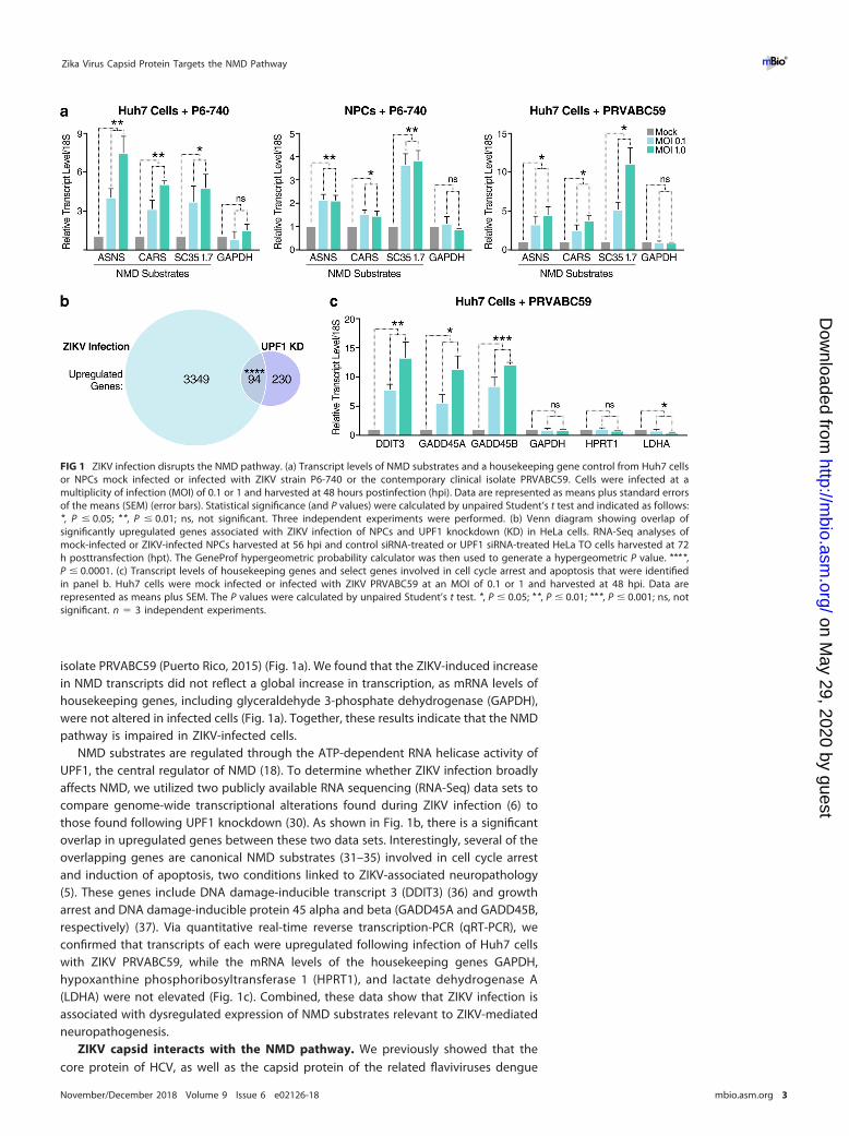
isolate PRVABC59 (Puerto Rico, 2015) (Fig. 1a). We found that the ZIKV-induced increasein NMD transcripts did not reflect a global increase in transcription, as mRNA levels ofhousekeeping genes, including glyceraldehyde 3-phosphate dehydrogenase (GAPDH),were not altered in infected cells (Fig. 1a). Together, these results indicate that the NMDpathway is impaired in ZIKV-infected cells.
NMD substrates are regulated through the ATP-dependent RNA helicase activity ofUPF1, the central regulator of NMD (18). To determine whether ZIKV infection broadlyaffects NMD, we utilized two publicly available RNA sequencing (RNA-Seq) data sets tocompare genome-wide transcriptional alterations found during ZIKV infection (6) tothose found following UPF1 knockdown (30). As shown in Fig. 1b, there is a significantoverlap in upregulated genes between these two data sets. Interestingly, several of theoverlapping genes are canonical NMD substrates (31–35) involved in cell cycle arrestand induction of apoptosis, two conditions linked to ZIKV-associated neuropathology(5). These genes include DNA damage-inducible transcript 3 (DDIT3) (36) and growtharrest and DNA damage-inducible protein 45 alpha and beta (GADD45A and GADD45B,respectively) (37). Via quantitative real-time reverse transcription-PCR (qRT-PCR), weconfirmed that transcripts of each were upregulated following infection of Huh7 cellswith ZIKV PRVABC59, while the mRNA levels of the housekeeping genes GAPDH,hypoxanthine phosphoribosyltransferase 1 (HPRT1), and lactate dehydrogenase A(LDHA) were not elevated (Fig. 1c). Combined, these data show that ZIKV infection isassociated with dysregulated expression of NMD substrates relevant to ZIKV-mediatedneuropathogenesis.
ZIKV capsid interacts with the NMD pathway. We previously showed that thecore protein of HCV, as well as the capsid protein of the related flaviviruses dengue
FIG 1 ZIKV infection disrupts the NMD pathway. (a) Transcript levels of NMD substrates and a housekeeping gene control from Huh7 cellsor NPCs mock infected or infected with ZIKV strain P6-740 or the contemporary clinical isolate PRVABC59. Cells were infected at amultiplicity of infection (MOI) of 0.1 or 1 and harvested at 48 hours postinfection (hpi). Data are represented as means plus standard errorsof the means (SEM) (error bars). Statistical significance (and P values) were calculated by unpaired Student’s t test and indicated as follows:*, P � 0.05; **, P � 0.01; ns, not significant. Three independent experiments were performed. (b) Venn diagram showing overlap ofsignificantly upregulated genes associated with ZIKV infection of NPCs and UPF1 knockdown (KD) in HeLa cells. RNA-Seq analyses ofmock-infected or ZIKV-infected NPCs harvested at 56 hpi and control siRNA-treated or UPF1 siRNA-treated HeLa TO cells harvested at 72h posttransfection (hpt). The GeneProf hypergeometric probability calculator was then used to generate a hypergeometric P value. ****,P � 0.0001. (c) Transcript levels of housekeeping genes and select genes involved in cell cycle arrest and apoptosis that were identifiedin panel b. Huh7 cells were mock infected or infected with ZIKV PRVABC59 at an MOI of 0.1 or 1 and harvested at 48 hpi. Data arerepresented as means plus SEM. The P values were calculated by unpaired Student’s t test. *, P � 0.05; **, P � 0.01; ***, P � 0.001; ns, notsignificant. n � 3 independent experiments.
Zika Virus Capsid Protein Targets the NMD Pathway ®
November/December 2018 Volume 9 Issue 6 e02126-18 mbio.asm.org 3
on May 29, 2020 by guest
http://mbio.asm
.org/D
ownloaded from
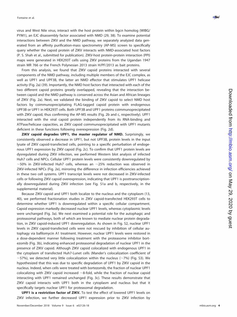
virus and West Nile virus, interact with the host protein within bgcn homolog (WIBG/PYM1), an EJC disassembly factor associated with NMD (29, 38). To examine potentialinteractions between ZIKV and the NMD pathway, we separately analyzed data gen-erated from an affinity purification-mass spectrometry (AP-MS) screen to specificallyquery whether the capsid protein of ZIKV interacts with NMD-associated host factors(P. S. Shah et al., submitted for publication). ZIKV-host protein-protein interaction (PPI)maps were generated in HEK293T cells using ZIKV proteins from the Ugandan 1947strain MR 766 or the French Polynesian 2013 strain H/PF/2013 as bait proteins.
From this analysis, we found that ZIKV capsid proteins interacted with severalcomponents of the NMD pathway, including multiple members of the EJC complex, aswell as UPF1 and UPF3B, the latter an NMD effector that stimulates UPF1 helicaseactivity (Fig. 2a) (39). Importantly, the NMD host factors that interacted with each of thetwo different capsid proteins greatly overlapped, revealing that the interaction be-tween capsid and the NMD pathway is conserved across the Asian and African lineagesof ZIKV (Fig. 2a). Next, we validated the binding of ZIKV capsid to select NMD hostfactors by coimmunoprecipitating FLAG-tagged capsid protein with endogenousUPF3B or UPF1 in HEK293T cells. Both UPF3B and UPF1 proteins coimmunoprecipitatedwith ZIKV capsid, thus confirming the AP-MS results (Fig. 2b and c, respectively). UPF1interacted with the viral capsid protein independently from its RNA-binding andATPase/helicase capacities, as ZIKV capsid coimmunoprecipitated with UPF1 mutantsdeficient in these functions following overexpression (Fig. 2d).
ZIKV capsid degrades UPF1, the master regulator of NMD. Surprisingly, weconsistently observed a decrease in UPF1, but not UPF3B, protein levels in the inputlysate of ZIKV capsid-transfected cells, pointing to a specific perturbation of endoge-nous UPF1 expression by ZIKV capsid (Fig. 2c). To confirm that UPF1 protein levels aredysregulated during ZIKV infection, we performed Western blot analysis of infectedHuh7 cells and NPCs. Cellular UPF1 protein levels were consistently downregulated by�50% in ZIKV-infected Huh7 cells, whereas an �25% reduction was observed inZIKV-infected NPCs (Fig. 2e), mirroring the difference in infection efficiencies achievedin these two cell systems. UPF1 transcript levels were not decreased in ZIKV-infectedcells or following ZIKV capsid overexpression, indicating that UPF1 is posttranscription-ally downregulated during ZIKV infection (see Fig. S1a and b, respectively, in thesupplemental material).
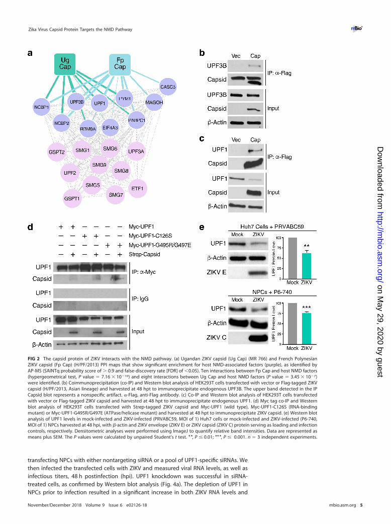
Because ZIKV capsid and UPF1 both localize to the nucleus and the cytoplasm (13,40), we performed fractionation studies in ZIKV capsid-transfected HEK293T cells todetermine whether UPF1 is downregulated within a specific cellular compartment.Capsid expression markedly decreased nuclear UPF1 levels, whereas cytoplasmic levelswere unchanged (Fig. 3a). We next examined a potential role for the autophagic andproteasomal pathways, both of which are known to mediate nuclear protein degrada-tion, in ZIKV capsid-induced UPF1 downregulation. As shown in Fig. S2, nuclear UPF1levels in ZIKV capsid-transfected cells were not rescued by inhibition of cellular au-tophagy via bafilomycin A1 treatment. However, nuclear UPF1 levels were restored ina dose-dependent manner following treatment with the proteasome inhibitor bort-ezomib (Fig. 3b), indicating enhanced proteasomal degradation of nuclear UPF1 in thepresence of ZIKV capsid. Although ZIKV capsid colocalized with endogenous UPF1 inthe cytoplasm of transfected Huh7-Lunet cells (Mander’s colocalization coefficient of�57%), we detected very little colocalization within the nucleus (�7%) (Fig. S3). Wehypothesized that this was due to specific degradation of UPF1 by ZIKV capsid in thenucleus. Indeed, when cells were treated with bortezomib, the fraction of nuclear UPF1colocalizing with ZIKV capsid increased �8-fold, while the fraction of nuclear capsidinteracting with UPF1 remained unchanged (Fig. 3c). These results demonstrate thatZIKV capsid interacts with UPF1 both in the cytoplasm and nucleus but that itspecifically targets nuclear UPF1 for proteasomal degradation.
UPF1 is a restriction factor of ZIKV. To test the effect of lowered UPF1 levels onZIKV infection, we further decreased UPF1 expression prior to ZIKV infection by
Fontaine et al. ®
November/December 2018 Volume 9 Issue 6 e02126-18 mbio.asm.org 4
on May 29, 2020 by guest
http://mbio.asm
.org/D
ownloaded from
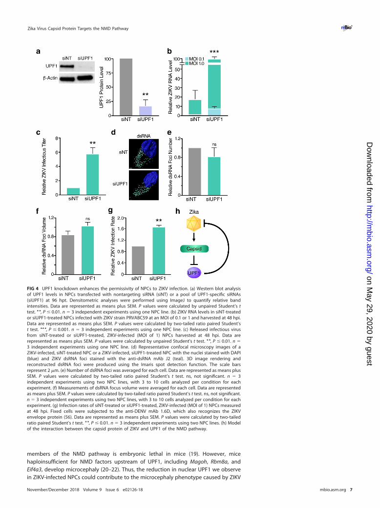
transfecting NPCs with either nontargeting siRNA or a pool of UPF1-specific siRNAs. Wethen infected the transfected cells with ZIKV and measured viral RNA levels, as well asinfectious titers, 48 h postinfection (hpi). UPF1 knockdown was successful in siRNA-treated cells, as confirmed by Western blot analysis (Fig. 4a). The depletion of UPF1 inNPCs prior to infection resulted in a significant increase in both ZIKV RNA levels and
FIG 2 The capsid protein of ZIKV interacts with the NMD pathway. (a) Ugandan ZIKV capsid (Ug Cap) (MR 766) and French PolynesianZIKV capsid (Fp Cap) (H/PF/2013) PPI maps that show significant enrichment for host NMD-associated factors (purple), as identified byAP-MS (SAINTq probability score of � 0.9 and false-discovery rate [FDR] of �0.05). Ten interactions between Fp Cap and host NMD factors(hypergeometrical test, P value � 7.16 � 10�10) and eight interactions between Ug Cap and host NMD factors (P value � 3.45 � 10�7)were identified. (b) Coimmunoprecipitation (co-IP) and Western blot analysis of HEK293T cells transfected with vector or Flag-tagged ZIKVcapsid (H/PF/2013, Asian lineage) and harvested at 48 hpt to immunoprecipitate endogenous UPF3B. The upper band detected in the IPCapsid blot represents a nonspecific artifact. �-Flag, anti-Flag antibody. (c) Co-IP and Western blot analysis of HEK293T cells transfectedwith vector or Flag-tagged ZIKV capsid and harvested at 48 hpt to immunoprecipitate endogenous UPF1. (d) Myc tag co-IP and Westernblot analysis of HEK293T cells transfected with Strep-tagged ZIKV capsid and Myc-UPF1 (wild type), Myc-UPF1-C126S (RNA-bindingmutant) or Myc-UPF1-G495R/G497E (ATPase/helicase mutant) and harvested at 48 hpt to immunoprecipitate ZIKV capsid. (e) Western blotanalysis of UPF1 levels in mock-infected and ZIKV-infected (PRVABC59, MOI of 1) Huh7 cells or mock-infected and ZIKV-infected (P6-740,MOI of 1) NPCs harvested at 48 hpi, with �-actin and ZIKV envelope (ZIKV E) or ZIKV capsid (ZIKV C) protein serving as loading and infectioncontrols, respectively. Densitometric analyses were performed using ImageJ to quantify relative band intensities. Data are represented asmeans plus SEM. The P values were calculated by unpaired Student’s t test. **, P � 0.01; ***, P � 0.001. n � 3 independent experiments.
Zika Virus Capsid Protein Targets the NMD Pathway ®
November/December 2018 Volume 9 Issue 6 e02126-18 mbio.asm.org 5
on May 29, 2020 by guest
http://mbio.asm
.org/D
ownloaded from
infectious virus production (Fig. 4b and c, respectively), indicating that expression ofUPF1 restricts ZIKV infection at or before the RNA replication stage. To differentiatebetween these two stages, we analyzed double-stranded RNA (dsRNA) intermediatesrepresenting presumed viral RNA replication centers in infected NPCs (Fig. 4d) (41).Using confocal microscopy and 3D reconstruction analyses, we observed no significantdifference in the number and size of dsRNA foci per cell when comparing ZIKV-infected,UPF1-depleted NPCs to ZIKV-infected cultures expressing UPF1 (Fig. 4e and f). Instead,we found a significant increase in the number of infected cells in NPC cultures whenUPF1 was depleted, indicating that UPF1 regulates permissivity of NPCs to ZIKVinfection at an early stage prior to viral RNA replication (Fig. 4g).
DISCUSSION
In summary, we identified the NMD pathway as a restriction mechanism for ZIKVinfection in human NPCs. NMD was partially inactivated in ZIKV-infected NPCs throughexpression of the viral capsid protein and the resulting degradation of host nuclearUPF1. As further weakening NMD by depleting UPF1 resulted in a marked increase inthe number of infected cells, we propose a model in which an evolutionary “arms race”between cellular NMD and ZIKV determines whether a cell is successfully infected(Fig. 4h).
Downregulation of UPF1 by ZIKV capsid is not complete and is likely limited by thedamaging effects of NMD disruption, as illustrated by the upregulation of genesinvolved in cell growth arrest and apoptosis. Indeed, knockout of Upf1 and other
FIG 3 ZIKV capsid degrades UPF1, the master regulator of NMD, via a proteasome-dependent mechanism. (a) Western blot analysis of UPF1 levels in subcellularfractionated HEK293T cells transfected with vector or Flag-tagged ZIKV capsid (H/PF/2013, Asian lineage) for 48 h. GAPDH was used as a cytoplasmic marker,and SP1 was used as a nuclear marker to ensure optimal fractionation. Densitometric analyses were performed using ImageJ to quantify relative bandintensities. Data are represented as means plus SEM. P values were calculated by unpaired Student’s t test. **, P � 0.01; ns, not significant. n � 3 independentexperiments. (b) Western blot analysis of nuclear UPF1 levels in fractionated HEK293T cells transfected with vector or Flag-tagged ZIKV capsid for 48 h. Cellswere treated with DMSO or increasing concentrations of the proteasome inhibitor bortezomib (Borte) for 24 h before harvest. Densitometric analyses wereperformed using ImageJ to quantify relative band intensities. Data are represented as means plus SEM. P values were calculated by one-way ANOVA withmultiple comparisons. *, P � 0.05; ns, not significant. n � 3 independent experiments. (c) Representative 3D confocal microscopy images of the nuclei ofHuh7-Lunet cells transfected with Strep-tagged ZIKV capsid. Cells were treated at 24 hpt with DMSO or 10 nM bortezomib and processed for immunostainingat 48 hpt with antibodies against Strep tag (turquoise) and endogenous UPF1 (purple). DAPI (blue) was used to stain and define the nuclei. Each channel wasreconstructed digitally for visualization of the 3D colocalization. The thresholded Mander’s correlation coefficients were determined, and P values werecalculated by unpaired Student’s t test. **, P � 0.01. n � 8 cells per condition. Scale bars represent 3 �m.
Fontaine et al. ®
November/December 2018 Volume 9 Issue 6 e02126-18 mbio.asm.org 6
on May 29, 2020 by guest
http://mbio.asm
.org/D
ownloaded from
members of the NMD pathway is embryonic lethal in mice (19). However, micehaploinsufficient for NMD factors upstream of UPF1, including Magoh, Rbm8a, andEif4a3, develop microcephaly (20–22). Thus, the reduction in nuclear UPF1 we observein ZIKV-infected NPCs could contribute to the microcephaly phenotype caused by ZIKV
FIG 4 UPF1 knockdown enhances the permissivity of NPCs to ZIKV infection. (a) Western blot analysisof UPF1 levels in NPCs transfected with nontargeting siRNA (siNT) or a pool of UPF1-specific siRNAs(siUPF1) at 96 hpt. Densitometric analyses were performed using ImageJ to quantify relative bandintensities. Data are represented as means plus SEM. P values were calculated by unpaired Student’s ttest. **, P � 0.01. n � 3 independent experiments using one NPC line. (b) ZIKV RNA levels in siNT-treatedor siUPF1-treated NPCs infected with ZIKV strain PRVABC59 at an MOI of 0.1 or 1 and harvested at 48 hpi.Data are represented as means plus SEM. P values were calculated by two-tailed ratio paired Student’st test. ***, P � 0.001. n � 3 independent experiments using one NPC line. (c) Released infectious virusfrom siNT-treated or siUPF1-treated, ZIKV-infected (MOI of 1) NPCs harvested at 48 hpi. Data arerepresented as means plus SEM. P values were calculated by unpaired Student’s t test. **, P � 0.01. n �3 independent experiments using one NPC line. (d) Representative confocal microscopy images of aZIKV-infected, siNT-treated NPC or a ZIKV-infected, siUPF1-treated NPC with the nuclei stained with DAPI(blue) and ZIKV dsRNA foci stained with the anti-dsRNA mAb J2 (teal). 3D image rendering andreconstructed dsRNA foci were produced using the Imaris spot detection function. The scale barsrepresent 2 �m. (e) Number of dsRNA foci was averaged for each cell. Data are represented as means plusSEM. P values were calculated by two-tailed ratio paired Student’s t test. ns, not significant. n � 3independent experiments using two NPC lines, with 3 to 10 cells analyzed per condition for eachexperiment. (f) Measurements of dsRNA focus volume were averaged for each cell. Data are representedas means plus SEM. P values were calculated by two-tailed ratio paired Student’s t test. ns, not significant.n � 3 independent experiments using two NPC lines, with 3 to 10 cells analyzed per condition for eachexperiment. (g) Infection rates of siNT-treated or siUPF1-treated, ZIKV-infected (MOI of 1) NPCs measuredat 48 hpi. Fixed cells were subjected to the anti-DENV mAb 1.6D, which also recognizes the ZIKVenvelope protein (56). Data are represented as means plus SEM. P values were calculated by two-tailedratio paired Student’s t test. **, P � 0.01. n � 3 independent experiments using two NPC lines. (h) Modelof the interaction between the capsid protein of ZIKV and UPF1 of the NMD pathway.
Zika Virus Capsid Protein Targets the NMD Pathway ®
November/December 2018 Volume 9 Issue 6 e02126-18 mbio.asm.org 7
on May 29, 2020 by guest
http://mbio.asm
.org/D
ownloaded from
infection in the fetal brain. While fetal and adult NPCs appear to be transcriptionallydistinct (42), it has been shown that adult NPCs are also permissive to ZIKV infection(43). As the NMD pathway is a ubiquitous cellular surveillance mechanism, it is likelythat ZIKV capsid targets UPF1 for degradation in any cell type that is susceptible to ZIKVinfection. Accordingly, we have found that UPF1 is degraded following infection ofboth NPCs and hepatic Huh7 cells.
Why ZIKV capsid specifically downregulates nuclear UPF1 and how nuclear UPF1contributes to ZIKV restriction remain unanswered. Several studies suggest that NMD isassociated with the nucleus, although this issue remains controversial. Multiple tran-scripts, such as those encoding T cell receptor beta, triosephosphate isomerase, andmouse major urinary protein, have been shown to be specifically degraded in purifiednuclei or reduced in nuclear fractions (44). These data support the model that selec-tively depleting nuclear UPF1 levels disrupts NMD function in ZIKV-infected cells. Inaddition, UPF1 is involved in several other processes within the nucleus, includingnucleus-associated RNA metabolism, cell cycle progression, and DNA replication (40).Therefore, by targeting nuclear UPF1, ZIKV could disrupt these processes and programtarget cells for viral replication. Notably, viral RNA replication is thought to occur solelywithin the cytoplasmic compartment (45, 46). Using confocal microscopy and 3Dreconstruction, we did not detect dsRNA foci localized within the nuclei of ZIKV-infected cells (data not shown), supporting our finding that UPF1 does not restrict viralRNA replication. While our results suggest a role for nuclear UPF1 in ZIKV restriction, itis possible that UPF1 also serves as a restriction factor of ZIKV within the cytoplasm.Previously, it was shown that UPF1 suppresses alphavirus replication by degrading theincoming viral RNA following uncoating in the cytosol (28). Thus, ZIKV may possess anadditional mechanism to prevent cytoplasmic UPF1 from targeting its incoming RNAgenome for destruction.
Our data reveal that nuclear UPF1 is degraded by ZIKV capsid in a proteasome-dependent manner. While the nuclear proteasome has not been specifically linked tomicrocephaly, it plays critical roles in the regulation of chromatin structure, geneexpression, DNA repair, and protein quality control (47). Thus, the co-opting of thenuclear proteasome by ZIKV capsid to degrade UPF1 could disrupt its normal protea-somal activity and further contribute to the cytopathic effects associated with ZIKVinfection. Furthermore, given that the capsid protein of the closely related dengue viruscan translocate across cell membranes (48), it is possible that capsid released fromapoptotic, ZIKV-infected cells can enter neighboring, uninfected cells to degrade UPF1,thus increasing permissivity of bystander cells to ZIKV infection. Studies are ongoing todetermine the precise molecular mechanism of ZIKV capsid-mediated UPF1 degrada-tion and how UPF1 depletion enhances ZIKV replication, directly or indirectly. Ulti-mately, these data may help inform new therapeutic approaches, as reinforcement ofthe antiviral properties of the NMD pathway is expected to enhance resistance of NPCsto ZIKV infection and to promote normal neurodevelopment in infected fetuses.
MATERIALS AND METHODSViruses and cells. Two Asian lineage strains of ZIKV, P6-740 (ATCC VR-1845) and PRVABC59 (ATCC
VR-1843), were used for all experiments. ZIKV stocks were propagated in Vero cells (ATCC), and titerswere determined by plaque assays on Vero cells. Huh7 cells (ATCC), Huh7-Lunet cells (Ralf Barten-schlager, Heidelberg University), and Vero cells were maintained in Dulbecco’s modified Eagle’s medium(DMEM) with 10% fetal bovine serum (FBS), 2 mM L-glutamine, 100 U/ml penicillin, and 100 �g/mlstreptomycin. HEK293T cells (ATCC) were maintained in DMEM/H21 medium supplemented with 10%FBS, 100 U/ml penicillin, 100 �g/ml streptomycin, and 1 mM sodium pyruvate or DMEM with 10% FBS,2 mM L-glutamine, 100 U/ml penicillin, and 100 �g/ml streptomycin. Human iPSC-derived NPCs weregenerated and maintained as described previously (49). All of the human fibroblast cell lines used togenerate iPSCs came from the Coriell Institute for Medical Research and Yale Stem Cell Center. The iPSCsused in these studies were the CTRL2493nXX, CS2518nXX, and Cs71iCTR-20nXX lines. CTRL2493nXX wasderived from the parental fibroblast line ND31845 that was biopsied from a healthy female at 71 yearsof age. CS2518nXX was derived from the parental fibroblast line ND30625 that was biopsied from ahealthy male at 76 years of age. CS71iCTR-20nXX was derived from the parental fibroblast line ND29971that was biopsied from a female at 61 years of age. For virus infections, NPCs plated on Matrigel-coated
Fontaine et al. ®
November/December 2018 Volume 9 Issue 6 e02126-18 mbio.asm.org 8
on May 29, 2020 by guest
http://mbio.asm
.org/D
ownloaded from
(Corning) multiwell plates or Huh7 cells were infected with ZIKV at a multiplicity of infection (MOI) of 0.1or 1 for 2 h at 37°C. Infected cells were harvested at 48 hpi for all analyses.
Affinity purification, mass spectrometry, and AP-MS scoring. The ZIKV capsid open readingframes (ORFs) from the Ugandan 1947 strain MR 766 or the French Polynesian 2013 strain H/PF/2013were cloned into pCDNA4_TO with a C-terminal 2xStrep II affinity tag for expression in human cells. Theviral capsid proteins (three biological replicates), as well as GFP (two biological replicates) and emptyvector (ten biological replicates) as negative controls, were expressed in HEK293T cells, and affinitypurifications were performed as previously described (50). Briefly, clarified lysates were incubated withStrep-Tactin Superflow (IBA) overnight at 4°C. Proteins were eluted with 50 mM Tris (pH 7.5), 150 mMNaCl, and 1 mM EDTA containing 2.5 mM Desthiobiotin (IBA) for 30 min at 4°C. Lysates and affinity-purified eluates were analyzed by Western blotting and silver stain PAGE to confirm expression andpurification. Purified protein eluates were digested with trypsin for LC-MS/MS analysis. Samples weredenatured and reduced in 2 M urea, 10 mM NH4HCO3, and 2 mM DTT for 30 min at 60°C and thenalkylated with 2 mM iodoacetamide for 45 min at room temperature. Trypsin (Promega) was added at a1:100 enzyme/substrate ratio and digested overnight at 37°C. Following digestion, samples wereconcentrated using C18 ZipTips (Millipore) according to the manufacturer’s specifications. Peptides wereresuspended in 15 �l of 4% formic acid and 3% ACN, and 1 to 2 �l of sample was loaded onto a 75-�m-IDcolumn packed with 25 cm of Reprosil C18 1.9-�m, 120-Å particles (Dr. Maisch GmbH). Peptides wereeluted into a Q-Exactive Plus (Thermo Fisher Scientific) mass spectrometer by gradient elution deliveredby an Easy1200 nLC system (Thermo Fisher). The gradient was from 4.5% to 32% acetonitrile over 53 min.All MS spectra were collected with orbitrap detection, while the 20 most abundant ions were fragmentedby higher energy collisional dissociation (HCD) and detected in the orbitrap. All data were searchedagainst the Swiss-Prot Human protein sequences, combined with ZIKV sequences and GFP. Peptide andprotein identification searches, as well as label-free quantitation, were performed using the MaxQuantdata analysis algorithm, and all peptide and protein identifications were filtered to a 1% false-discoveryrate (51, 52). SAINTq (53) was used to calculate the probability of bait-prey interactions for both UgandanZIKV capsid and French Polynesian ZIKV capsid against the negative controls, including GFP and emptyvector, with protein intensities as input values. We applied a combined threshold of probability ofinteraction (AvgP) greater than 0.90 and a Bayesian false-discovery rate of less than 0.05.
Quantitative real-time reverse transcription-PCR (qRT-PCR). Total cellular RNA was isolated fromHuh7 cells and NPCs using the RNeasy Mini kit (Qiagen). cDNA was synthesized with oligo(dT)18 (ThermoFisher Scientific) primers, random hexamer (Life Technologies) primers, and AMV reverse transcriptase(Promega). The cDNA was then used in SYBR green PCR master mix (Thermo Fisher Scientific) accordingto the manufacturer’s instructions and analyzed by qPCR (Bio-Rad ABI 7900). The primers used for ASNS,CARS, SC35 1.7 (1.7 kb mRNA), GAPDH, HPRT1, LDHA, and 18S rRNA have been described previously (29).The additional primers used were as follows: ZIKV PRVABC59 forward primer, 5=-GAG ACG AGA TGC GGTACA GG-3=; ZIKV PRVABC59 reverse primer, 5=-CGA CCG TCA GTT GAA CTC CA-3=; UPF1 forward primer,5=-CTG CAA CGG ACG TGG AAA TAC-3=; UPF1 reverse primer, 5=-ACA GCC GCA GTT GTA GCA C-3=; DDIT3forward primer, 5=- TG CTT CTC TGG CTT GGC TG-3=; DDIT3 reverse primer, 5=-GCT CTG GGA GGT GCT TGTGA-3=; GADD45A forward primer, 5=-GAG CTC CTG CTC TTG GAG AC-3=; GADD45A reverse primer, 5=-GCAGGA TCC TTC CAT TGA GA-3=; GADD45B forward primer, 5=-TGA CAA CGA CAT CAA CAT C-3=; GADD45Breverse primer, 5=-GTG ACC AGA GAC AAT GCA G-3=. The relative levels of each transcript werenormalized by the delta threshold cycle method to the abundance of 18S rRNA or GAPDH, withmock-infected cells or vector-transfected cells set at 1.
Western blot analysis. Cells were lysed in RIPA lysis buffer (50 mM Tris-HCl [pH 8], 150 mM NaCl, 1%NP-40, 0.5% sodium deoxycholate, 0.1% SDS, supplemented with Halt protease inhibitor cocktail[Thermo Fisher Scientific]) to obtain whole-cell lysates or lysed using the NE-PER nuclear and cytoplasmicextraction kit (Thermo Fisher Scientific) to obtain cytoplasmic and nuclear fractions. Proteins wereseparated by SDS-PAGE and transferred to nitrocellulose membranes (Bio-Rad). Blots were incubatedwith the indicated primary antibody: anti-UPF3B (ab134566; Abcam), anti-UPF1 (12040; Cell SignalingTechnology, Inc.), anti-ZIKV capsid (C) (GTX133304; GeneTex), anti-Flag (F7425; Sigma-Aldrich), anti-�-actin (A5316; Sigma-Aldrich), anti-ZIKV envelope (E) (GTX133314; GeneTex), anti-SP1 (sc-14027; SantaCruz Biotechnology), anti-GAPDH (5174; Cell Signaling Technology, Inc.), anti-Myc tag (ab9106; Abcam),anti-Strep tag (ab18422, Abcam), and anti-p62 (ab56416, Abcam). Proteins were visualized by chemilu-minescent detection with ECL and ECL Hyperfilm (Amersham). Differences in band intensity werequantified by densitometry using ImageJ.
Immunoprecipitations. Cells were lysed in either RIPA lysis buffer or IP lysis buffer (150 mM NaCl,50 mM Tris [pH 7.4], 1 mM EDTA, 0.5% NP-40 substitute, supplemented with Halt protease inhibitorcocktail [Thermo Fisher Scientific]) at 4°C and passed through a G23 needle. Clarified lysates wereimmunoprecipitated with Flag M2 agarose (Sigma), anti-Myc tag (ab9106; Abcam), or normal rabbit IgG(sc-2027; Santa Cruz Biotechnology) overnight, washed in lysis buffer, and resuspended in Laemmli bufferfor SDS-PAGE. Western blot analysis of immunoprecipitated proteins was performed as described above.
Immunofluorescence. Transfected Huh7-Lunet cells or infected NPCs were collected at 48 h andplated onto 22- by 22-mm no. 1.5 coverslips. Cells were fixed in 4% paraformaldehyde, permeabilizedwith 0.1% Triton X-100, and blocked in 3% bovine serum albumin. Cells were then immunostained withthe indicated antibodies: anti-Strep Tag (ab184224; Abcam), anti-UPF1 (ab109363; Abcam), humananti-dengue virus (DENV) MAb 1.6D (Sharon Isern and Scott Michael, Florida Gulf Coast University), whichrecognizes the ZIKV envelope protein, anti-dsRNA MAb J2 (SCICONS), and the appropriate fluorophore-conjugated secondary antibodies. Coverslips were mounted onto glass slides using Vectashield mount-ing medium with DAPI (Vector Laboratories) and analyzed by fluorescence microscopy (Zeiss Axio
Zika Virus Capsid Protein Targets the NMD Pathway ®
November/December 2018 Volume 9 Issue 6 e02126-18 mbio.asm.org 9
on May 29, 2020 by guest
http://mbio.asm
.org/D
ownloaded from
Observer ZI) or confocal microscopy (Zeiss LSM 880). For acquiring high-resolution images, cells wereimaged on the Zeiss LSM 880 with Airyscan using a 20�/0.8 or 63�/1.4 M27 oil immersion objective. Atotal of 15 to 20 (20� objective) or 60 to 80 (63� objective) Z-slices were acquired every 0.88 �m or0.15 �m, respectively. The resulting Z-stack was reconstructed and rendered in 3D using Imaris software(Bitplane). Viral dsRNA foci were reconstructed via the Imaris spot detection function, which provided ananalysis of total number and mean volume of foci within a cell, for images acquired using the 20�objective. Strep-tagged ZIKV capsid, UPF1, and dsRNA channels acquired using the 63� objective werereconstructed using the Imaris surfaces package. The Imaris colocalization function was used to deter-mine overlap of fluorescence. Thresholding for background fluorescence was determined by the Imarisautomatic thresholding tool that utilizes the Costes approach (54). The thresholded Mander’s correlationcoefficient (MCC) measures the fraction of voxels with fluorescence positive for one channel that alsocontains fluorescence from another channel. The MCC is typically more appropriate for analysis ofthree-dimensional colocalization (55).
Statistical analysis. Statistical differences between groups were analyzed using either a two-tailedunpaired Student’s t test or a two-tailed ratio paired Student’s t test as stated in the figure legends.Hypergeometrical tests were used to calculate the probability of an overlap in gene dysregulationbetween ZIKV-infected NPCs and UPF1-depleted cells and to calculate the probability of ZIKV capsidbait-prey interactions. Data are represented as means plus standard errors of the means (SEM). Statisticalsignificance was defined as follows: *, P � 0.05; **, P � 0.01; ***, P � 0.001; ****, P � 0.0001.
SUPPLEMENTAL MATERIALSupplemental material for this article may be found at https://doi.org/10.1128/mBio
.02126-18.FIG S1, PDF file, 0.2 MB.FIG S2, PDF file, 0.4 MB.FIG S3, PDF file, 10 MB.
ACKNOWLEDGMENTSWe thank all members of the Ott laboratory, as well as Roman Camarda, Marius
Walter, and Anna Maurer for helpful discussions and advice throughout the preparationof the manuscript. We thank Chia-Lin Tsou, the Gladstone Stem Cell Core, and MeredithCalvert from the Gladstone Microscopy Core for technical assistance, and Ralf Barten-schlager (Heidelberg University), Lynne Maquat (University of Rochester), and SharonIsern and Scott Michael (Florida Gulf Coast University) for reagents. We are grateful toVeronica Fonseca and Lauren Weiser for administrative support, John Carroll and TeresaRoberts for graphical design, and to Kathryn Claiborn, Eric Martens, and Gary Howardfor editorial assistance.
This work was supported by NIH/NIAID F32AI112262 to P.S.S., NIH/NINDS R01NS101996-01 to S.F., NIH/NIAID U19AI1186101 to N.J.K., DOD/DARPA HR0011-11-C-0094 (PROPHECY) to N.J.K., NIH/NIAID R01 AI097552 to M.O., BioFulcrum, and the JamesB. Pendleton Charitable Trust.
REFERENCES1. Fauci AS, Morens DM. 2016. Zika virus in the Americas–yet another
arbovirus threat. N Engl J Med 374:601– 604. https://doi.org/10.1056/NEJMp1600297.
2. Cugola FR, Fernandes IR, Russo FB, Freitas BC, Dias JLM, Guimarães KP,Benazzato C, Almeida N, Pignatari GC, Romero S, Polonio CM, Cunha I,Freitas CL, Brandão WN, Rossato C, Andrade DG, Faria DdP, Garcez AT,Buchpigel CA, Braconi CT, Mendes E, Sall AA, Zanotto PM, Peron JPS,Muotri AR, Beltrão-Braga PCB. 2016. The Brazilian Zika virus strain causesbirth defects in experimental models. Nature 534:267–271. https://doi.org/10.1038/nature18296.
3. Shao Q, Herrlinger S, Yang S, Lai F, Moore JM, Brindley MA, Chen J. 2016.Zika virus infection disrupts neurovascular development and results inpostnatal microcephaly with brain damage. Development 143:4127– 4136. https://doi.org/10.1242/dev.143768.
4. Souza BSF, Sampaio GLA, Pereira CS, Campos GS, Sardi SI, Freitas LAR,Figueira CP, Paredes BD, Nonaka CKV, Azevedo CM, Rocha VPC, BandeiraAC, Mendez-Otero R, Dos Santos RR, Soares MBP. 2016. Zika virusinfection induces mitosis abnormalities and apoptotic cell death ofhuman neural progenitor cells. Sci Rep 6:39775. https://doi.org/10.1038/srep39775.
5. Li C, Xu D, Xu Z, Ye Q, Hong S, Jiang Y, Liu X, Zhang N, Shi L, Qin C. 2016.
Zika virus disrupts neural progenitor development and leads to micro-cephaly in mice. Cell Stem Cell 19:120 –126. https://doi.org/10.1016/j.stem.2016.04.017.
6. Tang H, Hammack C, Ogden S, Wen Z, Qian X, Li Y, Yao B, Shin J, ZhangF, Lee E, Christian K, Didier R, Jin P, Song H, Ming G. 2016. Zika virusinfects human cortical neural progenitors and attenuates their growth.Cell Stem Cell 18:587–590. https://doi.org/10.1016/j.stem.2016.02.016.
7. Rasmussen SA, Jamieson DJ, Honein MA, Petersen LR. 2016. Zika virusand birth defects–reviewing the evidence for causality. N Engl J Med374:1981–1987. https://doi.org/10.1056/NEJMsr1604338.
8. Harris E, Holden KL, Edgil D, Polacek C, Clyde K. 2006. Molecular biologyof flaviviruses. Novartis Found Symp 277:253.
9. Lindenbach BD, Rice CM. 2003. Molecular biology of flaviviruses. AdvVirus Res 59:23– 61. https://doi.org/10.1016/S0065-3527(03)59002-9.
10. Oliveira ERA, Mohana-Borges R, de Alencastro RB, Horta BAC. 2017. Theflavivirus capsid protein: structure, function and perspectives towardsdrug design. Virus Res 227:115–123. https://doi.org/10.1016/j.virusres.2016.10.005.
11. den Boon JA, Diaz A, Ahlquist P. 2010. Cytoplasmic viral replicationcomplexes. Cell Host Microbe 8:77– 85. https://doi.org/10.1016/j.chom.2010.06.010.
Fontaine et al. ®
November/December 2018 Volume 9 Issue 6 e02126-18 mbio.asm.org 10
on May 29, 2020 by guest
http://mbio.asm
.org/D
ownloaded from
12. Colpitts TM, Barthel S, Wang P, Fikrig E. 2011. Dengue virus capsidprotein binds core histones and inhibits nucleosome formation in hu-man liver cells. PLoS One 6:e24365. https://doi.org/10.1371/journal.pone.0024365.
13. Slomnicki LP, Chung DH, Parker A, Hermann T, Boyd NL, Hetman M.2017. Ribosomal stress and Tp53-mediated neuronal apoptosis in re-sponse to capsid protein of the Zika virus. Sci Rep 7:1–15.
14. Tsuda Y, Mori Y, Abe T, Yamashita T, Okamoto T, Ichimura T, MoriishiK, Matsuura Y. 2006. Nucleolar protein B23 interacts with Japaneseencephalitis virus core protein and participates in viral replication.Microbiol Immunol 50:225–234. https://doi.org/10.1111/j.1348-0421.2006.tb03789.x.
15. Xu Z, Hobman TC. 2012. The helicase activity of DDX56 is required for itsrole in assembly of infectious West Nile virus particles. Virology 433:226 –235. https://doi.org/10.1016/j.virol.2012.08.011.
16. Rawlinson SM, Moseley GW. 2015. The nucleolar interface of RNA viruses.Cell Microbiol 17:1108 –1120. https://doi.org/10.1111/cmi.12465.
17. Peccarelli M, Kebaara BW. 2014. Regulation of natural mRNAs by thenonsense-mediated mRNA decay pathway. Eukaryot Cell 13:1126 –1135.https://doi.org/10.1128/EC.00090-14.
18. Hug N, Longman D, Cáceres JF. 2016. Mechanism and regulation of thenonsense-mediated decay pathway. Nucleic Acids Res 44:1483–1495.https://doi.org/10.1093/nar/gkw010.
19. Han X, Wei Y, Wang H, Wang F, Ju Z, Li T. 2018. Nonsense-mediatedmRNA decay: a ’nonsense’ pathway makes sense in stem cell biology.Nucleic Acids Res 46:1038 –1051. https://doi.org/10.1093/nar/gkx1272.
20. Silver DL, Watkins-Chow DE, Schreck KC, Pierfelice TJ, Larson DM, Bur-netti AJ, Liaw H, Myung K, Walsh CA, Gaiano N, Pavan WJ. 2010. The exonjunction complex component Magoh controls brain size by regulatingneural stem cell division. Nat Neurosci 13:551–558. https://doi.org/10.1038/nn.2527.
21. Mao H, Pilaz L, McMahon JJ, Golzio C, Wu D, Shi L, Katsanis N, Silver DL.2015. Rbm8a haploinsufficiency disrupts embryonic cortical develop-ment resulting in microcephaly. J Neurosci 35:7003–7018. https://doi.org/10.1523/JNEUROSCI.0018-15.2015.
22. Mao H, McMahon JJ, Tsai Y, Wang Z, Silver DL. 2016. Haploinsufficiencyfor core exon junction complex components disrupts embryonic neu-rogenesis and causes p53-mediated microcephaly. PLoS Genet 12:e1006282. https://doi.org/10.1371/journal.pgen.1006282.
23. Kurosaki T, Maquat LE. 2016. Nonsense-mediated mRNA decay in hu-mans at a glance. J Cell Sci 129:461– 467. https://doi.org/10.1242/jcs.181008.
24. Serquiña AKP, Das SR, Popova E, Ojelabi OA, Roy CK, Göttlinger HG. 2013.UPF1 is crucial for the infectivity of human immunodeficiency virus type1 progeny virions. J Virol 87:8853– 8861. https://doi.org/10.1128/JVI.00925-13.
25. Molleston JM, Cherry S. 2017. Attacked from all sides: RNA decay inantiviral defense. Viruses 9:2. https://doi.org/10.3390/v9010002.
26. Balistreri G, Bognanni C, Mühlemann O. 2017. Virus escape and manip-ulation of cellular nonsense-mediated mRNA decay. Viruses 9:24. https://doi.org/10.3390/v9010024.
27. Rigby RE, Rehwinkel J. 2015. RNA degradation in antiviral immunity andautoimmunity. Trends Immunol 36:179 –188. https://doi.org/10.1016/j.it.2015.02.001.
28. Balistreri G, Horvath P, Schweingruber C, Zünd D, McInerney G, Merits A,Mühlemann O, Azzalin C, Helenius A. 2014. The host nonsense-mediatedmRNA decay pathway restricts mammalian RNA virus replication. CellHost Microbe 16:403– 411. https://doi.org/10.1016/j.chom.2014.08.007.
29. Ramage HR, Kumar GR, Verschueren E, Johnson JR, Von Dollen J, John-son T, Newton B, Shah P, Horner J, Krogan NJ, Ott M. 2015. A combinedproteomics/genomics approach links hepatitis C virus infection withnonsense-mediated mRNA decay. Mol Cell 57:329 –340. https://doi.org/10.1016/j.molcel.2014.12.028.
30. Tani H, Imamachi N, Salam KA, Mizutani R, Ijiri K, Irie T, Yada T, Suzuki Y,Akimitsu N. 2012. Identification of hundreds of novel UPF1 target tran-scripts by direct determination of whole transcriptome stability. RNABiol 9:1370 –1379. https://doi.org/10.4161/rna.22360.
31. Weischenfeldt J, Damgaard I, Bryder D, Theilgaard-Mönch K, Thoren LA,Nielsen FC, Jacobsen SEW, Nerlov C, Porse BT. 2008. NMD is essential forhematopoietic stem and progenitor cells and for eliminating by-products of programmed DNA rearrangements. Genes Dev 22:1381–1396. https://doi.org/10.1101/gad.468808.
32. Li T, Shi Y, Wang P, Guachalla LM, Sun B, Joerss T, Chen Y, Groth M,Krueger A, Platzer M, Yang Y, Rudolph KL, Wang Z. 2015. Smg6/Est1
licenses embryonic stem cell differentiation via nonsense-mediatedmRNA decay. EMBO J 34:1630 –1647. https://doi.org/10.15252/embj.201489947.
33. Brazão TF, Demmers J, van IJcken W, Strouboulis J, Fornerod M, RomãoL, Grosveld FG. 2012. A new function of ROD1 in nonsense-mediatedmRNA decay. FEBS Lett 586:1101–1110. https://doi.org/10.1016/j.febslet.2012.03.015.
34. Chan W, Huang L, Gudikote JP, Chang Y, Imam JS, MacLean JA, WilkinsonMF. 2007. An alternative branch of the nonsense-mediated decay path-way. EMBO J 26:1820 –1830. https://doi.org/10.1038/sj.emboj.7601628.
35. Nelson JO, Moore KA, Chapin A, Hollien J, Metzstein MM. 2016. Degra-dation of Gadd45 mRNA by nonsense-mediated decay is essential forviability. Elife 5:e12876. https://doi.org/10.7554/eLife.12876.
36. Jauhiainen A, Thomsen C, Strömbom L, Grundevik P, Andersson C,Danielsson A, Andersson MK, Nerman O, Rörkvist L, Ståhlberg A, ÅmanP. 2012. Distinct cytoplasmic and nuclear functions of the stress inducedprotein DDIT3/CHOP/GADD153. PLoS One 7:e33208. https://doi.org/10.1371/journal.pone.0033208.
37. Salvador JM, Brown-Clay JD, Fornace AJ. 2013. Gadd45 in stress signal-ing, cell cycle control, and apoptosis. Adv Exp Med Biol 793:1–19.https://doi.org/10.1007/978-1-4614-8289-5_1.
38. Gehring NH, Lamprinaki S, Kulozik AE, Hentze MW. 2009. Disassembly ofexon junction complexes by PYM. Cell 137:536 –548. https://doi.org/10.1016/j.cell.2009.02.042.
39. Chamieh H, Ballut L, Bonneau F, Le Hir H. 2008. NMD factors UPF2 andUPF3 bridge UPF1 to the exon junction complex and stimulate its RNAhelicase activity. Nat Struct Mol Biol 15:85–93. https://doi.org/10.1038/nsmb1330.
40. Varsally W, Brogna S. 2012. UPF1 involvement in nuclear functions.Biochem Soc Trans 40:778 –783. https://doi.org/10.1042/BST20120052.
41. Klema VJ, Padmanabhan R, Choi KH. 2015. Flaviviral replication complex:coordination between RNA synthesis and 5=-RNA capping. Viruses7:4640 – 4656. https://doi.org/10.3390/v7082837.
42. Maisel M, Herr A, Milosevic J, Hermann A, Habisch H, Schwarz S, KirschM, Antoniadis G, Brenner R, Hallmeyer-Elgner S, Lerche H, Schwarz J,Storch A. 2007. Transcription profiling of adult and fetal human neuro-progenitors identifies divergent paths to maintain the neuroprogenitorcell state. Stem Cells 25:1231–1240. https://doi.org/10.1634/stemcells.2006-0617.
43. Li H, Saucedo-Cuevas L, Regla-Nava J, Chai G, Sheets N, Tang W, TerskikhA, Shresta S, Gleeson J. 2016. Zika virus infects neural progenitors in theadult mouse brain and alters proliferation. Cell Stem Cell 19:593–598.https://doi.org/10.1016/j.stem.2016.08.005.
44. Nickless A, Bailis JM, You Z. 2017. Control of gene expression throughthe nonsense-mediated RNA decay pathway. Cell Biosci 7:1–12. https://doi.org/10.1186/s13578-017-0153-7.
45. Cortese M, Goellner S, Acosta EG, Neufeldt CJ, Oleksiuk O, Lampe M,Haselmann U, Funaya C, Schieber N, Ronchi P, Schorb M, Pruunsild P,Schwab Y, Chatel-Chaix L, Ruggieri A, Bartenschlager R. 2017. Ultrastruc-tural characterization of Zika virus replication factories. Cell Rep 18:2113–2123. https://doi.org/10.1016/j.celrep.2017.02.014.
46. Grant A, Ponia SS, Tripathi S, Balasubramaniam V, Miorin L, Sourisseau M,Schwarz MC, Sánchez-Seco MP, Evans MJ, Best SM, García-Sastre A. 2016.Zika virus targets human STAT2 to inhibit type I interferon signaling. CellHost Microbe 19:882– 890. https://doi.org/10.1016/j.chom.2016.05.009.
47. von Mikecz A. 2006. The nuclear ubiquitin-proteasome system. J Cell Sci119:1977–1984. https://doi.org/10.1242/jcs.03008.
48. Freire JM, Veiga AS, Conceição TM, Kowalczyk W, Mohana-Borges R,Andreu D, Santos NC, Da Poian AT, Castanho MARB. 2013. Intracellularnucleic acid delivery by the supercharged dengue virus capsid protein.PLoS One 8:e81450. https://doi.org/10.1371/journal.pone.0081450.
49. HD iPSC Consortium. 2017. Developmental alterations in Huntington’sdisease neural cells and pharmacological rescue in cells and mice. NatNeurosci 20:648 – 660. https://doi.org/10.1038/nn.4532.
50. Jäger S, Gulbahce N, Cimermancic P, Kane J, He N, Chou S, D’Orso I,Fernandes J, Jang G, Frankel AD, Alber T, Zhou Q, Krogan NJ. 2011.Purification and characterization of HIV-human protein complexes.Methods 53:13–19. https://doi.org/10.1016/j.ymeth.2010.08.007.
51. Cox J, Hein MY, Luber CA, Paron I, Nagaraj N, Mann M. 2014. Accurateproteome-wide label-free quantification by delayed normalization andmaximal peptide ratio extraction, termed MaxLFQ. Mol Cell Proteomics13:2513–2526. https://doi.org/10.1074/mcp.M113.031591.
52. Cox J, Mann M. 2008. MaxQuant enables high peptide identificationrates, individualized p.p.b.-range mass accuracies and proteome-wide
Zika Virus Capsid Protein Targets the NMD Pathway ®
November/December 2018 Volume 9 Issue 6 e02126-18 mbio.asm.org 11
on May 29, 2020 by guest
http://mbio.asm
.org/D
ownloaded from
protein quantification. Nat Biotechnol 26:1367–1372. https://doi.org/10.1038/nbt.1511.
53. Teo G, Koh H, Fermin D, Lambert J, Knight JDR, Gingras A, Choi H.2016. SAINTq: scoring protein-protein interactions in affinity purifi-cation – mass spectrometry experiments with fragment or peptideintensity data. Proteomics 16:2238 –2245. https://doi.org/10.1002/pmic.201500499.
54. Costes SV, Daelemans D, Cho EH, Dobbin Z, Pavlakis G, Lockett S.2004. Automatic and quantitative measurement of protein-protein
colocalization in live cells. Biophys J 86:3993– 4003. https://doi.org/10.1529/biophysj.103.038422.
55. Dunn KW, Kamocka MM, McDonald JH. 2011. A practical guide toevaluating colocalization in biological microscopy. Am J Physiol CellPhysiol 300:C723–C742. https://doi.org/10.1152/ajpcell.00462.2010.
56. Paul LM, Carlin ER, Jenkins MM, Tan AL, Barcellona CM, Nicholson CO,Michael SF, Isern S. 2016. Dengue virus antibodies enhance Zika virusinfection. Clin Transl Immunol 5:e117. https://doi.org/10.1038/cti.2016.72.
Fontaine et al. ®
November/December 2018 Volume 9 Issue 6 e02126-18 mbio.asm.org 12
on May 29, 2020 by guest
http://mbio.asm
.org/D
ownloaded from