Embed Size (px)
Citation preview

CardiacConsult
The Changing Landscape of AAA Repair – p. 4
3-D Printing the Stenotic Aorta – p. 7
Choosing Graft Types for CABG: Rules of Thumb – p. 12
INSIDE THIS ISSUE
Heart and Vascular News from Cleveland Clinic | Fall 2016
Advancing Surgical and Interventional Strategies for Chronic Thromboembolic Pulmonary Hypertension
pp. 8-11
Keeping CTEPH in Check

Page 2 | Cardiac Consult | Fall 2016 |
Cardiac Consult offers updates on advanced
diagnostic and management techniques
from specialists in Cleveland Clinic’s Sydell
and Arnold Miller Family Heart & Vascular
Institute. Please direct correspondence to:
Medical Editors
Lars G. Svensson, MD, PhD
Amar Krishnaswamy, MD
W. Michael Park, MD
Michael Rocco, MD
Managing Editor
Glenn R. Campbell
Art Director
Michael Viars
Photography & Illustrations
Cleveland Clinic Center for
Medical Art and Photography
Cardiac Consult is written for physicians and
should be relied on for medical education
purposes only. It does not provide a complete
overview of the topics covered and should not
replace the independent judgment of a physi-
cian about the appropriateness or risks of a
procedure for a given patient.
© 2016 The Cleveland Clinic Foundation
Dear Colleagues:“Going into surgery with a plan A is never enough. We also need plans B, C and
D.” So writes our colleague Sudish Murthy, MD, PhD, in his piece on page 16
of this issue of Cardiac Consult recounting one remarkable case that stands out
among the 7,000 or so patients he’s treated as a thoracic surgeon.
Planning and resourcefulness served his patient very well in this case, and
these traits may be more important than ever as we all navigate the tech-
nological, regulatory and reimbursement changes that are now shaping car-
diovascular practice in unprecedented ways. In fact, the centrality of good
planning and preparedness is evident in almost every story in this issue.
Some examples are obvious, like our “Image of the Issue” feature on page 7
profiling how our cardiac imaging specialists are using 3-D printing to
develop novel functional models of severely stenotic aortic valves to indi-
vidualize the planning of surgical repair.
Other examples are less overt, like the discussion on pages 14-15 of how
heart transplant specialist Eileen Hsich, MD, is taking a lead role in sketch-
ing out how our nation can better align donor heart allocation policies to
bridge the yawning gap between supply and demand on the heart transplant
wait list. Carefully planned revisions based on diligent analysis are needed,
and Dr. Hsich and colleagues are helping guide the way.
Or consider our cover story package on chronic thromboembolic pulmo-
nary hypertension (CTEPH). While the first half focuses on the established
but challenging surgical treatment of this underrecognized condition, the
second half features a novel catheter-based procedure for inoperable cases
called balloon pulmonary angioplasty. Cleveland Clinic physicians diligently
planned and trained with Japanese specialists who pioneered this proce-
dure before we successfully performed it in two cases earlier this year. As
one of only three U.S. centers performing balloon pulmonary angioplasty in
this population to date, Cleveland Clinic is able to offer a resourcefulness in
managing CTEPH that few can match.
This tradition of careful and deliberate planning has been central to Cleveland
Clinic’s recognition by U.S. News & World Report as the nation’s No. 1 hospi-
tal for cardiology and heart surgery for 22 straight years, including 2016-17.
And it’s a tradition that we are committed to continuing. We thank you, our
colleagues and partners across the nation, for your enduring confidence in
collaborating with us on some of your most complex cases. We look forward to
together forging plans A, B, C and D for many more cases to come.
Respectfully,
Lars G. Svensson, MD, PhD Amar Krishnaswamy, MDChairman | Heart & Vascular Institute Staff Cardiologist | Invasive Cardiology
W. Michael Park, MD Michael Rocco, MDStaff Surgeon | Vascular Surgery Medical Director | Cardiac Rehabilitation and Stress Testing

| Cardiac Consult | Fall 2016 | Page 3Visit clevelandclinic.org /heart
Heart & Vascular Vitals: Focus on Cardiovascular MedicineA sampling of Cleveland Clinic Miller Family Heart & Vascular Institute volumes and outcomes. This issue’s
focus is cardiology. For more outcomes data from Cleveland Clinic, visit clevelandclinic.org/outcomes.
Cleveland Clinic achieved 4-star (highest) ratings
in two voluntary public reporting metrics — (1) use of
appropriate medications before and after PCI, and
(2) use of appropriate medications after ICD implant —
in the most recent reporting period.
(Source: American College of Cardiology [ACC] Na-
tional Cardiovascular Data Registry [NCDR®] database)
› 7 Number of consecutive quarters that 100%
of STEMI patients have had a door-to-balloon time
< 90 minutes (through Q2 2016)
› 2,027 Number of patient visits to Cleveland
Clinic’s Center for Pericardial Disease in 2015,
a doubling of annual volume since 2011
› 8,153 Diagnostic cardiac
catheterizations
› 1,435 Device implants
› 1,610 Interventional
cardiac procedures
› 1,559 EP ablations (863
for atrial fibrillation)
› 1,038 Lead extractions
› 303 TAVR procedures
Four-Star Achievement
Fast Facts
Selected Procedural Volumes (2015)
› 0.9% In-hospital mortality among patients undergoing PCI in
2015, vs. 1.8% average for comparable U.S. hospitals
(Source: ACC NCDR CathPCI Registry®)
› 0.97% Risk-adjusted ICD implant complication rate in 2015,
vs. 1.41% national median and 1.09% national 90th-
percentile threshold (Source: ACC NCDR ICD Registry™)
› 2.3% actual vs. 7.2% expectedIn-hospital mortality among TAVR patients in 2015
(Source: Vizient Clinical Data Base/Resource Manager™,
used by permission of Vizient. All rights reserved.)
Outcomes Snapshots
Page 4 | Cardiac Consult | Fall 2016 |
The Changing AAA Repair Landscape: Insights on 5 Issues to Help Navigate ItJust two decades ago, the only treatment for an abdominal aortic aneurysm (AAA) was open surgery.
Today, seven FDA-approved endograft devices provide many patients with a minimally invasive alternative.
Cleveland Clinic vascular surgeons partici-
pated in trials that led to the approval of
nearly every new AAA repair device on the
market, and they continue to contribute to
refinements in AAA management. Two of
those surgeons recently shared their insights
on five leading issues and developments in
AAA repair.
1. First Head-to-Head EVAR
Device Comparison
In a recent study (J Vasc Surg.
2014;60:876-883), Endologix’s AFX®
Endovascular AAA System extended the
proximal neck seal by about 5 mm in 70
percent of enrolled patients. Based on these
promising data, the multicenter LEOPARD
trial has been launched to compare the AFX
device with other endograft devices in a real-
world setting. In fact, LEOPARD will be the
first industry-sponsored randomized trial to
pit different FDA-approved devices for endo-
vascular aneurysm repair (EVAR) against one
another. Target enrollment is 800 patients
across up to 80 centers. Follow-up will con-
tinue for five years.
Cleveland Clinic vascular surgeon Lee Kirksey,
MD, believes LEOPARD has the potential to
answer questions about which device best
treats patients. “This is the first time we’re get-
ting a sense of head-to-head comparisons of innovative, mini-
mally invasive devices to treat aneurysmal disease,” he says.
2. Remaining Open to Open Surgery
While endografts have revolutionized AAA treatment, espe-
cially for community physicians, open surgery may still be
the best option for patients with complex aortic anatomy.
Cleveland Clinic performs many open surgeries for AAA pa-
tients with unfavorable anatomy or after endograft devices
have failed.
“We have the world’s largest experience in treating failures of
endovascular technologies,” says Sean Lyden, MD, Chairman
of the Department of Vascular Surgery. “We see two modes of
failure: early from use in unfavorable anatomy, and late from
aortic disease progression and device failure (J Vasc Surg.
2014;59:886-893). We are uniquely able to perform mini-
mally invasive fenestrated repair in some and open removal
in others (J Vasc Surg. 2014;59:1479-1487).”
Younger and healthier AAA patients may also find open
surgery to be a better option. For one, the lifelong surveillance

| Cardiac Consult | Fall 2016 | Page 5Visit clevelandclinic.org /heart
needed for endovascular repair poses a small but real cancer
risk due to radiation from CT scans (Acta Radiol. 2016 Jun
8 [Epub ahead of print]; and J Cardiovasc Surg (Torino).
2010;51:95-104). Additionally, endograft devices have been
tested to last through a simulated 10-year life cycle, but many
young and healthy AAA patients will live significantly longer
than that. “We are now seeing some devices failing after 10
years and requiring removal,” notes Dr. Lyden. “Yet removal
of the device is associated with more complications and risk
of death compared with open native AAA repair.”
3. The End of Type II Endoleaks?
About 15 to 20 percent of AAA patients receiving an endo-
graft will still have a flow into the aneurysm sac from the
lumbar arteries or the inferior mesenteric artery (i.e., a type
II endoleak) at one year. Type II endoleaks require treatment
when growth of the aneurysm is found. “Even when the aneu-
rysm is not growing, a type II endoleak can cause the patient
to be anxious about the durability of the repair,” explains Dr.
Lyden. “We spend a lot of time reassuring the patient that
treatment is not needed in many cases.”
However, a promising new EVAR device under investigation
at multiple sites, including Cleveland Clinic, has been shown
to markedly reduce the presence of type II endoleaks (J Vasc
Surg. 2016;63:23-31). The Nellix® EndoVascular Aneurysm
Sealing System (Endologix) employs a unique mechanism
using a polymer with the consistency of a pencil eraser to fill
and seal the sac. The polymer also takes the pressure off the
aortic wall, sealing the aneurysm sac.
“It’s difficult not to be enthusiastic about the Nellix device,”
Dr. Kirksey says. “The idea that we may substantially mitigate
the rate of type II endoleaks is exciting.”
Dr. Lyden notes that early data suggest that “the elimination
of concern over type II endoleaks” may be possible for many
patients. “The approach of creating a sealing of the sac is
unique,” he says. “Secondary embolization procedures for
type II endoleaks might become a thing of the past.”
4. Operation vs. Observation
It’s generally understood that patients with an AAA below a
certain diameter may not need immediate treatment. Dr. Lyden
says Cleveland Clinic surgeons generally treat aneurysms larger
than 5.5 cm in diameter in men, based on findings of the UK
Small Aneurysm Trial (Br J Surg. 2007;94:702-708) and the
ADAM trial (Arch Intern Med. 2000;160:1425-1430). He
treats aneurysms down to 5 cm in women, for whom better
data on when best to treat are lacking.
Dr. Kirksey, citing the PIVOTAL trial (J Vasc Surg. 2010;51:
1081-1087), says aneurysms in the 5- to 5.5-cm range can
be considered for treatment, especially given the low mortality
rates for minimally invasive surgery. But age matters too. “For
patients in their 50s or 60s,” he says, “you need to have a
frank discussion of the long-term risks of endovascular stent
graft failure, which loom larger for younger patients with
longer life expectancy (N Engl J Med. 2015;373:328-338).
Because you’re talking about the potential for problems over
a 20-year window, younger patients must fully understand all
their options and be offered the option of open repair.”
“We’ll continue to be involved in the development and evalua-
tion of new minimally invasive endovascular devices for treat-
ing aortic aneurysms,” adds Dr. Lyden. “However, the ideal
way to offer excellent care is by selecting the specific therapy
best suited to each patient’s individual needs, whether it’s an
endovascular approach, a larger open procedure or a com-
bined approach. By drawing on Cleveland Clinic’s decades
of experience as a national leader in the treatment of aortic
aneurysmal disease, we are equipped to offer patients which-
ever procedure they’re most likely to fare well with.”
5. To Go Off-Label or Not?
Patients who do receive a device are often treated out-
side FDA-approved instructions for use. Dr. Kirksey notes
that increasing evidence suggests that off-label use yields
inferior results (Surg Today. 2015;45:880-885). This was
first observed by former Cleveland Clinic vascular surgeon
Roy Greenberg, MD, who published findings (Circulation.
2011;123:2848-2855) that rates of compliance with EVAR
device guidelines were low and rates of post-EVAR aneurysm
sac enlargement were high, raising concern for long-term
risk of aneurysm rupture.
Dr. Kirksey says the success of off-label use depends on the
particular circumstances, with the proximal neck between the
renal arteries and the start of the aneurysm proving to be an
especially problematic region. “If that segment of the aorta is
diseased or doesn’t have adequate length or reasonable cir-
cumference to be treated with a standard endograft,” he notes,
“the patient is going to have more issues.”
He adds that going off-label might be reasonable for sicker
patients or those with limited life expectancy. But for healthy
patients, alternatives like open surgery or a fenestrated or
branched device should be considered.
Contact Dr. Kirksey at [email protected] and Dr. Lyden at [email protected].

Page 6 | Cardiac Consult | Fall 2016 |
CASE STUDIES IN COLLABORATION
Finding Strength in Numbers to Tackle EP Management ChallengesNovel retreat for affiliated providers promotes best practices.
One of the most interesting aspects of leading a network of allied and affiliated provider organizations
like the Cleveland Clinic Cardiovascular Specialty Network is the window it provides into which issues
are repeatedly cited as challenges by hospitals and health systems around the country.
It was that type of insight that led to
Cleveland Clinic’s first Electrophysi-
ology and Pacing Lab Management
Retreat, held April 5, 2016, on
Cleveland Clinic’s main campus. The
event drew representatives of hospitals
from around the country with whom
Cleveland Clinic’s Miller Family Heart &
Vascular Institute has forged affiliations
or alliances or entered into arrange-
ments to provide consulting services.
Born of Common Challenges
The idea stemmed from information
gathered at more than 25 consulting site
assessments performed by the Cleveland
Clinic team that manages these collabo-
rations (the “affiliate team”), made up
of both clinical and operational experts.
Although the assessments were done all
over the country, team members noticed
that a handful of themes and chal-
lenges related to electrophysiology (EP)
surfaced again and again. They figured
the best way to help these organizations
identify opportunities to improve man-
agement of their EP departments might
be to bring them together to share best
practices and learn from one another
at a retreat led by Cleveland Clinic EP
content experts.
Interest Was Intense
Clinical leaders and administrators
from over 20 hospitals across 13 states
came to the one-day retreat. The event
provided an opportunity for partici-
pants to network with EP leaders from
like-sized hospitals as well as learn
successful strategies implemented at
organizations of various sizes.
The agenda covered common themes
identified from the site assessments, as
listed in the box below. Panel discus-
sions provided a chance for participants
to share their struggles and discuss
potential solutions with EP experts and
colleagues from both larger and smaller
organizations.
Common EP Challenges Addressed at the Retreat
• Cardiac registries
• Staff onboarding
• Yearly competencies
• Role of the prep/recovery team
in the care of EP patients
• Using block scheduling
• Process improvement and use
of dashboards in the EP lab
• Hybrid OR utilization
• Running a device clinic
• Optimizing outpatient workflow
Dashboards Draw Special Attention
One topic that spurred significant discus-
sion was process improvement and the
use of homegrown dashboards in the EP
lab. Participants noted that as more and
more healthcare metrics are being pub-
licly reported, it’s more important than
ever to routinely track key metrics.
Attendees shared how using dashboards
to assess a department’s quality and
efficiency can be an effective, transpar-
ent way to stimulate engagement and
participation in improving key metrics.
One organization discussed how its
implementation of dashboards has
improved room turnover times, reduced
staff overtime, improved start times
and more. Many participants expressed
interest in learning more about how to
use business analytics to improve pro-
ductivity, efficiency and quality of care.
More Retreats to Come
Participant feedback consistently
described the retreat as informative and
valuable. The event’s success has al-
ready prompted more retreats focused on
recurring themes in other specialty areas
noted from site visits: an Echo Quality
Assurance Boot Camp was held in Sep-
tember, and a Cardiovascular Medicine
Retreat is being considered for spring
2017. These and others will retain the
focus on sharing best practices to pro-
mote efficiency, reduce healthcare costs
and enhance care quality nationwide.
“The best get better by collaborating and
learning from each other,” says Joseph
Cacchione, MD, Chair of Operations
and Strategy for Cleveland Clinic’s Miller
Family Heart & Vascular Institute. ■
For more on advisory services and affiliation opportunities with Cleveland Clinic, see affiliatenetwork.clevelandclinic.org.

An innovative research project at Cleveland Clinic is revealing
how three-dimensional (3-D) printing may enhance under-
standing of valvular pathophysiology in patients with atypical
forms of aortic stenosis.
Using 3-D printing, advanced cardiac imaging fellow Serge
Harb, MD, and several Cleveland Clinic colleagues developed
a functional model of a severely stenotic aortic valve. They
built a circuit around the valve and replicated the pressure
gradients obtained through echocardiography. Manipulating
the parameters allowed them to see how the valve would
behave under various hemodynamic conditions.
Their successful proof-of-concept study, among the first to
include a functional assessment of a 3-D-printed valve, was
presented at the American Society of Echocardiography’s
annual scientific sessions earlier this year.
The images above show key steps in preparing the
3-D-printed model:
• Left image: Creation of an initial digital model based
on a patient’s CT scans
• Center image: Generation of a 3-D-printed sample part
showing leaflet and calcium detail
• Right image: Generation of the full 3-D-printed model
with tubing connectors and pressure ports
“Printing the particular valve of a patient with an atypical
form of aortic stenosis may help us provide personalized
management,” says Dr. Harb, “and could be especially
helpful in planning surgery or TAVR procedures.”
“This is an exciting new field,” says cardiac imaging specialist
L. Leonardo Rodriguez, MD, staff adviser on the project.
“We hope that by creating a 3-D-printed model that simulates
various hemodynamic conditions, we’ll be able to refine the
diagnostic criteria for aortic stenosis.”
For more on the proof-of-concept study, see
consultqd.clevelandclinic.org /3Dvalve. ■
FOR MORE INFORMATION, CONTACT L. LEONARDO RODRIGUEZ, MD, AT [email protected].
Image of the Issue
HOW 3-D PRINTING PROMISES TO ENHANCE AORTIC STENOSIS CARE
| Cardiac Consult | Fall 2016 | Page 7Visit clevelandclinic.org /heart
CASE STUDIES IN COLLABORATION

Page 8 | Cardiac Consult | Fall 2016 |
Pulmonary Thromboendarterectomy for CTEPH: A Challenging but Highly Curative Approach to a ‘Vastly Underrecognized’ Condition
Chronic thromboembolic pulmonary hypertension (CTEPH) is a potentially deadly and underdiagnosed
condition that develops from unresorbed pulmonary emboli. A multidisciplinary team at Cleveland
Clinic is one of just a handful across the U.S. that treats CTEPH, using a specialized surgical procedure
called pulmonary thromboendarterectomy (PTE).
“We want to spread the word, because CTEPH is vastly
underrecognized,” says Gustavo Heresi-Davila, MD, Medical
Director of the Pulmonary Thromboendarterectomy Program
in Cleveland Clinic’s Department of Pulmonary and Critical
Care Medicine. “There are many people affected by CTEPH
who are not being diagnosed, yet there’s a highly effective
surgical procedure that can cure most of them.”
PTE: Grace Under Pressure Is a Must
Performing a PTE requires a dedicated team, a highly skilled
surgeon and extreme efficiency under pressure. It involves
quick yet painstaking removal of thin, scarred clot tissue lin-
ing the pulmonary arteries (Figure), with the patient rendered
hypothermic to allow for periods of circulatory arrest on a
heart-lung machine to enable a bloodless field.
Ideally, the procedure in each lung should be completed
within 20 minutes, as that’s the longest a patient can remain
in circulatory arrest before reperfusion is necessary. “There’s
no tolerance for error,” says Nicholas Smedira, MD, the
cardiothoracic surgeon who first performed PTE at Cleveland
Clinic, starting in the mid-1990s. “You have to do it fast. It’s
really, really hard surgery.”
In 2010, Dr. Heresi-Davila established a team at Cleveland
Clinic to standardize protocols for PTE patient selection,
preoperative evaluation, medical optimization and postopera-
tive follow-up. The team includes members from pulmonary
medicine, cardiothoracic surgery, nuclear medicine, radiology,
cardiology, anesthesiology and critical care medicine.
Cure Rates Above 90 Percent
Over the past 20 years, Dr. Smedira and the team have per-
formed more than 160 PTE procedures, with current CTEPH
cure rates of 90 to 95 percent. Since 2010, operative
mortality has dropped from around 12 percent to less than
4 percent. And rates of significant complications — such
as confusion and disorientation from neurologic injury, or
respiratory dysfunction due to lung injury — are now below
10 percent.
The improvement over time is due mainly to the protocol
and the team, both doctors say.
“Major changes have taken place in the management of
patients on the heart-lung machine [perfusion therapy],
anesthesiology and postoperative critical care management,”
notes Dr. Smedira. “Those have made a huge difference.
But the way I do the operation today isn’t much different
from 20 years ago.”
“It’s what happens before and after the operation that we
have improved,” Dr. Heresi-Davila adds. “It’s a whole pack-
age of medical optimization that improves outcomes.”
As one of the nation’s most experienced centers for PTE,
Cleveland Clinic has achieved PTE outcomes comparable
to those of the University of California, San Diego, which
pioneered PTE for CTEPH in the U.S. in the 1980s.
CTEPH: What Referring Physicians Need to Know
For referring clinicians, the first step is recognizing
CTEPH by considering it in the differential diagnosis of
pulmonary hypertension of unclear etiology, and even
in patients who merely have unexplained shortness of
breath or exercise limitation.
“So much of treating CTEPH is recognizing its presence,”
Dr. Smedira says. He notes that some patients do not ex-
hibit pulmonary hypertension at rest but have symptoms
during exercise, as on a stress echo. For those in whom
CTEPH is suspected, a lung ventilation/perfusion scan is
the gold standard for screening.
The estimated incidence of CTEPH within two years of
initial pulmonary embolism is about 4 percent, but that

| Cardiac Consult | Fall 2016 | Page 9Visit clevelandclinic.org /heart
doesn’t account for the fact that many pulmonary emboli
go unrecognized. In fact, some 30 percent of patients di-
agnosed with CTEPH have no history of pulmonary emboli
even though all are likely to have experienced one.
Of the half-million U.S. cases of pulmonary emboli per year,
conservative estimates place the number of CTEPH cases
between 2,400 and 5,000 annually. With only roughly 500
PTE operations performed annually in the U.S., thousands of
patients who might benefit from the procedure aren’t receiv-
ing it, Dr. Heresi-Davila notes.
Debunking Misconceptions Around PTE
Part of the reason, he continues, is that even when CTEPH
is diagnosed, several misconceptions prevent physicians
from referring patients for PTE.
Although about one-third of patients will have contraindica-
tions to surgery — most notably very distal and surgically
inaccessible clots or significant comorbidities — many other
factors are not contraindications, such as older age or obesity.
In fact, Dr. Smedira has performed successful PTEs in patients
in their 70s and 80s and even in morbidly obese patients.
And severe pulmonary hypertension is no longer a deal-
breaker. “There’s no degree of pulmonary hypertension above
which surgery is not feasible,” Dr. Heresi-Davila explains.
Another misconception is that CTEPH can be managed
medically. Although anticoagulants are indicated to prevent
further embolic events, they do not improve established
CTEPH or pulmonary hypertension. Although one medica-
tion, riociguat, was recently approved by the FDA to treat
CTEPH, it is indicated only for patients who are not surgical
candidates or who have residual or recurrent pulmonary
hypertension after surgery.
In fact, Dr. Heresi-Davila notes, there are no hard end
points that clearly identify nonoperable patients. “The
decision about operability is complex, largely subjective and
shaped by the team’s experience and expertise, which is
why it needs to be made at an expert center,” he says. “The
stakes are high. If the surgery is a possibility, it offers the
best outcome.” ■
Contact Dr. Smedira at [email protected] and Dr. Heresi-Davila at [email protected].
Figure. Pulmonary thromboendarterectomy for CTEPH involves quick but painstaking removal of thin, scarred clot tissue lining the pulmonary arteries. The residual scar is grasped and dissected from the lobar and segmental branches, as shown in the middle inset. The bottom inset shows an operative specimen. The procedure in each lung is ideally completed within 20 minutes to avoid the need for reperfusion.

Page 10 | Cardiac Consult | Fall 2016 |
Reviving Balloon Pulmonary Angioplasty to Offer New Hope for Inoperable CTEPH
Left untreated, chronic thromboembolic pulmonary hypertension (CTEPH) can lead to right heart
failure and death. Although pulmonary thromboendarterectomy is the gold-standard curative
treatment (see preceding article), surgery’s not an option for up to 40 percent of patients with
CTEPH, either because their clots are too distal or because they have too many comorbidities.
Reviving Balloon Angioplasty for the Lungs
For those patients, Cleveland Clinic specialists are explor-
ing a new option: balloon pulmonary angioplasty (BPA),
a catheter-based procedure well established in its applica-
tion for treating blocked vessels in the heart and brain.
Although the use of balloon angioplasty in the lungs dates
back 25 years, it was abandoned due to high rates of
severe complications, including perforation and reperfu-
sion edema.
Today, several medical centers in Japan have revived the
procedure using modern equipment and techniques,
with far better results.
From Japan to the U.S.
In the fall of 2015, Cleveland Clinic pulmonologist Gustavo
Heresi-Davila, MD, and interventional cardiologist Mehdi
Shishehbor, DO, MPH, PhD, traveled to Japan for two
weeks to learn the technique. And in early 2016 — with
the help of Japanese specialists who came to Cleveland
to assist — Dr. Shishehbor performed Cleveland Clinic’s
first BPA procedures on two patients with CTEPH, both
of whom were deemed inoperable due to distal disease.
Dr. Shishehbor treated each patient “without any compli-
cations and with immediate radiographic improvement in
pulmonary blood flow,” says Dr. Heresi-Davila.
Thus far, Cleveland Clinic is one of only three centers in
the U.S. that have performed the procedure.
“As we emerge as one of the premier CTEPH
centers in the country, BPA is a welcome
addition to the treatment armamentarium
for patients with inoperable disease,” Dr.
Heresi-Davila says.
A Complementary Procedure
Dr. Shishehbor, Director of Endovascular Ser-
vices at Cleveland Clinic, foresees expansion
of BPA here and at other specialized centers
in the U.S. for treatment of inoperable CTEPH,
but he doesn’t see it replacing surgery for
patients who can be treated surgically.
While BPA is far less invasive than thrombo-
endarterectomy and patients are awake
during the procedure, the arteries in the lung
are very fragile and vulnerable to perforation.
Moreover, to minimize the risk of reperfusion
injury, BPA must be performed in two to five
separate sessions — a process some patients
might find difficult.

| Cardiac Consult | Fall 2016 | Page 11Visit clevelandclinic.org /heart
“Even if we become very comfortable with angioplasty and
can prove it’s safe and efficacious, I still think it won’t
replace surgery,” Dr. Shishehbor explains. “I believe it will
always be complementary.”
Nonetheless, the Japanese experience has been
quite positive: Among approximately 700 BPA
procedures conducted in about 170 patients
over five years, there have been no deaths, a
complication rate of less than 2 percent and
only one case of restenosis. “They’re doing a
very good job and have improved significantly
over time,” Dr. Shishehbor observes.
Comprehensive Offerings Can
Optimize CTEPH Outcomes
Deciding whether a patient with CTEPH is a
surgical candidate isn’t always straightforward,
as it requires diverse expertise — cardiotho-
racic surgeon, pulmonologist, interventional-
ist, cardiologist and others — in concert with
patient and family input to determine whether
the correct path is medical therapy, surgery,
angioplasty or a combination.
“At Cleveland Clinic we take a global and multi-
disciplinary approach,” Dr. Shishehbor notes.
“These patients are vetted very carefully.”
Another key to success, he says, is limiting
procedures to a small number of surgeons,
interventionalists and institutions in order to
build expertise. For now, Dr. Shishehbor is
the only person performing BPA at Cleveland
Clinic, while a single surgeon performs the
thromboendarterectomies (see prior article).
“We are fortunate to have a high volume
of patients, which allows us to do a lot of
procedures and concentrate them so that
surgeons become experts,” Dr. Shishehbor
says. “We hope that by mastering these
techniques in specialized centers of excel-
lence, we can provide an alternative that
supplements surgical options for these patients.” ■
Contact Dr. Shishehbor at [email protected] and Dr. Heresi-Davila at [email protected].
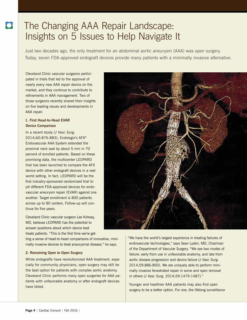
Figures. Imaging studies of the right lower lobe in one of the Cleveland Clinic CTEPH cases before (opposite page), during (left) and after (below) successful bal-loon pulmonary angioplasty.

Page 12 | Cardiac Consult | Fall 2016 |
SURGICAL DECISION-MAKING SERIES
Choosing Graft Types for CABGWeighing the conduit options means balancing a host of factors.
Coronary artery bypass grafting (CABG) remains the gold-
standard treatment for coronary artery disease. But which
grafts are best for bypass surgery?
The choice of bypass graft depends on a number of factors:
• Location of the blockage
• Extent of the blockage
• Size of the coronary arteries
• Availability of arteries and veins
• Patient medical factors
The success of CABG over
time depends on the long-term
patency of the arterial or venous
grafts used, notes Michael Zhen-
Yu Tong, MD, MBA, a cardiac
surgeon in Cleveland Clinic’s
Department of Thoracic and Cardio-
vascular Surgery.
“In general, arterial grafts are better
and more durable than veins,” he
says, pointing out that arterial grafts
are considered superior conduits
over saphenous vein grafts based
on experience using the left internal
mammary artery to bypass the left
anterior descending (LAD) coronary
artery. The efficacy of the radial
artery graft is less clear, he adds.
Cardiac Consult recently caught up with
Dr. Tong for the following summary of how
he weighs the pros and cons of various artery
and vein graft options in CABG procedures.
Internal Thoracic Artery
As the most commonly used bypass grafts, internal thoracic
(mammary) artery (ITA) grafts show the best long-term results.
In most cases, the artery is left intact at its origin, with the
opposite end sewn to the coronary artery below the site of the
blockage. Use of ITA grafts is considered a quality indicator
by the Society of Thoracic Surgeons (STS) and is factored into
STS star-rating calculations for cardiac surgery programs. “We
aim to use at least one ITA graft in 100 percent of patients
undergoing isolated coronary bypass surgery,” Dr. Tong notes.
ITA grafts are resistant to late failure. Studies of angiograms
performed after CABG show that not only do left ITA-LAD
grafts have a more than 90 percent chance of functioning
well early, but these grafts continue to function well over the
long term. Development of obstructions in these grafts has
been shown to be extremely uncommon.
Long-term follow-up studies done at Cleveland
Clinic from the 1980s showed that
these grafts have an important long-
term effect on clinical outcomes.
Over time, patients with left ITA-
LAD grafts were less likely to die
or need reoperation compared with
patients who received only vein
grafts. This is now a standard for
coronary bypass grafting.
In addition to the left ITA, the right
ITA is also often used in patients
age 65 or younger — as well as
in older but otherwise relatively
healthy patients when more than
one graft is needed, says Dr.
Tong. Long-term studies from
Cleveland Clinic found that
bilateral ITA grafts further de-
crease the long-term risks of
death and reoperation com-
pared with single ITA grafts.
Use of both ITAs as bypass
grafts is more complex and is
not appropriate for some patients.
“When more than one graft is needed in younger patients, we
will try to use the right and left mammary arteries,” says Dr.
Tong. “Exceptions are patients with coexisting obesity and
diabetes, as this can make wound healing more difficult due
to reduced blood flow to the sternum.” Bilateral grafting can
be considered in diabetics who are not obese and have good
blood sugar control, he adds.
In situ internal thoracic artery
Free internal thoracic artery
Radial artery

| Cardiac Consult | Fall 2016 | Page 13Visit clevelandclinic.org /heart
First CABG Guidelines on Selecting Arterial ConduitsFebruary 2016 saw the publication of the first set of guidelines
specifically focused on the choice of arterial conduits for CABG.
The guidelines were developed by a Society of Thoracic
Surgeons-convened expert writing group, including two
Cleveland Clinic cardiothoracic surgeons, based on a system-
atic literature review. The document, published in Annals of
Thoracic Surgery (2016;101:419-421), was described by an
accompanying editorial as part of a long-overdue “shift from
telling us when to operate to guiding us in how best to do so.”
The guidelines distinguish themselves from previous CABG-
related guideline documents in at least several notable ways:
• By recommending use of an internal thoracic artery (ITA),
rather than specifically the left ITA, for bypassing the left
anterior descending coronary artery
• By recommending consideration of a second arterial conduit
— either the right ITA or a radial artery — as an adjunct to
the left ITA in appropriate patients
• By recommending consideration of bilateral ITA grafting for
patients without excessive risk of sternal wound infection
• By endorsing consideration of a radial artery graft (as an adjunct
to left ITA grafting) for cases of “severe stenoses” rather than
specifying a percentage threshold of coronary artery stenosis
• By avoiding use of an age threshold for arterial revascularization
SURGICAL DECISION-MAKING SERIES
Radial Artery
Use of the radial artery (RA) in bypass surgery was revived in
the past decade after having been abandoned following high
rates of graft occlusion. The renewed interest stems from new
methods of preparation and drug treatment with antispas-
modic agents to improve long-term results.
Advantages of RA grafts include easy preparation and avail-
ability for use in most patients. Also, because RA grafts involve
arteries rather than veins, they are more resistant to develop-
ment of atherosclerosis, a problem that plagues vein grafts.
While medium-term results with RA grafts are good, these
grafts’ long-term patency and outcomes are not yet fully known.
RA grafts tend to work best when placed on an artery with a
blockage of at least 70 percent (and preferably higher).
RA grafts are recommended for young patients when a third ar-
terial graft is needed or if two arterial grafts are needed but the
right ITA is unsuitable, Dr. Tong notes. They’re also used in older
patients more cautiously when other grafts are unavailable.
Because the RA has a relatively muscular wall, it has a ten-
dency to go into spasm. If the RA is used as a graft, patients
are placed on a calcium channel blocker for several months
postoperatively to keep the artery open.
Before RA graft use, an Allen test is performed to determine
whether blood flow to the hand is sufficient. The artery can
be harvested minimally invasively through a small incision.
Wrist or hand numbness may occur as a side effect.
Gastroepiploic Artery
The gastroepiploic artery (GEA) has been used as a bypass graft,
usually to the right coronary artery. However, Dr. Tong notes
that this rarely used graft type comes into play only if no other
conduit is possible or when a fourth arterial graft is needed.
Bypass with a GEA graft is technically difficult and not a
popular choice among surgeons. Because it requires entry via
the abdomen, it is more invasive than other options but has
a high likelihood of good long-term functioning when used in
the right situation. In some patients a GEA graft represents an
advantage over vein grafts.
Saphenous Vein
The saphenous vein is a commonly used conduit for bypass
due to the ease of harvest, which is usually done through
minimally invasive procedures, with less scarring and faster
recovery. But the failure of vein grafts over the long term
remains a significant problem. Reasons for high failure rates
include variable vein quality and size, the presence of valves
within veins and the potential for areas of dilatation (varicosi-
ties) within veins. These and other factors can cause flow-
pattern disturbances within the veins that can lead to early
failure. The 10-year patency of vein grafts is approximately
60 percent, Dr. Tong notes.
Bottom-Line Recommendations
“Many factors go into the choice of conduit used for bypass,”
says Dr. Tong. “For older patients, an ITA graft and a vein graft
will likely be suitable. For medically unstable patients or older
patients, use of two or more arterial grafts may not be ideal
because it requires longer and more tedious surgery. But if the
patient is young and healthy and can tolerate a longer surgery,
using multiple arterial grafts gives the best long-term result.” ■
Contact Dr. Tong at [email protected].

Page 14 | Cardiac Consult | Fall 2016 |
Fresh Thinking on End-Stage Heart FailureRevisiting Distinctions in LVAD Therapy; New Ways to Balance Transplant Supply and Demand
Don’t mistake the multicenter MOMENTUM 3 trial as just another efficacy and safety study of a new left
ventricular assist device (LVAD). The trial also holds potential to shift paradigms about how and when
LVADs are used for patients with advanced heart failure.
Cleveland Clinic is among more than 60 centers participating
in MOMENTUM 3, which is randomizing over 1,000 patients
with advanced, refractory left ventricular heart failure to either
the Thoratec HeartMate 3™ LVAD — available in Europe
but still investigational in the U.S. — or Thoratec’s currently
marketed HeartMate II® LVAD. The new-generation device
incorporates magnetic suspension technology designed to of-
fer a more physiologic option for patients.
Moving Beyond the Bridge/Destination Distinction
Instead of categorizing LVAD use either as a bridge to heart
transplant or as destination therapy, the trial is using the pri-
mary end points of complication-free survival at six months
and two years, regardless of indication.
Indeed, the classification of “bridge” for patients awaiting
transplant versus “destination” for others doesn’t really make
sense for a number of reasons, says Nader Moazami, MD,
Director of Cleveland Clinic’s Cardiac Transplantation and
Ventricular Assist Device Therapy Program. These include
the fact that patients with LVADs on the transplant list may
become ineligible for transplant over time and end up
keeping their device in place long-term.
The HeartMate 3™ device. Reprinted with the permission of Thoratec Corporation.
“About a third of patients don’t fit neatly into either category,” Dr.
Moazami explains. “In this trial, all patients who are eligible for
an LVAD can get one. Some are transplant candidates in whom
device use may be short-term; if not, it becomes long-term.”
He adds that although the traditional “bridge” and “destination”
designations are highly arbitrary, for now the terminology is still
needed for record-keeping and reimbursement.
Will Magnets Mean Better Outcomes?
Unfortunately, Dr. Moazami notes, even with advances in
technology, many clinicians don’t refer for LVAD evaluation
until patients are severely impaired. “Referral patterns aren’t
changing much,” he says. “Often patients are referred later
in the course of heart failure than they should be,” in part
because of lingering concerns about complications despite
significant improvements in LVAD outcomes over the past
decade (see box on next page).
Depending on the results of MOMENTUM 3 and ultimate
FDA approval, the HeartMate 3’s advantages may help
diminish those concerns, he adds.
All LVADs work by the same basic mechanism, using an
impeller that rotates and augments blood flow. Differences
relate to how the impeller is suspended. Earlier LVADs used
solid bearings, while the newer ones — including the Heart-
Mate 3 — use magnetic suspension, which allows for wider
gaps between the impeller and the pump’s housing.
“It’s a tremendous engineering feat to develop a pump of
this size that’s fully magnetically suspended,” Dr. Moazami
says. “The idea is to cause less stress and trauma to blood
elements.” It also will likely reduce the risk of clot formation
and resultant stroke risk.
The magnetic “levitation” also allows for the pump’s action
to be slowed or accelerated, thereby generating pulsation.
“We think it’s more physiologic than those that can’t generate
pulsation,” Dr. Moazami adds. “It’s a much more biocompat-
ible pump.”

| Cardiac Consult | Fall 2016 | Page 15Visit clevelandclinic.org /heart
MOMENTUM 3 enrolled patients rapidly and could be
completed before its estimated completion date in 2018.
Dr. Moazami believes FDA approval may be possible based
on results at the six-month end point, before the trial is
fully finalized.
Curbing LVAD Demand with
Expanded Transplant Opportunities
Cleveland Clinic’s experience with LVADs dates to the early
1990s, when it was among the first centers to implant them.
Today Cleveland Clinic implants approximately 60 LVADs
every year.
Unfortunately, some of that LVAD volume is attributable to
static rates of heart transplantation — the most effective
treatment for end-stage heart failure — due to recent scarcity
of donor hearts across much of the U.S.
Cardiologist Eileen M. Hsich, MD, Associate Medical Director
of Cleveland Clinic’s Cardiac Transplantation Program, is help-
ing shape national efforts to overcome the shortfall in donor
hearts, as demonstrated by two recent papers on the subject.
The first is a review-style perspective article published by
Dr. Hsich in Circulation: Heart Failure (2016;9:e002679) in
the wake of two notable developments:
• Updated guidelines for transplant candidacy from the
International Society for Heart and Lung Transplantation
(J Heart Lung Transplant. 2016;35:1-23)
• A January 2016 proposal from the Organ Procurement
and Transplantation Network (OPTN) to change adult
heart allocation
While applauding these efforts, Dr. Hsich’s paper notes that
they address only parts of the problem. She argues that the
only way to properly match supply with demand in heart
transplantation is to simultaneously do the following:
• Increase the donor pool
• Reduce the wait list
• Improve the allocation system
Her paper explores each of these elements in detail,
comparing strategies in the U.S. with those in other countries.
She concludes that the U.S. heart transplant community
should consider a three-pronged strategy:
• Using more organs from “high-risk” or marginal donors
• Thoughtfully tightening wait-list eligibility (with wider
consideration of VADs as an alternative for appropriate pa-
tients) and standardizing eligibility criteria across centers
• Addressing disparities in the allocation system, in part by
incorporating more tiers based on medical urgency and
by creating an allocation score similar to those for liver
and lung transplants
“Increasing the donor pool, reducing the wait list and im-
proving the allocation system are necessary to better satisfy
demand for heart transplants,” says Dr. Hsich. “Although it’s
easier to focus on one strategy, pursuing all three simultane-
ously is key.” (For more on this paper, see consultqd.cleve-
landclinic.org/heartsupply.)
A Call to Factor in Type of Underlying Heart Disease
Dr. Hsich’s second paper is an analysis she led using data
from the Scientific Registry of Transplant Recipients to exam-
ine outcomes of all adult U.S. heart failure patients awaiting
transplantation between 2004 and 2014 (N = 30,747).
She and colleagues report in JACC Heart Failure (2016;4:689-
697) that the following conditions were associated with the
highest risk of death during the wait for transplant:
• Restrictive cardiomyopathy
• Congenital heart disease
• Prior heart transplantation
They conclude that donor heart allocation should be revised in
light of these findings. “Our data support a change in the allo-
cation system to prioritize restrictive cardiomyopathy, congeni-
tal heart disease and prior heart transplant,” says Dr. Hsich.
(For more on this analysis, see consultqd.clevelandclinic.org/
heartwaitlist.) ■
Contact Dr. Moazami at [email protected] and Dr. Hsich at [email protected].
› ~80% Two-year survival rates among Cleveland Clinic LVAD
recipients for the period 2012-2015
› 1.6% In-hospital mortality among Cleveland Clinic LVAD
recipients in 2015 (vs. 9.9% expected rate from
University HealthSystem Consortium database)
LVAD Outcome Snapshots

Page 16 | Cardiac Consult | Fall 2016 |
We All Have a Case that Stands Out from the Rest. Here’s Mine.Never go to the operating room without a plan B (and C and D).
By Sudish Murthy, MD, PhD
Of the approximately 7,000 patients I’ve treated as a thoracic surgeon, Danielle Abraham is the one that
stands out. In 2009, Danielle was an active 28-year-old — a happily married mother of an 8-month-old
daughter. Life was good, she said, until she contracted the H1N1 flu virus.
Since she was postpartum, it may have been her suppressed
immune system that allowed the influenza to trigger a dev-
astating case of bacterial pneumonia. A relentless cough and
severe pain sent her to the hospital near her home in Buffalo,
New York. For four weeks, she lay in the ICU in an induced
coma. When her right lung collapsed, and then her left lung,
she was flown to Cleveland Clinic.
When I first saw Danielle, the bacterial infection had begun
to dissolve her lung tissue. Pneumatoceles had developed
and had begun to rupture. Air was filling her pleural space.
Even multiple chest tubes couldn’t keep up with the vigor-
ous leak in her lungs. Air from the ventilator was coming
straight out of the tubes.
We knew she could not be ventilated much longer.
A New Spin on an Old Idea
It was late in the evening when we brought Danielle into
surgery. We didn’t expect her to make it to the next morning
on the ventilator.
At first, we tried reducing the pneumatoceles, but her lung
tissue was so thin and diseased that our attempts to seal one
hole just created more holes. The more we sewed or stapled,
the worse the problem became. Within 15 minutes of opening
her chest, we terminated our original plan and began brain-
storming other ways to stabilize her.
In the 1930s, ’40s and ’50s, thoracic surgeons treated
tuberculosis with collapse therapy, intentionally collapsing
the lung. They believed that the tuberculosis bacteria would
fail to thrive if denied oxygen, and lesions would heal if the
lung were immobilized. Historically, surgeons would insert
plombes, similar to plastic ping-pong balls, between the rib
cage and the pleura, compressing the lung.
The concept worked — at least to resolve the tuberculosis.
Unfortunately, it caused complications years later when the
plombes began to erode or move. Putting biologically inert
bodies into the thorax was not a lifelong solution.
While the technique was abandoned, the concept was still
valid. And it’s ultimately what we used to help Danielle.
We attempted to press the inner lining of her rib cage onto
her lung to control the air leak. Instead of using inert objects,
we balled up a biomaterial mesh that typically dissolves
in about six weeks. We hoped that would give Danielle’s
pneumatoceles enough time to close before the lining would
return to its normal position.
After closing up Danielle’s chest, we saw that the amount
of air escaping from her chest tubes had decreased by 95
percent. Our plan was working.
Danielle with her husband and daughter after a recent 5K walk.

| Cardiac Consult | Fall 2016 | Page 17Visit clevelandclinic.org /heart
A Shockingly Positive Outcome
Over the next several weeks, Danielle’s lungs healed and her
infection responded to antibiotics. After nearly three months
of being hospitalized, she returned to Buffalo to complete
months of rehabilitation.
Her recovery was heroic, one that many couldn’t have
endured. However, Danielle claims the hardest part was
being unable to see her daughter, including missing her first
Halloween, her first Thanksgiving and her first steps. I believe
Danielle’s family inspired her to fight as valiantly as she did.
When she returned for a follow-up months later, I was
shocked. I had only known her as a patient on a ventilator.
Now she was back to being the lively wife and mother she
had been before her illness.
I had thrown a medical Hail Mary, but she had caught it
and scored a touchdown. Her own drive and determination
had cured her.
CTs showed no residual evidence of our procedure. The balls of
mesh were completely gone. Although she did lose about 50
percent of her lung function due to the infection, she has been
able to lead a normal life with moderate physical activity.
When I last saw Danielle, she had recently walked a 5K (photo).
The Only Constant Is the Unexpected
Since Danielle’s surgery, we have used the same technique
successfully on other critically ill patients with similar problems.
Having performed thousands of operations in my career,
I wish I could say I’ve seen it all. But there will always be
unexpected circumstances to keep me — and all of us —
learning and innovating.
Going into surgery with a plan A isn’t enough. We also need
plans B, C and D. (For Danielle, plan C was to pack gauze
into her left lung, hoping her right lung would carry her until
cavities in the left lung scarred over. Plan D was to remove
her left lung. I doubt either of those plans would have had
outcomes as favorable.)
Everything we learn as physicians, no matter how seemingly
trivial or historically insignificant, could become vitally
important someday. When it comes to saving a life, some-
times we need every tool in the shed — including plain
common sense. ■
Dr. Murthy ([email protected]) is Section Head of Thoracic Surgery at Cleveland Clinic and Surgical Director of the Center of Major Airway Disease.
We hoped that balling up a dissolvable biomaterial mesh to control
the air leak in her lung would give the pneumatoceles enough time
to close before the lining returned to its normal position.

Page 18 | Cardiac Consult | Fall 2016 |
Research RoundupQuick Takes on Recent Cardiovascular Studies of Note
Centralized Telemetry Monitoring Slashes Alarm Fatigue, Saves Lives
For noncritically ill inpatients, off-site central monitoring
using standardized cardiac telemetry can result in monitored
patient census reductions without increasing — and actually
reducing — cardiopulmonary arrests. That’s the takeaway
from an analysis of the first 13 months of operation of Cleve-
land Clinic’s technician-staffed off-site telemetry monitoring
unit, developed to reduce the “alarm fatigue” endemic to
typical telemetry monitoring.
As reported in JAMA (2016;316:519-524), compared with the
13 months before the unit’s launch, telemetry standardization
in the off-site unit enabled a 15.5 percent weekly monitored
patient census reduction with no rise in cardiopulmonary ar-
rests. Central monitoring detected rate and rhythm changes in
79 percent of patients within one hour of emergency response
team activation, and discretionary direct notification was as-
sociated with successful resuscitation in 93 percent of patients
who coded — a rate that compares very favorably to national
benchmarks. “By eliminating low-risk patients from the monitor,
we can better concentrate our efforts on the patients who really
require our attention,” says lead author Daniel Cantillon, MD.
For more, see consultqd.clevelandclinic.org/telemetry.
Third Time’s No Charm for CETP Inhibitors
Lingering hopes for the CETP inhibitor class of lipid-modifying
drugs were dealt a major blow by data from the phase 3
ACCELERATE trial of evacetrapib, presented at the 2016
European Society of Cardiology meeting by Cleveland Clinic
cardiologist A. Michael Lincoff, MD. The study of patients with
high-risk vascular disease found that despite producing highly
significant HDL cholesterol increases and LDL cholesterol
reductions versus placebo, evacetrapib had no effect on major
cardiovascular events relative to placebo out to three years of
follow-up. The findings triggered early study termination.
“The outcome of this trial was counterintuitive,” says Dr.
Lincoff, adding that it also conflicted with phase 2 findings.
“This raises serious questions about whether we’ll ever see
cardiovascular benefit from this mechanism of lipoprotein
modification.” Evacetrapib is the third CETP inhibitor to
meet its demise in late-stage clinical trials. For more, see
consultqd.clevelandclinic.org/accelerate.
Study: Abandoning CIED Leads Isn’t Worth the Risk
As the popularity of cardiac implantable electronic devices
(CIEDs) surges, the number of infections linked to aban-
doned leads is growing fast. These unwanted leads compli-
cate management of CIED infections and result in worse out-
comes, including death. So finds a large prospective registry
study published online by JACC: Clinical Electrophysiology
in September.
Among 1,386 consecutive patients undergoing transvenous
lead extraction of infected CIEDs at Cleveland Clinic between
1996 and 2012, 23.3 percent had previously abandoned
leads and 76.7 percent did not. Failure to achieve the
study’s primary end point — successful removal of the device
and lead material from the vascular space without a major
complication — was significantly more common in patients
with abandoned leads (13.0 percent) than in those without
abandoned leads (3.7 percent). Lead author Oussama Wazni,
MD, says the study was done in response to frustration with
the high rate of lead abandonment: “It’s a common practice
that postpones risk now but gambles on the patient’s future.”
For more, see consultqd.clevelandclinic.org/abandonedleads.
RV Systolic Pressure Shapes Survival in Mitral Regurgitation
Elevated right ventricular systolic pressure (RVSP) is indepen-
dently associated with worse long-term survival in patients
with primary mitral regurgitation and preserved left ventricular
ejection fraction. So concludes an observational cohort study
of all 1,318 patients with primary myxomatous mitral regurgi-
tation evaluated at Cleveland Clinic between 2005 and 2008.
The study, published in Journal of the American College of
Cardiology (2016;67:2952-2961), is the largest of its type
by far and has the longest follow-up.
Corresponding author Milind Desai, MD, a Cleveland Clinic
cardiologist, says the findings should prompt closer scrutiny
of the optimal timing of mitral valve repair surgery in such
patients. “It appears that even mild RVSP elevation — 35
mm Hg or above — might be a marker of early decompensa-
tion,” he says, noting that waiting for RVSP to progress to 50
mm Hg or more before offering repair could worsen prognosis.
For more, see consultqd.clevelandclinic.org/rvsp.

| Cardiac Consult | Fall 2016 | Page 19Visit clevelandclinic.org /heart
24/7 ReferralsReferring Physician Center and Hotline
855.REFER.123clevelandclinic.org/Refer123
Physician Referral App Download today at the App Store or Google Play.
Physician Directory clevelandclinic.org/staff
Same-Day Appointments 216.444.CARE (2273) or 800.223.CARE (2273)
Track Your Patients’ Care Online Secure online DrConnect account at clevelandclinic.org/drconnect
Critical Care Transport Worldwide 216.448.7000 or 866.547.1467 clevelandclinic.org/criticalcaretransport
Outcomes Books clevelandclinic.org/outcomes
CME Opportunities ccfcme.org
Executive Education clevelandclinic.org/executiveeducation
Cleveland Clinic Way Book Series Lessons in excellence from one of the world’s leading healthcare organizations.
The Cleveland Clinic Way Toby Cosgrove, MD CEO and President | Cleveland Clinic
Communication the Cleveland Clinic Way Edited by Adrienne Boissy, MD, MA, and Tim Gilligan, MD, MS
Innovation the Cleveland Clinic Way Thomas J. Graham, MD Former Chief Innovation Officer | Cleveland Clinic
Service Fanatics James Merlino, MD Former Chief Experience Officer | Cleveland Clinic
About Cleveland Clinic Cleveland Clinic is an integrated healthcare delivery system with local, national and international reach. At Cleveland Clinic, more than 3,400 physicians and researchers represent 120 medical special-ties and subspecialties. We are a main campus, more than 150 northern Ohio outpatient locations (including 18 full-service family health centers and three health and wellness centers), Cleveland Clinic Florida, Cleveland Clinic Lou Ruvo Center for Brain Health in Las Vegas, Cleveland Clinic Canada, Sheikh Khalifa Medical City and Cleveland Clinic Abu Dhabi. In 2016, Cleveland Clinic was ranked the No. 2 hospital in America in U.S. News & World Report’s “Best Hospitals” survey. The survey ranks Cleveland Clinic among the nation’s top 10 hospitals in 13 specialty areas, and the top hospital in heart care for the 22nd consecutive year.
R E S O U R C E S F O R P H Y S I C I A N S
Stay Connected with Cleveland Clinic’s Heart & Vascular InstituteConsult QD — Heart & Vascular
A blog featuring insights and perspectives from
Cleveland Clinic experts. Visit today and join the
conversation.
consultqd.clevelandclinic.org/cardiovascular
Facebook for Medical Professionals
Facebook.com/CMEClevelandClinic
Follow us on Twitter
@CleClinicMD
Connect with us on LinkedIn
clevelandclinic.org/Heartlinkedin
On the web
clevelandclinic.org/heart

16-HRT-2075
The Cleveland Clinic Foundation9500 Euclid Ave./AC311Cleveland, OH 44195
CardiacConsultCME Alert: Live and OnlineValve Disease and Diastology SummitSat.-Mon., March 4-6, 2017
Eden Roc Hotel | Miami Beach, Florida
Information/registration: ccfcme.org/valve-disease-2017
SAVE THE DATE
Consensus & Controversies in the Management of Challenging PatientsThurs., March 16, 2017, 7-9:15 p.m.
Washington, D.C. (venue TBD)
An independent certified session at the ACC’s 66th Scientific Session (ACC.17)
This educational activity is not part of ACC.17,
but its content is reviewed and approved by
the ACC.17 Program Committee.
Free Online CME Available 24/7
• Visit ccfcme.org
• Click “Browse by Specialty”
• Choose “Cardiology”
Here’s a sampling of what you’ll find:
Masters’ Approach to Critical Limb Ischemia
A series of 19 focused webcasts on
diverse aspects of critical limb isch-
emia, peripheral artery disease and
wound care from a multidisciplinary
perspective. Webcasts range from 15
to 90 minutes each.
Online Journal CME from Cleveland Clinic Journal of Medicine
A steady supply of expert reviews on
timely and practical cardiology topics
and issues, such as:
• Dual antiplatelet therapy for acute
coronary syndromes: How long to
continue?
• Premature ventricular contractions:
Reassure or refer?
• When does asymptomatic aortic
stenosis warrant surgery?
Assessment techniques
• Many more
These activities have been approved for
AMA PRA Category 1 credit™.