Embed Size (px)
Citation preview
Nanomed. J. 6(2): 120-127, Spring 2019
RESEARCH PAPER
The comparison of the apoptosis effects of titanium dioxide nanoparticles into MDA-MB-231 cell line in microgravity and
gravity conditions Azadeh Hekmat 1*, Mohaddeseh Rabizadeh 1, Maliheh Safavi 2, Zahra Hajebrahimi 3
1 Department of Biology, Science and Research Branch, Islamic Azad University, Tehran, Iran 2 Department of Biotechnology, Iranian Research Organization for Science and Technology,
Tehran, Iran3Aerospace Research Institute, Ministry of Science Research and Technology, Tehran, Iran
* Corresponding Author Email: [email protected]. This manuscript was submitted on November 10, 2018;approved on February 5, 2019
How to cite this articleHekmat A, Rabizadeh M, Safavi M, Hajebrahimi Z. The comparison of the apoptosis effects of Titanium dioxide nanoparticles into MDA-MB-231 cell line in microgravity and gravity conditions. Nanomed J. 2019; 6(2): 120-127. DOI: 10.22038/nmj.2019.06.0006
ABSTRACTObjective (s): Gravity could affect some system features and perform directly as an organizing field factor. Recent investigations have examined the titanium dioxide nanoparticles (TiO2 NPs) in biomedical applications, mostly in the cancer treatment field. This study aimed to evaluate the effects of simulated microgravity combined with TiO2 NPs in MDA-MB-231 cells proliferation for the first time. In other words, this study examined the utility of the microgravity environment in nano-therapy. Materials and Methods: The MDA-MB-231 human breast cancer cell line and TiO2 NPs were purchased. The 2D clinostat was applied for the simulation of the microgravity. The morphological studies, MTT cytotoxicity assay, Acridine orange/Ethidium bromide double staining studies and flow cytometry analysis were utilized.Results: The MTT assay, the morphological studies, Acridine orange/Ethidium bromide double staining studies and flow cytometry analysis confirmed the apoptosis-inducing effect of microgravity in combination with TiO2 NPs. The IC50 of simulated microgravity in the presence of TiO2 NPs was determined to be 130 µM. Furthermore, MDA-MB-231 cells exposed to microgravity adopted a different phenotype. Conclusion: Based on our observation, although the relative mechanisms need to be explored further, microgravity can strictly affect the TiO2 NPs effects on MDA-MB-231 cells. The significance of this study lied in the fact that simulating microgravity can be a powerful physical cure for cancer therapy and open new horizons for the studies in the field of biology, biophysics, and medicine.
Keywords: Flow cytometry analyses, MDA-MB-231 cells, Simulated microgravity, Titanium dioxide nanoparticles
INTRODUCTIONHumans and living organisms are adapted
to the various conditions on Earth, including the constant force of gravity (9.8 m/s2) [1]. In recent decades, biophysical forces, such as flow, shear stress, and microgravity, are increasingly utilized as an innovative technology to manipulate cells and study the biological process accompanied by traditional techniques [2-4]. Microgravity, as a mechanical factor, has been approved to disturb cell function, cell morphology, and cell growth. Microgravity induces relevant variations in human
physiology, including cell function, cell morphology, and cell growth [5]. It has been previously thought that microgravity is too weak for contrasting the intermolecular forces; however, the direct effects on cells and signaling pathways inside the cell has been recognized [6]. It has been displayed that simulated microgravity can attenuate myogenic differentiation by controlling DNA methylation [7]. Moreover, it could decrease E-Cadherin in MCF7 cells (human breast adenocarcinoma) and generate multicellular spheroids [8]. Masiello et al. observed that simulated microgravity induces distinct phenotypic switch on MDA-MB-231 cells [9]. In fact, examinations on malignant cells exposed to microgravity reported conflicting
121Nanomed. J. 6(2): 120-127, Spring 2019
A. Hekmat et al. / Effects of TiO2 NPs into MDA-MB-231 cell line in microgravity and gravity
results, meaning that some authors have documented an inhibitory effect of microgravity on the proliferation of cancer cell [10, 11], while others have detected the opposite [12, 13].
In recent years, nanoparticle studies have indicated a broad scope in the medicine and biotechnology field attributed to their biological and physicochemical properties [14]. Since the 20th century, studies on the synthesis of TiO2 nanoparticles (TiO2 NPs) have been involved in large-scale production, such as self-cleaning, gas sensors sunscreen, food products, paints, cosmetics, and pharmacy [15, 16]. The studies on TiO2 NPs genotoxicity have been addressed a variety of issues ranging from no genotoxicity in some studies to overwhelming evidence of DNA destruction in other investigations. A rapid glance at the literature indicates that TiO2 NPs is genotoxic in terms of DNA strand breaks. Several studies have well-documented the ability of TiO2 NPs to produce ROS (reactive oxygen species), oxidative DNA damage, and apoptosis and/or necrosis in numerous cell lines [17-20]. Osman et al. indicated that TiO2 NPs initiated DNA destruction in Lymphocytes from healthy individuals to patients with respiratory diseases [21]. On the other hand, Uboldi et al. revealed that rutile TiO2 NPs seemed to be slightly genotoxic; however, anatase TiO2 NPs did not induce any significant genotoxic effect [22]. Carmo et al. showed that TiO2 NPs did not induce oxidative stress in freshwater fish gills, regardless of an increment in ROS production after two weeks of exposure and TiO2 NPs bioaccumulation [23]. Consequently, the effect of TiO2 NPs on proliferation and apoptosis of cells strongly depends on cellular types, TiO2 NPs physicochemical properties, and concentration of TiO2 NPs [24-26].
The mammalian cells could sense some environmental variations due to gravity affecting lots of biophysical parameters, namely viscosity, diffusion process upthrust (buoyancy), and shear forces. As a result, gravity could definitely affect some general features of the systems and perform directly as an organizing field factor. Furthermore, recent studies have examined the TiO2 NPs application in biomedical, particularly in the cancer treatment field. Accordingly, this investigation aimed to investigate the probable effects of simulated microgravity combined with TiO2 NPs in the proliferation of MDA-MB-231 cells. This is the first effort to examine the effects of simulated
microgravity combined with TiO2 NPs on cancer cells. The results acquired from this research can help to discover the microgravity effects on cancer cells and to find out whether or not biological process like cell morphology and cell growth, behave the same in space as it would behave on Earth. Besides, the application of microgravity environment has progressively increased in recent years. Furthermore, this study examined the utility of the microgravity environment in nano-therapy.
MATERIALS AND METHODSMaterials
The MDA-MB-231 human breast cancer cell line was purchased from the National Cell Bank of Iran, Pasteur Institute, Iran. The TiO2 NPs (Anatase phase, powder, less than 10 nanometers, 99% of purity, special surface area 150 m2/g) was purchased from Pars Lima Co., Iran. 3- [4, 5-Dimethylthiazol-2-yl]-2, 5-diphenyltetrazolium bromide (MTT) and DAPI were bought from Sigma Chemical Co., USA. The RPMI 1640, Trypsin-EDTA, streptomycin, penicillin and fetal bovine serum (FBS) were supplied by Gibco, USA. Ethidium bromide and Dimethylsulfoxide (DMSO) were prepared from Merck, Germany. Acridine orange was purchased from Hopkins and Williams Ltd., Chadwell Heath, England.
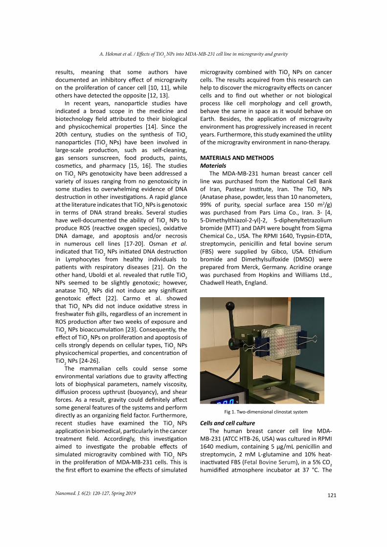
Fig 1. Two-dimensional clinostat system
Cells and cell cultureThe human breast cancer cell line MDA-
MB-231 (ATCC HTB-26, USA) was cultured in RPMI 1640 medium, containing 5 µg/mL penicillin and streptomycin, 2 mM L-glutamine and 10% heat-inactivated FBS (Fetal Bovine Serum), in a 5% CO2 humidified atmosphere incubator at 37 °C. The
1
122
A. Hekmat et al. / Effects of TiO2 NPs into MDA-MB-231 cell line in microgravity and gravity
Nanomed. J. 6(2): 120-127, Spring 2019
cells were grown routinely as monolayer culture.
Simulation of microgravity conditions The 2D clinostat (Aerospace Research Institute,
Tehran, Iran) was applied for the simulation of the microgravity (Fig 1).
This device prevents gravity from affecting cells solution through the rotation. Rotation causes gravity vector unrecognizable to cells. The clinostat was sterilized via UV light and 70% ethanol, and then it was put in a CO2 incubator at 37 °C. The clinostat rotation speed was 20 rpm and the rotation time was 48 hr.
The MDA-MB-231 cells were harvested with trypsin-EDTA when 90% confluence was reached in T25 flasks. In the next step, they were washed with PBS buffer and 1×10⁴ cells were seeded into each well of 96-well plates at growth medium of 180 µl and left to attach to the plates. Afterward, the MDA-MB-231 cells were divided into three groups, including one control group and two experimental groups.
The groups were (1) cells exposed to normal gravity (1g) in the presence of various concentrations of the sterilized TiO2 NPs (5, 10, 20, 40, 80, 100, 200, 400 µM) for 48 hr (control), (2) cells exposed to simulated microgravity and various concentrations of the sterilized TiO2 NPs (5, 10, 20, 40, 80, 100, 200, 400 µM) were added after 48 hr, (3) cells exposed to simulated microgravity in the presence of various concentrations of the sterilized TiO2 NPs (5, 10, 20, 40, 80, 100, 200, 400 µM) for 48 hr.
Due to the small size of each well, the surface tension between the inner vessel wall and medium molecules prevent the pouring of medium and reduce the probability of shear forces during rotation of plates. Furthermore, the culture medium was bubble-free during clinostat rotation due to the small size of wells, the surface tension force between fluid medium, and the inner vessel wall and low culture medium volume (180 µl).
MTT cytotoxicity assayAfter 48 hr of incubation, the medium of
each well was replaced via a fresh medium. Furthermore, 20 μL MTT (5 mg/mL in PBS buffer) was added into each well and incubated for 3 hr at 37 °C. Later, the insoluble formazan formed was dissolved in 100 µl of DMSO (Dimethyl sulfoxide).
The optical density (OD) of each well was quantified against reagent blank with multiwell
scanning spectrophotometer (ELISA reader, Model Expert 96, Asys Hitech) at 490 nm. Each experiment was repeated three times, in addition for each concentration performed in triplicate format.
Morphology changes in treated cellsEach cultured line was trypsinized and washed
with cold phosphate buffered saline (PBS). Afterwards, the cells were plated into 100-mm dishes (1×10⁵ cells/plate) in complete RPMI 1640 medium and incubated at 37°C. The cultures were photographed using a phase-contrast microscope (Olympus CK40).
Acridine orange (AO)/Ethidium bromide (EtBr) double staining
The MDA-MB-231 cells grown in 6-well plates (3×10⁵ cells/well) were washed 3 times by PBS, then 9 µL of cell suspension was stained with 1 µL of dye mixture (100 mg/mL EtBr and 100 mg/mL AO in PBS).
The stained cell suspension (10 µL) were placed on a clean microscope slide and covered with a coverslip and examined via fluorescence microscope (Axoscope 2 plus, Zeiss, Germany).
Flow cytometry analysesFlow cytometry was performed using the
Partec PAS (Germany) of EPICS. The Annexin V binding was used via the Annexin FITC kit, IQ product (Netherlands). Cells were plated into 24 well plates at a density of 106 cells/well for 48 hr. The MDA-MB-231 cells were exposed to simulated microgravity in the presence of 130 µM sterilized TiO2 NPs. After 48 hr, the cells were collected by centrifugation at 1000g for 5 min and washed by PBS twice and suspended in 100 µL of Annexin V binding buffer (10 mM HEPES/NaOH, pH 7.4, 2.5 mM CaCl2, 140 mM NaCl). Consequently, the cells were double stained with 10 µL FITC-labeled Annexin V and 10 µL PI (Propidium iodide) solution (50 µg/mL in PBS).
The samples were incubated at room temperature in the darkness for 20 min and then analyzed via flow cytometry, Partec PAS (Germany).
Statistical analysisStatistical correlation was performed using
independent samples t-test. P-value less than 0.05 were considered statistically significant. All experiments were replicated at least three times.
123Nanomed. J. 6(2): 120-127, Spring 2019
A. Hekmat et al. / Effects of TiO2 NPs into MDA-MB-231 cell line in microgravity and gravity
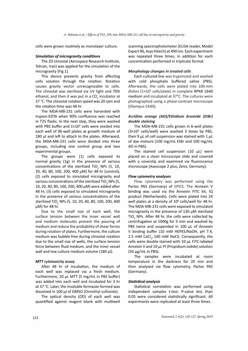
Fig 2. Growth inhibition of MDA-MB-231 cells after 48 hr treatment with TiO2 NPs in normal gravity and microgravity.
Values correspond to mean ±SD of three independent experiments. The signal * indicates a statistically significant
alteration in relation to the bars without * (P<0.05)
RESULTS Growth rates of MDA-MB-231 cells by MTT assay
To the best knowledge of the researchers, no studies have explored the combined effects of microgravity with TiO2 NPs simultaneously on cancer cells. As shown in Fig 2, TiO2 NPs decreased the viability of MDA-MB-231 cells slightly. In other words, TiO2 NPs was not toxic at these concentrations. Various concentrations of the sterilized TiO2 NPs were added when MDA-MB-231 cells were exposed to simulated microgravity for 48 hr. Furthermore, the viability of MDA-MB-231 cells decreased slightly. However, as shown in Fig 2, when MDA-MB-231 cells exposed to simulated microgravity in the presence of various concentrations of the sterilized TiO2 NPs for 48 h, the viability of cells decreased considerably. In addition, it is obvious that TiO2 NPs in combination with microgravity made dose-response suppression on the growth of the cell line. Consequently, the 50% inhibition concentration (IC50) of simulated microgravity in the presence of TiO2 NPs were determined to be 130 µM for MDA-MB-231 cells. As shown in Fig 2, 10% cell death occurred (90% cell growth occurred) at 130 µM of TiO2 NPs. Conversely, the cell death increased to 50% when 130 µM TiO2 NPs was used in the presence of simulated microgravity. The results suggested that microgravity in combination with TiO2 NPs had remarkable and specific antiproliferative effects on MDA-MB-231 cell line.
MDA-MB-231 morphology studies As morphological analysis displays the cell
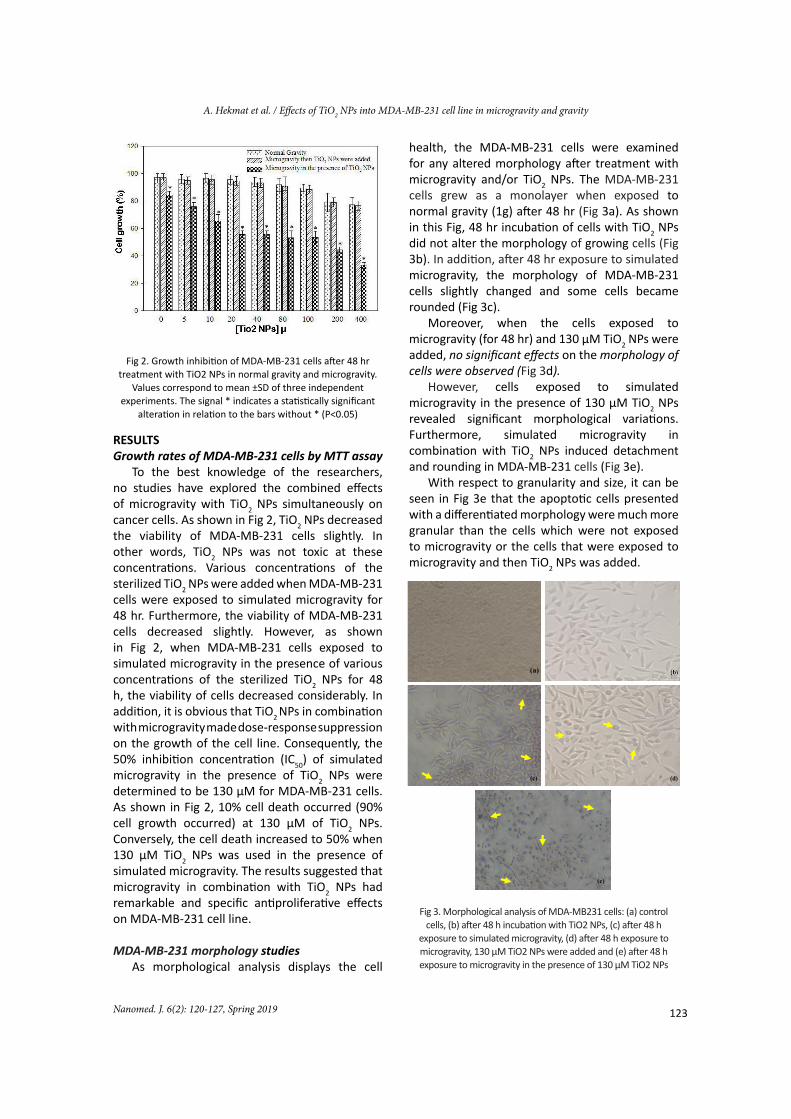
health, the MDA-MB-231 cells were examined for any altered morphology after treatment with microgravity and/or TiO2 NPs. The MDA-MB-231 cells grew as a monolayer when exposed to normal gravity (1g) after 48 hr (Fig 3a). As shown in this Fig, 48 hr incubation of cells with TiO2 NPs did not alter the morphology of growing cells (Fig 3b). In addition, after 48 hr exposure to simulated microgravity, the morphology of MDA-MB-231 cells slightly changed and some cells became rounded (Fig 3c).
Moreover, when the cells exposed to microgravity (for 48 hr) and 130 µM TiO2 NPs were added, no significant effects on the morphology of cells were observed (Fig 3d).
However, cells exposed to simulated microgravity in the presence of 130 µM TiO2 NPs revealed significant morphological variations. Furthermore, simulated microgravity in combination with TiO2 NPs induced detachment and rounding in MDA-MB-231 cells (Fig 3e).
With respect to granularity and size, it can be seen in Fig 3e that the apoptotic cells presented with a differentiated morphology were much more granular than the cells which were not exposed to microgravity or the cells that were exposed to microgravity and then TiO2 NPs was added.
Fig 3. Morphological analysis of MDA-MB231 cells: (a) control cells, (b) after 48 h incubation with TiO2 NPs, (c) after 48 h
exposure to simulated microgravity, (d) after 48 h exposure to microgravity, 130 µM TiO2 NPs were added and (e) after 48 h exposure to microgravity in the presence of 130 µM TiO2 NPs
3
(a)
(c) (d)
(e)
2
124
A. Hekmat et al. / Effects of TiO2 NPs into MDA-MB-231 cell line in microgravity and gravity
Nanomed. J. 6(2): 120-127, Spring 2019
Acridine Orange/Ethidium bromide double stainingTo acquire a better insight into the effects of
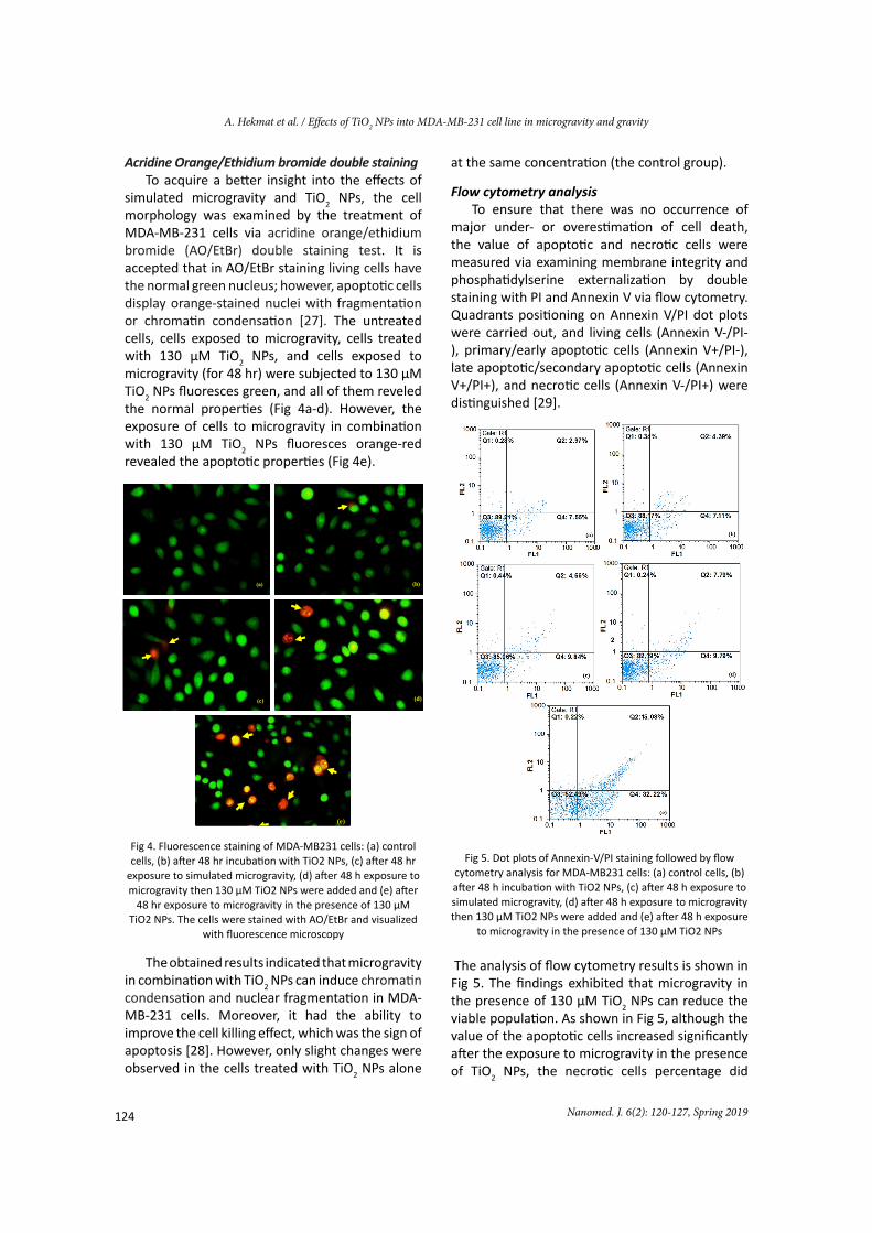
simulated microgravity and TiO2 NPs, the cell morphology was examined by the treatment of MDA-MB-231 cells via acridine orange/ethidium bromide (AO/EtBr) double staining test. It is accepted that in AO/EtBr staining living cells have the normal green nucleus; however, apoptotic cells display orange-stained nuclei with fragmentation or chromatin condensation [27]. The untreated cells, cells exposed to microgravity, cells treated with 130 µM TiO2 NPs, and cells exposed to microgravity (for 48 hr) were subjected to 130 µM TiO2 NPs fluoresces green, and all of them reveled the normal properties (Fig 4a-d). However, the exposure of cells to microgravity in combination with 130 µM TiO2 NPs fluoresces orange-red revealed the apoptotic properties (Fig 4e).
Fig 4. Fluorescence staining of MDA-MB231 cells: (a) control cells, (b) after 48 hr incubation with TiO2 NPs, (c) after 48 hr
exposure to simulated microgravity, (d) after 48 h exposure to microgravity then 130 µM TiO2 NPs were added and (e) after
48 hr exposure to microgravity in the presence of 130 µM TiO2 NPs. The cells were stained with AO/EtBr and visualized
with fluorescence microscopy
The obtained results indicated that microgravity in combination with TiO2 NPs can induce chromatin condensation and nuclear fragmentation in MDA-MB-231 cells. Moreover, it had the ability to improve the cell killing effect, which was the sign of apoptosis [28]. However, only slight changes were observed in the cells treated with TiO2 NPs alone
at the same concentration (the control group).
Flow cytometry analysisTo ensure that there was no occurrence of
major under- or overestimation of cell death, the value of apoptotic and necrotic cells were measured via examining membrane integrity and phosphatidylserine externalization by double staining with PI and Annexin V via flow cytometry. Quadrants positioning on Annexin V/PI dot plots were carried out, and living cells (Annexin V-/PI-), primary/early apoptotic cells (Annexin V+/PI-), late apoptotic/secondary apoptotic cells (Annexin V+/PI+), and necrotic cells (Annexin V-/PI+) were distinguished [29].
Fig 5. Dot plots of Annexin-V/PI staining followed by flow cytometry analysis for MDA-MB231 cells: (a) control cells, (b) after 48 h incubation with TiO2 NPs, (c) after 48 h exposure to simulated microgravity, (d) after 48 h exposure to microgravity then 130 µM TiO2 NPs were added and (e) after 48 h exposure
to microgravity in the presence of 130 µM TiO2 NPs
The analysis of flow cytometry results is shown in Fig 5. The findings exhibited that microgravity in the presence of 130 µM TiO2 NPs can reduce the viable population. As shown in Fig 5, although the value of the apoptotic cells increased significantly after the exposure to microgravity in the presence of TiO2 NPs, the necrotic cells percentage did
4
(a) (b)
(c) (d)
(e)
5
125Nanomed. J. 6(2): 120-127, Spring 2019
A. Hekmat et al. / Effects of TiO2 NPs into MDA-MB-231 cell line in microgravity and gravity
not display significant variations. This result was consistent with the data from AO/EtBr staining and showed the apoptosis-inducing effect of microgravity in combination with TiO2 NPs in MDA-MB-231 cells.
DISCUSSIONIn recent years, the direct effects of microgravity
on biological systems have been challenging. Currently, it has been revealed that microgravity can interrupt biological functions and cellular morphology in numerous cell types. These kind of examinations are critical for future Moon and Mars mission design and training. Importantly, the microgravity environment application has been widely used for various objectives [3].
The most common kind of female cancer is breast cancer. Breast cancer could be categorized into five subtypes, including luminal A, luminal B, basal, Claudin-low, and HER2. The MDA-MB-231 cells belong to claudin-low subtype. The claudin-low (CL) subtype is characterized via the lack of HER2, PR, and ERα expression. These tumors are also characterized via claudin-3 and claudinin-4 downregulation and the proliferation marker Ki67 low expression [30]. Claudin proteins are the tight junctions’ main component. There is some evidence that CL subtype are relatively resistant to conventional anticancer drugs and patients with CL tumors had a worse survival once compared to other subtypes [31]. Consequently, it is important to identify the new therapeutic strategies for breast cancer treatment especially for patients with CL tumors.
In this research, the results revealed that microgravity simulation after 48 h created slightly different morphological phenotypes in MDA-MB-231 cells. This finding was in agreement with the previous study, in which microgravity exposure led to the emergence of two morphologically distinct cell populations [9]. The obtained results of the current study were also in line with those of Gioia et al. [32] reporting that simulated microgravity on human primary osteoblasts could make a cellular regression. Moreover, the 48 h incubation of MDA-MB-231 cells with TiO2 NPs did not alter the morphology of growing cells. It was revealed that photoexcited TiO2 particles could induce cytotoxicity against cancer cells [33]. Garcia-Contreras et al. observed significant cell area expansion only on UV-irradiated plates, not on non-UV-irradiated Ti plates [34]. However, the TiO2
NPs utilized in this research was not active under UV light irradiation. The reason for this is that TiO2 NPs did not alter the morphology of MDA-MB-231 cells and the cells were significantly resistant to TiO2 NPs. Moreover, when the cells exposed to microgravity first and then TiO2 NPs was added, no significant effects on the morphology of cells were observed. Conversely, cells exposed to simulated microgravity in the presence of TiO2 NPs revealed significant morphological variations.
The MTT assay displayed microgravity and TiO2 NPs alone decreased the viability of MDA-MB-231 cells slightly. When MDA-MB-231 cells exposed to simulated microgravity and after 48 h various concentrations of TiO2 NPs were added, the viability of MDA-MB-231 cells decreased slightly. However, when MDA-MB-231 cells exposed to simulated microgravity in the presence of various concentrations of the sterilized TiO2 NPs for 48 hr, the viability of cells decreased noticeably. The IC50 of simulated microgravity in the presence of TiO2 NPs were determined as 130 µM. These results suggested that microgravity and TiO2 NPs had a remarkable and specific antiproliferative effects on MDA-MB-231 cell line. It is clear that microgravity and TiO2 NPs made dose-response suppression on the growth of MDA-MB-231 cells. Based on these results, it seemed that TiO2 NPs combined with microgravity could promote the mortality of cells in addition to those mortality effects induced via TiO2 NPs alone. This means that microgravity and TiO2 NPs can cooperatively kill more MDA-MB-231 cancer cells.
The AO/EB staining exposed to microgravity in combination with TiO2 NPs could induce chromatin condensation and nuclear fragmentation in MDA-MB-231 cells, which was the sign of apoptosis. Conversely, only the slight changes were observed in the cell in the control group exposed to simulated microgravity or treated with TiO2 NPs alone. This observation was consistent with MTT assay studies as mentioned above. The flow cytometry results demonstrated that microgravity in the presence of TiO2 NPs could reduce the viable population. The values of apoptotic cells were increased significantly after the exposure to microgravity in the presence of TiO2 NPs; however, the necrotic cells percentage did not indicate significant variations. This result was in line with the data from AO/EB staining and showed the apoptosis-inducing effect of microgravity in combination with TiO2 NPs in MDA-MB-231 cells.
126
A. Hekmat et al. / Effects of TiO2 NPs into MDA-MB-231 cell line in microgravity and gravity
Nanomed. J. 6(2): 120-127, Spring 2019
In humans, ABC (ATP-Binding Cassette) transporters proteins play many significant roles in drug-to-drug interactions, drug pharmacodynamics, and drug pharmacokinetics. The ABC transporters could be categorized into three classes, namely ABCB1 (MDR-1/P-gp), ABCG2 (BCRP) and ABCC1 (MRP-1). The overexpression of these transporters can initiate treatment failure due to the increased drug cell clearance and enhanced excretion of the drug from the body [35]. There is some evidence that microgravity can reduce MRP2 net transport activity and P-gp (P-glycoprotein) level [35, 36]. Furthermore, Goldermann and Hanke [37] showed that the activity of membrane-ion channels alters in microgravity condition. Based on our observations, microgravity and TiO2 NPs can cooperatively kill more MDA-MB-231 cancer cells. Consequently, although the present results were not enough to draw certain explanations to our observations, we are able to hypothesize that modified polar/apolar interactions caused by microgravity could affect vesicle’s lipid bi-layer membrane and impaired transporter-membrane interaction. However, additional investigations will be required to evaluate the ultra-structure of the transporter in microgravity.
On the other hand, when MDA-MB-231 cells exposed to microgravity obtained a different phenotype. As indicated previously, gravity, as an external field, dramatically influences the system in selecting one out of many other configurations. However, in microgravity condition, the system can access diverse attractor states without restrictions, and recover new configuration states or phenotypes [9, 38]. Variations in the shapes of the cell are associated with alteration in cytoskeleton architecture. The balance between the cytoskeleton architecture and tensional forces regulates numerous complex cell functions, such as apoptosis and proliferation directly [9]. Consequently, based on our observation, although the relative mechanisms need to be explored further, microgravity can strictly affect the TiO2 NPs effects on MDA-MB-231 cells. On the basis of the obtained results of this study, it can be recommended that microgravity can be a powerful physical treatment for cancer therapy, which opens a new horizon to novel strategies for the cancer treatment field. Furthermore, our observations open fascinating research lines in biology, biophysics, and medicine.
ACKNOWLEDGEMENTS We thank Ms. Evini at Institute of Biochemistry
and Biophysics of the University of Tehran for technical support.
CONFLICTS OF INTERESTThere are no conflicts of interest relating to the
manuscript and there were no extramural sources supporting this research.
REFERENCES1.Manzano AI, Herranz R, van Loon JJ, Medina FJ. A
hypergravity environment induced by centrifugation alters plant cell proliferation and growth in an opposite way to microgravity. Microgravity Sci Technol. 2012; 24(6): 373-381.
2.Blaber E, Sato K, Almeida EA. Stem cell health and tissue regeneration in microgravity. Stem Cells Dev. 2014; 23(S1): 73-78.
3.McPherson A, DeLucas LJ. Microgravity protein crystallization. NPJ Microgravity. 2015; 1: 1-20.
4.Shinde V, Brungs S, Henry M, Wegener L, Nemade H, Rotshteyn T, Acharya A. Baumstark-Khan C. Hellweg C.E. Hescheler J. Hemmersbach R. Sachinidis A. Simulated microgravity modulates differentiation processes of embryonic stem cells. Cell Physiol Biochem. 2016; 38(4): 1483-1499.
5.Buckey JC. Space physiology: Oxford University Press, USA; 2006.
6.Albrecht-Buehler G. Possible mechanisms of indirect gravity sensing by cells. ASGSB Bull. 1991;4(2): 25-34.
7.Furukawa T, Tanimoto K, Fukazawa T, Imura T, Kawahara Y, Yuge L. Simulated microgravity attenuates myogenic differentiation via epigenetic regulations. NPJ Microgravity. 2018; 4: 1-8.
8.Sahana J, Nassef MZ, Wehland M, Kopp S, KrKr Kawahara Y, Yuge L. Decreased E-Cadherin in MCF7 Human Breast Cancer Cells Forming Multicellular Spheroids Exposed to Simulated Microgravity. Proteomics. 2018; 28(3): e1800015.
9.Masiello MG, Cucina A, Proietti S, Palombo A, Coluccia P, D’Anselmi F, Dinicola S, Pasqualato A, Morini V, Bizzarri M. Phenotypic switch induced by simulated microgravity on MDA-MB-231 breast cancer cells. BioMed Res Int. 2014; 2014: 1-12.
10.Vassy J, Portet S, Beil M, Millot G, Fauvel-Lafeve F, Gasset G, Schoevaert D. Weightlessness acts on human breast cancer cell line MCF-7. Adv Space Res. 2003; 32(8): 1595-1603.
11.Qian A, Zhang W, Xie L, Weng Y, Yang P, Wang Z, Hu L, Xu H, Tian Z, Shang P. Simulated weightlessness alters biological characteristics of human breast cancer cell line MCF-7. Acta Astronaut. 2008; 63(7-10): 947-958.
12.Grigoryan E, Anton H, Mitashov V. Real and simulated microgravity can activate signals stimulating cells to enter the S phase during lens regeneration in urodelean amphibians. Adv Space Res. 2006; 38(6): 1071-1078.
13.Yuge L, Kajiume T, Tahara H, Kawahara Y, Umeda C, Yoshimoto R, Wu SL, Yamaoka K, Asashima M, Kataoka K, Ide T. Microgravity potentiates stem cell proliferation while sustaining the capability of differentiation. Stem Cells Dev. 2006; 15(6): 921-929.
127Nanomed. J. 6(2): 120-127, Spring 2019
A. Hekmat et al. / Effects of TiO2 NPs into MDA-MB-231 cell line in microgravity and gravity
14.Jin S, Leach JC, Ye K. Nanoparticle-mediated gene delivery. Micro and Nano Technologies in Bioanalysis: Springer; 2009: 547-557.
15.Lusvardi G, Barani C, Giubertoni F, Paganelli G. Synthesis and Characterization of TiO2 Nanoparticles for the Reduction of Water Pollutants. Materials (Basel). 2017; 10(10): 1-11.
16.Iavicoli I, Leso V, Fontana L, Bergamaschi A. Toxicological effects of titanium dioxide nanoparticles: a review of in vitro mammalian studies. Eur Rev Med Pharmacol Sci. 2011; 15(5): 481-508.
17.Hussain S, Hess K, Gearhart J, Geiss K, Schlager J. In vitro toxicity of nanoparticles in BRL 3A rat liver cells. Toxicol In Vitro. 2005; 19(7): 975-983.
18.Lai JC, Lai MB, Jandhyam S, Dukhande VV, Bhushan A, Daniels CK, Leung SW. Exposure to titanium dioxide and other metallic oxide nanoparticles induces cytotoxicity on human neural cells and fibroblasts. Int J Nanomedicine. 2008; 3(4): 533-545.
19.Shukla RK, Kumar A, Gurbani D, Pandey AK, Singh S, Dhawan A. TiO2 nanoparticles induce oxidative DNA damage and apoptosis in human liver cells. Nanotoxicology. 2013; 7(1): 48-60.
20.Hekmat A, Saboury AA, Divsalar A, Seyedarabi A. Structural effects of TiO2 nanoparticles and doxorubicin on DNA and their antiproliferative roles in T47D and MCF7 cells. Anticancer Agents Med Chem. 2013; 13(6): 932-951.
21.Osman IF, Najafzadeh M, Sharma V, Shukla RK, Jacob B, Dhawan A, Anderson D. TiO2 NPs Induce DNA Damage in Lymphocytes from Healthy Individuals and Patients with Respiratory Diseases-An Ex Vivo/In Vitro Study. J Nanosci Nanotechnol. 2018; 18(1): 544-555.
22.Uboldi C, Urbán P, Gilliland D, Bajak E, Valsami-Jones E, Ponti J, Rossi F. Role of the crystalline form of titanium dioxide nanoparticles: Rutile, and not anatase, induces toxic effects in Balb/3T3 mouse fibroblasts. Toxicol In Vitro. 2016; 31: 137-145.
23.do Carmo T, Azevedo V, de Siqueira P, Galvão T, Dos Santos F, Dos Reis Martinez C, Appoloni CR, Fernandes MN. Reactive oxygen species and other biochemical and morphological biomarkers in the gills and kidneys of the Neotropical freshwater fish, Prochilodus lineatus, exposed to titanium dioxide (TiO2) nanoparticles. Environ Sci Pollut Res Int. 2018; 25(23): 22963-22976.
24.Valdiglesias V, Costa C, Sharma V, Kiliç G, Pásaro E, Teixeira JP, Dhawan A, Laffon B. Comparative study on effects of two different types of titanium dioxide nanoparticles on human neuronal cells. Food Chem Toxicol. 2013; 57: 352-361.
25.Pandurangan M, Enkhtaivan G, Young JA, Hoon HJ, Lee
H, Lee S, Kim DH. In vitro therapeutic potential of TiO2 nanoparticles against human cervical carcinoma cells. Biol Trace Elem Res. 2016; 171(2): 293-300.
26.Rahmani NK, Rasmi Y, Abbasi A, Koshoridze N, Shirpoor A, Burjanadze G, Saboory E. Bio-Effects of TiO2 Nanoparticles on Human Colorectal Cancer and Umbilical Vein Endothelial Cell Lines. Asian Pac J Cancer Prev. 2018; 19(10): 2821-2829.
27.Kheirollahi A, Pordeli M, Safavi M, Mashkouri S, Naimi-Jamal MR, Ardestani SK. Cytotoxic and apoptotic effects of synthetic benzochromene derivatives on human cancer cell lines. Naunyn Schmiedebergs Arch Pharmacol. 2014; 387(12): 1199-1208.
28.Elmore S. Apoptosis: a review of programmed cell death. Toxicol Pathol. 2007; 35(4): 495-516.
29.Debnath A, McKerrow JH. Drug Development for Parasite-induced Diarrheal Diseases. Front Microbiol. 2017; 8: 1-3.
30.Holliday DL, Speirs V. Choosing the right cell line for breast cancer research. Breast Cancer Res. 2011; 13(4): 1-7.
31.Dias K, Dvorkin-Gheva A, Hallett RM, Wu Y, Hassell J, Pond GR, Levine M, Whelan T, Bane AL. Claudin-low breast cancer; clinical & pathological characteristics. PLoS One. 2017; 12(1): 1-17.
32.Gioia M, Michaletti A, Scimeca M, Marini M, Tarantino U, Zolla L, Coletta M. Simulated microgravity induces a cellular regression of the mature phenotype in human primary osteoblasts. Cell Death Discov. 2018; 4(1): 1-14.
33.Song M, Zhang R, Dai Y, Gao F, Chi H, Lv G, Chen B, Wang X. The in vitro inhibition of multidrug resistance by combined nanoparticulate titanium dioxide and UV irradition. Biomaterials. 2006; 27(23): 4230-4238.
34.Garcia-Contreras R, Kanagawa S, Beppu Y, Nagao T, Sakagami H, Nakajima H, Shimada J, Adachi K. Morphological features of osteoblasts cultured on ultraviolet-irradiated titanium plates. in vivo. 2011; 25(4): 649-655.
35.Vaquer S, Cuyàs E, Rabadán A, González A, Fenollosa F, de la Torre R. Active transmembrane drug transport in microgravity: a validation study using an ABC transporter model. F1000Research. 2014; 3: 1-15.
36.Lu SK, Bai S, Javeri K, Brunner LJ. Altered cytochrome P450 and P-glycoprotein levels in rats during simulated weightlessness. Aviat Space Environ Med. 2002; 73(2): 112-118.
37.Goldermann M, Hanke W. Ion channel are sensitive to gravity changes. Microgravity Sci Technol. 2001; 13(1): 35-38.
38.Nicolis G, Prigogine I. Symmetry breaking and pattern selection in far-from-equilibrium systems. Proc Natl Acad Sci U S A. 1981; 78(2): 659-663.