Embed Size (px)
Citation preview

647RESEARCH ARTICLE
INTRODUCTIONIn the vertebrate embryo, the segregation of cells into paraxialmesoderm, which forms the basis of skeletal muscle, and lateralmesoderm, which contributes cardiac myocytes, is thought tooccur during gastrulation. In vertebrates, myocardial progenitorsmigrate from the primitive streak anteriolaterally to form bilateralheart fields. During head-fold stages, these progenitors form thecardiac crescent, prior to the formation of the linear heart tube.Studies in chick embryos have demonstrated that subsequently,most of the outflow tract (OFT) is populated by myocardialprogenitors from the anterior or secondary heart field (AHF/SHF),which resides in pharyngeal mesoderm dorsal to the heart(Mjaatvedt et al., 2001; Waldo et al., 2001). In mouse embryos,retrospective lineage analysis has demonstrated the presence oftwo heart fields – the first and the second heart field – based ontheir timing of entry into the heart and their timing ofdifferentiation (Buckingham et al., 2005). The first heart fieldprimarily contributes to the left ventricle, whereas the second heartfield contributes most of the cells of the cardiac OFT and rightventricle (RV), a majority of cells in the atria, and some cellswithin the left ventricle (Black, 2007; Buckingham et al., 2005;Garry and Olson, 2006; Srivastava, 2006). In chick, the preciseboundaries and molecular identities of mesodermal ‘fields’ are less
clear, owing to a lack of genetic and lineage-specific markers forthese early progenitors, as well as differences between chick andmouse models (Abu-Issa and Kirby, 2007).
Heart development takes place in close apposition to thedeveloping head. The separation between the heart and the headcommences gradually, following heart-looping stages as the heartshifts caudally. The term ‘cardio-craniofacial morphogenetic field’reflects the intimate developmental relationship between the head,face and heart, which is also reflected in numerous cardiac andcraniofacial birth defects (Hutson and Kirby, 2003).
Cranial paraxial mesoderm (CPM) cells located anterior to thesomites, as well as prechordal mesoderm, provide the precursorsfor approximately 60 distinct skeletal muscles in the head, whichare used to facilitate food intake, move the eyeball, provide facialexpressions and aid speech in humans (Wachtler and Jacob, 1986).CPM cells stream into the neighboring branchial arches (BAs, alsoknown as pharyngeal arches), the templates of the adultcraniofacial structures. Within the BAs, cranial neural crest cellssurround the muscle anlagen (Noden, 1983; Trainor et al., 1994);these cells provide multiple signals that regulate cranial musclepatterning and differentiation (Rinon et al., 2007; Tzahor et al.,2003).
It is well accepted that distinct developmental programs controlskeletal muscle formation in the trunk and in the head (reviewed byBothe et al., 2007; Noden and Francis-West, 2006). Moreover,muscle myopathies are known to be differentially linked to aspecific trunk or cranial region (Emery, 2002). Similarly, within thehead musculature, eye muscles differ from branchiomeric muscles,and there is evidence that branchiomeric muscle developmentvaries among the different BAs (Dong et al., 2006; Kelly et al.,2004).
The contribution of Islet1-expressing splanchnic mesodermcells to distinct branchiomeric muscles reveals significantheterogeneity in head muscle developmentElisha Nathan1, Amir Monovich1, Libbat Tirosh-Finkel1, Zachary Harrelson2, Tal Rousso1, Ariel Rinon1,Itamar Harel1, Sylvia M. Evans2 and Eldad Tzahor1,*
During embryogenesis, paraxial mesoderm cells contribute skeletal muscle progenitors, whereas cardiac progenitors originate inthe lateral splanchnic mesoderm (SpM). Here we focus on a subset of the SpM that contributes to the anterior or secondary heartfield (AHF/SHF), and lies adjacent to the cranial paraxial mesoderm (CPM), the precursors for the head musculature. Molecularanalyses in chick embryos delineated the boundaries between the CPM, undifferentiated SpM progenitors of the AHF/SHF, anddifferentiating cardiac cells. We then revealed the regionalization of branchial arch mesoderm: CPM cells contribute to theproximal region of the myogenic core, which gives rise to the mandibular adductor muscle. SpM cells contribute to the myogeniccells in the distal region of the branchial arch that later form the intermandibular muscle. Gene expression analyses of thesebranchiomeric muscles in chick uncovered a distinct molecular signature for both CPM- and SpM-derived muscles. Islet1 (Isl1) isexpressed in the SpM/AHF and branchial arch in both chick and mouse embryos. Lineage studies using Isl1-Cre mice revealed thesignificant contribution of Isl1+ cells to ventral/distal branchiomeric (stylohyoid, mylohyoid and digastric) and laryngeal muscles. Bycontrast, the Isl1 lineage contributes to mastication muscles (masseter, pterygoid and temporalis) to a lesser extent, with virtually nocontribution to intrinsic and extrinsic tongue muscles or extraocular muscles. In addition, in vivo activation of the Wnt/�-cateninpathway in chick embryos resulted in marked inhibition of Isl1, whereas inhibition of this pathway increased Isl1 expression. Ourfindings demonstrate, for the first time, the contribution of Isl1+ SpM cells to a subset of branchiomeric skeletal muscles.
KEY WORDS: Anterior heart field, Splanchnic mesoderm, Myogenesis, Wnt/�-catenin
Development 135, 647-657 (2008) doi:10.1242/dev.007989
1Department of Biological Regulation, Weizmann Institute of Science, Rehovot76100, Israel. 2University of California–San Diego, Skaggs School of Pharmacy,La Jolla, CA 92093, USA.
*Author for correspondence (e-mail: [email protected])
Accepted 26 November 2007 DEVELO
PMENT
648
A previous study in chick embryos demonstrated that signalsfrom the dorsal neural tube (e.g. Wnt1 and Wnt3a) blockcardiogenesis in the adjacent CPM (Tzahor and Lassar, 2001). Wefurther demonstrated in vivo, also in chick embryos, that a subsetof CPM cells contributes to both myocardial and endocardial cellpopulations within the cardiac OFT (Tirosh-Finkel et al., 2006).These two studies revealed that CPM cells contribute to bothcardiac and skeletal muscle lineages, and illustrate the plasticity ofthese cells during embryogenesis. In accordance with these results,recent studies involving various transgenic mouse lines havedemonstrated an overlap in the progenitor populations contributingto branchiomeric and cardiac muscle (Dong et al., 2006; Kelly etal., 2001; Verzi et al., 2005) (reviewed by Grifone and Kelly,2007).
The LIM homeodomain protein Islet1 (Isl1) stands at anodal point in the self-renewal, differentiation and lineagespecification of distinct cardiovascular precursors, and is a majorplayer in the second heart field lineage during embryogenesis (Caiet al., 2003; Laugwitz et al., 2005; Moretti et al., 2006). Thistranscription factor marks undifferentiated progenitors of theSHF; its expression is downregulated with differentiation (Cai etal., 2003). Genetic removal of Isl1 in mice showed that Bmp4 (aswell as other Bmp and Fgf family members) is a target of Isl1in the AHF (Cai et al., 2003). We demonstrated in chickembryos that Bmp4 induces Isl1 expression in the CPM, whileblocking its expression in neuronal tissues (Tirosh-Finkel et al.,2006). More recently, it has been shown in mice that �-catenindirectly targets and activates Isl1 expression in the AHF (Lin etal., 2007).
In the present study, we characterized the nature of the Isl1+
cardio-craniofacial splanchnic mesoderm, using several lineage-tracing and gene expression techniques in both chick and mouseembryos. At both the cellular and molecular levels, the cardio-craniofacial mesoderm can be divided into two compartments: theCPM and splanchnic mesoderm (SpM), part of which comprisesthe AHF. Following linear heart tube stages, we found thatIsl1+/SpM cells contribute to the distal part of the pharyngeal(branchial) mesoderm, as well as to the cardiac OFT. Molecularand lineage analyses of the head musculature in chick and mouseembryos demonstrated distinct molecular and developmentalprograms for CPM and Isl1+ SpM-derived branchiomeric muscles.Furthermore, we demonstrate that the Wnt/�-catenin pathwayregulates Isl1 (and Nkx2.5) protein expression, presumably byfine-tuning boundary formation within the cardio-craniofacialmesoderm.
MATERIALS AND METHODSEmbryosFertilized white eggs from commercial sources were incubated for 1-3 daysat 38.5°C in a humidified incubator to HH stage (St.) 3-26 (Hamburger andHamilton, 1992).
Whole-mount in situ hybridizationWhole-mount in situ hybridization was performed using digoxigenin (dig)-labeled antisense riboprobes synthesized from total cDNA. A detailedprotocol, as well as specific primers for cDNA probes, are available uponrequest.
Double-fluorescence in situ hybridization (FISH) on paraffinsectionsParaffin sections were hybridized with two RNA probes, one labeled withdig-UTP and the other with fluorescein-UTP. Post-hybridization, each probewas developed separately using the FITC/Cy3 tyramide amplificationsystem (Perkin Elmer).
Sectioning and histologyFor cryosections, embryos were incubated overnight in 20% sucrose in PBS,and then embedded in 7.5% gelatin, 15% sucrose in PBS. Blocks weretrimmed and frozen and then sectioned at 20 �m.
Lineage tracing and dye injectionDiI/DiO (D282, C275, Molecular Probes) labeling experiments wereperformed on St. 8 embryos as described previously (Tirosh-Finkel et al.,2006).
Implantation of Fz8-CRD-IgG beadsAffi-Gel blue gel beads (150-300 �m; Bio-Rad) were soaked in 200 ng/�lFz8-CRD-IgG or BSA prior to in vivo implantation into the CPM of St. 8-9embryos.
Mutant mice and lacZ stainingIsl1-Cre and Rosa26R strains were crossed to generate embryos at E10.5,12.5 and 16.5, as previously described (Yang et al., 2006). �-gal staining wasperformed as previously described (Moretti et al., 2006). Embryos wereembedded in paraffin and 8 �m sections were counterstained with NuclearFast Red.
Immunofluorescence stainingSections were blocked with 5% goat serum, 1% BSA in PBS prior toincubation with the primary antibody: Nkx2.5 (Santa-Cruz), �-galactosidase(Sigma), chick MyoD (a gift from Prof. Yablonka-Reuveni, University ofWashington School of Medicine, Seattle, WA), Myf5 (a gift from Dr BrucePaterson, NIH, Bethesda, MD), Isl1, Pax7 and MF20 (DSHB). Secondaryantibodies used were Cy3 or Cy2-conjugated-anti-mouse or anti-rabbit IgG(Jackson ImmunoResearch).
ElectroporationDetailed protocols are available upon request.
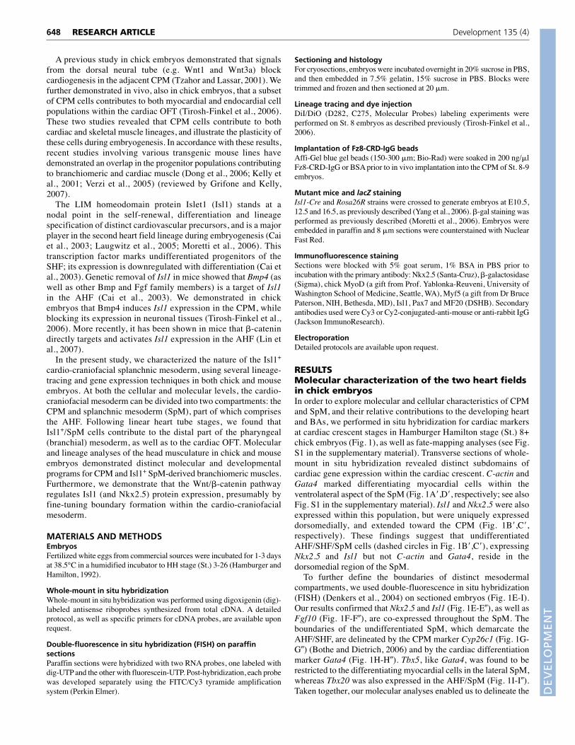
RESULTSMolecular characterization of the two heart fieldsin chick embryosIn order to explore molecular and cellular characteristics of CPMand SpM, and their relative contributions to the developing heartand BAs, we performed in situ hybridization for cardiac markersat cardiac crescent stages in Hamburger Hamilton stage (St.) 8+chick embryos (Fig. 1), as well as fate-mapping analyses (see Fig.S1 in the supplementary material). Transverse sections of whole-mount in situ hybridization revealed distinct subdomains ofcardiac gene expression within the cardiac crescent. C-actin andGata4 marked differentiating myocardial cells within theventrolateral aspect of the SpM (Fig. 1A�,D�, respectively; see alsoFig. S1 in the supplementary material). Isl1 and Nkx2.5 were alsoexpressed within this population, but were uniquely expresseddorsomedially, and extended toward the CPM (Fig. 1B�,C�,respectively). These findings suggest that undifferentiatedAHF/SHF/SpM cells (dashed circles in Fig. 1B�,C�), expressingNkx2.5 and Isl1 but not C-actin and Gata4, reside in thedorsomedial region of the SpM.
To further define the boundaries of distinct mesodermalcompartments, we used double-fluorescence in situ hybridization(FISH) (Denkers et al., 2004) on sectioned embryos (Fig. 1E-I).Our results confirmed that Nkx2.5 and Isl1 (Fig. 1E-E�), as well asFgf10 (Fig. 1F-F�), are co-expressed throughout the SpM. Theboundaries of the undifferentiated SpM, which demarcate theAHF/SHF, are delineated by the CPM marker Cyp26c1 (Fig. 1G-G�) (Bothe and Dietrich, 2006) and by the cardiac differentiationmarker Gata4 (Fig. 1H-H�). Tbx5, like Gata4, was found to berestricted to the differentiating myocardial cells in the lateral SpM,whereas Tbx20 was also expressed in the AHF/SpM (Fig. 1I-I�).Taken together, our molecular analyses enabled us to delineate the
RESEARCH ARTICLE Development 135 (4)
DEVELO
PMENT
boundaries between the CPM, undifferentiated SpM progenitors ofthe AHF/SHF, and differentiating cardiac cells in the SpM of St. 8chick embryos.
Utilizing fluorescent dyes as lineage tracers, we next explored thecontribution of differentiated cells within the lateral SpM (DiO,green) and undifferentiated AHF cells within the medial aspect ofthe SpM (DiI, red) in St. 8 chick embryos (see Fig. S1A in thesupplementary material). Control embryos sectioned immediatelyafter labeling were used to validate the accuracy of our method (seeFig. S1A� in the supplementary material). At St. 10, DiO-labeledcells were found in the looping heart tube (see Fig. S1B� in thesupplementary material), whereas DiI-labeled cells were locatedwithin the SpM, underneath the pharynx (see Fig. S1B� in thesupplementary material). At St. 12, both DiO- and DiI-labeled cellswere detected within the heart: DiO-labeled cells were detected inthe future ventricles, whereas the DiI-labeled AHF/SpM cells werefound in the OFT (see Fig. S1C-C� in the supplementary material).
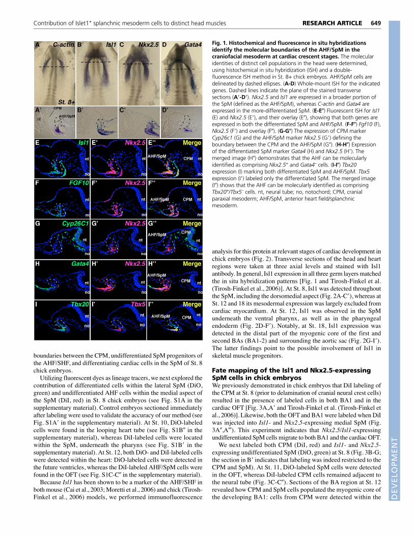
Because Isl1 has been shown to be a marker of the AHF/SHF inboth mouse (Cai et al., 2003; Moretti et al., 2006) and chick (Tirosh-Finkel et al., 2006) models, we performed immunofluorescence
analysis for this protein at relevant stages of cardiac development inchick embryos (Fig. 2). Transverse sections of the head and heartregions were taken at three axial levels and stained with Isl1antibody. In general, Isl1 expression in all three germ layers matchedthe in situ hybridization patterns [Fig. 1 and Tirosh-Finkel et al.(Tirosh-Finkel et al., 2006)]. At St. 8, Isl1 was detected throughoutthe SpM, including the dorsomedial aspect (Fig. 2A-C�), whereas atSt. 12 and 18 its mesodermal expression was largely excluded fromcardiac myocardium. At St. 12, Isl1 was observed in the SpMunderneath the ventral pharynx, as well as in the pharyngealendoderm (Fig. 2D-F�). Notably, at St. 18, Isl1 expression wasdetected in the distal part of the myogenic core of the first andsecond BAs (BA1-2) and surrounding the aortic sac (Fig. 2G-I�).The latter findings point to the possible involvement of Isl1 inskeletal muscle progenitors.
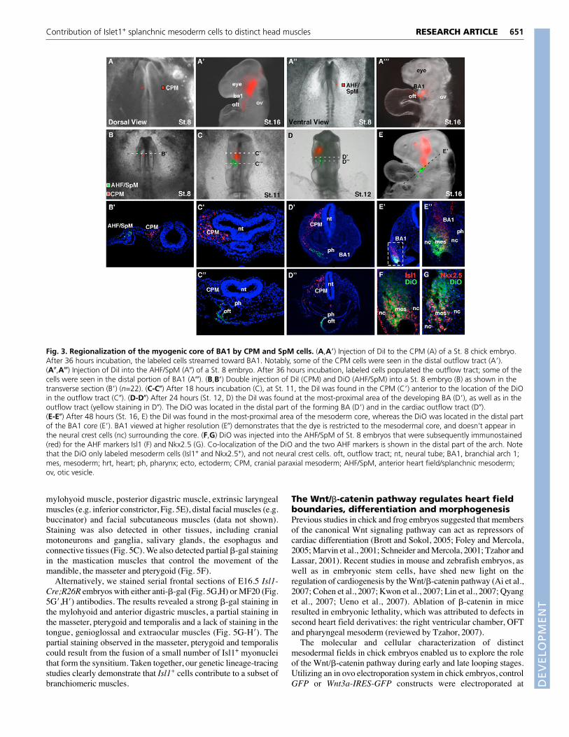
Fate mapping of the Isl1 and Nkx2.5-expressingSpM cells in chick embryosWe previously demonstrated in chick embryos that DiI labeling ofthe CPM at St. 8 (prior to delamination of cranial neural crest cells)resulted in the presence of labeled cells in both BA1 and in thecardiac OFT [Fig. 3A,A� and Tirosh-Finkel et al. (Tirosh-Finkel etal., 2006)]. Likewise, both the OFT and BA1 were labeled when DiIwas injected into Isl1- and Nkx2.5-expressing medial SpM (Fig.3A�,A�). This experiment indicates that Nkx2.5/Isl1-expressingundifferentiated SpM cells migrate to both BA1 and the cardiac OFT.
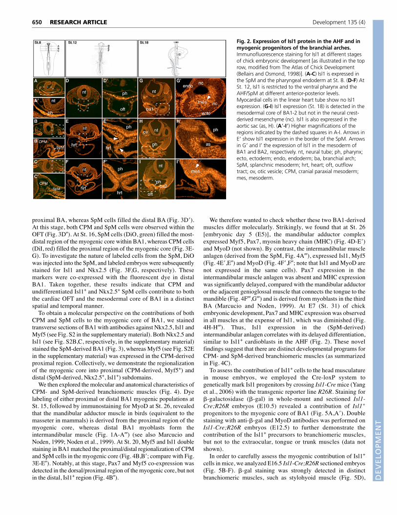
We next labeled both CPM (DiI, red) and Isl1- and Nkx2.5-expressing undifferentiated SpM (DiO, green) at St. 8 (Fig. 3B-G;the section in B� indicates that labeling was indeed restricted to theCPM and SpM). At St. 11, DiO-labeled SpM cells were detectedin the OFT, whereas DiI-labeled CPM cells remained adjacent tothe neural tube (Fig. 3C-C�). Sections of the BA region at St. 12revealed how CPM and SpM cells populated the myogenic core ofthe developing BA1: cells from CPM were detected within the
649RESEARCH ARTICLEContribution of Islet1+ splanchnic mesoderm cells to distinct head muscles
Fig. 1. Histochemical and fluorescence in situ hybridizationsidentify the molecular boundaries of the AHF/SpM in thecraniofacial mesoderm at cardiac crescent stages. The molecularidentities of distinct cell populations in the head were determined,using histochemical in situ hybridization (ISH) and a double-fluorescence ISH method in St. 8+ chick embryos. AHF/SpM cells aredelineated by dashed ellipses. (A-D) Whole-mount ISH for the indicatedgenes. Dashed lines indicate the plane of the stained transversesections (A�-D�). Nkx2.5 and Isl1 are expressed in a broader portion ofthe SpM (defined as the AHF/SpM), whereas C-actin and Gata4 areexpressed in the more-differentiated SpM. (E-E�) Fluorescent ISH for Isl1(E) and Nkx2.5 (E�), and their overlay (E�), showing that both genes areexpressed in both the differentiated SpM and AHF/SpM. (F-F�) Fgf10 (F),Nkx2.5 (F�) and overlay (F�). (G-G�) The expression of CPM markerCyp26c1 (G) and the AHF/SpM marker Nkx2.5 (G�) defining theboundary between the CPM and the AHF/SpM (G�). (H-H�) Expressionof the differentiated SpM marker Gata4 (H) and Nkx2.5 (H�). Themerged image (H�) demonstrates that the AHF can be molecularlyidentified as comprising Nkx2.5+ and Gata4– cells. (I-I�) Tbx20expression (I) marking both differentiated SpM and AHF/SpM. Tbx5expression (I�) labeled only the differentiated SpM. The merged image(I�) shows that the AHF can be molecularly identified as comprisingTbx20+/Tbx5– cells. nt, neural tube; no, notochord; CPM, cranialparaxial mesoderm; AHF/SpM, anterior heart field/splanchnicmesoderm.
DEVELO
PMENT
650
proximal BA, whereas SpM cells filled the distal BA (Fig. 3D�).At this stage, both CPM and SpM cells were observed within theOFT (Fig. 3D�). At St. 16, SpM cells (DiO, green) filled the most-distal region of the myogenic core within BA1, whereas CPM cells(DiI, red) filled the proximal region of the myogenic core (Fig. 3E-G). To investigate the nature of labeled cells from the SpM, DiOwas injected into the SpM, and labeled embryos were subsequentlystained for Isl1 and Nkx2.5 (Fig. 3F,G, respectively). Thesemarkers were co-expressed with the fluorescent dye in distalBA1. Taken together, these results indicate that CPM andundifferentiated Isl1+ and Nkx2.5+ SpM cells contribute to boththe cardiac OFT and the mesodermal core of BA1 in a distinctspatial and temporal manner.
To obtain a molecular perspective on the contributions of bothCPM and SpM cells to the myogenic core of BA1, we stainedtransverse sections of BA1 with antibodies against Nkx2.5, Isl1 andMyf5 (see Fig. S2 in the supplementary material). Both Nkx2.5 andIsl1 (see Fig. S2B,C, respectively, in the supplementary material)stained the SpM-derived BA1 (Fig. 3), whereas Myf5 (see Fig. S2Ein the supplementary material) was expressed in the CPM-derivedproximal region. Collectively, we demonstrate the regionalizationof the myogenic core into proximal (CPM-derived, Myf5+) anddistal (SpM-derived, Nkx2.5+, Isl1+) subdomains.
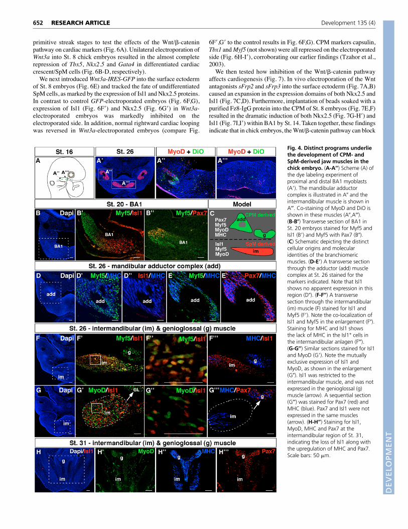
We then explored the molecular and anatomical characteristics ofCPM- and SpM-derived branchiomeric muscles (Fig. 4). Dyelabeling of either proximal or distal BA1 myogenic populations atSt. 15, followed by immunostaining for MyoD at St. 26, revealedthat the mandibular adductor muscle in birds (equivalent to themasseter in mammals) is derived from the proximal region of themyogenic core, whereas distal BA1 myoblasts form theintermandibular muscle (Fig. 1A-A�) (see also Marcucio andNoden, 1999; Noden et al., 1999). At St. 20, Myf5 and Isl1 doublestaining in BA1 matched the proximal/distal regionalization of CPMand SpM cells in the myogenic core (Fig. 4B,B�; compare with Fig.3E-E�). Notably, at this stage, Pax7 and Myf5 co-expression wasdetected in the dorsal/proximal region of the myogenic core, but notin the distal, Isl1+ region (Fig. 4B�).
We therefore wanted to check whether these two BA1-derivedmuscles differ molecularly. Strikingly, we found that at St. 26[embryonic day 5 (E5)], the mandibular adductor complexexpressed Myf5, Pax7, myosin heavy chain (MHC) (Fig. 4D-E�)and MyoD (not shown). By contrast, the intermandibular muscleanlagen (derived from the SpM, Fig. 4A�), expressed Isl1, Myf5(Fig. 4E�,E�) and MyoD (Fig. 4F�,F�; note that Isl1 and MyoD arenot expressed in the same cells). Pax7 expression in theintermandibular muscle anlagen was absent and MHC expressionwas significantly delayed, compared with the mandibular adductoror the adjacent genioglossal muscle that connects the tongue to themandible (Fig. 4F�,G�) and is derived from myoblasts in the thirdBA (Marcucio and Noden, 1999). At E7 (St. 31) of chickembryonic development, Pax7 and MHC expression was observedin all muscles at the expense of Isl1, which was diminished (Fig.4H-H�). Thus, Isl1 expression in the (SpM-derived)intermandibular anlagen correlates with its delayed differentiation,similar to Isl1+ cardioblasts in the AHF (Fig. 2). These novelfindings suggest that there are distinct developmental programs forCPM- and SpM-derived branchiomeric muscles (as summarizedin Fig. 4C).
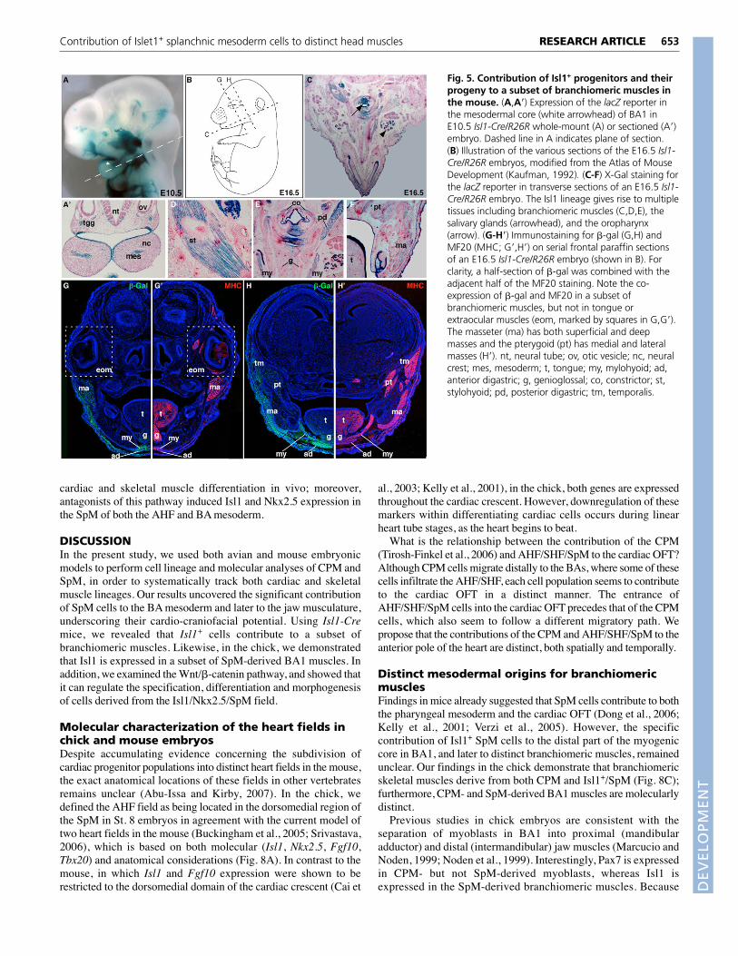
To assess the contribution of Isl1+ cells to the head musculaturein mouse embryos, we employed the Cre-loxP system togenetically mark Isl1 progenitors by crossing Isl1-Cre mice (Yanget al., 2006) with the transgenic reporter line R26R. Staining for�-galactosidase (�-gal) in whole-mount and sectioned Isl1-Cre;R26R embryos (E10.5) revealed a contribution of Isl1+
progenitors to the myogenic core of BA1 (Fig. 5A,A�). Doublestaining with anti-�-gal and MyoD antibodies was performed onIsl1-Cre;R26R embryos (E12.5) to further demonstrate thecontribution of the Isl1+ precursors to branchiomeric muscles,but not to the extraocular, tongue or trunk muscles (data notshown).
In order to carefully assess the myogenic contribution of Isl1+
cells in mice, we analyzed E16.5 Isl1-Cre;R26R sectioned embryos(Fig. 5B-F). �-gal staining was strongly detected in distinctbranchiomeric muscles, such as stylohyoid muscle (Fig. 5D),
RESEARCH ARTICLE Development 135 (4)
Fig. 2. Expression of Isl1 protein in the AHF and inmyogenic progenitors of the branchial arches.Immunofluorescence staining for Isl1 at different stagesof chick embryonic development [as illustrated in the toprow, modified from The Atlas of Chick Development(Bellairs and Osmond, 1998)]. (A-C) Isl1 is expressed inthe SpM and the pharyngeal endoderm at St. 8. (D-F) AtSt. 12, Isl1 is restricted to the ventral pharynx and theAHF/SpM at different anterior-posterior levels.Myocardial cells in the linear heart tube show no Isl1expression. (G-I) Isl1 expression (St. 18) is detected in themesodermal core of BA1-2 but not in the neural crest-derived mesenchyme (nc). Isl1 is also expressed in theaortic sac (as, H). (A�-I�) Higher magnifications of theregions indicated by the dashed squares in A-I. Arrows inE� show Isl1 expression in the border of the SpM. Arrowsin G� and I� the expression of Isl1 in the mesoderm ofBA1 and BA2, respectively. nt, neural tube; ph, pharynx;ecto, ectoderm; endo, endoderm; ba, branchial arch;SpM, splanchnic mesoderm; hrt, heart; oft, outflowtract; ov, otic vesicle; CPM, cranial paraxial mesoderm;mes, mesoderm.
DEVELO
PMENT
mylohyoid muscle, posterior digastric muscle, extrinsic laryngealmuscles (e.g. inferior constrictor, Fig. 5E), distal facial muscles (e.g.buccinator) and facial subcutaneous muscles (data not shown).Staining was also detected in other tissues, including cranialmotoneurons and ganglia, salivary glands, the esophagus andconnective tissues (Fig. 5C). We also detected partial �-gal stainingin the mastication muscles that control the movement of themandible, the masseter and pterygoid (Fig. 5F).
Alternatively, we stained serial frontal sections of E16.5 Isl1-Cre;R26R embryos with either anti-�-gal (Fig. 5G,H) or MF20 (Fig.5G�,H�) antibodies. The results revealed a strong �-gal staining inthe mylohyoid and anterior digastric muscles, a partial staining inthe masseter, pterygoid and temporalis and a lack of staining in thetongue, genioglossal and extraocular muscles (Fig. 5G-H�). Thepartial staining observed in the masseter, pterygoid and temporaliscould result from the fusion of a small number of Isl1+ myonucleithat form the synsitium. Taken together, our genetic lineage-tracingstudies clearly demonstrate that Isl1+ cells contribute to a subset ofbranchiomeric muscles.
The Wnt/�-catenin pathway regulates heart fieldboundaries, differentiation and morphogenesisPrevious studies in chick and frog embryos suggested that membersof the canonical Wnt signaling pathway can act as repressors ofcardiac differentiation (Brott and Sokol, 2005; Foley and Mercola,2005; Marvin et al., 2001; Schneider and Mercola, 2001; Tzahor andLassar, 2001). Recent studies in mouse and zebrafish embryos, aswell as in embryonic stem cells, have shed new light on theregulation of cardiogenesis by the Wnt/�-catenin pathway (Ai et al.,2007; Cohen et al., 2007; Kwon et al., 2007; Lin et al., 2007; Qyanget al., 2007; Ueno et al., 2007). Ablation of �-catenin in miceresulted in embryonic lethality, which was attributed to defects insecond heart field derivatives: the right ventricular chamber, OFTand pharyngeal mesoderm (reviewed by Tzahor, 2007).
The molecular and cellular characterization of distinctmesodermal fields in chick embryos enabled us to explore the roleof the Wnt/�-catenin pathway during early and late looping stages.Utilizing an in ovo electroporation system in chick embryos, controlGFP or Wnt3a-IRES-GFP constructs were electroporated at
651RESEARCH ARTICLEContribution of Islet1+ splanchnic mesoderm cells to distinct head muscles
Fig. 3. Regionalization of the myogenic core of BA1 by CPM and SpM cells. (A,A�) Injection of DiI to the CPM (A) of a St. 8 chick embryo.After 36 hours incubation, the labeled cells streamed toward BA1. Notably, some of the CPM cells were seen in the distal outflow tract (A�).(A�,A�) Injection of DiI into the AHF/SpM (A�) of a St. 8 embryo. After 36 hours incubation, labeled cells populated the outflow tract; some of thecells were seen in the distal portion of BA1 (A�). (B,B�) Double injection of DiI (CPM) and DiO (AHF/SpM) into a St. 8 embryo (B) as shown in thetransverse section (B�) (n=22). (C-C�) After 18 hours incubation (C), at St. 11, the DiI was found in the CPM (C�) anterior to the location of the DiOin the outflow tract (C�). (D-D�) After 24 hours (St. 12, D) the DiI was found at the most-proximal area of the developing BA (D�), as well as in theoutflow tract (yellow staining in D�). The DiO was located in the distal part of the forming BA (D�) and in the cardiac outflow tract (D�).(E-E�) After 48 hours (St. 16, E) the DiI was found in the most-proximal area of the mesoderm core, whereas the DiO was located in the distal partof the BA1 core (E�). BA1 viewed at higher resolution (E�) demonstrates that the dye is restricted to the mesodermal core, and doesn’t appear inthe neural crest cells (nc) surrounding the core. (F,G) DiO was injected into the AHF/SpM of St. 8 embryos that were subsequently immunostained(red) for the AHF markers Isl1 (F) and Nkx2.5 (G). Co-localization of the DiO and the two AHF markers is shown in the distal part of the arch. Notethat the DiO only labeled mesoderm cells (Isl1+ and Nkx2.5+), and not neural crest cells. oft, outflow tract; nt, neural tube; BA1, branchial arch 1;mes, mesoderm; hrt, heart; ph, pharynx; ecto, ectoderm; CPM, cranial paraxial mesoderm; AHF/SpM, anterior heart field/splanchnic mesoderm;ov, otic vesicle.
DEVELO
PMENT
652
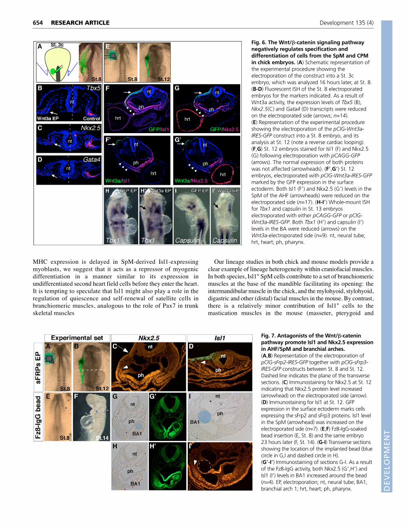
primitive streak stages to test the effects of the Wnt/�-cateninpathway on cardiac markers (Fig. 6A). Unilateral electroporation ofWnt3a into St. 8 chick embryos resulted in the almost completerepression of Tbx5, Nkx2.5 and Gata4 in differentiated cardiaccrescent/SpM cells (Fig. 6B-D, respectively).
We next introduced Wnt3a-IRES-GFP into the surface ectodermof St. 8 embryos (Fig. 6E) and tracked the fate of undifferentiatedSpM cells, as marked by the expression of Isl1 and Nkx2.5 proteins.In contrast to control GFP-electroporated embryos (Fig. 6F,G),expression of Isl1 (Fig. 6F�) and Nkx2.5 (Fig. 6G�) in Wnt3a-electroporated embryos was markedly inhibited on theelectroporated side. In addition, normal rightward cardiac loopingwas reversed in Wnt3a-electroporated embryos (compare Fig.
6F�,G� to the control results in Fig. 6F,G). CPM markers capsulin,Tbx1 and Myf5 (not shown) were all repressed on the electroporatedside (Fig. 6H-I�), corroborating our earlier findings (Tzahor et al.,2003).
We then tested how inhibition of the Wnt/�-catenin pathwayaffects cardiogenesis (Fig. 7). In vivo electroporation of the Wntantagonists sFrp2 and sFrp3 into the surface ectoderm (Fig. 7A,B)caused an expansion in the expression domains of both Nkx2.5 andIsl1 (Fig. 7C,D). Furthermore, implantation of beads soaked with apurified Fz8-IgG protein into the CPM of St. 8 embryos (Fig. 7E,F)resulted in the dramatic induction of both Nkx2.5 (Fig. 7G-H�) andIsl1 (Fig. 7I,I�) within BA1 by St. 14. Taken together, these findingsindicate that in chick embryos, the Wnt/�-catenin pathway can block
RESEARCH ARTICLE Development 135 (4)
Fig. 4. Distinct programs underliethe development of CPM- andSpM-derived jaw muscles in thechick embryo. (A-A�) Scheme (A) ofthe dye labeling experiment ofproximal and distal BA1 myoblasts(A�). The mandibular adductorcomplex is illustrated in A� and theintermandibular muscle is shown inA�. Co-staining of MyoD and DiO isshown in these muscles (A�,A�).(B-B�) Transverse section of BA1 inSt. 20 embryos stained for Myf5 andIsl1 (B�) and Myf5 with Pax7 (B�).(C) Schematic depicting the distinctcellular origins and molecularidentities of the branchiomericmuscles. (D-E�) A transverse sectionthrough the adductor (add) musclecomplex at St. 26 stained for themarkers indicated. Note that Isl1shows no apparent expression in thisregion (D�). (F-F�) A transversesection through the intermandibular(im) muscle (F) stained for Isl1 andMyf5 (F�). Note the co-localization ofIsl1 and Myf5 in the enlargement (F�).Staining for MHC and Isl1 showsthe lack of MHC in the Isl1+ cells inthe intermandibular anlagen (F�).(G-G�) Similar sections stained for Isl1and MyoD (G�). Note the mutuallyexclusive expression of Isl1 andMyoD, as shown in the enlargement(G�). Isl1 was restricted to theintermandibular muscle, and was notexpressed in the genioglossal (g)muscle (arrow). A sequential section(G�) was stained for Pax7 (red) andMHC (blue). Pax7 and Isl1 were notexpressed in the same muscles(arrow). (H-H�) Staining for Isl1,MyoD, MHC and Pax7 at theintermandibular region of St. 31,indicating the loss of Isl1 along withthe upregulation of MHC and Pax7.Scale bars: 50 �m.
DEVELO
PMENT
cardiac and skeletal muscle differentiation in vivo; moreover,antagonists of this pathway induced Isl1 and Nkx2.5 expression inthe SpM of both the AHF and BA mesoderm.
DISCUSSIONIn the present study, we used both avian and mouse embryonicmodels to perform cell lineage and molecular analyses of CPM andSpM, in order to systematically track both cardiac and skeletalmuscle lineages. Our results uncovered the significant contributionof SpM cells to the BA mesoderm and later to the jaw musculature,underscoring their cardio-craniofacial potential. Using Isl1-Cremice, we revealed that Isl1+ cells contribute to a subset ofbranchiomeric muscles. Likewise, in the chick, we demonstratedthat Isl1 is expressed in a subset of SpM-derived BA1 muscles. Inaddition, we examined the Wnt/�-catenin pathway, and showed thatit can regulate the specification, differentiation and morphogenesisof cells derived from the Isl1/Nkx2.5/SpM field.
Molecular characterization of the heart fields inchick and mouse embryosDespite accumulating evidence concerning the subdivision ofcardiac progenitor populations into distinct heart fields in the mouse,the exact anatomical locations of these fields in other vertebratesremains unclear (Abu-Issa and Kirby, 2007). In the chick, wedefined the AHF field as being located in the dorsomedial region ofthe SpM in St. 8 embryos in agreement with the current model oftwo heart fields in the mouse (Buckingham et al., 2005; Srivastava,2006), which is based on both molecular (Isl1, Nkx2.5, Fgf10,Tbx20) and anatomical considerations (Fig. 8A). In contrast to themouse, in which Isl1 and Fgf10 expression were shown to berestricted to the dorsomedial domain of the cardiac crescent (Cai et
al., 2003; Kelly et al., 2001), in the chick, both genes are expressedthroughout the cardiac crescent. However, downregulation of thesemarkers within differentiating cardiac cells occurs during linearheart tube stages, as the heart begins to beat.
What is the relationship between the contribution of the CPM(Tirosh-Finkel et al., 2006) and AHF/SHF/SpM to the cardiac OFT?Although CPM cells migrate distally to the BAs, where some of thesecells infiltrate the AHF/SHF, each cell population seems to contributeto the cardiac OFT in a distinct manner. The entrance ofAHF/SHF/SpM cells into the cardiac OFT precedes that of the CPMcells, which also seem to follow a different migratory path. Wepropose that the contributions of the CPM and AHF/SHF/SpM to theanterior pole of the heart are distinct, both spatially and temporally.
Distinct mesodermal origins for branchiomericmusclesFindings in mice already suggested that SpM cells contribute to boththe pharyngeal mesoderm and the cardiac OFT (Dong et al., 2006;Kelly et al., 2001; Verzi et al., 2005). However, the specificcontribution of Isl1+ SpM cells to the distal part of the myogeniccore in BA1, and later to distinct branchiomeric muscles, remainedunclear. Our findings in the chick demonstrate that branchiomericskeletal muscles derive from both CPM and Isl1+/SpM (Fig. 8C);furthermore, CPM- and SpM-derived BA1 muscles are molecularlydistinct.
Previous studies in chick embryos are consistent with theseparation of myoblasts in BA1 into proximal (mandibularadductor) and distal (intermandibular) jaw muscles (Marcucio andNoden, 1999; Noden et al., 1999). Interestingly, Pax7 is expressedin CPM- but not SpM-derived myoblasts, whereas Isl1 isexpressed in the SpM-derived branchiomeric muscles. Because
653RESEARCH ARTICLEContribution of Islet1+ splanchnic mesoderm cells to distinct head muscles
Fig. 5. Contribution of Isl1+ progenitors and theirprogeny to a subset of branchiomeric muscles inthe mouse. (A,A�) Expression of the lacZ reporter inthe mesodermal core (white arrowhead) of BA1 inE10.5 Isl1-Cre/R26R whole-mount (A) or sectioned (A�)embryo. Dashed line in A indicates plane of section.(B) Illustration of the various sections of the E16.5 Isl1-Cre/R26R embryos, modified from the Atlas of MouseDevelopment (Kaufman, 1992). (C-F) X-Gal staining forthe lacZ reporter in transverse sections of an E16.5 Isl1-Cre/R26R embryo. The Isl1 lineage gives rise to multipletissues including branchiomeric muscles (C,D,E), thesalivary glands (arrowhead), and the oropharynx(arrow). (G-H�) Immunostaining for �-gal (G,H) andMF20 (MHC; G�,H�) on serial frontal paraffin sectionsof an E16.5 Isl1-Cre/R26R embryo (shown in B). Forclarity, a half-section of �-gal was combined with theadjacent half of the MF20 staining. Note the co-expression of �-gal and MF20 in a subset ofbranchiomeric muscles, but not in tongue orextraocular muscles (eom, marked by squares in G,G�).The masseter (ma) has both superficial and deepmasses and the pterygoid (pt) has medial and lateralmasses (H�). nt, neural tube; ov, otic vesicle; nc, neuralcrest; mes, mesoderm; t, tongue; my, mylohyoid; ad,anterior digastric; g, genioglossal; co, constrictor; st,stylohyoid; pd, posterior digastric; tm, temporalis.
DEVELO
PMENT
654
MHC expression is delayed in SpM-derived Isl1-expressingmyoblasts, we suggest that it acts as a repressor of myogenicdifferentiation in a manner similar to its expression inundifferentiated second heart field cells before they enter the heart.It is tempting to speculate that Isl1 might also play a role in theregulation of quiescence and self-renewal of satellite cells inbranchiomeric muscles, analogous to the role of Pax7 in trunkskeletal muscles
Our lineage studies in both chick and mouse models provide aclear example of lineage heterogeneity within craniofacial muscles.In both species, Isl1+ SpM cells contribute to a set of branchiomericmuscles at the base of the mandible facilitating its opening: theintermandibular muscle in the chick, and the mylohyoid, stylohyoid,digastric and other (distal) facial muscles in the mouse. By contrast,there is a relatively minor contribution of Isl1+ cells to themastication muscles in the mouse (masseter, pterygoid and
RESEARCH ARTICLE Development 135 (4)
Fig. 6. The Wnt/�-catenin signaling pathwaynegatively regulates specification anddifferentiation of cells from the SpM and CPMin chick embryos. (A) Schematic representation ofthe experimental procedure showing theelectroporation of the construct into a St. 3cembryo, which was analyzed 16 hours later, at St. 8.(B-D) Fluorescent ISH of the St. 8 electroporatedembryos for the markers indicated. As a result ofWnt3a activity, the expression levels of Tbx5 (B),Nkx2.5(C) and Gata4 (D) transcripts were reducedon the electroporated side (arrows; n=14).(E) Representation of the experimental procedureshowing the electroporation of the pCIG-Wnt3a-IRES-GFP construct into a St. 8 embryo, and itsanalysis at St. 12 (note a reverse cardiac looping).(F,G) St. 12 embryos stained for Isl1 (F) and Nkx2.5(G) following electroporation with pCAGG-GFP(arrows). The normal expression of both proteinswas not affected (arrowheads). (F�,G�) St. 12embryos, electroporated with pCIG-Wnt3a-IRES-GFPmarked by the GFP expression in the surfaceectoderm. Both Isl1 (F�) and Nkx2.5 (G�) levels in theSpM of the AHF (arrowheads) were reduced on theelectroporated side (n=17). (H-I�) Whole-mount ISHfor Tbx1 and capsulin in St. 13 embryoselectroporated with either pCAGG-GFP or pCIG-Wnt3a-IRES-GFP. Both Tbx1 (H�) and capsulin (I�)levels in the BA were reduced (arrows) on theWnt3a-electroporated side (n=9). nt, neural tube;hrt, heart; ph, pharynx.
Fig. 7. Antagonists of the Wnt/�-cateninpathway promote Isl1 and Nkx2.5 expressionin AHF/SpM and branchial arches.(A,B) Representation of the electroporation ofpCIG-sFrp2-IRES-GFP together with pCIG-sFrp3-IRES-GFP constructs between St. 8 and St. 12.Dashed line indicates the plane of the transversesections. (C) Immunostaining for Nkx2.5 at St. 12indicating that Nkx2.5 protein level increased(arrowhead) on the electroporated side (arrow).(D) Immunostaining for Isl1 at St. 12. GFPexpression in the surface ectoderm marks cellsexpressing the sFrp2 and sFrp3 proteins. Isl1 levelin the SpM (arrowhead) was increased on theelectroporated side (n=7). (E,F) Fz8-IgG-soakedbead insertion (E, St. 8) and the same embryo23 hours later (F, St. 14). (G-I) Transverse sectionsshowing the location of the implanted bead (bluecircle in G,I and dashed circle in H).(G�-I�) Immunostaining of sections G-I. As a resultof the Fz8-IgG activity, both Nkx2.5 (G�,H�) andIsl1 (I�) levels in BA1 increased around the bead(n=4). EP, electroporation; nt, neural tube; BA1,branchial arch 1; hrt, heart; ph, pharynx. D
EVELO
PMENT
temporalis) or to the mandibular adductor complex in the chick.Furthermore, in both species, the intrinsic and extrinsic muscles ofthe tongue (e.g. genioglossal) and extraocular muscles are not
derived from the Isl1+ SpM lineage. The slightly broadercontribution of the Isl1+ lineage to branchiomeric muscle, observedin the mouse, could result from differences in the lineage allocationsbetween the two species. Similarly, the contribution of Isl1+ cells ofthe second heart field is broader in the mouse than that in the chick.Clearly, methodological and experimental differences affect lineagecomparisons between chick and mouse models. In our R26R musclelineage analyses in mice, it is important to appreciate that the fusionof a few �-gal+ myoblasts is likely to result in staining of the entiremyofiber (e.g. masseter, pterygoid and temporalis, Fig. 5).
Tbx1 (Kelly et al., 2004), as well as capsulin and MyoR (Lu et al.,2002), have been shown to act as upstream regulators ofbranchiomeric muscle development. In capsulin/MyoR doublemutants (Lu et al., 2002), the masseter, pterygoids and temporaliswere missing, whereas the distal BA1 muscles (e.g. anteriordigastric and mylohyoid, both derived from Isl1+ cells, Fig. 5) werenot affected. Our findings in both chick and mouse experimentalmodels, which reveal that jaw muscles are composed of at least twodistinct myogenic lineages (CPM-derived and Isl1+ SpM-derivedmuscles), provide a plausible developmental explanation for thisunique muscle phenotype.
It was recently demonstrated in mice that Isl1/Nkx2.5/Flk1-positive cells within the SpM are multipotent cardiovascularprogenitors that give rise to cardiac myocytes, smooth muscle andendothelial lineages within the heart (Moretti et al., 2006; Wu et al.,2006). We show that Isl1+ cells represent multipotential progenitorsof both cardiovascular and skeletal muscle lineages.
Wnt/�-catenin signaling and its effect on cardiacand skeletal muscle developmentWe previously demonstrated in the chick that signals emanatingfrom the neural tube (that can be mimicked by Wnt1 and Wnt3a)block cardiogenesis in the CPM (Tzahor and Lassar, 2001). Thesefindings, and those of two other studies (Marvin et al., 2001;Schneider and Mercola, 2001), suggest that Wnt/�-catenin signalinginhibits cardiogenesis during early embryogenesis. A subsequentstudy (Foley and Mercola, 2005) demonstrated that Wnt signalingmust be inhibited within the endoderm to induce secretion of an asyet unidentified cardiogenic-inducing factor. In fact, numerousstudies support the notion that inhibition of the Wnt/�-cateninpathway is required for proper heart development and repair(Barandon et al., 2003; Brott and Sokol, 2005; Lickert et al., 2002;Singh et al., 2007), whereas other studies, mostly in cultured EScells, suggest that the opposite is true (Nakamura et al., 2003).
Recent studies in mouse and zebrafish embryos, as well as inembryonic stem cells, demonstrate that the Wnt/�-catenin pathwayplays distinct, even opposing, roles during various stages and withindistinct tissues during cardiac development (reviewed by Tzahor,2007). The new loss-of-function studies of canonical Wnt signalingin the mouse (Ai et al., 2007; Cohen et al., 2007; Kwon et al., 2007;Lin et al., 2007; Qyang et al., 2007; Ueno et al., 2007) providecompelling evidence that this pathway is required within cardiacprogenitors and differentiating cardiac cells for the development ofthe second heart field (including AHF cells) and its derivatives: theright ventricular chamber, OFT and pharyngeal mesoderm. Thesestudies further demonstrate that Wnt signaling stimulates theproliferation of cardiac progenitors during mouse cardiogenesis.
Using electroporation of Wnt ligands or Wnt inhibitors in chickembryos, we observed either the inhibition of cardiac and skeletalmuscle differentiation markers, or the expansion of Isl1 and Nkx2.5,respectively. Similarly, bead implantation of a Wnt inhibitor into theCPM resulted in increased expression of Nkx2.5 and Isl1 within the
655RESEARCH ARTICLEContribution of Islet1+ splanchnic mesoderm cells to distinct head muscles
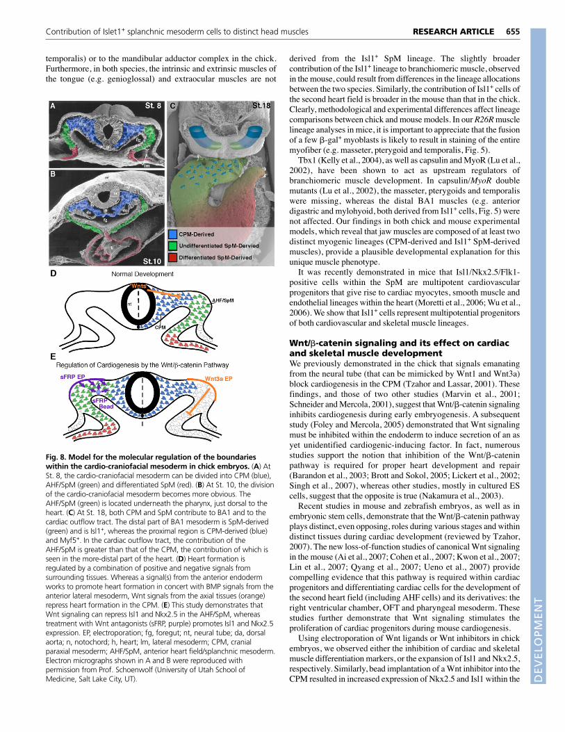
Fig. 8. Model for the molecular regulation of the boundarieswithin the cardio-craniofacial mesoderm in chick embryos. (A) AtSt. 8, the cardio-craniofacial mesoderm can be divided into CPM (blue),AHF/SpM (green) and differentiated SpM (red). (B) At St. 10, the divisionof the cardio-craniofacial mesoderm becomes more obvious. TheAHF/SpM (green) is located underneath the pharynx, just dorsal to theheart. (C) At St. 18, both CPM and SpM contribute to BA1 and to thecardiac outflow tract. The distal part of BA1 mesoderm is SpM-derived(green) and is Isl1+, whereas the proximal region is CPM-derived (blue)and Myf5+. In the cardiac outflow tract, the contribution of theAHF/SpM is greater than that of the CPM, the contribution of which isseen in the more-distal part of the heart. (D) Heart formation isregulated by a combination of positive and negative signals fromsurrounding tissues. Whereas a signal(s) from the anterior endodermworks to promote heart formation in concert with BMP signals from theanterior lateral mesoderm, Wnt signals from the axial tissues (orange)repress heart formation in the CPM. (E) This study demonstrates thatWnt signaling can repress Isl1 and Nkx2.5 in the AHF/SpM, whereastreatment with Wnt antagonists (sFRP, purple) promotes Isl1 and Nkx2.5expression. EP, electroporation; fg, foregut; nt, neural tube; da, dorsalaorta; n, notochord; h, heart; lm, lateral mesoderm; CPM, cranialparaxial mesoderm; AHF/SpM, anterior heart field/splanchnic mesoderm.Electron micrographs shown in A and B were reproduced withpermission from Prof. Schoenwolf (University of Utah School ofMedicine, Salt Lake City, UT). D
EVELO
PMENT

656
SpM-derived myogenic mesoderm of BA1. These results suggestthat canonical Wnt signaling can inhibit cardiac and cranial muscledifferentiation, which is consistent with findings in micedemonstrating that continuous Wnt signaling prolongs theprogenitor state and interferes with differentiation duringcardiogenesis.
The cardio-craniofacial mesodermTaken together, our past and present studies clearly demonstrate thatthe cardio-craniofacial mesoderm is tightly regulated by bothpositive and negative cues from surrounding tissues (Rinon et al.,2007; Tirosh-Finkel et al., 2006; Tzahor et al., 2003; Tzahor andLassar, 2001). Our findings highlight the heterogeneity ofdevelopmental programming among cranial muscles, and confirmthat craniofacial myogenesis is developmentally linked to cardiacdevelopment (this study) (Tirosh-Finkel et al., 2006; Tzahor andLassar, 2001) (reviewed by Grifone and Kelly, 2007), suggestingthat these tissues share a common evolutionary origin.
The striking parallel between a subset of branchiomeric musclesand the transcriptional networks involved in heart development (thisstudy) (Dong et al., 2006; Kelly et al., 2001; Verzi et al., 2005) isactually seen across vast phylogenetic distances. Nematodes do notpossess a heart, yet their pharyngeal muscle contracts like a heartand exhibits electrical activity similar to that of mammaliancardiomyocytes. Moreover, it has been shown that the developmentof the pharyngeal muscle in nematodes, and of cardiac muscle invertebrates and insects, is regulated by the homeobox gene Nkx2.5(Haun et al., 1998). Thus, unlike skeletal muscles in the trunk, headmuscles are likely to have evolved from an ancestral developmentalprogram that gave rise to a contractile tube used for feeding andcirculation. Insights into the genetic circuits that drive the evolutionand development of heart and craniofacial muscles might shed lighton general principles of organogenesis, as well as on the molecularbasis of cardiovascular and craniofacial myopathies in humans.
This work was supported by research grants to E.T. from the Estelle FunkFoundation for Biomedical Research, Ruth and Allen Ziegler, the Pasteur-Weizmann Foundation, the Helen and Martin Kimmel Institute for Stem CellResearch, the Y. Leon Benoziyo Institute for Molecular Medicine, a GermanIsraeli Foundation (GIF) Young Investigator Award, the Minerva Foundationwith funding from the Federal German Ministry for Education and Research,and the Association Française Contre les Myopathies (AFM). E.N. is a recipientof a travel fellowship from Development, and A.M. was supported by theLanda Center for Equal Opportunity Through Education. S.M.E. was supportedby research grants NIHRO1, HL074066 and an AHA (fellowship to Z.H.). Wethank Margaret Buckingham for her critical review of the manuscript; MichaelZagazki for the EM micrographs; Ori Brenner, Drew Noden and Giovanni Levifor histological and anatomical insights; Laura Burrus and Andrew McMahonfor Wnt-related DNA constructs for the electroporation; and our laboratoryteams for their insights and support.
Supplementary materialSupplementary material for this article is available athttp://dev.biologists.org/cgi/content/full/135/4/647/DC1
ReferencesAbu-Issa, R. and Kirby, M. L. (2007). Heart field: from mesoderm to heart tube.
Annu. Rev. Cell Dev. Biol. 23, 45-68.Ai, D., Fu, X., Wang, J., Lu, M. F., Chen, L., Baldini, A., Klein, W. H. and
Martin, J. F. (2007). Canonical Wnt signaling functions in second heart field topromote right ventricular growth. Proc. Natl. Acad. Sci. USA 104, 9319-9324.
Barandon, L., Couffinhal, T., Ezan, J., Dufourcq, P., Costet, P., Alzieu, P.,Leroux, L., Moreau, C., Dare, D. and Duplaa, C. (2003). Reduction of infarctsize and prevention of cardiac rupture in transgenic mice overexpressing FrzA.Circulation 108, 2282-2289.
Bellairs, R. and Osmond. M. (1998). The Atlas of Chick Development. London:Academic Press.
Black, B. L. (2007). Transcriptional pathways in second heart field development.Semin. Cell Dev. Biol. 18, 67-76.
Bothe, I. and Dietrich, S. (2006). The molecular setup of the avian headmesoderm and its implication for craniofacial myogenesis. Dev. Dyn. 235, 2845-2860.
Bothe, I., Ahmed, M. U., Winterbottom, F. L., von Scheven, G. and Dietrich,S. (2007). Extrinsic versus intrinsic cues in avian paraxial mesoderm patterningand differentiation. Dev. Dyn. 236, 2397-2409.
Brott, B. K. and Sokol, S. Y. (2005). A vertebrate homolog of the cell cycleregulator Dbf4 is an inhibitor of Wnt signaling required for heart development.Dev. Cell 8, 703-715.
Buckingham, M., Meilhac, S. and Zaffran, S. (2005). Building the mammalianheart from two sources of myocardial cells. Nat. Rev. Genet. 6, 826-835.
Cai, C. L., Liang, X., Shi, Y., Chu, P. H., Pfaff, S. L., Chen, J. and Evans, S.(2003). Isl1 identifies a cardiac progenitor population that proliferates prior todifferentiation and contributes a majority of cells to the heart. Dev. Cell 5, 877-889.
Cohen, E. D., Wang, Z., Lepore, J. J., Lu, M. M., Taketo, M. M., Epstein, D. J.and Morrisey, E. E. (2007). Wnt/beta-catenin signaling promotes expansion ofIsl-1-positive cardiac progenitor cells through regulation of FGF signaling. J. Clin.Invest. 117, 1794-1804.
Denkers, N., Garcia-Villalba, P., Rodesch, C. K., Nielson, K. R. and Mauch, T.J. (2004). FISHing for chick genes: triple-label whole-mount fluorescence in situhybridization detects simultaneous and overlapping gene expression in avianembryos. Dev. Dyn. 229, 651-657.
Dong, F., Sun, X., Liu, W., Ai, D., Klysik, E., Lu, M. F., Hadley, J., Antoni, L.,Chen, L., Baldini, A. et al. (2006). Pitx2 promotes development of splanchnicmesoderm-derived branchiomeric muscle. Development 133, 4891-4899.
Emery, A. E. (2002). The muscular dystrophies. Lancet 359, 687-695.Foley, A. C. and Mercola, M. (2005). Heart induction by Wnt antagonists
depends on the homeodomain transcription factor Hex. Genes Dev. 19, 387-396.
Garry, D. J. and Olson, E. N. (2006). A common progenitor at the heart ofdevelopment. Cell 127, 1101-1104.
Grifone, R. and Kelly, R. G. (2007). Heartening news for head muscledevelopment. Trends Genet. 23, 365-369.
Hamburger, V. and Hamilton, H. L. (1992). A series of normal stages in thedevelopment of the chick embryo. Dev. Dyn. 195, 231-272.
Haun, C., Alexander, J., Stainier, D. Y. and Okkema, P. G. (1998). Rescue ofCaenorhabditis elegans pharyngeal development by a vertebrate heartspecification gene. Proc. Natl. Acad. Sci. USA 95, 5072-5075.
Hutson, M. R. and Kirby, M. L. (2003). Neural crest and cardiovasculardevelopment: a 20-year perspective. Birth Defects Res. C Embryo Today 69, 2-13.
Kaufman, M. (1992). The Atlas of Mouse Development. London: AcademicPress.
Kelly, R. G., Brown, N. A. and Buckingham, M. E. (2001). The arterial pole ofthe mouse heart forms from Fgf10-expressing cells in pharyngeal mesoderm.Dev. Cell 1, 435-440.
Kelly, R. G., Jerome-Majewska, L. A. and Papaioannou, V. E. (2004). Thedel22q11.2 candidate gene Tbx1 regulates branchiomeric myogenesis. Hum.Mol. Genet. 13, 2829-2840.
Kwon, C., Arnold, J., Hsiao, E. C., Taketo, M. M., Conklin, B. R. andSrivastava, D. (2007). Canonical Wnt signaling is a positive regulator ofmammalian cardiac progenitors. Proc. Natl. Acad. Sci. USA 104, 10894-10899.
Laugwitz, K. L., Moretti, A., Lam, J., Gruber, P., Chen, Y., Woodard, S., Lin, L.Z., Cai, C. L., Lu, M. M., Reth, M. et al. (2005). Postnatal isl1+ cardioblastsenter fully differentiated cardiomyocyte lineages. Nature 433, 647-653.
Lickert, H., Kutsch, S., Kanzler, B., Tamai, Y., Taketo, M. M. and Kemler, R.(2002). Formation of multiple hearts in mice following deletion of beta-cateninin the embryonic endoderm. Dev. Cell 3, 171-181.
Lin, L., Cui, L., Zhou, W., Dufort, D., Zhang, X., Cai, C. L., Bu, L., Yang, L.,Martin, J., Kemler, R. et al. (2007). beta-Catenin directly regulates Islet1expression in cardiovascular progenitors and is required for multiple aspects ofcardiogenesis. Proc. Natl. Acad. Sci. USA 104, 9313-9318.
Lu, J. R., Bassel-Duby, R., Hawkins, A., Chang, P., Valdez, R., Wu, H., Gan, L.,Shelton, J. M., Richardson, J. A. and Olson, E. N. (2002). Control of facialmuscle development by MyoR and capsulin. Science 298, 2378-2381.
Marcucio, R. S. and Noden, D. M. (1999). Myotube heterogeneity in developingchick craniofacial skeletal muscles. Dev. Dyn. 214, 178-194.
Marvin, M. J., Di Rocco, G., Gardiner, A., Bush, S. M. and Lassar, A. B. (2001).Inhibition of Wnt activity induces heart formation from posterior mesoderm.Genes Dev. 15, 316-327.
Mjaatvedt, C. H., Nakaoka, T., Moreno-Rodriguez, R., Norris, R. A., Kern, M.J., Eisenberg, C. A., Turner, D. and Markwald, R. R. (2001). The outflowtract of the heart is recruited from a novel heart-forming field. Dev. Biol. 238,97-109.
Moretti, A., Caron, L., Nakano, A., Lam, J. T., Bernshausen, A., Chen, Y.,Qyang, Y., Bu, L., Sasaki, M., Martin-Puig, S. et al. (2006). Multipotentembryonic isl1+ progenitor cells lead to cardiac, smooth muscle, and endothelialcell diversification. Cell 127, 1151-1165.
RESEARCH ARTICLE Development 135 (4)
DEVELO
PMENT

Nakamura, T., Sano, M., Songyang, Z. and Schneider, M. D. (2003). A Wnt-and beta -catenin-dependent pathway for mammalian cardiac myogenesis. Proc.Natl. Acad. Sci. USA 100, 5834-5839.
Noden, D. M. (1983). The role of the neural crest in patterning of avian cranialskeletal, connective, and muscle tissues. Dev. Biol. 96, 144-165.
Noden, D. M. and Francis-West, P. (2006). The differentiation andmorphogenesis of craniofacial muscles. Dev. Dyn. 235, 1194-1218.
Noden, D. M., Marcucio, R., Borycki, A. G. and Emerson, C. P., Jr (1999).Differentiation of avian craniofacial muscles. I. Patterns of early regulatory geneexpression and myosin heavy chain synthesis. Dev. Dyn. 216, 96-112.
Qyang, Y., Martin-Puig, S., Chiravuri, M., Chen, S., Xu, H., Bu, L., Jiang, X.,Lin, L., Granger, A., Moretti, A. et al. (2007). The renewal and differentiationof Isl1+ cardiovascular progenitors are controlled by a Wnt/�-catenin pathway.Cell Stem Cell 1, 165-179.
Rinon, A., Lazar, S., Marshall, H., Buchmann-Moller, S., Neufeld, A.,Elhanany-Tamir, H., Taketo, M. M., Sommer, L., Krumlauf, R. and Tzahor,E. (2007). Cranial neural crest cells regulate head muscle patterning anddifferentiation during vertebrate embryogenesis. Development 134, 3065-3075.
Schneider, V. A. and Mercola, M. (2001). Wnt antagonism initiates cardiogenesisin Xenopus laevis. Genes Dev. 15, 304-315.
Singh, A. M., Li, F. Q., Hamazaki, T., Kasahara, H., Takemaru, K. and Terada,N. (2007). Chibby, an antagonist of the Wnt/beta-catenin pathway, facilitatescardiomyocyte differentiation of murine embryonic stem cells. Circulation 115,617-626.
Srivastava, D. (2006). Making or breaking the heart: from lineage determinationto morphogenesis. Cell 126, 1037-1048.
Tirosh-Finkel, L., Elhanany, H., Rinon, A. and Tzahor, E. (2006). Mesodermprogenitor cells of common origin contribute to the head musculature and thecardiac outflow tract. Development 133, 1943-1953.
Trainor, P. A., Tan, S. S. and Tam, P. P. (1994). Cranial paraxial mesoderm:
regionalisation of cell fate and impact on craniofacial development in mouseembryos. Development 120, 2397-2408.
Tzahor, E. (2007). Wnt/beta-catenin signaling and cardiogenesis: timing doesmatter. Dev. Cell 13, 10-13.
Tzahor, E. and Lassar, A. B. (2001). Wnt signals from the neural tube blockectopic cardiogenesis. Genes Dev. 15, 255-260.
Tzahor, E., Kempf, H., Mootoosamy, R. C., Poon, A. C., Abzhanov, A., Tabin,C. J., Dietrich, S. and Lassar, A. B. (2003). Antagonists of Wnt and BMPsignaling promote the formation of vertebrate head muscle. Genes Dev. 17,3087-3099.
Ueno, S., Weidinger, G., Osugi, T., Kohn, A. D., Golob, J. L., Pabon, L.,Reinecke, H., Moon, R. T. and Murry, C. E. (2007). Biphasic role for Wnt/beta-catenin signaling in cardiac specification in zebrafish and embryonic stem cells.Proc. Natl. Acad. Sci. USA 104, 9685-9690.
Verzi, M. P., McCulley, D. J., De Val, S., Dodou, E. and Black, B. L. (2005). Theright ventricle, outflow tract, and ventricular septum comprise a restrictedexpression domain within the secondary/anterior heart field. Dev. Biol. 287, 134-145.
Wachtler, F. and Jacob, M. (1986). Origin and development of the cranial skeletalmuscles. Bibl. Anat. 29, 24-46.
Waldo, K. L., Kumiski, D. H., Wallis, K. T., Stadt, H. A., Hutson, M. R., Platt,D. H. and Kirby, M. L. (2001). Conotruncal myocardium arises from asecondary heart field. Development 128, 3179-3188.
Wu, S. M., Fujiwara, Y., Cibulsky, S. M., Clapham, D. E., Lien, C. L.,Schultheiss, T. M. and Orkin, S. H. (2006). Developmental origin of abipotential myocardial and smooth muscle cell precursor in the mammalianheart. Cell 127, 1137-1150.
Yang, L., Cai, C. L., Lin, L., Qyang, Y., Chung, C., Monteiro, R. M., Mummery,C. L., Fishman, G. I., Cogen, A. and Evans, S. (2006). Isl1Cre reveals acommon Bmp pathway in heart and limb development. Development 133,1575-1585.
657RESEARCH ARTICLEContribution of Islet1+ splanchnic mesoderm cells to distinct head muscles
DEVELO
PMENT