Embed Size (px)
Citation preview
RESEARCH ARTICLE
The cup fungus Pestalopezia brunneopruinosa
is Pestalotiopsis gibbosa and belongs to
Sordariomycetes
Kyoko Watanabe1*, Shunsuke Nozawa1, Tom Hsiang2, Brenda Callan3
1 Graduate School of Agriculture, Tamagawa University, Machida, Tokyo, Japan, 2 Environmental Sciences,
University of Guelph, Guelph, Ontario, Canada, 3 Pacific Forestry Centre, Natural Resources Canada,
Victoria, British Columbia, Canada
Abstract
Pestalopezia brunneopruinosa, the type species of Pestalopezia in Leotiomycetes, pro-
duces typical cup-shaped ascomata. Because its asexual morph has conidia comprised of
five cells including apical and basal appendages and three pigmented median cells, it was
first described as Pestalotia gibbosa, which belongs to Sordariomycetes. This contradiction
has not been resolved due to the difficulty in isolating this fungus in culture. In this study, we
isolated separate strains from the sexual morph and the asexual morph for molecular analy-
sis. Phylogenetic trees of Sporocadaceae based on internal transcribed spacer, partial β-
tubulin, and partial translation elongation factor 1-alpha sequence datasets revealed that
both strains fall into the same taxon, in a clade in Pestalotiopsis sensu stricto alongside P.
gaultheriae and P. spathulata. We provide the first evidence that fungi producing cup-
shaped ascomata in Pestalotiopsis belong to Sordariomycetes, and we have proposed the
transfer of Pestalopezia brunneopruinosa to Pestalotiopsis gibbosa.
Introduction
Pestalopezia brunneopruinosa (Zeller) Seaver is a leaf spot pathogen on salal (Gaultheria shallonPursh) that produces asci on an apothecium as a sexual morph [1]. The asexual morph of Pes-talopezia brunneopruinosa resembles that of Pestalotiopsis sensu lato (s. lat.) and was first
described independently by Harkness [2] as Pestalotia gibbosa. Thus, it has been suspected that
Pestalopezia brunneopruinosa and Pestalotia gibbosa are the same fungus, because the two
fungi have been found in close proximity on the same leaves. Bonar [3] demonstrated that cul-
tures from germinated ascospores of Pestalopezia brunneopruinosa produced conidia that were
the same as that of Pestalotia gibbosa. Seaver [4] likewise concluded that Pestalopezia brunneo-pruinosa was the sexual morph of Pestalotia gibbosa. However, phylogenetic analyses of both
fungi to clarify their relationship has not been previously conducted.
The genus Pestalotia was established by De Notaris [5]. Subsequently, Steyaert [6] split the
genus Pestalotia into Pestalotia sensu stricto (s. str.) (conidia composed of 6 cells), Pestalotiop-sis (5 cells) and Truncatella (4 cells), although many species were still retained in Pestalotia s.
PLOS ONE | https://doi.org/10.1371/journal.pone.0197025 June 27, 2018 1 / 12
a1111111111
a1111111111
a1111111111
a1111111111
a1111111111
OPENACCESS
Citation: Watanabe K, Nozawa S, Hsiang T, Callan
B (2018) The cup fungus Pestalopezia
brunneopruinosa is Pestalotiopsis gibbosa and
belongs to Sordariomycetes. PLoS ONE 13(6):
e0197025. https://doi.org/10.1371/journal.
pone.0197025
Editor: Sabrina Sarrocco, Universita degli Studi di
Pisa, ITALY
Received: November 21, 2017
Accepted: April 24, 2018
Published: June 27, 2018
Copyright: © 2018 Watanabe et al. This is an open
access article distributed under the terms of the
Creative Commons Attribution License, which
permits unrestricted use, distribution, and
reproduction in any medium, provided the original
author and source are credited.
Data Availability Statement: All tree files are
available from the treebase database (http://www.
treebase.org; accession number S21431). All
sequence data are deposited in DDBJ database
(https://www.ddbj.nig.ac.jp/index-e.html;
accession numbers LC311584-LC31597, LC-3159-
LC311606). Additional data are available from the
MycoBank database (http://www.mycobank.org;
accession number MB#824630).
Funding: Funded by JSPS KAKENHI Grant Number
25440218 https://www.jsps.go.jp/.
lat. without reconsideration. Recently, Pestalotiopsis s. lat. was further split into three genera,
Pestalotiopsis s. st., Neopestalotiopsis, and Pseudopestalotiopsis, based on morphology and
molecular phylogeny [7]. These fungi belong to Sporocadaceae within Sordariomycetes [8].
The Harkness description of Pestalotia gibbosa conidia (three pigmented median cells in five-
celled versicoloured conidia, with septa darker than the rest of the cell), is similar to that of
Neopestalotiopsis. However, the current taxonomic position of Pestalotia gibbosa is unclear,
especially since the disposition of this fungus in Sordariomycetes was made without molecular
data support. The sexual morph of Pestalotiopsis s. lat. was determined by Barr [9] to be Pesta-losphaeria which produces three celled-ascospores and perithecial ascocarps. Reblova et al.
[10] proposed using the name Pestalotiopsis rather than Pestalosphaeria as the currently
accepted name, following recent botanical code changes, but there was no mention of the
name Pestalopezia in this argument. Pestalopezia, Pestalotiopsis, and Pestalosphaeria, are, how-
ever, included in a “without-prejudice list of generic names of fungi for protection under the
International Code”[11].
Pestalopezia brunneopruinosa, the sexual morph, was classified as a member of the Leotio-
mycetes [12] because it produces cup-shaped ascomata. Thus, the genus names of the sexual
and asexual morphs are currently forced into different taxonomic classes. Beimforde et al. [13]
conducted a phylogenetic analysis by combining fossil data and molecular data (18S rDNA,
28S rDNA, RPB1, and RPB2) and showed estimated lineages of both families diverged during
the Permian or Carboniferous periods and Leotiomycetes and Sordariomycetes are sister
clades. Their results indicate that these families, both of which produce inoperculate asci, are
closely related in the molecular phylogenetic tree. However, there is no report that fungi
belonging to Sordariomycetes can produce cup-shaped ascomata. The aim of this study was to
clarify the taxonomic position of Pestalopezia brunneopruinosa with respect to Pestalotia gib-bosa, and to determine the name for this fungus based on the concept of one fungus, one name
[14, 15].
Materials and methods
Sample collection and isolation
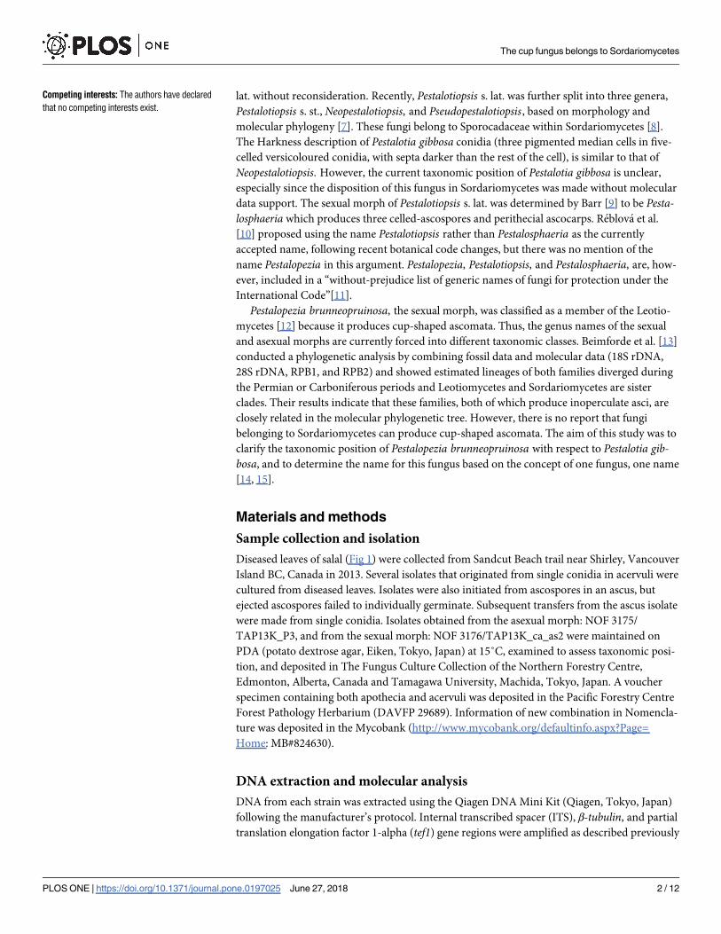
Diseased leaves of salal (Fig 1) were collected from Sandcut Beach trail near Shirley, Vancouver
Island BC, Canada in 2013. Several isolates that originated from single conidia in acervuli were
cultured from diseased leaves. Isolates were also initiated from ascospores in an ascus, but
ejected ascospores failed to individually germinate. Subsequent transfers from the ascus isolate
were made from single conidia. Isolates obtained from the asexual morph: NOF 3175/
TAP13K_P3, and from the sexual morph: NOF 3176/TAP13K_ca_as2 were maintained on
PDA (potato dextrose agar, Eiken, Tokyo, Japan) at 15˚C, examined to assess taxonomic posi-
tion, and deposited in The Fungus Culture Collection of the Northern Forestry Centre,
Edmonton, Alberta, Canada and Tamagawa University, Machida, Tokyo, Japan. A voucher
specimen containing both apothecia and acervuli was deposited in the Pacific Forestry Centre
Forest Pathology Herbarium (DAVFP 29689). Information of new combination in Nomencla-
ture was deposited in the Mycobank (http://www.mycobank.org/defaultinfo.aspx?Page=
Home: MB#824630).
DNA extraction and molecular analysis
DNA from each strain was extracted using the Qiagen DNA Mini Kit (Qiagen, Tokyo, Japan)
following the manufacturer’s protocol. Internal transcribed spacer (ITS), β-tubulin, and partial
translation elongation factor 1-alpha (tef1) gene regions were amplified as described previously
The cup fungus belongs to Sordariomycetes
PLOS ONE | https://doi.org/10.1371/journal.pone.0197025 June 27, 2018 2 / 12
Competing interests: The authors have declared
that no competing interests exist.
[16–19], using primers ITS1/ITS4, Bt2d/Bt2c, and pest_ef_f/EF1-1567R, respectively. These
primers target regions that are approximately 550 bp, 560 bp, and 530 bp in size, respectively.
To confirm the culture was isolated from the sexual morph, DNA was extracted from a sin-
gle apothecium from DAVFP 29689 by CTAB [20], and ITS was amplified using our designed
primer PES3 (5’-GGCCTACCCTGTAGCGCCTT-3’) and ITS4.
Polymerase chain reaction (PCR) products were purified using ExoSAP-IT (GE Healthcare
Japan, Tokyo, Japan) and sequenced using the ABI 310 DNA sequencer (ABI, Tokyo, Japan).
These sequences have been deposited in the DNA Data Bank of Japan (https://www.ddbj.nig.
ac.jp/index-e.html: accession numbers are shown in Table 1).
The results of the preliminary sequence homology search using BLAST were that the two
Vancouver Island salal isolates, NOF 3175/TAP13K_P3 and NOF 3176/TAP13K_ca_as2, fell
into Pestalotiopsis s. str. Additional sequence data for phylogenetic analysis were obtained
from 7 other previously unpublished strains (listed in bold in Table 1), and 43 other strains
published in previous studies [7, 21]. To generate phylogenies based on ITS, β-tubulin, and
tef1 sequences, Seiridium spp., members of Amphisphaeriaceae (outgroup) and Phlogicylin-
driaceae, were chosen because they are phylogenetically close to Sporocadaceae. The dataset of
each genomic region (ITS, β-tubulin, and tef1) was aligned using MAFFT [22]. All positions
containing gaps and missing data were deleted from the analysis. The strength of internal
branches from the resulting tree was tested using the bootstrap analysis [23] with 1,000
replications.
Sequence data comprising the aligned dataset were subjected to maximum-likelihood
(ML), neighbor-joining (NJ) and maximum-parsimony (MP) phylogenetic analyses using
MEGA software Version 7 [24]. Molecular analyses using the ML method were performed
using HKY+G+I nucleotide substitution model for ITS, β-tubulin, and tef1. Initial trees for the
heuristic searches were automatically generated by applying the NJ and BioNJ algorithms to a
matrix of pairwise distances estimated using the maximum composite likelihood approach
and then selecting the topology with a higher log-likelihood value. Evolutionary history was
inferred using the NJ method [25]. The tree was drawn to scale with branch-length units
equivalent to those of the evolutionary distances used to infer phylogeny. Evolutionary dis-
tances were computed using the Kimura 2-parameter method [26] as the number of base sub-
stitutions per site. MP trees were generated using the tree-bisection-regrafting (TBR)
algorithm and search level 3, which generates initial trees by randomly adding sequences (10
Fig 1. A diseased leaf of salal (Gaultheria shallon) from Sandcut beach trail, Vancouver Island, BC, Canada.
https://doi.org/10.1371/journal.pone.0197025.g001
The cup fungus belongs to Sordariomycetes
PLOS ONE | https://doi.org/10.1371/journal.pone.0197025 June 27, 2018 3 / 12
Table 1. Source of species for molecular analyses and the DNA database accession number.
Species Culture No. Location Host GenBank accession
ITS β-tubulin tef1Pestalotiopsis gibbosa (syn. Pestalotia gibbosa,
this study)
NOF 3175/TAP13K_P3 Canada Gaultheria shallon LC311589 LC311590 LC311591
P. gibbosa (syn. Pestalopezia brunneopruinosa,
this study)
NOF 3176/
TAP13K_ca_as2�Canada Gaultheria shallon LC311586 LC311587 LC311588
Pestalotiopsis adusta ICPM 6088 Fiji On refrigerator door PVC
gasket
JX399006 JX399037 JX399070
P. anacardiacearum IFRDCC 2397 China Mangifera indica KC247154 KC247155 KC247156
P. arceuthobii CBS 434.65 USA Arceuthobium campylopodum KM199341 KM199427 KM199516
P. arengae CBS 331.92 Singapore Arenga undulatifolia KM199340 KM199426 KM199515
P. australasiae CBS 114126 New Zealand Knightia sp. KM199297 KM199409 KM199499
P. chamaeropis CBS 186.71 Italy Chamaerops humilis KM199326 KM199391 KM199473
P. clavata MFLUCC 12–0268 China Buxus sp. JX398990 JX399025 JX399056
P. colombiensis CBS 118553 Colombia Eucalyptus eurograndis KM199307 KM199421 KM199488
P. diploclisiae CBS 115587 Hong Kong Diploclisia glaucescens KM199320 KM199419 KM199486
P. ericacearum IFRDCC 2439 China Rhododendron delavayi KC537807 KC537821 KC537814
P. furcata MFLUCC 12–0054 Thailand Camellia sinensis JQ683724 JQ683708 JQ683740
P. gaultheriae IFRD 411–014 China Gaultheria forrestii KC537805 KC537819 KC537812
P. grevilleae CBS 114127 Australia Grevillea sp. KM199300 KM199407 KM199504
P. hollandica CBS 265.33 The Netherlands Sciadopitys verticillata KM199328 KM199388 KM199481
P. humus CBS 336.97 Papua New
Guinea
Soil KM199317 KM199420 KM199484
P. kenyana CBS 442.67 Kenya Coffea sp. KM199302 KM199395 KM199502
P. monochaeta CBS 144.97 The Netherlands Quercus robur KM199327 KM199386 KM199479
P. neglecta (this study) TAP1100�/MAFF239735 Japan Quercus myrsinaefolia AB482220 LC311599 LC311600
P. novae-hollandiae CBS 130973 Australia Banksia grandis KM199337 KM199425 KM199511
P. oryzae CBS 353.69 Denmark Oryza sativa KM199299 KM199398 KM199496
P. papuana CBS 331.96 Papua New
Guinea
Coastal soil KM199321 KM199413 KM199491
P. parva CBS 265.37 - Delonix regia KM199312 KM199404 KM199508
P. pallidotheae MAFF 240993� Japan Pieris japonica NR111022 LC311584 LC311585
P. portugalica CBS 393.48 Portugal - KM199335 KM199422 KM199510
P. rhododendri IFRDCC 2399 China Rhododendron sinogrande KC537804 KC537818 KC537811
P. scoparia CBS 176.25 - Chamaecyparis sp. KM199330 KM199393 KM199478
P. spathulata CBS 356.86 Chile Gevuina avellana KM199338 KM199423 KM199513
P. telopeae CBS 114161 Australia Telopea sp. KM199296 KM199403 KM199500
Pestalotiopsis sp.1 (this study) TAP0K00Kin Japan Osmanthus fragrans var.
aurantiacusLC311595 LC311596 LC311597
Pestalotiopsis sp.2 (this study) TAP0E0SA� Japan Camellia sasanqua LC311592 LC311593 LC311594
Pseudopestalotiopsis cocos CBS 272.29 Java, Indonesia Cocos nucifera KM199378 KM199467 KM199553
Ps. theae MFLUCC 12-0055/CPC
20281
Thailand Camellia sinensis JQ683727 JQ683711 JQ683743
Ps. myanmarina NBRC 112264� Myanmar Averrhora carambola LC114025 LC114045 LC114065
Ps. vietnamensis NBRC 112252 Vietnam Fragaria sp. LC114034 LC114054 LC114074
Neopestalotiopsis australis CBS 114159 Australia Telopea sp. KM199348 KM199432 KM199537
N. cubana CBS 600.96 Cuba Leaf litter KM199347 KM199438 KM199521
N. foedans CGMCC 3.9123 China Mangrove plant JX398987 JX399022 JX399053
N. honoluluana CBS 114495 USA: Hawaii Telopea sp. KM199364 KM199457 KM199548
N. javaensis CBS 257.31 Indonesia: Java Cocos nucifera KM199357 KM199437 KM199543
(Continued)
The cup fungus belongs to Sordariomycetes
PLOS ONE | https://doi.org/10.1371/journal.pone.0197025 June 27, 2018 4 / 12
replicates). Consistency, retention, homoplasy, and composition indices were calculated for
parsimony-informative sites. The resulting trees were printed using TreeView v. 1.6.6 [27]
and, together with the alignments, deposited as S21431 in TreeBASE (https://www.treebase.
org/treebase-web/home.html).
Morphological observations
Morphological observations were made from symptomatic salal leaves collected in 2013
(DAVFP 29689) and from a single dried herbarium specimen DAVFP 11308. The latter was
collected in 1959, also from Vancouver Island, and determined as Pestalopezia brunneoprui-nosa by W. Ziller (S1 Fig). The asexual and sexual morphs were observed and measured in
water using light microscopy (BX 51, Olympus Tokyo, Japan).
Nomenclature
The electronic version of this article in Portable Document Format (PDF) in a work with an
ISSN or ISBN will represent a published work according to the International Code of Nomen-
clature for algae, fungi, and plants, and hence the new names contained in the electronic publi-
cation of a PLOS ONE article are effectively published under that Code from the electronic
edition alone, so there is no longer any need to provide printed copies.
In addition, new names contained in this work have been submitted to MycoBank from
where they will be made available to the Global Names Index. The unique MycoBank number
can be resolved and the associated information viewed through any standard web browser by
appending the MycoBank number [urn:lsid:mycobank.org:Mycobank:824630] contained in
this publication to the prefix http://www.mycobank.org/MB/. The online version of this work
is archived and available from the following digital repositories:PubMed Central, LOCKSS.
Results
Phylogenetic analysis
In addition to the Vancouver Island collections preliminarily identified as Pestalotia gibbosa(NOF 3175/TAP13K_P3, Culture from conidia) and Pestalopezia brunneopruinosa (NOF
Table 1. (Continued)
Species Culture No. Location Host GenBank accession
ITS β-tubulin tef1N. natalensis CBS 138.41 South Africa Acacia mollissima KM199377 KM199466 KM199552
N. piceana CBS 394.48 UK Picea sp. KM199368 KM199453 KM199527
N. protearum CBS 114178/STE-U 1765 UK Picea sp. KM199368 KM199453 KM199527
N. saprophytica MFLUCC 12–0282 China Magnolia sp. JX398982 JX399017 JX399048
N. surinamensis CBS 450.74 Zimbabwe Protea eximia KM199351 KM199465 KM199518
N. zimbabwana CBS 111495 Zimbabwe Leucospermum cunciforme JX556231 KM199456 KM199545
Seiridium camelliae SD096/MFLUCC 12–0647 China Camellia reticulata JQ683725 JQ683709 JQ683741
Seiridium sp. 1 (this study) TAP121 Japan Hamamelis japonica LC311607 LC311608 LC311609
Seiridium sp. 2 (this study) TAP1041 Japan Chamaecyparis obtusa LC311610 LC311611 LC311612
Seiridium sp. 3 (this study) TAP3355 Japan Tilia cordata LC311601 LC311602 LC311603
Seiridium sp. 4 (this study) TAP881 Japan Rhododendron keiskei LC311604 LC311605 LC311606
Bold accession numbers were obtained in this study.
� indicates strain producing sexual morph.
https://doi.org/10.1371/journal.pone.0197025.t001
The cup fungus belongs to Sordariomycetes
PLOS ONE | https://doi.org/10.1371/journal.pone.0197025 June 27, 2018 5 / 12
3176/TAP13K_ca_as2, Culture from ascospores), a total of 52 strains, including Pestalotiopsis(30 strains with two obtained from sexual morphs), Neopestalotiopsis (11 strains), and Pseudo-pestalotiopsis (4 strains including one obtained from the sexual morph), were examined (acces-
sion numbers shown in Table 1). The sequence matrix used for phylogenetic analyses
contained at least 1258 nucleotide positions for final data set from sequences 550 bp of ITS,
560 bp of β-tubulin, and 530 bp of tef1. In ML method, the highest log-likelihood was -6657.97.
The optimal tree generated using the NJ method had a branch-length of 0.665. An MP tree
had a length of 909, consistency index of 0.547, retention of 0.87 and composite index of 0.509.
Only the ML tree (Fig 2) is shown here, because the ML, NJ, and MP methods generated simi-
lar topologies.
Pestalotia gibbosa and Pestalopezia brunneopruinosa were placed in the same clade with Pes-talotiopsis gaultheriae (ML/NJ/MP: 100/100/100). Pestalotiopsis spathulata was also closely
placed to Pestalotia gibbosa and Pestalopezia brunneopruinosa with highly supported bootstrap
values (ML/NJ/MP: 100/100/99). Furthermore, the ITS sequence obtained from Pestalotia gib-bosa (NOF 3175/TAP13K_P3) and Pestalopezia brunneopruinosa (NOF 3176/TAP13K_-
ca_as2) were the same as the ITS sequence obtained from DNA extracted directly from an
apothecium of DAVFP 29689 (epitype specimen) (S2 Fig).
Morphological comparisons
Our observations of the apothecia from DAVFP 11308 and 29689 are similar to those of Sea-
ver’s description [4] of Pestalopezia brunneopruinosa, with few exceptions. Seaver’s ascospore
measurements were slightly larger than the Vancouver Island DAVFP (VI) specimens at 7–10
x 14–20 um, plus we observed in the VI collections that mature ascospores eventually darken
to brown rather than remaining hyaline (Fig 3, S3 Fig). We also observed a ring-shaped ascus
apparatus in DAVFP 29689 which stained blue in Melzer’s reagent, but only in scattered
mature asci. These morphological variations are relatively minor and likely reflective of the
state of maturity of Seaver’s material (S1 Table). We also compared our observations and mea-
surements of the conidial states of DAVFP 11308 and 29689 from leaves to published descrip-
tions and specimens of conidia of Pestalopezia brunneopruinosa, Pestalotiopsis gaultheriae, and
P. spathulata (Table 2). With the exception that P. spathulata has fewer and longer appendages
[7], all are morphologically very similar.
Taxonomy
Pestalotiopsis gibbosa (Harkn.) Kyoko Watan., Nozawa & B. Callan, comb. nov. [urn:lsid:
mycobank.org:Mycobank:824630]
= Pestalotia gibbosa Harkn. Bull. Calif. Acad. Sci. 2: 439, 1887 MB#191515
= Dermatea brunneopruinosa Zeller, Mycologia 26: 291, 1934 MB#259032
= Pestalopezia brunneopruinosa (Zeller) Seaver, Mycologia 34: 300, 1942 MB#289174
�Pestalotiopsis gaultheriae Y.M. Zhang, Maharachch. & K.D. Hyde, Sydowia 65: 121, 2013
MB#803236
Epitype (Fig 3)
DAVFP 29689, Sandcut Beach trail, Shirley, Vancouver Island BC, Canada, 48.4173˚N,
124.0185˚W March 5, 2013, on leaves of G. shallon Pursh collected by B. Callan and M.
The cup fungus belongs to Sordariomycetes
PLOS ONE | https://doi.org/10.1371/journal.pone.0197025 June 27, 2018 6 / 12
Brannigan. Ex-epitype NOF 3176/TAP13K_ca_as2 was isolated from a conidium transferred
from a colony originating from a single ascus.
Ascocarp: Apothecium developing on the upper surface of pale tan to light brown necrotic
areas of attached living leaves, sessile or with short stalk approximately 0.5–2 mm in diameter,
cup-shaped, with a wood brown to yellowish brown furfuraceous exterior. Hymenium fuscous
when immature, becoming black at maturity because of the dark tips of paraphyses forming
the epithelium; asci: 115–150 μm in length (including a short stalk) × 11–15 μm in diameter
(n = 20), eight-spored, unitunicate, cylindrical, with slightly pointed apex, apical apparatus
ring-shaped and staining blue in Melzer’s reagent, but only when fully mature; ascospores:
Fig 2. Maximum-likelihood (ML) tree with length 743 determined by analysis of the combined ITS, β-tubulin, and
tef1 sequence matrix. Numbers (ML/NJ/MP) and hyphens on the branches represent the bootstrap values (%) for each
node, calculated from 1,000 replicates; only values> 80% are shown. NJ: neighbor-joining, MP: Maximum-parsimony.�: ex-holotype cultures. Blue texts indicate strains producing sexual morphs.
https://doi.org/10.1371/journal.pone.0197025.g002
The cup fungus belongs to Sordariomycetes
PLOS ONE | https://doi.org/10.1371/journal.pone.0197025 June 27, 2018 7 / 12
5–8 × 11–16 μm (n = 20), ellipsoidal to ovate, at first hyaline, becoming dark brown when
mature, one-seriate; paraphyses: slender and clavate, light brown at their tips in Melzer’s
reagent.
Conidiomata: Acervuli erumpent through the upper surface of the leaf epidermis, fre-
quently in a zonate pattern in necrotic lesions. Lesions frequently coalescing, turning the leaf
Fig 3. Morphological characteristics of Pestalotiopsis gibbosa (Epitype). (A) Apothecia (arrowheads). (B) Acervuli
(arrows). (C) A vertical section of an apothecium. (D) Asci containing ascospores. (E) Apical ring of ascus tip staining
blue in Melzer’s reagent. (F) Mature ascospores (G) A Section of an acervulus. (H) Conidia. Bars (A), (B) = 2 mm, (C)–
(E) = 20 μm.
https://doi.org/10.1371/journal.pone.0197025.g003
Table 2. Morphological comparison of asexual morphs of Pestalotiopsis gibbosa and related species.
Species Three median cells Apical appendages
Size (length × width, μm) Length (μm) Colour Number Size (length, μm) Tip
Pestalopezia brunneopruinosa [2] 25–30 × 8–10.5 16–20 concolorous, olivaceous 2–4 (3) 30–60 knobbed
Pestalopezia brunneopruinosa (DAVFP 11308) 22.5–32 × 8–13.5 13–20 versicolorous, dark brown 1–4 (3) 20–48 knobbed
Pestalotiopsis gibbosa (DAVFP 29689) 24–31 × 7.5–10 15.5–22.5 versicolorous, dark brown 2–4 (3) 22–61 knobbed
Pestalotiopsis gaultheriae [28] 23–31 × 7–9.5 – versicolorous, dark brown 3 15–50 knobbed
Pestalotiopsis spathulata [7] 24–32 × 7.5–9.5 13–20 versicolour 2–5 17–25 knobbed
https://doi.org/10.1371/journal.pone.0197025.t002
The cup fungus belongs to Sordariomycetes
PLOS ONE | https://doi.org/10.1371/journal.pone.0197025 June 27, 2018 8 / 12
almost entirely brown while still attached to the stem. Conidiomata from leaves, subglobose to
oval, immersed, then erumpent, black, up to 150–219 μm wide (n = 10); Conidiogenous cells
directly lining the acervular wall, hyaline, cylindrical, annellidic; Conidia: 24–31 × 7.5–10 μm
(n = 30), pyriform, curved, four-septate and slightly constricted at the septa, which are darker
than the body of the cells; median three cells 15.5–22.5 μm long (n = 30) in total, pigmented;
two upper pigmented cells fuscous, darker than lower pigmented cell, 15.5–22.5 μm long
(n = 30); apical cell: hyaline, conical with two to four (mostly three) apical appendages arising
from the apical crest. Apical appendages typically swollen at the tip, unbranched, filiform, 22–
61 μm long (n = 30). Basal cell hyaline, conical, with a single, tubular, unbranched, centric
appendage.
Additional specimen examined: DAVFP 11308 (S3 Fig), Cowichan Lake, Vancouver Island,
BC, Canada, April 23, 1959, on leaves of G. shallon Pursh collected and determined as Pestalo-pezia brunneopruinosa by W. Ziller.
Note: The Holotype was O. S. C. Herb., 8096 in the original description of Dermatea by Zel-
ler in 1934 [1].This description did not mention the color of mature ascospores. DAVFP
11308 collected by Ziller (as Pestalopezia) in 1959 contains mature ascomata and is in suffi-
ciently intact state to observe brownish ascospores. However neither sample was suitable for
DNA extraction, and hence we established an epitype. Since obtaining cultures that originate
from single ascospores is difficult, we initiated our culture (NOF 3176/TAP13K_ca_as2) from
a monoconidial isolate that was obtained from hyphae grown from ascospores of a single
ascus. We were able to germinate single ascospores ejected from mature ascocarps onto agar,
but the resulting germinants failed to grow beyond an initial germ tube. We designated the
epitype of Pestalotiopsis gibbosa as DAVFP 29689. We consider P. gaultheriae Y.M. Zhang,
Maharchch. & K.D. Hyde [28] to be a synonym of P. gibbosa, but the authors [28] were unable
to obtain living cultures from the specimen of P. gaultheriae.
Discussion
Our morphological observations and sequence results confirm that Pestalopezia brunneoprui-nosa and Pestalotia gibbosa are the same fungus. Conidia of Pestalotia gibbosa are strikingly
similar to those of Neopestalotiopsis species because the three median cells of the conidia are
versicoloured, and they could be classified into the genus Neopestalotiopsis based on morphol-
ogy. However, in this study, we demonstrate by genomic analysis that P. gibbosa should be
transferred to Pestalotiopsis s. str., even though its sexual morph is an apothecium.
The majority of the more than 200 species associated with the well-known genus Pestalo-tiopsis s. lat. are typified by the asexual morph, while only a few (14) have known sexual states
producing perithecial ascocarps typified by the genus Pestalosphaeria [7, 21]. Reblova et al.
[10] have recommended use of Pestalotiopsis rather than Pestalosphaeria, but this recommen-
dation did not take into consideration the potential of either Neopestalotiosis or Pseudopestalo-tiopsis also having teleomorphs genetically related to Pestalosphaeria; and the small (three
known species), obscure genus Pestalopezia was not mentioned at all in this recommendation.
All species of Pestalosphaeria were considered to be linked to Pestalotiopsis s. str. after the three
genera Neopestalotiopsis, Pseudopestalotiopsis, and Pestalotiopsis were separated from Pestalo-tiopsis s. lat. [7]. Silverio et al [29] in 2016 and Nozawa et al. [17] in 2017, found the sexual
morphs of Neopestalotiopsis and Pseudopestalotiopsis, both in agreement with the description
of Pestalosphaeria. Hence, they reported that Pestalotiopsis s. str., Neopestalotiopsis, and Pseu-dopestalotiopsis produce the same sexual morph. However, the relationship of these fungi to
Pestalopezia, characterized by the production of apothecia, was not considered in these works.
In this study, we obtained strains from conidia of Pestalotia gibbosa and from ascospores of
The cup fungus belongs to Sordariomycetes
PLOS ONE | https://doi.org/10.1371/journal.pone.0197025 June 27, 2018 9 / 12
Pestalopezia. In phylogenetic analyses based on ITS, β-tubulin, and tef1, both strains were
placed with Pestalotiopsis s. str. (Fig 2) although the morphological characteristics of conidia
were strikingly similar to those of conidia of Neopestalotiopsis (Fig 3). Hence, the name of Pes-talotia gibbosa should be changed to Pestalotiopsis gibbosa. Although Pestalopezia Seaver 1942
precedes Pestalotiopsis Steyaert 1949, we recommend using Pestalotiopsis s. str. as this name is
more widely known and therefore likely to be better accepted. The species name gibbosa(1887) is older than brunneopruinosa (1942).With our strains, P. gaultheriae belongs to same
clade with high bootstrap values (MP/ML/NJ: 100/100/100, Fig 2). Pestalotiopsis gaultheriae
was established as a new species based on morphology and molecular data of ITS, β-tubulinand tef1 sequences, which were directly obtained from the fungi on a leaf of salal. However,
our sequence data demonstrated that P. gaultheriae was a synonym of Pestalotiopsis gibbosa. In
sordariomycetes, there is no fungi producing cup–shaped ascomata. According to results of
Zhuang et al [30] based on a phylogenetic tree of RNA secondary structures and on the esti-
mated morphologies from their phylogenetic tree, ascomata having exposed hymenia are esti-
mated as ancestral morphs. Even Pestalotiopsis s. lat. produces closed ascomata, and only the
clade of Pestalotiopsis gibbosa produces open ascomata, nested among other taxa with closed
ascomata. In this study, we were unable to determine whether this is the ancestral morph or a
reversion morph. Our results provide the first evidence that Sordariomycetes include species
that produce cup-shaped ascomata.
Supporting information
S1 Fig. Specimen of DAVFP 11308. This specimen is preserved in the Forest Pathology Her-
barium at the Pacific Forestry Center, Victoria, BC, Canada.
(TIF)
S2 Fig. Multiple alignment of ITS sequences among Pestalopezia brunneopruinosa (NOF
3176/TAP13K_ca_as2), Pestalotiopsis gibbosa (NOF 3175/TAP13K_P3), and extract DNA
directly from an apothecium on DAVFP 29689.
(TIF)
S3 Fig. Morphological characteristics of Pestalotia gibbosa (DAVFP 11308). (A) Apothecia;
(B) Acervuli; (C) Asci containing mature ascospores (arrow) on the layer of an apothecium;
(D) Asci and ascospores (stained with iodine); (E) Conidial formation on the upper layer of an
acervulus; and (F) Conidia. Bars (A), (B): 2 mm, (C): 100 μm, (D)–(F): 20 μm.
(TIF)
S1 Table. Morphological comparison of sexual morph of Pestalopezia brunneopruinosaand related species.
(DOCX)
Acknowledgments
This research was supported by JSPS KAKENHI Grant Number 25440218 to Kyoko
Watanabe.
Author Contributions
Conceptualization: Kyoko Watanabe, Brenda Callan.
Data curation: Kyoko Watanabe, Shunsuke Nozawa.
Formal analysis: Kyoko Watanabe.
The cup fungus belongs to Sordariomycetes
PLOS ONE | https://doi.org/10.1371/journal.pone.0197025 June 27, 2018 10 / 12
Visualization: Kyoko Watanabe, Shunsuke Nozawa.
Writing – original draft: Kyoko Watanabe, Shunsuke Nozawa, Brenda Callan.
Writing – review & editing: Kyoko Watanabe, Tom Hsiang, Brenda Callan.
References
1. Zeller SM. Some new or noteworthy fungi on ericaceous hosts in the Pacific Northwest. Mycologia.
1934; 26(4): 291–304.
2. Harkness HW. Fungi of the pacific coast. Bull Calif Acad Sci. 1887; 2: 438–447.
3. Bonar L. Studies on some California Fungi: II. Mycologia. 1942; 34(2): 180–192.
4. Seaver FJ. Photographs and descriptions of cup-fungi: XXXVI. A new species and genus. Mycologia.
1942; 34(3): 298–301.
5. De Notaris G. Micromycetes italici novi el minus cogniti. Taurini Dec Secundas. Mem Reale Accad Sci
Torino. 1841; 3: 80.
6. Steyaert RL. Contribution a l’etude monographique de Pestalotia de Not. et Monochaetia Sacc. (Trun-
catella gen. nov. et Pestalotiopsis gen. nov.). Bull Jard Bot Etat Bruxelles. 1949; 19(3): 285–354.
https://doi.org/10.2307/3666710
7. Maharachchikumbura SSN, Hyde KD, Groenewald JZ, Xu J, Crous PW. Pestalotiopsis revisited. Stud
Mycol. 2014; 79: 121–186. https://doi.org/10.1016/j.simyco.2014.09.005 PMID: 25492988
8. Jaklitsch WM, Gardiennet A, Voglmayr H. Resolution of morphology-based taxonomic delusions: Acro-
cordiella, Basiseptospora, Blogiascospora, Clypeosphaeria, Hymenopleella, Lepteutypa, Pseudapios-
pora, Requienella, Seiridium and Strickeria. Persoonia. 2016; 37(11): 82–105. https://doi.org/10.3767/
003158516X690475 PMID: 28100927
9. Barr ME. Pestalosphaeria, a new genus in the Amphisphaeriaceae. Mycologia. 1975; 67(1): 187–193.
https://doi.org/10.1139/b81-135
10. Reblova M, Miller AN, Rossman AY, Seifert KA, Crous PW, Hawksworth LD, et.al. Recommendations
for competing sexual-asexually typified generic names in Sordariomycetes (except Diaporthales, Hypo-
creales, and Magnaporthales). IMA Fungus. 2016; 7(1): 131–153. https://doi.org/10.5598/imafungus.
2016.07.01.08 PMID: 27433444
11. Kirk PM, Stalpers JA, Braun U, Crous PW, Hansen K, Hawksworth DL,et al. A without-prejudice list of
generic names of fungi for protection under the International Code of Nomenclature for algae, fungi, and
plants. IMA Fungus. 2013; 4(2): 381–443. https://doi.org/10.5598/imafungus.2013.04.02.17 PMID:
24563844
12. Kirk PM, Cannon PF, Minter DW, Stalpers JA. Dictionary of the Fungi, 10th Edition. Wallingford: CAB
International; 2008.
13. Beimforde C, Feldberg K, Nylinder S, Rikkinen J, Tuovila H, Dorfelt H, et.al. Estimating the phanerozoic
history of the ascomycota lineages: combining fossil and molecular data. Mol Phylogenet Evol. 2014;
78(9): 386–398. https://doi.org/10.1016/j.ympev.2014.04.024 PMID: 24792086
14. Taylor JW. One Fungus = One Name: DNA and fungal nomenclature twenty years after PCR. IMA Fun-
gus. 2011; 2(2): 113–120. https://doi.org/10.5598/imafungus.2011.02.02.01 PMID: 22679595
15. Wingfield MJ, Beer ZW, Slippers B, Wingfield BD, Groenewald JZ, Lombard L, et al. One fungus, one
name promotes progressive plant pathology. Mol Plant Pathol. 2012; 13(6): 604–613. https://doi.org/
10.1111/j.1364-3703.2011.00768.x PMID: 22146077
16. Glass NL, Donaldson GC. Development of primer sets designed for use with the PCR to amplify con-
served genes from filamentous Ascomycetes. Appl Environ Microbiol. 1995; 61(4): 1323–1330. PMID:
7747954
17. Nozawa S, Yamaguchi K, Yen LTH, Van Hop D, Phay Nyunt, Ando K, Watanabe K. Identification of two
new species and a sexual morph from the genus Pseudopestalotiopsis Mycosience. 2017; 58(5): 328–
337. https://doi.org/10.1016/j.myc.2017.02.008
18. Rehener SA, Buckley E: A Beauveria phylogeny inferred from nuclear ITS and EF1-a sequences: evi-
dence for cryptic diversification and links to Cordyceps teleomorphs. Mycologia. 2005; 97(1): 84–98.
https://doi.org/10.3852/mycologia.97.1.84 PMID: 16389960
19. White TJ, Bruns T, Lee S, Taylor J. Amplification and direct sequencing of fungal ribosomal RNA genes
for phylogenetics. In: Innis MA, Gelfand DH, Sninsky JJ, White TJ, editors. PCR Protocols: a guide to
methods and applications. San Diego: Academic Press; 1990. p. 315–322.
20. Tamari F, Hinkley CS, Ramprashad N. A comparison of DNA Extraction methods using Petunia hybrida
tissues. J Biomol Tech. 2013; 24(3):113–118. https://doi.org/10.7171/jbt.13-2403-001 PMID: 23997658
The cup fungus belongs to Sordariomycetes
PLOS ONE | https://doi.org/10.1371/journal.pone.0197025 June 27, 2018 11 / 12
21. Maharachchikumbura SSN, Guo LD, Cai L, Chukeatirote E, Wu WP, Sun X, et al. A multi-locus back-
bone tree for Pestalotiopsis, with a polyphasic characterization of 14 new species. Fungal Divers. 2012;
56(1): 95–129. https://doi.org/10.1007/s13225-012-0198-1
22. Katoh K, Rozewicki J, Yamada KD. 2017 MAFFT online service: multiple sequence alignment, interac-
tive sequence choice and visualization. Brief Bioinform. 1–7. https://doi.org/10.1093/bib/bbw003 PMID:
26868358
23. Felsenstein J. Confidence limits on phylogenies: An approach using the bootstrap. Evolution. 1985; 39
(4): 783–791. https://doi.org/10.1111/j.1558-5646.1985.tb00420.x PMID: 28561359
24. Kumar S, Stecher G, Tamura K. MEGA7: Molecular Evolutionary Genetics Analysis Version 7.0 for Big-
ger Datasets. Mol Biol Evol. 2016; 33(7):1870–4. https://doi.org/10.1093/molbev/msw054 PMID:
27004904
25. Saitou N, Nei M. The neighbor-joining method: a new method for reconstructing phylogenetic trees. Mol
Biol Evol. 1987; 4(4): 406–425. https://doi.org/10.1093/oxfordjournals.molbev.a040454 PMID: 3447015
26. Kimura M. A simple method for estimating evolutionary rate of base substitutions through comparative
studies of nucleotide sequences. J Mol Evol. 1980; 16(2): 111–120. https://doi.org/10.1007/bf01731581
PMID: 7463489
27. Page RDM. TreeView: An application to display phylogenetic trees on personal computers. Comput
Appl Biosci. 1966; 12(4): 357–358.
28. Zhang YM, Maharachchikumbura SSN, Tian Q, Hyde KD. Pestalotiopsis species on ornamental plants
in Yunnan Province, China. Sydowia 2013; 65(1): 113–128.
29. Silverio ML, Calvacanti MAQ, Silva GA, Oliveira RJV, Bezerra JL. A new epifoliar species of Neopesta-
lotiopsis from Brazil. Agrotropica. 2016; 28(2): 151–158. https://doi.org/10.21757/0103-3816.
2016v28n2p151-158
30. Zhuang W-Y, Liu C-Y. What an rRNA secondary structure tells about phylogeny of fungi in Ascomycota
with emphasis on evolution of major types of ascus. PLoS ONE. 2012; 7: e47546. https://doi.org/10.
1371/journal.pone.0047546 PMID: 23110078
The cup fungus belongs to Sordariomycetes
PLOS ONE | https://doi.org/10.1371/journal.pone.0197025 June 27, 2018 12 / 12