Embed Size (px)
Citation preview
Introduction
The term Amyotrophic lateral sclerosis (ALS) cov-ers a spectrum of neurodegenerative syndromes characterized by progressive degeneration of motor neurons (MNs). Other syndromes related to this spectrum of disorders include Progressive bul-bar palsy (PBP), Progressive muscular atrophy (PMA), Primary lateral sclerosis (PLS), Flail arm syndrome (Vulpian-Bernhardt syndrome), Flail leg syndrome, and ALS with multi-system involvement (e.g., ALS-Dementia). These syndromes share a common molecular and cellular pathology compris-ing of MN degeneration and the presence of charac-teristic ubiquitin-(Ub) and TDP-43-immunoreactive (TDP43) intraneuronal inclusions.
Definition and criteria relevantfor diagnosis
ALS can be defined as a neurodegenerative disorder characterized by progressive muscular paralysis reflecting degeneration of MNs in the primary motor cortex, brainstem, and spinal cord. “Amyotrophy” refers to the atrophy of muscle fibers, leading to weakness of affected muscles and fascicula-tions. “Lateral sclerosis” refers to hardening of the anterior and lateral corticospinal tracts as MNs in these areas degenerate and are replaced by gliosis (Rowland and Shneider, 2001). In the absence of any established biological marker, the diagnosis of ALS is based on the presence of characteristic clini-cal findings in conjunction with investigations to exclude “ALS-mimic” syndromes: cervical radicu-lomyelopathy leads to diagnostic error in 5-10%
The diagnosis of Amyotrophic Lateral SclerosisV. SILANI, S. MESSINA, B. POLETTI, C. MORELLI, A. DORETTI,
N. TICOZZI, L. MADERNA
Department of Neurology and Laboratory of Neuroscience, “Dino Ferrari” Center,Università degli Studi di Milano, IRCCS Istituto Auxologico Italiano, Milan, Italy
A B S T R A C T
The diagnosis of Amyotrophic lateral sclerosis (ALS) remains clinical with neurophysiological support in absence of specific biomarker(s). The disease is diverse in its presentation, cause, and progression. Treatable mimic syn-dromes must be excluded before the diagnosis is ascribed: ALS and its variants are recognized by neurologists, but 10% of patients are misdiagnosed. Delays in diagnosis are common. Less than 10% of cases are familial and associated with several interactive genes. The onset of ALS predates development of the clinical symptoms by an unknown interval which may extend several years. Prompt diagnosis, sensitive communication of the diagnosis, involvement of the patient and family, positive care plan, are pre-requisites for the good clinical management of ALS patients.
Key wordsAmyotrophic lateral sclerosis • Diagnosis • Neurophysiology • Neuroimaging • Genetics
Corresponding Author: Vincenzo Silani, MD, Dept. Neurology and Laboratory of Neuroscience, Università degli Studi di Milano, IRCCS Istituto Auxologico Italiano, piazzale Brescia 20, 20149 Milan, Italy - Tel.: +39 02 61911 2982/2937 - Fax: +39 02 61911 2937 - Email: [email protected]
Archives Italiennes de Biologie, 149: 5-27, 2011.
6 V. SILANI ET AL.
of cases (Davenport et al., 1994; Traynor et al., 2000a). Signs suggestive of combined upper (UMN) and lower motor neuron (LMN) impairment that cannot be explained by any other disease process (evident on electrophysiological, imaging, cerebro-spinal fluid (CSF), or serological studies), together with progression compatible with a neurodegenera-tive process, is invariably suggestive of ALS. Thus, neurophysiological results alone (e.g., evidence of chronic denervation on electromyography or EMG) are not adequate for achieving a diagnosis, and must be interpreted in light of the patient’s history and clinical findings.The World Federation of Neurology (WFN) Research Group on Motor Neuron Diseases have developed the 1994 “El Escorial” diagnostic criteria (Brooks et al., 1994) and the revised 2000 “Airlie House” criteria (Brooks et al., 2000) to aid in diag-nosing and classifying patients for research studies and drug trials. Based on these criteria patients can be classified into “Clinically definite”, “Clinically probable”, “Clinically probable - Laboratory sup-ported” and “Clinically possible” categories. In the previous 1994 classification, patients with a pure LMN syndrome were classified into the “Clinically suspected” category, which was removed from the revised criteria. However, it is well recognized that a significant number of patients who either have a pure LMN syndrome or who early in the course of the disease do not have obvious UMN signs, will undoubtedly have ALS (or a variant) but will not fall into these categories in the revised criteria. Therefore, these criteria are probably more useful for research purposes and therapeutic trials, rather than day-to-day clinical practice and early diag-nosis. A recent rationalization of the El Escorial Criteria with the Awaji consensus simplifies the criteria (de Carvalho et al., 2008). In contrast to previous criteria the so-called Awaji-Criteria are designed for everyday clinical practise. In essence it is recommended that electrophysiological evidence of chronic neurogenic changes should be taken as equivalent to clinical information in the recognition of LMN involvement and fasciculation potentials should be taken as equivalent of two signs of active denervation.
Relevant epidemiology for diagnosis
Epidemiology may offer useful informations to the clinicians, orienting early diagnosis: the incidence of sporadic ALS in the 1990’s was reported to be between 1.5 and 2.7 per 100,000 population/year (average 1.89 per 100,000/year) in Europe and North America (Worms, 2001). The point preva-lence ranges from 2.7 to 7.4 per 100,000 (average 5.2 per 100,000) in Western countries (Worms, 2001). The lifetime risk of SALS by the age of 70 has been estimated at 1 in 1,000 (Chancellor et al., 1993; Traynor et al., 1999) but a more accurate esti-mate is more likely to be 1 in 400 (Johnston et al., 2006). A consistent finding in studies is that there is a slight excess of males affected with an M:F ratio about 1.5:1. More recent data suggests that the gender ratio may be approaching equality (Worms, 2001; Abhinav et al., 2007; Zoccolella et al., 2008; Logroscino et al., 2008). Mortality rates of ALS in the 1990’s ranged from 1.54 to 2.55 per 100,000/year and a more recent study estimated the figure to be 1.84 per 100,000 persons (Worms, 2001; Sejvar et al., 2005). The mean age of onset for SALS var-ies between 55-65 years with a median age of onset of 64 years (Haverkamp et al., 1995). Only 5% of cases have an onset before the age of 30 years, although juvenile sporadic onset cases are being increasingly recognized (Gouveia and de Carvalho, 2007). Bulbar onset is commoner in women and in older age groups, with 43% of patients over the age of 70 presenting with bulbar symptoms compared to 15% below the age of 30 (Beghi et al., 2007; Forbes et al., 2004).Although most cases of ALS are sporadic, about 7% of cases have a family history of ALS (FALS). Often there is a Mendelian inheritance and high penetrance, with most cases having autosomal domi-nant (AD) pattern of inheritance, although autoso-mal recessive (AR) pedigrees have been reported (Mulder et al., 1986; Gros-Louis et al., 2006). The age of onset of FALS is about a decade earlier than for sporadic cases, affects males and female equally, and have a shorter survival (Li et al., 1988; Veltema et al., 1990). Age of onset in FALS has a normal Gaussian distribution, whereas SALS has an age dependant incidence (Strong et al., 1991). Juvenile onset ALS (JALS) is a term used when age of onset is less than 25 years (Ben Hamida et al., 1990).
DIAGNOSIS OF ALS 7
Most cases are AR although AD inheritance linked to chromosome 9q34 (ALS4, senataxin) has been reported (Chance et al., 1998). Recessive forms have been mapped to chromosome regions 2q33 (ALS2, alsin), and 15q12-21 (Hentati et al., 1994; 1998).Geographic loci of the Western Pacific form of ALS, where the prevalence is 50-100 times higher than elsewhere world have been reported, although the cause of these aggregations remains elusive (Steele and McGeer, 2008). These populations include the Chamorro people of Guam and Marianas island, the Kii peninsula of Honshu Island, and the Auyu and Jakai people of south west New Guinea, in whom ALS is associated with the Parkinsonism and dementia (ALS-PD complex). More recent studies however have shown a decrease in incidence of both ALS and PDC in these areas over the past 40 years, although the incidence of PDC slightly increased during the eighties and nineties (Steele and McGeer. 2008; Plato et al., 2003; Waring et al., 2004; Kuzuhara and Kokubo, 2005).
Relevant clinical features for early diagnosis
Approximately two thirds of patients with typical ALS have a spinal form of the disease: they present with symptoms related to focal muscle weakness, which may start either distally or proximally in the upper or lower limbs. Rarely, patients may notice muscle wasting before onset of weakness. Patients may have noticed fasciculations (involuntary muscle twitching) or cramps preceding the onset of weak-ness or wasting for some months or even years. The weakness is of insidious onset, usually asym-metrical, the controlateral limbs developing weak-ness and wasting sooner or later. Most patients go on to develop bulbar and eventually respiratory symptoms. Gradually, spasticity may develop in the weakened atrophic limbs, affecting manual dexter-ity and/or gait. Occasionally encountered symptoms include bladder dysfunction (such as urgency of micturition), sensory symptoms, and multi-system involvement (e.g. dementia, parkinsonism).Patients with bulbar onset ALS usually present with dysarthria. Patients develop dysphagia for solid or liquids after noticing speech disturbances. Limbs symptoms can develop almost simultaneously with
bulbar symptoms and in the vast majority of cases will occur within 1-2 years. Almost all patients with bulbar symptoms develop sialorrhoea with exces-sive drooling due to difficulty swallowing saliva and mild UMN type bilateral facial weakness, which affects the lower part of the face. “Pseudobulbar” symptoms such as emotional lability and excessive yawning are seen in a significant number of cases.About less than 5% of ALS patients present with respiratory weakness without previous limb or bulbar signs (de Carvalho et al., 1996; Chen et al., 1996), showing symptoms of respiratory failure or noctur-nal hypoventilation such as dyspnoea, orthopnoea, disturbed sleep, morning headaches, excessive day time somnolence, anorexia, decreased concentration, and irritability or mood changes (Polkey et al., 1999).Early examination in the course of limb onset disease reveals focal muscle atrophy especially involving the muscles of the hands forearms, or shoulders in the upper limbs, and proximal thigh or distal foot muscle in the lower limbs. Fasciculations are usually visible in more than one muscle group. Spasticity is evident in the upper limbs by increased tone and a supinator “catch”, and in the lower limbs with a patellar “catch” and clonus together with hypertonia. Tendon reflexes are brisk usually in a symmetrical manner, including the finger jerks in the upper limbs and positive crossed adductor reflex in the lower limbs with abnormal spread of tendon reflexes beyond the stimulated muscle group. The Hoffmann’s sign may be positive in the upper limbs and plantar response is often extensor. In patients with bulbar dysfunction, dysarthria may arise from either LMN pathology or UMN disorder (pseudo-bulbar palsy), leading to slow slurred speech or a nasal quality. The jaw jerk may be brisk, especially in bulbar-onset disease. An UMN type facial weak-ness affects the lower half of the face causing dif-ficulty with lip seal and blowing cheeks, but often varying degrees of UMN and LMN facial weakness coexist. The gag reflex is preserved and is often brisk while the soft palate may be weak. Patients develop fasciculations and wasting of the tongue. Tongue movements are slowed also due to spastic-ity. The rest of the cranial nerves remain intact, although in late stages of the disease patients may very rarely develop a supranuclear gaze palsy or oculomotor palsy (Okuda et al., 1992; Kobayashy et al., 1999). Sensory examination is usually unre-
8 V. SILANI ET AL.
markable. As disease progresses, patients develop the characteristic picture of the combination of UMN and LMN signs within the same central ner-vous system region, affecting the bulbar, cervical, thoracic and lumbar territories. Respiratory failure and other cardio-pulmonary complications are the usual cause of death in ALS.
Clinical features of variant disorders
Variants of ALS have differing clinical presenta-tions, rate of progression and prognosis. Opinion is divided as to whether these syndromes should be classed as separate entities from ALS, although there is evidence that they may be a common molec-ular pathology.PMA accounts for 5-10% of patients with ALS, and indicates a pure LMN syndrome without accompa-nying UMN signs (Norris et al., 1993). It is almost always of limb onset, but patients may eventually develop swallowing difficulties. It is reported that up to 50% of patients may develop UMN signs and go on to develop typical ALS picture (Traynor et al., 2000b).The “flail arm” and “flail leg” variants are initially localized forms with a predominantly lower motor neuron presentation. In the flail arm variant, which also is known as the Vulpian-Bernhardt syndrome and Brachial amyotrophic diplegia (Katz et al., 1999), weakness and wasting predominantly affect the proximal upper limb in a symmetrical pattern, leading to severe wasting around the shoulder girdle and the arms hanging flaccidly either side. Typically, the tendon reflexes in the upper limbs are depressed or absent, but patients may have retained reflexes or focal brisk reflexes especially in the unaffected limb when the disease is asymmetrical at the onset. The lower limbs remain strong for some years but even-tually spasticity and wasting develops. Swallowing difficulties and diaphragmatic weakness are usually late features (Hu et al., 1998; Couratier et al., 2000). In the flail leg syndrome, weakness and wasting begins in the distal lower limbs affecting both lower limbs in a symmetrical manner. Again the clinical features are of a LMN syndrome with hypotonia and depressed tendon reflexes. Pyramidal signs are usually absent, although it is not unusual for these patients to have focal brisk reflexes in the unaffected
limb when the disease is asymmetrical. The unusual clinical picture together with lack of neurophysi-ological evidence of denervation in other regions can lead to considerable diagnostic delays. These two variants characteristically show slower progres-sion compared to more typical forms of ALS (Hu et al., 1998; Guidetti et al., 1996; Katz et al., 1999; Talman et al., 2002).PLS is a clinically progressive pure UMN syndrome that cannot be attributed to another disease process. There is ongoing debate as to whether this syndrome is in fact an entirely separate disorder to ALS, but there is evidence from pathological studies that hall-marks of ALS such as ubiquitinated inclusions are present in this condition. Patients present with a pure UMN syndrome with either absent or minimal LMN signs. It can be difficult to differentiate PLS from ALS during the early stages as some patients with typical ALS may only manifest UMN signs. For this reason, some authors have suggested that LMN signs must be absent for 3 years from onset to confi-dently diagnose PLS (Pringle et al., 1992). However, there may be electrophysiological evidence of LMN involvement in PLS patients despite the absence of clinical LMN signs, and some patients may develop wasting of small muscles of the hands, adding to the diagnostic confusion (Le Forestier et al., 2001a,b), a condition called by some authors as “UMN-dominant ALS” (Gordon et al., 2006; Tartaglia et al., 2007). Prognosis for PLS is considerably better than for typical ALS (Gordon et al., 2006).
A new issue in ALS diagnosis:the cognitive impairment (ALSci)
The more recent heightened interest in ALS cogni-tive efficiency is partly due to the relevance played by patient cognitive integrity in giving an informed consent in the advanced stages of the disease when patients face important end-life decisions.Patients who have ALS also diagnosed with a cogni-tive disorder are characterized as having ALS with cognitive impairment, named ALSci. The impair-ment reflects frontal lobe dysfunction but must not be considered synonymous with the more severe dementia associated with FTD. Numerous studies show that the primary deficits in ALSci occur in the domains of attention (Frank et al., 1997), executive
DIAGNOSIS OF ALS 9
functions (Lomen-Hoerth et al., 2003; Phukan et al., 2007; Stukovnik et al., 2010), cognitive flexibility (Frank et al., 1997; Strong et al., 1999), word gen-eration, and retrieval (Abrahams et al., 2000; 2004).Many studies have identified impaired intrinsic response generation and particularly abnormalities in verbal fluency (Ludolph et al., 1992; Massman et al., 1996; Abrahams et al., 2000; Lomen-Hoerth et al., 2003; Abrahams et al., 2005; Ringholz et al., 2005; Murphy et al., 2007a,b), as also reviewed by Phukan et al., 2007 (see Table I).Consistent with a frontal syndrome, visuospatial functions, praxis, and memory storage are typi-cally spared (Massman et al., 1996; Abrahams et al., 2005; Ringholz et al., 2005), whereas reports suggesting that impaired memory may be found probably reflect deficiencies in retrieval processes associated with frontal lobe dysfunction rather than an amnestic process (Mantovan et al., 2003).Estimates of the prevalence of cognitive impair-ment in patients who have ALS are various, ranging from 10% (Poloni et al., 1986) to 75% (Frank et al., 1997). This difference is partly due to methodologi-cal issues and differences in the neuropsychological tests used. Moreover, neuropsychological testing is not always been adapted to motor or verbal impair-ments of ALS patients. In fact, where neuropsycho-logical testing is the gold standard for diagnosis, reports have used varied cognitive tests and different cut-offs for distinguishing normal from abnormal, resulting in different conclusions about the nature of the disease. As remarked by Phukan et al. (2007), even if untimed neuropsychological tests and control for lower motor speed have been suggested, exten-sive validation of these instruments is still needed.A single consensus on diagnostic criteria for ALSci has not been reached, but an increasingly common
threshold used in the field requires the presence of two or more neuropsychological scores at or below the fifth percentile compared with a normative group. This requirement assumes the tests are part of a comprehensive battery including measures that are distinct and sensitive to executive functioning and language processing. With this methodology, the incidence of ALSci within a typical tertiary ALS Center seems to be approximately 50% (Ludolph et al., 1992; Ringholz et al., 2005).Whether a specific clinical presentation predicts ALSci is not entirely clear and this may be critical in early diagnosis. Several studies have indicated an association of ALSci with bulbar involvement (Massman et al., 1996; Strong et al., 1999; Lomen-Hoerth et al., 2003). Studies suggesting a correlation between dysarthria and higher levels of distractibil-ity (Abrahams et al., 1996) or between pseudobulbar affection and cognitive impairment reflect similar conclusions (McCullagh et al., 1999). Pathologic studies have found that patients who have bulbar palsy may have a degenerative process that extends beyond the motor cortex into frontotemporal lobar regions (Neary et al., 2000). The relationship is not entirely clear, however, as other studies have failed to find correlations between ALSci and bul-bar involvement (Massman et al., 1996; Frank et al., 1997; Portet et al., 2001; Ringholz et al., 2005). The recent meta-analysis study of Raaphorst et al. (2010) was not able to support the suggested relation between bulbar ALS and cognitive deficits.The same meta-analysis study by Raaphorst et al. (2010), aiming to clarify the magnitude and pattern of cognitive impairment in non-demented ALS patients, analyzed 48 neuropsychological stud-ies: sixteen studies were eligible for inclusion in the meta-analysis that further confirmed that non-
Table I. - Cognitive deficits identified in Amyotrophic Lateral Sclerosis.
Attention and concentration
Executive functions
Planning/problem solving/abstract reasoning
Response inhibition
Cognitive flexibility
Language (i.e., verbal fluency, naming deficits)
Memory (i.e., working memory)
Visuoperceptual skills
10 V. SILANI ET AL.
demented ALS patients may suffer from decreased cognitive abilities, further corroborating previous observations that motor neuron disease is not solely confined to the upper and lower motor neuron tracts per se. Apparently, cognitive domains subserved by non-motor zones in the cerebral cortex resulted affected in ALS patients as well. In decreasing order of effect sizes the following domains are impaired in ALS patients: Mini Mental State Examination (MMSE), psychomotor speed, verbal fluency, lan-guage, visual memory, immediate verbal memory and executive functioning. No impairments were found for verbal IQ, delayed verbal memory, atten-tion, visuoperceptual and visuoconstructive func-tions. The heterogeneity between studies was fairly low, indicating that the observed pooled effects are reliable estimates. With the exception of the domains of language, psychomotor speed and executive func-tioning, clear indications for publication bias were not demonstrated. The results of the fluency and psy-chomotor speed domains may have been influenced by slight motor impairment (Raaphorst et al., 2010). New technologies such as eye-tracking (ET) or Brain Computer Interface (BCI) have been recently used to test cognition and frontal functions in ALS patients (Iversen et al., 2008; Poletti et al., 2009).The need for a validated neuropsychological battery that allows cognitive assessment from the onset to the late stages of the disease is a matter of primary importance. New technologies will allow neuropsy-chological testing of patients who are not testable at present.
Behavioral changes in ALS (ALSbi)
The term ALSbi describes behavioral impairment that does not meet diagnostic criteria for FTD, yet reflects a mood-independent change since ALS onset. Estimates of the prevalence of ALSbi vary depending on methodology and diagnostic criteria. One feature consistent across several studies is the presence of apathy that can occur despite significant cognitive impairments are present (Grossman et al., 2007; Murphy et al., 2007a,b) with abnormal levels of apathy reported in 55% of ALS patients (Grossman et al., 2007). Apathy correlated with deficits in verbal fluency but not with depression, disease duration, forced vital capacity, or ALS-FRS
scores. Similar to cognitive impairment in ALS, no current evidence shows that ALSbi progresses to ALS-FTD or that it is part of a continuum. However, disinhibition, which is a common feature in FTD, is not seen with high frequency as a behavioural mani-festation of ALS.Although apathy could develop on an organic basis, it is also known to be a manifestation of fatigue, respiratory weakness, impaired sleep, anxiety, or medication side effects. Apathy may also reflect a psychologic coping mechanism for patients who have terminal illness or paralysis. It occurs with a much higher frequency in ALS than depression, which occurs in 10% (Patten et al., 2007) to 26% (Wicks et al., 2007) of patients. Behavioural impairment is typically assessed through caregiver interview as opposed to direct patient questioning, primar-ily because patients who have behavioural changes may lack insight. Standardized measures for assess-ment include the Frontal Behavioural Inventory (Kertesz et al., 1997), Frontal Systems Behavioural Scale (Stout et al., 2003), and the Neuropsychiatric Inventory (Cummings et al., 1994). Diagnostic criteria that have been proposed for ALSbi include frontal lobe type behavioural impairment in two or more areas measured using one of these standard-ized caregiver interviews (Murphy et al., 2007a,b).
frontotemporal dementia in ALS(ALS-fTD)
The Neary criteria are most commonly used for diagnosing FTD (Neary et al., 1998). A diagnos-tic approach that relies solely on cognitive testing may fail to detect patients who have early or mild ALS-FTD, in whom these behavioural abnormali-ties can occur in the context of a relatively intact cognition (Neary et al., 2000). Varied frequencies of FTD subtypes within the ALS population have been reported. These discrepancies likely reflect biases introduced by the methodology used for diag-nosis and subjectivity in behavioural assessment. Using a combination of cognitive testing and the Neary criteria (Lomen-Hoerth et al., 2003), 65% of patients who where diagnosed with ALS-FTD had the behavioural (frontal) variant, characterized by apathy, disinhibition, and poor social monitoring. In contrast, another study (Ringholz et al., 2005) found
DIAGNOSIS OF ALS 11
that 63% of patients who had ALS and dementia had a language variant of FTD, more consistent with PNFA or semantic dementia.A population-based study of a sample of patients who had ALS estimated that 17% had frank demen-tia and 11% had clear aphasia (Rakowicz et al., 1998), whereas in tertiary care clinics, the preva-lence of impairment meeting criteria for overt dementia has ranged from 15% (Ringholz et al., 2005) to 41% (Lomen-Hoerth et al., 2003). Rippon et al. (2006) reported dementia in 23% of their ALS cohort but used Diagnostic and Statistical Manual of Mental Disorders, Fourth Edition (DSM-IV) crite-ria, which uses memory impairment as the defining feature of dementia.A small subset of patients, perhaps as low as 5%, presents with clear FTD. These patients differ dra-matically from those who have ALSci because of vast behavioural alterations, which usually begin before motor weakness becomes apparent. Many of these patients present initially to centers that focus on cognition, and they may be underrepresented in a center focusing on ALS.In contrast to ALSci, FTD symptoms typically occur before ALS symptoms. In a study of 24 patients who had motor neuron disease-FTD (MND-FTD) (Guedj et al., 2007), 58% experienced the onset of FTD more than 3 years before MND, whereas 38% reported simultaneous onset. A prospective study of patients who had ALS found that those who met criteria for a dementia exhibited cognitive or behav-ioural decline an average of 7 years and 7 months before motor symptoms (Murphy et al., 2007a,b).The incidence of clinical or EMG abnormalities suggestive of MND within an FTD clinic is approxi-mately 15% (Lomen-Hoerth et al., 2003) but no clear reports exist of patients in ALS centers devel-oping frank FTD during the course of motor degen-eration. Some experts hypothesize that patients who have ALS die before cognitive or behavioural impairments become apparent, in contrast to patients who develop FTD and have years to develop ALS. The loss of speech and movement in ALS may also mask the cognitive and behavioural degradation if they occur. An alternative explanation would be that patients who have typical ALS rarely develop FTD.Reports have suggested that FTD reflects one end of a disease continuum, with ALSci as the more benign, initial manifestation. The argument is dif-
ficult to support when considering temporal data. Prospective studies have failed to detect significant progression of ALSci over time (Moretti et al., 2002; Kilani et al., 2004; Abrahams et al., 2005; Screiber et al., 2005). Robinson et al. (2006) reported declines in cognitive test scores, defined by a 1 standard deviation change, but they did not specify whether performances declined to levels consistent with clinical impairment (i.e., below the fifth percentile). Strong et al. (1999) documented cognitive changes over 6 months in a small cohort of patients who had bulbar-onset but experienced no progression to typi-cal FTD. In contrast, patients who present to ALS clinics with frank FTD show clear progression of dementia along with motor decline. Moretti et al. (2002) found that among a cohort of patients docu-mented at baseline to have varying degrees of cog-nitive and behavioural impairment, progression was only evident in those initially diagnosed with FTD.
Diagnostic methods
The expert clinician after a careful neurological examination, in presence of UMN and LMN signs in the same anatomical segment with asymmetrical localization is able to suspect the diagnosis of ALS but he needs a neurophysiological confirmation per-formed by an expert colleague. The clinician and the neurophysiologist represent a monad and they have to relay on each other: they have the responsibility for the diagnosis of this deadly disease. According to different surveys, the time required to confirm the diagnosis of ALS from first signs/symptoms is approximately 1 year in Italy as in Europe (Chiò and Silani, 2001; Borasio et al., 2001).
Electrophysiological studiesElectrophysiological studies primarily have to docu-ment LMN dysfunction in clinically involved and uninvolved regions, and secondarily to exclude other disease processes. The first published crite-ria for electrodiagnosis of ALS were by Lambert in 1957 and 1969. The revised El-Escorial criteria (Brooks et al., 2000) have proposed electrophysi-ological criteria for the diagnosis of ALS, which have been further refined in December 2006 at a consensus conference on Awaji Island, Japan (de Carvalho et al., 2008). It is important to bear in mind
12 V. SILANI ET AL.
that clinical neurophysiological examination is used in the diagnosis of ALS when the diagnosis is clini-cally suspected, and suggestive neurophysiological abnormalities alone cannot clinch the diagnosis without the clinical support.
Nerve conduction studies(motor and sensory)
Nerve conduction studies are required for the diag-nosis principally to define and exclude other disor-ders of peripheral nerve, neuromuscular junction, and muscle that may mimic or confound the diag-nosis of ALS. These studies should generally be normal or near normal, unless the compound muscle potential is small (Brooks et al., 2000). In ALS, the distal motor latency (DML) and motor conduction velocity (MCV) remain almost normal, never fall-ing below 70% of the upper or lower limit of normal (Cornblath et al., 1992; Mills et al., 1998; de Carvalho and Swash, 2000). Motor studies are also important in excluding multifocal motor neuropathy, by the detection of partial conduction block. A marked reduction of proximal amplitude or negative-peak area as compared with the distal ones (over 50%), in short segments (excluding entrapment sites), implies partial conduction block (de Carvalho et al., 2001). F-wave studies are particularly useful in assessing proximal conduction and abnormalities have been reported in ALS. These include increased F-wave latency with normal frequency and increased ampli-tude, and slowing of F-wave velocity with decreased F-wave frequency. Prominent UMN features may be associated with an increased F-wave frequency (de Carvalho and Swash, 2000).The sensory nerve conduction studies can be abnor-mal in the presence of entrapment syndromes and coexisting peripheral nerve disease (Brooks et al., 2000). There is also recent evidence of sub-clinical involvement of the sensory system in 10-20% of patients with ALS, suggesting an additional poly-neuropathy or sensory ganglionopathy (Isaacs et al., 2007; Pugdahl et al., 2007).
Conventional electromyography
Concentric needle electromyography (EMG) pro-vides evidence of LMN dysfunction which is required to support the diagnosis of ALS, and should be found in at least two of the four CNS regions: brainstem (bulbar/cranial motor neurons), cervical,
thoracic, or lumbosacral spinal cord (anterior horn motor neurons). For the brainstem region it is suf-ficient to demonstrate EMG changes in one muscle (e.g. tongue, facial muscles, jaw muscles). For the thoracic spinal cord region it is sufficient to demon-strate EMG changes either in the paraspinal muscles at or below the T6 level or in the abdominal muscles. For the cervical and lumbosacral spinal cord regions at least two muscles innervated by different roots and peripheral nerves must show EMG changes (Brooks et al., 2000).The revised El-Escorial criteria require that both evi-dence of active or ongoing denervation and chronic partial denervation is required for the diagnosis of ALS, although relative proportions vary from muscle to muscle (Brooks et al., 2000).Signs of active denervation consist of:1. fibrillation potentials;2. positive sharp waves.Signs of chronic denervation consist of:1. large motor unit potentials of increased dura-
tion with an increased proportion of polyphasic potentials, often of increased amplitude;
2. reduced interference pattern with firing rates higher than 10 Hz (unless there is a significant UMN component, in which case the firing rate may be lower than 10 Hz);
3. unstable motor unit potentials.Fasciculation potentials are an important character-istic finding in ALS, although they can be seen in normal muscles (benign fasciculations) and are not present in all muscles in ALS patients. In benign fasciculations the morphology of the fascicula-tion potentials are normal, whereas in fasciculation potentials associated with neurogenic change there are abnormal and complex morphology (Janko et al., 1989; de Carvalho et al., 2008). The Awaji group suggest that the presence of abnormal com-plex fasciculation potentials in a muscle showing neurogenic change can be considered equivalent in importance to fibrillation potentials or positive sharp waves (de Carvalho et al., 2008).
Transcranial magnetic stimulationand Central motor conduction studies
Transcranial magnetic stimulation (TMS) allows a noninvasive evaluation of the corticospinal motor pathways, and allows detection of UMN lesions in patients who lack UMN signs. Motor amplitude,
DIAGNOSIS OF ALS 13
cortical threshold, central motor conduction time and silent periods can be easily evaluated using this method (Eisen and Shtybel, 1990). Central motor conduction time (CMCT) is often marginally pro-longed to muscles of at least one extremity in ALS patients. Electrophysiological features compatible with UMN involvement include (Brooks et al., 2000):1. up to a 30% increase in central motor conduction
time determined by cortical magnetic stimula-tion;
2. low firing rates of motor unit potentials on maxi-mal effort.
Marked prolongation in the CMCT is seen in FALS patients with D90A SOD1 mutations and patients with the flail arm and flail leg variants (Osei-Lah et al., 2004; Vucic and Kiernan, 2007; Cappellari et al., 2008).
Quantitative electromyography
Motor unit number estimation (MUNE) is a special electrophysiological technique that can provide a quantitative estimate of the number of axons inner-vating a muscle or group of muscles. MUNE con-sists of a number of different methods (incremental, multiple point stimulation, spike-triggered averag-ing, F-wave, and statistical methods), with each having specific advantages and limitations. Despite the lack of a perfect single method for performing MUNE, it may have value in the assessment of pro-gressive motor axon loss in ALS, and may have use as an end-point measure in clinical trials (Bromberg et al., 2008).
Neuroimaging studiesThe most important use of neuroimaging is in the diagnosis of ALS to exclude treatable struc-tural lesion that mimics ALS by producing varying degrees of UMN and LMN signs, especially in those with clinically probable or possible ALS. The WFN revised criteria state that imaging studies are not required in cases that have clinically definite disease with bulbar or pseudobulbar onset as it is unlikely that structural lesions can mimic clinically definite disease (Brooks et al., 2000). Magnetic resonance imaging (MRI) can be used in revealing lesions in the corticospinal tracts in ALS. The most character-istic finding in ALS is hyperintensity of the cortico-spinal tracts on T2-weighted, proton density weight-
ed and FLAIR-weighted MRI, and is best visualised in the brain and brainstem and at a less extent in the spinal cord (Goodin et al., 1988; Thorpe et al., 1996; Abe et al., 1997; Waragai, 1997). T2 weighted MRI may also show hypointensity of the primary motor cortex, usually along the posterior bank of the pre-central gyrus, although this is an inconsistent and non-specific finding (Oba et al., 1993).More advanced neuroimaging modalities such as magnetic resonance spectroscopy, diffusion weight-ed imaging (DWI), diffusion tensor imaging (DTI), magnetic resonance voxel-based morphometry and functional imaging techniques (fMRI, PET and SPECT) have a limited role in routine clinical practice but have shown promise in understanding pathophysiology of the disease in vivo, identification of potential biomarkers of disease progression and identifying disease changes earlier in the course of the disease facilitating earlier diagnosis (Ellis et al., 1998; 1999; Turner et al., 2000; Kalra and Arnold, 2003; Turner et al., 2004; 2009).
Neuroimaging in amyotrophic lateralsclerosis with cognitive impairment
Several imaging studies have described abnormali-ties outside the motor cortex in patients who have ALS compared with normal controls, and differenc-es between patients who have ALS and neuropsy-chological abnormalities and those who are cogni-tively normal (Hatazawa et al., 1988; Ohnishi et al., 1990; Ludolph et al., 1992; Abrahams et al., 1996; 2000; Lloyd et al., 2000; Neary et al., 2000; Guedj et al., 2007; Murphy et al., 2007a,b). ALS patients have significantly lower frontal and overall cortical metabolism (Dalakas et al., 1987; Hatazawa et al., 1988; Ohnishi et al., 1990; Ludolph et al., 1992), whereas regional cerebral blood flow to nonmotor areas is reduced in patients who have ALS regard-less of whether they have cognitive impairment (Kew et al., 1993; Lloyd et al., 2000). Larger ventri-cles have been reported compared with age-matched controls (Frank et al., 1997) with changes in frontal lobe white matter (Kew et al., 1993; Abrahams et al., 2005) and prefrontal and temporal gray matter, sug-gesting atrophy (Chang et al., 2005). Frontotemporal white matter changes have been described in cogni-tively normal patients who have ALS, although the changes were greater when cognitive impairment was present (Abrahams et al., 2005). These findings
14 V. SILANI ET AL.
have been used to support the notion of a continuum between ALS and FTD (Abe et al., 1997).A specific cause of ALSci can be gathered from vari-ous imaging studies that have attempted to localize abnormalities in verbal fluency, because this find-ing tends to correlate the highest with this cognitive syndrome. These studies have resulted in various findings. One study using positron emission tomog-raphy (PET) found that activity in the dorsolateral prefrontal cortex is decreased in patients who have ALS with cognitive impairment compared with those who are unimpaired and controls (Abrahams et al., 1996). Significantly reduced activation of the middle and inferior frontal gyri, anterior cingulate cortex, and parietal and temporal lobes was found in nonde-mented patients with ALS relative to controls during a letter fluency fMRI task. A confrontation naming fMRI task also revealed an impaired activation of a prefrontal region (including Broca area) and areas of the temporal, parietal, and occipital lobes. This pat-tern of dysfunction correlated with cognitive deficits on both word fluency and confrontation naming (Abrahams et al., 2004). Functional cortical changes have been observed in patients with ALS without cognitive/behavioral impairment during processing of socioemotional stimuli (i.e. pictures of persons in emotional situations). Compared with controls, patients with ALS showed an increased activation of the right supra-marginal gyrus (Lule et al., 2007).A subset of patients who had bulbar-onset who were shown to have progressive neuropsychological deficits over a 6-month interval were found to have structural changes localized to the anterior cingulate gyrus.The imaging correlates of patients who have definite ALS-FTD, however, seem to be more consistent with those of FTD in general. Voxel-based mor-phometry showed that frontal volume loss patterns were similar between ALS and ALS-frontotemporal lobar degeneration (FTLD) cohorts (Chang et al., 2005). However, volume reductions in patients who had ALS-FTLD were greater, particularly in the left hemisphere, than in patients who had ALS and no dementia.Another study found right temporal lobe volume loss was most predictive of classification in a fronto-temporal dysfunction group (Murphy et al., 2007). A single photon emission CT study (Guedj et al., 2007) compared patients who had FTD and MND-FTD to
age- and gender-matched controls. Clinically, all patients had frontal variant FTD, and patients who had FTD and MND-FTD had the same pattern of asymmetric anterior hypoperfusion involving fron-tal, temporal, cingular, and insular cortices. Bilateral hypoperfusion of the thalamus and striatum was also noted.A recent review further summarizes the most recent neuroimaging findings in ALS (Agosta et al., 2010).
Neuropsychological testingA number of screening batteries have been proposed for diagnosing cognitive impairment (Flaherty-Craig et al., 2006; Gordon et al., 2007; Phukan et al., 2007). Verbal fluency deficits are a sensitive measure of cognitive dysfunction if testing is appro-priately modified to account for physical deficits and results standardized to educational attainment and pre-morbid IQ (Phukan et al, 2007). More exten-sive full neuropsychological assessment is war-ranted if cognitive impairment has been suspected. Considering progression of cognitive impairment in the disease, a careful prospective population-based cohort study is required to determine prevalence and incidence of cognitive impairment in ALSci, ALSbi, the rate of progression and the degree of clinical overlap between the subgroups. The neu-rologist making diagnosis of ALS needs to realize that a frontotemporal syndrome occurs in up to 50% of ALS, is associated with a poorer prognosis and symptoms of cognitive dysfunction may appear before or after the onset of motor symptoms. The MMSE per se is an insensitive test for ALSci and ALSbi and rapid screening tools that include tests of verbal fluency can identify patients in whom more detailed neuropsychological evaluation is mandated. In all patients with frontal dysexecutive syndromes care needs to be taken to ensure informed consent during decision-making; capacity issues may need to be considered.
Muscle biopsy and neuropathologicalstudiesBiopsy of skeletal muscle or other tissues is not required for diagnosis, unless to rule out a mimic syndrome (e.g. Inclusion body myositis). In addi-tion, muscle biopsy may be used to demonstrate LMN dysfunction in a body region when clinical or electrophysiological findings do not support this evi-
DIAGNOSIS OF ALS 15
dence. Histological findings that are compatible with the diagnosis of ALS include (Brooks et al., 2000):– scattered hypertrophied muscle fibres;– no more than a moderate number of target or
targetoid fibres;– fibre type grouping of no more than mild-to-
moderate extent;– the presence of a small number of necrotic mus-
cle fibres.
Other laboratory studiesThere are a few other investigations that may be considered mandatory in the work-up of an ALS patient. Clinical laboratory tests that may be abnormal in otherwise typical case of ALS include (Brooks et al., 2000):– muscle enzymes (serum creatine kinase [unusual
above ten times upper limit of normal], ALT, AST, LDH);
– serum creatinine (related to loss of skeletal mus-cle mass);
– hypochloremia, increased bicarbonate (related to advanced respiratory compromise);
– elevated CSF protein (uncommonly more than 100 mg/dl).
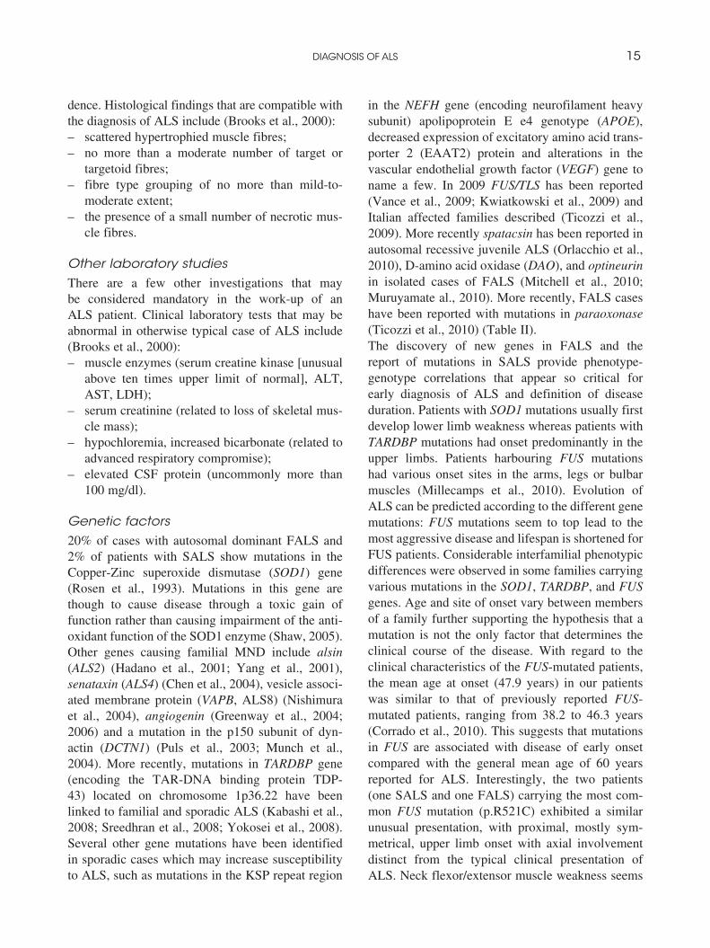
Genetic factors20% of cases with autosomal dominant FALS and 2% of patients with SALS show mutations in the Copper-Zinc superoxide dismutase (SOD1) gene (Rosen et al., 1993). Mutations in this gene are though to cause disease through a toxic gain of function rather than causing impairment of the anti-oxidant function of the SOD1 enzyme (Shaw, 2005). Other genes causing familial MND include alsin (ALS2) (Hadano et al., 2001; Yang et al., 2001), senataxin (ALS4) (Chen et al., 2004), vesicle associ-ated membrane protein (VAPB, ALS8) (Nishimura et al., 2004), angiogenin (Greenway et al., 2004; 2006) and a mutation in the p150 subunit of dyn-actin (DCTN1) (Puls et al., 2003; Munch et al., 2004). More recently, mutations in TARDBP gene (encoding the TAR-DNA binding protein TDP-43) located on chromosome 1p36.22 have been linked to familial and sporadic ALS (Kabashi et al., 2008; Sreedhran et al., 2008; Yokosei et al., 2008). Several other gene mutations have been identified in sporadic cases which may increase susceptibility to ALS, such as mutations in the KSP repeat region
in the NEFH gene (encoding neurofilament heavy subunit) apolipoprotein E e4 genotype (APOE), decreased expression of excitatory amino acid trans-porter 2 (EAAT2) protein and alterations in the vascular endothelial growth factor (VEGF) gene to name a few. In 2009 FUS/TLS has been reported (Vance et al., 2009; Kwiatkowski et al., 2009) and Italian affected families described (Ticozzi et al., 2009). More recently spatacsin has been reported in autosomal recessive juvenile ALS (Orlacchio et al., 2010), D-amino acid oxidase (DAO), and optineurin in isolated cases of FALS (Mitchell et al., 2010; Muruyamate al., 2010). More recently, FALS cases have been reported with mutations in paraoxonase (Ticozzi et al., 2010) (Table II).The discovery of new genes in FALS and the report of mutations in SALS provide phenotype-genotype correlations that appear so critical for early diagnosis of ALS and definition of disease duration. Patients with SOD1 mutations usually first develop lower limb weakness whereas patients with TARDBP mutations had onset predominantly in the upper limbs. Patients harbouring FUS mutations had various onset sites in the arms, legs or bulbar muscles (Millecamps et al., 2010). Evolution of ALS can be predicted according to the different gene mutations: FUS mutations seem to top lead to the most aggressive disease and lifespan is shortened for FUS patients. Considerable interfamilial phenotypic differences were observed in some families carrying various mutations in the SOD1, TARDBP, and FUS genes. Age and site of onset vary between members of a family further supporting the hypothesis that a mutation is not the only factor that determines the clinical course of the disease. With regard to the clinical characteristics of the FUS-mutated patients, the mean age at onset (47.9 years) in our patients was similar to that of previously reported FUS-mutated patients, ranging from 38.2 to 46.3 years (Corrado et al., 2010). This suggests that mutations in FUS are associated with disease of early onset compared with the general mean age of 60 years reported for ALS. Interestingly, the two patients (one SALS and one FALS) carrying the most com-mon FUS mutation (p.R521C) exhibited a similar unusual presentation, with proximal, mostly sym-metrical, upper limb onset with axial involvement distinct from the typical clinical presentation of ALS. Neck flexor/extensor muscle weakness seems
16 V. SILANI ET AL.
to be a further unusual clinical characteristic at dis-ease onset in the p.R521C-mutated SALS and FALS patients, usually being present in only about 0.7% of ALS patients (Gourie-Devi et al., 2003). The two previously reported FALS cases with p.R521C mutations (Ticozzi et al., 2009) presented similar clinical characteristics, with symmetrical, proximal and axial weakness at onset, further supporting the distinct FUS p.R521C clinical phenotype also characterized by prevalence of lower motor neuron signs. The accurate collection of clinical records reporting proximal, axial and neck muscle involve-ment at the disease onset is an excellent example of a genotype-phenotype correlation that, if early detected, can address the prompt diagnosis of ALS. For a further review of genetics see Ticozzi et al. (2011) in this journal.
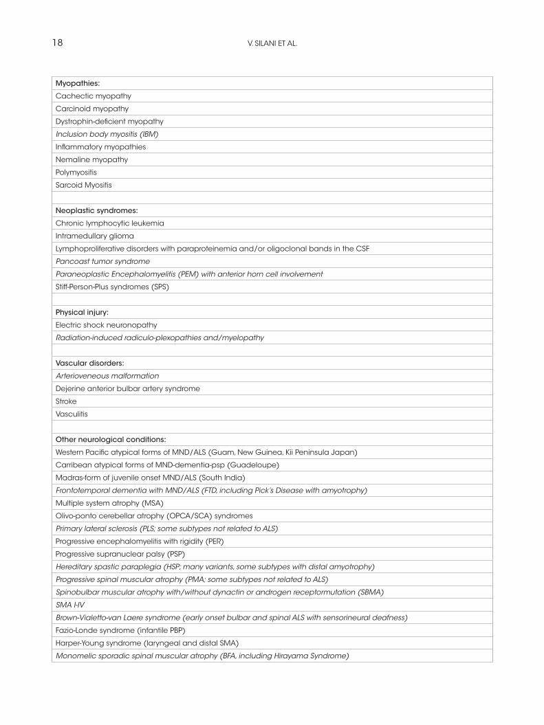
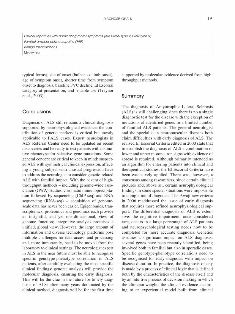
Differential diagnosis
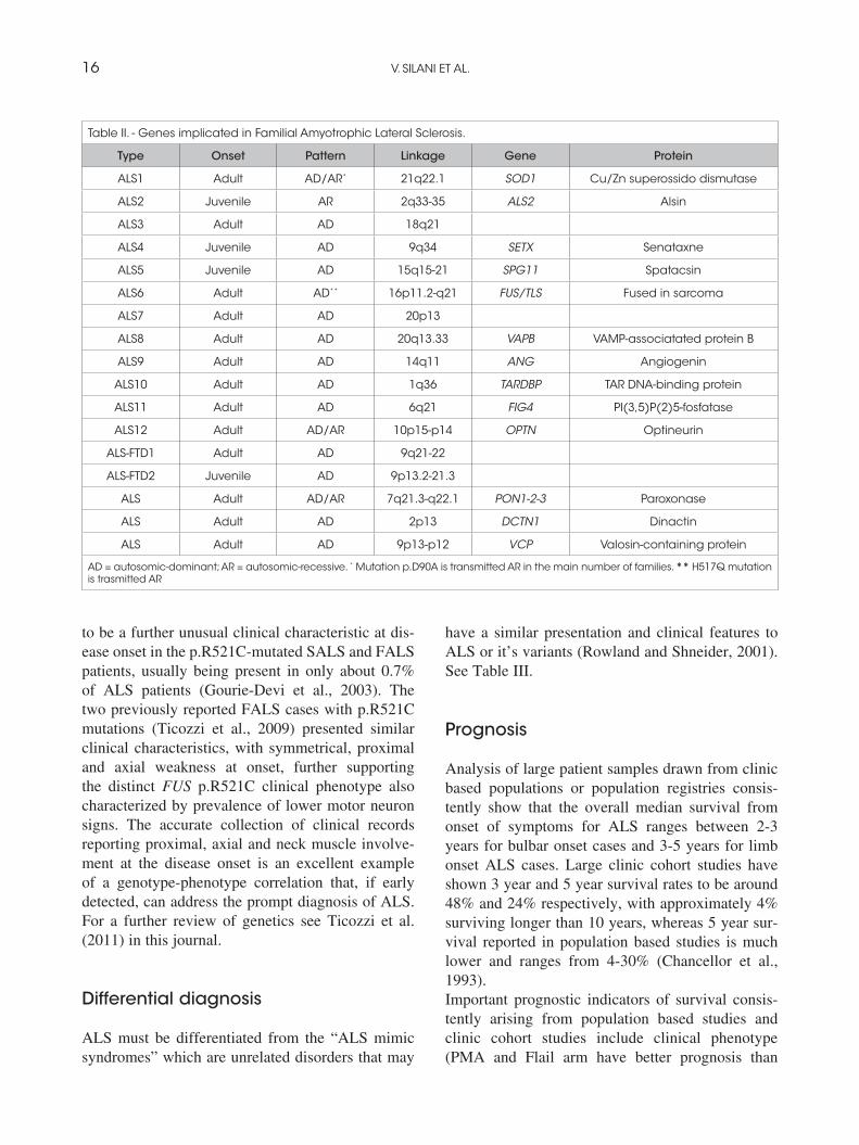
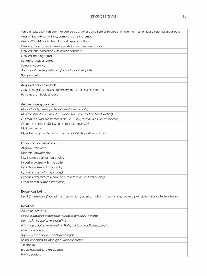
ALS must be differentiated from the “ALS mimic syndromes” which are unrelated disorders that may
have a similar presentation and clinical features to ALS or it’s variants (Rowland and Shneider, 2001). See Table III.
Prognosis
Analysis of large patient samples drawn from clinic based populations or population registries consis-tently show that the overall median survival from onset of symptoms for ALS ranges between 2-3 years for bulbar onset cases and 3-5 years for limb onset ALS cases. Large clinic cohort studies have shown 3 year and 5 year survival rates to be around 48% and 24% respectively, with approximately 4% surviving longer than 10 years, whereas 5 year sur-vival reported in population based studies is much lower and ranges from 4-30% (Chancellor et al., 1993).Important prognostic indicators of survival consis-tently arising from population based studies and clinic cohort studies include clinical phenotype (PMA and Flail arm have better prognosis than
Table II. - Genes implicated in Familial Amyotrophic Lateral Sclerosis.
Type Onset Pattern Linkage Gene Protein
ALS1 Adult AD/AR* 21q22.1 SOD1 Cu/Zn superossido dismutase
ALS2 Juvenile AR 2q33-35 ALS2 Alsin
ALS3 Adult AD 18q21
ALS4 Juvenile AD 9q34 SETX Senataxne
ALS5 Juvenile AD 15q15-21 SPG11 Spatacsin
ALS6 Adult AD** 16p11.2-q21 FUS/TLS Fused in sarcoma
ALS7 Adult AD 20p13
ALS8 Adult AD 20q13.33 VAPB VAMP-associatated protein B
ALS9 Adult AD 14q11 ANG Angiogenin
ALS10 Adult AD 1q36 TARDBP TAR DNA-binding protein
ALS11 Adult AD 6q21 FIG4 PI(3,5)P(2)5-fosfatase
ALS12 Adult AD/AR 10p15-p14 OPTN Optineurin
ALS-FTD1 Adult AD 9q21-22
ALS-FTD2 Juvenile AD 9p13.2-21.3
ALS Adult AD/AR 7q21.3-q22.1 PON1-2-3 Paroxonase
ALS Adult AD 2p13 DCTN1 Dinactin
ALS Adult AD 9p13-p12 VCP Valosin-containing protein
AD = autosomic-dominant; AR = autosomic-recessive. * Mutation p.D90A is transmitted AR in the main number of families. ** H517Q mutation is trasmitted AR
DIAGNOSIS OF ALS 17
Table III - Diseases that can masquerade as Amyotrophic Lateral Sclerosis (in italic the most critical differential diagnosis)
Anatomical abnormalities/compression syndromes:
Arnold-Chiari-1 and other hindbrain malformations
Cervical, foraman magnum or posterior fossa region tumors
Cervical disc herniation with osteochondrosis
Cervical meningeoma
Retropharyngeal tumour
Spinal epidural cyst
Spondylotic myelopathy and/or motor radiculopathy
Syringomyelia
Acquired enzyme defects:
Adult GM2 gangliosidosis (hexosaminidase-A or B deficiency)
Polyglucosan body disease
Autoimmune syndromes:
Monoclonal gammopathy with motor neuropathy
Multifocal motor neuropathy with/without conduction block (MMN)
Dysimmune LMN syndromes (with GM1, GD1b and asialo-GM1 antibodies)
Other dys-immune LMN syndromes including CIDP
Multiple sclerosis
Myasthenia gravis (in particular the anti-MuSK positive variant)
Endocrine abnormalities:
Allgrove Syndrome
Diabetic “amyotrophy”
Insulinoma causing neuropathy
Hyperthyroidism with myopathy
Hypothyroidism with myopathy
Hyperparathyroidism (primary)
Hyperparathyroidism (secondary due to vitamin D deficiency)
Hypokalemia (Conn’s syndrome)
Exogenous toxins:
Lead (?), mercury (?), cadmium, aluminium, arsenic, thallium, manganese, organic pesticides, neurolathyrism, konzo
Infections:
Acute poliomyelitis
Post-poliomyelitis progressive muscular atrophy syndrome
HIV-1 (with vacuolar myelopathy)
HTLV-1 associated myelopathy (HAM, tropical spastic paraplegia)
Neuroborreliosis
Syphilitic hypertrophic pachymeningitis
Spinal encephalitis lethargica, varicella-zoster
Trichinosis
Brucellosis, cat-scratch disease,
Prion disorders
18 V. SILANI ET AL.
Myopathies:
Cachectic myopathy
Carcinoid myopathy
Dystrophin-deficient myopathy
Inclusion body myositis (IBM)
Inflammatory myopathies
Nemaline myopathy
Polymyositis
Sarcoid Myositis
Neoplastic syndromes:
Chronic lymphocytic leukemia
Intramedullary glioma
Lymphoproliferative disorders with paraproteinemia and/or oligoclonal bands in the CSF
Pancoast tumor syndrome
Paraneoplastic Encephalomyelitis (PEM) with anterior horn cell involvement
Stiff-Person-Plus syndromes (SPS)
Physical injury:
Electric shock neuronopathy
Radiation-induced radiculo-plexopathies and/myelopathy
Vascular disorders:
Arterioveneous malformation
Dejerine anterior bulbar artery syndrome
Stroke
Vasculitis
Other neurological conditions:
Western Pacific atypical forms of MND/ALS (Guam, New Guinea, Kii Peninsula Japan)
Carribean atypical forms of MND-dementia-psp (Guadeloupe)
Madras-form of juvenile onset MND/ALS (South India)
Frontotemporal dementia with MND/ALS (FTD, including Pick’s Disease with amyotrophy)
Multiple system atrophy (MSA)
Olivo-ponto cerebellar atrophy (OPCA/SCA) syndromes
Primary lateral sclerosis (PLS; some subtypes not related to ALS)
Progressive encephalomyelitis with rigidity (PER)
Progressive supranuclear palsy (PSP)
Hereditary spastic paraplegia (HSP; many variants, some subtypes with distal amyotrophy)
Progressive spinal muscular atrophy (PMA; some subtypes not related to ALS)
Spinobulbar muscular atrophy with/without dynactin or androgen receptormutation (SBMA)
SMA I-IV
Brown-Vialetto-van Laere syndrome (early onset bulbar and spinal ALS with sensorineural deafness)
Fazio-Londe syndrome (infantile PBP)
Harper-Young syndrome (laryngeal and distal SMA)
Monomelic sporadic spinal muscular atrophy (BFA, including Hirayama Syndrome)
DIAGNOSIS OF ALS 19
typical forms), site of onset (bulbar vs. limb onset), age of symptom onset, shorter time from symptom onset to diagnosis, baseline FVC decline, El Escorial category at presentation, and riluzole use (Traynor et al., 2003).
Conclusions
Diagnosis of ALS still remains a clinical diagnosis supported by neurophysiological evidence: the con-tribution of genetic markers is critical but mostly applicable to FALS cases. Expert neurologists in ALS Referral Center need to be updated on recent discoveries and be ready to test patients with distinc-tive phenotype for selective gene mutations. Some general concept are critical to keep in mind: suspect-ed ALS with symmetrical clinical expression, affect-ing a young subject with unusual progression have to address the neurologist to consider genetic-related ALS with familial impact. With the advent of high-throughput methods – including genome-wide asso-ciation (GWA) studies, chromatin immunoprecipita-tion followed by sequencing (ChIP-seq) and RNA sequencing (RNA-seq) – acquisition of genome-scale data has never been easier. Epigenomics, tran-scriptomics, proteomics and genomics each provide an insightful, and yet one-dimensional, view of genome function; integrative analysis promises a unified, global view. However, the large amount of information and diverse technology platforms pose multiple challenges for data access and processing and, more importantly, need to be moved from the laboratory to clinical settings. The neurologist expert in ALS in the near future must be able to recognize specific genotype-phenotype correlation in ALS patients, after carefully evaluating the most specific clinical findings: genome analysis will provide the molecular diagnosis, ensuring the early diagnosis. This will be the clue in the future for timely diag-nosis of ALS: after many years dominated by the clinical method, diagnosis will be for the first time
supported by molecular evidence derived from high-throughput methods.
Summary
The diagnosis of Amyotrophic Lateral Sclerosis (ALS) is still challenging since there is no a single diagnostic test for the disease with the exception of mutations of identified genes in a limited number of familial ALS patients. The general neurologist and the specialist in neuromuscular diseases both claim difficulties with early diagnosis of ALS. The revised El Escorial Criteria edited in 2000 state that to establish the diagnosis of ALS a combination of lower and upper motoneuron signs with evidence of spread is required. Although primarily intended as an algorithm for entering patients into clinical and therapeutical studies, the El Escorial Criteria have been extensively applied. There was, however, a consensus among researchers, since certain clinical pictures and, above all, certain neurophysiological findings in some special situations were impossible to completion of diagnosis. The Awaji new criteria in 2006 readdressed the issue of early diagnosis that requires more refined neurophysiological sup-port. The differential diagnosis of ALS is exten-sive: the cognitive impairment, once considered rare, occurs in a large percentage of ALS patients and neuropsychological testing needs now to be completed for more accurate diagnosis. Genetics assumes a significant impact on ALS diagnosis: several genes have been recently identified, being involved both in familial but also in sporadic cases. Specific genotype-phenotype correlations need to be recognised for early diagnosis with impact on disease duration. In practice, the diagnosis of any is made by a process of clinical logic that is defined both by the characteristics of the disease itself and by an intuitive process of decision making in which the clinician weights the clinical evidence accord-ing to an experiential model built from clinical
Polyneuropathies with dominating motor symptoms (like HMSN type 2, HMN type 5)
Familial amyloid polyneuropathy (FAP)
Benign fasciculations
Myokymia
20 V. SILANI ET AL.
experience gained over time. In ALS this concept must be revisited because the recent impact of new genetic discoveries requires to the clinician a constant update to recognize specific genotype-phenotype correlations that unquestionably can accelerate early diagnosis of ALS, supported by genetic testing.
AcknowledgementsWe are grateful to the patients and their families. We thank Palmina Giannini that highly desired and inspired the ALS Meeting at IRCCS Neuromed on October 23, 2009. This study was supported by the Italian Ministry of Health (Ricerca Finalizzata 2007 no. 31) and AriSLA (EXOMEFALS, 2010).
References
Abe K., Fujimura H., Kobayashi Y., Fujita N., Yanagihara T. Degeneration of the pyramidal tracts in patients with amyotrophic lateral scle-rosis. A premortem and postmortem magnetic resonance imaging study. J. Neuroimaging, 7: 208-212, 1997.
Abhinav K., Stanton B., Johnston C., Hardstaff J., Orrell R.W., Howard R., Clarke J., Sakel M., Ampong M.A., Shaw C.E., Leigh P.N., Al-Chalabi A. Amyotrophic lateral sclerosis in South-East England: a population-based study. The South-East England register for amyotrophic lateral sclerosis (SEALS Registry). Neuroepidemiology, 29: 44-48, 2007.
Abrahams S., Goldstein L.H., Kew J.J.M. Brooks D.J., Lloyd C.M., Frith C.D., Leigh P N. Frontal lobe dysfunction in amyotrophic lateral sclerosis: a PET study. Brain, 119: 2105-2120, 1996.
Abrahams S., Leigh P.N, Harvey A., Vythelingum G.N., Grisé D, Goldstein L.H. Verbal fluency and executive dysfunction in amyotrophic lateral scle-rosis. Neuropsychologia, 38: 734-747, 2000.
Abrahams S., Goldstein L.H., Simmons A., Brammer M, Williams S.C., Giampietro V., Leigh P.N. Word retrieval in amyotrophic lateral sclerosis: a functional magnetic resonance imaging study. Brain, 127: 1507-1517, 2004.
Abrahams S., Leigh P.N., Goldstein L.H. Cognitive change in ALS: a prospective study. Neurology, 64: 1222-1226, 2005.
Agosta F., Chiò A., Cosottini M., De Stefano M., Falini A., Mascalchi M., Rocca M.A., Silani V.,
Tedeschi G., Filippi M. The present and the future of neuroimaging in amyotrophic lateral sclero-sis. Am. J. Neuroradiol., Apr 1 as 10.3174/ajnr.A2043., 2010.
Beghi E., Millul A., Micheli A., Vitelli E., Logroscino G. Incidence of ALS in Lombardy, Italy. Neurology, 68: 141-145, 2007.
Ben Hamida M., Hentati F., Ben Hamida C. Hereditary motor system diseases (chronic juvenile amyo-trophic lateral sclerosis). Conditions combining a bilateral pyramidal syndrome with limb and bulbar amyotrophy. Brain, 113: 347-363, 1990.
Borasio G.D., Shaw P.J. Hardiman O., Ludolph A., Sales Luis M.L., Silani V., for the European ALS Study Group. Standards of palliative care for patients with amyotrophic lateral sclerosis: results of a European survey. Amyotroph. Lateral Scler. Other Motor Neuron Disord., 2: 159-164, 2001.
Bromberg M.B. and Brownell A.A. Motor unit num-ber estimation in the assessment of performance and function in motor neuron disease. Physical Medicine and Rehabilitation Clinics of North America, 2008, 19: 509-532.
Brooks B.R. El Escorial World Federation of Neurology criteria for the diagnosis of amyo-trophic lateral sclerosis. Subcommittee on Motor Neuron Diseases/Amyotrophic Lateral Sclerosis of the World Federation of Neurology Research Group on Neuromuscular Diseases and the El Escorial “Clinical limits of amyotrophic lateral sclerosis” workshop contributors. J. Neurol. Sci., 124 (Suppl): 96-107, 1994.
Brooks B.R., Miller R.G., Swash M., Munsat T.L. El Escorial revisited: revised criteria for the diag-nosis of amyotrophic lateral sclerosis. Amyotroph Lateral Scler Other Motor Neuron Disord, 1: 293-299, 2000.
Cappellari A., Ciammola A., Silani V. The pseu-dopolyneuritic form of amyotrophic lateral scle-rosis (Patrikios’ disease). Electromyogr. Clin. Neurophysiol., 48: 75-81, 2008.
Chance P.F., Rabin B.A., Ryan S.G., Ding Y., Scavina M., Crain B., Griffin J.W. Cornblath D.R: Linkage of the gene for an autosomal dominant form of juvenile amyotrophic lateral sclerosis to chromosome 9q34. Am. J. Hum. Genet., 62: 633-640, 1998.
Chancellor A.M., Slattery J.M., Fraser H., Swingler R.J., Holloway S.M., Warlow C.P. The prognosis of adult-onset motor neuron disease: a prospective study based on the Scottish Motor Neuron Disease Register. J. Neurol., 240: 339-346, 1993.
DIAGNOSIS OF ALS 21
Chang J.L., Lomen-Hoerth C., Murphy J., Henry R.G., Kramer J.H., Miller B.L., Gorno-Tempini M.L. Avoxel-based morphometry study of pat-terns of brain atrophy in ALS and ALS/FTLD. Neurology, 5: 75-80, 2005.
Chen R., Grand’Maison F., Strong M.J., Ramsay D.A., Bolton C.F. Motor neuron disease presenting as acute respiratory failure: a clinical and patho-logical study. J. Neurol. Neurosurg. Psychiatry, 60: 455-458, 1996.
Chen Y.Z., Bennett C.L., Huynh H.M., Blair I.P., Puls I., Irobi J., Dierick I., Abel A., Kennerson M.L., Rabin B.A., Nicholson G.A. Auer-Grumbach M., Wagner K., De Jonghe P., Griffin J.W., Fischbeck K.H., Timmerman V., Cornblath D.R., Chance P.F. DNA/RNA helicase gene mutations in a form of juvenile amyotrophic lateral sclerosis (ALS4). Am. J. Hum. Genet., 74: 1128-1135, 2004.
Chiò A. and Silani V. Amyotrophic Lateral Sclerosis (ALS) care in Italy: a nationwide study in neuro-logical centers. J. Neurol. Sci., 191: 145-150, 2001.
Couratier P., Truong C., Khalil M., Deviere F., Vallat J.M. Clinical features of flail arm syndrome. Muscle Nerve, 23: 646-648, 2000.
Cornblath D.R., Kuncl R.W., Mellits E.D., Quaskey S.A., Clawson L., Pestronk A., Drachman D.B. Nerve conduction studies in amyotrophic, lateral sclerosis. Muscle Nerve, 15: 1111-1115, 1992.
Corrado L., Del Bo R., Castellotti B., Ratti A., Cereda C., Penco S., Soraru G., Carlomagno Y., Grezzi S., Pensato V., Colombrita C., Gagliardi S., Cozzi L., Orsetti V., Mancuso M., Siciliano
G., Mazzini L., Comi G.P., Gellera C., Ceroni M., D’Alfonso S., Silani V. Mutations of FUS Gene in Sporadic Amyotrophic Lateral Sclerosis. J. Medical Genetics, 47: 190-194, 2010.
Cummings J.L., Mega M., Gray K., Rosenberg-Thompson S., Carusi D.A., Gornbein J. The neuro-psychiatric inventory: comprehensive assessment of psychopathology in dementia. Neurology, 44: 2308-2314, 1994.
Dalakas M.C., Hatazawa J., Brooks R.A., Di Chiro G. Lowered cerebral glucose utilization in amyo-trophic lateral sclerosis. Ann. Neurol., 22: 471-474. 1987.
Davenport R.J., Swingler R.J., Chancellor A.M., Warlow C.P. Avoiding false positive diagnoses of motor neuron disease: lessons from the Scottish Motor Neuron Disease Register. J. Neurol. Neurosurg. Psychiatry, 60: 147-151, 1996.
de Carvalho M., Matias T., Coelho F., Evangelista T., Pinto A., Luis M.L. Motor neuron disease presen-
ting with respiratory failure. J. Neurol. Sci., 139: 117-122, 1996.
de Carvalho M. and Swash M. Nerve conduction studies in amyotrophic lateral sclerosis. Muscle Nerve, 23: 344-352, 2000.
de Carvalho M., Johnsen B., Fuglsang-Frederiksen A. Medical technology assessment. Electrodiagnosis in motor neuron diseases and amyotrophic lateral sclerosis. Neurophysiol. Clin., 31: 341-348, 2001.
de Carvalho M., Dengler R., Eisen A., England J.D., Kaji R., Kimura J., Mills K., Mitsumoto H., Nodera H., Shefner J., Swash M. Electrodiagnostic criteria for diagnosis of ALS. Clin. Neurophysiol., 119: 497-503, 2008.
Eisen A.A. and Shtybel W. AAEM minimonograph #35: Clinical experience with transcranial mag-netic stimulation. Muscle Nerve, 13: 995-1011, 1990.
Ellis C.M., Simmons A., Andrews C., Dawson J.M., Williams S.C., Leigh P.N. A proton magnetic reso-nance spectroscopic study in ALS: correlation with clinical findings. Neurology, 51: 1104-1109, 1998.
Ellis C.M., Simmons A., Jones D.K., Bland J., Dawson J.M., Horsfield M.A., Williams S.C., Leigh P.N. Diffusion tensor MRI assesses cor-ticospinal tract damage in ALS. Neurology, 53: 1051-1058, 1999.
Forbes R.B., Colville S., Swingler R.J. The epide-miology of amyotrophic lateral sclerosis (ALS/MND) in people aged 80 or over. Age Ageing, 33: 131-134, 2004.
Flaherty-Craig C., Eslinger P., Stephens B., Simmons Z. A rapid screening battery to identify frontal dysfunction in patients with ALS. Neurology, 67: 2070-72, 2006.
Frank B., Haas J., Heinze H., Stark E,. Münte T.F. Relation of neuropsycological and magnetic reso-nance findings in amyotrophic lateral sclerosis: evidence for subgroups. Clin. Neurol. Neurosurg., 99: 79-86, 1997.
Goodin D.S., Rowley H.A., Olney R.K. Magnetic resonance imaging in amyotrophic lateral sclero-sis. Ann. Neurol., 23: 418-420, 1988.
Gordon P.H., Cheng B., Katz I.B., Pinto M., Hays A.P., Mitsumoto H., Rowland L.P. The natural history of primary lateral sclerosis. Neurology, 66: 647-653, 2006.
Gordon P.H., WangY., Doorish C., Lewis M., Battista V., Mitsumoto H,. Marder K. A screening assess-ment of cognitive impairment in patients with ALS. Amyotroph. Lateral Scler., 8: 362-365, 2007.
22 V. SILANI ET AL.
Gourie-Devi M., Nalini A., Sandhya S. Early or late appearance of “dropped head syndrome” in amyo-trophic lateral sclerosis. J. Neurol. Neurosurg. Psychiatry, 74: 683-686, 2003.
Gouveia L.O. and De Carvalho M. Young-onset sporadic amyotrophic lateral sclerosis: A distinct nosological entity? Amyotroph. Lateral Scler., 8: 323-327, 2007.
Greenway M.J., Alexander M.D., Ennis S., Traynor B.J., Corr B., Frost E., Green A., Hardiman O. A novel candidate region for ALS on chromosome 14q11.2. Neurology, 63: 1936-1938, 2004.
Greenway M.J., Andersen P.M., Russ C., Ennis S., Cashman S., Donaghy C., Patterson V., Swingler R., Kieran D., Prehn J., Morrison K.E., Green A., Acharya K.R, Brown R.H. Jr., Hardiman O. ANG mutations segregate with familial and ‘sporadic’ amyotrophic lateral sclerosis. Nat. Genet., 38: 411-413, 2006.
Gros-Louis F., Gaspar C., Rouleau G.A. Genetics of familial and sporadic amyotrophic lateral sclerosis. Biochim. Biophys. Acta, 1762: 956-972, 2006.
Grossman A.B., Woolley-Levine S., Bradley W.G., Miller R.G. Detecting neurobehavioral changes in amyotrophic lateral sclerosis. Amyotroph. Lateral Scler., 8: 56-61, 2007.
Guedj E., Ber I., Lacomblez L., Dubois B., Verpillat P., Didic M., Salachas F., Vera P., Hannequin D., Lotterie J.A., Puel M., Decousus M., Thomas-Antérion C., Magne C., Vercelletto M., Bernard A.M., Golfier V., Pasquier J., Michel B.F., Namer I., Sellal F., Bochet J., Volteau M., Brice A., Meininger V., French Research Network on FTD/FTD-MND, Habert M.O. Brain spect perfusion of frontotemporal dementia associated with motor neuron disease. Neurology, 69: 488-490, 2007.
Guidetti D., Bondavalli M., Sabadini R., Marcello N., Vinceti M., Cavalletti S., Marbini A., Gemignani F., Colombo A., Ferrari A., Vivoli G., Solimè F. Epidemiological survey of amyotrophic lat-eral sclerosis in the province of Reggio Emilia, Italy: influence of environmental exposure to lead. Neuroepidemiology, 15: 301-312, 1996.
Hadano S., Hand C.K., Osuga H., Yanagisawa Y., Otomo A., Devon R.S., Miyamoto N., Showguchi-Miyata J., Okada Y., Singaraja R., Figlewicz D.A., Kwiatkowski T., Hosler B.A., Sagie T., Skaug J., Nasir J., Brown R.H. Jr., Scherer S.W., Rouleau G.A., Hayden M.R., Ikeda J.E. A gene encoding a putative GTPase regulator is mutated in familial amyotrophic lateral sclerosis 2. Nat. Genet., 29: 166-173, 2001.
Hatazawa J., Brooks R.A., Dalakas M.C., Mansi L., Di Chiro G. Cortical motorsensory hypometabo-lism in amyotrophic lateral sclerosis: a PET study. J. Comput. Assist. Tomogr., 12: 630-636, 1988.
Haverkamp L.J., Appel V., Appel S.H: Natural his-tory of amyotrophic lateral sclerosis in a database population. Validation of a scoring system and a model for survival prediction. Brain, 118 (Pt 3): 707-719, 1995.
Hentati A., Bejaoui K., Pericak-Vance M.A., Hentati F., Speer M.C., Hung W.Y., Figlewicz D.A., Haines J., Rimmler J., Ben Hamida C., Ben Hamida M., Brown R.H. Jr, Siddique T. Linkage of recessive familial amyotrophic lateral sclerosis to chromosome 2q33-q35. Nat. Genet., 7: 425-428, 1994.
Hentati A., Ouahchi K., Pericak-Vance M.A., Nijhawan D., Ahmad A., Yang Y., Rimmler J., Hung W., Schlotter B., Ahmed A., Yang Y., Rimmler J., Hung W., Schlotter B., Ahmed A., Ben Hamida M., Hentati F., Siddique T. Linkage of a commoner form of recessive amyotrophic lat-eral sclerosis to chromosome 15q15-q22 markers. Neurogenetics, 2: 55-60, 1998.
Hu M.T., Ellis C.M., Al-Chalabi A., Leigh P.N., Shaw C.E. Flail arm syndrome: a distinctive vari-ant of amyotrophic lateral sclerosis. J. Neurol. Neurosurg. Psychiatry, 65: 950-951, 1998.
Isaacs J.D., Dean A.F., Shaw C.E., Al-Chalabi A., Mills K.R., Leigh P.N. Amyotrophic lateral sclero-sis with sensory neuropathy: part of a multisystem disorder? J. Neurol. Neurosurg. Psychiatry, 78: 750-753, 2007.
Iversen I.H., Ghanayim N., Kübler A., Neumann N., Birbaumer N., Kaiser J. A brain-computer interface tool to assess cognitive functions in completely paralyzed patients with amyotrophic lateral sclerosis, Clinical Neurophysiology, 119: 2214-2223, 2008.
Janko M., Trontelj J.V., Gersak K. Fasciculations in motor neuron disease: discharge rate reflects extent and recency of collateral sprouting. J. Neurol. Neurosurg. Psychiatry, 52: 1375-1381, 1989.
Johnston C.A., Stanton B.R., Turner M.R., Gray R., Blunt A.H., Butt D., Ampong M.A., Shaw C.E., Leigh P.N., Al-Chalabi A. Amyotrophic lateral sclerosis in an urban setting: a population based study of inner city London. J. Neurol., 253: 1642-1643, 2006.
Kabashi E., Valdmanis P.N., Dion P., Spiegelman D., McConkey B.J., Velde C. Vande, Bouchard J.P.,
DIAGNOSIS OF ALS 23
Lacomblez L., Pochigaeva K., Salachas F, Pradat P.F., Camu W., Meininger V., Dupre N., Rouleau G.A. TARDBP mutations in individuals with spo-radic and familialamyotrophic lateral sclerosis. Nat. Genet., 40: 572-574, 2008.
Kalra S. and Arnold D. Neuroimaging in amyotrophic lateral sclerosis. Amyotrophic Lateral Sclerosis, 4: 243-248, 2003.
Katz J.S., Wolfe G.I., Andersson P.B., Saperstein D.S., Elliott J.L., Nations S.P., Bryan W.W., Barohn R.J. Brachial amyotrophic diplegia: a slow-ly progressive motor neuron disorder. Neurology, 53: 1071-1076, 1999.
Kertesz A., Davidson W., Fox H. Frontal behav-ioural inventory: diagnostic criteria for frontal lobe dementia. Can. J. Neurol. Sci., 24: 29-36, 1997.
Kew J.J., Goldstein L.H., Leigh P.N., Abrahams S., Cosgrave N., Passingham R.E., Frackowiak RS, Brooks DJ. The relationship between abnormali-ties of cognitive function and cerebral activation in amyotrophic lateral sclerosis. A neuropsycho-logical and positron emission tomography study. Brain, 116: 1399-1423, 1993.
Kilani M., Micallef J., Soubrouillard C., Rey-Lardiller D., Dematteï C., Dib M., Philippot P., Ceccaldi M., Pouget J., Blin O. A longitudinal study of the evolution of cognitive function and affective state in patients with amyotrophic lateral sclerosis. Amyotroph. Lateral Scler. Other Motor Neuron Disord., 5: 46-54, 2004.
Kobayashi M., Ikeda K., Kinoshita M., Iwasaki Y. Amyotrophic lateral sclerosis with supranuclear ophthalmoplegia and rigidity. Neurol. Res., 21: 661-664, 1999.
Kuzuhara S. and Kokubo Y. Atypical parkinsonism of Japan: amyotrophic lateral sclerosis parkin-sonism-dementia complex of the Kii peninsula of Japan (Muro disease): an update. Mov. Disord., 20 (Suppl 12): S108-113, 2005.
Kwiatkowski T.J. Jr, Bosco D.A., Leclerc A.L., Tamrazian E., Vanderburg C.R., Russ C., Davis A., Gilchrist J., Kasarskis E.J., Munsat T., Valdmanis P., Rouleau G.A., Hosler B.A., Cortelli P., de Jong P.J., Yoshinaga Y., Haines J.L., Pericak-Vance M.A., Yan J., Ticozzi N., Siddique T., McKenna-Yasek D., Sapp P.C., Horvitz H.R., Landers J.E., Brown R.H. Jr. Mutations in the FUS/TLS gene on chromosome 16 cause familial amyotrophic lateral sclerosis. Science, 323: 1205-1208, 2009.
Lambert E.H. and Mulder D.W. Electromyographic studies in amyotrophic lateral sclerosis. Proc. Staff Meet. Mayo Clin., 32: 441-446, 1957.
Le Forestier N., Maisonobe T., Piquard A., Rivaud S., Crevier-Buchman L., Salachas F., Pradat P.F., Lacomblez L., Meininger V. Does primary lateral sclerosis exist? A study of 20 patients and a review of the literature. Brain, 124: 1989-1999, 2001a.
Le Forestier N., Maisonobe T., Spelle L., Lesort A., Salachas F., Lacomblez L., Samson Y., Bouche P., Meininger V. Primary lateral sclerosis: further clarification. J. Neurol. Sci., 185: 95-100, 2001b.
Li T.M., Alberman E., Swash M. Comparison of sporadic and familial disease amongst 580 cases of motor neuron disease. J. Neurol. Neurosurg. Psychiatry, 51: 778-784, 1988.
Lloyd C.M., Richardson M.P., Brooks D.J., Al-Chalabi A., Leigh P.N. Extramotor involve-ment in ALS: pet studies with the GABA(A) ligand [(11)C]flumazenil. Brain, 123: 2289-96, 2000.
Logroscino G., Traynor B.J., Hardiman O., Chiò A., Couratier P., Mitchell J.D., Swingler R.J., Beghi E., for EURALS. Descriptive epidemiology of amyotrophic lateral sclerosis: new evidence and unsolved issues. J. Neurol. Neurosurg. Psychiatry, 79: 6-11, 2008.
Lomen-Hoerth C., Murphy J., Langmore S., Kramer J.H., Olney R.K., Miller B. Are amyotrophic lateral sclerosis patients cognitively normal? Neurology, 60:1094-1097, 2003.
Ludolph A.C., Langen K.J., Regard M., Herzog H., Kemper B., Kuwert T., Böttger I.G., Feinendegen L. Frontal lobe function in amyotrophic lateral sclerosis: a neurocognitive and positron emission tomography study. Acta Neurol. Scand., 85: 81-89, 1992.
Lule D., Diekmann V., Anders S., Kassubek J., Kübler A., Ludolph A.C, Birbaumer N. Brain responses to emotional stimuli in patients with amyotrophic lateral sclerosis (ALS). J. Neurol., 254: 519-527, 2007.
Mantovan M.C., Baggio L., Barba G., Smith P., Pegoraro E., Sorarù G., Bonometto P., Angelini C. Memory deficits and retrieval processes in ALS. Eur. J. Neurol., 10: 221-227, 2003.
Maruyama H., Morino H., Ito H., Izumi Y., Kato H., Watanabe Y., Kinoshita Y., Kamada M., Nodera H., Suzuki H., Komure O., Matsuura S., Kobatake K., Morimoto N., Abe K., Suzuki N., Aoki M., Kawata A., Hirai T., Kato T., Ogasawara K., Hirano A., Takumi T., Kusaka H., Hagiwara K., Kaji R., Kawakami H. Mutations of optineurin in amyotrophic lateral sclerosis. Nature, 465: 223-226, 2010.
24 V. SILANI ET AL.
Massman P.J., Sims J., Cooke N., Haverkamp L.J., Appel V., Appel S.H. Prevalence and correlates of neuropsychological deficits in amyotrophic lateral sclerosis. J. Neurol. Neurosurg. Psychiatry, 61: 450-455, 1996.
McCullagh S., Moore M., Gawel M., Feinstein A. Pathological laughing and crying in amyotrophic lateral sclerosis: an association with prefrontal cog-nitive dysfunction. J. Neurol. Sci., 169: 43-48, 1999.
Millecamps S., Salachas F., Cazeneuve C., Gordon P., Bricka B., Camuzat A., Guillot-Noël L., Russaouen O., Bruneteau G., Pradat P.F., Le Forestier N., Vandenberghe N., Danel-Brunaud V., Guy N., Thauvin-Robinet C., Lacomblez L., Couratier P., Hannequin D., Seilhean D., Le Ber I., Corcia P., Camu W., Brice A., Rouleau G., Leguern E., Meininger V. SOD1, ANG, VAPB, TARDBP, and FUS muattions in familial amyo-trophic lateral sclerosis: genotype-phenotype cor-relations. J. Med. Genet., 47: 554-560, 2010.
Mills K.R. and Nithi K.A. Peripheral and central motor conduction in amyotrophic lateral sclerosis. J. Neurol. Sci., 159: 82-87, 1998.
Mitchell J., Paul P., Chen H.J., Morris A., Payling M., Falchi M., Habgood J., Panoutsou S., Winkler S., Tisato V., Hajitou A., Smith B., Vance C., Shaw C., Mazarakis N.D., de Belleroche J. Familial amyotrophic lateral sclerosis is associated with a mutation in D-amino acid oxidase. Proc. Natl. Acad. Sci. U.S.A., 107: 7556-7561, 2010.
Moretti R., Torre P., Antonello R.M., De Masi R., Cazzato G. Complex cognitive disruption in motor neuron disease. Dement. Geriatr. Cogn. Disord., 14: 141-150, 2002.
Mulder D.W., Kurland L.T., Offord K.P., Beard C.M. Familial adult motor neuron disease: amyotrophic lateral sclerosis. Neurology, 36: 511-517, 1986.
Munch C., Sedlmeier R., Meyer T., Homberg V., Sperfeld A.D., Kurt A., Prudlo J., Peraus G., Hanemann C.O., Stumm G., Ludolph A.C. Point mutations of the p150 subunit of dynactin (DCTN1) gene in ALS. Neurology, 63: 724-726, 2004.
Murphy J.M., Henry R.G., Lomen-Hoerth C. Establishing subtypes of the continuum of frontal lobe impairment in amyotrophic lateral sclerosis. Arch. Neurol., 64: 330-334, 2007a.
Murphy J.M., Henry R.G., Langmore S., Kramer J.H., Miller B.L., Lomen-Hoerth C. Continuum of frontal lobe impairment in amyotrophic lateral sclerosis. Arch. Neurol., 64: 530-534, 2007b.
Neary D., Snowden J.S, Gustafson L. Passant U., Stuss D., Black S., Freedman M., Kertesz A.,
Robert P.H., Albert M., Boone K., Miller B.L., Cummings J., Benson D.F. Frontotemporal lobar degeneration: a consensus on clinical diagnostic criteria. Neurology, 51: 1546-1554, 1998.
Neary D., Snowden J.S., Mann D.M.A. Cognitive change in motor neuron disease/amyotrophic lat-eral sclerosis. J. Neurol. Sci., 180: 15-20, 2000.
Nishimura A.L., Mitne-Neto M., Silva H.C., Richieri-Costa A., Middleton S., Cascio D., Kok F., Oliveira J.R., Gillingwater T., Webb J., Skehel P., Zatz M. A mutation in the vesicle-trafficking protein VAPB causes late onset spinal muscular atrophy and amyotrophic lateral sclerosis. Am. J. Hum. Genet., 75: 822-883, 2004.
Norris F., Shepherd R., Denys E., UK, Mukai E., Elias L., Holden D., Norris H. Onset, natural his-tory and outcome in idiopathic adult motor neuron disease. J. Neurol. Sci., 118: 48-55, 1993.
Oba H., Araki T., Ohtomo K., Monzawa S., Uchiyama G., Koizumi K., Nogata Y., Kachi K., Shiozawa Z., Kobayashi M. Amyotrophic lateral sclerosis: T2 shortening in motor cortex at MR imaging. Radiology, 189: 843-846, 1993.
Okuda B., Yamamoto T., Yamasaki M., Maya K., Imai T. Motor neuron disease with slow eye movements and vertical gaze palsy. Acta Neurol. Scand., 85: 71-76, 1992.
Ohnishi T., Hoshi H., Jinnouchi S., Nagamachi S., Watanabe K., Mituyama Y. The utility of cerebral blood flow imaging in patients with the unique syndrome of progressive dementia with motor neuron disease. J. Nucl. Med., 31: 688-691, 1990.
Orlacchio A., Babalini C., Borreca A., Patrono C., Massa R., Basaran S., Munhoz R.P., Rogaeva E.A., St George-Hyslop P.H., Bernardi G, Kawarai T. SPATACSIN mutations cause autosomal reces-sive juvenile amyotrophic lateral sclerosis. Brain, 133 (Pt 2), 591-598, 2010.
Osei-Lah A.D., Turner M.R., Andersen P.M., Leigh P.N., Mills K.R. A novel central motor conduction abnormality in D90Ahomozygous patients with amyotrophic lateral sclerosis. Muscle Nerve, 29: 790-794, 2004.
Patten S.B., Svenson L.W, White C.M., Khaled S.M., Metz L.M. Affective disorders in motor neuron disease: a population-based study. Neuroepidemiology, 28: 1-7, 2007.
Plato C.C., Garruto R.M., Galasko D., Craig U.K., Plato M., Gamst A., Torres J.M., Wiederholt W. Amyotrophic lateral sclerosis and parkinsonism-dementia complex of Guam: changing incidence
DIAGNOSIS OF ALS 25
rates during the past 60 years. Am. J. Epidemiol., 157: 149-157, 2003.
Polkey M.I., Lyall R.A., Moxham J., Leigh P.N. Respiratory aspects of neurological disease. J. Neurol. Neurosurg. Psychiatry, 66: 5-15. 1999.
Poloni M., Capitani E., Mazzini L, Ceroni M. Neuropsychological measures in amyotrophic lat-eral sclerosis and their relationship with CT scan-assessed cerebral atrophy. Acta Neurol. Scand., 74: 257-260, 1986.
Poletti B., Maringelli F., Lafronza A., Lombardi C., Meriggi P., Silani V. Anti-saccade paradigm in the cognitive frontal assessment of Amyotrophic Lateral Sclerosis. Amyotroph. Lateral Scler. other Motor Neuron Disord., 10 (suppl 1): 45, 2009.
Portet F., Cadilhac C., Touchon J., Camu W. Cognitive impairment in motor neuron disease with bulbar onset. Amyotroph. Lateral Scler. Other Motor Neuron Disord., 2: 23-29, 2001.
Pringle C.E., Hudson A.J., Munoz D.G., Kiernan J.A., Brown W.F., Ebers G.C. Primary lateral scle-rosis. Clinical features, neuropathology and diag-nostic criteria. Brain, 115 (Pt 2): 495-520, 1992.
Phukan J., Pender N.P., Hardiman O. Cognitive impairment in amyotrophic lateral sclerosis. Lancet Neurol., 6: 994-1003, 2007.
Pugdahl K., Fuglsang-Frederiksen A., de Carvalho M., Johnsen B., Fawcett P.R., Labarre-Vila A., Liguori R., Nix W.A., Schofield I.S. Generalised sensory system abnormalities in amyotrophic lat-eral sclerosis: a European multicentre study. J. Neurol. Neurosurg. Psychiatry, 78: 746-749, 2007.
Puls I., Jonnakuty C., LaMonte B.H., Holzbaur E.L., Tokito M., Mann E., Floeter M.K., Bidus K., Drayna D., Oh S.J., Brown R.H.Jr., Ludlow C.L., Fischbeck K.H. Mutant dynactin in motor neuron disease. Nat. Genet., 33: 455-456, 2003.
Raaphorst J., De Visser M., Linssen W.H., De Haan R.J., Schmand B. The cognitive profile of amyotrophic lateral sclerosis: A meta-analysis. Amyotroph. Lateral Scler., 11: 27-37, 2010.
Rakowicz W.P. and Hodges J.R. Dementia and apha-sia in motor neuron disease: an underrecognised association? J. Neurol. Neurosurg. Psychiatry, 65: 881-889, 1998.
Ringholz G.M., Appel S.H., Bradshaw M., Cooke N.A., Mosnik D.M., Schulz P.E. Prevalence and patterns of cognitive impairment in sporadic ALS. Neurology, 65: 586-590, 2005.
Rippon G.A., Scarmeas N., Gordon P.H., Murphy P.L., Albert S.M., Mitsumoto H., Marder K.,
Rowland L.P., Stern Y. An observational study of cognitive impairment in amyotrophic lateral scle-rosis. Arch. Neurol., 63: 345-352, 2006.
Robinson K.M., Lacey S.C., Grugan P., Glosser G., Grossman M., McCluskey L.F. Cognitive functioning in sporadic amyotrophic lateral scle-rosis: a six month longitudinal study. J. Neurol. Neurosurg. Psychiatry, 77: 668-670, 2006.
Rosen D.R., Siddique T., Patterson D., Figlewicz D.A., Sapp P., Hentati A., Donaldson D., Goto J., O’Regan J.P., Deng H.X., Rahmani Z., Krizus A., McKenna-Yasek D., Cayabyab A., Gaston S.M., Berger R., Tanzi R.E., Halperin J.J., Herzfeldt B., Van den Bergh R., Wu-Yen H., Bird T., Deng G., Mulder D.W., Smyth C., Laing N.G., Soriano E., Pericak-Vance M.A., Haines J., Rouleau G.A., Gusella J.S., Horvitz R., Brown R.H. Jr. Mutations in Cu/Zn superoxide dismutase gene are associated with familial amyotrophic lateral sclerosis. Nature, 362: 59-62, 1993.
Rowland L.P. and Shneider N.A. Amyotrophic lateral sclerosis. N. Engl. J. Med., 344: 1688-1700, 2001.
Schreiber H., Gaigalat T., Wiedemuth-Catrinescu U., Graf M., Uttner I., Muche R., Ludolph A.C. Cognitive function in bulbar- and spinal-onset amyotrophic lateral sclerosis. A longitudinal study in 52 patients. J. Neurol., 52: 772-781, 2005.
Sejvar J.J., Holman R.C., Bresee J.S., Kochanek K.D., Schonberger L.B. Amyotrophic lateral scle-rosis mortality in the United States, 1979-2001. Neuroepidemiology, 25: 144-152, 2005.
Shaw P.J. Molecular and cellular pathways of neuro-degeneration in motor neurone disease. J. Neurol. Neurosurg. Psychiatry, 76: 1046-1057, 2005.
Sreedharan J., Blair I.P., Tripathi V.B., Hu X., Vance C., Rogelj B., Ackerley S., Durnall J.C., Williams K.L., Buratti E., Baralle F., de Belleroche J., Mitchell J.D., Leigh P.N., Al-Chalabi A., Miller C.C., Nicholson G., Shaw C.E. TDP-43 mutations in familial and sporadic amyotrophic lateral scle-rosis. Science, 319: 1668-1672, 2008.
Steele J.C. and McGeer P.L. The ALS/PDC syndrome of Guam and the cycad hypothesis. Neurology, 70: 1984-1990, 2008.
Stout J.C., Ready R.E., Grace J., Malloy P.F., Paulsen J.S. Factor analysis of the frontal systems behavior scale (FrSBe). Assessment, 10: 79-85, 2003.
Strong M.J., Hudson A.J., Alvord W.G. Familial amyotrophic lateral sclerosis, 1850-1989: a sta-tistical analysis of the world literature. Can. J. Neurol. Sci., 18: 45-58, 1991.
Strong M.J., Grace G.M., Orange J.B., Leeper H.A., Menon R.S., Aere C. A prospective study of cog-
26 V. SILANI ET AL.
nitive impairment in ALS. Neurology, 53: 1665-1670, 1999.
Stukovnik V., Zidar J., Podnar S., Repovs G. Amyotrophic lateral sclerosis patients show execu-tive impairments on standard neuropsychological measures and an ecologically valid motor-free test of executive functions. J. Clin. Exp. Neuropsychol., 24: 1-15, 2010.
Talman P., Forbes A., Mathers S. Clinical pheno-types and natural progression for motor neuron disease: Analysis from an Australian database. Amyotrophic Lateral Sclerosis, 10: 79-84, 2009.
Tartaglia M.C., Rowe A., Findlater K., Orange J.B., Grace G., Strong M.J. Differentiation between primary lateral sclerosis and amyotrophic lateral sclerosis: examination of symptoms and signs at disease onset and during follow-up. Arch. Neurol., 64: 232-236, 2007.
Thorpe J.W., Moseley I.F., Hawkes C.H., MacManus D.G., McDonald W.I., Miller D.H. Brain and spi-nal cord MRI in motor neuron disease. J. Neurol. Neurosurg. Psychiatry, 61: 314-317, 1996.
Ticozzi N., Silani V., LeClerc A.L., Keagle P., Gelelra C., Ratti A., TAroni F., Kwiatkowski T.J., Jr., McKenna-Yasek D.M., Sapp P.C., Brown R.H.Jr., Landers J.E. Analysis of FUS gene mutation in familial amyotrophic laterale sclerosis within an Italian cohort. Neurology, 73: 1180-1185, 2009.
Ticozzi N., LeClerc A.L., Keagle P.J., Glass J.D., Wills A.M., van Blitterswijk M, Bosco D.A., Rodriguez-Leyva I., Gellera C., Ratti A., Taroni F., McKenna-Yasek D., Sapp P.C., Silani V., Furlong C.E., Brown R.H.Jr, Landers J.E. Paroxonase gene mutations in amyotrophic lateral sclerosis. Ann. Neurol., 68: 102-107, 2010.
Ticozzi N., Tiloca C., Morelli C., Colombrita C., Poletti B., Doretti A., Maderna L., Messina S., Ratti A., Silani V. Genetics of Familial Amyotrophic Lateral Sclerosis. Arch. Ital. Biol., 149: 65-82, 2011 (this issue).
Traynor B.J., Codd M.B., Corr B., Forde C., Frost E., Hardiman O. Incidence and prevalence of ALS in Ireland, 1995-1997: a population-based study. Neurology, 52: 504-509, 1999.
Traynor B.J., Codd M.B., Corr B., Forde C., Frost E., Hardiman O. Amyotrophic lateral sclerosis mimic syndromes: a population-based study. Arch. Neurol., 57: 109-113, 2000a.
Traynor B.J., Codd M.B., Corr B., Forde C., Frost E., Hardiman O.M. Clinical features of amyo-trophic lateral sclerosis according to the El Escorial and Airlie House diagnostic criteria: A
population-based study. Arch. Neurol., 57: 1171-1176, 2000b.
Traynor B.J., Alexander M., Corr B., Frost E., Hardiman O. An outcome study of riluzole in amyotrophic lateral sclerosis - a population-based study in Ireland, 1996-2000. J. Neurol., 250: 473-479, 2003.
Turner M.R. and Leigh P.N. Positron emission tomog-raphy (PET) - its potential to provide surrogate markers in ALS. Amyotroph. Lateral Scler. Other Motor Neuron Disord., 1 (Suppl 2): S17-22, 2000.
Turner M.R., Cagnin A., Turkheimer F.E., Miller C.C., Shaw C.E., Brooks D.J., Leigh P.N., Banati R.B. Evidence of widespread cerebral microg-lial activation in amyotrophic lateral sclerosis: an[11C](R)-PK11195 positron emission tomog-raphy study. Neurobiol. Dis., 15: 601-609, 2004.
Turner M.R., Kiernan M.C., Leigh P.N., Talbot K. Biomarkers in amyotrophic lateral sclerosis. The Lancet Neurology, 8: 94-109, 2009.
Vance C., Rogelj B., Hortobagyi T., De Vos K.J., Nishimura A.L., Sreedharan J., Hu X., Smith B., Ruddy D., Wright P., Ganesalingam J., Williams K.L., Tripathi V., Al-Saraj S., Al-Chalabi A., Leigh P.N., Blair I.P., Nicholson G., de Belleroche J., Gallo J.M., Miller C.C., Shaw C.E. Mutations in FUS, an RNA processing protein, cause familial amyotrophic lateral sclerosis type 6. Science, 323: 1208-1211, 2009.
Veltema A.N., Roos R.A., Bruyn G.W. Autosomal dominant adult amyotrophic lateral sclerosis. A six generation Dutch family. J. Neurol. Sci., 97: 93-115, 1990.
Vucic S., and Kiernan M.C. Abnormalities in cortical and peripheral excitability in flail arm variant amy-otrophic lateral sclerosis. J. Neurol. Neurosurg. Psychiatry., 78: 849-852, 2007.
Waragai M. MRI and clinical features in amyotrophic lateral sclerosis. Neuroradiology, 39: 847-851, 1997.
Waring S.C., Esteban-Santillan C., Reed D.M., Craig U.K., Labarthe D.R., Petersen R.C., Kurland L.T. Incidence of amyotrophic lateral sclerosis and of the parkinsonism-dementia complex of Guam, 1950-1989. Neuroepidemiology, 23: 192-200, 2004.
Wicks P., Abrahams S., Masi D., Hejda-Forde S., Leigh P.N., Goldstein L.H. Prevalence of depres-sion in a 12-month consecutive sample of patients with ALS. Eur. J. Neurol., 14: 993-1001, 2007.
Worms P.M. The epidemiology of motor neuron dis-eases: a review of recent studies. J. Neurol. Sci., 191: 3-9, 2001.
DIAGNOSIS OF ALS 27
Yang Y., Hentati A., Deng H.X., Dabbagh O., Sasaki T., Hirano M., Hung W.Y., Ouahchi K., Yan J., Azim A.C., Cole N., Gascon G., Yagmour A., Ben-Hamida M., Pericak-Vance M., Hentati F., Siddique T. The gene encoding alsin, a protein with three guanine-nucleotide exchange factor domains, is mutated in a form of recessive amyotrophic lateral sclerosis. Nat. Genet., 29: 160-165, 2001.
Yokoseki A., Shiga A., Tan C.F., Tagawa A., Kaneko H., Koyama A., Eguchi H., Tsujino A., Ikeuchi T.,
Kakita A., Okamoto K., Nishizawa M., Takahashi H., Onodera O. TDP-43 mutation in familial amyotrophic lateral sclerosis. Ann. Neurol., 63: 538-542, 2008.
Zoccolella S., Beghi E., Palagano G., Fraddosio A., Guerra V., Samarelli V., Lepore V., Simone I.L., Lamberti P., Serlenga L., Logroscino G. Analysis of survival and prognostic factors in amyotrophic lateral sclerosis: a population based study. J. Neurol. Neurosurg. Psychiatry, 79: 33-37, 2008.