Embed Size (px)
Citation preview

The Ecology and Evolution of Coral-Associated Apicomplexans
by
Nathan Lauer Kirk
A dissertation submitted to the Graduate Faculty of Auburn University
in partial fulfillment of the requirements for the Degree of
Doctor of Philosophy
Auburn, Alabama December 8, 2012
Keywords: Coral, Apicomplexa, Prevalence, Seasonality, Larvae, Phylogenetics
Copyright 2012 by Nathan Lauer Kirk
Approved by
Scott R. Santos, Chair, Associate Professor of Biology Frank F. Bartol, Professor of Anatomy, Physiology and Pharmacology
Nanette E. Chadwick, Associate Professor of Biology Christine A. Sundermann, Professor of Biology

ii
Abstract
Apicomplexans are protists within the eukaryotic supergroup S.A.R. that infect a wide-
range of animal species and can cause disease. Although apicomplexans are an important
parasitic group, little is published regarding those associated with many invertebrates, such as
reef-building scleractinian corals. There is a single described species of coral-associated
apicomplexans, Gemmocystis cylindrus, which is hypothesized to diverge early within the
coccidian lineage. This group contains many opportunistic human and livestock pathogens. To
resolve the potential effect of apicomplexans on coral health, it is first necessary to further
describe this enigmatic group of putative parasites and determine their prevalence among host
species. This study utilized previously collected seasonal samples from four Caribbean
scleractinian coral species (Montastraea annularis, Montastraea faveolata, Siderastrea siderea,
and Porites astreoides) over a nine-year period (May 2000-2008) from two reefs in the Florida
Keys as well as over five-and-a-half years (May 2001-Nov 2005) for two Bahamian reefs. Using
PCR-based screening, these colonies exhibited consistent infection for the sampling duration.
There was a significant effect of season and species, with apicomplexans more likely to be
associated during the winter and less likely associated with S. siderea. High prevalence may be
partially explained by life-history traits as apicomplexans were found associated with planulae
larvae of P. astreoides, indicating vertical transmission in species that brood (i.e. undergo
fertilization internally). Conversely, apicomplexans are not associated with larvae of
broadcasting (i.e.

iii
undergo external fertilization) species, implying horizontal transmission in these species. To
determine the evolutionary history of these as well as coral-associated apicomplexans from an
additional 16 coral hosts, small subunit (18S) ribosomal DNA (rDNA) was utilized to generate
phylogenetic trees. This group of apicomplexans forms a monophyletic clade with strong
bootstrap support near the coccidians. Altogether, these data provide insights into the symbiotic
association between coral hosts and apicomplexans.
iv
Acknowledgments I would like to sincerely thank my committee, Drs. Scott. R. Santos, Frank F. (Skip)
Bartol, Nanette E. Chadwick, and Christine A. Sundermann, for all their encouragement, advice
and help completing this dissertation. Specifically, I would like to thank my advisor, Scott R.
Santos, for tireless work reading this dissertation and throughout my career at Auburn.
Throughout my tenure in the department of biological sciences, Scott has been a tremendous
asset, wealth of knowledge, discerning voice of reason, patient instructor and even a good friend.
These papers would not be possible without his support. I will always appreciate all the help
through my Undergraduate and Master’s degrees as well. I would also like to thank Christine A.
Sundermann for generously providing teaching release during the summer of 2011, which
allowed me to present at several meetings, and for teaching me everything I know about
parasites. I thank Drs. Sundermann, Bartol, Kenneth M. Halanych, and Mark A. Liles for
generously allowing me access to their laboratories throughout these projects. I would also like
to extend my gratitude to Ken and Daniel J. Thornhill for providing feedback on all grants,
publications, and presentations. Their comments greatly influenced the scope and increased the
quality of this work. I also thank Kevin T. Fielman for help generating these ideas and Stephen
A. (Ash) Bullard for kindly agreeing to be the outside reader.
The fieldwork would not be possible without the help of many people including Mary
Alice Coffroth, Todd C. LaJeunesse, Valerie J. Paul, Raphael Ritson-Williams, Margaret W.

v
Miller, Nicole D. Fogarty, William K. Fitt, Gregory W. Schmidt, Daniel J. Thornhill, Dustin W.
Kemp, Clint Oakley, Daniel F. Gleason, Jan Slapeta, Michael Evans, Clifford Ross, and Noel
Leigh. Samples for the Chapter 3 were kindly provided by Drs. Dan Thornhill, Bill Fitt, and
Dusty Kemp. Likewise, I would like to thank the staffs at the Keys Marine Lab, MOTE Marine
Lab, and Carrie Bow Caye notably including Cynthia Lewis and Erich Bartels. I am truly
indebted to all. I also would like to extend my gratitude to Stephen Landers, Esther Peters and
Kevin Wakeman, who were kind enough to review papers prior to submission, as well. Their
expertise was greatly appreciated.
Likewise, I extend extreme gratitude to everyone that helped with lab work. I thank Anne
Wiley, Dori Miller, Karen Wolfe, Mike Miller, Atoska Gentry, Shanna Hanes, Christine
Sundermann, and Frank Bartol for help with histology and laser-capture microdissection. I would
also like to thank my labmates, David Weese, Justin Havird, Yu Xiang, Jessica Stephens, and
Stephanie Irvin for all their support and help. I would also like to thank the other members of the
Molette Biology Laboratory for Environmental and Climate Change Studies, Alexis Janosik,
Kevin Kocot, Johanna Cannon, Damien Waits, Amanda Shaver, Pamela Brannock, and Matthew
Galaska, for advice and suggestions. I would also like to thank Todd D. Steury, Skip Bartol, and
Mark Carpenter for statistical advice. I am indebted to the Auburn University library system for
tracking down numerous, obscure sources utilized in the review of marine apicomplexans
(Chapter 2).
I was generously supported by grants and fellowships from Auburn University-Cellular
and Molecular Biosciences Graduate Research Assistantship, the PADI foundation and by the
Auburn University Graduate School. This work was also partially funded by my advisor through
grants from the National Science Foundation.

vi
I would also like to thank Nanette, Christine, Bob Boyd, Steve Dobson, Jack Feminella,
Sharon Hermann, Mark Liles, and Laura Silo-Suh for mentoring me as an educator. It is through
your help that I have vastly improved my teaching and lecturing ability. Thank you for allowing
me to cover lectures and providing feedback.
Finally, I would like to thank all my friends, family, and wife for all their advice, support,
and making Auburn and this experience much more enjoyable. I could not have gotten through
without your friendships and cannot thank you enough. I particularly want to acknowledge Mark
Liles, Larissa Parsley, Erin Carroll, Nancy Capps, Kathy Morrow, Debbie Folkerts, Shawn
Jacobsen, David Weese, Justin and Jennifer Havird-Parker, Joie Cannon, Alexis Janosik, Brad
Schneid, Stephen Sefick, Susan Balenger, Shanna Hanes, Molli and Shelby Newman, Joe
Krieger, Joey Szczebak, Lindsay Huebner, Ben Titus, Nobuo Ueda, Reni Kaul, Richard Mitchell,
John Steffan, Carol Johnston, Matt Galaska, Damien Waits, Dan Holt, Tom Barnum, Brian Lowe
and others I have almost certainly forgotten. I appreciate all the help and love you have given me
along the long journey. I am also extremely fortunate to have two loving parents and brother that
have supported me throughout this arduous process. I could not have asked for a better, more-
loving family. I would also like to thank my wonderful wife, Emily E. Kirk, for supporting and
encouraging me to continue on and finish this dissertation. I understand how hard it must have
been to endure me throughout the writing and I thank you for your extreme patience. It is to my
loving family and wife that I dedicate this body of work.

vii
Table of Contents
Abstract ......................................................................................................................................... ii
Acknowledgments........................................................................................................................ iv
List of Tables ................................................................................................................................ x
List of Illustrations ..................................................................................................................... xiii
List of Abbreviations .................................................................................................................. xv
Chapter 1. Introduction ............................................................................................................... 1
References ....................................................................................................................... 6
Chapter 2. The Diversity, Prevalence, and Importance of Marine Apicomplexans ................... 14
Abstract ......................................................................................................................... 14
Introduction ................................................................................................................... 14
Descriptive Taxonomy of Apicomplexans ................................................................... 16
Evolution of Apicomplexa ............................................................................................. 18
Marine Apicomplexan Taxa ......................................................................................... 20
Marine Hosts of Apicomplexans .................................................................................. 31
Infection and Host Preference ......................................................................................... 31
Biogeography and Transport........................................................................................... 34
Abundance and Pathology .............................................................................................. 36
Conclusions ................................................................................................................... 38

viii
References ..................................................................................................................... 54
Chapter 3. Persistent Associations Between Apicomplexan Symbionts and Caribbean Reef Corals .............................................................................................................................. 89
Abstract ......................................................................................................................... 89
Introduction ................................................................................................................... 90
Methods and Materials .................................................................................................. 93
Results ........................................................................................................................... 98
Discussion ................................................................................................................... 102
References .................................................................................................................... 124
Chapter 4. Tracking Transmission of Apicomplexan Symbionts in diverse Caribbean Corals ............................................................................................................................ 141
Abstract ....................................................................................................................... 141
Introduction ................................................................................................................. 142
Methods ....................................................................................................................... 145
Results ......................................................................................................................... 153
Discussion ................................................................................................................... 158
References .................................................................................................................... 176
Chapter 5. Evolutionary History of the Coral-Associated Apicomplexans ............................ 190
Abstract ....................................................................................................................... 190
Introduction ................................................................................................................. 191
Methods and Materials ................................................................................................ 194
Results ......................................................................................................................... 199
Discussion ................................................................................................................... 201
References .................................................................................................................... 212

ix
Chapter 6. Conclusions ........................................................................................................... 224
References .................................................................................................................... 232
Appendix 1 .............................................................................................................................. 242
Appendix 2 .............................................................................................................................. 246

x
List of Tables
Chapter 2. The Diversity, Prevalence, and Importance of Marine Apicomplexans
Table 1. A list of apicomplexan taxonomic groups that infect marine hosts. The taxonomic rankings of Perkins and colleagues were used (2000) with modifications explained in the text............................................................................................................................................... 40
Table 2. Identity of apicomplexan genera infecting marine hosts. Host location, habitat, tissue preference (Location in host) and climate is annotated. ............................................................. 43 Chapter 3. Persistent Associations Between Apicomplexan Symbionts and Caribbean Reef Corals Table 1. Sampling locations and corresponding start and end dates for all species collected at a site. ............................................................................................................................................ 115 Table 2. Amplification results (amp) of 47 Symbiodinium cultures tested with the primers 18N-F2 and 18N-R1. Provided are culture ID, host species (Host), location and Ocean/Sea the culture was isolated from as well as the Symbiodinium clade (CLA) ................................................... 116 Table 3. Blast report for the 19 haplotypes (Hap) found among the 100 sequenced samples. The number of times a haplotype was recovered (# Hap), the length (in basepairs), description and accession number (Hit-Name) are provided. The type species (sp), Reef, Season and year are provided as are the sp, reef, season and year of all (Total) samples sharing identical haplotypes. Abbreviations for reef and season are provided in Figure 2. Mf=M. faveolata, Ma=M. annularis, Pa=P. astreoides, and Ss=S. siderea. Numbers in parentheses are the total number sharing that characteristic. The most common species, reef, season or year sharing the haplotype are in bold ........................................................................................................................................... 118 Table 4. Estimates of apicomplexan prevalence on a reef with corresponding 95% confidence intervals (95% C I) for the Montastraea annularis colonies on Admiral Reef for 3 months during 2003. The sample size and number of colonies infected (Num. Inf) are provided. ................. 122 Table 5. Partial likelihood ratio tests (lrt) comparing the full model to those combining two species at a time (top), those combining two months at a time (middle) and those combining two reefs at a time (bottom). See text for more explanation. These values model a χ2 distribution with 1 degree of freedom. Bold values are significant at α = 0.05 after Bonferroni correction for sequential comparisons. .......................................................................................................... 123

xi
Chapter 4. Tracking Transmission of Apicomplexan Symbionts in diverse Caribbean Corals Table 1. Species of brooding species collected in Belize. Depth is the collection depth, Collect Ad is the date the adults were collected, Collect Spn is the period of release of brooded larvae, No. Adults and No. Spawn are the number of colonies collected total and the number that released brooded larvae............................................................................................................. 167 Table 2. Broadcast spawning species collected in Florida (FLA) and Belize (BEL). Date of gamete collection and fertilization (Fert) is noted as it whether Eggs (E), sperm (S), both (E/S), or none (0) were collected. The number of parents utilized in the gamete cross and the Parental Reef with corresponding GPS coordinates are also given ........................................................ 168 Table 3. The blastn report for the larvae of brooding species samples from Florida (Fla) and Belize (Bel) amplified with the 18S rDNA apicomplexan-specific primers. Blast scores (Score), percent identity (% Id) and E-values are provided as well as the NCBI GenBank accession numbers (Acc #) and species and molecule (Top Hit) of the top blastn hit.............................. 169 Table 4. The number of times apicomplexan DNA was detected (Present) in larvae a) or gametes b) from broadcasting spawner species in Florida or the Bahamas. The parental reef (Reef) and time after fertilization (Fert Time) is noted for the larvae. The larvae were generated from pooled gametes from all corals, so the sample size is always 1. The total number of colonies providing gametes (Total) also are reported. The 95% confidence interval (C.I.) for prevalence were calculated using Sterne’s exact method for all colonies that were PCR positive for apicomplexan DNA in at least one sample. Florida Reefs: HR=Horseshoe Reef, AR=Alligator Reef, LK=Looe Key, EB=Elbow Reef. Belize: CBC=Carrie Bow Cay ............................................................. 171 Table 5. The blastn report for the larvae (L), eggs (E) and sperm (S) broadcasting spawner samples from Florida (Fla) and Belize (Bel) amplified with the COI primers. Blast Scores, percent identity (% Id) and E-Values are provided as well as the NCBI GenBank accession numbers (Acc #) and species and molecule (Top Hit) of the top blastn hit.............................. 172 Table 6. The apicomplexan prevalence in clones of A. palmata and A. cervicornis. For each clone the number of ramets (i.e. individuals per clone; N ramet) and the number infected (N infect) is given as is the 95% confidence interval (C.I.) ......................................................... 173 Table 7. Pairwise comparison of prevalence between larvae of brooding species in Belize. P values from Fisher’s exact test are presented. None were significant before or after Bonferroni correction .................................................................................................................................. 174 Table 8. Pairwise comparison of prevalence between eggs (top) and sperm (bottom) of broadcasting spawning species in Belize. P values from Fisher’s exact test are presented. None were significant before or after Bonferroni correction ............................................................. 175 Chapter 5. Evolutionary History of the Coral-Associated Apicomplexans
xii
Table 1. Samples utilized in the study. Given are the host species, Host family (Fam), Clade of the coral host, and sample collection location. FLA=Florida, BAH=Bahamas, BAR=Barbados, CUR=Curaçao. .......................................................................................................................... 209 Table 2. Hypotheses for the constraint trees generated. ........................................................... 210
Table 3. Results of the SH test comparing constraint trees (Model) and their log likelihood (Ln likelihood) to that of the best ML tree (-19823.78). The difference, standard deviation (Std Dev) and significance is provided. The hypotheses (H #) are detailed in Table 2 ............................ 211

xiii
List of Illustrations
Chapter 3. Persistent Associations Between Apicomplexan Symbionts and Caribbean Reef Corals
Figure 1. a) Locations of the reefs sampled in this study with enlargements of b) Florida and c) the Bahamas. Sampled reefs in Florida: Admiral Patch Reef (ADM) and Little Grecian Reef (LG). Sampled reefs in the Bahamas: North Norman’s Pond (NNP) and South Perry Reef (SP) ........................................................................................................................................... 111 Figure 2. Apicomplexan prevalence by sampling date, species for Admiral Patch Reef (ADM) and Little Grecian Reef (LG) in Florida. Seasons are abbreviated: Winter=W, Spring=P, Summer=S, Fall=F ................................................................................................................... 112 Figure 3. Apicomplexan prevalence by sampling date, species for North Norman’s Pond (NNP) and South Perry Reef (SP) in the Bahamas. Seasons are abbreviated: Winter=W, Spring=P, Summer=S, Fall=F ................................................................................................................... 113 Figure 4. Cumulative apicomplexan prevalence with corresponding 95% Confidence intervals of all samples of a given species collected at Admiral Patch Reef (ADM) and Little Grecian Reef (LG) in Florida, as well as South Perry Reef (SP) and North Norman’s Pond (NNP) in the Bahamas. M. faveolata=white, M. annularis=grey, P. astreoides=stippled, S. siderea= checkered. ................................................................................................................................. 114 Chapter 4. Tracking Transmission of Apicomplexan Symbionts in diverse Caribbean Corals Figure 1. Map of sampling locations with inset of Florida reefs. Elbow Reef=EB, Horseshow Reef=HR, Alligator Reef=AR, Rubble Piles=RP, Looe Key=LK, Wonderland Reef=WR .... 165 Figure 2. a) Light microscopy image and b) transmission electon micrographs of putative apicomplexans associated with Porites astreoides larvae. A= potential amylopectin granule....................................................................................................................................... 166 Chapter 5. Evolutionary History of the Coral-Associated Apicomplexans
Figure 1. Maximum likelihood (ML) tree constructed from nearly-complete 18S rDNA sequences of Apicomplexa (-ln L= -19823.78 and shape parameter (α)=0.35). Inset is a blow-up of the coral-associated apicomplexan clade and connects to the main tree at the red triangle. Coral-associated apicomplexans are denoted by their host species. Nodes with >85% bootstrap support (BS) are denoted with a black dot and BS support <65% is not provided. Other values on

xiv
nodes are specifically mentioned in text. Parallel lines indicate a break in the branch length (i.e. the mean number of substitutions per site) to aid in visualization. Substitution rates were higher for transitions than transversions and ranged from 0.72 (C ⇔ G) to 5.97 (C ⇔ T) and base frequencies were estimated (πA=0.277; πC=0.188; πG=0.254; πT=0.281) ................................. 207 Figure 2. Bayesian phylogeny constructed from nearly-complete 18S rDNA sequences of Apicomplexa based on 50% consensus. Inset is a close-up of the coral-associated clade and connects back the main tree at the red triangle. Coral-associated apicomplexans are denoted by their host species. Nodes >0.90 posterior probability (PP) are denoted with a black dot and values <0.65 are not provided. Parallel lines indicate a break in the branch length (i.e. the mean number of substitutions per site) to aid in visualization ........................................................... 208

xv
List of Abbreviations
ADM Admiral Patch Reef (Florida)
LG Little Grecian Reef (Florida)
SP South Perry Reef (The Bahamas)
NNP North Norman’s Pond Reef (The Bahamas)
ML Maximum Likelihood
BS Bootstrap Support PP Posterior Probability

1
Chapter 1. Introduction
Symbiosis, defined by Anton de Bary as the intimate “living together of two differently
named organisms”, has helped shape the evolution of eukaryotic life (Douglas 1994; Margulis
1998). Such interactions between partners can range from harmful to beneficial to the host
species, with mutualistic symbionts providing or facilitating nutrition (Muscatine et al. 1981;
Baumann et al. 1995; Little and Currie 2007; Thornhill et al. 2008), access to resources
(Pirozynski and Malloch 1975; Lesser et al. 2004), intraspecific communication (Sasaki et al.
2003), defense (Ferrari et al. 2004; Kaltenpoth et al. 2005; Kroiss et al. 2010) and camouflage
(Nyholm and McFall-Ngai 2004), to name a few. In addition, symbioses can alter the
physiological requirements of hosts, increasing both their range and niche (Pirozynski and
Malloch 1975; Dunbar et al. 2007; Piscart et al. 2007; Kranner et al. 2008). However, not all
symbionts positively affect the host and a majority of all species on earth are hypothesized to be
parasitic (Poulin 1996; Windsor 1998). One such group of parasites is the Apicomplexa.
Apicomplexans are alveolates (Adl et al. 2005) and taxonomically part of the S. A. R.
eukaryotic supergroup, which phylogenetically groups the stramenopiles, alveolates, and
Rhizaria together (Walker et al. 2011). Well-known apicomplexans include the causative agents
of human malaria, cryptosporidiosis, and toxoplasmosis as well as coccidiosis, babesiosis, and
theileriosis that can result in severe illness and death in livestock (Fayer et al. 2000; Bishop et al.
2004; Kim and Weiss 2004; Snow et al. 2005; Morris and Gasser 2006; El Hussein et al. 2012;
Vannier and Krause 2012). There are currently ~6000 described species of apicomplexans

2
(Perkins et al. 2000), but this is likely a gross underestimate of the total that exist (Levine
1973,1988). Part of the discrepancy is due to poor sampling among hosts, especially of
invertebrate taxa. Determining the occurrence, transmission, and diversity of these
apicomplexans can potentially identify previously unknown species (e.g. Rueckert and Leander
2008; 2009), their intermediate and definitive hosts (e.g. Mathew et al. 1998; Cooper et al.
2009), source pools for known species (e.g. Miller et al. 2008; Vilcins et al. 2009; Winiecka-
Krusnell et al. 2009), elucidate cases of host-switching (e.g. Duval et al. 2007; Vilcins et al.
2009), and reveal new models for studying closely related parasites (e.g. Leander 2008).
Resolving the relationships among and between the apicomplexans is crucial as many
invertebrate parasites may represent intermediate “links” between groups of well-known human
pathogens (Kopecna et al. 2006; Leander 2008). Therefore, understanding the association of
apicomplexans and invertebrates will help clarify both ecological distributions of known
parasites and evolutionary history regarding the taxa potentially leading to new areas of applied
research (e.g. Laurent and Pietra 2006).
Scleractinian corals are considered the foundation of the tropical reef ecosystem,
providing such services as nutrition and shelter to a wide-range of organisms including
apicomplexans (Knowlton and Jackson 1994; Plaisance et al. 2011). However, little is known
regarding the prevalence, host range, transmission, and diversity of these particular symbionts. A
single coral-associated apicomplexan species, Gemmocystis cylindrus, has been described
infecting six species of Caribbean corals (Upton and Peters 1986). Later, apicomplexan DNA
was detected in a majority (~90%) of Montastraea faveolata and Montastraea annularis (Toller
et al. 2002), two common Caribbean corals, as well as several species of gorgonian corals
(Goulet and Coffroth 2003). In this light, this dissertation focuses on apicomplexan parasites

3
infecting both scleractinian (i.e. hard) and gorgonian (i.e. soft) corals and will further describe
this enigmatic group. The objectives of my dissertation were to 1) review the current state of
knowledge of marine apicomplexans in general, 2) assess the prevalence of apicomplexans
within several Caribbean scleractinian coral species across distance and time, 3) examine the
transmission patterns of these symbionts in Caribbean scleractinian coral species having different
reproductive modes, and 4) infer the evolutionary relationships among coral-associated
apicomplexans and their placement within the apicomplexan phylogeny.
While human and agricultural parasites are well known, a number of apicomplexans
infect invertebrates and marine taxa in particular. Notably, many apicomplexan taxa are only
found in the marine environment (Perkins et al. 2000). Describing and understanding marine
apicomplexans has been influential in resolving phylogenetic relationships and the evolutionary
history of the group in general (Leander 2008; Rueckert et al. 2010; Rueckert et al. 2011) as well
as uncovering the first known mutualistic apicomplexan (Saffo 1988; Saffo et al. 2010). In
addition, several human pathogens, such as Toxoplasma gondii and Cryptosporidium parva, have
caused “reverse” zoonoses in marine species (Fayer et al. 2004; Conrad et al. 2005; Miller et al.
2008). Chapter 2 is a systematic review of marine apicomplexan taxonomy and evolution. This
review encompasses apicomplexan specificity for host taxa, transmission, and pathology
associated with infection. Human induced transport and introduction of novel apicomplexans
also are discussed.
The remainder of this dissertation focuses on coral-associated apicomplexans in the
context of their ecology and evolutionary history. Chapter 3 examined apicomplexan prevalence
in four species of Caribbean scleractinian corals: M. annularis, M. faveolata, Porites astreoides,
and Siderastrea siderea. Prevalence was assessed using a PCR-based screening technique for 6

4
marked colonies of each species on two reefs each in Florida and the Bahamas over a 9 and 5.5-
yr period, respectively. These colonies were examined seasonally to test the hypothesis that
prevalence would increase in summer months as is seen in other groups of apicomplexans
(Sawyer et al. 1973; Tuntiwaranuruk et al. 2008; Alvarez-Pellitero et al. 2009) and scleractinian
symbionts (Chen et al. 2005; Koren and Rosenberg 2006; Jones et al. 2008; Suwa et al. 2008;
Cavada et al. 2011; Chen et al. 2011).
Another important aspect of this symbiosis is transmission between host individuals.
Corals offer a unique perspective into symbiont transmission, because they harbor numerous
symbionts and exhibit one of two different reproductive modes: brooding and broadcasting.
Brooding corals undergo internal fertilization and generally release larger and well-provisioned
larvae. Conversely, broadcasting species spawn gametes into the water column and the larvae
tend to be small and without symbionts. Additionally, corals are able to propagate asexually via
fragmentation potentially contributing to symbiont transmission. Scleractinians make an
excellent study system for symbiont transmission, because they 1) harbor many different
symbionts, 2) possess varying reproductive modes via either internal external or external internal
fertilization, and 3) can propagate asexually. Scleractinian corals usually exhibit one of two
modes of sexual reproduction depending on whether fertilization occurs internally (brooding) or
externally (broadcasting) (Baird et al. 2009). Brooding species produce larvae that are generally
larger and provisioned with symbionts (i.e. vertical transmission), while larvae of broadcasting
species are typically smaller and exclude symbionts (i.e. horizontal transmission) (Baird et al.
2009). Chapter 4 examined the brooded larvae of P. astreoides taken from two reefs in Florida
and from a reef in Belize to determine if apicomplexan acquisition pattern mirrors that of other
symbionts. To further examine the generality of vertical transmission in brooding species,

5
planulae larvae were collected from an additional four brooding species in Belize. Along with
this, larvae and adult tissue was sampled from five species of broadcasting species on the same
reef tracts as the brooding species to determine if apicomplexans associated with the adults but
not larvae, which would be indicative of horizontal transmission.
This study also attempted to determine the host range and phylogeny of the coral-
associated apicomplexans. As mentioned previously, only a single species of apicomplexan has
been formally described from coral species. This species was taxonomically placed as an early
branching lineage within the coccidians (Levine 1988), which include numerous economically
important parasites (Perkins et al. 2000). However, oocyst size and shape differences are
observed among apicomplexans isolated from different hosts (Upton and Peters 1986), implying
potential undiscovered diversity as these characters have been utilized to describe novel species
(e.g. Duszynski 1974). Furthermore, Peters (1984) observed another clade of apicomplexans
(gregarines) within the tissues of the Caribbean scleractinian Porites porites. Chapter 5 used a
molecular approach to examine the diversity within the coral-associated clade and to determine
the phylogeny. 20 coral species, encompassing both scleractinians and gorgonians, were
collected from three locations in the Caribbean Sea. P. porites, P. astreoides, M. faveolata, M.
annularis, and S. siderea were all included in the analysis. Small subunit (18S) ribosomal DNA
(18S rDNA) was utilized to determine the evolutionary history of the group and search for
evidence of co-evolution between the coral hosts and apicomplexan symbionts.
Finally, chapter 6 reflects on the previous chapters and outlines outstanding research
questions and the future direction of this system. First and foremost is the nature of the
interaction between host and symbiont. Various techniques, such as fluorescence in situ
hybridization (FISH) (e.g. Ainsworth and Hoegh-Guldberg 2009), quantitative PCR (e.g.

6
Thurber et al. 2008) and enumeration via next-generation sequencing (e.g. Medinger et al. 2010),
could be utilized to estimate differences in relative abundance between coral samples. In this
context, increased parasite load often negatively correlates with fitness, which could be measured
as growth or fecundity. Finally, to examine diversity within this lineage of coral-associated
apicomplexans, morphological data and fine-scale molecular markers should be utilized.
References
Adl SM, Simpson AGB, Farmer MA, Andersen RA, Anderson OR, Barta JR, Bowser SS,
Brugerolle G, Fensome RA, Fredericq S, James TY, Karpov S, Kugrens P, Krug J, Lane
CE, Lewis LA, Lodge J, Lynn DH, Mann DG, McCourt RM, Mendoza L, Moestrup O,
Mozley-Standridge SE, Nerad TA, Shearer CA, Smirnov AV, Spiegel FW, Taylor MFJR
(2005) The new higher level classification of eukaryotes with emphasis on the taxonomy
of protists. J Eukaryot Microbiol 52:399-451
Ainsworth TD, Hoegh-Guldberg O (2009) Bacterial communities closely associated with coral
tissues vary under experimental and natural reef conditions and thermal stress. Aquat
Biol 4:289-296
Alvarez-Pellitero P, Perez A, Quiroga MI, Redondo MJ, Vázquez S, Riaza A, Palenzuela O,
Sitjà-Bobadilla A, Nieto JM (2009) Host and environmental risk factors associated with
Cryptosporidium scophthalmi (Apicomplexa) infection in cultured turbot, Psetta maxima
(L.) (Pisces, Teleostei). Vet Parasitol 165:207-215
Baird AH, Guest JR, Willis BL (2009) Systematic and biogeographical patterns in the
reproductive biology of scleractinian corals. Annu Rev Ecol Evol Syst 40:551-571

7
Baumann P, Lai C-Y, Baumann L, Rouhbakhsh D, Moran NA, Clark MA (1995) Mutualistic
associations of aphids and porkaryotes: biology of the genus Buchnera. Appl Environ
Microbiol 61:1-7
Bishop R, Musoke A, Morzaria S, Gardner M, Nene V (2004) Theileria: intracellular protozoan
parasites of wild and domestic ruminants transmitted by ixodid ticks. Parasitology
129:S271-283
Cavada F, Ayala R, Troccoli L, Cruz-Motta JJ (2011) Microalgae from the mucus layer of two
massive corals: more than sunken plankton. Mar Biol 158:2495-2504
Chen C-P, Tseng C-H, Chen CA, Tang S-L (2011) The dynamics of microbial partnerships in the
coral Isopora palifera. ISME J 5:728-740
Chen CA, Wang J-T, Fang L-S, Yang Y-W (2005) Fluctuating algal symbiont communities in
Acropora palifera (Scleractinia: Acroporidae) from Taiwan. Mar Ecol Prog Ser 295:113-
121
Conrad PA, Miller MA, Kreuder C, James ER, Mazet J, Dabritz HA, Jessup D, Gulland F, Grigg
ME (2005) Transmission of Toxoplasma: clues from the study of sea otters as sentinels of
Toxoplasma gondii flow into the marine environment. Int J Parasit 35:1155-1168
Cooper RD, Waterson DGE, Frances SP, Beebe NW, Pluess B, Sweeney AW (2009) Malaria
vectors of Papua New Guinea. Int J Parasit 39:1495-1501
Douglas AE (1994) Symbiotic Interactions. Oxford University Press, Oxford
Dunbar HE, Wilson ACC, Ferguson NR, Moran NA (2007) Aphid thermal tolerance is governed
by a point mutation in bacterial symbionts. PLoS Biol 5:e96
Duszynski DW (1974) More information on the coccidian parasites (Protozoa: Eimeriidae) of the
Colorado Pika, Ochotona princeps, with a key to the species. J Wildlife Dis 10:94-100

8
Duval L, Robert V, Csorba G, Hassanin A, Randrianarivelojosia M, Walston J, Nhim T,
Goodman SM, Ariev F (2007) Multiple host-switching of Haemosporidia parasites in
bats. Malaria J 6:157
El Hussein AM, Hassan SM, Salih DA (2012) Current situation of tropical theileriosis in the
Sudan. Parasitol Res 111:503-508
Fayer R, Morgan U, Upton SJ (2000) Epidemiology of Cryptosporidium: transmission, detection
and identification. Int J Parasit 30:1305-1322
Fayer R, Dubey JP, Lindsay DS (2004) Zoonotic protozoa: from land to sea. Trends Parasitol
20:531-536
Ferrari J, Darby AC, Daniell TJ, Godfray HCJ, Douglas AE (2004) Linking the bacterial
community in pea aphids with host-plant use and natural enemy resistance. Ecol Entomol
29:60-65
Goulet TL, Coffroth MA (2003) Genetic composition of zooxanthellae between and within
colonies of the octocoral Plexaura kuna, based on small subunit rDNA and multilocus
DNA fingerprinting. Mar Biol 142:233-239
Jones AM, Berkelmans R, van Oppen MJH, Mieog JC, Sinclair W (2008) A community change
in the algal endosymbionts of a scleractinian coral following a natural bleaching event:
field evidence of acclimatization. Proc R Soc Lond B 275:1359-1365
Kaltenpoth M, Göttler W, Herzner G, Strohm E (2005) Symbiotic bacteria protect wasp larvae
from fungal infestation. Curr Biol 15:475-479
Kim K, Weiss LM (2004) Toxoplasma gondii: the model apicomplexan. Int J Parasit 34:423-432
Knowlton N, Jackson JBC (1994) New taxonomy and niche partitioning on coral reefs: jack of
all trades or master of some? Trends Biotechnol 9:7-9

9
Kopecna J, Jirku M, Obornik M, Tokarev YS, Lukes J, Modry D (2006) Phylogenetic analysis of
coccidian parasites from invertebrates: search for missing links. Protist 157:173-183
Koren O, Rosenberg E (2006) Bacteria associated with mucus and tissues of the coral Oculina
patagonica in summer and winter. Appl Environ Microbiol 72:5254–5259
Kranner I, Beckett R, Hochman A, Nash TH (2008) Desiccation-tolerance in lichens: a review.
The Bryologist 111:576-593
Kroiss J, Kaltenpoth M, Schneider B, Schwinger M-G, Hertweck C, Maddula RK, Strohm E,
Svatos A (2010) Symbiotic streptomycetes provide antibiotic combination prophylaxis
for wasp offspring. Nature Chemical Biology 6:261-263
Laurent D, Pietra F (2006) Antiplasmodial marine natural products in the perspective of current
chemotherapy and prevention of malaria. A review. Mar Biotechnol (NY) 8:433-447
Leander BS (2008) Marine gregarines: evolutionary prelude to the apicomplexan radiation?
Trends Parasitol 24:60-67
Lesser MP, Mazel CH, Gorbunov MY, Falkowski PG (2004) Discovery of symbiotic nitrogen-
fixing cyanobacteria in corals. Science 305:997-1000
Levine ND (1973) Introduction, history, and taxonomy. In: Hammond DM, Long PL (eds) The
Coccidia: Eimeria, Isospora, Toxoplasma, and related genera. University Park Press,
Baltimore, pp1-22
Levine ND (1988) The Protozoan Phylum Apicomplexa. CRC Press, Inc, Boca Raton
Little AEF, Currie CR (2007) Symbiotic complexity: discovery of a fifth symbiont in the attine
ant-microbe symbiosis. Biol Lett 3:501-504
Margulis L (1998) Symbiotic Planet. Basic Books, New York

10
Mathew JS, Ewing SA, Panciera RJ, Woods JP (1998) Experimental transmission of Hepatozoon
americanum Vincent-Johnson et al., 1997 to dogs by the Gulf Coast tick, Amblyomma
maculatum Koch. Vet Parasitol 80:1-14
Medinger R, Nolte V, Pandey RV, Jost S, Ottenwälder B, Schlötterer C, Boenigk J (2010)
Diversity in a hidden world: potential and limitation of next-generation sequencing for
surveys of molecular diversity of eukaryotic microorganisms. Mol Ecol 19:32-40
Miller MA, Miller WA, Conrad PA, James ER, Melli A, Leutenegger CM, Dabritz HA,
Packham AE, Paradies D, Harris M, Ames J, Jessup D, Worchester K, Grigg ME (2008)
Type X Toxoplasma gondii in a wild mussel and terrestrial carnivores from coastal
California: New linkages between terrestrial mammals, runoff and toxoplasmosis of sea
otters. Int J Parasit 38:1319-1328
Morris GM, Gasser RB (2006) Biotechnological advances in the diagnosis of avian coccidiosis
and the analysis of genetic variation in Eimeria Biotechnol Adv 24:590-603
Muscatine L, McCoskey LR, Marian RE (1981) Estimating the daily contribution of carbon from
zooxanthellae to coral animal respiration. Limnol Oceanog 26:601-611
Nyholm SV, McFall-Ngai MJ (2004) The winnowing: establishing the squid-vibrio symbiosis.
Nat Rev Microbiol 2:632-642
Perkins FO, Barta JR, Clopton RE, Peirce MA, Upton SJ (2000) Phylum Apicomplexa. In: Lee
JJ, Leedale GF, Bradbury P (eds) An Illustrated Guide to the Protozoa: Second Edition.
Allen Press, Lawrence, KS,
Peters EC (1984) A survey of cellular reactions to environmental stress and disease in Caribbean
scleractinian corals. Helgolander Meeresun 37:113-137

11
Pirozynski KA, Malloch DW (1975) The origin of land plants: a matter of mycotrophism.
BioSystems 6:153-164
Piscart C, Webb D, Beisel JN (2007) An acanthocephalan parasite increases the salinity
tolerance of the freshwater amphipod Gammarus roeseli (Crustacea: Gammaridae).
Naturwissenschaften 94:741-747
Plaisance L, Caley MJ, Brainard RE, Knowlton N (2011) The diversity of coral reefs: what are
we missing? PLoS One 6:e25026
Poulin R (1996) How many parasite species are there: are we close to answers? Int J Parasit
26:1127-1129
Rueckert S, Leander BS (2008) Morphology and phylogenetic position of two novel marine
gregarines (Apicomplexa, Eugregarinorida) from the intestines of North-eastern Pacific
ascidians. Zool Scr 37:637-645
Rueckert S, Leander BS (2009) Molecular phylogeny and surface morphology of marine
archigregarines (Apicomplexa), Selenidium spp., Filipodium phascolosomae n. sp., and
Platyproteum n. g. and comb. from North-Eastern Pacific peanut worms (Sipuncula). J
Eukaryot Microbiol 56:428-439
Rueckert S, Chantangsi C, Leander BS (2010) Molecular systematics of marine gregarines
(Apicomplexa) from North-eastern Pacific polychaetes and nemerteans, with descriptions
of three novel species: Lecudina phyllochaetopteri sp. nov., Difficilina tubulani sp. nov.
and Difficilina paranemertis sp. nov. Int J Syst Evol Microbiol 60:2681-2690
Rueckert S, Simdyanov TG, Aleoshin VV, Leander BS (2011) Identification of a divergent
environmental DNA sequence clade using the phylogeny of gregarine parasites
(Apicomplexa) from crustacean hosts. PLoS One 6:e18163

12
Saffo MB (1988) Nitrogen waste of nitrogen source? Urate degration in the renal sac of mogulid
tunicates. Biol Bull 175:403-409
Saffo MB, McCoy AM, Rieken C, Slamovits CH (2010) Nephromyces, a beneficial
apicomplexan symbiont in marine animals. Proc Natl Acad Sci U S A 107:16190-16195
Sasaki A, Ikejima K, Aoki S, Azuma N, Kashimura N, Wada M (2003) Field evidence for
bioluminescent signaling in the pony fish, Leiognathus elongatus. Environ Biol Fish
66:307-311
Sawyer TK, Newman MW, Otto SA (1973) Seasonal pathology in the American oyster
associated with a gregarine-like intestinal parasite. J Protozool 20:511
Snow RW, Guerra CA, Noor AM, Myint HY, Hay SI (2005) The global distribution of clinical
episodes of Plasmodium falciparum malaria. Nature 434:214-217
Suwa R, Hirose M, Hidaka M (2008) Seasonal fluctuation in zooxanthellar genotype
composition and photophysiology in the corals Pavona divaricata and P. decussata. Mar
Ecol Prog Ser 361:129-137
Thornhill DJ, Fielman KT, Santos SR, Halanych KM (2008) Siboglinid-bacteria endosymbiosis.
Commun Integr Biol 1:163–166
Thurber RV, Barott KL, Hall D, Liu H, Rodriguez-Mueller B, Desnues C, Edwards RA, Haynes
M, Angly FE, Wegley L, Rohwer FL (2008) Metagenomic analysis indicates that
stressors induce production of herpes-like viruses in the coral Porites compressa. Proc
Natl Acad Sci U S A 105:18413–18418
Toller WW, Rowan R, Knowlton N (2002) Genetic evidence for a protozoan (phylum
Apicomplexa) associated with corals of the Montastrea annularis species complex. Coral
Reefs 21:143-146

13
Tuntiwaranuruk C, Chalermwat K, Pongsakchat V, Meepool A, Upatham ES, Kruatrachue M
(2008) Infection of Nematopsis oocysts in different size classes of the farmed mussel
Perna viridis in Thailand. Aquaculture 281:12-16
Upton SJ, Peters EC (1986) A new and unusual species of coccidium (apicomplexa:
agamococcidiorida) from Caribbean scleractinian corals. J Invertebr Pathol 47:184-193
Vannier E, Krause PJ (2012) Human Babesiosis. New Engl J Med 366:2397-2407
Vilcins I-ME, Ujvari B, Old JM, Deane E (2009) Molecular and morphological description of a
hepatozoon species in reptiles and their ticks in the Northern Territory, Australia. J
Parasitol 95:434-442
Walker G, Dorrell RG, Schlacht A, Dacks JB (2011) Eukaryotic systematics: a user’s guide for
cell biologists and parasitologists. Parasitology 138:1638-1663
Windsor DA (1998) Most of the species on Earth are parasites. Int J Parasit 28:1939-1941
Winiecka-Krusnell J, Dellacasa-Lindberg I, Dubey JP, Barragan A (2009) Toxoplasma gondii:
Uptake and survival of oocysts in free-living amoebae. Exp Parasitol 121:124-131

14
Chapter 2. The Diversity, Prevalence, and Importance of Marine Apicomplexans
Abstract: Apicomplexans are unicellular eukaryotic parasites infecting numerous metazoan
hosts. The most well-known include the causative agents of human malaria, toxoplasmosis and
cryptosporidiosis. Epidemics generated by these parasites significantly affect the human
condition and cost billions of dollars in damages. However, these human pathogens represent a
small minority of total biodiversity. Apicomplexa likely originated in the marine environment
and unparalleled diversity exists within several exclusively maritime lineages. Thus a better
understanding of apicomplexan evolutionary history starts by further discovering and describing
these marine species.
Introduction: The evolution of life was shaped when α-proteobacteria and cyanobacteria
became the mitochondrion and plastid respectively (reviewed in Margulis & Chapman 1998).
Although considerable debate remains regarding the timing (Javaux et al. 2001, Katz et al. 2004,
Yoon et al. 2004, Berney & Pawlowski 2006, Knoll et al. 2006, Cavalier-Smith 2009) and
progenitor host (Cavalier-Smith 2006, Esser & Martin 2007, Pisani et al. 2007, Poole & Penny
2007, Rivera 2007, Saruhashi et al. 2008, Yutin et al. 2008), these symbioses almost certainly
occurred in the marine environment (Raven 1997, Anbar & Knoll 2002, Mentel & Martin 2008).
The success of these early symbioses has led to tremendous diversity within the eukaryote
lineage. This symbiogenesis was replayed millions of years later when secondary and tertiary
acquisition of plastids occurred giving rise to the chlorarachniophytes, euglenids, and alveolates

15
(reviewed in Archibald & Keeling 2002, Palmer 2003, Keeling 2009). Thus for billions of years,
symbioses have facilitated evolution in the oceans.
Although these extreme mutualisms have lead to symbiogenesis, parasitism is not in the
evolutionary best interest of the host. Therefore, parasites must evade host resistance and even
temper their own virulence to optimize survival and future propagation (Frank 1993, Ebert &
Herre 1996). Infection requires encounters with suitable host species and can either be vertical or
horizontal, where parasites are inherited directly or acquired from the environment each
generation, respectively. In the marine environment, parasite transmission tends to be more open
(McCallum et al. 2004) with infection success dependent on abundance and range of suitable
hosts (González & Oliva 2009), host dispersal ability (Palm & Klimpel 2007, Marcogliese 2009),
environmental tolerance (Poulin & Rohde 1997, Ford & Chintala 2006), presence of competing
parasites (Dobson 1985) as well as accidental hosts (Thieltges et al. 2008), and recently has been
linked to climate change and other anthropogenic factors (reviewed in Harvell et al. 1999,
Harvell et al. 2002, Lafferty et al. 2004). However, this has not prevented millions of parasitic
symbionts from evolving numerous times within bacteria, protists, and metazoans (reviewed in
Baker 1994, Poulin & Morand 2000, Moya et al. 2008) and the taxonomic diversity reflects their
widespread success. In fact, it has been hypothesized that the number of parasite species
surpasses all others utilizing different life history strategies (Windsor 1998). Parasitism is
ubiquitous in marine environments and contributes greatly to the community composition
(Dobson et al. 2008). As in terrestrial systems, parasite species encompass a wide range of
metazoan phyla including Platyhelminthes, Nematoda, Annelida, Mollusca, Arthropoda,
Porifera, and Chordata to name a few (reviewed in Rohde 1993, de Meeûs & Renaud 2002). In

16
addition, numerous groups of parasitic protists have been discovered in marine hosts (reviewed
in Rohde 1993, de Meeûs & Renaud 2002).
One protistan group of interest is the Apicomplexa, within the Alveolata (Adl et al.
2005). All of the ~6000 apicomplexan species described to date are obligate endosymbiotic
parasites (Levine 1988a). Although first seen in 1674 by Antony van Leeuwenhoek, they were
not described until later (See below) and largely dismissed as irrelevant until Plasmodium
species were implicated as the causative agent of malaria in the late nineteenth century (Levine
1973b, 1988a). Other notable taxonomic members include Cryptosporidium spp. and
Toxoplasma gondii, which are opportunistic pathogens of humans (reviewed in Barta &
Thompson 2006, Dubey 2008). Numerous other species cause epizootics in livestock (reviewed
in Bishop et al. 2004, Bock et al. 2004, Morris & Gasser 2006) resulting in the loss of millions of
dollars annually. In addition to terrestrial chordates, thousands of species have been described
associated with strictly marine hosts and the scope of this review will focus on these parasites.
Descriptive Taxonomy of Apicomplexans:
As unicellular protists, it is not surprising that apicomplexans were discovered only after
microscopes were invented. Nor is it unexpected that the first description by Cavolini in 1787
was of a gregarine, as they can reach several mm in length (Minchin 1903). However, they were
erroneously interpreted as larval tapeworms and half a century passed before von Kolliker
correctly identified them as protists in 1845 (Minchin 1903, Levine 1973b). Once accurately
described, thousands of species have been named based on several criteria. Gross morphology
and ultrastructure have been used to characterize groups within Apicomplexa (reviewed in

17
Clopton 2004, Leander 2008, Clopton 2009, Dubremetz & Ferguson 2009). Oocyst morphology
and the number of infective sporozoites per cyst has delineated several groups (reviewed in
Levine 1973b). In addition, life-cycle patterns have differentiated clades. For example, Grassé
(1953) split the subclass Gregarinasina into three orders initially based on the presence or
absence of schizogony, the ability to reproduce asexually by multiple fissions inside the parent -
schizont- cell (but see Schrével 1971). The agammococcidians also are defined by lacking both
schizogony and gametogony (the creation of gametes), reproducing solely by asexual sporogony
(Levine 1979a). Host range has been implemented as an important method for differentiating
taxa. Some apicomplexans, such as most gregarines and eimeriids, utilize a single host species
and are termed homoxenous. Others, including the Sarcocystidae and haemosporidians, are
heteroxenous requiring at least two host species: one for sexual (definitive host) and another for
asexual reproduction (intermediate host) (reviewed in Vivier & Desportes 1990, Perkins et al.
2000). Often different species of Apicomplexa will inhabit distinct definitive hosts. For example,
two closely-related eucoccidians are distinguished as Toxoplasma gondii undergoes sexual
recombination in felines and Neospora caninum requires a canine host (Dubey et al. 2002).
Cross-infection studies also have been utilized to confirm that morphologically similar Eimeria
spp. isolated from different hosts are distinct (e.g. Upton et al. 1992). Recently, molecular
markers have been applied to further explore differences between intragenic species (e.g. Yang
et al. 2002, Satoh & Nakai 2007) and relatedness between and among taxa (e.g. Hnida &
Duszynski 1999, Barta et al. 2001, Morrison et al. 2004). They also have been used in
collaboration with morphology and life-cycle traits to elucidate phylogeny (e.g. Jakes et al. 2003,
Barta & Thompson 2006, Clopton 2009). Finally, movements of feeding stages (trophozoites)

18
have been used to differentiate archigregarines from eugregarines (Schrével 1970, Leander et al.
2006, Rueckert & Leander 2009a).
Evolution of Apicomplexa:
Recently, the former classification system of protists and animals has been modified to
reflect current phylogenetic thought regarding the evolution of eukaryotes. To this end, several
putatively monophyletic “supergroups” have been proposed (Simpson & Roger 2004, Burki et al.
2008, Parfrey et al. 2010, Walker et al. 2011). The recently erected SAR supergroup unites the
stramenopiles and Rhizaria with the apicomplexan-containing alveolates (Burki et al. 2007,
Burki et al. 2008, Walker et al. 2011). A common secondary acquisition of red-algal plastid has
been hypothesized within Alveolata and relatives (Cavalier-Smith 1999, Keeling 2009).
Although considerable debate remains (Palmer 2003, Bodyl 2005, Cavalier-Smith 2009), this
hypothesis has been supported by several independent lines of evidence including monophyletic
grouping utilizing nuclear (Baldauf et al. 2000) and plastid genes (Bachvaroff et al. 2005,
Parfrey et al. 2006), rare genomic changes (Fast et al. 2001, Harper & Keeling 2003, Patron et al.
2004), and biochemical pathways for polysaccharide storage (Coppin et al. 2005). Thus the
ancestor to Apicomplexa was likely photosynthetic, possibly with a secondarily acquired red
algal plastid. This is corroborated by the detection of a relict plastid in several apicomplexan
groups (McFadden et al. 1996, Denny et al. 1998, Lang-Unnasch et al. 1998) and by the exciting
discovery of Chomera velia, a photosynthetic alveolate basal to the apicomplexan group (Moore
et al. 2008). This cumulative evidence implies that photosynthetic loss within the apicomplexans
was widespread as they switched to heterotrophy (Obornik et al. 2009), which may have

19
occurred in several other groups of alveolates. Genes associated with plastids have now been
discovered inside non-photosynthetic alveolates including the ciliates (Reyes-Prieto et al. 2008),
dinoflagellates (Sanchez-Puerta et al. 2007), and their relatives Perkinsus spp. (Teles-Grilo et al.
2007, Matsuzaki et al. 2008) and Oxyrrhis marina (Slamovits & Keeling 2008).
Unlike the supergroups, relationships within the phylum Alveolata are much less
contentious with three large groups encompassed: Ciliophora, Dinozoa, and Apicomplexa (Adl
et al. 2005). The alveolates are united by possessing cortical alveoli – flattened membraneous
sacs beneath the plasmalemma – , a micropore, and tubular/ampulliform cristae inside the
mitochondria (Cavalier-Smith 1991, 1993, Adl et al. 2005). Within the phylum, dinoflagellates
are strongly supported as a sister taxon to the apicomplexans (Escalante & Ayala 1995, Fast et al.
2002, Leander et al. 2003b, Cavalier-Smith & Chao 2004, Moore et al. 2008). Although they
most likely share a photosynthetic past, apicomplexans are strictly parasitic with the exception of
Nephromyces, a potentially mutualistic symbiont of tunicates (Saffo et al. 2010). Characteristic
of a parasitic lifestyle, the apicomplexans all have an anterior apical complex during at least one
life-cycle stage (Levine 1988b). Despite the secondary loss of components in some groups, the
apical complex usually consists of a microtubule-associated polar ring(s) together with a closed
conoid (Morrissette & Sibley 2002) and other invasion organelles such as rhopteries (Sam-
Yellowe 1996, Boothroyd & Dubremetz 2008), micronemes (Tomley & Soldati 2001), and dense
granules (Mercier et al. 2005). The apical complex and affiliated organelles have been attributed
to active invasion, host cell attachment, feeding, movement, and creation of the intracellular
parasitophorous vacuole within which many parasites reside (Tomley & Soldati 2001, Huang et
al. 2004, Sibley 2004, Kats et al. 2008).

20
Apicomplexa is subdivided into two groups depending on the presence or absence of the
conoid: Aconoidasida, containing the haemosporidians and the piroplasms, and the Conoidasida
(Levine 1988b, Perkins et al. 2000). The latter comprises the gregarines and coccidians,
encompassing a majority of the known species. Within the gregarines, it has been hypothesized
that archigregarines are early branching among apicomplexan lineages as they share many
pleisomorphic traits including using the apical complex as a myzocytotic –sucking of host
cytoplasm- feeding apparatus similar to colpodellids (Simdyanov & Kuvardina 2007),
extracellular attachment, homoxenous life-cycle, and exclusive residence in the digestive tract of
marine invertebrates (reviewed in Vivier & Desportes 1990, Cox 1994, Leander & Keeling 2003,
Leander et al. 2003b, Leander 2008, Rueckert & Leander 2009a). This hypothesis was recently
examined utilizing molecular phylogenies of the 18S small subunit ribosomal DNA (18S rDNA),
but lack of resolution among the basal nodes led to low bootstrap support and alternate
topographies (Leander et al. 2003a, Cavalier-Smith & Chao 2004, Leander et al. 2006, Leander
2007). Despite the ambiguous phylogeny and lack of fossil record, it still seems likely that
apicomplexans originated in the marine environment (Leander 2008), arose ~350-930 mya as
estimated from sequence divergence (Escalante & Ayala 1995, Douzery et al. 2004), and
parasitized invertebrate hosts (Kopecna et al. 2006).
Marine Apicomplexan Taxa:
Although ~6000 species have been formally named, this is likely a gross underestimate
with millions more remaining to be discovered (Adl et al. 2007). Other factors clearly bias the
rate of discovery of parasites including location of ecosystem and scientific researchers, size of

21
the organisms (Poulin 1996, Takahashi et al. 2004), relevance to human health, and economic
importance of hosts. In addition, relatively less attention has been given to invertebrate hosts
(Kopecna et al. 2006). It is beyond the scope of this review to provide a list of all species
infecting marine hosts. Instead, it will focus on groups associated with marine organisms. For
detailed species lists within these groups, the reader is referred elsewhere (Levine 1988b, c,
Duszynski et al. 1998, Perkins et al. 2000). A summarized list of marine apicomplexan taxa is
provided below (Table 1).
Gregarine taxa: The gregarines are unique among the Apicomplexa by exclusively
parasitizing invertebrate hosts and forming equal numbers of male and female gametes in
separate gamonts during gametogony (Théodoridès 1984, Adl et al. 2005). Most gregarines are
homoxenous and many are extracellular parasites (Schrével & Philippe 1993). Three groups
(orders) are described within Gregarinasina: Archigregarinorida, Eugregarinorida, and
Neogregarinorida (Grassé 1953). The archigregarines solely infect the digestive tract of marine
hosts, specifically polychaetes, sipunculids, enteropneust hemichordates, urochordates, and are
strictly homoxenous (Harant 1931, Levine 1971, 1988b). The taxonomy of Archigregarinorida is
still in flux and was historically split from Eugregarinorida based on the presence of schizogony
observed during the life-cycle (Levine 1971, 1988b, Perkins et al. 2000). However, schizogony
may simply be rare and difficult to observe in some taxa making it a poor taxonomic character.
Worse, it may not accurately reflect evolutionary history (Rueckert & Leander 2009a). Recent
ultrastructure and phylogenetic studies have found several pleisomorphic characters that support
its original placement in the archigregarines (Kuvardina & Simdyanov 2002, Leander et al.
2003a, Leander et al. 2006, Leander 2007, Simdyanov & Kuvardina 2007, Rueckert & Leander
2009a). This modification might also include moving Digyalum oweni, an intestinal parasite of

22
Littorina gastropods tentatively placed within this family, potentially increasing the host range of
this group (Dyson et al. 1993, 1994). Trophozoites of this parasite exhibit non-progressive
coiling and pulsating movements characteristic of other archigregarines, although further work is
needed to place this species (Koura et al. 1990). Thus they appear to be an early-branching
paraphyletic stem group within the gregarines (Rueckert & Leander 2008, 2009a, b, Rueckert et
al. 2010). Regardless of the current taxonomic uncertainty, archigregarines are prevalent in
marine polychaetes (Ray 1930a, MacKinnon & Ray 1933, Dibb 1938, Schrével 1970, 1971,
Leander 2006) and perhaps understudied due to their potential phylogenetic position basal
compared to other Apicomplexa taxa (Leander 2008).
Eugregarines are speciose with ~1600 divided into ~250 genera as of the late 1990’s
(Perkins et al. 2000). This group is differentiated from the archigregarines by likely method of
nutrient acquisition, motility, ultrastructure, location within a host, and lack of schizogony
(reviewed in Leander et al. 2006). In brief, different eugregarines specialize in infecting
intestines, reproductive tissues, and coelomic cavities of hosts. Therefore, the ultrastructure and
movements have evolved to exploit the distinct microhabitats (Leander et al. 2006).
Eugregarinorida has been traditionally split into three groups namely the blastogregarines,
aseptate (acephaline) and septate (cephaline) gregarines (Levine 1988b). The Blastogregarinorina
contain a single genus –Siedleckia - and four described species all of which infect the intestines
of marine polychaetes (Chatton & Villenueve 1936a, b, Perkins et al. 2000). Due to their peculiar
life-cycle in which the trophozoite feeding-form buds off gametes (reviewed in Grassé 1953),
they have been grouped into Eugregarinorida simply because they abstain from schizogony
(Levine 1988b, Perkins et al. 2000) or ranked as a unique gregarine order (Vivier & Desportes
1990). Unfortunately, blastogregarines have been largely ignored in studies since their initial
23
descriptions and require further taxonomic attention. Septate eugregarines are characterized by a
septum that separates the anterior and posterior of gamont cells and aseptates have no partition
(Levine 1988b). Although convenient, molecular data do not support this clean distinction as
both groups are polyphyletic (Rueckert et al. 2011). Even so, Rueckert and colleagues (2011),
utilizing 18S rDNA sequence data, have shown differences between “aquatic” septate and
aseptate gregarines after the exclusion of all “terrestrial” clades. Thus, we retain these taxonomic
terms in this review, although the phylogeny remains to be elucidated and many groups will
likely be rearranged in light of new molecular evidence.
Within the Aseptatorina, ~450 species have been characterized within nine groups of
classical families, three of which contain marine parasites: Monocystidae, Urosporidae and
Lecudinidae (reviewed in Vivier & Desportes 1990, Perkins et al. 2000). Within Monocystidae,
only two genera containing four species associate with marine annelid hosts (Bhatia & Setna
1938, Noble 1938). The Urosporidae almost exclusively contain marine species that infect a
wide range of taxa including mollusks (e.g. Tuzet 1931) echinoderms (e.g. Minchin 1893, Pixell-
Goodrich 1915, Coulon & Jangoux 1987), nemertean ribbon worms (reviewed in McDermott
2006), sipunculids (e.g. Pixell-Goodrich 1950), and other polychaetes (e.g. Pixell-Goodrich
1916, Porchet-Henneré & Fischer 1973, Levine 1977a, Landers 1991). Traditionally, the
Lecudinidae encompass both terrestrial and marine parasites, the latter inhabiting tunicates (e.g.
Harant 1931, Ciancio et al. 2001, Rueckert & Leander 2008) crustaceans (e.g. Théodoridès &
Desportes 1972) and turbellarian flatworms (Cannon & Jennings 1988) in addition to the host
range of the Urosporidae (MacKinnon & Ray 1931b, a, Ganapati 1946, Schrével 1963, Levine
1976, 1977b, Desportes & Théodoridès 1986, Rueckert et al. 2010). Aseptate gregarine species
are likely homoxenous and can reside in the host coelomic fluid (Brownell & McCauley 1971,

24
De Ridder & Jangoux 1984, Landers 2002), the respiratory tree (Pixell-Goodrich 1929) or hemal
system of marine echinoderms (Pixell-Goodrich 1925), and gastrointestinal tract (Kuriyama et al.
2005). Some spend different stages of their life-cycles in separate tissues (Coulon & Jangoux
1987).
The septates, sub-order Septatorina, comprise the majority of gregarines with ~1200
species partitioned into 23 families, eight of which include marine taxa (Perkins et al. 2000,
Clopton 2009, Rueckert et al. 2011). Two of these families contain few thalassic genera:
Thalicola and Tricystis within Actinocephalidae and Cirrigregarina within Gregarinidae, which
infect pelagic salps, arrowworms, and barnacles, respectively (Hamon 1951, Théodoridès &
Desportes 1975, Levine 1979b, 1988b, Perkins et al. 2000). The family Ganymedidae
incorporates only two species infecting marine crustaceans to date (Prokopowicz et al. 2010,
Rueckert et al. 2011). Another family, Cephalolobidae, consists of only five species that infect
the intestines of crustaceans (Sprague & Couch 1971, Perkins et al. 2000, Chávez-Sánchez et al.
2002), including planktonic forms (Théodoridès & Desportes 1975). Most marine septate
gregarines are characterized into the following three families: Porosporidae,
Cephaloidophoridae, and Uradiophoridae. The Porosporidae are uniquely heteroxenous, utilizing
a mollusk as an intermediate host and a crustacean as the definitive host where sexual
reproduction occurs (reviewed in Prytherch 1940, Cheng 1967). Thus this family has received
much attention due to the shellfish industry as several economically important species harbor
these parasites (Prytherch 1940, Landau & Galtsoff 1951, Canestri-Trotti et al. 2000, Jiménez et
al. 2002, Tuntiwaranuruk et al. 2008, Francisco et al. 2010). Species within Cephaloidophoridae
are homoxenous and parasitize crustaceans (e.g. Ball 1959, 1963, Takahashi et al. 2004).
Members of this group have been found inside the digestive tract of Antarctic krill (Takahashi et

25
al. 2004, Takahashi et al. 2008), pelagic amphipods (Pixell-Goodrich 1949, Théodoridès &
Desportes 1975) and barnacles (Lacombe et al. 2002). The final marine septate family,
Uradiophoridae, has ~20 species that mostly infect the intestines of decapods, amphipods, and
barnacles (Tuzet & Ormières 1964, Théodoridès & Laird 1970, Sprague & Couch 1971,
Rueckert et al. 2011).
No marine taxa exist in the final gregarine order, Neogregarinorida. However, this is a
tautology as the neogregarines are partially defined by only infecting insects (Grassé 1953,
Levine 1988b).
Cryptosporidium: Cryptosporidiidae has traditionally been considered a Eucoccidian family
within the Eimeriorina (see below) based on morphology, life-cycle, and anisogamy, which
among other characteristics define this group (reviewed in Egyed et al. 2003). However, as they
are resistant to anti-coccidian drugs and have unique feeding morphology, this placement is
tenuous (Huang et al. 2004, Barta & Thompson 2006). Recently, a number of molecular studies
have grouped Cryptosporidium species outside of the coccidia and often with gregarines (Barta
& Thompson 2006, Bachvaroff et al. 2011). Therefore, this group will be considered separately
in this review. Cryptosporidium spp. have been found in the digestive tract of several marine
chordates including fishes (Alvarez-Pellitero & Sitjà-Bobadilla 2002, Reid et al. 2010), turtles
(Graczyk et al. 1997), seabirds (Bogomolni et al. 2008), pinnipeds (Deng et al. 2000, Santin et al.
2005) and other marine mammals (Morgan et al. 2000). In addition, oocysts have been isolated
from bivalves (Miller et al. 2005, Schets et al. 2007), although it has been hypothesized that
these filter-feeders have accumulated the water-resistant spores from nearby anthropogenic

26
runoff (Miller et al. 2005) as no signs of pathology were seen in the shellfish (Potasman et al.
2002).
Coccidian taxa:
The coccidians differ from the gregarines by creating an unequal number of male
(microgametes) and female (macrogametes) gametes during gametogony; in coccidians, the male
and female gamonts develop many smaller microgametes and a single larger macrogamete,
respectively (Vivier & Desportes 1990). Although there are some exceptions, coccidians tend to
infect vertebrates, residing intracellularly within a host and parasite derived parasitophorous
vacuole membrane (PVM) (reviewed in Sibley 2004). The coccidians are a speciose group with a
majority of the ~2,000 species residing within Eucoccidiorida, by far the largest of the orders
(Levine 1988b).
Species within the order Eucoccidiorida exhibit sporogony, gametogony, and schizogony
stages in their life-cycles (Levine 1988b). This order is further split into two groups:
Adeleinorina and Eimeriorina (Perkins et al. 2000). Although most studies have focused on
terrestrial systems, marine taxa exist within each. Species within Adeleinorina are unique among
coccidians by pairing gamonts before gametogony in a process called syzygy (Levine 1988b).
Interestingly, this is commonly seen in most gregarine taxa. Marine species are restricted to just
three heteroxenous families, Haemogregarinidae, Dactylosomatidae, and Adeleidae (Perkins et
al. 2000). Historically many species lumped into the haemogregarines were incompletely
described often solely from the vertebrate intermediate host species that they were isolated
(reviewed in Siddall 1995, Perkins et al. 2000). In an attempt to clarify the taxonomy of this

27
group of apicomplexans, Siddall and colleagues (1995) used morphological and life-cycle
characters to determine possible evolutionary relationships. Therefore, species have been
rearranged among different genera, some of which may still be in flux (e.g. Smit et al. 2003,
Davies et al. 2004). All marine haemogregarines infect erythrocytes from a wide range of fish
hosts (e.g. Khan 1978, Paperna 1981, Davies et al. 2004) and require leeches (Siddall & Desser
1992, Siddall & Burreson 1994) or parasitic copepods (Davies et al. 1994, Davies et al. 2004) for
sexual reproduction. Within Dactylosomatidae, species also have been found inside red blood
cells of marine fish (Saunders 1960). This group also requires a leech definitive host for
fulfillment of its life-cycle (Barta & Desser 1989). The final family, Adeleidae, contains a single
genus infecting marine bivalves and gastropods (Buchanan 1979).
The Eimeriorina encompass seven families, but the majority of marine taxa reside within
just three: Aggregatidae, Eimeriidae, and Calyptosporidae (Perkins et al. 2000). The
Aggregatidae are mostly heteroxenous and have many sporocysts –containing the infective
sporozoites- within the oocysts, which differentiates them from the Eimeriidae and
Calyptosporidae. Cephalopods and crustaceans are the definitive and intermediate hosts,
respectively (Pixell-Goodrich 1914, Sardella & Martorelli 1997, Gestal et al. 2002a). These
species develop intracellularly inside gastro-intestinal tissues of both hosts (Sardella &
Martorelli 1997, Gestal et al. 2002a, Gestal et al. 2005). The homoxenous species infect echiurid
annelids (MacKinnon & Ray 1937), ascidian urochordates (Harant 1931) and mollusks
(Buchanan 1979). The Eimeridae consists of many genera that are almost completely
homoxenous (Perkins et al. 2000). A vast number of marine fish parasites were historically
described within the genus Eimeria (Dyková & Lom 1981, 1983), but differences in the oocyst
ultrastructure have sub-divided some species into the genus Goussia (Dyková & Lom 1981,

28
Gestal & Azevedo 2006). Although this split has not been accepted by all (Duszynski et al. 1998,
Perkins et al. 2000), it is corroborated by molecular evidence in 18S rDNA phylogenies, albeit
Goussia appears paraphyletic (Jirku et al. 2002, Jirku et al. 2009). Eimeria spp. have been
discovered in the testes of menhaden (Hardcastle 1944), air bladder of haddock (Morrison et al.
1993), and the digestive tract of rays (Lom & Dyková 1981, Upton et al. 1986, Upton et al.
1988), eels (Hine 1975, Lom & Dyková 1995), pipefish (Upton et al. 2000), and other marine
fishes (Lom & Dyková 1981, Diouf & Toguebaye 1994, Lom & Dyková 1995). In addition, this
genus has been isolated from enteropneust hemichordates (Léger & Duboscq 1917, Fernandez &
Benito 1983, Fernandez et al. 1989) as well as marine mammals and birds (Upton et al. 1989,
McClelland 1993, Golemansky 2011). Goussia spp. have also been isolated from the liver (Costa
& MacKenzie 1994), swim bladder (Levine 1983) spleen (Jones 1990), kidneys (Morrison &
Poynton 1989) and intestines (Alvarez-Pellitero et al. 1997) of several species of marine fishes.
Other eimeriids have been discovered inside eggs of echiurid worms (MacKinnon & Ray 1929,
1937), the gut of polychaetes worms (Ray 1930b) and priapulids (McLean 1984, Levine 1985),
and the kidneys of mollusks (Friedman et al. 1993, Friedman et al. 1995).
The final family, Calyptosporidae, was split from Eimeriidae after it was determined that
members of this group were heteroxenous (Overstreet et al. 1984). For example, Calyptospora
funduli infect several definitive atheriniform fish hosts (Fournie & Overstreet 1993), residing
within the liver and pancreas cells (Duszynski et al. 1979). For completion of their life-cycle,
oocysts need to be ingested by palemonid shrimps, where the infective sporozoites are released
(Fournie & Overstreet 1983, Fournie et al. 2000). Although few species exist within this family,
it has been suggested that several other species within Eimeriidae should belong as they likely

29
have undiscovered intermediate hosts (Overstreet 1981, Overstreet et al. 1984). However,
without more substantiated results, this group remains small (Perkins et al. 2000).
Three additional groups (orders) exist within the Coccidiasina: Agamococcidiorida,
Ixoheorida, and Protococcidiorida (Levine 1988b). Although most coccidians consist of a life-
cycle containing three stages – gametogony, schizogony, and sporogony – these groups are
defined by the lack of one or more stages (Levine 1988b, Perkins et al. 2000). However, all of
these orders consist almost exclusively of marine taxa and warrant consideration.
Agamococcidiorida, distinguished by the absence of gametogony and schizogony, contain two
gerena and few species (Levine 1979a, Upton & Peters 1986). Rhytidocytis spp. infect the
gastrointestinal tract of polychaetes worms (Leander & Ramey 2006, Rueckert & Leander
2009b) and Gemmocystis cylindrus was discovered in the digestive tissues of scleractinian corals
(Peters 1984, Upton & Peters 1986). Within Ixoheorida, there is only one species, Ixorheis
psychroptae (Levine 1984). I. psychroptae is missing the gametogony stage and infects blood
vessels and the gastrointestinal system of a deep-sea sea cucumber (Massin et al. 1978). The
Protococcidiorida is larger, composed of four families and seven genera (Perkins et al. 2000).
Members of this order lack the asexual replicative phase of schizogony. These species infect the
intestines and coelomic cavities of marine polychaetes (e.g. Chatton & Villenueve 1936b,
Ganapati 1941, 1952, Henneré 1967, Porchet-Henneré 1967, Levine 1973a).
Aconoidasida taxa:
As the name implies, members of this group lack a conoid in the apical complex and are
heteroxenous (Perkins et al. 2000, Adl et al. 2005). This clade contains the haemosporidians and

30
the piroplasms, infamously encompassing the causative agents of malaria and babesiosis,
respectively. Although this group harbors ~1500 named species and has been intensely studied,
almost all infect terrestrial species and no members of Haemosporida inhabit strictly marine
hosts (Levine 1988c). However, Plasmodium and Haemoproteus spp. have been recovered from
the blood of three species of seabirds (Quillfeldt et al. 2010).
Several species within the genus Haemohormidium have been categorized as “piroplasms” inside
fish erythrocytes (So 1972, Davies 1980) with leeches hypothesized as the intermediate host
(reviewed in Molnár 1995). This taxonomic position has been considered tenuous, because
micropores and invasion organelles associated with an apical complex were not observed in
some parasites inside host erythrocytes (Siddall et al. 1994). In addition to ultrastructure
differences, Siddall and colleagues (1994) argue that the absence of schizogony and the ability of
fish in captivity to remain infected without intermediate hosts should invalidate their placement
and perhaps remove them from Apicomplexa altogether. Although ongoing debate remains
involving this group of parasites (Davies et al. 2003), piroplasms have been discovered in an
ascidian urochordate as well (Van Gaver & Stephan 1907, Ciancio et al. 2008). Cardiosporidium
cionae was isolated from the pericardial body of the common marine tunicate, Ciona intestinalis.
In this study, ultrastructure and molecular phlogeny of 18S rDNA group this apicomplexan
within other piroplasms (Ciancio et al. 2008). The definitive host remains unknown. Finally,
molecular evidence has placed the enigmatic symbiont, Nephromyces, within Apicomplexa sister
to C. cionae (Saffo et al. 2010). This symbiont, which was initially described as a fungus, is an
intriguing addition to the apicomplexan lineage as it appears to be a mutualist breaking down
nitrogenous waste in tunicate host renal glands (Saffo 1988, Saffo et al. 2010). To our
knowledge, this would be the first record of a beneficial (i.e. mutualisitic) apicomplexan.

31
Marine Hosts of Apicomplexans:
There are ~35 recognized animal phyla of which almost all (~33) are present in the
marine environment (Ruppert et al. 2004). Apicomplexans have been found in at least 15 of
these including some of the most speciose (Table 2): Cnidaria (Upton & Peters 1986), Mollusca
(Azevedo & Padovan 2004), Annelida (Schrével 1971), Arthropoda (Prasadan & Janardanan
2001), Echinodermata (Jangoux 1987), and Chordata (Dyková & Lom 1981). They also have
been found associated with Platyhelminthes (Cannon & Jennings 1988), crustaceans (Davies et
al. 2004) and Hirudinea (Siddall & Desser 1993) as hyperparasites. Oocysts can even be taken up
by ameobae and rotifers, which may be utilized as reservoir hosts (Fayer et al. 2000, Gómez-
Couso et al. 2007, Winiecka-Krusnell et al. 2009).
Infection and Host Preference:
Each generation, apicomplexans must find a suitable host and have evolved to facilitate
transmission. In homoxenous species, juvenile hosts have to be infected each generation. The
aseptate eugregarine, Diplauxis hatti, has synchronized the timing of its life-cycle with its host,
the polychaete worm Perinereis cultrifera (Prensier et al. 2008). Inside the worm, D. hatti
remains paired in syzygy for over two years before maturing into oocysts and sticking to the
gelatinous coat of host eggs, which the larval worm ingests upon hatching (Prensier et al. 2008).
Other species rely on transmission via the water column. The uptake of viable oocysts can occur
with frequent inhalation of seawater for respiration and sustenance (Pixell-Goodrich 1929, e.g.

32
Miller et al. 2005). It has been hypothesized that oral consumption of oocysts, necrotic fish, or
fecal matter infects marine fish (Hardcastle 1944, Molnár 1995, Paperna 1995, Sitjà-Bobadilla et
al. 2005). For example, fish fed intraspecific stomach tissue infected with Cryptosporidium
molnari developed infection in all individuals (Sitjà-Bobadilla & Alvarez-Pellitero 2003). In
captivity, oocysts of Pseudoklossia haliotis from abalone can be transferred to uninfected
individuals via the water column as well (Friedman et al. 1993).
Other groups of Apicomplexa have utilized a second host species to ensure infection.
Blood parasites are often transmitted by hematophagous intermediates, such as leeches and
crustaceans (Khan 1980, Siddall & Burreson 1994, Davies et al. 2004). In addition,
apicomplexans exploit prey/predator cycles. For example, oocysts of Nematopsis spp. (a septate
eugregarine) are found in the gills, mantle, and the adductor muscles of bivalves (Landau &
Galtsoff 1951, Azevedo & Cachola 1992). This has been hypothesized to weaken the definitive
host making it easier to transfer infective stages to predatory crab hosts (Prytherch 1940, but see
below, Pathology). Similarly, the oocysts of the eucoccidian genus Aggregata can accumulate in
cephalopod hosts, which may make the octopus susceptible to predation and disease (Gestal et al.
2002b, Mladineo & Bocina 2007). The intermediate hosts are scavenger crustaceans and would
likely ingest the necrotic tissues and fecal matter (Gestal et al. 2002a). A final example involves
the eucoccidian, Calyptospora funduli, a parasite of killifishes (Overstreet et al. 1984). C. funduli
can not be transferred directly from fish to fish (Solangi & Overstreet 1980), instead it requires a
shrimp host to consume the oocysts (Fournie & Overstreet 1983). Only inside the intermediate
host, do the oocysts rupture releasing the infective sporozoites (Fournie et al. 2000).
Remarkably, the shrimp hosts harbor viable sporozoites up to 200 days post-infection increasing
the probability of predation by killifish (Fournie & Overstreet 1983). Paratenic hosts, which are

33
not required for the full life-cycle of the apicomplexan, can also be utilized to increase the
chances of infection. Sporozoites of the eimeriid Goussia carpelli can remain viable inside
tubificid oligochaetes at least 57 days before infecting the host carp (Steinhagen & Korting
1990). Likewise, oocysts of another coccidian, T. gondii, remained infective to mice following
experimental passage through sardine and anchovy hosts (Massie et al. 2010).
In addition, several parasites have increased infection success by increasing suitable host
range. For example, the fish haemogregine, Haemogregarina bigemina, has been isolated in 96
fish species spanning 34 families (reviewed in Davies et al. 2004). However, the authors admit
that cross-infection studies need to be conducted to ensure it is the same species. The abalone
parasite, Pseudoklossia haliotis, was found in all six western United States Haliotis spp.
examined (Friedman 1991) and co-infection could occur between some of the species in tank
experiments (Friedman et al. 1993). Conversely, Goussia (formerly Eimeria) gadi was found
infecting up to 58% of haddock populations off of Nova Scotia, but not in cod, pollock, or hake
species (Odense & Logan 1976, but see below, Abundance). This is a bit surprising, as they were
originally described in the air bladder of cod and this result may be caused by lower sample sizes
of cod (n=90). Laboratory experiments have shown some hosts are not suitable. For example,
cuttlefish became infected with Aggregata eberthi when fed carrying host shrimps Palaemon
Elegans and P. adpersus, but not closely related P. serratus or other crustaceans (Gestal et al.
2002a). Calyptospora funduli could only be transferred to the definitive killifish host via
palaemonid shrimp vectors, but not penaeid shrimp, amphipods, decopods, or copepods (Fournie
& Overstreet 1983). Additionally, when C. eberthi was introduced to a non-native host, Rivulus
marmoratus, degeneration was observed in 95% of the parasites 47 days after infection (Fournie
& Overstreet 1993, see below, Pathology). Most gregarines are considered stenoxenous, in which

34
the parasite can only persist and grow within a few intragenic species (Perkins et al. 2000). This
is true of several terrestrial and freshwater eugregarines (Clopton & Gold 1996, Wise et al. 2000,
e.g. Reyes-Villanueva et al. 2003, Detwiler & Janovy 2008, Smith & Cook 2008), but cross-
infection or molecular studies are needed to confirm in marine systems (Wagenbach et al. 1983).
Biogeography and Transport:
Apicomplexans have been observed in marine hosts near all seven continents (Molnár &
Rohde 1988, Landers 1991, Diouf & Toguebaye 1994, Sardella et al. 2000, Ciancio et al. 2001,
Tuntiwaranuruk et al. 2004) residing in a wide range of latitudes and habitats. Apicomplexans
have been recovered from hosts inhabiting rocky (Dyson et al. 1993, Pomory & Lares 1998) as
well as sandy (Buchanan 1979, Landers 2002) intertidal zones, estuaries (Prasadan & Janardanan
2001), and even deep-ocean seeps and vents (Takishita et al. 2007, Gestal et al. 2010). Although
commonly found associated with benthic fauna, apicomplexans also infect pelagic hosts
(Théodoridès & Desportes 1975, Jones 1990). Species have been isolated from polar (Takahashi
et al. 2008), temperate (Costa & MacKenzie 1994), subtropical (Saunders 1960), and tropical
waters (Diouf & Toguebaye 1996) worldwide. Therefore, this parasitic group has been highly
successful over evolutionary timescales. Although stages of the apicomplexan life-cycle are
motile (Baum et al. 2006), transport is almost certainly facilitated.
Motile hosts can increase the geographic range of apicomplexans. One of the most
spectacularly successful apicomplexans is the circumglobal blood parasite, Haemogregarina
bigemina, which has been isolated from many fish hosts (see above) in habitats including coral
reefs, open ocean, coastal, and even brackish water (reviewed in Davies et al. 2004). The

35
eimeriid eucoccidian, Goussia auxidis, was found associated with pelagic tuna species across the
southern Pacific Ocean (Jones 1990). Although it is likely that large pelagic fish species can
transport apicomplexan parasites long distances, this is not restricted to vertebrate hosts.
Cephaloidophora pacifica, a septate eugregarine, is associated with krill at all sampled locations
across the Antarctic Ocean (Takahashi et al. 2004, Takahashi et al. 2008). These gregarines are
homoxenous and have utilized the hosts’ circumpolar range to expand their own. This trend was
also seen in the eucoccidian Aggregata andresi that was isolated from the digestive tract of the
circumpolar squid, Martialia hyadesi. This coccidian was observed in 96% (55/57) of samples
collected from the Southwest Atlantic Ocean (Gestal et al. 2005).
In addition, anthropogenic activity has likely facilitated the spread of some
apicomplexans. Transport of exotic host species will have unintended consequences regarding
their parasite loads. For example, Sparus aurata, a Mediterranean fish brought to Mexico for
farming recently has been found outside the enclosures (Balart et al. 2009). This fish species is a
known vector for Cryptosporidium molnari, as well as other parasites (Alvarez-Pellitero & Sitjà-
Bobadilla 2002), which may have increased their geographic range. Commercial oysters
historically transplanted from the Chesapeake Bay to New York City in the United States were
infected with a Nematopsis sp. common in the former area, but native oysters were not as of
1950 (Landau & Galtsoff 1951). Apicomplexans also can be spread to marine systems via run-
off. As mentioned above, terrestrial species, Cryptosporidium parvum, C. felis, and C. andersoni,
have been isolated from mussels on the coast of California, which may serve as reservoirs
(Miller et al. 2005). Oocysts of Cryptosporidium were still viable and infective after being
exposed to seawater for up to 12 weeks (reviewed in Fayer et al. 2004), which indicates that
epizootics could occur in marine mammals from run-off. Toxoplasma gondii is another terrestrial

36
apicomplexan found in marine mussels (Miller et al. 2008) as well as marine mammals
(reviewed in Fayer et al. 2004). In fact, an autopsy of over 100 California sea otters found dead
between 1998 and 2001 revealed that over 60% of deaths were likely caused by T. gondii
(Conrad et al. 2005). Infections of T. gondii and Sarcocystis neurona were implicated in another
mass-mortality event along the California coast in spring of 2004 (Miller et al. 2010). Therefore,
human activities have contributed to the spread of both these potential pathogens.
Abundance and Pathology:
It is not unusual for apicomplexan parasites to be prevalent, especially in filter-feeding
hosts (e.g. Ray 1930b, Jones 1990, Takahashi et al. 2004). Nematopsis spp. can have infection
rates higher than 80% in many commercially important bivalves species from coastal waters near
Ecuador (Jiménez et al. 2002), Spain (Azevedo & Cachola 1992), Italy (Canestri-Trotti et al.
2000), Thailand (Tuntiwaranuruk et al. 2004), and the United States (Landau & Galtsoff 1951).
High infection prevalence also was seen in the farmed fish, Sparus aurata, with over 80% of fish
containing Eimeria sp. in their intestines (Alvarez-Pellitero et al. 1995). Local populations can
differ in relative abundance of parasites. For example, Goussia gadi prevalence ranged from 4-
50% in two different fishing banks off of Nova Scotia, Canada (Odense & Logan 1976). Strong
seasonal and yearly differences in infection rate and severity also were observed in the same
sampling area (Odense & Logan 1976, Scott 1981). In the stingray Urolophus halleri, prevalence
of Eimeria chollaensis varied based on the location the host was caught: 52% (26/50) in estuaries
and 20% (10/51) in open water (Upton et al. 1988). Finally, age of the host can influence parasite
quantities. Eimeria sp. were absent in the smallest whiting fish examined (n=50), present in the

37
intermediate fish size (12% had sporulated oocysts: 9/55), and infected all adults examined
(n=308).
It is clear that apicomplexans are prevalent; however, the pathogenicity associated with
these infections is less well known. Conflicting observations have been made regarding the
infection of mollusk and crustacean hosts with Nematopsis species. High infections were
observed in the adductor muscles of bivalves, leading to paralysis and failure to close the shell
(Prytherch 1940). This genus was observed to cause cell death and degeneration in gill
(Tuntiwaranuruk et al. 2004) and gut (Jiménez et al. 2002) cells. Nematopsis spp. also were
thought to cause slow growth and mortality in farmed shrimp (Fajer-Ávila et al. 2005). However,
Landau and Galtsoff (1951) found intensity of infection did not correlate with mortality or
visibly affect oyster health. Destruction of host tissues and irregular growth were commonly seen
in histological studies of other apicomplexan species (e.g. Solangi & Overstreet 1980, Gestal &
Azevedo 2006, Mladineo & Bocina 2007, Gjurcevic et al. 2008), occasionally leading to
observable host malaise. The eucoccidian Aggregata octopiana inhabits the intestines of
octopuses, which led to measurable decreases in food absorption (Gestal et al. 2002b) and
muscle protein concentrations correlating with high levels of infection (Gestal et al. 2007) that
increases with age (Pascual et al. 2010). Infection by Eimeria sp. can lead to loss of 6-10% of the
host fish weight (MacKenzie 1981). Haemogregarines even cause anemia in fish hosts with a
corresponding 20% reduction of erythrocytes (Kirmse 1978). There is often a host response to
infections and lymphocytes and phagocytes are often seen in highly damaged areas (e.g. Costa &
MacKenzie 1994, Gestal & Azevedo 2006, Tuntiwaranuruk et al. 2008). Host immune cells are
particularily adept at removing non-natural species. For example, Calyptospora funduli can
destroy up to 85% of the liver and pancreas of definitive host killifish (Solangi & Overstreet

38
1980, Overstreet 1981). However, in a non-natural host, there is a general inflammatory response
and macrophages are associated with degeneration of 50-95% of parasite oocysts (Fournie &
Overstreet 1993).
Direct fitness costs of the host are associated with apicomplexans infecting reproductive
tissue. In addition, it has been hypothesized that parasitizing host germ cells will lead to optimal
fitness of a horizontally acquired symbiont as this will increase host survival and thus parasite
transmission (Obrebski 1975, Ebert & Herre 1996). Eimeriorina eucoccidians have been found
infecting the testes of male fishes (Hardcastle 1944, Diouf & Toguebaye 1994) and this can lead
to complete castration (Molnár 2005). This group of coccidians also parasitize the ovary and
eggs of bivalve mollusks (Buchanan 1979) and echiurid annelids (MacKinnon & Ray 1929,
1937). Infection of the eggs leads to degredation of the ovum and a decrease in host fecundity
(MacKinnon & Ray 1929, 1937). This was also seen in the aseptate eugregarine, Gonospora
goodrichae, which infects eggs of the marine annelid, Arenicola ecaudata (Goodrich & Pixell-
Goodrich 1920). Therefore, both coccidians and gregarines are capable of inhabiting and
destroying host reproductive cells.
Conclusions:
Symbiosis has shaped the course of evolution in marine environments, and parasitism has
arisen as an extremely successful life history strategy (Windsor 1998). One such group of
parasites is the apicomplexans, which are commonly found in association with many different
taxa of marine hosts (Levine 1988b, c). Apicomplexans are ubiquitous with prevalence
approaching 100% in some hosts and localities (e.g. Fournie & Overstreet 1983, Jones 1990,

39
Gestal et al. 2005). As apicomplexans can infect many species harvested as food, it is important
to understand the pathogenicity inside the hosts, which is often unknown and requires further
attention. This effect may be exacerbated when individuals are crowded in aquaculture farms
(Kirmse 1980, Alvarez-Pellitero et al. 1995, Fajer-Ávila et al. 2005, Pascual et al. 2006, Gestal et
al. 2007). Furthermore, marine epizootics appear to be rising and some may be a consequence of
human activities (Harvell et al. 1999, Harvell et al. 2002, Lafferty et al. 2004). Reservoirs of
oocysts of human apicomplexan parasites can accumulate in filter-feeding shellfish and even
infect marine mammals (Fayer et al. 2004). These newly introduced pathogens can cause
significant mortality in some animals (reviewed in Conrad et al. 2005). Therefore, it is important
to catalogue existing biodiversity of potential pathogens and prevent any possible new sources
from occurring. Finally, it is important to study marine apicomplexans as they may be vital for
understanding the evolutionary history of this sub-phylum (Kopecna et al. 2006). As the
ancestral types were likely marine (reviewed in Leander & Keeling 2003, Leander 2008),
including these organisms may help elucidate phylogenetic information, which has been elusive
in this group to date (Morrison 2008).

40
Table 1: A list of apicomplexan taxonomic groups that infect marine hosts. The taxonomic rankings of Perkins and colleagues were used (2000) with modifications explained in the text. Apicomplexan Taxon Host Taxon Example Citation Gregarinasina
Archigregarinorida Hemichordata: enteroptneust Leger and Duboscq, 1917 Urochordata: Ascidians Harant, 1931 Annelida: Sipuncula Levine, 1971 Flabelligera Levine, 1971 Terribelliformia Ray, 1930b Spionids Ganapati, 1946 Sabellids Levine, 1971 Serpulids Levine, 1976 Amphinomidae Bhatia and Setna, 1938
Eugregarinorida Blastogregarinorina Annelida:
Spionids Chatton and Villeneuve,
1936a; b Aseptatorina Echinoidermata:
Spatangoids Pixell-Goodrich, 1915 Holothuroideans Pixell-Goodrich, 1925 Arthropoda:
Copopoda Theodorides and Desportes,
1972
Decapoda Theodorides and Desportes,
1972
Amphipoda Theodorides and Desportes,
1972 Mollusca: Gastropods Tuzet, 1931 Platyhelminthes: Cannon and Jennings, 1988 Urochordata: Ascidians Rueckert and Leander, 2008 Chaetognatha: Levine, 1976 Nemertea: Vinckier, 1972 Annelida: Echiura Mackinnon and Ray, 1931a Sipuncula Pixell-Goodrich, 1950 Flabelligera Levine, 1976 Terribelliformia Levine, 1977a
Phyllodicida Desportes and Theodorides,
1986

41
Eunicida Bhatia and Setna, 1938 Spionids Levine, 1971 Capitellids Levine, 1976 Opheliiidae Levine, 1977a
Septatorina Cnidaria: Scleractinian Corals Peters, 1984 Arthropoda: Cirripedia Levine, 1988c Decapoda Sprague and Couch, 1971
Amphipoda Theodorides and Desportes,
1975 Mollusca: Gastropods Azevedo and Padovan, 2004 Bivalves Azevedo and Cachola, 1992 Urochordata:
Thaliaceans Theodorides and Desportes,
1975 Chaetognatha: Levine, 1988c Annelida: Eunicida Bhatia and Setna, 1938
Cryptosporidium Mollusca: Bivalves Miller et al., 2005 Chordata:
Osteichthyes Alvarez-Pellitero and Sitja-
Bobadilla, 2002 Mammalia Morgan et al., 2000 Reptilia Graczyk et al., 1997
Coccidiasina Agamococcidiorida Cnidaria:
Scleractinian Corals Upton and Peters, 1986 Annelida: Opheliiidae Levine, 1979b
Ixoreiorida Echinoidermata: Holothuroideans Massin et al., 1978
Protococcidiorida Annelida: Phyllodicida Porchet-Hennere, 1967 Eunicida Levine, 1973
Spionids Chatton and Villeneuve, 1936b
Sabellids Hennere, 1967 Eucoccodiorida
Adeleorina Arthropoda: Isopoda Davies et al., 2004 Mollusca: Gastropods Levine, 1988c
Bivalves Buchanan, 1979
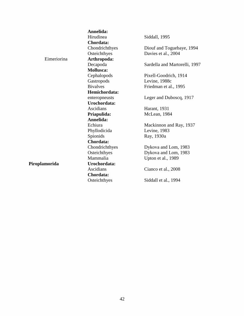
42
Annelida: Hirudinea Siddall, 1995 Chordata:
Chondrichthyes Diouf and Toguebaye, 1994 Osteichthyes Davies et al., 2004
Eimeriorina Arthropoda: Decapoda Sardella and Martorelli, 1997 Mollusca: Cephalopods Pixell-Goodrich, 1914 Gastropods Levine, 1988c Bivalves Friedman et al., 1995 Hemichordata: enteropneusts Leger and Duboscq, 1917 Urochordata: Ascidians Harant, 1931 Priapulida: McLean, 1984 Annelida: Echiura Mackinnon and Ray, 1937 Phyllodicida Levine, 1983 Spionids Ray, 1930a Chordata: Chondrichthyes Dykova and Lom, 1983 Osteichthyes Dykova and Lom, 1983 Mammalia Upton et al., 1989 Piroplamorida Urochordata: Ascidians Cianco et al., 2008 Chordata: Osteichthyes Siddall et al., 1994

43
Table 2: Identity of apicomplexan genera infecting marine hosts. Host location, habitat, tissue preference (Location in host) and climate is annotated.
Host Taxon Apicomplexa Taxon
Species Geographic Location
Host Habitat Location in Host
Citation
Invertebrates Cnidaria: Scleractinian Corals
Agamococcidian
Gemmocystis cylindrus
Caribbean Sea Tropical Gastroderm
Upton and Peters, 1986
Septate Eugregarine Nematopsis sp.
Caribbean Sea Tropical Calicodermis Peters, 1984
Echinoidermata: Spatangoids
Aseptate Eugregarine Lithocystis spp.
North Sea/ English
Channel/ NE Pacific Temperate
Coelomic/ Intrahemal
Pixell-Goodrich, 1915; Theodorides and Laird, 1970;Coulon and Jangoux, 1987
Aseptate Eugregarine Urospora spp.
North Sea/ Mediterran
ean Sea/ English Channel Temperate
Coelomic/ Intrahemal
Coulon and Jangoux, 1987; Pixell-Goodrich, 1915
Holothuroideans
Aseptate Eugregarine Urospora spp.
NW Atlantic/
Mediterranean Sea/ Sea of Japan
Temperate/ Subtropical
Blood/ Coelom/
Gastrointestinal
Pixell-Goodrich, 1925; Levine, 1977a
Aseptate Eugregarine
Gonospora spp.
English Channel/ Barents
Sea Arctic/ Temperate
Blood Vessels/ Gonads/ Coelom
Minchin, 1893; Levine, 1977a
Aseptate Eugregarine Lithocystis spp.
NW Atlantic, English Channel Temperate
Coelomic/ Gastrointesti
nal/ Respiratory
Tree
Pixell-Goodrich, 1925; Levine, 1977a
Ixoreiorida coccidian
Ixorheis psychropotae
NE Atlantic
Temperate (4000 m)
Blood Vessels/ Coelom
Massin et al., 1978
Arthropoda: Crustecea: Cirripedia
Septate Eugregarine
Cirrigregarina spp.
NE Pacific/ Sea of Japan
Temperate/ Subtropical
Gastrointestinal
Levine 1979a
Septate Eugregarine
Cephaloidophora spp. NE Pacific Temperate
Gastrointestinal
Levine, 1988c
Septate
Eugregarine Pyxiniodes spp.
NW Atlantic/
NE Pacific/ Temperate/ Subtropical
Gastrointestinal
Theodorides and Laird, 1970;

44
Sea of Japan
Levine 1988c
Septate Eugregarine
Nematoides fusiformis
Mediterranean Sea Temperate
Gastrointestinal
Levine, 1988c
Septate
Eugregarine Bifilida rara Europe Temperate Gastrointesti
nal
Tuzet and Ormieres, 1964
Copopoda
Aseptate Eugregarine
Paraophioidina spp.
Mediterranean Sea Temperate
Gastrointestinal
Theodorides and Desportes, 1972; Levine, 1977b
Aseptate Eugregarine
Cephaloidophora petiti
Mediterranean Sea Temperate
Gastrointestinal
Levine, 1988c
Malacostraca Decapoda
Aseptate Eugregarine
Paraophioidina spp.
Mediterranean Sea Temperate
Gastrointestinal
Theodorides and Desportes, 1972; Levine, 1977b
Septate Eugregarine
Cephalolobus penaeus
Caribbean Sea/
Mediterranean Sea
Temperate/ Tropical
Gastrointestinal
Sprague and Couch, 1971; Chavez-Sanchez, 2002
Septate Eugregarine
Nematopsis spp.
Caribbean Sea/ Sea of
Japan/ Mediterran
ean Sea/ NE Pacific/
NW Atlantic
Temperate/ Subtropical/
Tropical Gastrointesti
nal
Chavez-Sanchez, 2002
Septate Eugregarine Porospora spp.
English Channel/
NW Atlantic Temperate
Gastrointestinal
Sprague and Couch, 1971; Theodorides and Laird, 1970
Septate Eugregarine
Pachyporospora spp.
Mediterranean Sea/ Sea of Japan
Temperate/ Subtropical
Gastrointestinal
Sprague and Couch, 1971
Septate Eugregarine
Cephaloidophora spp.
Mediterranean Sea/ Arabian
Sea/ Southern
Ocean
Antarctic/ Temperate/
Tropical Gastrointesti
nal
Ball, 1959; Theodorides and Desportes, 1975; Takahashi et al., 2008
Septate Caridohabitans Arabian Tropical Gastrointesti Ball, 1959;

45
Eugregarine spp. Sea nal Levine, 1988c
Eimeriorina
Selenococcidium intermedium
Atlantic Ocean Temperate
Gastrointestinal
Perkins et al., 2000
Eimeriorina Aggregata spp.
SW Atlantic/
Mediterranean Sea Temperate
Gastrointestinal
Theodorides and Desportes, 1975; Sardella and Martorelli, 1997
Amphipoda
Aseptate Eugregarine
Lateroprotomeritus conicus
Mediterranean Sea Temperate
Gastrointestinal
Theodorides and Desportes, 1976
Aseptate Eugregarine
Paraophioidina spp.
Mediterranean Sea Temperate
Gastrointestinal
Theodorides and Desportes, 1972; Levine, 1977b
Septate Eugregarine
Callyntrochlamys phronimae
Mediterranean Sea Temperate
Gastrointestinal
Theodorides and Desportes, 1975
Septate Eugregarine
Cephalolobus spp.
Mediterranean Sea Temperate
Gastrointestinal
Theodorides and Desportes, 1975
Septate
Eugregarine Uradiophora
spp. NW
Atlantic Temperate Gastrointesti
nal
Theodorides and Laird, 1970
Septate Eugregarine
Nematopsis spp.
Gastrointestinal
Prasadan and Janardanan, 2001
Septate Eugregarine
Cephaloidophora spp.
Mediterranean Sea Temperate
Gastrointestinal
Theodorides and Desportes, 1975; Levine, 1988c
Septate Eugregarine Rotundula spp.
English Channel Temperate
Gastrointestinal
Pixell-Goodrich, 1949; Levine 1988c
Septate Eugregarine
Isopoda Adeleid Eucoccidian
Haemogregarina spp. Global Global Blood Cells
Davies et al., 2004
Mollusca: Cephalopods
Eimeriorina Aggregata spp.
SW Atlantic/
NE Temperate Gastrointesti
nal/ Gills
Pixell-Goodrich, 1914;

46
Atlantic/ English
Channel/ Mediterran
ean Sea
Sardella et al., 2000; Pascual et al., 2006
Gastropods Aseptate
Eugregarine Digyalum
owenii
North Sea/NW Atlantic Temperate
Gastrointestinal
Dyson et al., 1993
Aseptate
Eugregarine Gonospora
spp. Mediterran
ean Sea Temperate Testes Tuzet, 1931
Septate
Eugregarine Nematopsis
spp. SW
Atlantic Tropical Phagocytes in Mantle
Azevedo and Padovan, 2004
Adeleid
Eucoccidian Klossia spp. NE
Atlantic Tropical Renal Tissue Levine, 1988c
Eimeriorina Merocystis
kathae Europe Temperate Kidney Levine, 1988c
Bivalves
Septate Eugregarine
Nematopsis spp.
Gulf of Thailand/
Mediterranean Sea/
NE Atlantic
Temperate/ Tropical Gills
Azevedo and Cachola, 1992; Tuntiwaranuruk et al., 2004
Adeleid
Eucoccidian Klossia spp.
NE Atlantic/
North Sea Temperate Renal Tissue Buchanan, 1979
Cryptosporid
ium Cryptosporidiu
m sp. NE Pacific Temperate
Haemolymph in adductor
muscle Miller et al., 2005
Eimeriorina Pseudoklossia
spp.
NE Pacific/ NE
Atlantic/ North Sea Temperate
Ovary/ Kidney
Buchanan, 1979; Levine, 1988c; Friedman et al., 1995
Platyhelminthes:
Aseptate Eugregarine
Monocystella spp.
Mediterranean Sea/
Coral Sea Temperate/
Tropical Gastrointesti
nal
Levine, 1977b; Cannon and Jennings, 1988
Hemichordata: enteropneusts
Archigregarine
Selenidium metchnikovi
Mediterranean Sea Temperate
Gastrointestinal
Leger and Duboscq, 1917
Eimeriorina Eimeria spp.
NE Atlantic Ocean/
Mediterranean Sea Temperate
Hepatic caeca/
Epithelial cells
Leger and Duboscq, 1917; Fernandez et al., 1989
Urochordata:
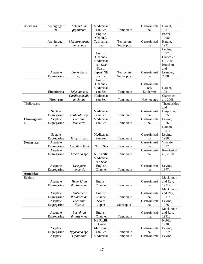
47
Ascidians Archigregarine
Selenidium giganteum
Mediterranean Sea Temperate
Gastrointestinal
Harant, 1931
Archigregarine
Merogregarina amaroucii
English Channel/
Tasmanian Sea
Temperate/ Subtropical
Gastrointestinal
Porter, 1908; Harant, 1931
Aseptate Eugregarine
Lankesteria spp.
English Channel/
Mediterranean Sea/ Sea of
Japan/ NE Pacific
Temperate/ Subtropical
Gastrointestinal
Levine, 1977b; Cianco et al., 2001; Rueckert and Leander, 2008
Eimeriorina Selysina spp.
English Channel/
Mediterranean Sea Temperate
Gastrointestinal/
Epidermis Harant, 1931
Piroplasm
Cardiosporidium cionae
Mediterranean Sea Temperate Haemocytes
Cianco et al., 2008
Thaliaceans
Septate Eugregarine Thalicola spp.
Mediterranean Sea Temperate
Gastrointestinal
Theodorides and Desportes, 1975
Chaetognatha:
Aseptate Eugregarine
Lecudina leuckartii
Mediterranean Sea Temperate
Gastrointestinal
Levine, 1976
Septate Eugregarine Tricystis spp.
Mediterranean Sea Temperate
Gastrointestinal
Hamon, 1951; Levine, 1988c
Nemertea: Aseptate Eugregarine Lecudina linei North Sea Temperate
Gastrointestinal
Vinckier, 1972
Aseptate Eugregarine Difficilina spp. NE Pacific Temperate
Gastrointestinal
Rueckert et al., 2010
Aseptate Eugregarine
Urospora nemertis
Mediterranean Sea/ English Channel Temperate
Gastrointestinal
Levine, 1977a
Annelida: Echiura
Aseptate Eugregarine
Hyperidion thalassemae
English Channel Temperate
Gastrointestinal
Mackinnon and Ray, 1931a
Aseptate
Eugregarine Hentschelia thalassemae
English Channel Temperate
Gastrointestinal
Mackinnon and Ray, 1931b
Aseptate Eugregarine
Lecudina fluctus
Sea of Japan Subtropical
Gastrointestinal
Levine, 1976
Aseptate
Eugregarine Lecythion
thalassemae English Channel Temperate
Gastrointestinal
Mackinnon and Ray, 1931b
Aseptate Eugregarine Zygosoma spp.
NE Pacific Ocean/
Meriterranean Sea Temperate
Gastrointestinal
Noble, 1938; Levine, 1977b
Aseptate Ophiodina Mediterran Temperate Gastrointesti Levine,

48
Eugregarine bonelliae ean Sea nal 1977b Aseptate
Eugregarine Echiurocystis
spp. NE Pacific
Ocean Temperate Gastrointesti
nal Noble, 1938
Eimeriorina Ovivora
thalassemae English Channel Temperate Gonads
Mackinnon and Ray, 1937
Sipuncula
Archigregarine
Selenidium spp.
NE Atlantic/ Sea of
Japan/ NE Pacific Temperate
Gastrointestinal
Levine, 1971; Leander, 2006
Aseptate
Eugregarine Lithocystis lankesteri
Mediterranean Sea Temperate Coelom
Pixell-Goodrich, 1950
Aseptate
Eugregarine Urospora spp. Mediterran
ean Sea Temperate Coelom
Pixell-Goodrich, 1950
Aseptate
Eugregarine Filipodium
spp.
Caribbean Sea/ Sea of
Japan/ Mediterran
ean Sea
Subtropical/ Tropical/
Temperate Gastrointesti
nal Levine, 1977b
Aseptate
Eugregarine Extremocystis dendrostomi
Indian Ocean Tropical Coelom
Levine, 1977b
Flabelligera Archigregari
ne Selenidium
spp. Mediterran
ean Sea Temperate Gastrointesti
nal Levine, 1971
Aseptate
Eugregarine Lecudina zimmeri
NE Pacific Ocean Temperate
Gastrointestinal
Levine, 1976
Terribelliformia
Archigregarine
Selenidium spp.
English Channel Temperate
Gastrointestinal Ray, 1930b
Aseptate Eugregarine
Gonospora varia
English Channel Temperate Coelom
Levine, 1977a
Aseptate Eugregarine
Paragonospora typica North Sea Temperate Coelom
Levine, 1977a
Phyllodicida
Aseptate Eugregarine
Cygnicollum lankesteri
Mediterranean Sea/
Iles Crozet Temperate Gastrointesti
nal
Desportes and Theodorides, 1986
Aseptate Eugregarine Lecudina spp.
Sea of Japan/
English Channel/
Indian Ocean/
NW Atlantic/
Mediterranean Sea
Temperate/ Subtropical/
Tropical Gastrointesti
nal
Theodorides and Laird, 1970; Levine, 1976
Aseptate
Eugregarine Ulivina spp.
SW Atlantic Ocean Tropical
Gastrointestinal
Levine, 1977b
Aseptate Eugregarine Diplauxis spp.
Mediterranean Sea Temperate Coelom
Porchet-Hennere and Fischer, 1973;

49
Levine, 1977b
Aseptate Eugregarine
Gonospora spp.
Mediterranean Sea/ Sea of Japan
Temperate/ Subtropical Coelom
Pixell-Goodrich, 1916; Levine, 1977a
Aseptate Eugregarine
Ceratospora mirabilis France Temperate Coelom
Levine, 1977a
Protococcidia
n Coelotropha
spp. France Temperate Coelom
Porchet-Hennere, 1967
Agamococcidian
Rhytidocystis sthenelais
NE Atlantic/ English Channel Temperate Coelom
Levine, 1979b
Eimeriorina
Defretinella eulaliae
English Channel Temperate Epidermis
Levine, 1983
Eunicida
Aseptate Eugregarine Lecudina spp.
Sea of Japan/ Indian Ocean/
Mediterranean Sea/ English
Channel, North Sea
Temperate/ Subtropical/
Tropical Gastrointesti
nal
Bhatia and Setna, 1938; Schrevel, 1963; Levine, 1976
Aseptate Eugregarine
Ulivina eunicae
Andaman Sea Tropical
Gastrointestinal
Bhatia and Setna, 1938; Levine, 1977b
Aseptate
Eugregarine Bhatiella spp.
Andaman Sea/ Sea of
Japan Tropical Gastrointesti
nal Levine, 1977b
Aseptate Eugregarine
Viviera marphysae
English Channel Temperate
Gastrointestinal
Levine, 1988c
Aseptate
Eugregarine Cochleomeritu
s spp.
NE Pacific Ocean/ Sea
of Japan Temperate/ Subtropical
Gastrointestinal
Levine, 1977b
Aseptate Eugregarine
Contortiocorpa prashadi
Andaman Sea Tropical
Gastrointestinal
Levine, 1977b
Aseptate Eugregarine
Stomatophora primitiva
Andaman Sea Tropical
Gastrointestinal
Bhatia and Setna, 1938
Septate Eugregarine
Deuteromera cleava
Andaman Sea Tropical
Gastrointestinal
Bhatia and Setna, 1938
Protococcidian
Grellia ophryotrochae
Mediterranean Sea Temperate Coelom
Levine, 1973
Spionids
Archigregarine
Selenidium spp.
Bay of Bengal Tropical
Gastrointestinal
Ganapati, 1946; Levine, 1971
Blastogregarine Siedleckia spp.
English Channel/ Bay of Bengal
Temperate/ Tropical
Gastrointestinal
Chatton and Villeneuve, 1936a; b

50
Aseptate Eugregarine
Lecudina bogolepova
Sea of Japan Subtropical
Gastrointestinal
Levine, 1976
Aseptate Eugregarine Ulivina spp.
Mediterranean Sea Temperate
Gastrointestinal
Levine, 1977b
Aseptate Eugregarine
Selenocystis foliata
English Channel Temperate
Gastrointestinal Dibb, 1938
Aseptate Eugregarine
Polyrhabdina spp.
English Channel/
Indian Ocean
Temperate/ Tropical
Gastrointestinal
Mackinnon and Ray, 1931b; Ganapati, 1946
Aseptate Eugregarine Sycia spp.
Sea of Japan/
Mediterranean Sea/ Bay of Bengal
Subtropical/Temperate
Gastrointestinal
Ganapti, 1946; Levine, 1977b
Aseptate
Eugregarine Ditrypanocysti
s spp.
English Channel/ North Sea Temperate
Gastrointestinal
Levine, 1971
Aseptate Eugregarine Urospora spp.
Mediterranean Sea Temperate Unknown
Levine, 1977a
Aseptate Eugregarine
Gonospora varia
English Channel Temperate Coelom
Levine, 1977a
Protococcidia
n Eleutheroschiz
on dubosqui English Channel Temperate
Gastrointestinal
Chatton and Villeneuve, 1936b
Protococcidian
Myriospora spp.
Bay of Bengal Tropical
Coelom/ Gastrointesti
nal
Ganapati, 1941; Ganapati, 1952
Eimeriorina
Dorisiella scolelepidis
English Channel Temperate
Gastrointestinal Ray, 1930a
Sabellids
Archigregarine
Selenidium spp.
NE Atlantic/
NE Pacific Temperate Gastrointesti
nal
Levine, 1971; Ray, 1930b; Leander, 2007
Protococcidian
Myriosporides amphiglenae
English Channel/
Mediterranean Sea Temperate Coelom
Hennere, 1967
Serpulids
Archigregarine
Selenidium spp.
English Channel/
Mediterranean Sea Temperate
Gastrointestinal
Levine, 1971
Capitellids Aseptate
Eugregarine Lecudina capitellae
SW Atlantic Ocean Tropical
Gastrointestinal
Levine, 1976
Aseptate Eugregarine Ancora spp.
SW Atlantic Ocean/
Mediterranean Sea/ Bay of
Tropical/ Temperate
Gastrointestinal
Ganapati, 1946; Levine, 1977b
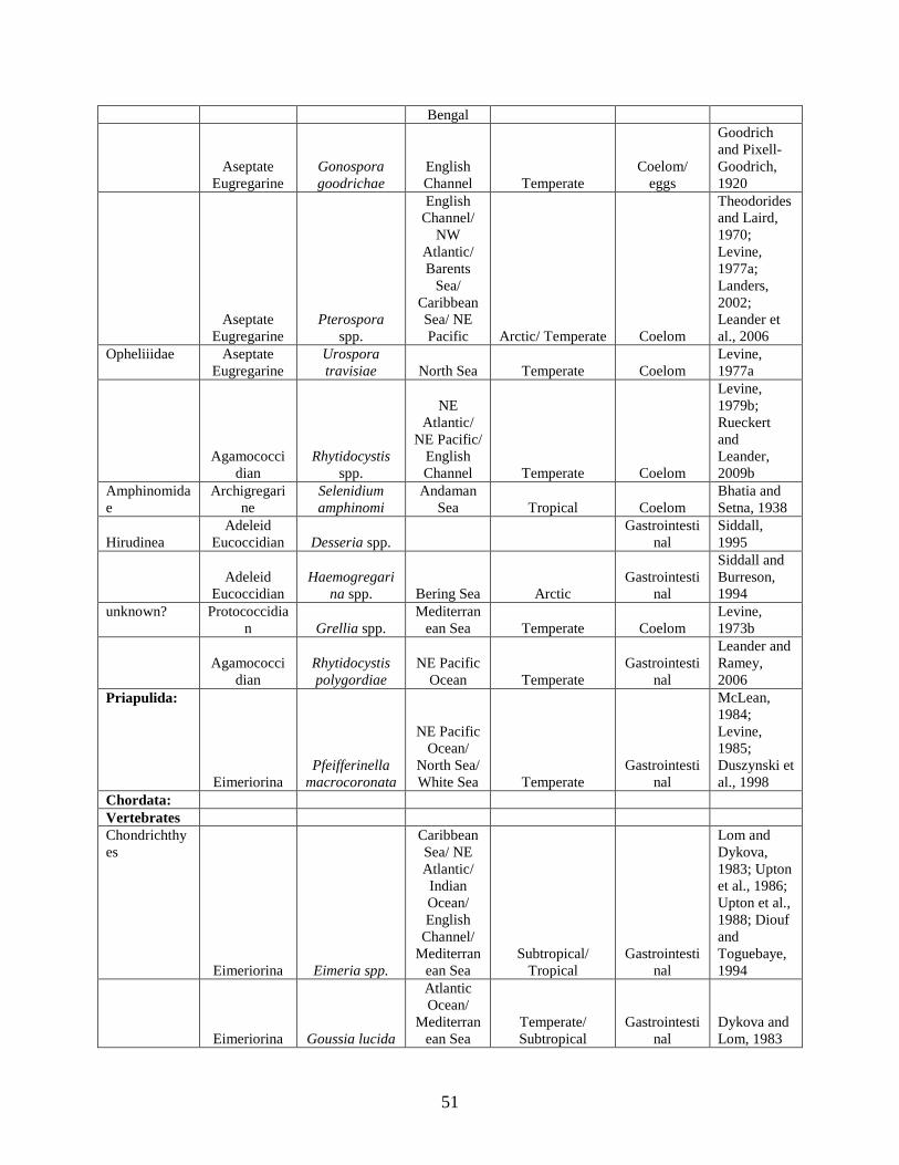
51
Bengal
Aseptate Eugregarine
Gonospora goodrichae
English Channel Temperate
Coelom/ eggs
Goodrich and Pixell-Goodrich, 1920
Aseptate Eugregarine
Pterospora spp.
English Channel/
NW Atlantic/ Barents
Sea/ Caribbean Sea/ NE Pacific Arctic/ Temperate Coelom
Theodorides and Laird, 1970; Levine, 1977a; Landers, 2002; Leander et al., 2006
Opheliiidae Aseptate Eugregarine
Urospora travisiae North Sea Temperate Coelom
Levine, 1977a
Agamococcidian
Rhytidocystis spp.
NE Atlantic/
NE Pacific/ English Channel Temperate Coelom
Levine, 1979b; Rueckert and Leander, 2009b
Amphinomidae
Archigregarine
Selenidium amphinomi
Andaman Sea Tropical Coelom
Bhatia and Setna, 1938
Hirudinea Adeleid
Eucoccidian Desseria spp. Gastrointesti
nal Siddall, 1995
Adeleid
Eucoccidian Haemogregari
na spp. Bering Sea Arctic Gastrointesti
nal
Siddall and Burreson, 1994
unknown? Protococcidian Grellia spp.
Mediterranean Sea Temperate Coelom
Levine, 1973b
Agamococci
dian Rhytidocystis polygordiae
NE Pacific Ocean Temperate
Gastrointestinal
Leander and Ramey, 2006
Priapulida:
Eimeriorina Pfeifferinella
macrocoronata
NE Pacific Ocean/
North Sea/ White Sea Temperate
Gastrointestinal
McLean, 1984; Levine, 1985; Duszynski et al., 1998
Chordata: Vertebrates Chondrichthyes
Eimeriorina Eimeria spp.
Caribbean Sea/ NE Atlantic/ Indian Ocean/ English
Channel/ Mediterran
ean Sea Subtropical/
Tropical Gastrointesti
nal
Lom and Dykova, 1983; Upton et al., 1986; Upton et al., 1988; Diouf and Toguebaye, 1994
Eimeriorina Goussia lucida
Atlantic Ocean/
Mediterranean Sea
Temperate/ Subtropical
Gastrointestinal
Dykova and Lom, 1983

52
Adeleid
Eucoccidian Haemogregari
na spp.
NW Atlantic Ocean Temperate Blood Cells So, 1972
Osteichthyes Adeleid
Eucoccidian Haemogregari
na spp. Global
Temperate/ Subtropical/
Tropical Blood Cells Davies et al., 2004
Adeleid
Eucoccidian Cyrilia
uncinata
NW Atlantic Ocean Temperate Blood Cells Khan, 1978
Adeleid Eucoccidian Desseria spp. SE Atlantic Subtropical Blood Cells
Siddall, 1995; Smit and Davies, 2006
Cryptosporid
ium Cryptosporidiu
m molnari
Mediterranean Sea/
NE Atlantic Temperate
Gastrointestinal
Alvarez-Pellitero and Sitja-Bobadilla, 2002
Adeleid
Eucoccidian Dactylosoma
spp.
Red Sea/ Indian Ocean Subtropical Blood Cells
Saunders, 1960; Levine, 1988c
Adeleid
Eucoccidian Babesiosoma
spp.
NW Atlantic
Ocean/ Red Sea
Temperate/ Subtropical Blood Cells
Saunders, 1960; Paperna, 1981
Eimeriorina Eimeria spp. Global
Temperate/ Subtropical/
Tropical
Gastrointestinal/ Testes/
Liver/ Kidney
Dykova and Lom, 1983; Lom and Dykova, 1995; Diouf and Toguebaye, 1994
Eimeriorina Goussia spp.
Sea of Japan/ N Atlantic Ocean/
Mediterranean Sea/
Caribbean Sea/
Tasman Sea/ Baltic
Sea Subtropical/
Tropical
Gastrointestinal/ Liver/
Swim bladder
Levine, 1983; Upton et al., 1989; Lom and Dykova, 1995; Gestal and Azevedo, 2006
Eimeriorina Calyptospora
spp.
Gulf of Mexico,
SW Atlantic Subtropical
Liver/ Hepatocytes
Overstreet et al., 1984
Piroplasm Haemohormidi
um spp.
NW and NE
Atlantic Ocean/
Southern Ocean
Temperate/ Subtropical Blood Cells
Khan, 1980; Siddall et al., 1994; Davies et al., 2003

53
Mammalia Sarcocystida
e Toxoplasma
gondii
North-Eastern Pacific Temperate
Gastrointestinal
Millet et al., 2008
Cryptosporid
ium Cryptosporidiu
m sp.
South-Western Pacific Subtropical
Gastrointestinal
Morgan et al., 2000
Eimeriorina Eimeria spp.
NW Atantic Ocean/
Caribbean Sea
Temperate/ Subtropical
Gastrointestinal
Upton et al., 1989, McClelland, 1993
Reptilia Cryptosporidium
Cryptosporidium sp. Hawaii Tropical
Gastrointestinal
Graczyk et al., 1997

54
References:
Adl SM, Leander BS, Simpson AGB, Archibald JM, Anderson OR, Bass D, Bowser SS,
Brugerolle G, Farmer MA, Karpov S, Kolisko M, Lane CE, Lodge DJ, Mann DG,
Meisterfeld R, Mendoza L, Moestrup O, Mozley-Standridge SE, Smirnov AV, Spiegel
FW (2007) Diversity, nomenclature, and taxonomy of protists. Syst Biol 56:684-689
Adl SM, Simpson AGB, Farmer MA, Andersen RA, Anderson OR, Barta JR, Bowser SS,
Brugerolle G, Fensome RA, Fredericq S, James TY, Karpov S, Kugrens P, Krug J, Lane
CE, Lewis LA, Lodge J, Lynn DH, Mann DG, McCourt RM, Mendoza L, Moestrup O,
Mozley-Standridge SE, Nerad TA, Shearer CA, Smirnov AV, Spiegel FW, Taylor MFJR
(2005) The new higher level classification of eukaryotes with emphasis on the taxonomy
of protists. J Eukaryot Microbiol 52:399-451
Alvarez-Pellitero P, Palenzuela O, Sitjà-Bobadilla A (1997) Ultrastructure and cytochemistry
study of Goussia sparis (Protozoa: Apicomplexa) stages from the intestine of the gilthead
sea bream Sparus aurata L. (Pisces: Teleostei). Parasitol Res 83:24-33
Alvarez-Pellitero P, Sitjà-Bobadilla A (2002) Cryptosporidium molnari n. sp. (Apicomplexa:
Cryptosporidiidae) infecting two marine fish species, Sparus aurata L. and
Dicentrarchus labrax L. Int J Parasit 32:1007-1021
Alvarez-Pellitero P, Sitjà-Bobadilla A, Franco-Sierra A, Palenzuela O (1995) Protozoan parasites
of gilthead sea bream, Sparus aurata L., from different culture systems in Spain. J Fish
Dis 18:105-115
Anbar AD, Knoll AH (2002) Proterozoic ocean chemistry and evolution: a bioinorganic bridge?
Science 297:1137-1142

55
Archibald JM, Keeling PJ (2002) Recycled plastids: a ‘green movement’ in eukaryotic evolution.
Trends Genet 18:577-584
Azevedo C, Cachola R (1992) Fine structure of the apicomplexa oocyst of Nematopsis sp. of two
marine bivalve molluscs. Dis Aquat Org 14:69-73
Azevedo C, Padovan I (2004) Nematopsis gigas n. sp. (Apicomplexa), a parasite of Nerita
ascencionis (Gastropoda, Neritidae) from Brazil. J Eukaryot Microbiol 51:214-219
Bachvaroff TR, Handy SM, Place AR, Delwiche CF (2011) Alveolate phylogeny inferred using
concatenated ribosomal proteins. J Eukaryot Microbiol DOI: 10.1111/j.1550-
7408.2011.00555.x
Bachvaroff TR, Puerta MVS, Delwiche CF (2005) Chlorophyll c-containing plastid relationships
based on analyses of a multigene data set with all four chromalveolate lineages. Mol Biol
Evol 22:1772-1782
Baker JR (1994) The origins of parasitism in the protists. Int J Parasit 24:1131-1137
Balart EF, Pérez-Urbiola JC, Campos-Dávila L, Monteforte M, Ortega-Rubio A (2009) On the
first record of a potentially harmful fish, Sparus aurata in the Gulf of California. Biol
Invasions 11:547-550
Baldauf SL, Roger AJ, Wenk-Siefert I, Doolittle WF (2000) A kingdom-level phylogeny of
eukaryotes based on combined protein data. Science 290:972-977
Ball GH (1959) Some gregarines from crustaceans taken near Bombay, India. J Protozool 6:8-13
Ball GH (1963) Cephaloidophora carpilodei n. sp. and C. pinguis n. sp., gregarines of a xanthid
crab in Hawaii. J Protozool 10:321-327

56
Barta JR, Desser SS (1989) Development of Babesiosoma stableri (Dactylosomatidae; Adeleina;
Apicomplexa) in its leech vector (Batracobdella picta) and the relationship of the
dactylosomatids to the piroplasms of higher vertebrates. J Protozool 36:241-253
Barta JR, Martin DS, Carreno RA, Siddall ME, Profous-Juchelka H, Hozza M, Powles MA,
Sundermann CA (2001) Molecular phylogeny of the other tissue coccidia: Lankesterella
and Caryospora. J Parasitol 87:121-127
Barta JR, Thompson RCA (2006) What is Cryptosporidium? Reappraising its biology and
phylogenetic affinities. Trends Parasitol 22:463-468
Baum J, Richard D, Healer J, Rug M, Krnajski Z, Gilberger T-W, Green JL, Holder AA,
Cowman AF (2006) A conserved molecular motor drives cell invasion and gliding
motility across malaria life cycle stages and other apicomplexan parasites. J Biol Chem
281:5197-5208
Berney C, Pawlowski J (2006) A molecular time-scale for eukaryote evolution recalibrated with
the continuous microfossil record. Proc R Soc Lond B 273:1867-1872
Bhatia BL, Setna SB (1938) On some gregarine parasites from certain polychete worms from the
Andaman Islands. Proc Ind Acad Sci B 8:231-242
Bishop R, Musoke A, Morzaria S, Gardner M, Nene V (2004) Theileria: intracellular protozoan
parasites of wild and domestic ruminants transmitted by ixodid ticks. Parasitology
129:S271-283
Bock R, Jackson L, De Vos A, Jorgensen W (2004) Babesiosis of cattle. Parasitology 129:S247-
269
Bodyl A (2005) Do plastid-related characters support the chromalveolate hypothesis? J Phycol
41:712-719

57
Bogomolni AL, Gast RJ, Ellis JC, Dennett M, Pugliares KR, Lentell BJ, Moore MJ (2008)
Victims or vectors: a survey of marine vertebrate zoonoses from coastal waters of the
Northwest Atlantic. Dis Aquat Org 81:13-38
Boothroyd JC, Dubremetz JF (2008) Kiss and spit: the dual roles of Toxoplasma rhoptries. Nat
Rev Microbiol 6:79-88
Brownell CL, McCauley JE (1971) Two new parasites (Protozoa: Telosporea) from the
spatangoid urchin Brisaster latifrons. Zool Anz 186:141-147
Buchanan JS (1979) On two new species of coccidia from the marine bivalve, Tellina tenuis (Da
Costa)- an adeliorine from the renal organ and an eimeriorine from the ovary. Haliotis
8:57-64
Burki F, Shalchian-Tabrizi K, Minge MA, Skjaeveland A, Nikolaev SI, Jakobsen KS, Pawlowski
J (2007) Phylogenomics reshuffles the eukaryotic supergroups. PLoS One 2:e790
Burki F, Shalchian-Tabrizi K, Pawlowski J (2008) Phylogenomics reveals a new ‘megagroup’
including most photosynthetic eukaryotes. Biol Lett 4:366-369
Canestri-Trotti G, Baccarani EM, Paesanti F, Turolla E (2000) Monitoring of infections by
protozoa of the genera Nematopsis, Perkinsus and Porospora in the smooth venus clam
Callista chione from the North-Western Adriatic Sea (Italy). Dis Aquat Org 42:157-161
Cannon LRG, Jennings JB (1988) Monocystella epibatis n.sp., a new aseptate gregarine
hyperparasite of Rhabdocoel turbellarians parasitic in the crown of thorns starfish,
Acanthaster planci Linnaeus, from the great barrier reef. Arch Protintenkd 136:267-272
Cavalier-Smith T (1991) Cell diversification in heterotrophic flagellates. In: Patterson DJ,
Larson J (eds) The biology of free-living heterotrophic flagellates. Clarendon Press,
Oxford

58
Cavalier-Smith T (1993) Kingdom protozoa and its 18 phyla. Microbiol Rev 57:953-994
Cavalier-Smith T (1999) Principles of protein and lipid targeting in secondary symbiogenesis:
euglenoid, dinoflagellate, and sporozoan plastid origins and the Eukaryote family tree. J
Eukaryot Microbiol 46:347-366
Cavalier-Smith T (2006) Cell evolution and Earth history: stasis and revolution. Phil Trans R
Soc B 361:969-1006
Cavalier-Smith T (2009) Predation and eukaryote cell origins: A coevolutionary perspective. Int
J Biochem Cell Biol 41:307-322
Cavalier-Smith T, Chao EE (2004) Protalveolate phylogeny and systematics and the origins of
Sporozoa and dinoflagellates (phylum Myzozoa nom. nov.). Eur J Protistol 40:185-212
Chatton E, Villenueve F (1936a) La sexualité et le cycle évolutif des Seidleckia d'après l'étude de
S. caulleryi, n. sp. Hologrégarines et Blastogrégarines. Sporozoaires Hologamétogènes et
Blastogamétogènes. C R Acad Sci Paris 203:505-508
Chatton E, Villenueve F (1936b) Le cycle évolutif de l'Eleutheroschizon duboscqui Brasil.
Preuve expérimentale de l'absence de schizogonie chez cette forme et chez la Siedleckia
caulleryi Ch. et Villen. C R Acad Sci Paris 203:833-836
Chávez-Sánchez MC, Hernández-Martínez M, Abad-Rosales S, Fajer-Ávila EJ, Montoya-
Rodríguez L, Álvarez-Torres P (2002) A survey if infectious diseases and parasites of
Penaeid shrimp from the Gulf of Mexico. J World Aquacult Soc 33:316-329
Cheng TC (1967) Marine molluscs as hosts for symbioses with a review of known parasites of
commercially important sp., Vol 5. Academic Press Inc, London

59
Ciancio A, Scippa S, Cammarano M (2001) Ultrastructure of trophozoites of the gregarine
Lankesteria ascidiae (Apicomplexa: Eugregarinida) parasitic in the ascidian Ciona
intestinalis (Protochordata). Eur J Protistol 37:327-336
Ciancio A, Scippa S, Finetti-Sialer M, De Candia A, Avallone B, De Vincentiis M (2008)
Redescription of Cardiosporidium cionae (Van Gaver and Stephan, 1907) (Apicomplexa:
Piroplasmida), a plasmodial parasite of ascidian haemocytes. Eur J Protistol 44:181-196
Clopton RE (2004) Standard nomenclature and metrics of plane shapes for use in gregarine
taxonomy. Comp Parasitol 71:130-140
Clopton RE (2009) Phylogenetic relationships, evolution, and systematic revision of the septate
gregarines (Apicomplexa: Eugregarinorida: Septatorina). Comp Parasitol 76:167-190
Clopton RE, Gold RE (1996) Host specificity of Gregarina blattarum von Siebold, 1839
(Apicomplexa: Eugregarinida) among five species of domiciliary cockroaches. J
Invertebr Pathol 67:219-223
Conrad PA, Miller MA, Kreuder C, James ER, Mazet J, Dabritz HA, Jessup D, Gulland F, Grigg
ME (2005) Transmission of Toxoplasma: clues from the study of sea otters as sentinels of
Toxoplasma gondii flow into the marine environment. Int J Parasit 35:1155-1168
Coppin A, Varré J-S, Lienard L, Dauvillée D, Guérardel Y, Soyer-Gobillard M-O, Buléon A,
Ball S, Tomavo S (2005) Evolution of plant-like crystalline storage polysaccharide in the
protozoan parasite Toxoplasma gondii argues for a red alga ancestry. J Mol Evol 60:257-
267
Costa G, MacKenzie K (1994) Histopathology of Goussia clupearum (Protozoa: Apicomplexa:
Coccidia) in some marine fish from Scottish waters. Dis Aquat Org 18:195-202

60
Coulon P, Jangoux M (1987) Gregarine species (Apicomplexa) parasitic in the burrowing
echinoid Echinocardium cordatum: occurrence and host reaction. Dis Aquat Org 2:135-
145
Cox FEG (1994) The evolutionary expansion of the Sporozoa. Int J Parasit 24:1301-1316
Davies AJ (1980) Some observations on Haemohormidium cotti Henry 1910, from the marine
fish Cottus bubalis Euphrasen. Z Parasitenkd 62:31-38
Davies AJ, Eiras JC, Austin RTE (1994) Investigations into the transmission of Haemogregarina
bigemina Laveran and Mesnil, 1901 (Apicomplexa: Adeleorina) between intertidal fishes
in Portugal. J Fish Dis 17:283-289
Davies AJ, Reed CC, Smit NJ (2003) An unusual intraerythrocytic parasite of Parablennius
cornutus. J Parasitol 89:913-917
Davies AJ, Smit NJ, Hayes PM, Seddon AM, Wertheim D (2004) Haemogregarina bigemina
(Protozoa: Apicomplexa: Adeleorina) -past, present and future. Folia Parasitol 51:99-108
de Meeûs T, Renaud F (2002) Parasites within the new phylogeny of eukaryotes. Trends
Parasitol 18:247-251
De Ridder C, Jangoux M (1984) Intracoelomic parasitic sporozoa in the burrowing spatangoid
echinoid Echinocardium cordatum: coelomocyte reaction and formation of brown bodies.
Helgolander Meeresun 37:225-231
Deng M-Q, Peterson RP, Cliver DO (2000) First findings of Cryptosporidium and Giardia in
California sea lions (Zalophus californianus). J Parasitol 86:490-494
Denny P, Preiser P, Williamson D, Wilson I (1998) Evidence for a single origin of the 35 kb
plastid DNA in apicomplexans. Protist 149:51-59

61
Desportes I, Théodoridès J (1986) Cygnicollum lankesteri n. sp., grégarine (Apicomplexa,
Lecudinidae) parasite des annélides polychètes Laetmonice hystrix et L. producta;
particularités de l'appareil de fixation et implications taxonomiques. Protistologica 22:47-
60
Detwiler J, Janovy J (2008) The role of phylogeny and ecology in experimental host specificity:
insights from a eugregarine-host system. J Parasitol 94:7-12
Dibb MJ (1938) Selenocystis foliata (Ray) from Scolelepis fuliginosa Clpde. and its identity with
Haplozoon sp. . Parasitology 30:296-308
Diouf JN, Toguebaye BS (1994) Study of some marine fish coccidia of the genus Eimeria
Schneider, 1815 (Apicomplexa, Coccidia) from Senegal coasts. Acta Protozool 33:239-
250
Diouf JN, Toguebaye BS (1996) Eimeria spari n. sp., (Apicomplexa, Eimeriidae) parasite de
Sparus caeruleostictus (Valenciennes, 1830), (Poisson, Sparidae) des cotes Sénégalaises.
Parasite 4:351-355
Dobson AP (1985) The population dynamics of competition between parasites. Parasitology
91:317-347
Dobson AP, Lafferty KD, Kuris AM, Hechinger RF, Jetz W (2008) Homage to Linnaeus: How
many parasites? How many hosts? Proc Natl Acad Sci U S A 105:11482-11489
Douzery EJP, Snell EA, Bapteste E, Delsuc F, Philippe H (2004) The timing of eukaryotic
evolution: Does a relaxed molecular clock reconcile proteins and fossils? Proc Natl Acad
Sci U S A 101:15386–15391
Dubey JP (2008) The history of Toxoplasma gondii—The first 100 years. J Eukaryot Microbiol
55:467-475

62
Dubey JP, Barr BC, Barta JR, Bjerkas I, Bjorkman C, Blagburn BL, Bowman DD, Buxton D,
Ellis JT, Gottstein B, Hemphill A, Hill DE, Howe DK, Jenkins MC, Kobayashi Y,
Koudela B, Marsh AE, Mattsson JG, McAllister MM, Modry D, Omata Y, Sibley LD,
Speer CA, Trees AJ, Uggla A, Upton SJ, Williams DJL, Lindsay DS (2002)
Redescription of Neospora caninum and its differentiation from related coccidia. Int J
Parasit 32:929-946
Dubremetz JF, Ferguson DJ (2009) The role played by electron microscopy in advancing our
understanding of Toxoplasma gondii and other apicomplexans. Int J Parasit 39:883-893
Duszynski DW, Solangi MA, Overstreet RM (1979) A new and unusual eimerian (Protozoa:
Eimeriidae) from the liver of the gulf killifish, Fundulus grandis. J Wildlife Dis 15:543-
552
Duszynski DW, Upton SJ, Couch L (1998) The Coccidia of the World.
http://biology.unm.edu/biology/coccidia/home.html
Dyková I, Lom J (1981) Fish coccidia: critical notes on life cycles, classification and
pathogenicity. J Fish Dis 4:487-505
Dyková I, Lom J (1983) Fish coccidia: an annotated list of described species. Folia Parasitol
30:193-208
Dyson J, Grahame J, Evennett PJ (1993) The mucron of the gregarine Digyalum oweni
(Protozoa: Apicomplexa), parasitic in Littorina species (Mollusca: Gastropoda). J Nat
Hist 27:557-564
Dyson J, Grahame J, Evennett PJ (1994) The apical complex of the gregarine Digyalum oweni
(Protozoa: Apicomplexa). J Nat Hist 28:1-7
Ebert D, Herre EA (1996) The evolution of parasitic diseases. Parasitol Today 12:96-101

63
Egyed Z, Sreter T, Szell Z, Varga I (2003) Characterization of Cryptosporidium spp. -recent
developments and future needs. Vet Parasitol 111:103-114
Escalante AA, Ayala FJ (1995) Evolutionary origin of Plasmodium and other Apicomplexa
based on rRNA genes. Proc Natl Acad Sci U S A 92:5793-5797
Esser C, Martin W (2007) Supertrees and symbiosis in eukaryote genome evolution. Trends
Microbiol 15:435-437
Fajer-Ávila EJ, Covarrubias MSM, Abad-Rosales S, Roque A, Meza-Bojórquez P, Hernández-
González C (2005) Effectiveness of oral Elancobank™ and Avimix-ST™ against
Nematopsis (Apicomplexa: Porosporidae) gametocyts infecting the shrimp Litopenaeus
vannamei. Aquaculture 244:11-18
Fast N, Kissinger JC, Roos DS, Keeling PJ (2001) Nuclear-encoded, plastid-targeted genes
suggest a single common origin for apicomplexan and dinoflagellate plastids. Mol Biol
Evol 18:418-426
Fast NM, Xue L, Bingham S, Keeling PJ (2002) Re-examining alveolate evolution using
multiple protein molecular phylogenies. J Eukaryot Microbiol 49:30-37
Fayer R, Dubey JP, Lindsay DS (2004) Zoonotic protozoa: from land to sea. Trends Parasitol
20:531-536
Fayer R, Trout JM, Walsh E, Cole R (2000) Rotifers ingest oocysts of Cryptosporidium parvum.
J Eukaryot Microbiol 47:161-163
Fernandez I, Benito J (1983) New contributions on parasitic coccidia of the hepatic region of
Glossobalanus minutus kow. (Enteropneusta, Hemichordata). Acta Protozool 22:241-250

64
Fernandez I, Pardos F, Benito J (1989) Fine structure of merozoites of Eimeria ? beauchampi
Léger and Duboscq, 1917 in Glossobalanus minutus (Kow.) (Enteropneusta). Ann
Parasitol Hum Comp 64:10-19
Ford SE, Chintala MM (2006) Northward expansion of a marine parasite: Testing the role of
temperature adaptation. J Exp Mar Biol Ecol 339:226-235
Fournie JW, Overstreet RM (1983) True intermediate hosts for Eimeria funduli (Apicomplexa)
from estuarine fishes. J Protozool 30:672-675
Fournie JW, Overstreet RM (1993) Host specificity of Calyptospora funduli (Apicomplexa:
Calyptosporidae) in atheriniform fishes. J Parasitol 79:720-727
Fournie JW, Vogelbein WK, Overstreet RM, Hawkins WE (2000) Life cycle of Calptospora
funduli (Apicomplexa: Calyptosporidae). J Parasitol 86:501-505
Francisco CJ, Hermida MA, Santos MJ (2010) Parasites and symbionts from Mytilus
galloprovincialis (Lamark, 1819) (Bivalves: Mytilidae) of the Aveiro Estuary Portugal. J
Parasitol 96:200-205
Frank SA (1993) Evolution of the host-parasite diversity. Evolution 47:1721-1732
Friedman CS (1991) Coccidiosis of California abalone, Haliotis spp. J Shellfish Res 10:236
Friedman CS, Gardner GR, Hedrick RP, Stephenson M, Cawthorn RJ, Upton SJ (1995)
Pseudoklossia haliotis sp. n. (Apicomplexa) from the kidney of California abalone,
Haliotis spp. (Mollusca). J Invertebr Pathol 66:33-38
Friedman CS, Roberts W, Kismohandaka G, Hedrick RP (1993) Transmissibility of a coccidian
parasite of abalone, Haliotis spp. . J Shellfish Res 12:201-205

65
Ganapati PN (1941) The development and sporogony of a coccidian Myriospora gopalai n.sp.,
parasitic in the gut of the polychaete Cirratulus filiformis Keferstein. Proc Ind Acad Sci
B 13:144-162
Ganapati PN (1946) Notes on some gregarines from polychaetes of the Madras Coast. Proc Ind
Acad Sci B 23:228-247
Ganapati PN (1952) The morphology and life-history of a coccidina Myriospora polydorae n.
sp., in the body-cavity of Polydora ciliata Johnston. Arch Protintenkd 98:71-90
Gestal C, Azevedo C (2006) Ultrastructural aspects of hepatic coccidiosis caused by Goussia
lusca n. sp. (Apicomplexa: Coccidia) infecting Trisopterus luscus (Gadidae) from the NE
Atlantic Ocean. Dis Aquat Org 71:25-31
Gestal C, Guerra A, Pascual S (2007) Aggregata octopiana (Protista: Apicomplexa): a dangerous
pathogen during commercial Octopus vulgaris ongrowing. ICES J Mar Sci 64:1743-1748
Gestal C, Guerra A, Pascual S, Azevedo C (2002a) On the life cycle of Aggregata eberthi and
observations on Aggregata octopiana (Apicomplexa, Aggregatidae) from Galicia (NE
Atlantic). Eur J Protistol 37:427-435
Gestal C, Nigmatullin CM, Hochberg FG, Guerra A, Pascual S (2005) Aggregata andresi n. sp.
(Apicomplexa: Aggregatidae) from the ommastrephid squid Martialia hyadesi in the SW
Atlantic Ocean and some general remarks on Aggregata spp. in cephalopod hosts. Syst
Parasitol 60:65-73
Gestal C, Páez de la Cadena M, Pascual S (2002b) Malabsorption syndrome observed in the
common octopus Octopus vulgaris infected with Aggregata octopiana (Protista:
Apicomplexa). Dis Aquat Org 51:61-65

66
Gestal C, Pascual S, Hochberg FG (2010) Aggregata bathytherma sp. nov. (Apicomplexa:
Aggregatidae), a new coccidian parasite associated with a deep-sea hydrothermal vent
octopus. Dis Aquat Org 91:237-242
Gjurcevic E, Kozaric Z, Bambir S, Petrinec Z, Kuzir S, Gudan A, Bazdaric B (2008)
Histological investigations of Eimeria infection in large-scaled gurnards, Lepidotrigla
cavillone (Lacepède, 1801) from the Novigrad Sea, Croatia. Acta Protozool 53:81-84
Golemansky V (2011) Coccidian parasites (Apicomplexa) of penguins (Pygoscelis ssp.) from
Livingston Island and King George Island, the Antarctic. Pol Polar Res 32:263-268
Gómez-Couso H, Paniagua-Crespo E, Ares-Mazás E (2007) Acanthamoeba as a temporal
vehicle of Cryptosporidium. Parasitol Res 100:1151-1154
González MT, Oliva ME (2009) Is the nestedness of metazoan parasite assemblages of marine
fishes from the southeastern Pacific coast a pattern associated with the geographical
distributional range of the host? Parasitology 136:401-409
Goodrich ES, Pixell-Goodrich HLM (1920) Gonospora minchinii, n. sp., a gregarine inhabiting
the egg of Arenicola. Q J Microsc Sci 65:157-161
Graczyk TK, Balazs GH, Work T, Aguirre AA, Ellis DM, Murakawa SKK, Morris R (1997)
Cryptosporidium sp. infections in green turtles, Chelonia mydas, as a potential source of
marine waterborne oocysts in the Hawaiian Islands. Appl Environ Microbiol 63:2925-
2927
Grassé PP (1953) Classe de Grégarinomorphes. In: Traité de Zoologie: Anatomie, Systématique,
Biologie, Book 1. Masson et Cie, Paris
Hamon M (1951) Note sur une Grégarine parasite du tube digestif de Sagitta lyra. Bull Soc Hist
Nat Afr Nord 42:11-14
67
Harant H (1931) Les ascidies et leurs parasites II; Les parasites des ascidies (Protozoa). Ann Inst
Oceanogr 8:347-365
Hardcastle AB (1944) Eimeria brevoortiana, a new sporozoan parasite from menhaden
(Brevoortia tyrannus), with observations on its life history. J Parasitol 30:60-68
Harper JT, Keeling PJ (2003) Nucleus-encoded, plastid-targeted glyceraldehyde-3-phosphate
dehydrogenase (GAPDH) indicates a single origin for chromalveolate plastids. Mol Biol
Evol 20:1730-1735
Harvell CD, Kim K, Burkholder JM, Colwell RR, Epstein PR, Grimes DJ, Hofmann EE, Lipp
EK, Osterhaus ADME, Overstreet RM, Porter JW, Smith GW, Vasta GR (1999)
Emerging marine diseases-climate links and anthropogenic factors. Science 285:1505-
1510
Harvell CD, Mitchell CE, Ward JR, Altizer S, Dobson AP, Ostfeld RS, Samuel MD (2002)
Climate Warming and disease risks for terrestrial and marine biota. Science 296:2158-
2162
Henneré E (1967) Etude cytologique des premiers stades du developpement d'une coccidie:
Myriosporides amphiglenae. J Protozool 14:27-39
Hine PM (1975) Eimeria anguillae Léger and Hollande, 1922 parasitic in New Zealand eels. N Z
J Mar Freshw Res 9:239-243
Hnida JA, Duszynski DW (1999) Taxonomy and systematics of some Eimeria species of murid
rodents as determined by the ITS1 region of the ribosomal gene complex. Parasitology
119:349-357

68
Huang BQ, Chen X-M, LaRusso NF (2004) Cryptosporidium parvum attachment to and
internalization by human biliary epithelia in vitro: a morphological study. J Parasitol
90:212-221
Jakes K, O'Donoghue PJ, Cameron SL (2003) Phylogenetic relationships of Hepatozoon
(Haemogregarina) boigae, Hepatozoon sp., Haemogregarina clelandi and Haemoproteus
chelodina from Australian reptiles to other Apicomplexa based on cladistic analyses of
ultrastructural and life-cycle characters. Parasitology 126:555-559
Jangoux M (1987) Diseases of Echinodermata. I. Agents microorganisms and protistans. Dis
Aquat Org 2:147-162
Javaux EJ, Knoll AH, Walter MR (2001) Morphological and ecological complexity in early
eukaryotic ecosystems. Nature 412:66-69
Jiménez R, de Barniol L, Machuca M (2002) Nematopsis marinus n. sp., a new septate gregarine
from cultured penaeoid shrimp Litopenaeus vannamei (Boone), in Ecuador. Aquac Res
33:231-240
Jirku M, Jirku M, Oborník M, Lukes J, Modry D (2009) Goussia Labbé, 1896 (Apicomplexa,
Eimeriorina) in Amphibia: diversity, biology, molecular phylogeny and Comments on the
status of the genus. Protist 160:123-136
Jirku M, Modry D, Slapeta JR, Koudela B, Lukes J (2002) The phylogeny of Goussia and
Choleoeimeria (Apicomplexa: Eimeriorina) and the evolution of excystation structures in
Coccidia. Protist 153:379-390
Jones JB (1990) Goussia auxidis (Dogiel, 1948) (Apicomplexa: Calyptosporidae) from tuna
(Pices: Scombridae) in the South Pacific. J Fish Dis 13:215-223

69
Kats LM, Cooke BM, Coppel RL, Black CG (2008) Protein trafficking to apical organelles of
malaria parasites- building an invasion machine. Traffic 9:176-186
Katz ME, Finkel ZV, Grzebyk D, Knoll AH, Falkowski PG (2004) Evolutionary trajectories and
biogeochemical impacts of marine eukaryotic phytoplankton. Annu Rev Ecol Evol Syst
35:523-556
Keeling PJ (2009) Chromalveolates and the evolution of plastids by secondary endosymbiosis. J
Eukaryot Microbiol 56:1-8
Khan RA (1978) A new hemogregarine from marine fishes. J Parasitol 64:35-44
Khan RA (1980) The leech as a vector of a fish piroplasm. Can J Zool 58:1631-1637
Kirmse PD (1978) Haemogregarina sachai n. sp. from cultured turbot Scophthalmus maximus
(L.) in Scotland. J Fish Dis 1:337-342
Kirmse PD (1980) Observations on the pathogenicity of Haemogregarina sachai Kirmse, 1978,
in farmed turbot Scophthalmus maximus (L.). J Fish Dis 3:101-114
Knoll AH, Javaux EJ, Hewitt D, Cohen P (2006) Eukaryotic organisms in Proterozoic oceans.
Phil Trans R Soc B 361:1023-1038
Kopecna J, Jirku M, Obornik M, Tokarev YS, Lukes J, Modry D (2006) Phylogenetic analysis of
coccidian parasites from invertebrates: search for missing links. Protist 157:173-183
Koura EA, Grahame J, Owen RW, Kamel EG (1990) Digyalum oweni, gen. nov., sp. nov., a new
and unusual gregarin protozoan from the gut of mollusc Littorina obtusata (Prosobranchi:
Gastropoda). Journal of the Egyptian Society of Parasitology 20:53-59
Kuriyama R, Besse C, Geze M, Omoto CK, Schrével J (2005) Dynamic organization of
microtubules and microtubule-organizing centers during the sexual phase of a parasitic

70
protozoan, Lecudina tuzetae (Gregarine, Apicomplexa). Cell Motil Cytoskeleton 62:195-
209
Kuvardina ON, Simdyanov TG (2002) Fine structure of syzygy in Selenidium pennatum
(Sporozoa, Archigregarinida). Protistol 2:169-177
Lacombe D, Jakowska S, Silva E (2002) Gregarine Cephaloidophora communis Mawrodiadi,
1908 in the Barnacle Euraphia rhyzophorae, Oliveira, 1940 from Brazil. Mem Inst
Oswaldo Cruz 97:1057-1061
Lafferty KD, Porter JW, Ford SE (2004) Are diseases increasing in the ocean? Annu Rev Ecol
Evol Syst 35:31-54
Landau H, Galtsoff PS (1951) Distribution of Nematopsis infection on the oyster grounds of the
Chesapeake Bay and in other waters of the Atlantic and Gulf states. Tex J Sci 3:115-130
Landers SC (1991) Pterospora demodendrion sp. nov. and Pterospora clymenellae, acephaline
eugregarines from coastal North Carolina. Eur J Protistol 27:55-59
Landers SC (2002) The fine structure of the gamont of Pterospora floridiensis (Apicomplexa:
Eugregarinida). J Eukaryot Microbiol 49:220-226
Lang-Unnasch N, Reith ME, Munholland J, Barta JR (1998) Plastids are widespread and ancient
in parasites of the phylum Apicomplexa. Int J Parasit 28:1743-1754
Leander BS (2006) Ultrastructure of the archigregarine Selenidium vivax (Apicomplexa) - A
dynamic parasite of sipunculid worms (host: Phascolosoma agassizii). Mar Biol Res
2:178-190
Leander BS (2007) Molecular phylogeny and ultrastructure of Selenidium serpulae
(Apicomplexa, Archigregarinia) from the calcareous tubeworm Serpula vermicularis
(Annelida, Polychaeta, Sabellida). Zool Scr 36:213-227

71
Leander BS (2008) Marine gregarines: evolutionary prelude to the apicomplexan radiation?
Trends Parasitol 24:60-67
Leander BS, Clopton RE, Keeling PJ (2003a) Phylogeny of gregarines (Apicomplexa) as
inferred from small-subunit rDNA and b-tubulin. Int J Syst Evol Microbiol 53:345-354
Leander BS, Keeling PJ (2003) Morphostasis in alveolate evolution. Trends Ecol Evol 18:395-
402
Leander BS, Kuvardina ON, Aleshin VV, Mylnikov AP, Keeling PJ (2003b) Molecular
phylogeny and surface morphology of Colpodella edax (Alveolata): insights into the
phagotrophic ancestry of apicomplexans. J Eukaryot Microbiol 50:334-340
Leander BS, Lloyd SAJ, Marshall W, Landers SC (2006) Phylogeny of marine gregarines
(Apicomplexa) - Pterospora, Lithocystis and Lankesteria- and the origin(s) of coelomic
parasitism. Protist 157:45-60
Leander BS, Ramey PA (2006) Cellular identity of a novel small subunit rDNA sequence clade
of apicomplexans: description of the marine parasite Rhytidocystis polygordiae n. sp.
(Host: Polygordius sp., Polychaeta). J Eukaryot Microbiol 53:280-291
Léger L, Duboscq O (1917) Sporozoaires de Glossobalanus minutus Kow. Eimeria epidermica
n. sp.; Eimeria beauchampi n. sp. Selenidium metchnikovi n. sp. Annales de l'Institut
Pasteur 31:60-72
Levine ND (1971) Taxonomy of the Archigregarinorida and Selenidiidae (Protozoa:
Apicomplexa). J Protozool 18:704-717
Levine ND (1973a) Grellia gen. n. for Eucoccidium of Grell (1953) preoccupied. J Protozool
20:548-549

72
Levine ND (1973b) Introduction, history, and taxonomy. In: Hammond DM, Long PL (eds) The
Coccidia: Eimeria, Isospora, Toxoplasma, and related genera. University Park Press,
Baltimore
Levine ND (1976) Revision and checklist of the species of the aseptate gregarine genus
Lecudina. Trans Am Micros Soc 95:695-702
Levine ND (1977a) Checklist of the species of the aspetate gregarine family Urosporidae. Int J
Parasit 7:101-108
Levine ND (1977b) Revision and checklist of the species (other than Lecudina) of the aseptate
gregarine family Lecudinidae. J Protozool 24:41-52
Levine ND (1979a) Agamococcidiorida ord. n. and Rhytidocystidae fam. n. for the coccidian
genus Rhytidocystis Henneguy, 1907. J Protozool 26:167-168
Levine ND (1979b) New genera and higher taxa of septate gregarines (Protozoa, Apicomplexa).
J Protozool 26:532-536
Levine ND (1983) The genera Barrouxia, Defretinella, and Goussia of the coccidian family
Barrouxiidae (Protozoa, Apicomplexa). J Protozool 30:542-547
Levine ND (1984) Nomenclatural corrections and new taxa in the apicomplexan protozoa. Trans
Am Micros Soc 103:195-204
Levine ND (1985) Species of the coccidian genus Alveocystis. J Protozool 32:368-369
Levine ND (1988a) Progress in taxonomy of the apicomplexan protozoa. J Protozool 35:518-520
Levine ND (1988b) The Protozoan Phylum Apicomplexa., Vol 1. CRC Press, Inc, Boca Raton
Levine ND (1988c) The Protozoan Phylum Apicomplexa., Vol 2. CRC Press, Inc, Boca Raton
Lom J, Dyková I (1981) New species of the genus Eimeria (Apicomplexa: Coccidia) from
marine fish. Z Parasitenkd 66:207-220

73
Lom J, Dyková I (1995) Studies on protozoan parasites of Australian fishes. Notes on coccidian
parasites with description of three new species. Syst Parasitol 31:147-156
MacKenzie K (1981) The effect of Eimeria sp. infection on the condition of blue whiting,
Micromesistius poutassou (Risso). J Fish Dis 4:473-486
MacKinnon DL, Ray HN (1929) Lankester's 'gregarine' from the eggs of Thalassema neptuni.
Nature 124:877
MacKinnon DL, Ray HN (1931a) A new protozoan, Hyperidion thalassemae n. gen., n.sp., from
the intestine of Thalassema neptuni Gartner. Q J Microsc Sci 74:467-475
MacKinnon DL, Ray HN (1931b) Observations on dicystid gregarines from marine worms. Q J
Microsc Sci 74:439-466
MacKinnon DL, Ray HN (1933) The life cycle of two species of "Selenidium" from the
polychaete worm Potamilla reniformis. Parasitology 25:143-162
MacKinnon DL, Ray HN (1937) A coccidian from the eggs of Thalassema neptuni Gaertner.
Parasitology 29:457-468
Marcogliese DJ (2009) Evolution of parasitic life in the ocean: paratenic hosts enhance lateral
incorporation. Trends Parasitol 23:519-521
Margulis L, Chapman MJ (1998) Endosymbioses: cyclical and permanent in evolution. Trends
Microbiol 6:342-345
Massie GN, Ware MW, VIllegas EN, Black MW (2010) Uptake and transmission of Toxoplasma
gondii oocysts by migratory, filter-feeding fish. Vet Parasitol 169:296-303
Massin C, Jangoux M, Sibuet M (1978) Description d'Ixoreis psychropotae, nov. gen., nov. sp.,
coccidie parasite du tube digestif de l'holothurie abyssale Psychropotes longicauda Theel.
Protistologica 14:253-259

74
Matsuzaki M, Kuroiwa H, Kuroiwa T, Kita K, Nozaki H (2008) A cryptic algal group unveiled:
a plastid biosynthesis pathway in the oyster parasite Perkinsus marinus. Mol Biol Evol
25:1167-1179
McCallum HI, Kuris A, Harvell CD, Lafferty KD, Smith GW, Porter JW (2004) Does terrestrial
epidemiology apply to marine systems? Trends Ecol Evol 19:585-591
McClelland G (1993) Eimeria phocae (Apicomplexa: Eimeriidae) in harbour seals Phoca
vitulina from Sable Island, Canada. Dis Aquat Org 17:1-8
McDermott JJ (2006) Nemerteans as hosts for symbionts: a review. J Nat Hist 40:1007-1020
McFadden GI, Reith ME, Munholland J, Lang-Unnasch N (1996) Plastid in human parasites.
Nature 381:482
McLean N (1984) Ultrastructure of a coccidium (Apicomplexa: Sporozoea: Coccidia) in
Priapulus caudatus (Priapulida). J Protozool 31:241-247
Mentel M, Martin W (2008) Energy metabolism among eukaryotic anaerobes in light of
proterozoic ocean chemistry. Phil Trans R Soc B 363:2717-2729
Mercier C, Adjogble KDZ, Däubener W, Delauw MFC (2005) Dense granules: Are they key
organelles to help understand the parasitophorous vacuole of all apicomplexa parasites?
Int J Parasit 35:829-849
Miller MA, Conrad PA, Harris M, Hatfield B, Langlois G, Jessup DA, Magargal SL, Packham
AE, Toy-Choutka S, Melli AC, Murray MA, Gulland FM, Grigg ME (2010) A protozoal-
associated epizootic impacting marine wildlife: Mass-mortality of southern sea otters
(Enhydra lutris nereis) due to Sarcocystis neurona infection. Vet Parasitol 172:183-194
Miller MA, Miller WA, Conrad PA, James ER, Melli A, Leutenegger CM, Dabritz HA,
Packham AE, Paradies D, Harris M, Ames J, Jessup D, Worchester K, Grigg ME (2008)

75
Type X Toxoplasma gondii in a wild mussel and terrestrial carnivores from coastal
California: New linkages between terrestrial mammals, runoff and toxoplasmosis of sea
otters. Int J Parasit 38:1319-1328
Miller WA, Miller MA, Gardner IA, Atwill ER, Harris M, Ames J, Jessup D, Melli A, Paradies
D, Worchester K, Olin P, Barnes N, Conrad PA (2005) New genotypes and factors
associated with Cryptosporidium detection in mussels (Mytilus spp.) along the California
coast. Int J Parasit 35:1103-1113
Minchin EA (1893) Observations on the gregarines of holothurians. Q J Microsc Sci 34:279-310
Minchin EA (1903) Sporozoa. In: Lankester ER (ed) Treatise on Zoology: Introduction and
Protozoa, Book I. Adam and Charles Black, London
Mladineo I, Bocina I (2007) Extraintestinal gamogony of Aggregata octopiana in the reared
common octopus (Octopus vulgaris) (Cephalopoda: Octopodidae). J Invertebr Pathol
96:261-264
Molnár K (1995) Phylum Apicomplexa. In: Woo PTK (ed) Fish Disease and Disorders: Volume
1 Protozoan and Metazoan Infections, Book 1. CAB International, Cambridge
Molnár K (2005) Apicomplexa (sporozoans). In: Rohde K (ed) Marine Parasitology. CSIRO
Publishing, Collington, Australia
Molnár K, Rohde K (1988) Seven new coccidian species from marine fishes in Australia. Syst
Parasitol 11:19-29
Moore RB, Obornik M, Janouskovec J, Chrumdimsky T, Vancova M, Green DH, Wright SW,
Davies NW, Bolch CJS, Heimann K, Slapeta JR, Hoegh-Guldberg O, Logsdon JM,
Carter DA (2008) A photosynthetic alveolate closely related to apicomplexan parasites.
Nature 452:959-963

76
Morgan UM, Xiao L, Hill BD, O'Donoghue PJ, Limor J, Lal AA, Thompson RCA (2000)
Detection of the Cryptosporidium parvum ‘‘Human’’ Genotype in a Dugong (Dugong
dugon). J Parasitol 86:1352-1354
Morris GM, Gasser RB (2006) Biotechnological advances in the diagnosis of avian coccidiosis
and the analysis of genetic variation in Eimeria Biotechnol Adv 24:590-603
Morrison CM, Leger JP, Morrison CA (1993) Gametogenesis of Goussia gadi (Apicomplexa:
Coccidia) in the swimbladder wall of haddock Melanogrammus aeglefinus. Dis Aquat
Org 17:127-135
Morrison CM, Poynton SL (1989) A new species of Goussia (Apicomplexa, Coccidia) in the
kidney tubules of the cod, Gadus morhua L. J Fish Dis 12:533-560
Morrison DA (2008) Prospects for elucidating the phylogeny of the Apicomplexa. Parasite
15:191-196
Morrison DA, Bornstein S, Thebo P, Wernery U, Kinne J, Mattsson JG (2004) The current status
of the small subunit rRNA phylogeny of the coccidia (Sporozoa). Int J Parasit 34:501-514
Morrissette NS, Sibley LD (2002) Cytoskeleton of apicomplexan parasites. Microbiol Mol Biol
Rev 66:21-38
Moya A, Peretó J, Gil R, Latorre A (2008) Learning how to live together: genomic insights into
prokaryote–animal symbioses. Nat Rev Genet 9:218-229
Noble ER (1938) A new gregarine from Urechis caupo. Trans Am Micros Soc 57:142-146
Obornik M, Janouskovec J, Chrumdimsky T, Lukes J (2009) Evolution of the apicoplast and its
hosts: From heterotrophy to autotrophy and back again. Int J Parasit 39:1-12
Obrebski S (1975) Parasitic reproductive strategy and evolution of castration of hosts by
parasites. Science 188:1314-1316

77
Odense PH, Logan VH (1976) Prevalence and morphology of Eimeria gadi (Fiebirger, 1913) in
the Haddock. J Protozool 23:564-571
Overstreet RM (1981) Species of Eimeria in nonepithelial sites. J Protozool 28:258-260
Overstreet RM, Hawkins WE, Fournie JW (1984) The coccidian genus Calyptospora n. g. and
family Calyptosporidae n. fam. (Apicomplexa), with members infecting primarily fishes.
J Protozool 31:332-339
Palm HW, Klimpel S (2007) Evolution of parasitic life in the ocean. Trends Parasitol 23:10-12
Palmer JD (2003) The symbiotic birth and spread of plastids: how many times and whodunit? J
Phycol 39:4-11
Paperna I (1981) Dactylosoma hannesi n. sp. (Dactylosomatidae, Piroplasmia) found in the
blood of Grey Mullets (Mugilidae) from South Africa. J Protozool 28:486-491
Paperna I (1995) Ultrastructural and developmental affinities of piscine coccidia. Dis Aquat Org
22:67-76
Parfrey LW, Barbero E, Lasser E, Dunthorn M, Bhattacharya D, Patterson DJ, Katz LA (2006)
Evaluating support for the current classification of eukaryotic diversity. PLoS Genet
2:e220
Parfrey LW, Grant J, Tekle YI, Lasek-Nesselquist E, Morrison HG, Sogin ML, Patterson DJ,
Katz LA (2010) Broadly sampled multigene analyses yield a well-resolved eukaryotic
tree of life. Syst Biol 59:518-533
Pascual J, González AF, Guerra A (2010) Coccidiosis during octopus senescence: Preparing for
parasite outbreak. Fisheries Research 106:160-162
Pascual S, González AF, Guerra A (2006) Unusual sites of Aggregata octopiana infection in
octopus cultured in floating cages. Aquaculture 254:21-23

78
Patron NJ, Rogers MB, Keeling PJ (2004) Gene replacement of fructose-1,6-bisphosphate
aldolase supports the hypothesis of a single photosynthetic ancestor of chromalveolates.
Eukaryot Cell 3:1169-1175
Perkins FO, Barta JR, Clopton RE, Peirce MA, Upton SJ (2000) Phylum Apicomplexa. In: Lee
JJ, Leedale GF, Bradbury P (eds) An Illustrated Guide to the Protozoa: Second Edition,
Book 1. Allen Press, Lawrence, KS
Peters EC (1984) A survey of cellular reactions to environmental stress and disease in Caribbean
scleractinian corals. Helgolander Meeresun 37:113-137
Pisani D, Cotton JA, McInerney JO (2007) Supertrees disentangle the chimerical origin of
eukaryotic genomes. Mol Biol Evol 24:1752-1760
Pixell-Goodrich HLM (1914) The sporogony and systematic position of the Aggregatidae. Q J
Microsc Sci 60:159-173
Pixell-Goodrich HLM (1915) On the life-history of the Sporozoa of spatangoids, with
observations on some allied forms. Q J Microsc Sci 61:81-105
Pixell-Goodrich HLM (1916) The gregarines of Glycera siphonostoma. Q J Microsc Sci 61:205-
216
Pixell-Goodrich HLM (1925) Observations on the gregarines of Chiridota. Q J Microsc Sci
69:619-628
Pixell-Goodrich HLM (1929) The gregarines of Cucumaria: Lithocystis minchinii Woodc. and
Lithocystis cucumariae n.sp. Q J Microsc Sci s2-73:275-287
Pixell-Goodrich HLM (1949) Heliospora n. g. and Rotundula n. g., gregarines of Gammarus
pulex. Q J Microsc Sci 90:27-35
Pixell-Goodrich HLM (1950) Sporozoa of Sipunculus. Q J Microsc Sci 91:469-476

79
Pomory CM, Lares MT (1998) Gonospora holoflora: a new species of gregarine protozoan
parasite (Apicomplexa) in Holothuria floridana (Echinodermata: Holothuroidea) from
the Florida Keys. Bull Mar Sci 62:213-218
Poole AM, Penny D (2007) Evaluating hypotheses for the origin of eukaryotes. BioEssays
29:74-84
Porchet-Henneré E (1967) Etude des premiers stades de développement de la coccidie
Coelotropha durchoni. Z Zellforsch 80:556-569
Porchet-Henneré E, Fischer A (1973) Diplauxis schreveli, n. sp., grégarine parasite du coelome
de l'annélide polychète Platynereis dumerilii: cycle, et étude ultrastructurale préliminaire.
Protistologica 9:437-454
Potasman I, Paz A, Odeh M (2002) Infectious outbreaks associated with bivalve shellfish
consumption: a worldwide perspective. Clin Infect Dis 35:921-928
Poulin R (1996) How many parasite species are there: are we close to answers? Int J Parasit
26:1127-1129
Poulin R, Morand S (2000) The diversity of parasites. Q Rev Biol 75:277-293
Poulin R, Rohde K (1997) Comparing the richness of metazoan ectoparasite communities of
marine fishes: controlling for host phylogeny. Oecol 110:278-283
Prasadan PK, Janardanan KP (2001) Three new species of gregarines (Apicomplexa: Sporozoea:
Porosporidae) in the estuarine crabs from Kerala, India. Acta Protozool 40:303-309
Prensier G, Dubremetz JF, Schrével J (2008) The unique adaptation of the life cycle of the
coelomic gregarine Diplauxis hatti to its host Perinereis cultrifera (Annelida,
Polychaeta): an experimental and ultrastructural study. J Eukaryot Microbiol 55:541-553
80
Prokopowicz AJ, Rueckert S, Leander BS, Michaud J, Fortier L (2010) Parasitic infection of the
hyperiid amphipod Themisto libellula in the Canadian Beaufort Sea (Arctic Ocean), with
a description of Ganymedes themistos sp. n. (Apicomplexa, Eugregarinorida). Polar Biol
33:1339-1350
Prytherch HF (1940) The life cycle and morphology of Nematopsis ostrearum, sp. nov. a
gregarine parasite of the mud crab and oyster. J Morph 66:39-65
Quillfeldt P, Matrínez J, Hennicke J, Ludynia K, Gladbach A, Masello JF, Riou S, Merino S
(2010) Hemosporidian blood parasites in seabirds—a comparative genetic study of
species from Antarctic to tropical habitats. Naturwissenschaften 97:809-817
Raven JA (1997) The role of marine biota in the evolution of terrestrial biota: gases and genes.
Biogeochemistry 39:139-164
Ray HN (1930a) Studies on some sporozoa in polychaete worms. I. Gregarines of the genus
Selenidium. Parasitology 22:370-398
Ray HN (1930b) Studies on some sporozoa in polychaete worms. II. Dorisiella scolelepidis, n.
gen., n. sp. Parasitology 22:471-480
Reid A, Lymbery A, Ng J, Tweedle S, Ryan U (2010) Identification of novel and zoonotic
Cryptosporidium species in marine fish. Vet Parasitol 168:190-195
Reyes-Prieto A, Moustafa A, Bhattacharya D (2008) Multiple genes of apparent algal origin
suggest cilliates may once have been photosynthetic. Curr Biol 18:956-962
Reyes-Villanueva F, Becnel JJ, Butler JF (2003) Susceptibility of Aedes aegypti and Aedes
albopictus larvae to Ascogregarina culicis and Ascogregarina taiwanensis
(Apicomplexa: Lecudinidae) from Florida. J Invertebr Pathol 84:47-53

81
Rivera MC (2007) Genomic analyses and the origin of the eukaryotes. Chem Biodivers 4:2631-
2638
Rohde K (1993) Ecology of marine parasites, Vol. CAB International, Wallingford
Rueckert S, Chantangsi C, Leander BS (2010) Molecular systematics of marine gregarines
(Apicomplexa) from North-eastern Pacific polychaetes and nemerteans, with descriptions
of three novel species: Lecudina phyllochaetopteri sp. nov., Difficilina tubulani sp. nov.
and Difficilina paranemertis sp. nov. Int J Syst Evol Microbiol 60:2681-2690
Rueckert S, Leander BS (2008) Morphology and phylogenetic position of two novel marine
gregarines (Apicomplexa, Eugregarinorida) from the intestines of North-eastern Pacific
ascidians. Zool Scr 37:637-645
Rueckert S, Leander BS (2009a) Molecular phylogeny and surface morphology of marine
archigregarines (Apicomplexa), Selenidium spp., Filipodium phascolosomae n. sp., and
Platyproteum n. g. and comb. from North-Eastern Pacific peanut worms (Sipuncula). J
Eukaryot Microbiol 56:428-439
Rueckert S, Leander BS (2009b) Phylogenetic position and description of Rhytidocystis cyamus
sp. n. (Apicomplexa, Rhytidocystidae): a novel intestinal parasite of the north-eastern
Pacific ‘stink worm’ (Polychaeta, Opheliidae, Travisia pupa). Mar Biodiv 39:227-234
Rueckert S, Simdyanov TG, Aleoshin VV, Leander BS (2011) Identification of a divergent
environmental DNA sequence clade using the phylogeny of gregarine parasites
(Apicomplexa) from crustacean hosts. PLoS One 6:e18163
Ruppert EE, Fox RS, Barnes RD (2004) Invertebrate Zoology, Vol. Thomson Brooks/Cole,
Belmont, CA

82
Saffo MB (1988) Nitrogen waste of nitrogen source? Urate degration in the renal sac of mogulid
tunicates. Biol Bull 175:403-409
Saffo MB, McCoy AM, Rieken C, Slamovits CH (2010) Nephromyces, a beneficial
apicomplexan symbiont in marine animals. Proc Natl Acad Sci U S A 107:16190-16195
Sam-Yellowe TY (1996) Rhoptery organelles of the Apicomplexa: their role in host cell invasion
and intracellular survival. Parasitol Today 12:308-316
Sanchez-Puerta MV, Lippmeier JC, Apt KE, Delwiche CF (2007) Plastid genes in a non-
photosynthetic dinoflagellate. Protist 158:105-117
Santin M, Dixon BR, Fayer R (2005) Genetic characterization of Cryptosporidium isolates from
ringed seals (Phoca hispida) in Northern Quebec, Canada. J Parasitol 91:712-716
Sardella NH, Martorelli SR (1997) Occurrence of merogony of Aggregata Frenzel 1885
(Apicomplexa) in Pleoticus muelleri and Artemesia longinaris (Crustacea: Natantia) from
Patagonian waters (Argentina). J Invertebr Pathol 70:198-202
Sardella NH, Ré ME, Timi JT (2000) Two new Aggregata species (Apicomplexa: Aggregatidae)
infecting Octopus tehuelchus and Enteroctopus megalocyathus (Mollusca: Octopodidae)
in Patagonia, Argentina. J Parasitol 86:1107-1113
Saruhashi S, Hamada K, Miyata D, Horiike T, Shinozawa T (2008) Comprehensive analysis of
the origin of eukaryotic genomes. Genes Genet Syst 83:285-291
Satoh M, Nakai Y (2007) Discrimination of Cryptosporidium species by denaturing gradient gel
electrophoresis. Parasitol Res 101:463-466
Saunders DC (1960) A survey of the blood parasites in the fishes of the Red Sea. Trans Am
Micros Soc 79:239-252
83
Schets FM, van den Berg HHJL, Engels GB, Lodder WJ, Husman AMR (2007)
Cryptosporidium and Giardia in commercial and non-commercial oysters (Crassostrea
gigas) and water from the Oosterschelde, the Netherlands. Int J Food Microbiol 113:189-
194
Schrével J (1963) Grégarines parasites de quelques Eunicidae et Gyceridae (Annélides
Polychètes). C R Soc Biol Paris 157:568-571
Schrével J (1970) Contribution a l'étude des Selenidiidae parasites d'annélides polychètes I -
cycles biologiques. Protistologica 6:389-426
Schrével J (1971) Observations biologiques et ultrastructurales sur les Selenidiidae et leurs
conséquences sur la systématique des Grégarinomorphes. J Protozool 18:448-470
Schrével J, Philippe M (1993) The gregarines. In: Kreier JP (ed) Parasitic Protozoa, Book 4.
Academic Press, Inc., San Diego
Scott JS (1981) Alimentary tract parasites of haddock (Melanogrammus aeglefinus L.) on the
Scotian shelf. Can J Zool 59:2244-2252
Sibley LD (2004) Intracellular parasite invasion strategies. Science 204:248-253
Siddall ME (1995) Phylogeny of adeleid blood parasites with a partial systematic revision of the
haemogregarine complex. J Eukaryot Microbiol 42:116-125
Siddall ME, Burreson EM (1994) The develpment of a hemogregarine of Lycodes raridens from
Alaska in its definitive leech host. J Parasitol 80:569-575
Siddall ME, Desser SS (1992) Ultrastructure of gametogenesis and sporogony of
Haemogregarina (sensu lato) myoxocephali (Apicomplexa: Adeleina) in the marine leech
Malmiana scorpii. J Protozool 39:545-554

84
Siddall ME, Desser SS (1993) Ultrastructure of merogonic development of Haemogregarina
(sensu lato) myoxocephali (Apicomplexa: Adeleina) in the marine leech Malmiana
scorpii and localization of infective stages in the salivary cells. Eur J Protistol 29:191-201
Siddall ME, Desser SS, Measures LN (1994) Light and electron microscopoc examination of so
called piroplasms of fishes from Atlantic Canada and systematic revision of the
Haemohormiidae (Incertae Sedis). J Parasitol 80:1018-1025
Simdyanov TG, Kuvardina ON (2007) Fine structure and putative feeding mechanism of the
archigregarine Selenidium orientale (Apicomplexa: Gregarinomorpha). Eur J Protistol
43:17-25
Simpson AGB, Roger AJ (2004) The real 'kingdoms' of eukaryotes. Curr Biol 14:R693-R696
Sitjà-Bobadilla A, Alvarez-Pellitero P (2003) Experimental transmission of Cryptosporidium
molnari (Apicomplexa: Coccidia) to gilthead sea bream (Sparus aurata L.) and European
sea bass (Dicentrarchus labrax L.). Parasitol Res 91:209-214
Sitjà-Bobadilla A, Padrós F, Aguilera C, Alvarez-Pellitero P (2005) Epidemiology of
Cryptosporidium molnari in Spanish gilthead sea bream (Sparus aurata L.) and European
sea bass (Dicentrarchus labrax L.) cultures: from hatchery to market size. Appl Environ
Microbiol 71:131-139
Slamovits CH, Keeling PJ (2008) Plastid-derived genes in the nonphotosynthetic alveolate
Oxyrrhis marina. Mol Biol Evol 25:1297-1306
Smit NJ, Van As JG, Davies AJ (2003) Taxonomic re-evaluation of the South African fish
haemogregarine, Desseria fragilis. J Parasitol 89:151-153
Smith AJ, Cook TJ (2008) Host specificity of five species of Eugregarinida among six species of
cockroaches (Insecta: Blattodea). Comp Parasitol 75:288-291
85
So BKF (1972) Marine fish haematozoa from Newfoundland waters. Can J Zool 50:543-554
Solangi MA, Overstreet RM (1980) Biology and pathogenesis of the coccidian Eimeria funduli
infecting killifishes. J Parasitol 66:513-526
Sprague V, Couch J (1971) An annotated list of protozoan parasites, hyperparasites, and
commensals of decopod crustacea. J Protozool 18:526-537
Steinhagen D, Korting W (1990) The role of tubificid oligochaetes in the transmission of
Goussia carpelli. J Parasitol 76:104-107
Takahashi KT, Kawaguchi S, Kobayashi M, Toda T (2004) The variability in abundance of
eugregarines living in the Antarctic krill. Polar Biosci 17:16-25
Takahashi KT, Kobayashi M, Kawaguchi S, Saigusa J, Tanimura A, Fukuchi M, Naganobu M,
Toda T (2008) Circumpolar occurrence of eugregarinid protozoan Cephaloidophora
pacifica associated with Antarctic krill, Euphausia superba. Ant Sci 20:437-440
Takishita K, Yubuki N, Kakizoe N, Inagaki Y, Maruyama T (2007) Diversity of microbial
eukaryotes in sediment at a deep-sea methane cold seep: surveys of ribosomal DNA
libraries from raw sediment samples and two enrichment cultures. Extremophiles 11:563-
576
Teles-Grilo ML, Tato-Costa J, Duarte SM, Maia A, Casal G, Azevedo C (2007) Is there a plastid
in Perkinsus atlanticus (Phylum Perkinsozoa). Eur J Protistol 43:163-167
Théodoridès J (1984) The phylogeny of the Gregarinia (Sporozoa). Origins of Life 13:339-342
Théodoridès J, Desportes I (1972) Mise en évidence de nouveaux représentants de la famille des
Ganymedidae Huxley, grégarines parasites de Crustacés. C R Acad Sc Paris 274:3251-
3253

86
Théodoridès J, Desportes I (1975) Sporozoaires d'invertébrés pélagiques de Villefranche-Sur-
Mer (étude descriptive et faunistique). Protistologica 11:205-220
Théodoridès J, Laird M (1970) Quelques eugrégarines parasites d'invertébrés marins de St.
Andrew (Nouveau-Brunswick). Can J Zool 48:1013-1017
Thieltges DW, Bordalo MD, Hernandez AC, Prinz K (2008) Ambient fauna impairs parasite
transmission in a marine parasite-host system. Parasitology 135:1111-1116
Tomley FM, Soldati D (2001) Mix and match modules: structure and function of microneme
proteins in apicomplexan parasites. Trends Parasitol 17:81-88
Tuntiwaranuruk C, Chalermwat K, Pongsakchat V, Meepool A, Upatham ES, Kruatrachue M
(2008) Infection of Nematopsis oocysts in different size classes of the farmed mussel
Perna viridis in Thailand. Aquaculture 281:12-16
Tuntiwaranuruk C, Chalermwat K, Upatham ES, Kruatrachue M, Azevedo C (2004)
Investigation of Nematopsis spp. oocyts in 7 species of bivalves from Chonburi Province,
Gulf of Thailand. Dis Aquat Org 58:47-53
Tuzet O (1931) Une grégarine parasite de Bythinia tentaculata L., Gonospora duboscqui n. sp.
appareil de golgi, mitochondries, vacuome. Archives de Zoologie Expérimentale et
Générale 71:16-20
Tuzet O, Ormières R (1964) Sur Cephaloidophora communis Mavrodiadi (1908). Pyxinioides
chthamali (Lég. Dub.) (1909) et Bifilida rara, n. g., n. sp. eugrégarines parasites de
Cirripèdes. Leurs sporocystes. Archives de Zoologie Expérimentale et Générale
104:153-161

87
Upton SJ, Bristol JR, Gardner SL, Duszynski DW (1986) Eimeria halleri sp. n. (Apicomplexa:
Eimeriidae) from the round stingray, Urolophus halleri (Rajiformes: Dasyatidae). Proc
Helminthol Soc Wash 53:110-112
Upton SJ, Gardner SL, Duszynski DW (1988) The round stingray, Urolophus halleri
(Rajiformes: Dasyatidae), as a host for Eimeria chollaensis sp. nov. (Apicomplexa:
Eimeriidae). Can J Zool 66:2049-2052
Upton SJ, McAllister CT, Brillhart DB, Duszynski DW, Wash CW (1992) Cross-transmission
studies with Eimeria arizonensis-like oocysts (Apicomplexa) in New World rodents of
the genera Baiomys, Neotoma, Onychomys, Peromyscus, and Reithrodontomys
(Muridae). J Parasitol 78:406-413
Upton SJ, Odell DK, Bossart GD, Walsh MT (1989) Description of the oocysts of two new
species of Eimeria (Apicomplexa: Eimeriidae) from the Florida Manatee, Trichechus
manatus (Sirenia: Trichechidae). J Protozool 36:87-90
Upton SJ, Peters EC (1986) A new and unusual species of coccidium (apicomplexa:
agamococcidiorida) from Caribbean scleractinian corals. J Invertebr Pathol 47:184-193
Upton SJ, Stamper MA, Osborn AL, Mumford SL, Zwick L, Kinsel MJ, Overstreet RM (2000)
A new species of Eimeria (Apicomplexa, Eimeriidae) from the weedy sea dragon
Phyllopteryx taeniolatus (Osteichthyes: Syngnathidae). Dis Aquat Org 43:55-59
Van Gaver F, Stephan P (1907) Cardiosporidium cionae, sporozoaire nouveau parasite du corps
péricardique de Ciona intestinalis. C R Soc Biol Paris 62:556-557
Vivier E, Desportes I (1990) Phylum Apicomplexa. In: Margulis L, Corliss JO, Melkonian M,
Chapman DJ (eds) Handbook of Protoctista. Jones and Bartlett Pub, Boston

88
Wagenbach GE, Grassle JP, Mills SW (1983) Host specificity of intestinal gregarines (Protozoa,
Apicomplexa) in two sympatric species of Capitella (Polychaeta). Biol Bull 165:538
Walker G, Dorrell RG, Schlacht A, Dacks JB (2011) Eukaryotic systematics: a user’s guide for
cell biologists and parasitologists. Parasitology 138:1638-1663
Windsor DA (1998) Most of the species on Earth are parasites. Int J Parasit 28:1939-1941
Winiecka-Krusnell J, Dellacasa-Lindberg I, Dubey JP, Barragan A (2009) Toxoplasma gondii:
Uptake and survival of oocysts in free-living amoebae. Exp Parasitol 121:124-131
Wise MR, Janovy J, Wise JC (2000) Host specificity in Metamera sillasenorum, n. sp., a
gregarine parasite of the leech Helobdella triserialis with notes on tranmission dynamics.
J Parasitol 86:602-606
Yang Z-Q, Li Q-Q, Zuo Y-X, Chen X-W, Chen Y-J, Nie L, Wei C-G, Zen J-S, Attwood SW,
Zhang X-Z, Zhang Y-P (2002) Characterization of Sarcocystis species in domestic
animals using a PCR-RFLP analysis of variation in the 18S rRNAgene: a cost-effective
and simple technique for routine species identification. Exp Parasitol 102:212-217
Yoon HS, Hackett JD, Ciniglia C, Pinto G, Bhattacharya D (2004) A molecular timeline for the
origin of photosynthetic eukaryotes. Mol Biol Evol 21:809-818
Yutin N, Makarova KS, Mekhedov SL, Wolf YI, Koonin EV (2008) The deep archaeal roots of
eukaryotes. Mol Biol Evol 25:1619-1630

89
Chapter 3. Persistent Associations Between Apicomplexan Symbionts and Caribbean Reef Corals
Abstract Although apicomplexans are a widely recognized and important parasitic
group, little is known about those associated with invertebrates, such as reef-building
scleractinian corals. To resolve the potential impact of apicomplexans on coral health, it is first
necessary to further describe this group of putative parasites and determine their prevalence
among host species. Here, it was hypothesized that apicomplexan prevalence would increase in
summer and decrease in winter, similar to what occurs in other marine apicomplexans as well as
some coral symbionts. To test this, samples were examined from the Caribbean scleractinian
species Porites astreoides, Montastraea annularis, M. faveolata, and Siderastrea siderea that
had been collected seasonally from two reefs each in the Florida Keys and the Bahamas over
nine and five and a half year periods, respectively. Utilizing a PCR-based screening assay,
apicomplexan DNA was detected from most Floridian (80.1%: n = 555/693) and Bahamian
(90.7%: n = 311/343) coral tissue samples collected over these multi-year periods. Furthermore,
apicomplexan DNA was detected from nearly all (98.7%: n = 78/79) single-polyps sampled at
multiple locations within six M. faveolata colonies, indicating little to no intracolonial variation
in the screening assay. Mixed-model logistic regression was utilized to determine the effects of
season, host species, and reef on apicomplexan prevalence. The model identified a significant
seasonal effect, with the highest apicomplexan prevalence occurring during winter, contrary to
the hypothesis. There also was a large effect of host species, with apicomplexan prevalence
significantly lower among S. siderea colonies relative to the other species. While reef did not

90
have a significant effect in the full model, there was a significant difference in apicomplexan
prevalence between Floridian and Bahamian reefs for S. siderea, implying regional differences in
this host species. Despite seasonal and species-specific differences in prevalence, apicomplexans
are ubiquitous constituents of these particular scleractinian coral species from Florida and the
Bahamas.
Introduction
Tropical coral reefs are a highly productive ecosystem occurring in oligotrophic waters. The
physiology and metabolism of scleractinian corals, the foundation of this ecosystem, is largely
dependent on symbioses with dinoflagellates in the genus Symbiodinium (reviewed in
Yellowlees et al. 2008). While this symbiosis is well known, many other organisms reside on, or
within, coral tissues and over the last 15 years, histological and molecular techniques have
detected a wide range of eukaryotic, as well as prokaryotic symbionts (Humes 1985; Rohwer et
al. 2002; Toller et al. 2002; Knowlton and Rohwer 2003; Lesser et al. 2004; Wegley et al. 2004;
Wilson et al. 2005; Cróquer et al. 2006; Ritchie 2006; Bourne et al. 2008; Qiu et al. 2010). These
members of the “holobiont”, which include the coral colony itself and the assemblage of other
organisms on and within it (i.e., Archaea, Eubacteria, Protista, etc.), are hypothesized to
contribute significantly to host physiology and health (Reshef et al. 2006; Rosenberg et al. 2007).
Despite advances in identifying the lesser-known constituents of the holobiont, many remain
undescribed and understudied and their interactions with the host are only beginning to be
hypothesized. One such group of coral symbionts is the apicomplexans.

91
Apicomplexans are classified within the eukaryotic supergroup S.A.R., which unites the
stramenopiles, alveolates, and rhizaria (Parfrey et al. 2010; Walker et al. 2011), and are the sister
group to the dinoflagellates (Cavalier-Smith 1999; Adl et al. 2007; Bachvaroff et al. 2011).
Notable apicomplexans include Plasmodium falciparum, a causative agent of human malaria
(Snow et al. 2005), as well as the opportunistic pathogens Toxoplasma gondii and
Cryptosporidium parvum that can cause severe illness or death in immuno-compromised humans
(Kim and Weiss 2004; Wanyiri and Ward 2006). Epidemics caused by apicomplexans affect
millions of people and cost billions of dollars in damages (Sachs and Malaney 2002;
Abrahamsen et al. 2004; Snow et al. 2005; Alonso 2006; Jones et al. 2007; Khan et al. 2007;
Jones et al. 2008b; Snow et al. 2008). Although apicomplexans are an important parasitic group,
considerably less is known about their interactions with invertebrates, particularly scleractinian
corals. Apicomplexans were first observed in apparently healthy Caribbean corals using light
microscopy (Peters 1984; Upton and Peters 1986). Later, it was determined that ~ 90% (n = 52)
of examined Montastraea annularis colonies harbored apicomplexans according to DNA-based
molecular markers (Toller et al. 2002). Therefore, it is plausible that apicomplexans are
widespread in a variety of corals (e.g. Toller et al. 2002; Goulet and Coffroth 2004) and infection
may not be associated with visible symptoms of disease.
One important aspect of elucidating the nature of coral-apicomplexan interactions is to
establish patterns of prevalence (i.e., the percentage of infected host individuals within a sample)
and seasonality (i.e., differences in prevalence among seasons) across scleractinians since these
generally vary according to the particular host and/or symbiont taxa being examined. For
example, seasonal stability in prevalence has been identified for several groups of hosts and their
apicomplexans (Smallridge and Bull 2000; Amo et al. 2005; Prokopowicz et al. 2010; Godfrey et

92
al. 2011). In contrast, seasonal and interannual variation in apicomplexan prevalence has been
reported for a number of terrestrial vertebrates and invertebrates (Weatherhead and Bennett
1992; Leinwand et al. 2005; Vezzani and Wisnivesky 2006; Albicócco and Vezzani 2009;
Locklin and Vodopich 2010; Schultz et al. 2011; Forbes et al. 2012; Morsy et al. 2012). In
marine systems, Goussia gadi, an apicomplexan associated with fish, exhibits large differences
in prevalence both seasonally and geographically in Canada (Odense and Logan 1976; Scott
1981). Other examples of seasonality include the marine apicomplexans Cephaloidophora alii
and Aggregata sp. that associate with shrimp in the Mediterranean Sea (Théodoridès and
Desportes 1975) as well as in an unknown gregarine sp. infecting the American oyster,
Crassostrea virginica, in the Chesapeake Bay (Sawyer et al. 1973). In this latter example,
seasonal prevalence is apparently driven by rapid springtime growth followed by clearing of the
symbiont via the host immune response (Sawyer et al. 1973). Other instances of seasonality,
which may correlate with temperature, have been observed in aquaculture stocks of mussels and
fish (Tuntiwaranuruk et al. 2008; Alvarez-Pellitero et al. 2009). Thus, variation in prevalence
due to seasonality, geography and/or host species appears to be characteristic of symbioses
involving apicomplexans.
Here, patterns of prevalence and seasonality in scleractinian apicomplexan symbionts
were assessed via examination of repeatedly-collected temporal samples of colonies belonging to
four Caribbean coral species. These colonies were previously utilized in quantifying seasonal
variation in tissue biomass as well as density and genetic identity of their Symbiodinium (i.e.,
mutualistic dinoflagellate) populations (Thornhill et al. 2006a; Thornhill et al. 2006b; Thornhill
et al. 2011). Thus, estimates of apicomplexan prevalence can be put into the larger context of
tissue biomass, which is indicative of host energy reserves (Fitt et al. 2000; Thornhill et al.
93
2011), in addition to Symbiodinium complement and density. Based on the seasonality patterns
observed in other apicomplexan associations, it was hypothesized that apicomplexan prevalence
would correlate with seasonal changes in temperature, specifically being higher in summer and
lower in winter. Such changes in prevalence have been observed in other members of the coral
holobiont, including particular Symbiodinium-host combinations (Chen et al. 2005; Jones et al.
2008a; Suwa et al. 2008), microalgal assemblages (Cavada et al. 2011) as well as mutualistic and
pathogenic bacteria (Koren and Rosenberg 2006; Chen et al. 2011). Furthermore, previously-
collected samples from two reefs each in the Florida Keys and Bahamas were examined towards
determining whether general aspects of apicomplexan prevalence among scleractinians were
attributable to 1) host species and/or 2) geography.
Materials and Methods
Sample collection
This study took advantage of previously collected samples from individual colonies of four
species of Caribbean scleractinian corals (Thornhill et al. 2006a; Thornhill et al. 2006b). All
samples utilized in this study were previously collected to determine the “Symbiodinium” type
present and were preserved in 95% ethanol. The sibling species Montastraea faveolata and M.
annularis were previously sampled from two reefs in the Florida Keys (Little Grecian Reef [LG;
3-4 m depth] and Admiral Patch Reef, [ADM; 1-2 m]) and from two reefs in the Bahamas (South
Perry Reef, [SP; 12-15 m] and North Norman’s Patch Reef, [NNP; 2-4 m]) (see Fig 1, Thornhill
et al. 2009). A third species, Porites astreoides, was only collected from ADM and NNP in

94
Florida and the Bahamas, respectively (Thornhill et al. 2006a). The final species, Siderastrea
siderea, was collected from both Floridian reefs and at NNP in the Bahamas (Thornhill et al.
2006b). Six tagged colonies of each species were sampled at four different time periods annually
(Winter: Jan-Mar, Spring: Apr-Jun, Summer: Jul-Sep, and Fall: Oct-Dec) for a majority of the
multi-year study periods in Florida and the Bahamas (Table 1). Due to circumstances, such as
inclement weather, precluding complete sampling on an annual basis, the number of seasonal
collections ranged from two to four per year. A total of 1,157 samples were examined across all
species, reefs, seasons and years.
Along with the above, additional samples of a single scleractinian coral species were
examined in order to estimate prevalence among individuals on a reef. Specifically, the sample
size of M. annularis was increased from 6 to 18 individuals in March (Winter) and 12
individuals during May (Spring) and August (Summer) of 2003 (i.e., timepoints representing
midway through the overall study period) at ADM in Florida.
To determine if location within a colony influenced the detectability of apicomplexans,
previously-collected samples utilizing the microsampling technique of Kemp et al. (2008) to
survey multiple polyps from a single colony at a given time point were screened. For this, two M.
faveolata colonies from NNP collected in Sep 2005, one from SP in Nov 2005, and three
colonies from LG in Aug 2006 were selected. From these collections, 79 were haphazardly
selected from the top, middle and bottom of these six colonies to include the widest breadth of
single-polyp samples across these colonies.
Testing specificity of PCR primers for apicomplexans

95
For this study, the presence or absence of apicomplexans in any given sample was ascertained
via PCR using primers 18N-F2 and 18N-R1, which specifically target an 860 bp fragment of the
small subunit (18S) ribosomal DNA (18S rDNA) (Toller et al. 2002). Prior to being utilized on
field samples, primers were first compared to 18S rDNA sequences from closely related protists
that associate with scleractinian corals to determine their specificity in silico. These comparisons
identified seven and ten mismatches between the photosynthetic alveolate, Chromera velia
(Moore et al. 2008) and the forward and reverse primers, respectively. Additionally, the primers
were designed to anneal to the 18S rDNA in apicomplexan-specific insertion-deletion (indel)
regions that should preclude amplification of Symbiodinium DNA (Toller et al. 2002).
Nevertheless, to ensure that 18N-F2 and 18N-R1 did not amplify Symbiodinium DNA, the
primers were screened against 47 cultures, representing 5 of the major sub-generic clades in the
genus (Table 2). These included 30 cultures isolated from Caribbean hosts as well as 13
specifically derived from Symbiodinium populations of M. faveolata. Nucleic acids from
Symbiodinium cultures were extracted using a 2xCTAB protocol (Coffroth et al. 1992) and
reactions were conducted in 10 µL volumes with 10 mM Tris-HCl, 1.5 mM MgCl2, 50 mM KCl,
0.2 mM dNTPs, 0.3 µM of each primer, 1 U of Taq DNA polymerase, and ~5 ng of template
DNA. A touchdown PCR was employed, with a 5 min initial denaturation step at 94°C, 45 sec at
94°C, 45 sec at 60°C, decreasing 1°C/cycle until 50°C was reached, and a 1 min extension at
72°C. This was followed by 30 additional cycles of 94°C for 45 sec, 50°C for 45 sec, 72°C for 1
min, and a 5 min final extension at 72°C. PCR products were run in a 1% w/v agarose gel,
visualized with ethidium bromide and scored for presence or absence of amplicons (see Results).
Presence/absence screening for apicomplexans in scleractinian corals
96
Extraction of coral holobiont DNA utilized the Wizard® (Promega) DNA preparation protocol
and was conducted previously at the University of Georgia (Thornhill et al. 2006b; Kemp et al.
2008). All 1,157 samples, and the additional 24 M. annularis for estimating prevalence, were
shipped to Auburn University in Fall 2008 and screened via PCR for apicomplexan DNA as
described above. Reactions were repeated (at least) once for cases were a sample did not initially
amplify with primers 18N-F2 and 18N-R1. If a sample consistently failed to amplify with these
apicomplexan-specific primers, it was subsequently screened with the alveolate-specific primers
SS5 and SS3Z, which targets both apicomplexan and Symbiodinium DNA (Toller et al. 2002;
Goulet and Coffroth 2004), using previously described PCR protocols (Rowan and Powers
1991). Screening of such samples with the SS5 and SS3Z primer pair was done to confirm the
presence of suitable template and to eliminate the possibility that DNA degradation was the
underlying cause of amplification attempts scored as apicomplexan “absent”.
To verify that primers 18N-F2 and 18N-R1 were in fact amplifying apicomplexan DNA
from the scleractinian corals being screened, 100 apicomplexan “present” reactions were selected
using a random number generator as a representative sub-sample from across all host species,
reefs, seasons and years for sequencing using 18N-F2. Electropherograms were trimmed using
Sequencher v5.0.1 and IUPAC ambiguity codes were substituted for nucleotide positions
possessing multiple peaks. Identical sequences were collapsed into a unique haplotype, each of
which were submitted to the NCBI GenBank database using blastn (Altschul et al. 1990) to
identify their most similar matches. Unique haplotype sequences were deposited to GenBank
under accession numbers JX943865- JX943883.

97
Prevalence comparisons among groups and statistical modeling
Apicomplexan prevalence among M. annularis individuals at ADM in Florida was calculated
with corresponding 95% confidence intervals using Sterne’s exact method, as corrected by
Reiczigel (2003), in qp3.0 (Rózsa et al. 2000). Due to the low number of samples where
apicomplexan DNA was not detected (see Results), estimates of prevalence between the three
months were compared using Fisher’s exact test (Rózsa et al. 2000). Likewise, this test also was
applied to compare apicomplexan prevalence of the single-polyp samples among the six different
M. faveolata colonies.
To test the hypothesis of seasonality, and to examine affects of host species and reef on
prevalence, apicomplexan presence was modeled as a binary response variable in a mixed-model
logistic regression (Jaeger 2008). Season, host species, and reef were considered categorical
explanatory fixed variables. Individual colony hierarchically nested within species, followed by
within reef, location, and year were the random factors included in the statistical mixed model.
The lme4 package (Bates et al. 2011) was used to create the models in the R statistical
environment (ver. 2.13.2; R Development Core Team, 2011). Generalized variance inflation
factors (GVIF) were calculated to examine any collinearity between the variables using the car
package in R (Fox and Weisberg 2011).
Season, host species, and reef also were coded as dummy variables to examine
differences in apicomplexan prevalence according to these three variables. Pairwise differences
among seasons, host species and reef were evaluated with partial likelihood ratio tests (lrt)
comparing the full model (i.e., all seasons separately) to those combining two seasons (e.g.,
Summer + Spring). This was then repeated for all possible pairwise combinations of seasons
98
(e.g., Summer + Winter, etc.). The comparison was structured in this manner to enable the
difference between the paired models to be fully attributed to the differences between the two
combined seasons. Similarly, all pairwise combinations of reefs and host species were compared
to the full model using this approach. All possible pairwise comparisons were performed and α
was adjusted by Bonferroni correction (Legendre and Legendre 1998) for each set of fixed
variables (i.e., season, host species, or reef) as they represent separate hypotheses (Ruxton and
Beauchamp 2008).
Logistic regression cannot be used when all the binary response variables in one group
are identical. For example, when apicomplexan prevalence is 0 or 100% within a group, it cannot
be included in the model. Although this does not affect the previously described model,
interactions between reef, season, and host species could not be determined due to numerous
instances of ~100% prevalence when categories are broken down further (see Results). Thus, to
examine the interaction between reef and host species, it was necessary to use Fisher’s exact test
instead. To control for multiple tests, α was adjusted to (P = 6.25x10⁻³) by the Bonferroni
correction.
Results
Primer specificity and DNA quality
The apicomplexan-specific primer pair 18N-F2 and 18N-R1 amplified four (4) of 47 (8.5%)
Symbiodinium cultures (Table 2), producing amplicons that could not be distinguished via size
from reactions scored as apicomplexan “present”. Two of these cultures were members of

99
Symbiodinium clade F, a lineage typically harbored by foraminifera and uncommonly associated
with scleractinian corals (Pochon et al. 2006). The other two were cultures belonging to
Symbiodinium clade A. Notably, an additional eleven Symbiodinium clade A cultures did not
amplify, including four isolated from Florida Montastraea faveolata.
As an additional test of primer specificity for apicomplexans, a random sub-set of 100
samples scored as apicomplexan “present” were sequenced. Nineteen unique haplotypes of 514-
690 bp were identified, with 16 being recovered individually from 2-13 samples and the
remaining three being singletons (Table 3). The blastn search identified all 19 haplotypes as
being most similar (97-100% identical) in sequence to apicomplexans previously associated with
scleractinian corals (Table 3). Given this, as well as the low false positive rate from
Symbiodinium (see above), PCRs yielding an amplicon of ~860 bp were considered “positive”
for apicomplexan template DNA in the scleractinian coral samples.
Estimates of apicomplexan prevalence
A total of 1,157 field-collected coral tissue samples were examined for the presence of
apicomplexans, with 719 and 438 coming from Florida and the Bahamas, respectively. Of these,
10.6% (n = 123) of all samples failed to amplify with either the apicomplexan-specific or the
alveolate-general (i.e., SS5 and SS3Z) primer sets. Specifically, the failure rate among Florida
samples was 3.6% (n = 26/719) and 22.1% (n = 97/438) for the Bahamas, especially during a
six-month period from June 2002 to Jan 2003 (69.8%: n = 67/96). Excluding these two years, the
failure rate for the Bahamas samples was 8.7% (n = 30/342).

100
The majority of scleractinian coral samples from both Florida (80.1%: n = 555/693) and
the Bahamas (90.7%: n = 311/343) tested positive for apicomplexan DNA (Fig. 2, 3).
Apicomplexan prevalence ranged from 83.3-100% of M. annularis colonies sampled from ADM
in Florida during March (Winter), May (Spring), and August (Summer) of 2003 (Table 4). There
was no significant difference in prevalence between sampling months (P = 0.153).
No statistical difference in prevalence was detected among polyps taken from transects
along multiple colonies of M. faveolata. At LG reef in Florida, 96% (n = 24/25) of single polyps
sampled from one M. faveolata colony had detectable apicomplexan DNA, as did all polyps from
two other M. faveolata colonies (n = 7 and 12, respectively). In the Bahamas, apicomplexan
DNA was detected in all single-polyps (n = 18 and 5, respectively) from two colonies at NNP,
although two samples from the first colony failed to amplify with both primer sets. Similarly,
apicomplexan DNA was detected in all single-polyps (n = 7) sampled from a M. faveolata
colony at SP. In this latter case, seven samples failed with either primer set (50% failure rate).
Overall, there was no significant difference in pairwise comparisons of apicomplexan prevalence
between all M. faveolata colonies from which single polyps were screened (P = 1.00 for all
intercolonial comparisons).
Statistical modeling of apicomplexan prevalence by season, host species, and reef
Calculated generalized variance inflation factors (GVIF) ranged from 1.31-1.61, indicating low
collinearity between season, reef, and host species. A significant difference in apicomplexan
prevalence was identified for winter relative to fall, spring, and summer (Table 5). Specifically,
apicomplexan DNA was 4.38 times as likely (1.72-11.12, 95% Confidence Limit [C. L.]) to be

101
detected during winter compared to summer. Likewise, apicomplexan DNA was detected 5.28
(1.97-14.12, 95% C. L.) and 9.21 (3.72-22.84, 95% C. L.) times less frequently during spring and
fall, respectively, relative to winter.
A significant difference in apicomplexan prevalence also was found between S. siderea
and the other examined host species (Table 5). Relative to S. siderea, apicomplexan DNA was 58
(10.83-310.57, 95% C. L.), 602.09 (86.49-4191.42, 95% C. L.), and 605.95 (42.39-8662.86, 95%
C. L.) times more likely to be detected in M. annularis, M. faveolata and P. astreoides,
respectively. Over all reefs, there was no significant difference in apicomplexan prevalence
detected between P. astreoides and either M. annularis (lrt: χ2 = 3.73, d.f. = 1, P = 0.053, Table
5) or M. faveolata (lrt: χ2= 0.00, d.f.=1, P = 0.996, Table 5). Likewise, there was no significant
difference between M. annularis and M. faveolata following Bonferroni correction (lrt: χ2 = 6.87,
d.f. = 1, P = 0.0087, Table 5).
There was no statistical difference between total apicomplexan prevalence at any of the
reefs (Table 5). Total prevalence was lowest at ADM in Florida (79.2%: 294/371) and colonies
were 2.40 (0.47-12.26; 95% C. L.), 2.01 (0.22-18.70; 95% C. L.), and 3.52 (0.59-21.13; 95%
C.L.) times as likely to harbor apicomplexans at LG, SP, and NNP, respectively.
On a reef-by-reef basis, there was a significant difference in apicomplexan prevalence
between the two Montastraea species at ADM in Florida (P = 4.67-10, Fig. 4) while no difference
was recovered at any other reef (LG, P = 0.314; SP, P = 0.0960; NNP, P = 0.702) in Florida or
the Bahamas. There also was a significant difference between apicomplexan prevalence of S.
siderea between Floridian and Bahamian reefs (ADM vs. NNP: P = 1.20x10⁻⁴, LG vs. NNP: P =
1.27x10⁻⁸), but not between colonies at the two Florida Reefs after Bonferroni correction (ADM

102
vs. LG: P = 0.0478). Finally, there was no significant difference in apicomplexan prevalence
between Floridian (ADM) and Bahamian (NNP) P. astreoides (P = 0.0943).
Discussion
The data presented here imply that members of the Apicomplexa associate with multiple
scleractinian coral species in the Western Atlantic, Florida and the Caribbean Sea. Furthermore,
these protists appear to be persistent members of the holobiont since their DNA was detected in
multiple samplings of the same colonies from Florida and the Bahamas over time periods of 5.5-
9 years. In the case of M. annularis, estimated prevalence ranged from 83.3-100% of colonies on
the same reef. This corroborates a previous report where 90% of apparently-healthy M. annularis
and M. faveolata colonies in Panama harbored apicomplexans according to a similar PCR assay
(Toller et al. 2002). Furthermore, apicomplexan oocysts were reported in histological samples of
eight scleractinian coral species from Jamaica and Puerto Rico, encompassing 65.6% (n = 21/32)
of examined colonies (Peters 1984; Upton and Peters 1986). To date, apicomplexans have been
documented from eleven Caribbean scleractinian, three Caribbean gorgonian, and two temperate
Pacific scleractinians (Upton and Peters 1986; Toller et al. 2002; Goulet and Coffroth 2004, this
study) species, signifying a widespread and persistent cnidarian-apicomplexan symbiosis.
Here, screening was conducted using a PCR-based assay with both alveolate (Rowan and
Powers 1991) and apicomplexan specific primers (Toller et al. 2002). Such methodology can
rapidly and accurately estimate apicomplexan prevalence in other systems (Morris and Gasser
2006; Schall and Smith 2006; Hide et al. 2009; Vilcins et al. 2009; Miller et al. 2010). In this
context, it is important to note that a negative PCR result does not necessarily demonstrate that

103
apicomplexan DNA was absent from a sample since it could be below the template threshold for
successful amplification. Furthermore, it is also possible that some apicomplexan symbionts of
scleractinian corals do not amplify with the primer set 18N-F2 and 18N-R1. Therefore, these
data should be interpreted as minimum estimates of apicomplexan prevalence in the scleractinian
species examined here.
Consistent detection of apicomplexans within corals in general is indicative of a stable
symbiosis, a conclusion that is congruent with the histological evidence and the infection
process. Apicomplexans have been observed intracellularly in the gastrodermis (Peters 1984)
Thus, these endosymbionts are likely actively entering and inhabiting tissues. In other hosts,
apicomplexans actively invade host cells and persist within a parasitophorous vacuole that
prevents lysosomal fusion and subsequent degradation (Sibley 2004). They can further
manipulate host cellular machinery and gene expression to facilitate their growth and
reproduction (Entzeroth et al. 1998; Gilbert et al. 2007; Saeij et al. 2007; Boothroyd and
Dubremetz 2008; Plattner and Soldati-Favre 2008). Therefore, apicomplexans associating with
scleractinian corals likely use similar means of active entry and proliferation within a colony.
Seasonal prevalence of apicomplexans in scleractinian corals
Although most individual coral colonies were consistently infected throughout multiyear
timespans, exceptions are evident. For instance, a statistically significant seasonal effect was
identified, with apicomplexan detection more likely in winter compared to other seasons.
Notably, higher winter prevalence was contrary to the initial hypothesis (see Introduction),
wherein it was predicted that these symbionts would be more prevalent in summer when coral

104
colonies are generally more susceptible to opportunistic pathogens (Edmunds 1991; Lafferty et
al. 2004; Willis et al. 2004; Lesser et al. 2007; Sato et al. 2009). This hypothesis is based on the
assumptions that (1) decreased host resistance in the summer would be coupled with increased
parasite growth and (2) apicomplexans associating with corals are parasites, analogous to the role
these protists play in other organisms (see below).
Several possible explanations may account for the increased winter prevalence of
apicomplexans across scleractinian coral species. Firstly, the presence and prevalence of
organisms in the holobiont have been shown to be influenced by host- and symbiont-derived
antimicrobial compounds (Kim et al. 2000; Ritchie 2006; Shnit-Orland and Kushmaro 2009;
Mydlarz et al. 2010; Rypien et al. 2010; Gantar et al. 2011), with season and temperature altering
the number, abundance, and efficacy of such compounds (Alker et al. 2001; Gochfeld et al.
2006; Ward et al. 2007; Rypien et al. 2010). Furthermore, some of these antimicrobial
metabolites, as well as wandering amoebocytes which are involved in induction of the immune
response (Mydlarz et al. 2008), are upregulated during high temperatures (Ward et al. 2007;
Mydlarz et al. 2009; Mydlarz et al. 2010). Taken together, such “defenses” could alter the
symbiont assemblage of a given holobiont, leading to lower summer and higher winter patterns
of prevalence in scleractinian coral-associated apicomplexans.
Secondly, increases in host resource availability during winter could influence
seasonality in apicomplexan prevalence. For instance, seasonal variability in host-derived
nutrients is a hypothesized driver for the presence or absence of particular microeukaryotes
within the mucous layer of scleractinian corals (Cavada et al. 2011). Furthermore, previous work
on the coral colonies examined here demonstrated significant seasonal fluctuations in
Symbiodinium density and maximum quantum yield of charge separation of the photosystem II

105
as well as host tissue biomass, with all three of these parameters being highest during winter
months (Fitt et al. 2000; Warner et al. 2002; Thornhill et al. 2011). Thus, the correlation between
a peak in host tissue biomass and increased prevalence may suggest apicomplexans are
exploiting the higher levels of host energy reserves during this time of the year. It also may
simply be a function of greater biomass leading to increased habitat availability for symbiont
colonization and/or reproduction (and hence, detection) of these apicomplexans. Interestingly,
this pattern was not observed in S. siderea (see below), whose biomass and Symbiodinium
densities are not correlated (Thornhill et al. 2011).
Thirdly, it is possible that sampling position within a colony may explain summer
decrease in apicomplexan detection. All samples in this study, except those collected from
single-polyps, were collected from the unshaded colony tops (Thornhill et al. 2006b), potentially
a more variable location (e.g., in terms of light exposure and oxidative stress) for detecting the
presence of apicomplexans. Although apicomplexans are not photosynthetic (McFadden et al.
1996), they may be sensitive to differences in microenvironment throughout the colony, such as
those generated by Symbiodinium (Lesser 2006; Ulstrup et al. 2008). The release of reactive
oxygen species by Symbiodinium spp. may cause apicomplexans to vacate the high-light
environment of the top and take refuge in the shaded periphery of the colonies. Higher
apicomplexan mortality within sun-exposed surfaces of the colony also would explain this
pattern. Upton and Peters (1986) observed decreased apicomplexan prevalence in tissues
collected from the middle of P. astreoides colonies in the spring and summer, but not in samples
collected from the periphery of the colonies, supporting this hypothesis. However, it is important
to note that nearly all the single-polyp samples (98.7%; n = 73/74) from multiple locations on
single M. faveolata colonies indicated the presence of apicomplexan DNA, which were

106
specifically chosen to include locations from unshaded top and peripheral polyps. Thus, the
consistent detection of apicomplexan DNA from these positions indicates that summer sampling
location within the colony would not have affected this PCR-based assay while demonstrating
that apicomplexan DNA can be detected from single scleractinian polyps in non-bleaching years.
Differences in apicomplexan prevalence among scleractinian coral species and reefs
Significant differences in apicomplexan prevalence were also detected among the four
scleractinian coral species examined in this study. For example, apicomplexans were 48 to 582
times less likely to be detected in S. siderea compared to the other three species examined. As
noted above, biomass does not positively correlate with increased densities of Symbiodinium in
S. siderea colonies (Thornhill et al. 2011). Thus, for this host species, tissue biomass does not
correlate with habitat availability for symbionts in general and other factors likely influence the
prevalence patterns of this particular coral-apicomplexan association.
Each host species combines taxon-specific differences in metabolism, physiology,
immune response and symbiotic assemblage, which can all influence the habitat for a given
symbiont through both facilitation and inhibition (Engel et al. 2002). For example, extracts of S.
siderea significantly increased the survivorship of the human gut-bacteria Serratia marcescens
when compared to extracts of M. faveolata or Acropora palmata (Looney et al. 2010). This
indicates that metabolites isolated from S. siderea facilitate growth of this eubacteria, a causative
agent of white pox disease in Acropora spp. (Sutherland et al. 2004; Sutherland et al. 2011).
Conversely, coral extracts from many different species have varied inhibitory activity against
other marine bacteria (Koh 1997; Shnit-Orland and Kushmaro 2009). Furthermore, different
107
coral species harbor different symbiont assemblages (Frias-Lopez et al. 2002; LaJeunesse 2002;
Rohwer et al. 2002; van Oppen et al. 2005; Brück et al. 2007; Cavada et al. 2011; Gutner-Hoch
and Fine 2011) and cultured bacteria isolated from coral colonies can potentially inhibit growth
of other bacterial constituents of the holobiont (Nissimov et al. 2009; Rypien et al. 2010; Gantar
et al. 2011; Kvennefors et al. 2012). It is possible that the symbiotic assemblages of P. astreoides
and the two Montastraea spp. are more amenable to apicomplexans than S. siderea, contributing
to observed differences in apicomplexan prevalence. In addition, apicomplexan prevalence in S.
siderea was significantly lower in Florida (ADM and LG) than the Bahamas (NNP). Differences
in the dominant Symbiodinium type were observed in these same individuals with C3
consistently associated with all colonies at NNP, and with B5a, C3, or a combination of the two,
were found associated with colonies at LG and ADM (Thornhill et al. 2006b). Likewise, site-
specific differences in the bacterial symbiont assemblage are observed in Australian Acropora
spp. and Stylophora pistillata (Littman et al. 2009; Kvennefors et al. 2010). Thus, site-specific
difference in the holobiont or host genetic background may facilitate or inhibit apicomplexans in
their association with S. siderea.
Although there was not an overall significant effect of reef location in the full model, a
significant difference in apicomplexan prevalence between the two Montastraea species at ADM
was identified. Specifically, apicomplexan DNA was not detected in any M. annularis colonies
prior to the winter of 2002 and subsequent detection starting in the following spring may
represent a colonization event. Interestingly, for colonies of the congeneric species, M. faveolata,
apicomplexans were detected throughout this same time period on the same reef. This may imply
genetic differences between the apicomplexan symbionts harbored by these species. Previous
work on M. annularis and M. faveolata, as well as their associated Symbiodinium populations, at

108
ADM demonstrated that both are genetically distinct (Thornhill et al. 2009). Although there are
examples of host specificity in many groups of apicomplexans (Upton et al. 1992; Garcia et al.
1994; Clopton and Gold 1996; Jäkel et al. 1996; Molnár et al. 2005; Kvicerova et al. 2007; Smith
and Cook 2008), the 18S rDNA haplotypes recovered in this study were virtually identical across
host species (Table 3). This is not surprising given that 18S rDNA is generally conserved due to
its relatively slow rate of evolution driven by purifying selection and concerted evolution (Hillis
and Dixon 1991; Gagnon et al. 1996; Barta 1997). Molecular markers under less selective
constraint, such as the internal transcribed spacer regions of the ribosomal region (i.e., ITS1
and/or ITS2), have be used to distinguish between closely-related apicomplexan species and sub-
species (Olivier et al. 2001; Morales et al. 2005; Boullianne et al. 2007; Motriuk-Smith et al.
2009) as well as other constituents of the coral holobiont (van Oppen et al. 2001; LaJeunesse
2002). Thus, markers with higher substitution rates are likely needed to potentially resolve
specificity within this group of apicomplexans.
Potential interaction of apicomplexans in the holobiont
Although the exact nature of the association between apicomplexans and scleractinian corals is
unknown, the interaction between this group of symbionts and other hosts spans the range of
interactions, from mutualism to parasitism. For example, gregarines can be benign compared to
other apicomplexans and have been described as commensal (Kamm 1922). Furthermore, while
apicomplexan mutualisms are rare, there is a single documented case between the apicomplexan
genus Nephromyces and its tunicate host (Saffo et al. 2010). In this association, the
apicomplexans reside within the tunicate’s renal sac breaking-down and recycling nitrogenous

109
wastes (Saffo 1982). However, barring this unusual and interesting example, the vast majority of
apicomplexan interactions are considered parasitisms. Thus, the likeliest scenario is that
apicomplexan-coral associations are also parasitic in nature. In general, apicomplexan parasites
destroy host cells and tissues (Solangi and Overstreet 1980; Jiménez et al. 2002), decrease food
absorption (Gestal et al. 2002), retard somatic and reproductive growth (MacKinnon and Ray
1937; MacKenzie 1981; Field and Michiels 2005), decrease muscle protein content (Gestal et al.
2007), and/or cause host malaise and even death. Furthermore, increased parasite load negatively
affects host health for many groups of apicomplexans (Dyková and Lom 1981; MacKenzie 1981;
Collantes-Fernández et al. 2004; Gestal et al. 2007; Tuntiwaranuruk et al. 2008). This includes
gregarines in the genus Monocystis sp., which infect the earthworm Lumbricus terrestris, and
have previously been described as “commensal”, “benign”, and “completely non-pathogenic”
(Field and Michiels 2005). As a result, the fitness costs of harboring apicomplexan parasites
could potentially be significant for scleractinian and other coral species. Previous work has
shown that three Caribbean coral species (Montastraea cavernosa, Agaricia agaricites, and
Meandrina meandrites) exhibited signs of partial bleaching and tissue necrosis when associated
with apicomplexans; however, it is uncertain if apicomplexans were causative, as three
additional host species (Porites porites, P. astreoides, and Dendrogyra cylindrus) showed none
of these same signs (Upton and Peters 1986). Although not measured in this study, fitness costs
of harboring apicomplexans have been assessed in other systems by correlating parasite load to
growth, body condition, survivorship, and/or fecundity (Landau and Galtsoff 1951; Siva-Jothy
and Plaistow 1999; Collantes-Fernández et al. 2004; Amo et al. 2005; Field and Michiels 2005;
Leinwand et al. 2005). Corals, especially small brooding species like P. astreoides, are amenable
to measurements of both growth and fecundity as they predictably release brooded larvae on the

110
new moon in May and June (McGuire 1998; Edmunds et al. 2001). Correlating infection
intensity to larvae produced and/or adult colony growth rates would greatly improve our
understanding of this symbiosis.

111
Fig. 1 a) Locations of the reefs sampled in this study with enlargements of b) Florida and c) the Bahamas. Sampled reefs in Florida: Admiral Patch Reef (ADM) and Little Grecian Reef (LG). Sampled reefs in the Bahamas: North Norman’s Pond (NNP) and South Perry Reef (SP).

112
Fig. 2 Apicomplexan prevalence by sampling date, species for Admiral Patch Reef (ADM) and Little Grecian Reef (LG) in Florida. Seasons are abbreviated: Winter=W, Spring=P, Summer=S, Fall=F.

113
Fig. 3 Apicomplexan prevalence by sampling date, species for North Norman’s Pond (NNP) and South Perry Reef (SP) in the Bahamas. Seasons are abbreviated: Winter=W, Spring=P, Summer=S, Fall=F.

114
Fig. 4 Cumulative apicomplexan prevalence with corresponding 95% Confidence intervals of all samples of a given species collected at Admiral Patch Reef (ADM) and Little Grecian Reef (LG) in Florida, as well as South Perry Reef (SP) and North Norman’s Pond (NNP) in the Bahamas. M. faveolata=white, M. annularis=grey, P. astreoides=stippled, S. siderea=checkered.

115
Table 1 Sampling locations and corresponding start and end dates for all species collected at a site.
Reef Species Start End Florida Admiral Patch Reef (ADM) Porites astreoides May 2002 Aug 2007
(25.04˚ N, 80.39˚ W) Montastraea annularis May 2000 Mar 2006 Montastraea faveolata May 2000 Mar 2006 Siderastrea siderea May 2002 Mar 2006 Little Grecian Reef (LG) M. annularis May 2000 Mar 2008 (25.12˚ N, 80.30˚ W) M. faveolata Mar 2000 Mar 2008 S. siderea May 2002 Mar 2006
Bahamas North Norman's Patch (NNP) P. astreoides Jun 2002 Sep 2005 (23.79˚ N, 76.14˚ W) M. annularis Mar 2002 Nov 2005
M. faveolata Aug 2000 Nov 2005 S. siderea Jun 2002 Nov 2005 South Perry Reef (SP) M. annularis May 2001 Nov 2005 (23.78˚ N, 76.09˚ W) M. faveolata Nov 2000 Nov 2005

116
Table 2 Amplification results (amp) of 47 Symbiodinium cultures tested with the primers 18N-F2 and 18N-R1. Provided are culture ID, host species (Host), location and Ocean/Sea the culture was isolated from as well as the Symbiodinium clade (CLA). culture Host Location Ocean/Sea CLA amp
1 CassELI Cassiopea sp. Hawaii C. Pacific A no 2 CasskB8 Cassiopea sp. Hawaii C. Pacific A no 3 Cx Cassiopea xamachana Jamacia Caribbean A YES
4 Flap1 10ab Aiptasia pallida Florida Caribbean A no
5 Mf 13.14 Montastrea faveolata Florida Caribbean A no 6 Mf10.1 Montastrea faveolata Florida Caribbean A no 7 Mf11.4 Montastrea faveolata Florida Caribbean A no 8 Zs Zoanthus sociatus Jamacia Caribbean A YES 9 9 Plexaura kuna Florida Caribbean A no
10 11 Plexaura kuna Florida Caribbean A no 11 Mf10.2a Montastrea faveolata Florida Caribbean A no 12 Fla Cass Cassiopea xamachana Florida Caribbean A no 13 Y109 Unknown host Okinawa W. Pacific A no 14 Ap Aiptasia pulchella Hawaii C. Pacific B no 15 Cane Unknown anemone Hawaii C. Pacific B no 16 FLAp3 Aiptasia pallida Florida Caribbean B no 17 FLCass Cassiopea xamachana Florida Caribbean B no 18 Gv5.6a Gorgonia ventalina Florida Caribbean B no 19 Mf1.5b Montastrea faveolata Florida Caribbean B no 20 Mf11.5b1 Montastrea faveolata Florida Caribbean B no 21 P P flex Plexaura flexuosa Florida Caribbean B no 22 PtBr Briareum sp. Florida Caribbean B no 23 Schypus Unknown host Hawaii C. Pacific B no
24 FLAp3 10ab Aiptasia pallida Florida Caribbean B+ no
25 FLAp4 Aiptasia pallida Florida Caribbean B+ no 26 Mf 6.1T Montastrea faveolata Florida Caribbean C no 27 Mf 7.5T Montastrea faveolata Florida Caribbean C no 28 Mf 8.5Tb Montastrea faveolata Florida Caribbean C no
29 Mf 8.5TB.2 Montastrea faveolata Florida Caribbean C no
30 Mf8.3 TC4 Montastrea faveolata Florida Caribbean C no
31 Mp Mastigia paupa Palau W. Pacific C no 32 Pa44b Porites divaricata Florida Caribbean C no 33 Pa45a Porites divaricata Florida Caribbean C no 34 A001 Acropora sp-0 Okinawa W. Pacific D no 35 A006 Acropora sp-3 Okinawa W. Pacific D no 36 A013 Porites annae Okinawa W. Pacific D no

117
37 A014 Porites australiensis Okinawa W. Pacific D no 38 Ap32 Unknown anemone Okinawa W. Pacific D no 39 Ap37 Unknown anemone Okinawa W. Pacific D no 40 Mf10.8a Montastrea faveolata Florida Caribbean D no
41 CCMP 421 From CCMP
New Zealand W. Pacific E no
42 Mv Montipora verrucosa Hawaii C. Pacific F YES 43 Sin Sinularia sp. Guam W. Pacific F YES 44 711 Plexaura kuna Panama Caribbean Het no 45 JN120.1 Plexaura kuna Panama Caribbean Het no 46 Mf13.3b Montastrea faveolata Florida Caribbean Het no
47 Pdam3 N Pocillopora damnicornis Hawaii C. Pacific Het no

118
Table 3 Blast report for the 19 haplotypes (Hap) found among the 100 sequenced samples. The number of times a haplotype was recovered (# Hap), the length (in basepairs), description and accession number (Hit-Name) are provided. The type species (sp), Reef, Season and year are provided as are the sp, reef, season and year of all (Total) samples sharing identical haplotypes. Abbreviations for reef and season are provided in Figure 2. Mf=M. faveolata, Ma=M. annularis, Pa=P. astreoides, and Ss=S. siderea. Numbers in parentheses are the total number sharing that characteristic. The most common species, reef, season or year sharing the haplotype are in bold.
Hap
# Hap
Length (bp)
Hit-Name Description
Type Sp
Type Reef
Type Sea
Type Yr
Total Sp
Total Reef
Total Sea
Total Loc
Total yrs
1 13 636 Af238265
Unidentified symbiont Type N clone N:0-2 18S ribosomal RNA gene, partial sequence Mf SP P
2005
Ma (6), Mf (6), Ss
NNP (6), SP (7)
W (7), P, S, F (4)
B (13)
2001 (4), 2003 (2), 2004 (3), 2005 (4)
2 12 636 Af238265
Unidentified symbiont Type N clone N:0-2 18S ribosomal RNA gene, partial sequence Ma LG S
2005
Ma (4), Mf (8)
ADM (4), LG (6), NNP (2)
W (2), P, S (3), F (6)
F (10), B (2)
2000 (2), 2002 (4), 2005 (5), 2006
3 10 636 AF238264
Unidentified symbiont Type N clone N:0-1 18S ribosomal RNA gene, partial sequence Mf LG S
2003
Ma (5), Mf (5)
ADM (3), LG (6), NNP
W, P, S (5), F (2)
F (9), B
2001, 2002 (4), 2003 (2), 2004 (2), 2005
4 9 593 AF238264
Unidentified symbiont Type N clone N:0-1 18S ribosomal RNA gene, partial sequence Ma LG W
2005
Ma (2), Mf (2), Pa (3), Ss (2)
ADM (7), LG (2)
W (6), P (2), F
F (9)
2002 (2), 2003, 2004, 2005 (2), 2006 (2), 2007
5 8 668 Af238265
Unidentified symbiont Type N clone N:0-2 18S Pa
ADM P
2003
Mf, Pa (7)
ADM (7), LG
W (3), P (4), F
F (8)
2002, 2003 (4), 2005

119
ribosomal RNA gene, partial sequence
, 2006 (2)
6 6 593 AF238264
Unidentified symbiont Type N clone N:0-1 18S ribosomal RNA gene, partial sequence Mf LG P
2002
Ma, Mf (5)
LG (5), SP (1)
W (4), P, S
F (5), B
2002, 2004 (3), 2005, 2008
7 6 688 AF238264
Unidentified symbiont Type N clone N:0-1 18S ribosomal RNA gene, partial sequence Ma LG W
2006
Ma (2), Mf (4)
ADM (5), LG
W (4), S (2)
F (6)
2004 (3), 2005, 2006 (2)
8 5 688 AF238264
Unidentified symbiont Type N clone N:0-1 18S ribosomal RNA gene, partial sequence Ma
ADM S
2005
Ma (2), Mf (3)
ADM (2), LG (3)
W (2), P, S (2)
F (5)
2003, 2005, 2006, 2007, 2008
9 5 637 Af238265
Unidentified symbiont Type N clone N:0-2 18S ribosomal RNA gene, partial sequence Ss
ADM W
2006
Ss (5)
ADM (5)
W, S (2), F (2)
F (5)
2002 (2), 2004, 2005, 2006
10 5 648 AF238264
Unidentified symbiont Type N clone N:0-1 18S ribosomal RNA gene, partial sequence Ma
ADM S
2003
Ma (4), Mf
ADM (3), SP (2)
W (4), S
F (3), B (2)
2003, 2004, 2005, 2006
11 4 690 AF238264
Unidentified symbiont Type N clone N:0-1 18S ribosomal RNA gene, Pa NNP S
2002
Mf, Pa (3)
LG, NNP (3)
P, S (3)
F, B (3)
2002 (4)

120
partial sequence
12 4 636 AF238264
Unidentified symbiont Type N clone N:0-1 18S ribosomal RNA gene, partial sequence Mf
ADM F
2000
Ma, Mf, Ss (2)
ADM, LG, NNP (2)
W (3), F
F (2), B (2)
2000, 2004 (3)
13 3 636 Af238265
Unidentified symbiont Type N clone N:0-2 18S ribosomal RNA gene, partial sequence Mf NNP F
2004
Ma, Mf (2)
NNP (3)
P (2), S
B (3)
2003, 2004, 2005
14 3 652 AF238264
Unidentified symbiont Type N clone N:0-1 18S ribosomal RNA gene, partial sequence Ss NNP W
2005
Ss (3)
NNP (3)
W, P, S
B (3)
2002 (2), 2005
15 2 517 AF238264
Unidentified symbiont Type N clone N:0-1 18S ribosomal RNA gene, partial sequence Mf
ADM S
2002
Mf, Pa
ADM (2)
W, S
F (2)
2002, 2007
16 2 636 AF238264
Unidentified symbiont Type N clone N:0-1 18S ribosomal RNA gene, partial sequence Pa
ADM S
2006
PA (2)
ADM (2) S, F
F (2)
2005, 2006
17 1 514 AF238264
Unidentified symbiont Type N clone N:0-1 18S ribosomal RNA gene, partial sequence Ma NNP P
2005

121
18 1 593 AF238264
Unidentified symbiont Type N clone N:0-1 18S ribosomal RNA gene, partial sequence Ma SP F
2005
19 1 593 AF238264
Unidentified symbiont Type N clone N:0-1 18S ribosomal RNA gene, partial sequence Pa NNP S
2005

122
Table 4 Estimates of apicomplexan prevalence on a reef with corresponding 95% confidence intervals (95% C I) for the Montastraea annularis colonies on Admiral Reef for 3 months during 2003. The sample size and number of colonies infected (No. Inf) are provided.
Date Sample Size No. Inf Prevalence 95% C I Mar 2003 18 18 100% 83.73-100% May 2003 12 12 100% 79.17-100% Aug 2003 12 10 83.3% 58.32-95.47%

123
Table 5 Partial likelihood ratio tests (lrt) comparing the full model to those combining two species at a time (top), those combining two months at a time (middle) and those combining two reefs at a time (bottom). See text for more explanation. These values model a χ2 distribution with 1 degree of freedom. Bold values are significant at α = 0.05 after Bonferroni correction for sequential comparisons.
M. annularis M. faveolata P. astreoides S. siderea
Spring XX 6.8734 3.7301 14.99 M. annularis Summer 0.2006 XX 0.0517 30.248 M. faveolata
Fall 2.3557 3.7803 XX 23.538 P. astreoides Winter 12.019 10.659 27.827 XX S. siderea
Spring Summer Fall Winter FLORIDA BAHAMAS ADM LG SP NNP XXX 1.1673 0.4049 1.9225 ADM XXX 0.0245 0.1618 LG XXX 0.2159 SP XXX NNP

124
References
Abrahamsen MS, Templeton TJ, Enomoto S, Abrahante JE, Zhu G, Lancto CA, Deng M, Liu C,
Widmer G, Tzipori S, Buck GA, Xu P, Bankier AT, Dear PH, Konfortov BA, Spriggs
HF, Iyer L, Anantharaman V, Aravind L, Kapur V (2004) Complete Genome Sequence
of the Apicomplexan, Cryptosporidium parvum. Science 304:441-445
Adl SM, Leander BS, Simpson AGB, Archibald JM, Anderson OR, Bass D, Bowser SS,
Brugerolle G, Farmer MA, Karpov S, Kolisko M, Lane CE, Lodge DJ, Mann DG,
Meisterfeld R, Mendoza L, Moestrup O, Mozley-Standridge SE, Smirnov AV, Spiegel
FW (2007) Diversity, nomenclature, and taxonomy of protists. Syst Biol 56:684-689
Albicócco AP, Vezzani D (2009) Further study on Ascogregarina culicis in temperate Argentina:
Prevalence and intensity in Aedes aegypti larvae and pupae. J Invertebr Pathol 101:210-
214
Alker AP, Smith GW, Kim K (2001) Characterization of Aspergillus sydowii (Thom et Church),
a fungal pathogen of Caribbean sea fan corals. Hydrobiologia 460:105-111
Alonso PL (2006) Malaria: deploying a candidate vaccine (RTS,S/AS02A) for an old scourge of
humankind. Int Microbiol 9:83-93
Altschul SF, Gish W, Miller W, Myers EW, Lipman DJ (1990) Basic local alignment search
tool. J Mol Biol 215:403-410
Alvarez-Pellitero P, Perez A, Quiroga MI, Redondo MJ, Vázquez S, Riaza A, Palenzuela O,
Sitjà-Bobadilla A, Nieto JM (2009) Host and environmental risk factors associated with
Cryptosporidium scophthalmi (Apicomplexa) infection in cultured turbot, Psetta maxima
(L.) (Pisces, Teleostei). Vet Parasitol 165:207-215

125
Amo L, Fargallo JA, Martínez-Padilla J, Millán J, López P, Martín J (2005) Prevalence and
intensity of blood and intestinal parasites in a field population of a Mediterranean lizard,
Lacerta lepida. Parasitol Res 96:413-417
Bachvaroff TR, Handy SM, Place AR, Delwiche CF (2011) Alveolate phylogeny inferred using
concatenated ribosomal proteins. J Eukaryot Microbiol DOI: 10.1111/j.1550-
7408.2011.00555.x
Barta JR (1997) Investigating phylogenetic relationships within the Apicomplexa using sequence
data: the search for homology. Methods 13:81-88
Bates D, Maechler M, Bolker B (2011) lme4: Linear mixed-effects models using S4 classes (R
package) Available at: http://cranr-projectorg/web/packages/lme4/indexhtml
Boothroyd JC, Dubremetz JF (2008) Kiss and spit: the dual roles of Toxoplasma rhoptries. Nat
Rev Microbiol 6:79-88
Boullianne B, Evans RC, Smith TG (2007) Phylogentic analysis of Hepatozoon species
(Apicomplexa: Adeleorina) infecting frogs of Nova Scotia, Canada, determined by ITS-1
sequences. J Parasitol 93:1435-1441
Bourne DG, Boyett HV, Henderson ME, Muirhead A, Willis BL (2008) Identification of a ciliate
(Oligohymenophorea: Scuticociliatia) associated with brown band disease on corals of
the Great Barrier Reef. Appl Environ Microbiol 74:883-888
Brück TB, Brück WM, Santiago-Vázquez LZ, McCarthy PJ, Kerr RG (2007) Diversity of
bacterial communities associated with the azooxanthellate deep water octocorals
Leptogorgia minimata, Iciligorgia schrammi, and Swiftia exertia. Mar Biotechnol (NY)
9:561-576

126
Cavada F, Ayala R, Troccoli L, Cruz-Motta JJ (2011) Microalgae from the mucus layer of two
massive corals: more than sunken plankton. Mar Biol 158:2495-2504
Cavalier-Smith T (1999) Principles of protein and lipid targeting in secondary symbiogenesis:
euglenoid, dinoflagellate, and sporozoan plastid origins and the Eukaryote family tree. J
Eukaryot Microbiol 46:347-366
Chen C-P, Tseng C-H, Chen CA, Tang S-L (2011) The dynamics of microbial partnerships in the
coral Isopora palifera. ISME J 5:728-740
Chen CA, Wang J-T, Fang L-S, Yang Y-W (2005) Fluctuating algal symbiont communities in
Acropora palifera (Scleractinia: Acroporidae) from Taiwan. Mar Ecol Prog Ser 295:113-
121
Clopton RE, Gold RE (1996) Host specificity of Gregarina blattarum von Siebold, 1839
(Apicomplexa: Eugregarinida) among five species of domiciliary cockroaches. J
Invertebr Pathol 67:219-223
Coffroth MA, Lasker HR, Diamond ME, Bruenn JA, Bermingham E (1992) DNA fingerprints of
a gorgonian coral: a method for detecting clonal structure in a vegetative species. Mar
Biol 114:317-325
Collantes-Fernández E, Alvarez-García G, Pérez-Pérez V, Pereira-Bueno J, Ortega-Mora LM
(2004) Characterization of pathology and parasite load in outbred and inbred mouse
models of chronic Neospora caninum infection. J Parasitol 90:579-583
Cróquer A, Bastidas C, Lipscomb D, Rodríguez-Martínez RE, Jordan-Dahlgren E, Guzman HM
(2006) First report of folliculinid ciliates affecting Caribbean scleractinian corals. Coral
Reefs 25:187-191

127
Dyková I, Lom J (1981) Fish coccidia: critical notes on life cycles, classification and
pathogenicity. J Fish Dis 4:487-505
Edmunds PJ (1991) Extent and effect of black band disease on a Caribbean reef. Coral Reefs
10:161-165
Edmunds PJ, Gates RD, Gleason DF (2001) The biology of larvae from the reef coral Porites
astreoides, and their response to temperature disturbances. Mar Biol 139:981-989
Engel S, Jensen PR, Fenical W (2002) Chemical ecology of marine microbial defense. J Chem
Ecol 28:1971-1985
Entzeroth R, Mattig FR, Werner-Meier R (1998) Structure and function of the parasitophorous
vacuole in Eimeria species. Int J Parasit 28:1015-1018
Field SG, Michiels NK (2005) Parasitism and growth in the earthworm Lumbricus terrestris:
fitness costs of the gregarine parasite Monocystis sp. Parasitology 130:397-403
Fitt WK, McFarland FK, Warner ME, Chilcoat GC (2000) Seasonal patterns of tissue biomass
and densities of symbiotic dinoflagellates in reef corals and relation to coral bleaching.
Limnol Oceanog 45:677-685
Forbes MR, Mlynarek JJ, Allison J, Hecker KR (2012) Seasonality of gregarine parasitism in the
damselfly, Nehalennia irene: understanding unimodal patterns. Parasitol Res 110:245-
250
Fox J, Weisberg S (2011) An R Companion to Applied Regression. Sage, Thousand Oaks CA
Frias-Lopez J, Zerkle AL, Bonheyo GT, Fouke BW (2002) Partitioning of bacterial communities
between seawater and healthy, black band diseased, and dead coral surfaces. Appl
Environ Microbiol 68:2214-2228

128
Gagnon S, Bourbeau D, Levesque (1996) Secondary structures and features of the 18S, 5.8s and
26s ribosomal RNAs from the Apicomplexan parasite Toxoplasma gondii. Gene 173:129-
135
Gantar M, Kaczmarsky LT, Stanic D, Miller AW, Richardson LL (2011) Antibacterial activity of
marine and black band disease cyanobacteria against coral-associated bacteria. Mar
Drugs 9:2089-2105
Garcia JJ, Fukuda T, Becnel JJ (1994) Seasonality, prevalence and pathogenicity of the gregarine
Ascogregarina taiwanensis (Apicomplexa: Lecudinidae) in mosquitoes from Florida. J
Am Mosq Control Assoc 10:413-418
Gestal C, Páez de la Cadena M, Pascual S (2002) Malabsorption syndrome observed in the
common octopus Octopus vulgaris infected with Aggregata octopiana (Protista:
Apicomplexa). Dis Aquat Org 51:61-65
Gestal C, Guerra A, Pascual S (2007) Aggregata octopiana (Protista: Apicomplexa): a dangerous
pathogen during commercial Octopus vulgaris ongrowing. ICES J Mar Sci 64:1743-1748
Gilbert LA, Ravindran S, Turetzky JM, Boothroyd JC, Bradley PJ (2007) Toxoplasma gondii
targets a protein phosphatase 2C to the nuclei of infected host cells. Eukaryot Cell 6:73-
83
Gochfeld DJ, Olson JB, Slattery M (2006) Colony versus population variation in susceptibility
and resistance to dark spot syndrome in the Caribbean coral Siderastrea siderea. Dis
Aquat Org 69:53-65
Godfrey SS, Nelson NJ, Bull CM (2011) Ecology and dynamics of the blood parasite,
Hepatozoon tuatarae (Apicomplexa), in Tuatara (Sphenodon punctatus) on Stephens
Island, New Zealand. J Wildlife Dis 47:126-139

129
Goulet TL, Coffroth MA (2004) The genetic identity of dinoflagellate symbionts in Caribbean
octocorals. Coral Reefs 23:465-472
Gutner-Hoch E, Fine M (2011) Genotypic diversity and distribution of Ostreobium quekettii
within scleractinian corals. Coral Reefs 30:643-650
Hide G, Morley EK, Hughes JM, Gerwash O, Elmahaishi MS, Thomasson D, Wright EA,
Williams RH, Murphy RG, Smith JE (2009) Evidence for high levels of vertical
transmission in Toxoplasma gondii. Parasitology 136:1877-1885
Hillis DM, Dixon MT (1991) Ribosomal DNA: molecular evolution and phylogenetic inference.
Q Rev Biol 66:411-453
Humes AG (1985) Cnidarians and copepods: a success story. Trans Am Micros Soc 104:313-320
Jaeger TF (2008) Categorical data analysis: Away from ANOVAs (transformation or not) and
towards logit mixed models. J Mem Lang 59:434-446
Jäkel T, Burgstaller H, Frank W (1996) Sarcocystis singaporensis: studies on host specificity,
pathogenicity, and potential use as a biocontrol agent of wild rats. J Parasitol 82:280-287
Jiménez R, de Barniol L, Machuca M (2002) Nematopsis marinus n. sp., a new septate gregarine
from cultured penaeoid shrimp Litopenaeus vannamei (Boone), in Ecuador. Aquac Res
33:231-240
Jones AE, Wort UU, Morse AP, Hastings IM, Gagnon AS (2007) Climate prediction of El Niño
malaria epidemics in north-west Tanzania. Malaria J 6:162
Jones AM, Berkelmans R, van Oppen MJH, Mieog JC, Sinclair W (2008a) A community change
in the algal endosymbionts of a scleractinian coral following a natural bleaching event:
field evidence of acclimatization. Proc R Soc Lond B 275:1359-1365

130
Jones C, Abeku TA, Rapuoda B, Okia M, Cox J (2008b) District-based malaria epidemic early
warning systems in East Africa: perceptions of acceptability and usefulness among key
staff at health facility, district and central levels. Soc Sci Med 67:292-300
Kamm MW (1922) A List of the New Gregarines Described from 1911 to 1920. Trans Am
Micros Soc 41:122-152
Kemp DW, Fitt WK, Schmidt GW (2008) A microsampling method for genotyping coral
symbionts. Coral Reefs 27:289-293
Khan A, Fux B, Su C, Dubey JP, Darde ML, Ajioka JW, Rosenthal BM, Sibley LD (2007)
Recent transcontinental sweep of Toxoplasma gondii driven by a single monomorphic
chromosome. Proc Natl Acad Sci U S A 104:14872-14877
Kim K, Weiss LM (2004) Toxoplasma gondii: the model apicomplexan. Int J Parasit 34:423-432
Kim K, Kim PD, Alker AP, Harvell CD (2000) Chemical resistance of gorgonian corals against
fungal infections. Mar Biol 137:393-401
Knowlton N, Rohwer FL (2003) Multispecies microbial mutualisms on coral reefs: the host as a
habitat. Amer Nat 162:S51-S61
Koh EGL (1997) Do scleractinian corals engage in chemical warfare against microbes? J Chem
Ecol 23:379-398
Koren O, Rosenberg E (2006) Bacteria associated with mucus and tissues of the coral Oculina
patagonica in summer and winter. Appl Environ Microbiol 72:5254–5259
Kvennefors ECE, Sampayo E, Ridgway T, Barnes AC, Hoegh-Guldberg O (2010) Bacterial
communities of two ubiquitous Great Barrier Reef corals reveals both site- and species-
specificity of common bacterial associates. PLoS One 5:e10401

131
Kvennefors ECE, Sampayo E, Kerr C, Vieira G, Roff G, Barnes AC (2012) Regulations of
bacterial communities through antimicrobial activity by the coral holobiont. Microb Ecol
63:605-618
Kvicerova J, Ptackova P, Modry D (2007) Endogenous development, pathogenicity and host
specificity of Eimeria cahirinensis Couch, Blaustein, Duszynski, Shenbrot and Nevo,
1997 (Apicomplexa: Eimeriidae) from Acomys dimidiatus (Cretzschmar 1826) (Rodentia:
Muridae) from the Near East. Parasitol Res 100:219-226
Lafferty KD, Porter JW, Ford SE (2004) Are diseases increasing in the ocean? Annu Rev Ecol
Evol Syst 35:31-54
LaJeunesse TC (2002) Diversity and community structure of symbiotic dinoflagellates from
Caribbean coral reefs. Mar Biol 141:387-400
Landau H, Galtsoff PS (1951) Distribution of Nematopsis infection on the oyster grounds of the
Chesapeake Bay and in other waters of the Atlantic and Gulf states. Tex J Sci 3:115-130
Legendre P, Legendre L (1998) Numerical Ecology. Elsevier Science, Amsterdam
Leinwand I, Kilpatrick AM, Cole N, Jones CG, Daszak P (2005) Patterns of coccidial prevalence
in lizards of Mauritius. J Parasitol 91:1103-1108
Lesser MP (2006) Oxidative stress in marine environments: Biochemistry and physiological
ecology. Annu Rev Physiol 68:253-278
Lesser MP, Mazel CH, Gorbunov MY, Falkowski PG (2004) Discovery of symbiotic nitrogen-
fixing cyanobacteria in corals. Science 305:997-1000
Lesser MP, Bythell JC, Gates RD, Johnstone RW, Hoegh-Guldberg O (2007) Are infectious
diseases really killing corals? Alternative interpretations of the experimental and
ecological data. J Exp Mar Biol Ecol 346:36-44

132
Littman RA, Willis BL, Pfeffer C, Bourne DG (2009) Diversities of coral-associated bacteria
differ with location, but not species, for three acroporid corals on the Great Barrier Reef.
FEMS Microbiol Ecol 68:152-163
Locklin JL, Vodopich DS (2010) Patterns of gregarine parasitism in dragonflies: host, habitat,
and seasonality. Parasitol Res 107:75-87
Looney EE, Sutherland KP, Lipp EK (2010) Effects of temperature, nutrients, organic matter and
coral mucus on the survival of the coral pathogen, Serratia marcescens PDL100. Environ
Microbiol 12:2479-2485
MacKenzie K (1981) The effect of Eimeria sp. infection on the condition of blue whiting,
Micromesistius poutassou (Risso). J Fish Dis 4:473-486
MacKinnon DL, Ray HN (1937) A coccidian from the eggs of Thalassema neptuni Gaertner.
Parasitology 29:457-468
McFadden GI, Reith ME, Munholland J, Lang-Unnasch N (1996) Plastid in human parasites.
Nature 381:482
McGuire MP (1998) Timing of larval release by Porites astreoides in the northern Florida Keys.
Coral Reefs 17:369-375
Miller MA, Conrad PA, Harris M, Hatfield B, Langlois G, Jessup DA, Magargal SL, Packham
AE, Toy-Choutka S, Melli AC, Murray MA, Gulland FM, Grigg ME (2010) A protozoal-
associated epizootic impacting marine wildlife: Mass-mortality of southern sea otters
(Enhydra lutris nereis) due to Sarcocystis neurona infection. Vet Parasitol 172:183-194
Molnár K, Ostoros G, Baska F (2005) Cross-infection experiments confirm the host specificity
of Goussia spp. (Eimeriidae: Apicomplexa) parasitizing cyprinid fish. Acta Protozool
44:43-49

133
Moore RB, Obornik M, Janouskovec J, Chrudimsky T, Vancova M, Green DH, Wright SW,
Davies NW, Bolch CJ, Heimann K, Slapeta J, Hoegh-Guldberg O, Logsdon JM, Carter
DA (2008) A photosynthetic alveolate closely related to apicomplexan parasites. Nature
451:959-963
Morales ME, Ocampo CB, Cadena H, Copeland CS, Termini M, Wesson DM (2005) Differential
identification of Ascogregarina species (Apicomplexa: Lecudinidae) in Aedes Aegypti
and Aedes albopictus (Diptera: Culicidae) by polymerase chain reaction. J Parasitol
91:1352-1356
Morris GM, Gasser RB (2006) Biotechnological advances in the diagnosis of avian coccidiosis
and the analysis of genetic variation in Eimeria Biotechnol Adv 24:590-603
Morsy K, Bashtar A-R, Abdel-Ghaffar F, Mehlhorn H, Quraishy SA, Al-Ghamdi A, Koura E,
Maher S (2012) Sarcocystis acanthocolubri sp. n. infecting three lizard species of the
genus Acanthodactylus and the problem of host specificity. Light and electron
microscopic study. Parasitol Res 110:355-362
Motriuk-Smith D, Seville RS, Oliver CE, Hofmann DL, Smith AW (2009) Species of Eimeria
(Apicomplexa: Eimeriidae) from tree squirrels (Sciurus niger) (Rodentia: Sciuridae) and
analysis of the ITS1, ITS2, and 5.8S rDNA. J Parasitol 95:191-197
Mydlarz LD, McGinty ES, Harvell CD (2010) What are the physiological and immunological
responses of coral to climate warming and disease? J Exp Biol 213:934-945
Mydlarz LD, Holthouse SF, Peters EC, Harvell CD (2008) Cellular responses in sea fan corals:
granular amoebocytes react to pathogen and climate stressors. PLoS One 3:e1811

134
Mydlarz LD, Couch CS, Weil E, Smith G, Harvell CD (2009) Immune defenses of healthy,
bleached and diseased Montastraea faveolata during a natural bleaching event. Dis Aquat
Org 87:67-78
Nissimov J, Rosenberg E, Munn CB (2009) Antimicrobial properties of resident coral mucus
bacteria of Oculina patagonica. FEMS Microbiol Lett 292:210-215
Odense PH, Logan VH (1976) Prevalence and morphology of Eimeria gadi (Fiebirger, 1913) in
the Haddock. J Protozool 23:564-571
Olivier C, van de Pas S, Lepp PW, Yoder K, Relman DA (2001) Sequence variability in the first
internal transcribed spacer region within and among Cyclospora species is consistent
with polyparasitism. Int J Parasit 31:1475-1487
Parfrey LW, Grant J, Tekle YI, Lasek-Nesselquist E, Morrison HG, Sogin ML, Patterson DJ,
Katz LA (2010) Broadly sampled multigene analyses yield a well-resolved eukaryotic
tree of life. Syst Biol 59:518-533
Peters EC (1984) A survey of cellular reactions to environmental stress and disease in Caribbean
scleractinian corals. Helgolander Meeresun 37:113-137
Plattner F, Soldati-Favre D (2008) Hijacking of host cellular functions by the Apicomplexa.
Annu Rev Microbiol 62:471-487
Pochon X, Montoya-Burgos JI, Stadelmann B, Pawlowski J (2006) Molecular phylogeny,
evolutionary rates, and divergence timing of the symbiotic dinoflagellate genus
Symbiodinium. Mol Phylogenet Evol 38:20-30
Prokopowicz AJ, Rueckert S, Leander BS, Michaud J, Fortier L (2010) Parasitic infection of the
hyperiid amphipod Themisto libellula in the Canadian Beaufort Sea (Arctic Ocean), with

135
a description of Ganymedes themistos sp. n. (Apicomplexa, Eugregarinorida). Polar Biol
33:1339-1350
Qiu D, Huang L, Huang H, Yang J, Lin S (2010) Two functionally distinct ciliates dwelling in
Acropora corals in the South China Sea near Sanya, Hainan Province, China. Appl
Environ Microbiol 76:5639-5643
Reiczigel J (2003) Confidence intervals for the binomial parameter: some new considerations.
Statistics in Medicine 22:611-621
Reshef L, Koren O, Loya Y, Zllber-Rosenberg I, Rosenberg E (2006) The coral probiotic
hypothesis. Environ Microbiol 8:2068-2073
Ritchie KB (2006) Regulation of microbial populations by coral surface mucus and mucus-
associated bacteria. Mar Ecol Prog Ser 322:1-14
Rohwer FL, Seguritan V, Azam F, Knowlton N (2002) Diversity and distribution of coral-
associated bacteria. Mar Ecol Prog Ser 243:1-10
Rosenberg E, Koren O, Reshef L, Efrony R, Rosenberg I-K (2007) The role of microorganisms
in coral health, disease and evolution. Nat Rev Microbiol 5:355-362
Rowan R, Powers DA (1991) Molecular genetic identification of symbiotic dinoflagellates
(zooxanthellae). Mar Ecol Prog Ser 71:65-73
Rózsa L, Reiczigel J, Majoros G (2000) Quantifying parasites in samples of hosts. J Parasitol
86:228-232
Ruxton GD, Beauchamp G (2008) Time for some a priori thinking about post hoc testing.
Behavioral Ecology 19:690-693
Rypien KL, Ward JR, Azam F (2010) Antagonistic interactions among coral-associated bacteria.
Environ Microbiol 12:28-39

136
Sachs J, Malaney P (2002) The economic and social burden of malaria. Nature 415:680-685
Saeij JPJ, Coller S, Boyle JP, Jerome ME, White MW, Boothroyd JC (2007) Toxoplasma co-opts
host gene expression by injection of a polymorphic kinase homologue. Nature 445:324-
327
Saffo MB (1982) Distribution of the endosymbiont Nephromyces Giard within the ascidian
family Molgulidae. Biol Bull 162:95-104
Saffo MB, McCoy AM, Rieken C, Slamovits CH (2010) Nephromyces, a beneficial
apicomplexan symbiont in marine animals. Proc Natl Acad Sci U S A 107:16190-16195
Sato Y, Bourne DG, Willis BL (2009) Dynamics of seasonal outbreaks of black band disease in
an assemblage of Montipora species at Pelorus Island (Great Barrier Reef, Australia).
Proc R Soc Lond B 276:2795-2803
Sawyer TK, Newman MW, Otto SA (1973) Seasonal pathology in the American oyster
associated with a gregarine-like intestinal parasite. J Protozool 20:511
Schall JJ, Smith TC (2006) Detection of malaria parasite (Plasmodium mexicanum) in
ectoparasites (mites and ticks), and possible significance for transmission. J Parasitol
92:413-415
Schultz A, Underhill LG, Earlé R, Underhil G (2011) Seasonality, distribution and taxonomic
status of avian haemosporidian parasites within the Greater Cape Town area, South
Africa. Ostrich 82:141-154
Scott JS (1981) Alimentary tract parasites of haddock (Melanogrammus aeglefinus L.) on the
Scotian shelf. Can J Zool 59:2244-2252
Shnit-Orland M, Kushmaro A (2009) Coral mucus - associated bacteria: a possible first line of
defense. FEMS Microbiol Ecol 67:371-380

137
Sibley LD (2004) Intracellular parasite invasion strategies. Science 204:248-253
Siva-Jothy MT, Plaistow SJ (1999) A fitness cost of eugregarine parasitism in a damselfly. Ecol
Entomol 24:465-470
Smallridge CJ, Bull CM (2000) Prevalence and intensity of the blood parasite Hemolivia mariae
in a field population of the skink Tiliqua rugosa. Parasitol Res 86:655-660
Smith AJ, Cook TJ (2008) Host specificity of five species of Eugregarinida among six species of
cockroaches (Insecta: Blattodea). Comp Parasitol 75:288-291
Snow RW, Guerra CA, Mutheu JJ, Hay SI (2008) International funding for malaria control in
relation to populations at risk of stable Plasmodium falciparum transmission. PLoS Med
5:e142
Snow RW, Guerra CA, Noor AM, Myint HY, Hay SI (2005) The global distribution of clinical
episodes of Plasmodium falciparum malaria. Nature 434:214-217
Solangi MA, Overstreet RM (1980) Biology and pathogenesis of the coccidian Eimeria funduli
infecting killifishes. J Parasitol 66:513-526
Sutherland KP, Porter JW, Torres C (2004) Disease and immunity in Caribbean and Indo-Pacific
zooxanthellate corals. Mar Ecol Prog Ser 266:273-302
Sutherland KP, Shaban S, Joyner JL, Porter JW, Lipp EK (2011) Human pathogen shown to
cause disease in the threatened elkhorn coral Acropora palmata. PLoS One 6:e23468
Suwa R, Hirose M, Hidaka M (2008) Seasonal fluctuation in zooxanthellar genotype
composition and photophysiology in the corals Pavona divaricata and P. decussata. Mar
Ecol Prog Ser 361:129-137
Théodoridès J, Desportes I (1975) Sporozoaires d'invertébrés pélagiques de Villefranche-Sur-
Mer (étude descriptive et faunistique). Protistologica 11:205-220

138
Thornhill DJ, Fitt WK, Schmidt GW (2006a) Highly stable symbioses among western Atlantic
brooding corals. Coral Reefs 25:515-519
Thornhill DJ, Xiang Y, Fitt WK, Santos SR (2009) Reef endemism, host specificity and temporal
stability in populations of symbiotic dinoflagellates from two ecologcially dominant
Caribbean corals. PLoS One 4:e6262
Thornhill DJ, LaJeunesse TC, Kemp DW, Fitt WK, Schmidt GW (2006b) Multi-year, seasonal
genotypic surveys of coral-algal symbioses reveal prevalent stability or post-bleaching
reversion. Mar Biol 148:711-722
Thornhill DJ, Rotjan RD, Todd BD, Chilcoat GC, Iglesias-Prieto R, Kemp DW, LaJeunesse TC,
Reynolds JM, Schmidt GW, Shannon T, Warner ME, Fitt WK (2011) A connection
between colony biomass and death in Caribbean reef-building corals. PLoS One
6:e29535
Toller WW, Rowan R, Knowlton N (2002) Genetic evidence for a protozoan (phylum
Apicomplexa) associated with corals of the Montastrea annularis species complex. Coral
Reefs 21:143-146
Tuntiwaranuruk C, Chalermwat K, Pongsakchat V, Meepool A, Upatham ES, Kruatrachue M
(2008) Infection of Nematopsis oocysts in different size classes of the farmed mussel
Perna viridis in Thailand. Aquaculture 281:12-16
Ulstrup KE, Hill R, van Oppen MJH, Larkum AWD, Ralph PJ (2008) Seasonal variation in the
photo-physiology of homogeneous and heterogeneous Symbiodinium consortia in two
scleractinian corals. Mar Ecol Prog Ser 361:139-150
Upton SJ, Peters EC (1986) A new and unusual species of coccidium (apicomplexa:
agamococcidiorida) from Caribbean scleractinian corals. J Invertebr Pathol 47:184-193

139
Upton SJ, McAllister CT, Brillhart DB, Duszynski DW, Wash CW (1992) Cross-transmission
studies with Eimeria arizonensis-like oocysts (Apicomplexa) in New World rodents of
the genera Baiomys, Neotoma, Onychomys, Peromyscus, and Reithrodontomys
(Muridae). J Parasitol 78:406-413
van Oppen MJH, Palstra FP, Piquet AM, Miller DJ (2001) Patterns of coral-dinoflagellate
associations in Acropora: significance of local availability and physiology of
Symbiodinium strains and host-symbiont selectivity. Proc Biol Sci 268:1759-1767
van Oppen MJH, Mieog JC, Sanchez CA, Fabricius KE (2005) Diversity of algal endosymbionts
(zooxanthellae) in octocorals: the roles of geography and host relationships. Mol Ecol
14:2403-2417
Vezzani D, Wisnivesky C (2006) Prevalence and seasonality of Ascogregarina culicis
(Apicomplexa: Lecudinidae) in natural populations of Aedes aegypti (Diptera: Culicidae)
from temperate Argentina. J Invertebr Pathol 91:183-187
Vilcins I-ME, Old JM, Deane E (2009) Detection of a Hepatozoon and spotted fever group
Rickettsia species in the common marsupial tick (Ixodes tasmani) collected from wild
Tasmanian devils (Sarcophilus harrisii), Tasmania. Vet Parasitol 162:23-31
Walker G, Dorrell RG, Schlacht A, Dacks JB (2011) Eukaryotic systematics: a user’s guide for
cell biologists and parasitologists. Parasitology 138:1638-1663
Wanyiri J, Ward H (2006) Molecular basis of Cryptosporidium-host cell interactions: recent
advances and future prospects. Future Microbiol 1:201-208
Ward JR, Kim K, Harvell CD (2007) Temperature affects coral disease resistance and pathogen
growth. Mar Ecol Prog Ser 329:115-121

140
Warner ME, Chilcoat GC, McFarland FK, Fitt WK (2002) Seasonal fluctuations in the
photosynthetic capacity of photosystem II in symbiotic dinoflagellates in the Caribbean
reef-building coral Montastraea. Mar Biol 141:31-38
Weatherhead PJ, Bennett GF (1992) Ecology of parasitism of brown-headed cowbirds by
haematozoa. Can J Zool 70:1-7
Wegley L, Yu Y, Breitbart M, Casas V, Kline DI, Rohwer FL (2004) Coral-associated Archaea.
Mar Ecol Prog Ser 273:89-96
Willis BL, Page CA, Dinsdale EA (2004) Coral disease in the Great Barrier Reef. In: Rosenberg
E, Loya Y (eds) Coral Health and Disease. Springer-Verlag, New York, pp69-104
Wilson WH, Dale AL, Davy JE, Davy SK (2005) An enemy within? Observations of virus-like
particles in reef corals. Coral Reefs 24:145-148
Yellowlees D, Rees TAV, Leggat W (2008) Metabolic interactions between algal symbionts and
invertebrate hosts. Plant Cell Environ 31:679-694

141
Chapter 4. Tracking Transmission of Apicomplexan Symbionts in Diverse Caribbean Corals Abstract
In each generation, symbionts must reassociate with their hosts to ensure continuity of the
relationship. Scleractinian corals make an excellent study system for understanding patterns of
symbiont transmission because they harbor many diverse symbionts, possess varying
reproductive modes via either internal (brooding) or external (broadcast spawning) fertilization,
and can propagate clonally, potentially passing symbionts to new individuals in the process.
Here, it was hypothesized that apicomplexans (Chromalvaeolata: Alveolata), a widely
recognized and important group of parasites, would be vertically and horizontally transmitted in
brooding and broadcasting species, respectively, which are patterns consistent with many other
coral symbionts. Utilizing a PCR-based screening assay, apicomplexan DNA was detected from
all larvae produced by the majority (93.9%; n = 62/66) of Porites astreoides colonies, a brooding
species examined in Florida and Belize. Apicomplexan DNA also was detected in planula larvae
from all colonies of four additional brooding species, Agaricia agaricites, Agaricia tenuifolia,
Favia fragum and Mycetophyllia ferox, collected in Belize. In contrast, no apicomplexan DNA
was detected from planulae larvae of three (Acropora palmata, Diploria strigosa, and
Montastraea faveolata) and four (Acropora cervicornis, A. palmata, D. strigosa, and M.
faveolata) broadcast spawning species collected in the Florida Keys and Belize, respectively, and
rarely from gametes of these same species. Notably, tissue samples from nearly all (92.0%; n =
81/88) colonies of M. faveolata, A. cervicornis, and A. palmata that either provided gametes for

142
planulae crosses, or were from wild populations, harbored apicomplexan DNA. Lastly, almost all
individuals and clones of A. cervicornis (100%; n = 7/7) and A. palmata (84.2%; n = 16/19) were
associated with apicomplexans. Taken together, these data suggest brooding species transmit
their apicomplexans vertically while broadcast spawning species acquire these symbionts
horizontally, patterns that are consistent with other scleractinian coral symbionts.
Introduction
Symbioses have helped shape the evolution of eukaryotic life (Douglas 1994; Margulis
1998) and their ubiquity (Poulin 1996; Windsor 1998) and antiquity (Simon et al. 1993; Lake
2009) demonstrates the widespread success of such relationships in general. Of importance to
any symbiosis is the continuity of the relationship, particularly across each generation. In this
context, symbionts may be passed vertically from parents to offspring or acquired horizontally
via a vector or from the local environment. For the symbiont, there is an obvious benefit from
vertical transmission, as a new host individual is guaranteed. However, the fate of the symbiont
is often tied to the extinction or local extirpation of their host species in strictly vertical systems
(Stork and Lyal 1993; Koh et al. 2004; Poulin and Keeney 2008). On the other hand, horizontal
transmission requires the symbiont encounter a suitable host each generation. It is hypothesized
that vertical transmission will lead to more benevolent symbionts (Ewald 1987; Herre et al.
1999) and this has been demonstrated in manipulation studies (Bull et al. 1991; Stewart et al.
2005; Sachs and Wilcox 2006; Magalon et al. 2010). While exceptions to these generalizations
exist, it is in the evolutionary interest of the symbiont to occasionally transfer horizontally (if

143
possible) in order to maximize fitness while preventing the accumulation of deleterious
mutations due to inbreeding (Hamilton and May 1977; Lipsitch et al. 1995; Frank 1996b).
Serving as the foundation of the tropical reef ecosystem, scleractinian corals, within the
phylum Cnidaria, provide services such as nutrition and shelter to a wide-range of other
organisms (Knowlton and Jackson 1994; Plaisance et al. 2011). Furthermore, scleractinian corals
are an ideal system to study symbiont acquisition as they harbor numerous, diverse symbionts.
As hosts, corals form symbioses with members from all three domains of life: Eubacteria,
Archaea, and Eukaryota (Knowlton and Rohwer 2003). Their most well-known relationship
involves dinoflagellates in the genus Symbiodinium, which translocate up to 90% of the
photosynthetically fixed carbon they produce to the host (Muscatine and Porter 1977; Muscatine
et al. 1981). Along with Symbiodinium, other mutualists as well as parasites of scleractinian
corals influence host health and physiology in both positive and negative ways (Aeby 1991;
Lesser et al. 2004; Reshef et al. 2006; Ritchie 2006; Nissimov et al. 2009; Rosenberg and Zilber-
Rosenberg 2011).
Symbiont transmission in scleractinian corals is often related to the reproductive mode of
the particular host species (reviewed in Stat et al. 2006; Baird et al. 2009), with different modes
dependent upon whether syngamy occurs internally or externally (Fadlallah 1983; Szmant 1986;
Richmond and Hunter 1990; Baird et al. 2009). For example, species possessing internal
fertilization produce planula larvae that develop within the maternal colony prior to release,
termed “brooding”. These host species tend to provision symbionts like Symbiodinium vertically
(Baird et al. 2009). Conversely, species releasing gametes into the water column, in a process
called “broadcasting”, have external fertilization and the resulting planulae tend to obtain
symbionts, such as Symbiodinium, horizontally upon settling and metamorphosis (Babcock et al.

144
1986; Little et al. 2004; Baird et al. 2009). While the transmission patterns of other coral
symbionts are beginning to be described, this general trend appears to apply to Eubacteria
(Apprill et al. 2009; Sharp et al. 2010; Sharp et al. 2012) and eukaryotic stramenopiles as well
(Siboni et al. 2010). Finally, corals are modular organisms capable of clonal growth and
propagation. Over time, they add additional polyps to create single colonies defined as ramets
(Begon et al. 1996; Lasker and Coffroth 1999) and individual ramets derived from a single
zygote represent a genet (Begon et al. 1996). Colony fragmentation and subsequent growth can
lead to clonal individuals becoming abundant on some reefs (Highsmith 1982; Jackson and
Hughes 1985; Ayre and Willis 1988; Ayre and Hughes 2000). This is exemplified by Caribbean
corals like Acropora cervicornis and A. palmata, both of which grow rapidly (Gladfelter et al.
1978) and readily form clonal aggregates due to frequent fragmentation (Gilmore and Hall 1976;
Bak and Engel 1979; Tunnicliffe 1981; Highsmith 1982; Lirman 2000). Such a situation can
increase habitat availability for symbionts while potentially transporting them across a reef. This
can also lead to increases in the source pools of symbionts that are horizontally transmitted.
This study focuses on elucidating the transmission patterns of another group of coral
symbionts, the eukaryotic apicomplexans. Evolutionarily, this large clade (~6,000 described
species) of protists is sister to the dinoflagellates and almost exclusively comprised of parasites,
including the causative agents of malaria and toxoplasmosis (Snow et al. 2005; Dubey 2008).
Apicomplexans were first documented in coral hosts and described as a single species,
Gemmocystis cylindrus, based on morphology and life-cycle (Peters 1984; Upton and Peters
1986) and have been subsequently detected in Caribbean scleractinian corals and gorgonians
using molecular markers (Toller et al. 2002; Goulet and Coffroth 2004). However, the impacts
these symbionts have on their scleractinian coral hosts, such as fitness costs, remain unknown.

145
Furthermore, their route of transmission among host individuals, which could be vertical,
horizontal or both, is unresolved. Given the transmission patterns of other symbionts like
Symbiodinium (see above), we hypothesized that 1) planulae from brooding species of
scleractinian corals would harbor apicomplexans; 2) gametes and/or planulae of broadcasting
scleractinian corals would lack apicomplexans, and; 3) asexual propagation would lead to high
prevalence of apicomplexans among individual ramets belonging to the same genet. To test these
hypotheses, gametes, planulae and/or adults were examined from multiple species of brooding
and broadcasting scleractinian corals in both the Florida Keys and Belize.
Methods
Ethics Statement
Collection of all scleractinian coral larvae and adult tissue samples was permitted through
appropriate regulatory bodies and in accordance to the permits and laws of the issuing body.
Specifically, Florida samples were collected in accordance to the following permits from the
Florida Keys National Marine Sanctuary: 2010 Porites astreoides adult and larvae colonies:
(FKNMS-2010-039); 2011 P. astreoides larvae: (FKNMS-2010-023): broadcasting gametes and
larvae (FKNMS-2009-081-A and FKNMS-2010-055). In Belize, all colonies and larvae were
collected (by permit of Belize Fisheries) and imported according to CITES permits (131, 385,
1817, 1818).
Collection of Planulae and Adults from the Brooding Species Porites astreoides in Florida

146
In May 2010, 30 colonies of the brooding scleractinian coral Porites astreoides were
collected at 4-6m depth using a hammer and chisel from an artificial patch reef established in
1986 from Long Key viaduct rubble in the Middle Keys (Bureau of Marine Fisheries
Management (1999); Rubble Piles [RP]: N 24.7428°, W 80.8147°, Fig. 1). Individual colonies
were at least 7.7 cm in diameter ( =13.1±2.2) in order to maximize the probability of being
reproductive (Chornesky and Peters 1987) and collections occurred three days prior to the new
moon (13 May) when P. astreoides was predicted to spawn (Chornesky and Peters 1987;
McGuire 1998). Colonies were brought to the Keys Marine Laboratory and kept outside in
shaded raceways with constant seawater flow. Daily and prior to dusk, colonies were
individually placed into planulae collection buckets (Jokiel 1985; Jokiel et al. 1985) and flow
was decreased and diverted into each container. Released brooded planulae were collected the
following mornings over the subsequent three days and preserved in 95% ethanol for
downstream molecular analyses. Additionally, two 1 cm2 tissue samples were removed from the
edge of all maternal colonies and preserved as above to test whether adults harbored
apicomplexan symbionts.
As few colonies spawned in 2010 (see Results), the experiment was repeated in 2011 in
the lower Florida Keys (Wonderland Reef [WR]: N 24.5603°, W 81.5013°, Fig. 1). Fifty-one P.
astreoides colonies were collected from a patch reef in April 2011, five days prior to the new
moon, and transported to MOTE Marine Laboratory in the Lower Keys (Fig. 1). Here colonies
were kept in shaded raceways until dusk when they were placed in collection buckets as before.
Planulae were collected in the morning of first release and preserved as described above.
Furthermore, to determine whether apicomplexans were present in all planulae or just in those

147
that were released on the first day of spawning, collections were made from five maternal
colonies over three consecutive days, which was the duration of the April 2011 spawning period.
Collection of Planulae from Other Brooding Species in Belize
To determine if apicomplexans were present in planulae of other Caribbean brooding
corals, 15-40 colonies from four additional species as well as P. astreoides (Table 1) were
collected from reefs surrounding Carrie Bow Cay (CBC: N 16.8025°, W 88.0819°, Fig. 1) in the
Belizean Barrier Reef and transported to the water adjacent to the dock at Carrie Bow Cay
Marine Laboratory. Nightly, colonies were placed in individual buckets on the dock and larvae
were collected the following morning and batch preserved in 95% ethanol. Specifically, colonies
of P. astreoides and Agaricia tenuifolia were collected on 1 June 2011 (the night of the full
moon when both were predicted to spawn) from the reef flat directly adjacent and northeast of
CBC and from a small patch reef ~200 m north of CBC, respectively. The following day (2 June
2011), colonies of Mycetophyllia ferox and Agaricia agaricites were collected from the fore-reef
~200 m east of CBC. Twenty colonies of Favia fragum were collected on 8 June and again on 10
August from the same patch reef as A. tenuifolia. This species was collected later in the lunar
cycle when individuals have a higher likelihood of releasing brooded planulae (Szmant-Froelich
et al. 1985). All planulae were collected the morning following release and preserved as
described above.
Collection of Gametes, Planulae and Adults from Broadcasting Species in Florida and Belize
Gametes and planulae were collected from three and five Caribbean broadcasting coral
species in the Florida Keys and Belize, respectively (Table 2). In August 2011, gametes were

148
collected in the Florida Keys 2-4 hours after sunset by tenting individual colonies prior to
release. Gamete bundles were returned the laboratory, allowed to break apart and then mixed
with bundles from different colonies to increase fertilization success (Ritson-Williams et al.
2010). Planulae were reared in filtered seawater for between 18 hrs and 9 days and then
preserved in 95% ethanol prior to metamorphic competence. All Floridian Acropora palmata
planulae came from a single parental cross-fertilization involving a single genet (i.e. clone) from
Horseshoe Reef and a single colony from Elbow Reef (Fig 1, Table 2). Additionally, eggs were
separated from sperm prior to fertilization in this species and both preserved separately in 95%
ethanol. Similarly, all Floridian Diploria strigosa planulae came from a single bi-parental cross
of individuals from Horseshoe Reef. Finally, three separate batches of Montastraea faveolata
larvae were generated from reef-specific, multi-parental crosses using gametes collected from
three reefs in the Florida Keys: Looe Key (10-15 adults), Horseshoe Reef (3-5 adults), and
Alligator Reef (10-15 adults) (Fig 1, Table 2).
The hypothesis that apicomplexans are transmitted horizontally in broadcasting coral
species assumes that adult colonies in the population (including those not contributing to the
gametic pool) would be found associating with these symbionts. Thus, it was necessary to
sample adults in the population. In Florida, however, it was not possible to sample the exact
colonies providing gametes for planula generation due to logistic difficulties. Instead, single
polyps from the top, middle and bottom of 30 Floridian M. faveolata colonies were sampled at
Alligator Reef (one of the sampling sites where gametes were collected) a few days after the
spawning event using the syringe technique of Correa et al. (2009) and preserved in 95%
ethanol.

149
In Belize, all colonies of the four broadcasting species were collected from the reef 50-
100 m south of CBC (Table 2). The only exception was the deeper (~20 m) water colonies of
Montastraea franksi, which were from a reef ~1.5 km south of CBC (N 16.7797°, W 88.0753°).
Once detached from the reef, colonies were transported to the lab and placed in individual 5-
gallon containers similar to the brooding species (see above). Planulae were generated for all
species in August and September 2011 from multi-parental crosses of gametes similar to the
Florida samples, except eggs were separated from sperm prior to crossing using 100 μM Nitex
mesh (Table 2). Since gametes from all adults were mixed for the fertilization attempts, it was
impossible to track the exact parental colonies of the planulae. Therefore, gametes were also
preserved for comparison to that of adult colonies for all species except D. strigosa, where the
few bundles obtained were utilized solely for generating planulae. Lastly, gametes were
harvested from M. franksi by hand, separated, and preserved as previously described. However,
no viable planulae were obtained from this species.
At CBC in Belize, tissue from the six A. cervicornis and three A. palmata colonies that
provided gametes for crosses were sampled and preserved in CHAOS buffer (Fukami et al.
2004). Additionally, all A. cervicornis and A. palmata individuals from the same reef flat as the
colonies providing gametes for crosses were sampled prior to spawning towards determining 1)
prevalence of apicomplexans among reproductive colonies and 2) levels of host clonality on the
reef. From these, 30 colonies of each Acropora species were selected by a random number
generator to include those of reproductive age using a size cutoff metric of colony length x
width; the minimum threshold sizes for A. palmata and A. cervicornis colonies included in this
study were 2,500 cm2 and 600 cm2, respectively. If the colonies providing gametes were not
selected as part of this random subset, they were also included into the sampling scheme. This

150
increased the number of individuals examined (N) to 31 and 33 for A. palmata and A.
cervicornis, respectively.
DNA extraction and Presence/Absence Screening for Apicomplexans
Preliminary experiments determined that 3-5 brooding and 20 broadcasting planulae
consistently provided sufficient template DNA to produce a strong amplicon via PCR (data not
shown). Therefore, DNA was extracted from single batches of either 5 planulae for each adult
sampled of all brooding species or 20 planulae for each conducted cross of broadcasting
gametes. Additionally, the presence of apicomplexans in gametes prior to syngamy was assessed
in Belize by combining all sperm collected from an individual or ~100 eggs for downstream
molecular analyses. DNA was extracted from these egg and sperm samples for all colonies
providing gametes in Belize, except D. strigosa (Table 2, see above). The majority of DNA
extractions were done solely using 2X CTAB buffer, with tissue homogenization by pestles and
bead-beat prior to standard phenol:chloroform extraction (Coffroth et al. 1992). However, due to
co-precipitation of inhibitors using the 2X CTAB protocol, all P. astreoides samples taken from
adult colonies also were gel purified using Spin-X filters (Corning Costar®). For the Acropora
colonies from Belize, DNA was isolated using the protocol described in Levitan et al. (2011). To
control for potential contamination during DNA isolation, no-larvae controls were included
during all extractions; these controls utilized all the same buffers, plastic consumables, and
protocol steps as the other samples, except planulae were not included.
To confirm the presence of amplifiable DNA in all samples, the small subunit ribosomal
DNA (18S rDNA) “universal” primer SS5 was coupled with two reverse primers, the “universal”
SS3 and the alveolate-specific primer SS3z, using previously described PCR conditions and

151
cycling (Rowan and Powers 1991). This was followed by screening for apicomplexans via a
presence/absence PCR based assay utilizing the apicomplexan-specific 18S rDNA primers 18N-
F2 and 18N-R1 (Toller et al. 2002). To ensure that these latter amplicons were derived from
apicomplexan DNA, twenty samples from the brooded planulae dataset were randomly selected
as described above and sequenced in the forward direction with the primer 18N-F2 (Toller et al.
2002). This set of random samples was representative of all examined brooding species: Florida
and Belize P. astreoides as well as A. agaricites, A. tenuifolia, F. fragum, and M. ferox from
Belize. Lastly, twelve gamete or planulae samples also were randomly selected from the Florida
and Belize broadcasting species and an ~710 bp fragment of the mitochondrial cytochrome
oxidase subunit I (COI) gene amplified utilizing the universal primers of Folmer et al. (1994) and
protocol of Craft et al. (2008). These twelve samples were sequenced using the forward primer
LCO1490 (Folmer et al. 1994). All sequences were trimmed in Sequencher v5.0.1 prior to being
submitted to GenBank’s non-redundant (nr) database using blastn (Altschul et al. 1990) to
identify their most similar matches. Sequences were submitted to GenBank under accession
numbers (###-###).
Assessing Clonality of Parental Acropora sp. in Belize
Both A. cervicornis and A. palmata commonly undergo clonal propagation by
fragmentation (Tunnicliffe 1981; Lirman 2000). To assess the clonality among adult colonies
included here, samples were genotyped for three polymorphic microsatellite loci: 181, 182, and
187 (Baums et al. 2005a). All samples were amplified using the protocol of Fogarty et al. (2012)
and separated on an ABI 3100 xl (Applied Biosystems®) capillary sequencer. Allele sizes were
determined using Genescan v3.1 and Genotyper v3.7 software (Applied Biosystems). The

152
probability of identity (P.I.), which represents the potential that identical and shared genotypes
are by chance and not common descent (i.e. ramets of the same genet), was calculated for each of
the three loci in Gimlet v1.3.3 (Valiére 2002). The combined probability of identifying such
clonemates utilizing three loci was calculated by multiplying the three individual locus
probabilities (Baums et al. 2005b). The number of genets (Ng) and ratio of genets to ramets (N;
i.e., the sample size examined) also were calculated.
Light and Transmission Electron Microscopy
To visualize potential apicomplexans in the planulae of brooding scleractinian corals,
twenty Florida P. astreoides planulae collected on 1 Jun 2011 were fixed in 2.5% gluteraldehyde
for 24 hrs prior to being transferred to Millonig’s phosphate buffer. These were then post-fixed
in 2% osmium tetraoxide for 2 hrs and embedded in Epon/Araldite resin (kit, EMS). Sectioning
at 1 μm was done on a Reichert Ultracut S ultramicrotome followed by staining with toluidine
blue and examination via light microscopy. Samples with suspected apicomplexans were cut into
ultra-thin sections, stained with uranyl acetate and viewed on a Philips 301 transmission electron
microscope.
Statistical Analyses
Prevalence (i.e., the number of times apicomplexan DNA was detected divided by the
number of samples examined, expressed as a percentage) was calculated along with their 95%
Confidence Intervals (C.I.) using Sterne’s exact method in qp v3.0 (Rózsa et al. 2000).
Prevalence was compared among planulae of the brooding species in Belize using Fisher’s exact
test as there were few samples were apicomplexans were not detected (see Results) and

153
significance was adjusted by the Bonferroni correction (Legendre and Legendre 1998). Likewise,
prevalence was compared between all pairs of broadcasting species in Belize for sperm and egg
samples. Fisher’s exact test also was utilized to determine whether apicomplexan prevalence
between brooding and broadcasting species were significantly different. Specifically,
comparisons were made between all brooding planulae from Belize (n = 25) and sperm (n = 23),
eggs (n = 23) and all batches of planulae (n = 4) from the broadcasting species in Belize.
Results
Apicomplexan Screening of Porites astreoides from Florida
In May 2010, 14 of 30 Porites astreoides colonies released brooding planulae over a six-
day period. The number of planulae released ranged from 2-105 per colony ( = 27.7±26.6) and
apicomplexan DNA was detected via PCR in planulae collected from all of these colonies (n =
14/14; 76.2-100%, 95% C.I.). Furthermore, 96.2% of the adult colonies (n = 25/26; 81.2-99.8%,
95% C.I.), including all (n = 14) that released planulae, tested positive for the presence of
apicomplexan DNA. Template DNA from four (4) of the 30 colonies failed to amplify with any
of the three primer sets (i.e., universal, alveolate-specific, and apicomplexan-specific) and were
thus excluded from the prevalence calculation, as they were unsuitable for PCR. In 2011, 50 of
51 (98%) P. astreoides colonies from the lower Florida Keys population produced planulae. In
this case, planulae from 92.0% of these colonies (n = 46; 81.21-97.22%, 95% C.I.) screened
positive for apicomplexans. Overall, there was no significant difference in apicomplexan
prevalence of P. astreoides planulae between the two years (P = 0.568). Two and nine amplicons
from these apicomplexan-specific PCR reactions were sequenced for the 2010 and 2011 larvae,

154
respectively, (Supplementary Table 1) and all 11 sequences (142-750 bp in length; = 559 bp)
were most similar (97-100%) to that of a coral-associated apicomplexan (GenBank Accession
#AF23826) from Toller et al. (2002). Apicomplexan DNA also was detected in batches of 5
planulae collected from five colonies on three consecutive mornings from 1 June-3 June 2011.
Thus, apicomplexans are apparently associated with brooded planulae over the course of a
spawning event. Lastly, no amplicons were produced from either the planulae-free extractions or
negative (i.e., no template added) PCR controls with any of the three primer sets, implying that
contamination was not a contributing factor to the above results.
Putative apicomplexans were observed inside the ectoderm and the endoderm of P.
astreoides planulae collected in 2011 (Fig. 2). These symbionts were located within membrane-
bound vacuoles inside host cells. The TEM micrographs of one of the putative apicomplexans
have numerous electron-lucent bodies, resembling the amylopectin granules found in some
stages of members of the group (Wang et al. 1975; Asai and Tomavo 2007; Breshears et al.
2009).
Apicomplexan Screening of P. astreoides and Other Brooding Species from Belize
In June 2011, larvae were collected from 6 of 22 Belizean P. astreoides colonies
throughout a six-day period, starting two days after the new moon (1 June). Apicomplexan DNA
was detected in planulae of all releasing colonies (n = 6/6; 58.9-100%, 95% C.I.). Batches of
planulae also were collected from five colonies of A. agaricites, two colonies of A. tenuifolia,
and eleven colonies of F. fragum and in all cases, apicomplexan DNA was detected via the PCR
assay from planula of these four brooding species. Here, nine sequences generated from a
random selection of these samples with the 18S rDNA apicomplexan-specific primers were most

155
similar (97-100%) to the same GenBank accession (Table 3) from Toller et al. (2002). Again,
planulae-free extraction and negative PCR controls for these samples were amplicon-free using
all three primer sets.
Apicomplexan Screening of Broadcasting Species from Florida and Belize
In contrast to the brooding scleractinian species, apicomplexan DNA was not detected via
the PCR assay from any planulae in the three broadcasting species, A. palmata, M. faveolata and
D. strigosa, of the Florida Keys (Table 4a). However, amplicons could be generated using the
18S rDNA universal primers (i.e., SS5 and SS3), implying the presence of suitable template and
an absence (or levels incapable of generating a PCR product) of apicomplexan DNA.
Furthermore, eggs from one of the A. palmata colonies used in the cross-fertilization also
produced a negative result for the presence of apicomplexans. Notably, M. faveolata planulae
from the cross between individuals collected at Alligator and Horseshoe Reefs were the only
instance where the alveolate-specific primers (i.e., SS5 and SS3z) produced an amplicon,
possibly indicating the presence of Symbiodinium in these planulae.
All colonies of M. faveolata at Alligator Reef were PCR-positive for apicomplexan DNA
(n = 24/24, 100%; 86.1-100, 95% C.I.) from at least one of the three polyps sampled.
Specifically, apicomplexan DNA was detected in 21, 22, and 22 of each of 30 samples taken
from the bottom, middle and top, respectively, and all but one colony (n = 23/24, 95.8%; 78.9-
99.9, 95% C.I.) was positive for the presence of apicomplexans in at least two of the three polyps
taken from a colony.
Similarly, apicomplexan DNA was not detected via the PCR assay in any planulae of the
four broadcasting species from Belize while all of these same samples produced amplicons using

156
the universal 18S rDNA primers, again implying the existence of amplifiable DNA.
Unexpectedly, apicomplexan DNA was detected in gametes collected from some of the Belize
colonies. Specifically, these included sperm from an A. cervicornis colony (n = 1/6; 16.7%, 0-
58.86% C.I.), eggs from an A. palmata colony (n = 1/3; 33.3%, 0-86.46% C.I.), sperm from two
M. faveolata colonies (n = 2/7; 28.6%, 0-70.96% C.I.), and eggs from one of these same colonies
(n = 1/7; 14.3%, 0-55.42% C.I.) (Table 4b).
As an additional test of DNA template integrity, twelve samples were randomly selected
from the complete set of broadcasting planulae and gamete samples for PCR of a partial region
of the mitochondrial COI gene (see Materials and Methods). Subsequent sequencing and BLAST
searches of these amplicons found all to be 100% identical to GenBank accessions for corals of
their respective genera (Table 5).
Apicomplexan Prevalence and Clonality of Acropora sp. in Belize
Apicomplexan DNA was detected in 87.9% (n = 29/33; 72.4-95.9, 95% C.I.) of the
examined A. cervicornis colonies from Belize. This includes five of six colonies that contributed
gametes for planula generation. Furthermore, the PCR assay was positive for apicomplexan
DNA in 90.3% (n = 28/31; 74.5-97.3, 95% C.I.) of the A. palmata colonies on the same reef,
including all three from which gametes were collected. There was no significant difference
between apicomplexan prevalence in adults of the two species (P = 0.786).
Utilizing three microsatellite loci, the biased and unbiased P.I. between colonies of A.
cervicornis were 1.36x10⁻² and 3.65x10⁻², respectively, while the same estimates for A. palmata
were 3.37x10⁻⁴ and 1.37x10⁻⁴, respectively. Thus, these microsatellite loci have sufficient power
to discriminate between ramets belonging to different genets of both species present on the same

157
reef. From the 33 A. cervicornis and 31 A. palmata individuals (N) sampled, seven (genet to
ramet [Ng/N] ratio= 0.212) and 19 (Ng/N = 0.623) unique genets (Ng) were identified for A.
cervicornis and A. palmata, respectively. For A. cervicornis, 66.7% (n = 22/33) of the examined
individuals were ramets belonging to a single genet (Table 6). Overall, all (n = 7/7) A.
cervicornis and 84.2% of the A. palmata (n = 16/19) genets tested positive for apicomplexan
DNA. A single ramet represented the genets (n = 3/19) of A. palmata where apicomplexan DNA
was not detected in this data set. However, there was no statistical difference between the
apicomplexan prevalence of genets represented once and those comprised of multiple ramets (P
= 0.530).
Statistical Comparison of Apicomplexan Prevalence in Brooding and Broadcasting Species in
Belize
Statistical pairwise comparisons found no significant differences in apicomplexan
prevalence between planulae of the brooding coral species examined from Belize before, or after,
sequential Bonferroni correction (Table 7). Likewise, no significant difference was identified
between all species of broadcasting species when comparing among egg or sperm samples
(Table 8). Thus, samples from each category were combined so as to compare apicomplexan
prevalence between planulae and/or gametes from all brooding and broadcasting species (i.e.,
brooding planulae [total prevalence = 100%; n = 25/25], broadcasting eggs [8.7%; n = 2/23],
broadcasting sperm [13.0%; n = 3/23], and broadcasting planulae [0%, n = 0/4]). For this overall
comparison, significant differences in apicomplexan prevalence were apparent between brooded
planulae relative to eggs (P < 0.001), sperm (P < 0.001), and planulae (P < 0.001) of the
broadcasting coral species.

158
Discussion
This study detected the presence of apicomplexan DNA in nearly all brooded planulae of
P. astreoides from the Florida Keys and Belize. Similar results were obtained from planulae of
four other scleractinian species with the same reproductive mode. Additionally, sequences
derived from a randomly selected subset of these samples were most similar to a previously
identified apicomplexan harbored by Caribbean corals (Toller et al. 2002), implying that primers
18N-F2 and 18N-R1 are specific to these symbionts. For coral species that brood their planulae,
vertical transmission is the most parsimonious explanation for this pattern, particularly since
apicomplexan DNA also was detected among sampled adult colonies. Conversely, apicomplexan
DNA was not detected in any planulae, and at a low frequency in gametes, from five
broadcasting scleractinian species despite being present in a majority of adult colonies (Peters
1984; Upton and Peters 1986; Toller et al. 2002; Kirk et al. In Prep). Given this, apicomplexans
associated with broadcasting species must be horizontally acquired via another means, such as
from the sediment, water-column, or a paratenic (i.e. transport) and/or intermediate host.
Vertical Transmission of Apicomplexan in Brooding Corals
Apicomplexans (by virtue of their DNA) were found to be associated with planulae from
a majority of P. astreoides colonies (n = 62/66, 93.9%) sampled in two different years on
Floridian and Belizean reefs as well as from planulae of four additional brooding species in
Belize. To our knowledge, this represents the first evidence of apicomplexans associating with
this particular life stage of scleractinian corals. In addition, apicomplexans were detected from

159
96% of adult P. astreoides at the Middle Keys site as well as a majority of individuals of this
species sampled elsewhere in the Florida Keys along with the Bahamas, Jamaica and Puerto Rico
(Kirk et al., in prep, Peters 1984; Upton and Peters 1986). Thus, this symbiosis appears to be
widespread in the Caribbean Sea, with prevalence reaching 100% of host individuals on certain
reefs. Apicomplexans were associated with P. astreoides larvae collected up to three days after
first release, which may represent a form of bet-hedging (sensu Begon et al. 1996) by these
symbionts. Constant association with planulae, which represent a highly variable environment
(i.e., they exhibit high mortality), would assure symbiont perpetuation via successful host
recruits. Interestingly, P. astreoides has been increasing in relative abundance throughout the
Caribbean Sea (Green et al. 2008), with relatively high numbers of recruits and juveniles seen on
many reefs (Bak and Engel 1979; Smith 1992; Vermeij et al. 2011). Taken together, vertical
symbiont transmission coupled with high rates of successful recruitment may offer one
explanation for the apicomplexan prevalence reported here among colonies of P. astreoides.
In general, the planulae of brooding scleractinian species are larger and provisioned with
mutualistic dinoflagellates (Baird et al. 2009) as evident by the fact that Symbiodinium has been
reported in planulae of the five brooding species examined in this study (Szmant-Froelich et al.
1985; Gleason and Wellington 1995; Edmunds et al. 2005; Gleason et al. 2009). Furthermore,
vertical transmission of bacterial symbionts also has been documented for P. astreoides (Sharp et
al. 2012). Thus, transmission of apicomplexans in a vertical fashion for these brooding
scleractinian species is parsimonious with both the inheritance pattern of their other symbionts as
well as with other host-apicomplexan systems (Bergeron et al. 2000; Hide et al. 2009).
Specifically, apicomplexans are directly passed in zygotes and larvae of other aquatic species
(Prensier et al. 2008; Fellous and Koella 2009) and in one case, the eugregarine Diplauxis hatti is

160
capable of arresting development to couple its reproduction with that of its host (Prensier et al.
2008). It remains to be determined, however, whether brooding scleractinian species have the
ability to acquire these symbionts horizontally as well.
One intriguing aspect of vertical transmission is how coral larvae cope with a potential
parasitic infection as larval and juvenile mortality is high (Vermeij and Sandin 2008). In this
light, there are several potential explanations. Firstly, infection intensity is positively correlated
to virulence in many groups of parasites (Field and Michiels 2005; Hill and Su 2012).
Apicomplexan DNA was not detected regularly (~1/20) within single brooded planulae of P.
astreoides, but they were detected in almost all samples (n = 66/70, 94.3%) after pooling 3-5
larvae. Low abundance within planulae is consistent with this observation and may explain the
future survival of the planulae. Secondly, apicomplexans are capable of forming resting stages
(e.g. tissue cysts and oocysts) that remain viable in the environment, paratenic and, in some
cases, aberrant hosts (Fayer et al. 1998; Lindsay et al. 2003; Fayer et al. 2004; Winiecka-
Krusnell et al. 2009; Massie et al. 2010). For example, bradyzoites (i.e. cells within tissue cysts)
of the apicomplexan Toxoplasma gondii decrease growth and enter a quiescent stage, which is
likely caused by slowing the cell cycle (Weiss and Kim 2007). Furthermore, bradyzoites
accumulate and utilize amylopectin granules, which serve as a source of energy for survival,
sporulation, invasion, and initial development inside the host (Guérardel et al. 2005; Weiss and
Kim 2007). Oocysts have been observed in coral tissues (Peters 1984; Upton and Peters 1986)
and the putative apicomplexans observed in this study appear to have these amylopectin
granules. Thus the coral-associated apicomplexans may not utilize the larval energy stores in
favor of their own. Finally, it is hypothesized that vertical transmission will lead to more
benevolent symbionts (Ewald 1987; Bull et al. 1991; Mangin et al. 1995; Herre et al. 1999;

161
Sachs and Wilcox 2006; Magalon et al. 2010). Virulence must be diminished within species that
are strictly transmitted vertically or there would be a severe fitness cost for infected individuals,
leading to strong selection for those that are unparasitized (Lipsitch et al. 1995; Mangin et al.
1995). Low abundance, forming resting stages (i.e. slowing metabolism), and an evolutionary
decrease in pathogenicity are not mutually exclusive hypotheses, and a combination of these
scenarios (or others) may explain the predominance of apicomplexans harbored by brooded
planulae.
Horizontal Transmission of Apicomplexans in Broadcasting Spawners
In contrast to the brooding species, apicomplexans were not detected in the planulae of
any broadcasting scleractinian species examined in Florida or Belize. As most of the adult
colonies in this and others studies (Upton and Peters 1986; Toller et al. 2002) harbor
apicomplexans, they must be acquired horizontally. This mirrors the transmission pattern of
other symbionts by most broadcasting scleractinian species (Baird et al. 2009; Sharp et al. 2010),
including Symbiodinium and bacterial symbionts for the five hosts examined here (Bassim and
Sammarco 2003; Wellington and Fitt 2003; Schwarz et al. 2008; Sharp et al. 2010; Mason and
Cohen 2012).
Horizontal transmission involves the encounter of a suitable host and a symbiont, either
in the environment and/or via paratenic (i.e., intermediate) hosts. Since apicomplexan DNA was
detected in a few gamete samples from three of the broadcasting species in Belize (n = 4/54,
7.4%), this may represent one mechanism of horizontal transmission. Specifically, the five
broadcasting species examined here are hermaphroditic, releasing positively buoyant gametes in
bundles that break apart and cross-fertilize in surface waters (Szmant 1986). These bundles are

162
bound in a mucous covering, allowing negatively-buoyant sperm to float (Padilla-Gamiño et al.
2011). Given that apicomplexans were not detected in larvae generated from the gametes in these
bundles, it is unlikely they are inside the oocytes or spermatozoa themselves. Instead, they are
most likely associated with the mucous covering of the gamete bundles prior to fertilization.
Corallivorous fish may also play a role in horizontal transmission by acting as paratenic hosts.
For example, fish predation of broadcasting gamete bundles is significant (Babcock et al. 1986;
Pratchett et al. 2001) and ingestion of the oocysts may occur at this stage. Fish defecation of
viable cells upon the surface of coral colonies may help initiate this symbiosis. Such a route of
transmission has been hypothesized for these apicomplexans (Upton and Peters 1986) and other
coral symbionts like Symbiodinium (Muller-Parker 1984; Porto et al. 2008). It may also be
possible that apicomplexans are freely transported in the water column as viable oocysts from
terrestrial host species are detected in filter-feeding mussels near sources of runoff (e.g. Miller et
al., 2005). Lastly, established colonies and benthic sediments may act as horizontal sources of
apicomplexans for broadcasting planulae recruiting into a population. For the latter,
Symbiodinium capable of forming symbioses (Carlos et al. 1999; Coffroth et al. 2006) as well as
the alveolate parasites of bivalves, Perkinsus spp. (Bushek et al. 2002; Audemard et al. 2004;
Park et al. 2010), have been recovered from this habitat. Thus, apicomplexans, either as
physiologically active cells or in resting stages (i.e. oocysts), may reside in the benthos until
being horizontally acquired by newly settled planulae of broadcasting scleractinian species.
Although there are drawbacks to horizontal transmission (i.e. finding a suitable host),
there are advantages. Namely, horizontally acquired symbionts tend to be more virulent than
those transmitted vertically with virulence balancing a tradeoff among parasite growth,
transmission, and host mortality (Anderson and May 1979; Frank 1996a; Lipsitch et al. 1996).

163
Thus, growth and consequent virulence of the symbiont is able to increase, which can be a
selective advantage. In addition, avoiding the higher mortality stages of broadcast spawning
corals might increase survivorship of these apicomplexans. Szmant (1986) hypothesized that
brooder planulae would have higher survival than broadcasted planulae as they are larger and
reach competency sooner, thus avoiding long planktonic duration that can lead to high mortality.
A combination of increased virulence and decreased planulae size, which decreases survivorship
(Isomura and Nishihira 2001), may make vertical transmission an unstable strategy in planulae of
broadcasting spawners.
Clonality and Host as a Symbionts’ Habitat
Estimates of clonality reported here for the two Acropora species are consistent with
those from reefs in the Caribbean Sea (Baums et al. 2006; Vollmer and Palumbi 2007). In fact,
the high prevalence of clonal growth in these species can lead to >20 individuals on some reefs
being ramets derived from a single genet (Neigel and Avise 1983; Baums et al. 2005b; Baums et
al. 2006). In this context, nearly all ramets of a given Acropora spp. genet harbored
apicomplexans. Transmission of symbionts among individuals due to clonal reproduction of a
host also has been documented in other systems. For example, bacterial symbionts of aphids and
flatworms are transmitted to clones during asexual reproduction (Sandström et al. 2001; Dirks et
al. 2012). In addition, myxozoan parasites are propagated in freshwater bryozoans by
fragmentation of the host (Morris and Adams 2006), leading to high prevalence (Hill and
Okamura 2007). Thus, clonal propagation increases symbiont transmission and can contribute

164
towards the high apicomplexan prevalence seen among ramets of the same scleractinian coral
genet.
It has been demonstrated here that scleractinian corals can acquire apicomplexan
symbionts via three routes of transmission (i.e., vertically, horizontally, and clonal propagation)
over two different life stages (i.e., larval and adult). While this information furthers our
knowledge regarding this widespread, but under-recognized symbiosis, numerous questions
remained to be addressed. For example, are there persistent populations of scleractinian-
associated apicomplexans present in the environment? Are intermediate/paratenic hosts involved
in these relationships and, if so, do they potentially provide transportation to symbionts
dispersing between and among reefs? Do apicomplexans infect broadcast spawning corals as
recruits, as Symbiodinium does, or adults? Lastly, do coral-associated apicomplexans exhibit host
specificity or are they “generalists”, being shared between both brooding and broadcasting
species? Given that these apicomplexans have been found in coral species throughout the
Caribbean Sea (Peters 1984; Upton and Peters 1986; Goulet and Coffroth 2003) and the Eastern
Pacific Ocean (Toller et al. 2002), transmission appears to be efficient. Therefore, mechanisms
of long distance transport for apicomplexans associated with broadcasting spawner species
remains to be elucidated.

165
Fig 1. Map of sampling locations with inset of Florida reefs. Elbow Reef=EB, Horseshow Reef=HR, Alligator Reef=AR, Rubble Piles=RP, Looe Key=LK, Wonderland Reef=WR.

166
Fig 2. a) Light microscopy image and b) transmission electron micrographs of putative apicomplexans associated with Porites astreoides larvae. A= potential amylopectin granule.
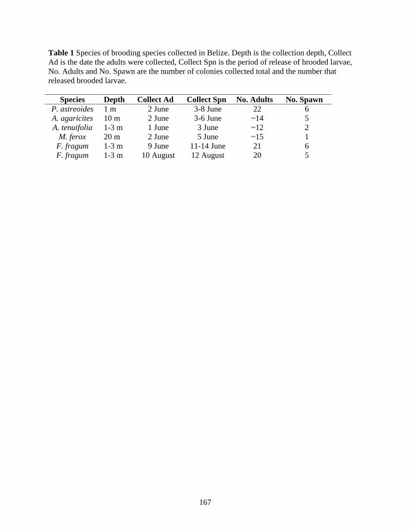
167
Table 1 Species of brooding species collected in Belize. Depth is the collection depth, Collect Ad is the date the adults were collected, Collect Spn is the period of release of brooded larvae, No. Adults and No. Spawn are the number of colonies collected total and the number that released brooded larvae.
Species Depth Collect Ad Collect Spn No. Adults No. Spawn P. astreoides 1 m 2 June 3-8 June 22 6 A. agaricites 10 m 2 June 3-6 June ~14 5 A. tenuifolia 1-3 m 1 June 3 June ~12 2
M. ferox 20 m 2 June 5 June ~15 1 F. fragum 1-3 m 9 June 11-14 June 21 6 F. fragum 1-3 m 10 August 12 August 20 5

168
Table 2 Broadcast spawning species collected in Florida (FLA) and Belize (BEL). Date of gamete collection and fertilization (Fert) is noted as it whether Eggs (E), sperm (S), both (E/S), or none (0) were collected. The number of parents utilized in the gamete cross and the Parental Reef with corresponding GPS coordinates are also given.
Loc Species Fert E/S # Par
Parental Reef GPS
FLA D. Strigosa 19-Aug 0 2 Horseshoe Reef N 25.1399°, W 80.2944° M. faveolata 20-Aug 0 3-5 Horseshoe Reef
M. faveolata 19-Aug 0 10-15 Looe Key N 24.5449°, W 81.4094°
M. faveolata 19-Aug 0
10-15 Alligator Reef N 24.8129°, W 80.6695°
A. palmata 16-Aug E 1 Elbow Reef N 25.1435°, W 80.2574° E 1 Horseshoe Reef A. palmata 17-Aug 0 1 Molasses Reef N 25.0102°, W 80.3733° 0 1 Horseshoe Reef
BEL D. Strigosa 19-Sep 0 3 CBC Reef N 16.8025°, W 88.0819° A. palmata 16-Aug E/S 5 CBC Reef A. cervicornis 17-Aug E/S 7 CBC Reef M. faveolata 19-Sep E/S 7 CBC Reef M. franksi 19-Aug E/S 5 CBC Wall N 16.7797°, W 88.0753°

169
Table 3 The blastn report for the larvae of brooding species samples from Florida (Fla) and Belize (Bel) amplified with the 18S rDNA apicomplexan-specific primers. Blast scores (Score), percent identity (% Id) and E-values are provided as well as the NCBI GenBank accession numbers (Acc #) and species and molecule (Top Hit) of the top blastn hit.
Fla/Bel Query Species E-Values % Id Score Acc # Top Hit
Bel A. agaricites 0 100% 583
AF238264 Coral symbiont (18S rDNA)
Bel A. tenuifolia 0 99% 704
AF238264 Coral symbiont (18S rDNA)
Bel F. fragum 0 100% 672
AF238264 Coral symbiont (18S rDNA)
Bel F. fragum 0 100% 622
AF238264 Coral symbiont (18S rDNA)
Bel F. fragum 0 100% 728
AF238264 Coral symbiont (18S rDNA)
Bel F. fragum 2.0x10-
104 97% 210 AF238264 Coral symbiont (18S
rDNA) Fla P. astreoides
0 99% 704 AF238264 Coral symbiont (18S
rDNA) Fla P. astreoides
7.0x10-54 99% 119 AF238264 Coral symbiont (18S
rDNA) Fla P. astreoides
0 99% 557 AF238264 Coral symbiont (18S
rDNA) Fla P. astreoides 5.0x10-
105 99% 211 AF238264 Coral symbiont (18S
rDNA) Fla P. astreoides
0 98% 599 AF238264 Coral symbiont (18S
rDNA) Fla P. astreoides
1.0x10-56 99% 124 AF238264 Coral symbiont (18S
rDNA) Fla P. astreoides
0 97% 588 AF238264 Coral symbiont (18S
rDNA) Fla P. astreoides
0 97% 586 AF238264 Coral symbiont (18S
rDNA) Fla P. astreoides
0 99% 723 AF238264 Coral symbiont (18S
rDNA) Fla P. astreoides
4.0x10-56 99% 123 AF238264 Coral symbiont (18S
rDNA) Fla P. astreoides
3.0x10-57 99% 125 AF238264 Coral symbiont (18S
rDNA) Bel M. ferox
0 99% 737 AF238264 Coral symbiont (18S
rDNA) Bel P. astreoides
0 99% 539 AF238264 Coral symbiont (18S
rDNA) Bel P. astreoides 6.0x10-65 99% 139 AF238264 Coral symbiont (18S

170
rDNA)

171
Table 4 The number of times apicomplexan DNA was detected (Present) in larvae a) or gametes b) from broadcasting spawner species in Florida or the Bahamas. The parental reef (Reef) and time after fertilization (Fert Time) is noted for the larvae. The larvae were generated from pooled gametes from all corals, so the sample size is always 1. The total number of colonies providing gametes (Total) also are reported. The 95% confidence interval (C.I.) for prevalence were calculated using Sterne’s exact method for all colonies that were PCR positive for apicomplexan DNA in at least one sample. Florida Reefs: HR=Horseshoe Reef, AR=Alligator Reef, LK=Looe Key, EB=Elbow Reef. Belize: CBC=Carrie Bow Cay. Species Reef Fert Time Present a) Florida D. Strigosa HR 40 hr 0 4 d 0 M. faveolata AR 6 d 0 HR 18 hr 0 40 hr 0 LK 4 d 0 7 d 0 A. palmata HR x EB 18 hrs 0 5 d 0 9 d 0 Species Reef Gamete Present Total 95% C. I. b) Florida A. palmata HR Eggs 0 2 Belize A. palmata CBC Eggs 1 3 0-86.46% CBC Sperm 0 3 A. cervicornis CBC Eggs 0 6 CBC Sperm 1 6 0-58.86% M. faveolata CBC Eggs 1 7 0-55.42% CBC Sperm 2 7 0-70.96% M. franksi CBC Eggs 0 5 (August) CBC Sperm 0 5 M. franksi CBC Eggs 0 5 (September) CBC Sperm 0 5

172
Table 5 The blastn report for the larvae (L), eggs (E) and sperm (S) broadcasting spawner samples from Florida (Fla) and Belize (Bel) amplified with the COI primers. Blast Scores, percent identity (% Id) and E-Values are provided as well as the NCBI GenBank accession numbers (Acc #) and species and molecule (Top Hit) of the top blastn hit.
Fla/Bel Query Species E/S/L
E-Values % Id Score Acc # Top Hit
Bel A. cervicornis S
1.0x10-
143 100% 280 AY451340 Acropora cervicornis (COI)
Bel A. cervicornis E 0 100% 553 AY451340
Acropora cervicornis (COI)
Bel A. cervicornis E 0 100% 549 AY451340
Acropora cervicornis (COI)
Bel A. palmata E 0 100% 613 AY451341 Acropora palmata (COI)
Bel D. strigosa L 0 100% 578 AY451349 Diploria strigosa (COI)
Fla M. faveolata L 0 100% 374 HQ203282 Montastraea annularis (COI)
Bel M. faveolata E 0 100% 522 HQ203283 Montastraea annularis (COI)
Bel M. faveolata L 0 100% 549 HQ203284 Montastraea annularis (COI)
Bel M. faveolata E 0 100% 454 HQ203285 Montastraea annularis (COI)
Fla M. faveolata L 0 100% 506 HQ203286 Montastraea annularis (COI)
Fla M. faveolata L 0 100% 554 HQ203287 Montastraea annularis (COI)
Bel M. franksi S 0 100% 426 HQ203288 Montastraea annularis (COI)

173
Table 6 The apicomplexan prevalence in clones of A. palmata and A. cervicornis. For each clone the number of ramets (i.e. individuals per clone; N ramet) and the number infected (N infect) is given as is the 95% confidence interval (C.I.). Acropora palmata (n=31) Clone N ramet N infect prevalence 95% C.I.
1 8 8 1 .6354-1 2 3 3 1 .3685-1 3 2 2 1 .2237-1 4 2 2 1 .2237-1 5 2 2 1 .2237-1 6 1 1 1 0.05-1 7 1 1 1 0.05-1 8 1 1 1 0.05-1 9 1 1 1 0.05-1 10 1 1 1 0.05-1 11 1 1 1 0.05-1 12 1 1 1 0.05-1 13 1 1 1 0.05-1 14 1 0 0 0-.9499 15 1 1 1 0.05-1 16 1 0 0 0-.9499 17 1 0 0 0-.9499 18 1 1 1 0.05-1 19 1 1 1 0.05-1
Acropora cervicornis (n=33) Clone N ramet N infect prevalence 95% C.I.
1 22 18 0.818 .6111-.9354 2 3 3 1 .3685-1 3 3 3 1 .3685-1 4 2 2 1 .2237-1 5 1 1 1 0.05-1 6 1 1 1 0.05-1 7 1 1 1 0.05-1

174
Table 7 Pairwise comparison of prevalence between larvae of brooding species in Belize. P values from Fisher’s exact test are presented. None were significant before or after Bonferroni correction.
P. astreoides M. ferox F. fragum A. agaricites A. tenuifolia
XXX 1.000 1.000 1.000 1.000 P. astreoides XXX 1.000 1.000 1.000 M. ferox XXX 1.000 1.000 F. fragum XXX 1.000 A. agaricites XXX tenuifolia

175
Table 8 Pairwise comparison of prevalence between eggs (top) and sperm (bottom) of broadcasting spawning species in Belize. P values from Fisher’s exact test are presented. None were significant before or after Bonferroni correction. M. faveolata M. franksi A. cervicornis A. palmata
XXX 1.000 1.000 1.000 M. faveolata 0.205 XXX 1.000 0.177 M. franksi 1.000 0.483 XXX 0.258 A. cervicornis 1.000 1.000 1.000 XXX A. palmata

176
References
Aeby G (1991) Behavioral and ecological relationships of a parasite and its hosts within a coral
reef system. Pac Sci 45:263-269
Altschul SF, Gish W, Miller W, Myers EW, Lipman DJ (1990) Basic local alignment search
tool. J Mol Biol 215:403-410
Anderson RM, May RM (1979) Population biology of infectious diseases: Part I. Nature
280:361-367
Apprill AM, Marlow HQ, Martindale MQ, Rappé MS (2009) The onset of microbial associations
in the coral Pocillopora meandrina. ISME J 3:685-699
Asai T, Tomavo S (2007) Biochemistry and metabolism of Toxoplasma gondii. In: Weiss LM,
Kim K (eds) Toxoplasma gondii the model apicomplexan: perspectives and methods.
Academic Press, Amsterdam, pp185-206
Audemard C, Reece KS, Burreson EM (2004) Real-time PCR for detection and quantification of
the protistan parasite Perkinsus marinus in environmental waters. Appl Environ
Microbiol 70:6611-6618
Ayre DJ, Willis BL (1988) Population structure in the coral Pavona cactus: clonal genotypes
show littel phenotypic plasticity. Mar Biol 99:495-505
Ayre DJ, Hughes TP (2000) Genotypic diversity and gene flow in brooding and spawning corals
along the Great Barrier Reef, Australia. Evolution 54:1590-1605
Babcock RC, Bull GD, Harrison PL, Heyward AJ, Oliver JK, Wallace CC, Willis BL (1986)
Synchronous spawnings of 105 scleractinian coral species on the Great Barrier Reef. Mar
Biol 90:379-394

177
Baird AH, Guest JR, Willis BL (2009) Systematic and biogeographical patterns in the
reproductive biology of scleractinian corals. Annu Rev Ecol Evol Syst 40:551-571
Bak RPM, Engel MS (1979) Distribution, abundance and survival of juvenile hermatypic corals
(Scleractinia) and the importance of life history strategies in the parent coral community.
Mar Biol 54:341-352
Bassim KM, Sammarco PW (2003) Effects of temperature and ammonium on larval
development and survivorship in a scleractinian coral (Diploria strigosa). Mar Biol
142:241-252
Baums IB, Hughes CR, Hellberg ME (2005a) Mendelian microsatellite loci for the Caribbean
coral Acropora palmata. Mar Ecol Prog Ser 288:115-127
Baums IB, Miller MW, Hellberg ME (2005b) Regionally isolated populations of an imperiled
Caribbean coral, Acropora palmata. Mol Ecol 14:1377-1390
Baums IB, Miller MW, Hellberg ME (2006) Geographic variation in clonal structure in a reef-
building Caribbean coral, Acropora palmata. Ecol Monogr 76:503-519
Begon M, Harper JL, Townsend CR (1996) Ecology Blackwell Science Ltd, Oxford
Bergeron N, Fecteau G, Paré J, Martineau R, Villeneuve A (2000) Vertical and horizontal
transmission of Neospora caninum indairy herds in Québec. Canadian Veterinary Journal
41:464-467
Breshears MA, Kocan KM, Johnson EM, Panciera RJ (2009) Light and transmission electron
microscopic characteristics of a novel Hepatozoon spp. in naturally infected cotton rats
(Sigmodon hispidus). Parasitol Res 105:1327-1332
Bull JJ, Molineux IJ, Rice WR (1991) Selection of benevolence in a host-parasite system.
Evolution 45:875-882

178
Bushek D, Ford SE, Chintala MM (2002) Comparison of in vitro-cultured and wild-type
Perkinsus marinus. III. Fecal elimination and its role in transmission. Dis Aquat Org
51:217-225
Carlos AA, Baillie BK, Kawachi M, Maruyama T (1999) Phylogenetic position of Symbiodinium
(dinophyceae) isolates from tridacnids (bivalvia), cardids (bivalvia), a sponge (porifera),
a soft coral (anthozoa), and a free-living strain. J Phycol 35:1054-1062
Chornesky EA, Peters EC (1987) Sexual reproduction and colony growth in the scleractinian
coral Porites astreoides. Biol Bull 172:161-177
Coffroth MA, Lewis CF, Santos SR, Weaver JL (2006) Environmental populations of symbiotic
dinoflagellates in the genus Symbiodinium can initiate symbioses with reef cnidarians.
Curr Biol 16:R985-987
Coffroth MA, Lasker HR, Diamond ME, Bruenn JA, Bermingham E (1992) DNA fingerprints of
a gorgonian coral: a method for detecting clonal structure in a vegetative species. Mar
Biol 114:317-325
Correa AMS, Brandt ME, Smith TB, Thornhill DJ, Baker AC (2009) Symbiodinium associations
with diseased and healthy scleractinian corals. Coral Reefs 28:437-448
Craft JD, Russ AD, Yamamoto MN, Iwai TY, Hau S, Kahiapo J, Chong CT, Ziegler-Chong S,
Muir C, Fujita Y, Polhemus DA, Kinzie RA, Santos SR (2008) Islands under islands: The
phylogeography and evolution of Halocaridina rubra Holthuis, 1963 (Crustacean:
Decapoda: Atyidae) in the Hawaiian archipelago. Limnol Oceanog 53:675-689
Dirks U, Gruber-Vodicka HR, Leisch N, Bulgheresi S, Egger B, Ladurner P, Ott JA (2012)
Bacterial symbiosis maintenance in the asexually reproducing and regenerating flatworm
Paracatenula galateia. PLoS One 7:e34709
179
Douglas AE (1994) Symbiotic Interactions. Oxford University Press, Oxford
Dubey JP (2008) The history of Toxoplasma gondii—The first 100 years. J Eukaryot Microbiol
55:467-475
Edmunds PJ, Gates RD, Leggat W, Hoegh-Guldberg O, Allen-Requa L (2005) The effect of
temperature on the size and population density of dinoflagellates in larvae of the reef
coral Porites astreoides. Invertebr Biol 124:185-193
Ewald PW (1987) Transmission modes and evolution of the parasitism-mutualism continuum.
Ann NY Acad Sci 503:295-306
Fadlallah YH (1983) Sexual reproduction, development and larval biology in scleractinian
corals. Coral Reefs 2:129-150
Fayer R, Dubey JP, Lindsay DS (2004) Zoonotic protozoa: from land to sea. Trends Parasitol
20:531-536
Fayer R, Graczyk TK, Lewis EJ, Trout JM, Farley CA (1998) Survival of infectious
Cryptospordium parvum oocysts in seawater and eastern oysters (Crassostrea virginica)
in the Chesapeake Bay. Appl Environ Microbiol 64:1070-1074
Fellous S, Koella JC (2009) Different transmission strategies of a parasite in male and female
hosts. J Evol Biol 22:582-588
Field SG, Michiels NK (2005) Parasitism and growth in the earthworm Lumbricus terrestris:
fitness costs of the gregarine parasite Monocystis sp. Parasitology 130:397-403
Fogarty ND, Vollmer SV, Levitan DR (2012) Weak prezygotic isolating mechanisms in
threatened Caribbean Acropora corals. PLoS One 7:e30486

180
Folmer O, Black M, Hoeh W, Lutz R, Vrijenhoek R (1994) DNA primers for amplification of
mitochondrial cytochrome C oxidase subunit I from diverse metazoan invertebrates.
Molecular Marine Biology and Biotechnology 3:294-299
Frank SA (1996a) Models of parasite virulence. Q Rev Biol 71:37-78
Frank SA (1996b) Host-symbiont conflict over the mixing of symbiotic lineages. Proc R Soc
Lond B 263:339-344
Fukami H, Budd AF, Levitan DR, Jara J, Kersanach R, Knowlton N (2004) Geographic
differences in species boundaries among members of the Montastraea annularis complex
based on molecular and morphological markers. Evolution 58:324-337
Gilmore MD, Hall BR (1976) life history, growth habits, and constuctional roles of Acropora
cervicornis in the patch reef environment. J Sediment Petrol 46:519-522
Gladfelter EH, Monahan RK, Gladfelter WB (1978) Growth rates of five reef-building corals in
the Northeastern Caribbean. Bull Mar Sci 28:728-734
Gleason DF, Wellington GM (1995) Variation in UVB sensitivity of planula larvae of the coral
Agaricia agaricites along a depth gradient. Mar Biol 123:693-703
Gleason DF, Danilowicz BS, Nolan CJ (2009) Reef waters stimulate substratum exploration in
planulae from brooding Caribbean corals. Coral Reefs 28:549-554
Goulet TL, Coffroth MA (2003) Genetic composition of zooxanthellae between and within
colonies of the octocoral Plexaura kuna, based on small subunit rDNA and multilocus
DNA fingerprinting. Mar Biol 142:233-239
Goulet TL, Coffroth MA (2004) The genetic identity of dinoflagellate symbionts in Caribbean
octocorals. Coral Reefs 23:465-472

181
Green DH, Edmunds PJ, Carpenter RC (2008) Increasing relative abundance of Porites
astreoides on Caribbean reefs mediated by an overall decline in coral cover. Mar Ecol
Prog Ser 359:1-10
Guérardel Y, Leleu D, Coppin A, Liénard L, Slomianny C, Strecker G, Ball S, Tomavo S (2005)
Amylopectin biogenesis and characterization in the protozoan parasite Toxoplasma
gondii, the intracellular development of which is restricted in the HepG2 cell line. Microb
Infect 7:41-48
Hamilton WD, May RM (1977) Dispersal in stable habitats. Nature 269:578-581
Herre EA, Knowlton N, Mueller UG, Rehner SA (1999) The evolution of mutualisms: exploring
the paths between conflict and cooperation. Trends Ecol Evol 14:49-53
Hide G, Morley EK, Hughes JM, Gerwash O, Elmahaishi MS, Thomasson D, Wright EA,
Williams RH, Murphy RG, Smith JE (2009) Evidence for high levels of vertical
transmission in Toxoplasma gondii. Parasitology 136:1877-1885
Highsmith RC (1982) Reproduction by fragmentation in corals. Mar Ecol Prog Ser 7:207-226
Hill RD, Su C (2012) High tissue burden of Toxoplasma gondii is the hallmark of acute
virulence in mice. Vet Parasitol 187:36-43
Hill SLL, Okamura B (2007) Endoparasitism in colonial hosts: patterns and processes.
Parasitology 134:841-852
Isomura N, Nishihira M (2001) Size variation of planulae and its effect on the lifetime of
planulae in three pocilloporid corals. Coral Reefs 20:309-315
Jackson JBC, Hughes TP (1985) Adaptive strategies of coral-reef invertebrates. Am Sci 73:265-
274

182
Jokiel PL (1985) Lunar periodicity of planula release in the reef coral Pocillopora damicornis in
relation to various environmental factors. Proceedings of the 5th International Coral Reef
Congress, Tahiti, 1985 4:307-312
Jokiel PL, Ito RY, Liu PM (1985) Night irradiance and synchronization of lunar release of
planula larvae in the reef coral Pocillopora damicornis. Mar Biol 88:167-174
Kirk NL, Thornhill DJ, Kemp DW, Fitt WK, Santos SR (In Prep) Persistent association between
apicomplexan symbionts and Caribbean reef corals
Knowlton N, Jackson JBC (1994) New taxonomy and niche partitioning on coral reefs: jack of
all trades or master of some? Trends Biotechnol 9:7-9
Knowlton N, Rohwer FL (2003) Multispecies microbial mutualisms on coral reefs: the host as a
habitat. Amer Nat 162:S51-S61
Koh LP, Dunn RR, Sodhi NS, Colwell RK, Proctor HC, Smith VS (2004) Species coextinctions
and the biodiversity crisis. Science 305:1632-1634
Lake JA (2009) Evidence for an early prokaryotic endosymbiosis. Nature 460:967-971
Lasker HR, Coffroth MA (1999) Responses of clonal reef taxa to environmental change. Amer
Zool 39:92-103
Legendre P, Legendre L (1998) Numerical Ecology. Elsevier Science, Amsterdam
Lesser MP, Mazel CH, Gorbunov MY, Falkowski PG (2004) Discovery of symbiotic nitrogen-
fixing cyanobacteria in corals. Science 305:997-1000
Levitan DR, Fogarty ND, Jara J, Lotterhos KE, Knowlton N (2011) Genetic, spatial, and
temporal components of precise spawning synchrony in reef building corals of the
Montastraea annularis species complex. Evolution 65:1254-1270

183
Lindsay DS, Collins MV, Mitchell SM, Cole RA, Flick GJ, Wetch CN, Lindquist A, Dubey JP
(2003) Sporulation and survival of Toxoplasma gondii oocysts in seawater. J Eukaryot
Microbiol S1:687-688
Lipsitch M, Nowak MA, Siller S (1996) The evolution of virulence in pathogens with vertical
and horizontal transmission. Evolution 50:1729-1741
Lipsitch M, Nowak MA, Ebert D, May RM (1995) The population dynamics of vertically and
horizontally transmitted parasites. Proc R Soc Lond B 260:321-327
Lirman D (2000) Fragmentation in the branching coral Acropora palmata (Lamarck): growth,
survivorship, and reproduction of colonies and fragments. J Exp Mar Biol Ecol 251:41-57
Little AF, van Oppen MJH, Willis BL (2004) Flexibility in algal endosymbioses shapes growth
in reef corals. Science 304:1492-1494
Magalon H, Nidelet T, Martin G, Kaltz O (2010) Experimental evolution of life history and
virulence of a parasite with vertical and horizontal transmission. Evolution 64:2126-2138
Mangin KL, Lipsitch M, Ebert D (1995) Virulence and transmission modes of two microsporidia
in Daphnia magna. Parasitology 111:133-142
Margulis L (1998) Symbiotic Planet. Basic Books, New York
Mason BM, Cohen JH (2012) Long-wavelength photosensitivity in coral planula larvae. Biol
Bull 222:88-92
Massie GN, Ware MW, VIllegas EN, Black MW (2010) Uptake and transmission of Toxoplasma
gondii oocysts by migratory, filter-feeding fish. Vet Parasitol 169:296-303
McGuire MP (1998) Timing of larval release by Porites astreoides in the northern Florida Keys.
Coral Reefs 17:369-375

184
Miller WA, Miller MA, Gardner IA, Atwill ER, Harris M, Ames J, Jessup D, Melli A, Paradies
D, Worchester K, Olin P, Barnes N, Conrad PA (2005) New genotypes and factors
associated with Cryptosporidium detection in mussels (Mytilus spp.) along the California
coast. Int J Parasit 35:1103-1113
Morris DJ, Adams A (2006) Transmission of freshwater myxozoans during the asexual
propagation of invertebrate hosts. Int J Parasit 36:371-377
Muller-Parker G (1984) Dispersal of zooxanthellae on coral reefs by predators on cnidarians.
Biol Bull 167:159
Muscatine L, Porter JW (1977) Reef corals: mutualistic symbioses adapted to nutrient-poor
environments. Bioscience 27:454-460
Muscatine L, McCoskey LR, Marian RE (1981) Estimating the daily contribution of carbon from
zooxanthellae to coral animal respiration. Limnol Oceanog 26:601-611
Neigel JE, Avise JC (1983) Clonal diversity and population structure in a reef-building coral,
Acropora cervicornis: self-recognition analysis and demographic interpretation.
Evolution 37:437-453
Nissimov J, Rosenberg E, Munn CB (2009) Antimicrobial properties of resident coral mucus
bacteria of Oculina patagonica. FEMS Microbiol Lett 292:210-215
Padilla-Gamiño JL, Weatherby TM, Waller RG, Gates RD (2011) Formation and structural
organization of the egg–sperm bundle of the scleractinian coral Montipora capitata.
Coral Reefs 30:371-380
Park K-I, Yang H-S, Kang H-S, Cho M, Park K-J, Choi K-S (2010) Isolation and identification
of Perkinsus olseni from feces and marine sediment using immunological and molecular
techniques. J Invertebr Pathol 105:261-269

185
Peters EC (1984) A survey of cellular reactions to environmental stress and disease in Caribbean
scleractinian corals. Helgolander Meeresun 37:113-137
Plaisance L, Caley MJ, Brainard RE, Knowlton N (2011) The diversity of coral reefs: what are
we missing? PLoS One 6:e25026
Porto I, Granados C, Restrepo JC, Sanchez JA (2008) Macroalgal-associated dinoflagellates
belonging to the genus Symbiodinium in Caribbean reefs. PLoS One 3:e2160
Poulin R (1996) How many parasite species are there: are we close to answers? Int J Parasit
26:1127-1129
Poulin R, Keeney DB (2008) Host specificity under molecular and experimental scrutiny. Trends
Parasitol 24:24-28
Pratchett MS, Gust N, Goby G, Klanton SO (2001) Consumption of coral propagules represents
a significant trophic link between corals and reef fish. Coral Reefs 20:13-17
Prensier G, Dubremetz JF, Schrével J (2008) The unique adaptation of the life cycle of the
coelomic gregarine Diplauxis hatti to its host Perinereis cultrifera (Annelida,
Polychaeta): an experimental and ultrastructural study. J Eukaryot Microbiol 55:541-553
Reshef L, Koren O, Loya Y, Zllber-Rosenberg I, Rosenberg E (2006) The coral probiotic
hypothesis. Environ Microbiol 8:2068-2073
Richmond RH, Hunter CL (1990) Reproduction and recruitment of corals: comparisons among
the Caribbean, the Tropical Pacific, and the Red Sea. Mar Ecol Prog Ser 60:185-203
Ritchie KB (2006) Regulation of microbial populations by coral surface mucus and mucus-
associated bacteria. Mar Ecol Prog Ser 322:1-14

186
Ritson-Williams R, Paul VJ, Arnold SN, Steneck RS (2010) Larval settlement preferences and
post-settlement survival of the threatened Caribbean corals Acropora palmata and A.
cervicornis. Coral Reefs 29:71-81
Rosenberg E, Zilber-Rosenberg I (2011) Symbiosis and development: the hologenome concept.
Birth Defects Res (Part C) 93:56-66
Rowan R, Powers DA (1991) Molecular genetic identification of symbiotic dinoflagellates
(zooxanthellae). Mar Ecol Prog Ser 71:65-73
Rózsa L, Reiczigel J, Majoros G (2000) Quantifying parasites in samples of hosts. J Parasitol
86:228-232
Sachs JL, Wilcox TP (2006) A shift to parasitism in the jellyfish symbiont Symbiodinium
microadriaticum. Proc R Soc Lond B 273:425-429
Sandström JP, Russell JA, White JP, Moran NA (2001) Independent origins and horizontal
transfer of bacterial symbionts of aphids. Mol Ecol 10:217-228
Schwarz JA, Brokstein PB, Voolstra C, Terry AY, Miller DJ, Szmant AM, Coffroth MA,
Medina M (2008) Coral life history and symbiosis: Functional genomic resources for two
reef building Caribbean corals, Acropora palmata and Montastraea faveolata. BMC
Genomics 9:97
Sharp KH, Distel DL, Paul VJ (2012) Diversity and dynamics of bacterial assemblages
associated with the Caribbean coral Porites astreoides. ISME J 6:790-801
Sharp KH, Ritchie KB, Schupp PJ, Ritson-Williams R, Paul VJ (2010) Bacterial acquisition in
juveniles of several broadcast spawning coral species. PLoS One 5:e10898

187
Siboni N, Rasoulouniriana D, Ben-Dov E, Kramarsky-Winter E, Sivan A, Loya Y, Hoegh-
Guldberg O, Kushmaro A (2010) Stamenopile microorganisms associated with the
massive coral Favia sp. J Eukaryot Microbiol 57:236-244
Simon L, Bousquet J, Lévesque RC, Lalonde M (1993) Origin and diversification of
endomycorrhizal fungi and coincidence with vascular land plants. Nature 363:67-69
Smith SR (1992) Patterns of coral recruitment and post-settlement mortality on Bermuda's reefs:
comparisons to Caribbean and Pacific Reefs. Amer Zool 32:663-673
Snow RW, Guerra CA, Noor AM, Myint HY, Hay SI (2005) The global distribution of clinical
episodes of Plasmodium falciparum malaria. Nature 434:214-217
Stat M, Carter D, Hoegh-Guldberg O (2006) The evolutionary history of Symbiodinium and
scleractinian hosts— Symbiosis, diversity, and the effect of climate change. Perspect
Plant Ecol Evol Syst 8:23-43
Stewart AD, Logsdon JM, Kelley SE (2005) An empirical study of the evolution of virulence
under both horizontal and vertical transmission. Evolution 59:730-739
Stork NE, Lyal CHC (1993) Extinction or 'co-extinction' rates? Nature 366:307
Szmant AM (1986) Reproductive ecology of Caribbean reef corals. Coral Reefs 5:43-54
Szmant-Froelich A, Reutter M, Riggs L (1985) Sexual reproduction of Favia fragum (Esper):
lunar patterns of gametogenesis, embryogenesis and planulation in Puerto Rico. Bull Mar
Sci 37:880-892
Toller WW, Rowan R, Knowlton N (2002) Genetic evidence for a protozoan (phylum
Apicomplexa) associated with corals of the Montastrea annularis species complex. Coral
Reefs 21:143-146

188
Tunnicliffe V (1981) Breakage and propagation of the stony coral Acropora cervicornis. Proc
Natl Acad Sci U S A 78:2427-2431
Upton SJ, Peters EC (1986) A new and unusual species of coccidium (apicomplexa:
agamococcidiorida) from Caribbean scleractinian corals. J Invertebr Pathol 47:184-193
Valiére N (2002) GIMLET: a computer program for analysing genetic individual identification
data. Mol Ecol Notes 2:377-379
Vermeij MJA, Sandin SA (2008) Density-dependent settlement and mortality structure the
earliest life phases of a coral population. Ecology 89:1994-2004
Vermeij MJA, Bakker J, van der Hal N, Bak RPM (2011) Juvenile coral abundance has
decreased by more than 50% in only three decades on a small Caribbean island. Diversity
3:296-307
Vollmer SV, Palumbi SR (2007) Restricted gene flow in the Caribbean staghorn coral Acropora
cervicornis: implications for the recovery of endangered reefs. Journal of Heredity 98:40-
50
Wang CC, Weppelman RM, Lopez-Ramos B (1975) Isolation of amylopectin granules and
identification of amylopectin phosphorylase in the oocysts of Eimeria tenella. J Protozool
22:560-564
Weiss LM, Kim K (2007) Bradyzoite development. In: Weiss LM, Kim K (eds) Toxoplasma
gondii the model apicomplexan: perspectives and methods. Academic Press, Amsterdam,
pp341-366
Wellington GM, Fitt WK (2003) Influence of UV radiation on the survival of larvae from
broadcast-spawning reef corals. Mar Biol 143:1185-1192
Windsor DA (1998) Most of the species on Earth are parasites. Int J Parasit 28:1939-1941

189
Winiecka-Krusnell J, Dellacasa-Lindberg I, Dubey JP, Barragan A (2009) Toxoplasma gondii:
Uptake and survival of oocysts in free-living amoebae. Exp Parasitol 121:124-131

190
Chapter 5. Evolutionary History of the Coral-Associated Apicomplexans
ABSTRACT. Apicomplexans (Alveolata: Apicomplexa) are an important parasitic group,
but phylogenetic relationships among the major lineages are only now being elucidated in part
due to poor taxon sampling. One of the most overlooked sources of novel, as well as recognized,
apicomplexan lineages are invertebrates, and corals in particular. A single coral-associated
species has been formally described and based on life history, taxonomically united within the
apicomplexan order Agamococcidiorida with the rhytidocystids, which are symbionts of
polychaete worms. To test this phylogenetic hypothesis and to determine the evolutionary
relationships among coral-associated symbionts, 18S rDNA sequences were generated from a
broad sampling of 16 scleractinian coral and 3 gorgonian host species. Maximum likelihood and
Bayesian phylogenetic trees recovered a monophyletic clade containing all coral apicomplexan
symbionts with strong support. This coral-associated clade was closely related to the coccidian
families Sarcocystidae and Eimeriidae, which both contain economically important taxa.
However, the coral-associated clade was phylogenetically distinct from the rhytidocystids and a
constraint tree forcing such a grouping was significantly worse than the best tree. Within the
clade there was little evidence of specificity and constraint trees enforcing monophyly of
apicomplexans isolated from five of the seven same coral families also were significantly worse
than the best tree. Likewise, a constraint tree binning coral-associated apicomplexans by the two
major scleractinian coral clades (complex and robust) also was rejected. Thus, there was little

191
support for Agamococcidiorida or specificity within the clade, which may be indicative of host-
switching and/or incomplete lineage sorting.
INTRODUCTION
Molecular sequence data have considerably improved our understanding of eukaryotic
evolutionary relationships over the last 25 years (Simpson and Roger 2004; Adl et al. 2005;
Keeling et al. 2005; Adl et al. 2007). Six putatively monophyletic “supergroups”, based on
ultrastructural, phylogenetic and genomic data, have been erected to reflect this current
classification system (Adl et al. 2005). However, the relationships between and among
supergroups remain in flux (Parfrey et al. 2006; Burki et al. 2007; Burki et al. 2008; Roger and
Simpson 2009; Parfrey et al. 2010; Walker et al. 2011). A prime example is the former
supergroup Chromalveolata, first hypothesized by Cavalier-Smith (1999) to involve a secondary
endosymbiotic event between a photosynthetic red-alga lineage and the precursor to several
eukaryotic groups. Today, both phylogenomics and other genetic evidence suggest
Chromalveolata and another supergroup, Rhizaria (Burki et al. 2007; Hackett et al. 2007; Burki
et al. 2008; Elias et al. 2009; Burki et al. 2010; Cavalier-Smith 2010) belong to a single, new
supergroup uniting the stramenopiles, alveolates and rhizaria (SAR) (Burki et al. 2007). This has
important implications in understanding the evolutionary history of the encompassed taxa as
many have lost their photosynthetic capacity and evolved into obligate parasites. One group is
the apicomplexans (Alveolata: Apicomplexa), who retain a vestigial plastid (McFadden et al.
1996; Wilson and Williamson 1997). However, as significant as it is to understand the
relationships between eukaryotic supergroups, it is equally important to determine the phylogeny

192
of member clades as well. Due to their parasitic nature and impact in areas such as the quality of
human life, elucidating the evolutionary history of the apicomplexans can be considered of
utmost importance.
Historically, Apicomplexa has been split into four major lineages, the haemosporidians,
piroplasms, coccidians, and gregarines, based on motility, life-cycle, host range, tissue
localization, and morphology (Perkins et al. 2000). Along with this, small subunit (18S)
ribosomal DNA (rDNA) sequence data have supported some clades while highlighting
morphological synapomorphies that unite particular groups. Conversely, these data also have
reassigned taxonomic placements. For example, Cryptosporidium parvum, an opportunistic
human pathogen, was moved from the coccidians to the gregarines (Barta and Thompson 2006;
Saffo et al. 2010; Rueckert et al. 2011b), based on analyses of 18S rDNA data. However,
evolutionary relationships among many of the major apicomplexan groups remain elusive in part
due to poor taxon sampling (Leander 2008; Morrison 2008,2009). Historically, most
phylogenetic studies focused on taxa infecting hosts with human economic importance and little
attention has been paid to others, such as invertebrates (Leander et al. 2003; Kopecna et al. 2006;
Leander 2008; Clopton 2009; Rueckert et al. 2010; Rueckert et al. 2011a). This is unfortunate
since many apicomplexans from invertebrates represent previously unknown species (Rueckert
and Leander 2008; Dubey et al. 2009; Rueckert and Leander 2009b). Thus, describing the
diversity of apicomplexans in invertebrates will clarify both ecological distributions of known
parasites and the evolutionary history of the involved (and other) taxa.
One understudied group of host invertebrates is the scleractinian and gorgonian corals
(Cnidaria: Anthozoa). Scleractinians (Hexacorallia: Scleractinia) are the foundation of the coral
reef ecosystem and phylogenetically group into two clades: complex and robust (Romano and

193
Palumbi 1996). Within these clades, multiple phylogenetic studies have clarified our
understanding of coral evolutionary history (Kerr 2005; Fukami et al. 2008; Kitahara et al.
2010). Likewise, gorgonians (Octocorallia: Gorgonacea) are numerically dominant on shallow
Caribbean reefs and their phylogenetic relationships also have been well-elucidated (Sanchez et
al. 2003; McFadden et al. 2006). Scleractinians and gorgonians harbor numerous bacterial and
protist symbionts, including apicomplexans (Upton and Peters 1986; Toller et al. 2002;
Knowlton and Rohwer 2003; Goulet and Coffroth 2004). A single apicomplexan species,
Gemmocystis cylindrus, was formally described and due to its unusual life-cycle (i.e. no
merogony or sporogony), it was placed within the coccidian order Agamococcidiorida (Upton
and Peters 1986), which included a single other genus, Rhytodocystis (Levine 1979). Although
no 18S rDNA data exist for G. cylindrus, Rhytodocystis spp. have recently been described with
corresponding sequence data and found to phylogenetically group with a symbiont isolated from
a giant clam in a clade called the “rhytidocystids” (Leander and Ramey 2006; Rueckert and
Leander 2009a). Therefore, these taxa can be utilized towards determining whether coral-
associated apicomplexans and Rhytodocystis spp. share a most recent common ancestor, as
hypothesized by their mutual inclusion in Agamococcidiorida.
To test this hypothesis, 18S rDNA sequences were utilized to resolve phylogenetic
relationships 1) among apicomplexans associated with scleractinian and gorgonian hosts in the
Caribbean Sea and 2) determine their evolutionary placement within the Apicomplexa. Sixteen
coral species, including both host clades (i.e. complex and robust) as well as three gorgonian
species, were chosen to provide wide-breadth of diversity and to test for potential specificity
between hosts and their symbionts. The host species Dendrogyra cylindrus also was specifically
targeted since it is the host of the type species, G. cylindrus. Lastly, apicomplexan 18S rDNA

194
sequences isolated from other hosts of the same ecosystem were included to determine if
symbionts from corals and other coral reef taxa belong to a common lineage. These samples
encompassed two coccidians from coral reef fishes and an apicomplexan from a giant clam,
which has previously grouped with the rhytidocystids.
MATERIALS AND METHODS
Coral samples utilized. Samples were collected from 16 Caribbean scleractinian coral species
representing four families in the robust and complex clades, respectively (Table 1). Two or three
species were selected from each of the families to test the hypothesis of specificity, which
assumes monophyly within the families. The one exception was Siderastreidae, which contains
only two Caribbean species. Apicomplexan DNA was not detected in any colonies of Siderastrea
radians examined (n = 15) and this scleractinian family was confined to symbionts exclusively
from Siderastrea siderea. As several host families are paraphyletic, host species were chosen to
include members that exhibited common ancestry in a recent host phylogeny (Fukami et al.
2008). Two colonies of Porites astreoides were included to determine if symbionts from the
same host were identical and/or most closely related to each other. Samples also were collected
from three gorgonian species commonly found in the Caribbean Sea (Kinzie 1973; Jordán-
Dahlgren 2002). These host species were selected to include sister genera, Gorgonia and
Pseudopterogorgia (Family Gorgoniidae) as well as a more distant related genus, Muriceopsis
(Family Plexauridae) (Sanchez et al. 2003).
DNA isolation, PCR amplification, and cloning. Scleractinian tissue was removed from
the skeleton using a water-pik (LaJeunesse 2002) and DNA isolated using a modified Promega

195
Wizard protocol as described in LaJeunesse et al. (2003). DNA was extracted from gorgonian
samples using the 2xCTAB method (Coffroth et al. 1992).
To specifically target apicomplexan DNA while excluding other coral symbionts, an
~860 bp fragment of the 18S small subunit ribosomal DNA (18S rDNA) molecule was amplified
using the apicomplexan-specific primers 18N-F2 and 18N-R1 under previously published
conditions (Toller et al. 2002) and a touchdown PCR protocol with a 5 min initial denaturation
step at 94°C, followed by 45 sec at 94°C, 45 sec at 60°C, decreasing 1°C/cycle until 50°C was
reached, and a 1 min extension at 72°C. This was immediately followed by 30 additional cycles
of 94°C for 45 sec, 50°C for 45 sec, 72°C for 1 min, and a 30 min final extension at 72°C.
Amplicons from all 20 samples were sequenced using the forward primer (18N-F2) to determine
the dominant (i.e. most abundant) 18S rDNA copy from each coral host.
To acquire nearly full-length (~1,800 bp) 18S rDNA sequences for the coral hosts, two
overlapping segments of the molecule were individually amplified. Specifically, samples were
amplified using the universal eukaryotic primers 18S-82F and 18S-1498R (Slapeta et al. 2005)
coupled with 18N-R1 and 18N-F2 (Toller et al. 2002), respectively. The first half of the
molecule (18Sa) was amplified using a 5 min denaturation step, and 35 cycles of 15 sec at 94° C,
15 sec at 55° C, 30 sec at 72° C, followed by a 30 min extension at 72° C. The second half of the
molecule (18Sb) was amplified utilizing the touchdown PCR protocol detailed above. PCR
products from 18Sa and 18Sb were immediately cloned using the pCR® 2.1 TOPO vector and
TOP10 chemically competent cells (Invitrogen, Life Technologies, USA). Twenty bacterial
colonies were haphazardly picked and PCR screened for insert size. From these, five clones per
coral host sample were chosen by a random number generator. Clones were grown overnight in

196
LB broth, plasmids were purified by alkaline lysis extraction (Birnboim 1983), and then
sequenced bi-directionally by HtSeq (Seattle, Washington).
Assembly and alignment of 18S rDNA sequences. Vector sequence was removed and
bi-direction reads assembled for 18Sa and 18Sb separately using Geneious (Drummond et al.
2011). To identify potential PCR chimeras within the dataset, sequences of 18Sa and 18Sb were
assessed using a 300 bp sliding window and compared to all other sequences on both sides of the
breakpoint with a partial tree-building approach based on distance (i.e. sequence similarity) in
Bellerophon (Huber et al. 2004). Conflicting topologies derived from the sequence to the left or
right of the breakpoint are indicative of chimeras and were removed from the dataset. The 18Sa
and 18Sb sequences were then aligned to the dominant apicomplexan-copy from the same coral
sample if they contained 100% sequence identity and no gaps to generate the almost full-length
18S rDNA. These consensus sequences were rechecked using Bellerophon to ensure no chimeric
sequences were artificially generated in this process.
The following Apicomplexa 18S rDNA sequences were downloaded from GenBank to
place those associating with corals into an evolutionary context with other taxa from the group:
Adelina bambarooniae (AF494058), Adelina dimidiata (DQ096835), Adelina grylli
(DQ096836), Aggregata eberthi (DQ096838), Ascogregarina taiwanensis (JX131297), Babesia
rodhaini (M87565), Besnoitia besnoiti (AF109678), Caryospora bigenetica (AF060976),
Choleoeimeria sp. (AY043207), Cryptosporidium parvum (DQ898159), Cryptosporidium
serpentis (AF093502), Cystoisospora felis (L76471), Dactylosoma ranarum (HQ224957),
Eimeria leucisci (GU479649), Eimeria intestinalis (EF694012), Eimeria maxima (DQ538348),
Frenkelia glareoli (AF009245), Goussia janae (AY043206), Gregarina chortiocetes (L31841),
Gregarina niphandrodes (AF129882), Hepatozoon ayorgbor (EF157822), Hepatozoon canis

197
(EU289222), Hepatozoon felis (AY628681), Isospora orlovi (AY365026), Isospora suis
(U97523), Klossia helicina (HQ224955), Lankesterella minima (AF080611), Lankesteria abbotti
(DQ093796), Lecudina polymorpha (AY196706), Lecudina tuzetae (AF457128), Leidyana
migrator (AF457130), Lithocystis sp. (DQ093795), Mattesia geminata (AY334568), Monocystis
agilis (AF457127), Neospora caninum (U03069), Pterospora floridiensis (DQ093794),
Pterospora schizosoma (DQ093793), Rhytidocystis cyamus (GQ149767), Rhytidocystis
polygordiae (DQ273988), Sarcocystis canis (DQ146148), Sarcocystis hominis (AF176945),
Theileria annulata (DQ287944), Theileria parva (HQ684067), and Toxoplasma gondii
(U03070). Additionally, sequences from unidentified apicomplexan species from aquatic
environments (AF372780, GQ330636, AY664971, AY426932, GU825688), the coral reef
environment (two isolated from fishes, Lutjanus kasmira and Mulloidichthys pfluegeri,
(HM117907 and HM117908) and one from a giant clam, Tridacna crocea (AB000912), and
three apicomplexan 18S rDNA clones (AF238264-6), originally isolated in Panama from the
coral Montastraea sp., were included.
The final data matrix encompassed 76 18S rDNA sequences including Chromera velia
(Genbank accession number JN986789), a photosynthetic alveolate branching near the base of
the apicomplexans (Moore et al. 2008; Janouskovec et al. 2011), which served as the outgroup in
phylogenetic analyses. Sequences were aligned using the L-INS-in algorithm with 1000 iterative
refinements in MAFFT v6.861b (Katoh and Toh 2008). The alignment was inspected by eye and
further refined in Mesquite v2.75 (Maddison and Maddison 2011), as recommended for
improved accuracy (Edgar and Batzoglou 2006). To increase the likelihood of comparing
homologous characters in the alignment, ambiguously aligned regions were removed using
Gblocks v0.91b (Castresana 2000). “Relaxed” parameters were used to trim the alignments as

198
they performed best on simulated datasets of similar length (Talavera and Castresana 2007).
Under these parameters, gaps were allowed in half of the sequences, which permitted inclusion
of shorter environmental sequences (e.g. marine water column sample: GU825688, 1095 bp)
without truncating the final alignment to that length. This also allowed the inclusion of
characters within insertion/deletion events (indels) that might provide additional phylogenetic
information.
Phylogenetic analyses. The most appropriate model of evolution for the alignment was
selected using hierarchical likelihood ratio tests (hLRT) and Akaike Information Criterion
corrected for sample size (i.e. number of sites in the alignment: AICc (Hurvich and Tsai 1989))
in jModelTest v0.1.1 (Posada 2008). Both methods selected the general time reversible model
with rate variation across sites as modeled by the gamma distribution (GTR+γ) (Sullivan and
Joyce 2005) and this model of evolution was utilized in all phylogenetic analyses. Maximum
likelihood (ML) trees were inferred in RAxML v.7.0.4 (Stamatakis 2006), with nodal confidence
assessed using 1,000 bootstrap replicates. Trees also were generated using Bayesian inference
(Huelsenbeck et al. 2001) in MrBayes v3.2.1 (Huelsenbeck and Ronquist 2001; Ronquist and
Huelsenbeck 2003). All Bayesian phylogenies comprised of the best tree from 2 runs (with 4
chains per run) of 10x106 generations, sampling every 10x103 steps after removing the first 25%
(i.e. 2.5x10⁵) as burnin. The runs were terminated after 10x106 generations as the average
standard deviation between them was less than 0.02, suggesting both converged on a similar
topology despite starting in different, randomly-selected, areas of tree space (Ronquist et al.
2009).
Hypothesis testing of phylogenetic trees. To test the hypothesis that the coral-associated
apicomplexans are the sister taxa to the rhytidocystids, which also included the clam symbiont,

199
the topology of the best ML tree was compared to a constraint tree (i.e. coral symbionts and
rhytidocystids forced to be sister taxa a priori). Likewise, a constraint tree uniting the coral-
associated apicomplexans and coccidians infecting reef fishes was created to test whether they
belong to a single lineage. Additionally, the monophyly of apicomplexans collected from the
coral families belonging to the complex clade (i.e., Acroporidae, Agariciidae, Poritidae,
Siderastreidae) and to the robust clade (i.e., Faviidae, Meandrinidae, Pocilloporidae, Mussidae)
separately to determine if specificity was apparent in this group. Separate trees constraining
apicomplexans specifically to each of the seven (no Siderastreidae test was possible, as only one
species was examined) coral families also were tested. Lastly, three previously sequenced
apicomplexans from Montastraea sp. (Toller et al. 2002) were included within the Faviidae to
test monophyly of this group. ML constraint trees were generated for all ten hypotheses (Table 2)
using the GTR+γ model and compared to the most likely ML tree generated above using SH tests
(Shimodaira and Hasegawa 1999) in RAxML v.7.0.4.
RESULTS
Final data matrix. Twenty coral-associated apicomplexan 18S rDNA consensus
sequences generated in this study were used in all analyses, ranging in size from 1705-1712 bp.
The alignment, which included the other apicomplexans and C. velia, was 2014 bp including
gaps. This included 1031 parsimony-informative, 340 uninformative, and 643 constant (i.e.
invariable) characters. Following removal of ambiguously aligned regions, the final data matrix
contained 1436 total characters with included gaps, with 727 parsimony-informative, 237
uninformative, and 472 constant characters.

200
Phylogenetic analysis. Strong support (99% bootstrap [BS] support) was found for a
single clade containing all coral-associated apicomplexans, as well as a single environmental
sample from the Southern Caribbean Sea, in the ML tree (Fig. 1). This clade grouped with the
Sarcocystidae and the Eimeriidae, two coccidian families, with moderate (71% BS) support.
Three distinct groups, based on >85% BS support, were identified within the coral-associated
apicomplexans. Firstly, apicomplexans associated with gorgonians represented Group I with
strong (92% BS) support. Notably, a symbiont of P. astreoides was basal to this group with
moderate (77% BS) support. The second group united symbionts from Madracis decactis,
Agaricia grahamae, Scolymia sp. and a Mycetophyllia sp. (90% BS, Group II). The third group
(Group III) encompassed apicomplexans from Dendrogyra cylindrus, Madracis mirabilis,
Acropora palmata, Mussa sp., Porites porites, and the other symbiont from P. astreoides with
strong (86% BS) support. None of the symbionts grouped with others isolated from con-familial
corals (Fig. 1).
The phylogeny based on Bayesian inference also recovered a monophyletic clade of
coral-associated apicomplexans and the same environmental sample (Fig. 2). Once again, the
coral-associated apicomplexan clade was united with the Eimeriidae and the Sarcocystidae with
a posterior probability (PP) of 0.84 (Fig. 2). Likewise, the three clades of coral-associated
apicomplexans were recovered with strong support (PPs of 1.00, 0.98, and 1.00, respectively).
Thus, while the Bayesian tree possessed minor differences in its basal topology relative to the
ML tree (e.g., placement of the coccidian Aggregata eberthi; see Discussion), the inferred
position of the coral-associated clade, and the groups within it, were identical across
methodologies (Fig. 2).

201
Hypothesis testing. There were significant differences between the best ML tree and 8 of
the 10 hypothesis-testing constraint trees. A clade uniting the coral-associated apicomplexans
with the rhytidocystids significantly decreased the likelihood of the phylogeny (Table 3).
Similarly, the SH test rejected a clade containing the coral symbionts and apicomplexans isolated
from coral-reef fish. Within the coral-associated clade, trees grouping apicomplexans based on
whether their host came from the complex or robust clade were significantly worse. There also
were significant differences between the best ML tree and constraint trees when grouping
symbionts from five of the seven coral families tested (Table 3). The two exceptions were the
coral families Acroporidae and Faviidae, where no significant difference was found between the
most likely topology and those constraining these families (Table 3).
DISCUSSION
Here, coral-associated apicomplexans were found to form a strongly supported
monophyletic clade. Furthermore, trees inferred via both ML and Bayesian approaches were
largely congruent with previous studies in regards to the phylogenetic placement of other
apicomplexan taxa. For example, the Bayesian phylogeny of the adeleid coccidians mirrored that
of a targeted evolutionary study for this group (Barta et al. 2012). While this relationship was not
found in the ML tree, with A. eberthi being within the clade, this alternative topology (A. eberthi
+ the adeleid clade) has also been identified in another phylogenetic study (Kopecna et al. 2006).
Branching within the Sarcocystidae and Eimeriidae also agreed with published phylogenies
containing these groups (Jirku et al. 2002; Morrison et al. 2004; Jirku et al. 2009; Barta et al.
2012). Finally, the “marine eugregarine” clade from Rueckert et al. (2010) and the “terrestrial

202
gregarine” clades I and II from Rueckert et al. (2011b) were also recovered in the ML and
Bayesian trees presented here. Taken together, the high congruence among multiply phylogenies
provides strong support for the inferred phylogenetic placement of the coral-associated clade
presented here.
Evolutionary placement of the coral-associated clade. There was moderate support for
coral-associated apicomplexans as being a sister group to the eucoccidian families Sarcocystidae
and Eimeriidae. The Sarcocystidae are mostly heteroxenous, requiring an intermediate host in
their life-cycle and infecting mammals, birds and occasionally reptiles (Smith 1981; Olias et al.
2011). Although some marine taxa host sarcocystid apicomplexans, all documented cases
involve mammals (Miller et al. 2008; Miller et al. 2010; Colegrove et al. 2011; Carlson-Bremer
et al. 2012) and they likely originate from terrestrial sources (Fayer et al. 2004). On the other
hand, Eimeriidae are generally homoxenous, and can infect a wider range of marine animals
including mammals (Hsu et al. 1974), fishes (Dyková and Lom 1981), hemichordates (Fernandez
et al. 1989), polychaetes (Ray 1930), and mollusks (Friedman et al. 1995). Therefore, while
coral-associated apicomplexans are likely coccidians, they do not belong within either the
Sarcocystidae or Eimeriidae.
In the phylogenies presented here, the symbiont found within the hemolymph of the giant
clam Tridacna crocea grouped with polychaete symbionts of the genus Rhytidocystis. This
relationship was first described by Leander and Ramey (2006) and has been recovered in
subsequent phylogenies (Rueckert and Leander 2009a; Saffo et al. 2010). As the genus
Rhytidocystis was placed taxonomically within the coccidian order Agamococcidiorida (Levine
1979), which contains the coral-associated genus Gemmocystis (Perkins et al. 2000), this study

203
tested this hypothesized evolutionary relationship. While Leander and Ramey (2006)
hypothesized that the Tridacna crocea symbiont may be closer related to those from corals, the
coral-associated clade of apicomplexans are distinct from the rhytidocystids with strong (100%
bootstrap, 1.00 posterior probability) support. Furthermore, forcing the rhytidocystids to group
with the coral apicomplexans significantly decreased the likelihood of the tree (Table 3).
Although it is uncertain whether the morphological description and host tissue localization of G.
cylindrus is representative of all members of the coral-associated clade in this study, it is clear
these symbionts, harbored by a diverse scleractinian coral species throughout the Caribbean Sea,
including several gorgonians, 1) do not share a recent common ancestor with Rhytodocystis spp.
and 2) should potentially be removed from within the Agamococcidiorida.
The coral-associated clade is also phylogenetically distinct from apicomplexans of other
host species in the coral reef environment as well. For example, two coccidians infecting the
kidneys and spleen of Hawaiian reef fishes (Work et al. 2003) formed a monophyletic clade with
two additional fish symbionts, Eimeria leucisci (Molnár 1996) and Goussia janae (Lukes and
Dyková 1990), basal to the Sarcocystidae/Eimeriidae split. This position of G. janae has been
recovered in other published phylogenies (Jirku et al. 2002; Morrison et al. 2004; Jirku et al.
2009), so it appears that this group of fish parasites are a distinct lineage relative to the
Sarcocystidae and Eimeriidae. Again, similar to the Tridacna symbiont, these symbionts are
phylogenetically discrete from the coral symbionts with strong (100% bootstrap; 1.00 posterior
probability) support and constraint trees grouping the fish parasites with the coral symbionts
were significantly longer than the best ML tree (Table 3).

204
Phylogenetic relationships among the coral-associated apicomplexans. There was
little to no support for specificity between members of the coral-associated apicomplexan clade
and their hosts. Specifically, a constraint tree parsing apicomplexans associated with complex
corals and those from robust corals was significantly worse than the best ML topology (Table 3).
The complex/robust division among host taxa has been strongly supported using different
molecular markers and thus likely represents the evolutionary history for the group (Romano and
Palumbi 1996; Kerr 2005; Medina et al. 2006; Fukami et al. 2008; Kitahara et al. 2010).
Furthermore, symbionts did not branch following host familial lineages and most constraint trees
forcing monophyly of symbionts from coral families also were significantly worse than the best
ML tree. The two exceptions were the Faviidae and Acroporidae. However, it seems unlikely
that this represents specificity within the family, as apicomplexans associating with Montastraea
spp. 1) were scattered throughout the clade and 2) shared identical sequences with those hosted
by S. siderea and P. astreoides (See Chapter 3). This potentially suggests rapid host switching
and/or incomplete sorting (sensu Page and Charleston 1998) within the coral-associated clade
(Johnson and Clayton 2004; Ricklefs et al. 2004).
Three groups of coral-associated apicomplexans were identified and consistently
recovered in both the ML and Bayesian trees. One (Group I) united all sequences recovered from
gorgonians and shared a most recent common ancestor with symbionts from P. astreoides. This
group was nested within the coral-associated clade with strong support, despite encompassing
distantly-related hosts (Kitahara et al. 2010). One potential explanation for this pattern is again
host-switching within the symbiotic lineage. Within the gorgonian-associated apicomplexans,
there was equivocal support for the groupings. The Gorgoniidae (Pseudopterogorgia americana
+ Gorgonia ventalina) was recovered in the Bayesian, but not the ML tree. Once again this is a

205
strongly supported host division (Sanchez et al. 2003). Although two additional clades were
detected (Clades II and III), host phylogeny does not fully explain the groups. This could
indicate the coral-associated clade are “generalist” symbionts or that 18S rDNA lacks the
appropriate resolution to identify potential host specificity. This has been seen in other groups of
apicomplexans, where markers, such as the internal transcriber spacer (ITS) regions of the
ribosomal array, have resolved host specificity whereas the 18S rDNA could not (Hnida and
Duszynski 1999; Motriuk-Smith et al. 2009). Likewise, while 18S rDNA data groups the coral
dinoflagellate symbiont Symbiodinium into eight distinct clades (Pochon and Gates 2010) that
appear to be “generalists” (Rowan and Powers 1992), ITS2 data have uncovers many subgroup
“types” displaying host-specificity (LaJeunesse 2002; Coffroth and Santos 2005). Thus, its is
suggested that future studies employ genetic markers with higher resolution to address questions
of specificity in this coral-associated clade of apicomplexans.
Finally, a single environmental sample from the Caribbean Sea grouped with the coral-
associated apicomplexan clade with strong support in all trees. The specific 18S rDNA sequence
came from the “anoxic water column” of the Cariaco Basin, which is in the Southern Caribbean
near Venezuela (Edgcomb et al. 2011) and contained three indels (5 bp, 5 bp, and 9 bp in length)
distinguishing it from all other sequences. Notably, these rare genomic changes (sensu Rokas
and Holland 2000) were also unique relative to all other coccidian sequences included in this
study. Therefore, they are not phylogenetically informative as characters other than to
differentiate this environmental sequence from those of the coral-associated apicomplexans.
However, given that apicomplexans associate with corals throughout the Caribbean Sea, they
likely are transported via currents and/or intermediate/paratenic hosts (see Chapter 4). Thus,
further sampling of environmental samples could facilitate understanding of apicomplexan

206
transport and potentially recover novel 18S rDNA sequences that would further improve the
phylogenies presented here.

207
Figure 1. Maximum likelihood (ML) tree constructed from nearly-complete 18S rDNA sequences of Apicomplexa (-ln L= -19823.78 and shape parameter (α)=0.35). Inset is a blow-up of the coral-associated apicomplexan clade and connects to the main tree at the red triangle. Coral-associated apicomplexans are denoted by their host species. Nodes with >85% bootstrap support (BS) are denoted with a black dot and BS support <65% is not provided. Other values on nodes are specifically mentioned in text. Parallel lines indicate a break in the branch length (i.e. the mean number of substitutions per site) to aid in visualization. Substitution rates were higher for transitions than transversions and ranged from 0.72 (C ⇔ G) to 5.97 (C ⇔ T) and base frequencies were estimated (πA=0.277; πC=0.188; πG=0.254; πT=0.281).

208
Figure 2. Bayesian phylogeny constructed from nearly-complete 18S rDNA sequences of Apicomplexa based on 50% consensus. Inset is a close-up of the coral-associated clade and connects back the main tree at the red triangle. Coral-associated apicomplexans are denoted by their host species. Nodes >0.90 posterior probability (PP) are denoted with a black dot and values <0.65 are not provided. Parallel lines indicate a break in the branch length (i.e. the mean number of substitutions per site) to aid in visualization.

209
Table 1. Samples utilized in the study. Given are the host species, Host family (Fam), Clade of the coral host, and sample collection location. FLA=Florida, BAH=Bahamas, BAR=Barbados, CUR=Curaçao. Host Fam Host Species Clade Loc Scleractinia 1 Poritidae Porites porites Complex CUR 2 Poritidae Porites astreoides
1 Complex FLA
3 Poritidae Porites astreoides 2
Complex FLA
4 Acroporidae Acropora palmata Complex BAH 5 Acroporidae Acropora
cervicornis Complex BAH
6 Agaricidae Agaricia agaricites
Complex BAH
7 Agaricidae Agaricia graheami Complex CUR 8 Siderastrea Siderastrea
siderea Complex BAH
9 Pocilloporidae Madracis mirabilis
Robust BAH
10 Pocilloporidae Madracis decactis Robust CUR 11 Meandrinidae Dendrogyra
cylindrus Robust BAR
12 Meandrinidae Meandrina meandrites
Robust BAR
13 Mussidae Mussa sp. Robust BAR 14 Mussidae Mycetophyllia sp. Robust CUR 15 Mussidae Scolymia sp. Robust BAR 16 Faviidae Montastraea
annularis Robust BAH
17 Faviidae Montastraea faveolata
Robust FLA
Gorgonacea 18 Gorgoniidae Gorgonia ventalina
FLA
19 Gorgoniidae Pseudopterogoria americana
FLA
20 Plexauridae Muriceopsis sp. FLA

210
Table 2. Hypotheses for the constraint trees generated. Hypothesis Constraint 1 Coral symbionts + Rhytidocystids are
monophyletic (All coral symbionts + Tridacna symbiont + Rhytidocystis spp.)
2 Coral symbionts + fish coccidians are monophyletic
(All coral symbionts + 2 fish coccidians)
3 Monophyly of symbionts from Acroporidae
(A. palmata + A. cervicornis)
4 Monophyly of symbionts from Agariciidae
(A. agaricites + A. graheami)
5 Monophyly of symbionts from Faviidae (M. faveolata + M. annularis + Montastraea sp. 1, 2, and 3)
6 Monophyly of symbionts from Meandrinidae
(M. meandrites + D. cylindrus)
7 Monophyly of symbionts from Mussidae (Mussa sp. + Mycetophyllia sp. + Scolymia sp.)
8 Monophyly of symbionts from Pocilloporidae
(M. decactis + M. mirablis)
9 Monophyly of symbionts from Poritidae (P. astreoides I + II + P. porites) 10 Dichotomy of complex vs. robust corals Complex and robust corals separate Complex: (P. astreoides I, II + P. porites + A.
palmata +A. cervicornis + A. agaricites + A. graheami + S. siderea)
Robust: (M. mirabilis + M. decactis + D. cylindrus + M. meandrites + Mussa sp. + Mycetophyllia sp. + Scolymia sp. + Montastraea sp.)

211
Table 3. Results of the SH test comparing constraint trees (Model) and their log likelihood (Ln likelihood) to that of the best ML tree (-19823.78). The difference, standard deviation (Std Dev) and significance is provided. The hypotheses (H #) are detailed in Table 2.
H # Model Ln likelihood Difference Std Dev Significantly worse
1 Coral + Rhytidocystids -19873.03286 -49.252119 19.36463 Yes
2 Coral + Fish -19883.88225 -60.101505 22.072439 Yes 3 Acroporidae -19857.84557 -34.064821 19.565327 No 4 Agaricidae -19873.8407 -50.059958 16.902246 Yes 5 Faviidae -19865.47634 -41.695593 22.315588 No 6 Meandrinidae -19884.54512 -60.764374 23.579406 Yes 7 Mussidae -19876.42379 -52.643041 20.453795 Yes 8 Pocilloporidae -19888.5194 -64.738659 21.972723 Yes 9 Poritidae -19881.92729 -58.146546 18.81824 Yes 10 (Complex) +
(Robust) -19941.18074 -117.399991 30.446318 Yes

212
References
Adl SM, Leander BS, Simpson AGB, Archibald JM, Anderson OR, Bass D, Bowser SS,
Brugerolle G, Farmer MA, Karpov S, Kolisko M, Lane CE, Lodge DJ, Mann DG,
Meisterfeld R, Mendoza L, Moestrup O, Mozley-Standridge SE, Smirnov AV, Spiegel
FW (2007) Diversity, nomenclature, and taxonomy of protists. Syst Biol 56:684-689
Adl SM, Simpson AGB, Farmer MA, Andersen RA, Anderson OR, Barta JR, Bowser SS,
Brugerolle G, Fensome RA, Fredericq S, James TY, Karpov S, Kugrens P, Krug J, Lane
CE, Lewis LA, Lodge J, Lynn DH, Mann DG, McCourt RM, Mendoza L, Moestrup O,
Mozley-Standridge SE, Nerad TA, Shearer CA, Smirnov AV, Spiegel FW, Taylor MFJR
(2005) The new higher level classification of eukaryotes with emphasis on the taxonomy
of protists. J Eukaryot Microbiol 52:399-451
Barta JR, Thompson RCA (2006) What is Cryptosporidium? Reappraising its biology and
phylogenetic affinities. Trends Parasitol 22:463-468
Barta JR, Ogedengbe JD, Martin DS, Smith TG (2012) Phylogenetic position of the adeleorinid
Coccidia (Myzozoa, Apicomplexa, Coccidia, Eucoccidiorida, Aderleorina) inferred using
18S rDNA sequences. J Eukaryot Microbiol 59:171-180
Birnboim HC (1983) A rapid alkaline extraction method for the isolation of plasmid DNA.
Methods in Enzymology 100:243-255
Burki F, Shalchian-Tabrizi K, Pawlowski J (2008) Phylogenomics reveals a new ‘megagroup’
including most photosynthetic eukaryotes. Biol Lett 4:366-369
Burki F, Shalchian-Tabrizi K, Minge MA, Skjaeveland A, Nikolaev SI, Jakobsen KS, Pawlowski
J (2007) Phylogenomics reshuffles the eukaryotic supergroups. PLoS One 2:e790

213
Burki F, Kudryavtsev A, Matz MV, Aglyamova GV, Bulman S, Fiers M, Keeling PJ, Pawlowski
J (2010) Evolution of Rhizaria: new insights from phylogenomic analysis of uncultivated
protists. BMC Evol Biol 10:377
Carlson-Bremer D, Johnson CK, Miller RH, Gulland FMD, Conrad PA, Wasmuth JD, Colgrove
KM, Grigg ME (2012) Identification of two novel coccidian species shed by California
sea lions (Zalophus californianus). J Parasitol 98:347-354
Castresana J (2000) Selection of conserved blocks from multiple alignments for their use in
phylogenetic analysis. Mol Biol Evol 17:540-552
Cavalier-Smith T (1999) Principles of protein and lipid targeting in secondary symbiogenesis:
euglenoid, dinoflagellate, and sporozoan plastid origins and the Eukaryote family tree. J
Eukaryot Microbiol 46:347-366
Cavalier-Smith T (2010) Kingdoms Protozoa and Chromista and the eozoan root of the
eukaryotic tree. Biol Lett 6:342-345
Clopton RE (2009) Phylogenetic relationships, evolution, and systematic revision of the septate
gregarines (Apicomplexa: Eugregarinorida: Septatorina). Comp Parasitol 76:167-190
Coffroth MA, Santos SR (2005) Genetic diversity of symbiotic dinoflagellates in the genus
Symbiodinium. Protist 156:19-34
Coffroth MA, Lasker HR, Diamond ME, Bruenn JA, Bermingham E (1992) DNA fingerprints of
a gorgonian coral: a method for detecting clonal structure in a vegetative species. Mar
Biol 114:317-325
Colegrove KM, Grigg ME, Carlson-Bremer D, Miller RH, Gulland FMD, Ferguson DJP,
Rejmanek D, Barr BC, Nordhausen R, Melli AC, Conrad PA (2011) Discovery of three

214
novel coccidian parasites infecting California sea lions (Zalophus californianus), with
evidence of sexual reproduction and interspecies pathogenicity. J Parasitol 97:868-877
Drummond AJ, Ashton B, Buxton S, Cheung M, Cooper A, Heled J, Kearse M, Moir R, Stones-
Havas S, Sturrock S, Thierer T, Wilson A (2011) Geneiousv5.5 available from
http://www.geneious.com
Dubey JP, Mergl J, Gehring E, Sundar N, Velmurugan GV, Kwok OCH, Grigg ME, Su C,
Martineau D (2009) Toxoplasmosis in captive dolphins (Tursiops truncatus) and walrus
(Odobenus rosmarus). J Parasitol 95:82-85
Dyková I, Lom J (1981) Fish coccidia: critical notes on life cycles, classification and
pathogenicity. J Fish Dis 4:487-505
Edgar RC, Batzoglou S (2006) Multiple sequence alignment. Current Opinion in Structural
Biology 16:368-373
Edgcomb V, Orsi W, Bunge J, Jeon S, Christen R, Leslin C, Holder M, Taylor GT, Suarez P,
Varela R (2011) Protistan microbial observatory in the Cariaco Basin, Caribbean. I.
Pyrosequencing vs Sanger insights into species richness. ISME J 5:1344-1356
Elias M, Patron NJ, Keeling PJ (2009) The RAB Family GTPase Rab1A from Plasmodium
falciparum Defines a Unique Paralog Shared by Chromalveolates and Rhizaria. J
Eukaryot Microbiol 56:348-356
Fayer R, Dubey JP, Lindsay DS (2004) Zoonotic protozoa: from land to sea. Trends Parasitol
20:531-536
Fernandez I, Pardos F, Benito J (1989) Fine structure of merozoites of Eimeria ? beauchampi
Léger and Duboscq, 1917 in Glossobalanus minutus (Kow.) (Enteropneusta). Ann
Parasitol Hum Comp 64:10-19
215
Friedman CS, Gardner GR, Hedrick RP, Stephenson M, Cawthorn RJ, Upton SJ (1995)
Pseudoklossia haliotis sp. n. (Apicomplexa) from the kidney of California abalone,
Haliotis spp. (Mollusca). J Invertebr Pathol 66:33-38
Fukami H, Chen CA, Budd AF, Collins AG, Wallace CC, Chuang Y-Y, Chen C, Dai C-F, Iwao
K, Sheppard C, Knowlton N (2008) Mitochondrial and nuclear genes suggest that stony
corals are monophyletic but most families of stony corals are not (Order Scleractinia,
Class Anthozoa, Phylum Cnidaria). PLoS One 3:e3222
Goulet TL, Coffroth MA (2004) The genetic identity of dinoflagellate symbionts in Caribbean
octocorals. Coral Reefs 23:465-472
Hackett JD, Yoon HS, Li S, Reyes-Prieto A, Rummele SE, Bhattacharya D (2007) Phylogenetic
analysis supports the monophyly of cryptophytes and haptophytes and the association of
Rhizaria with chromalveolates. Mol Biol Evol 24:1702-1713
Hnida JA, Duszynski DW (1999) Taxonomy and systematics of some Eimeria species of murid
rodents as determined by the ITS1 region of the ribosomal gene complex. Parasitology
119:349-357
Hsu C-K, Melby EC, Altman NH (1974) Eimeria phocae sp. n. from the Harbor Seal (Phoca
vitulina Concolor). J Parasitol 60:399-402
Huber T, Faulkner G, Hugenholtz P (2004) Bellerophon: a program to detect chimeric sequences
in multiple sequence alignments. Bioinformatics 14:2317-2319
Huelsenbeck JP, Ronquist F (2001) MRBAYES: Bayesian inference of phylogenetic trees.
Bioinformatics 17:754-755
Huelsenbeck JP, Ronquist F, Nielsen R, Bollback JP (2001) Bayesian inference of phylogeny
and its impact on evolutionary biology. Science 294:2310-2314

216
Hurvich CM, Tsai C-L (1989) Regression and time series model selection in small samples.
Biometrika 76:297-307
Janouskovec J, Horák A, Oborník M, Lukes J, Keeling PJ (2011) A common red algal origin of
the apicomplexan, dinoflagellate, and heterokont plastids. Proc Natl Acad Sci U S A
107:10949–10954
Jirku M, Modry D, Slapeta JR, Koudela B, Lukes J (2002) The phylogeny of Goussia and
Choleoeimeria (Apicomplexa: Eimeriorina) and the evolution of excystation structures in
Coccidia. Protist 153:379-390
Jirku M, Jirku M, Oborník M, Lukes J, Modry D (2009) Goussia Labbé, 1896 (Apicomplexa,
Eimeriorina) in Amphibia: diversity, biology, molecular phylogeny and Comments on the
status of the genus. Protist 160:123-136
Johnson KP, Clayton DH (2004) Untangling coevolutionary history. Syst Biol 53:92-94
Jordán-Dahlgren E (2002) Gorgonian distribution patterns in coral reef environments of the Gulf
of Mexico: evidence of sporadic ecological connectivity? Coral Reefs 21:205-215
Katoh K, Toh H (2008) Recent developments in the MAFFT multiple sequence alignment
program. Briefings in Bioinformatics 9:286-298
Keeling PJ, Burger G, Durnford DG, Lang BF, Lee RW, Pearlman RE, Roger AJ, Gray MW
(2005) The tree of eukaryotes. Trends Ecol Evol 20:670-675
Kerr AM (2005) Molecular and morphological supertree of stony corals (Anthozoa: Scleractinia)
using matrix representation parsimony. Biol Rev 80:543-558
Kinzie RA (1973) The zonation of West Indian gorgonians. Bull Mar Sci 23:93-155

217
Kitahara MV, Cairns SD, Stolarski J, Blair D, Miller DJ (2010) A comprehensive phylogenetic
analysis of the Scleractinia (Cnidaria, Anthozoa) based on mitochondrial CO1 sequence
data. PLoS One 5:e11490
Knowlton N, Rohwer FL (2003) Multispecies microbial mutualisms on coral reefs: the host as a
habitat. Amer Nat 162:S51-S61
Kopecna J, Jirku M, Obornik M, Tokarev YS, Lukes J, Modry D (2006) Phylogenetic analysis of
coccidian parasites from invertebrates: search for missing links. Protist 157:173-183
LaJeunesse TC (2002) Diversity and community structure of symbiotic dinoflagellates from
Caribbean coral reefs. Mar Biol 141:387-400
LaJeunesse TC, Loh WKW, van Woesik R, Hoegh-Guldberg O, Schmidt GW, Fitt WK (2003)
Low symbiont diversity in southern Great Barrier Reef corals, relative to those of the
Caribbean. Limnol Oceanog 48:2046-2054
Leander BS (2008) Marine gregarines: evolutionary prelude to the apicomplexan radiation?
Trends Parasitol 24:60-67
Leander BS, Ramey PA (2006) Cellular identity of a novel small subunit rDNA sequence clade
of apicomplexans: description of the marine parasite Rhytidocystis polygordiae n. sp.
(Host: Polygordius sp., Polychaeta). J Eukaryot Microbiol 53:280-291
Leander BS, Clopton RE, Keeling PJ (2003) Phylogeny of gregarines (Apicomplexa) as inferred
from small-subunit rDNA and b-tubulin. Int J Syst Evol Microbiol 53:345-354
Levine ND (1979) Agamococcidiorida ord. n. and Rhytidocystidae fam. n. for the coccidian
genus Rhytidocystis Henneguy, 1907. J Protozool 26:167-168
Lukes J, Dyková I (1990) Goussia janae n. sp. (Apicomplexa, Eimeriorina) in dace Leuciscus
leuciscus and chub L. cephalus. Dis Aquat Org 8:85-90
218
Maddison WP, Maddison DR (2011) Mesquite: a modular system for evolutionary analysis.
Version 2.75. http://mesquiteproject.org
McFadden CS, France SC, Sanchez JA, Alderslade P (2006) A molecular phylogenetic analysis
of the Octocorallia (Cnidaria: Anthozoa) based on mitochondrial protein-coding
sequences. Mol Phylogenet Evol 41:513-527
McFadden GI, Reith ME, Munholland J, Lang-Unnasch N (1996) Plastid in human parasites.
Nature 381:482
Medina M, Collins AG, Takaoka TL, Kuehl JV, Boore JL (2006) Naked corals: skeleton loss in
Scleractinia. Proc Natl Acad Sci U S A 103:9096–9100
Miller MA, Conrad PA, Harris M, Hatfield B, Langlois G, Jessup DA, Magargal SL, Packham
AE, Toy-Choutka S, Melli AC, Murray MA, Gulland FM, Grigg ME (2010) A protozoal-
associated epizootic impacting marine wildlife: Mass-mortality of southern sea otters
(Enhydra lutris nereis) due to Sarcocystis neurona infection. Vet Parasitol 172:183-194
Miller MA, Miller WA, Conrad PA, James ER, Melli A, Leutenegger CM, Dabritz HA,
Packham AE, Paradies D, Harris M, Ames J, Jessup D, Worchester K, Grigg ME (2008)
Type X Toxoplasma gondii in a wild mussel and terrestrial carnivores from coastal
California: New linkages between terrestrial mammals, runoff and toxoplasmosis of sea
otters. Int J Parasit 38:1319-1328
Molnár K (1996) Remarks on the morphology, site of infection and validity of some coccidian
species from fish. Acta Veterinaria Hungarica 44:295-307
Moore RB, Obornik M, Janouskovec J, Chrudimsky T, Vancova M, Green DH, Wright SW,
Davies NW, Bolch CJ, Heimann K, Slapeta J, Hoegh-Guldberg O, Logsdon JM, Carter

219
DA (2008) A photosynthetic alveolate closely related to apicomplexan parasites. Nature
451:959-963
Morrison DA (2008) Prospects for elucidating the phylogeny of the Apicomplexa. Parasite
15:191-196
Morrison DA (2009) Evolution of the Apicomplexa: where are we now? Trends Parasitol
25:375-382
Morrison DA, Bornstein S, Thebo P, Wernery U, Kinne J, Mattsson JG (2004) The current status
of the small subunit rRNA phylogeny of the coccidia (Sporozoa). Int J Parasit 34:501-514
Motriuk-Smith D, Seville RS, Oliver CE, Hofmann DL, Smith AW (2009) Species of Eimeria
(Apicomplexa: Eimeriidae) from tree squirrels (Sciurus niger) (Rodentia: Sciuridae) and
analysis of the ITS1, ITS2, and 5.8S rDNA. J Parasitol 95:191-197
Olias P, Schade B, Mehlhorn H (2011) Molecular pathology, taxonomy and epidemiology of
Besnoitia species (Protozoa: Sarcocystidae). Infect Genet Evol 11:1564-1576
Page RDM, Charleston MA (1998) Trees within trees: phylogeny and historical associations.
Trends Ecol Evol 13:356-359
Parfrey LW, Barbero E, Lasser E, Dunthorn M, Bhattacharya D, Patterson DJ, Katz LA (2006)
Evaluating support for the current classification of eukaryotic diversity. PLoS Genet
2:e220
Parfrey LW, Grant J, Tekle YI, Lasek-Nesselquist E, Morrison HG, Sogin ML, Patterson DJ,
Katz LA (2010) Broadly sampled multigene analyses yield a well-resolved eukaryotic
tree of life. Syst Biol 59:518-533

220
Perkins FO, Barta JR, Clopton RE, Peirce MA, Upton SJ (2000) Phylum Apicomplexa. In: Lee
JJ, Leedale GF, Bradbury P (eds) An Illustrated Guide to the Protozoa: Second Edition.
Allen Press, Lawrence, KS,
Pochon X, Gates RD (2010) A new Symbiodinium clade (Dinophyceae) from soritid foraminifera
in Hawai’i. Mol Phylogenet Evol 56:492-497
Posada D (2008) jModeltest: phylogenetic model averaging. Mol Biol Evol 25:1253-1256
Ray HN (1930) Studies on some sporozoa in polychaete worms. II. Dorisiella scolelepidis, n.
gen., n. sp. Parasitology 22:471-480
Ricklefs RE, Fallon SM, Bermingham E (2004) Evolutionary relationships, cospeciation, and
host-switching in avian malaria parasites. Syst Biol 53:111-119
Roger AJ, Simpson AGB (2009) Evolution: revisiting the root of the eukaryote tree. Curr Biol
19:R165-167
Rokas A, Holland PW (2000) Rare genomic changes as a tool for phylogenetics. Trends Ecol
Evol 15:454-459
Romano SL, Palumbi SR (1996) Evolution of scleractinian corals inferred from molecular
systematics Science 271:640-642
Ronquist F, Huelsenbeck JP (2003) MrBayes 3: Bayesian phylogenetic inference under mixed
models. Bioinformatics 19:1572-1574
Ronquist F, van der Mark P, Huelsenbeck JP (2009) Bayesian phylogenetic analysis using
MrBayes. In: Lemey P, Salemi M, Vandamme A-M (eds) The Phylogenetic Handbook.
University Press, Cambridge, pp210-266
Rowan R, Powers DA (1992) Ribosomal RNA sequences and the diversity of symbiotic
dinoflagellates (zooxanthellae). Proc Natl Acad Sci U S A 89:3639-3643

221
Rueckert S, Leander BS (2008) Morphology and phylogenetic position of two novel marine
gregarines (Apicomplexa, Eugregarinorida) from the intestines of North-eastern Pacific
ascidians. Zool Scr 37:637-645
Rueckert S, Leander BS (2009a) Phylogenetic position and description of Rhytidocystis cyamus
sp. n. (Apicomplexa, Rhytidocystidae): a novel intestinal parasite of the north-eastern
Pacific ‘stink worm’ (Polychaeta, Opheliidae, Travisia pupa). Mar Biodiv 39:227-234
Rueckert S, Leander BS (2009b) Molecular phylogeny and surface morphology of marine
archigregarines (Apicomplexa), Selenidium spp., Filipodium phascolosomae n. sp., and
Platyproteum n. g. and comb. from North-Eastern Pacific peanut worms (Sipuncula). J
Eukaryot Microbiol 56:428-439
Rueckert S, Chantangsi C, Leander BS (2010) Molecular systematics of marine gregarines
(Apicomplexa) from North-eastern Pacific polychaetes and nemerteans, with descriptions
of three novel species: Lecudina phyllochaetopteri sp. nov., Difficilina tubulani sp. nov.
and Difficilina paranemertis sp. nov. Int J Syst Evol Microbiol 60:2681-2690
Rueckert S, Villette PMAH, Leander BS (2011a) Species boundaries in gregarine apicomplexan
parasites: a case study— comparison of morphometric and molecular variability in
Lecudina cf. tuzetae (Eugregarinorida, Lecudinidae). J Eukaryot Microbiol 58:275-283
Rueckert S, Simdyanov TG, Aleoshin VV, Leander BS (2011b) Identification of a divergent
environmental DNA sequence clade using the phylogeny of gregarine parasites
(Apicomplexa) from crustacean hosts. PLoS One 6:e18163
Saffo MB, McCoy AM, Rieken C, Slamovits CH (2010) Nephromyces, a beneficial
apicomplexan symbiont in marine animals. Proc Natl Acad Sci U S A 107:16190-16195

222
Sanchez JA, McFadden CS, France SC, Lasker HR (2003) Molecular phylogenetic analyses of
shallow-water Caribbean octocorals. Mar Biol 142:975–987
Shimodaira H, Hasegawa M (1999) Multiple comparisons of log-likelihoods with applications to
phylogenetic inference. Mol Biol Evol 16:1114-1116
Simpson AGB, Roger AJ (2004) The real 'kingdoms' of eukaryotes. Curr Biol 14:R693-R696
Slapeta J, Moreira D, López-García P (2005) The extent of protist diversity: insights from
molecular ecology of freshwater eukaryotes. Proc R Soc Lond B 272:2073–2081
Smith DD (1981) The sarcocystidae: Sarcocystis, Frenkelia, Toxoplasma, Besnoitia,
Hammondia, and Cystoisospora. J Protozool 28:262-266
Stamatakis A (2006) RAxML-VI-HPC: maximum likelihood-based phylogenetic analyses with
thousands of taxa and mixed models. Bioinformatics 22:2688-2690
Sullivan J, Joyce P (2005) Model selection in phylogenetics. Annu Rev Ecol Syst 36:445-466
Talavera G, Castresana J (2007) Improvement of phylogenies after removing divergent and
ambiguously aligned blocks from protein sequence alignments. Syst Biol 56:564-577
Toller WW, Rowan R, Knowlton N (2002) Genetic evidence for a protozoan (phylum
Apicomplexa) associated with corals of the Montastrea annularis species complex. Coral
Reefs 21:143-146
Upton SJ, Peters EC (1986) A new and unusual species of coccidium (apicomplexa:
agamococcidiorida) from Caribbean scleractinian corals. J Invertebr Pathol 47:184-193
Walker G, Dorrell RG, Schlacht A, Dacks JB (2011) Eukaryotic systematics: a user’s guide for
cell biologists and parasitologists. Parasitology 138:1638-1663
Wilson RJ, Williamson DH (1997) Extrachomosomal DNA in the apicoplast. Microbiol Mol
Biol Rev 61:1-16

223
Work TM, Rameyer RA, Takata G, Kent ML (2003) Protozoal and epitheliocystis-like infections
in the introduced bluestripe snapper Lutjanus kasmira in Hawaii. Dis Aquat Org 57:59-66

224
Chapter 6. Conclusions
This dissertation demonstrates that apicomplexans are intimately associated with
scleractinian (and to a lesser extent, gorgonian) corals throughout the Caribbean Sea. This
association is stable through time, although there is a significant decrease in prevalence during
the summer (Chapter 3). High prevalence can be partially explained by vertical transmission in
brooding hosts and by the clonal propagation exhibited by some species (Chapter 4). In this
largely unexplored system, there are a number of outstanding research directions. First and
foremost is the nature of this symbiosis and the interactions between these potential pathogens
and their host species. Second, it is important to determine the drivers of variation in prevalence
among hosts. Third, the functional diversity of the coral-associated apicomplexan clade warrants
further description. Although there is little evidence of coevolution (Chapter 5), host-symbiont
relationships, such as specificity, may still exist. Finally, as anthropogenic climate change
occurs, the corresponding increase in host stressors will likely alter the interaction, potentially
for the worse.
Interaction between coral and apicomplexan symbionts
Although the nature of the coral/apicomplexan symbiosis has yet to be determined,
interactions between corals and their other symbionts have been elucidated. In general, alveolates
form symbioses ranging from mutualism to parasitism. Ciliates can be food (Ferrier-Pagès et al.

225
1998), predators to recruits (Cooper et al. 2007), commensals (Qiu et al. 2010), and opportunistic
pathogens (Willis et al. 2004) in scleractinian corals. For example, the recently described brown
band syndrome involves a ciliate engulfing Symbiodinium adjacent to denuded skeleton (Willis
et al. 2004; Bourne et al. 2008). Additionally, the folliculinid ciliate, Halofolliculina corallasia,
has been implicated in skeletal eroding band (SEB) disease, which can spread quickly (up to 2
cm/day) and lead to necrosis and erosion of the skeleton (Winkler et al. 2004; Page and Willis
2008). This ciliate can rapidly colonize injured tissue, but has limited capacity to invade intact
coral tissues (Page and Willis 2008). This indicates it is likely opportunistic and may require
additional organisms, stressors, or insults to become pathogenic. In fact, there is an increase in
number and change in species composition of ascomycetes fungi assemblages associated with
both BrB and SEB (Yarden et al. 2007). It should be noted that this is only correlative as the
etiology of these fungi groups and potential synergistic effects with ciliates remain unknown.
Dinoflagellates are also symbionts with corals and can be parasites, commensals and mutualists.
The most intimate and well characterized of coral mutualisms involves species within the genus
Symbiodinium, which can provide their hosts with up to 90% of its photosynthetically fixed
carbon (Muscatine et al. 1981). This symbiosis allows corals to survive in the oligotrophic waters
of tropical reefs. However, even these symbionts have been hypothesized to become parasitic in
response to either changing environmental conditions or host acquisition method (Sachs and
Wilcox 2006; Thornhill et al. 2008).
One potential way to assess the impact of the interaction would be to correlate growth or
fecundity to parasite load as is done in other apicomplexan systems (e.g. Field and Michiels
2005). Thus, it is important to determine the abundance (sensu Rózsa et al. 2000) of
apicomplexans within a given host colony. Fluorescence in situ hybridization (FISH) and

226
quantitative PCR have both been utilized to detect and enumerate symbionts in corals
(Ainsworth et al. 2006; Loram et al. 2007; Thurber et al. 2008; Ainsworth and Hoegh-Guldberg
2009). Another potential way to enumerate symbionts within a host involves “next-generation”
technologies such as Illumina sequencing where hyper-variable domains of the 18S rDNA (e.g.
V4 or V9; Dunthorn et al. 2012) are enumerated, detecting millions of copies of this gene within
a sample (Gloor et al. 2010). With this technique, seasonal changes in protist relative abundance
in an aquatic environment have already been identified (Nolte et al. 2010); however, it is
important to note that the 18S rDNA is a multicopy gene and correlation to the number of
individuals present is difficult. Therefore, single copy genes have been recommended for this
approach (Medinger et al. 2010) and published genomes of other coccidians, such as Toxoplasma
gondii (Kissinger et al. 2003), may help identify such single-copy markers. Symbioses represent
a unique challenge, as the host and other symbionts will also amplify with universal primers. To
avoid this problem, Troedsson et al. (2008) added a “blocking probe” that was specific to their
particular host species to the PCR reaction. This probe specifically annealed to the host DNA and
prevented amplification, resulting in PCR products only from the symbiont. Each of these
techniques, separately or in combination, could be used to estimate abundance of coral-
associated apicomplexans.
To determine the effects of apicomplexan association and transmission patterns in the
broadcast spawners, it would be helpful to remove these protists from their coral hosts. It may be
possible to use anticoccidial drugs to clear apicomplexan infections in scleractinian corals. A
number of anticoccidial drugs designed to treat avian and mammalian coccidiosis have been
effective in clearing apicomplexan parasites in freshwater and marine fishes (Solangi and
Overstreet, 1980; Molnár and Ostoros 2007). If infection intensity (i.e. parasitic load) can be

227
manipulated, growth rates and fecundity (e.g. % reproductive mesentaries, number of
oocytes/cm2 of tissue, number of brooded planulae released, etc.) can be measured to assess
differences between the infection treatments. These metrics were used to show a negative effect
of white plague and yellow band diseases on the fecundity of Montastraea annularis colonies
(Weil et al. 2009; Borger and Colley 2010). In addition, planula larvae from broadcast spawning
species could be a source of uninfected individuals. Infection rates can be estimated by tracking
recruits placed into the field as has been done for Symbiodinium (Little et al. 2004; Coffroth et al.
2006) and other apicomplexan systems (Mladineo et al. 2009). Additionally, placing
aposymbiotic larvae in tanks with apicomplexan-associated adults will potentially address
whether these symbionts can be passed through the water-column. Waterborne transmission has
been demonstrated in other apicomplexan symbioses using this approach (Steinhagen and
Körting 1988; Friedman et al. 1993).
Immune response of corals and corresponding differences in prevalence and abundance
Apicomplexans are found in a variety of Caribbean corals, but their prevalence also is
host dependent (Chapter 3). Furthermore, apicomplexans did not appear to correlate with visible
signs of disease in gross morphology nor at the tissue level in half of the species examined
(Upton and Peters 1986). Thus, there is a species-specific host response to apicomplexans that
warrants future study. Apicomplexans, as well as other symbionts, must enter host cells though a
defensive coral mucous layer (Reed et al. 2010), avoid digestion, grow, and reproduce (van
Woesik and Jordán-Garza 2011; Davy et al. 2012). This is not a trivial task as corals have an
innate immunological response capable of differentiating self from non-self (Theodor 1970;

228
Hildemann et al. 1977; Salter-Cid and Bigger 1991; Jokiel and Bigger 1994). Moreover, corals
have some degree of immune memory, where they are able to react quicker to subsequent
challenges (Hildemann et al. 1977; Hildemann et al. 1980; Salter-Cid and Bigger 1991; Reed et
al. 2010). In addition, some infectious agents that have previously caused disease are no longer
able to infect hosts on the same reefs (Reshef et al. 2006; Rosenberg et al. 2007) as there is
intercolonial variation in disease resistance (Vollmer and Kline 2008). This is potentially
influenced and modulated by other microorganisms present in the coral holobiont (Reshef et al.
2006; Rosenberg et al. 2007; Rosenberg and Zilber-Rosenberg 2011). Thus, understanding
differences in prevalence and pathology associated with different host/apicomplexan interactions
could provide insight in the innate immune response or scleractinian corals in general.
In the context of the immune response, clonality could explain high prevalence of
apicomplexans in some corals (Chapter 4). Abundant clonal individuals are more likely to be
infected with parasites following a time-lag (Dybdahl and Lively 1995,1998; Jokela et al. 2009).
Consistent apicomplexan infection indicates these symbionts are well adapted to live inside their
coral hosts. Thus if apicomplexans are well adapted to avoid digestion and persist within host
cells in one colony, they would likely be able to infect other ramets (i.e. individuals of the same
clone). The host would be potentially naïve to infection while the symbiont was conditioned in
another ramet of the same genet (i.e. clone). This hypothesis could be tested with the species
Acropora cervicornis and Acropora palmata, as their population structure varies throughout the
Caribbean in terms of clonality (Baums et al. 2006; Vollmer and Palumbi 2007). Correlating
apicomplexan prevalence with clonality could further our understanding disease dynamics on
reefs.

229
Diversity within the coral-associated clade
Apicomplexans have traditionally been classified based on morphology, ultrastructure,
life-cycle and host range (Chapter 2). To compare the species description of G. cylindrus to the
18S-rDNA sequences generated in this dissertation, morphological data will have to be collected.
Utilizing adult samples for histology is relatively difficult due to the presence of a calcium
carbonate skeleton. However, coral larvae of brooding species can be used since PCR assays
have detected apicomplexans in this life stage in Florida and Belize (Chapter 4). In addition,
apicomplexans should be easier to detect in larval histological sections as they represent a
smaller surface area to search. A combination of FISH and light microscopy would enable
visualization for enumeration and determining location within the larvae. Currently,
apicomplexans have been in observed within the gastrodermis and rarely the calicodermis of
adult colonies (Peters 1984; Upton and Peters 1986). However, their location within larvae in
unpublished and likely unknown. Light microscopy of coral-associated apicomplexans is
essential for taxonomy as most coccidian species are described in part due to oocyst morphology
(Perkins et al. 2000). Therefore, larval samples are ideally suited for comparing morphological
and molecular data necessary for species descriptions.
While complete 18S-rDNA sequence data was able to infer the likely phylogenetic
position and relationships among coral-associated apicomplexans, further differentiation within
this clade will require molecular genetic markers with the appropriate resolution, such as the
internal transcribed spacer (ITS) regions of the ribosomal array. These regions are transcribed,
but are excised and degraded, during rRNA maturation. Thus, they are under less selective
constraint at the nucleotide level than the 18S-rDNA and typically more variable (LaJeunesse

230
2001; Coleman 2003). These markers have distinguished closely related species in the genus
Eimeria corresponding to different species of host rodents (Hnida and Duszynski 1999;
Kvicerova et al. 2008; Motriuk-Smith et al. 2009). Additionally, this spacer region was capable
of resolving differences between species and subspecies of other apicomplexans (Barta et al.
1998; Zahler et al. 1998; Boullianne et al. 2007). Developing more variable molecular genetic
markers is important for addressing ecological concepts such as host-specificity, population
genetics and connectivity.
Impending climate change and its affect on this symbiosis
A worldwide decline in coral reefs has been documented and is predicted to worsen. An
increase in marine diseases over the last 30 years, and in corals in particular (Porter et al. 2001;
Ward and Lafferty 2004; Bourne et al. 2009), has been noted with increasing water temperatures
associated with global climate change implicated as a possible cause (Harvell et al. 1999; Harvell
et al. 2002). A specific example is the unprecedented decline of Caribbean A. cervicornis and A.
palmata over this time period, partially caused by outbreaks of white-band disease (Aronson and
Precht 2001; Sutherland et al. 2004). Historically, no record of a decline on this scale can be
found in the entire fossil record since the Holocene (Aronson and Precht 2001). These two
species exhibit low recruitment (Bak and Engel 1979; Williams et al. 2008), but have come to
dominate reefs via fragmentation and clonal propagation (Tunnicliffe 1981; Lirman 2000,2003).
Apicomplexans were associated with ~85% of adult colonies of A. cervicornis and A. palmata in
Belize. Although the interaction between corals and apciomplexans likely predates the
Caribbean-wide decrease in Acropora abundances, environmental change can change the nature

231
of this symbiosis. For example, the marine fungal pathogen Aspergillus sydowii increases growth
rates with increasing temperatures. This is accompanied by a decrease in the efficacy of the host
anti-fungal activity, which would lead to an increase in pathogenicity of this coral symbiont
(Alker et al. 2001).
Visible signs of disease are variable in corals associated with apicomplexans, with
several species displaying no effects, even in cases of high parasite load (Upton and Peters 1986;
Toller et al. 2002). That suggests that these symbionts do not appear to be causative agents of
disease or mortality in corals (i.e. they may be commensal). Nevertheless, changes in host
physiology or environment can lead to proliferation of opportunistic pathogens (Le Campion-
Alsumard et al. 1995; Alverdy et al. 2005; Marçais and Bréda 2006; Lesser et al. 2007) and/or
perturb the nature of symbiotic interactions (Sachs and Simms 2006). Thus, altering
environmental conditions, as is predicted in many global climate change scenarios (Donner et al.
2005; Donner et al. 2007; Hoegh-Guldberg et al. 2007), may also affect the interaction between
apicomplexans and their coral hosts, potentially for the worse. In order to explore such
possibilities and determine the nature of this association, it was first necessary to establish that
apicomplexans are common, persistent constituents of coral holobionts as has been demonstrated
here.

232
References:
Ainsworth TD, Hoegh-Guldberg O (2009) Bacterial communities closely associated with coral
tissues vary under experimental and natural reef conditions and thermal stress. Aquat
Biol 4:289-296
Ainsworth TD, Fine M, Blackall LL, Hoegh-Guldberg O (2006) Fluorescence in situ
hybridization and spectral imaging of coral-associated bacterial communities. Appl
Environ Microbiol 72:3016-3020
Alker AP, Smith GW, Kim K (2001) Characterization of Aspergillus sydowii (Thom et Church),
a fungal pathogen of Caribbean sea fan corals. Hydrobiologia 460:105-111
Alverdy J, Zaborina O, Wu L (2005) The impact of stress and nutrition on bacterial–host
interactions at the intestinal epithelial surface. Curr Opin in Clin Nutr Metab Care 8:205-
209
Aronson RB, Precht WF (2001) White-band disease and the changing face of Caribbean coral
reefs. Hydrobiologia 460:25-38
Bak RPM, Engel MS (1979) Distribution, abundance and survival of juvenile hermatypic corals
(Scleractinia) and the importance of life history strategies in the parent coral community.
Mar Biol 54:341-352
Barta JR, Coles BA, Schito ML, Fernando MA, Martin A, Danforth HD (1998) Analysis of
infraspecific variation among five strains of Eimeria maxima from North America. Int J
Parasit 28:485-492
Baums IB, Miller MW, Hellberg ME (2006) Geographic variation in clonal structure in a reef-
building Caribbean coral, Acropora palmata. Ecol Monogr 76:503-519

233
Borger JL, Colley S (2010) The effects of a coral disease on the reproductive output of
Montastraea faveolata (Scleractinia: Faviidae). Rev Biol Trop 58:99-110
Boullianne B, Evans RC, Smith TG (2007) Phylogentic analysis of Hepatozoon species
(Apicomplexa: Adeleorina) infecting frogs of Nova Scotia, Canada, determined by ITS-1
sequences. J Parasitol 93:1435-1441
Bourne DG, Boyett HV, Henderson ME, Muirhead A, Willis BL (2008) Identification of a ciliate
(Oligohymenophorea: Scuticociliatia) associated with brown band disease on corals of
the Great Barrier Reef. Appl Environ Microbiol 74:883-888
Bourne DG, Garren M, Work TM, Rosenberg E, Smith GW, Harvell CD (2009) Microbial
disease and the coral holobiont. Trends Microbiol 17:554-562
Coffroth MA, Lewis CF, Santos SR, Weaver JL (2006) Environmental populations of symbiotic
dinoflagellates in the genus Symbiodinium can initiate symbioses with reef cnidarians.
Curr Biol 16:R985-987
Coleman AW (2003) ITS2 is a double-edged tool for eukaryote evolutionary comparisons.
Trends Genet 19:370-375
Cooper W, Lirman D, Schmale M, Lipscomb D (2007) Consumption of coral spat by histophagic
ciliates. Coral Reefs 26:249
Davy SK, Allemand D, Weis VM (2012) Cell biology of cnidarian-dinoflagellate symbiosis.
Microbiol Mol Biol Rev 76:229-261
Donner SD, Knutson TR, Oppenheimer M (2007) Model-based assessment of the role of human-
induced climate change in the 2005 Caribbean coral bleaching event. Proc Natl Acad Sci
U S A 104:5483-5488

234
Donner SD, Skirving WJ, Little CM, Oppenheimer M, Hoegh-Guldberg O (2005) Global
assessment of coral bleaching and required rates of adaptation under climate change.
Global Change Biol 11:2251–2265
Dunthorn M, Klier J, Bunge J, Stoeck T (2012) Comparing the hyper-variable V4 and V9
regions of the small subunit rDNA for assessment of ciliate environmental diversity. J
Eukaryot Microbiol 59:185-187
Dybdahl MF, Lively CM (1995) Host-parasite interactions: infection of common clones in
natural populations of a freshwater snail (Potamopyrgus antipodarum). Proc R Soc Lond
B 260:99-103
Dybdahl MF, Lively CM (1998) Host-parasite coevolution: evidence for rare advantage and
time-lagged selection in a natural population. Evolution 52:1057-1066
Ferrier-Pagès C, Allemand D, Gattuso J-P, Jaubert J, Rassoulzadegan F (1998)
Microheterotrophy in the zooxanthellate coral Stylophora pistillata: Effects of light and
ciliate density. Limnol Oceanog 43:1639-1648
Field SG, Michiels NK (2005) Parasitism and growth in the earthworm Lumbricus terrestris:
fitness costs of the gregarine parasite Monocystis sp. Parasitology 130:397-403
Friedman CS, Roberts W, Kismohandaka G, Hedrick RP (1993) Transmissibility of a coccidian
parasite of abalone, Haliotis spp. J Shellfish Res 12:201-205
Gloor GB, Hummelen R, Macklaim JM, Dickson RJ, Fernandes AD, MacPhee R, Reid G (2010)
Microbiome profiling by illumina sequencing of combinatorial sequence-tagged PCR
products. PLoS One 5:e15406

235
Harvell CD, Mitchell CE, Ward JR, Altizer S, Dobson AP, Ostfeld RS, Samuel MD (2002)
Climate Warming and disease risks for terrestrial and marine biota. Science 296:2158-
2162
Harvell CD, Kim K, Burkholder JM, Colwell RR, Epstein PR, Grimes DJ, Hofmann EE, Lipp
EK, Osterhaus ADME, Overstreet RM, Porter JW, Smith GW, Vasta GR (1999)
Emerging marine diseases-climate links and anthropogenic factors. Science 285:1505-
1510
Hildemann WH, Jokiel PL, Bigger CH, Johnston IS (1980) Allogeneic polymorphism and
alloimmune memory in the coral, Montipora verrucosa. Transplantation 30:297-301
Hildemann WH, Raison RL, Cheung G, Hull CJ, Akaka L, Okamoto J (1977) Immunological
specificity and memory in a scleractinian coral. Nature 270:219-223
Hnida JA, Duszynski DW (1999) Taxonomy and systematics of some Eimeria species of murid
rodents as determined by the ITS1 region of the ribosomal gene complex. Parasitology
119:349-357
Hoegh-Guldberg O, Mumby PJ, Hooten AJ, Steneck RS, Greenfield P, Gomez E, Harvell CD,
Sale PF, Edwards AJ, Caldeira K, Knowlton N, Eakin CM, Iglesias-Prieto R, Muthiga N,
Bradbury RH, Dubi A, Hatziolos ME (2007) Coral reefs under rapid climate change and
ocean acidification. Science 318:1737-1742
Jokela J, Dybdahl MF, Lively CM (2009) The maintenance of sex, clonal dynamics, and host-
parasite coevolution in a mixed population of sexual and asexual snails. Amer Nat
174:S43-S53
Jokiel PL, Bigger CH (1994) Aspects of histocompatibility and regeneration in the solitary reef
coral Fungia scutaria. Biol Bull 186:72-80

236
Kissinger JC, Gajria B, Li L, Paulsen IT, Roos DS (2003) ToxoDB: accessing the Toxoplasma
gondii genome. Nucleic Acids Res 31:234-236
Kvicerova J, Pakandl M, Hypsa V (2008) Phylogenetic relationships among Eimeria spp.
(Apicomplexa, Eimeriidae) infecting rabbits: evolutionary significance of biological and
morphological features. Parasitology 135:443-452
LaJeunesse TC (2001) Investigating the biodiversity, ecology, and phylogeny of endosymbiotic
dinoflagellates in the genus Symbiodinium using the ITS region: in search of a "species"
level marker. J Phycol 37:866-880
Le Campion-Alsumard T, Golubic S, Priess K (1995) Fungi in corals: symbiosis or disease?
Interaction between polyps and fungi causes pearl-like skeleton biomineralization. Mar
Ecol Prog Ser 117:137-147
Lesser MP, Bythell JC, Gates RD, Johnstone RW, Hoegh-Guldberg O (2007) Are infectious
diseases really killing corals? Alternative interpretations of the experimental and
ecological data. J Exp Mar Biol Ecol 346:36-44
Little AF, van Oppen MJH, Willis BL (2004) Flexibility in algal endosymbioses shapes growth
in reef corals. Science 304:1492-1494
Lirman D (2000) Fragmentation in the branching coral Acropora palmata (Lamarck): growth,
survivorship, and reproduction of colonies and fragments. J Exp Mar Biol Ecol 251:41-57
Lirman D (2003) A simulation model of the population dynamics of the branching coral
Acropora palmata effects of storm intensity and frequency. Ecol Model 161:169-182
Loram JE, Boonham N, O'Toole P, Trapido-Rosenthal HG, Douglas AE (2007) Molecular
Quantification of symbiotic dinoflagellate algae of the genus Symbiodinium. Biol Bull
212:259-268

237
Marçais B, Bréda N (2006) Role of an opportunistic pathogen in the decline of stressed oak trees.
J Ecol 94:1214-1223
Medinger R, Nolte V, Pandey RV, Jost S, Ottenwälder B, Schlötterer C, Boenigk J (2010)
Diversity in a hidden world: potential and limitation of next-generation sequencing for
surveys of molecular diversity of eukaryotic microorganisms. Mol Ecol 19:32-40
Mladineo I, Trumbic Z, Jozic S, Segvic T (2009) First report of Cryptosporidium sp. (Coccidia,
Apicomplexa) oocysts in the black mussel (Mytilus galloprovincialis) reared in the
Maliston Bay, Adriatic Sea. J Shellfish Res 28:541-546
Molnár K, Ostoros G (2007) Efficacy of some anticoccidial drugs for treating coccidial enteritis
of the common carp caused by Goussia carpelli (Apicomplexa: Eimeriidae). Acta
Veterinaria Hungarica 55:67-76
Motriuk-Smith D, Seville RS, Oliver CE, Hofmann DL, Smith AW (2009) Species of Eimeria
(Apicomplexa: Eimeriidae) from tree squirrels (Sciurus niger) (Rodentia: Sciuridae) and
analysis of the ITS1, ITS2, and 5.8S rDNA. J Parasitol 95:191-197
Muscatine L, McCoskey LR, Marian RE (1981) Estimating the daily contribution of carbon from
zooxanthellae to coral animal respiration. Limnol Oceanog 26:601-611
Nolte V, Pandey RV, Jost S, Medinger R, Ottenwälder B, Boenigk J, Schlötterer C (2010)
Contrasting seasonal niche separation between rare and abundant taxa conceals the extent
of protist diversity. Mol Ecol 19:2908-2915
Page CA, Willis BL (2008) Epidemiology of skeletal eroding band on the Great Barrier Reef and
the role of injury in the initiation of this widespread coral disease. Coral Reefs 27:257-
272

238
Perkins FO, Barta JR, Clopton RE, Peirce MA, Upton SJ (2000) Phylum Apicomplexa. In: Lee
JJ, Leedale GF, Bradbury P (eds) An Illustrated Guide to the Protozoa: Second Edition,
Book 1. Allen Press, Lawrence, KS
Peters EC (1984) A survey of cellular reactions to environmental stress and disease in Caribbean
scleractinian corals. Helgolander Meeresun 37:113-137
Porter JW, Dustan P, Jaap WC, Patterson KL, Kosmynin V, Meier OW, Patterson ME, Parsons
M (2001) Patterns of spread of coral disease in the Florida Keys. Hydrobiologia 460:1-24
Qiu D, Huang L, Huang H, Yang J, Lin S (2010) Two functionally distinct ciliates dwelling in
Acropora corals in the South China Sea near Sanya, Hainan Province, China. Appl
Environ Microbiol 76:5639-5643
Reed KC, Muller EM, van Woesik R (2010) Coral immunology and resistance to disease. Dis
Aquat Org 90:85-92
Reshef L, Koren O, Loya Y, Zllber-Rosenberg I, Rosenberg E (2006) The coral probiotic
hypothesis. Environ Microbiol 8:2068-2073
Rosenberg E, Zilber-Rosenberg I (2011) Symbiosis and development: the hologenome concept.
Birth Defects Res (Part C) 93:56-66
Rosenberg E, Koren O, Reshef L, Efrony R, Rosenberg I-K (2007) The role of microorganisms
in coral health, disease and evolution. Nat Rev Microbiol 5:355-362
Rózsa L, Reiczigel J, Majoros G (2000) Quantifying parasites in samples of hosts. J Parasitol
86:228-232
Sachs JL, Simms EL (2006) Pathways to mutualism breakdown. Trends Ecol Evol 21:585-592
Sachs JL, Wilcox TP (2006) A shift to parasitism in the jellyfish symbiont Symbiodinium
microadriaticum. Proc R Soc Lond B 273:425-429

239
Salter-Cid L, Bigger CH (1991) Alloimunity in the gorgonian coral Swiftia exserta. Biol Bull
181:127-134
Solangi MA, Overstreet RM (1980) Biology and pathogenesis of the coccidian Eimeria funduli
infecting killifishes. J Parasitol 66:513-526
Steinhagen D, Körting W (1988) Experimental transmission of Goussia carpelli (Leger and
Stankovitch, 1921; Protista: Apicomplexa) to common carp, Cyprinus carpio L. Bull Eur
Ass Fish Pathol 8:111-114
Sutherland KP, Porter JW, Torres C (2004) Disease and immunity in Caribbean and Indo-Pacific
zooxanthellate corals. Mar Ecol Prog Ser 266:273-302
Theodor JL (1970) Distinction between "self" and "not-self" in lower invertebrates. Nature
227:690-692
Thornhill DJ, Kemp DW, Bruns BU, Fitt WK, Schmidt GW (2008) Correspondence between
cold tolerance and temperate biogeography in a western Atlantic Symbiodinium
(Dinophyta) lineage. J Phycol 44:1126-1135
Thurber RV, Barott KL, Hall D, Liu H, Rodriguez-Mueller B, Desnues C, Edwards RA, Haynes
M, Angly FE, Wegley L, Rohwer FL (2008) Metagenomic analysis indicates that
stressors induce production of herpes-like viruses in the coral Porites compressa. Proc
Natl Acad Sci U S A 105:18413–18418
Toller WW, Rowan R, Knowlton N (2002) Genetic evidence for a protozoan (phylum
Apicomplexa) associated with corals of the Montastrea annularis species complex. Coral
Reefs 21:143-146
Troedsson C, Lee RF, Walters T, Stokes V, Brinkley K, Naegele V, Frisher ME (2008)
Detection and discovery of crustacean parasites in blue crabs (Callinectes sapidus) by
240
using 18S rRNA gene-targeted denaturing high-proformance liquid chromatography.
Appl Environ Microbiol 74:4346–4353
Tunnicliffe V (1981) Breakage and propagation of the stony coral Acropora cervicornis. Proc
Natl Acad Sci U S A 78:2427-2431
Upton SJ, Peters EC (1986) A new and unusual species of coccidium (apicomplexa:
agamococcidiorida) from Caribbean scleractinian corals. J Invertebr Pathol 47:184-193
van Woesik R, Jordán-Garza AG (2011) Coral populations in a rapidly changing environment. J
Exp Mar Biol Ecol 408:11-20
Vollmer SV, Palumbi SR (2007) Restricted gene flow in the Caribbean staghorn coral Acropora
cervicornis: implications for the recovery of endangered reefs. Journal of Heredity 98:40-
50
Vollmer SV, Kline DI (2008) Natural disease resistance in threatened staghorn corals. PLoS One
3:e3718
Ward JR, Lafferty KD (2004) The elusive baseline of marine disease: are diseases in ocean
ecosystems increasing? PLoS Biol 2:e120
Weil E, Cróquer A, Urreiztieta, I (2009) Yellow band disease compromises the reproductive
output of the Caribbean reef-building coral Montastraea faveolata (Anthozoa,
Scleractinia). Dis Aquat Org 87:45-55
Williams DE, Miller MW, Kramer KL (2008) Recruitment failure in Florida Keys Acropora
palmata, a threatened Caribbean coral. Coral Reefs 27:697-705
Willis BL, Page CA, Dinsdale EA (2004) Coral disease in the Great Barrier Reef. In: Rosenberg
E, Loya Y (eds) Coral Health and Disease. Springer-Verlag, New York, pp69-104

241
Winkler R, Antonius A, Renegar DA (2004) The skeleton eroding band disease on coral reefs of
Aqaba, Red Sea. Marine Ecology 25:129-144
Yarden O, Ainsworth TD, Roff G, Leggat W, Fine M, Hoegh-Guldberg O (2007) Increased
prevalence of ubiquitous ascomycetes in an acropoid coral (Acropora formosa) exhibiting
symptoms of brown band syndrome and skeletal eroding band disease. Appl Environ
Microbiol 73:2755-2757
Zahler M, Schein E, Rinder H, Gothe R (1998) Characteristic genotypes discriminate between
Babesia canis isolates of differing vector specificity and pathogenicity to dogs. Parasitol
Res 84:544-548

242
Appendix 1
Ecological modeling allows complex processes to be reduced to a few chosen variables.
To address differences in apicomplexan prevalence (i.e., the percentage of infected host
individuals within a sample), a logistic regression model was employed with four independent
variables (See Chapter 3 for more detail):
Location: (Bahamas [BAH] or Florida [FLA]) Reef: (FLA: Little Grecian Reef [LG], Admiral Reef [ADM]; BAH: South Perry Reef [SP], North Norman’s Pond [NNP]) Season: (Winter [win], Spring [spg], Summer [sum], Fall) Host Species (sp): (Montastraea annularis [Ma], Montastraea faveolata [MF], Porites astreoides [Pa], Siderastrea siderea [Ss]). Thus, all were categorical and coded as dummy variables to explore the effects of combining
them 2 at a time in the model. For example, each season was entered separately and a sample
from winter would be coded as Win=1, SPG=0, SUM=0, FAL=0. In this way, each of these
seasons also could serve as the reference in the model. The R statistical environment was utilized
to build all models (ver. 2.13.2; R Development Core Team, 2011).
As a first step, all variables were included in a linear model to determine if there was
collinearity among them using the car package (Fox and Weisberg 2011). In brief, if collinearity
exists between variables, then they are correlated with each other and can either be confounding
(i.e. negatively correlated) or redundant (i.e. positively correlated). This increases the variance
associated with the variable, making it more difficult to identify significance. The easiest way to
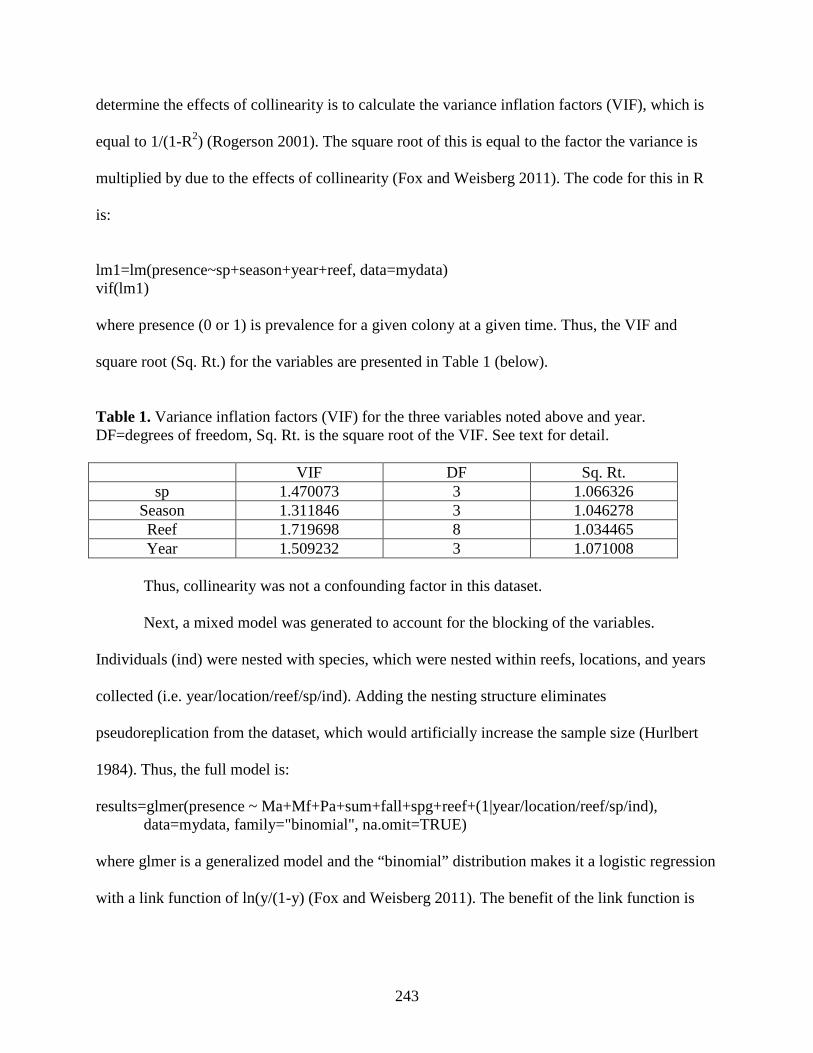
243
determine the effects of collinearity is to calculate the variance inflation factors (VIF), which is
equal to 1/(1-R2) (Rogerson 2001). The square root of this is equal to the factor the variance is
multiplied by due to the effects of collinearity (Fox and Weisberg 2011). The code for this in R
is:
lm1=lm(presence~sp+season+year+reef, data=mydata) vif(lm1) where presence (0 or 1) is prevalence for a given colony at a given time. Thus, the VIF and
square root (Sq. Rt.) for the variables are presented in Table 1 (below).
Table 1. Variance inflation factors (VIF) for the three variables noted above and year. DF=degrees of freedom, Sq. Rt. is the square root of the VIF. See text for detail. VIF DF Sq. Rt.
sp 1.470073 3 1.066326 Season 1.311846 3 1.046278 Reef 1.719698 8 1.034465 Year 1.509232 3 1.071008
Thus, collinearity was not a confounding factor in this dataset. Next, a mixed model was generated to account for the blocking of the variables.
Individuals (ind) were nested with species, which were nested within reefs, locations, and years
collected (i.e. year/location/reef/sp/ind). Adding the nesting structure eliminates
pseudoreplication from the dataset, which would artificially increase the sample size (Hurlbert
1984). Thus, the full model is:
results=glmer(presence ~ Ma+Mf+Pa+sum+fall+spg+reef+(1|year/location/reef/sp/ind), data=mydata, family="binomial", na.omit=TRUE) where glmer is a generalized model and the “binomial” distribution makes it a logistic regression
with a link function of ln(y/(1-y) (Fox and Weisberg 2011). The benefit of the link function is

244
that it restricts the range of y from 0-1, as is appropriate for presence/absence data, and does not
restrict the coefficients of the variables in the model (Rogerson 2001).
To determine if there were significant differences among the different host species,
season, and reef, these variables had to be combined. For example, the full model takes into
account each variable separately (Table 2).
Table 2. Estimates of the coefficients (Coeff) associated with the full model along with the standard errors (S.E.), z scores (Z), p values (P) and whether it was significant (**) or not (NS). Coeff S.E Z value P Sig (Intercept) -0.1297 1.0192 -0.127 0.898712 NS Ma 4.0604 0.839 4.84 1.30E-06 ** Mf 6.4004 0.9702 6.597 4.20E-11 ** Pa 6.4068 1.33 4.817 1.46E-06 ** sum -1.4762 0.4661 -3.167 0.001539 ** fall -2.2207 0.454 -4.891 1.00E-06 ** spg -1.6638 0.4918 -3.383 0.000716 ** reefLG 0.8774 0.8143 1.077 0.281272 NS reefNNP 1.2581 0.8962 1.404 0.160343 NS reefSP 0.6994 1.1146 0.628 0.530321 NS To examine the differences between prevalence in M. annularis vs. M. faveolata, the model was
specified to combine Ma + Mf as a single variable (Table 3).
results=glmer(presence ~ I(Ma+Mf)+Pa+sum+fall+spg+reef+ (1|year/location/reef/sp/ind), data=mydata, family="binomial", na.omit=TRUE) Table 3. Estimates of the coefficients (Coeff) associated with the model combining Ma and Mf along with the standard errors (S.E.), z scores (Z), p values (P) and whether it was significant (**) or not (NS).
Coeff S.E Z value P Sig (Intercept) 0.2933 1.016 0.289 0.77279 NS (Ma+Mf) 4.8944 0.9658 5.068 4.02E-07 *** Pa 6.9078 1.7349 3.982 6.84E-05 *** sum -1.4878 0.4782 -3.112 0.00186 ** fall -2.1377 0.4601 -4.646 3.39E-06 *** spg -1.5443 0.5011 -3.082 0.00206 ** reefLG 1.2754 1.0564 1.207 0.22732 NS reefNNP 1.211 1.1345 1.067 0.28576 NS

245
reefSP 0.728 1.3994 0.52 0.60291 NS At this point the models could be compared using a likelihood ratio test and all differences
between the models could be attributed to the differences between Ma and Mf (Table 4).
Table 4. Likelihood ratio test, which examines the log likelihoods (LogLik) of the full model and a model combining M. annularis and M. faveolata (Ma + Mf). There is one degree of freedom (Df) and the test statistic models a chi-square distribution.
logLik Chisq Df P value (Ma + Mf) -240.99 Full model -237.56 6.8734 1 0.008749 After the Bonferroni correction, α=0.008. Therefore, there was no significant difference between
prevalence in M. faveolata and M. annularis (See Chapter 3 for more detail). This test was done
for all possible combinations of species as well as reef and season.
References
Fox J, Weisberg S (2011) An R Companion to Applied Regression. Sage, Thousand Oaks CA
Hurlbert SH (1984) Pseudoreplication and the design of ecological field experiments. Ecol
Monogr 54:187-211
Rogerson PA (2001) Statistical Methods for Geography. Sage Publications, London
246
Appendix 2
Two different protocols were followed to determine the best way to fix coral larval
samples for transmission electron microscopy (TEM). The first (1) fixative consisted of 2.5%
glutaraldehyde, 0.14 M NaCl2 and 0.2 M Millonig’s (monobasic) phosphate and the second (2)
was 2% glutaraldehyde, 0.14 M NaCl2, 0.05 sodium cacodylate, and 0.2% tannic acid, which is a
protocol developed for preserving marine oocysts (Landers 2011). Larvae were preserved
separately in both fixatives for 24 hours and transferred to Millonig’s phosphate buffer (0.2%
with 0.3 M NaCl2) and (0.05 M Na cacodylate and 0.3 M 0.3 M NaCl2) for 1 and 2, respectively.
Samples were kept at 4° C for 1 week prior to osmication and embedding.
Samples were washed in their respective wash buffers (see above) and post-fixed in OsO4
for 24 hrs (there was no difference between those fixed for 2hrs). The samples from the first
preservative were fixed in 2% OsO4 and 0.2 M Millonig’s phosphate and the second were fixed
in 0.5% OsO4, 0.05 M Na cacodylate, and 1% potassium ferrocyanide followed by 2x washes in
their respective buffers for 15 min. Following this step, both sets of samples were treated
identically. Samples were transferred to 0.2 μm filtered water for 15 min and sequentially
transferred to 30, 50, 70, 80 and 95% ethanol for 10 min at each step. They were then dehydrated
in 2 rinses of absolute (100%) ethanol for 15 min each and rinsed twice with propylene oxide
(PO) for 15 min.
Samples were infiltrated and embedded in Araldite 502/epon (Embed-812) (EMS) resin.
For infiltration, 1:1 (PO : resin) solution was added to the sample and incubated at room
temperature for 1 hr in a rotator. This was followed by another 1 hr incubation in a 1:3 (PO :

247
resin) solution. Finally, samples was incubated at room temperature in 100% resin for 6 hrs,
transferred to molds containing new resin and hardened at 60° C for 24 hrs.
Samples were trimmed, 1.5 μm sections cut on a Reichert Ultracut S ultramicrotome and
dried prior to staining in Toluidine Blue (2% toluidine blue, 2% Sodium borate). Ultrathin (95
nm) sections were stained in uranyl acetate prior to visualization with a Philips 301 transmission
electron microscope.
Overall, the samples fixed in phosphate buffer had the best fixation and figures are shown
within (Chapter 4, Fig. 2). Samples fixed in the cacodylate buffer appeared brittle and sections
contained numerous cracks originating in the ectoderm and spreading through to the endoderm.
Therefore, this fixation protocol is not recommended for coral larvae.
References
Landers SC (2011) Improving TEM fixation with additives. Microscopy Today 19:40-42

248