Embed Size (px)
Citation preview

Available online at www.sciencedirect.com
ScienceDirect
Tanta Dental Journal 11 (2014) 122e129www.elsevier.com/locate/tdj
The effect of adding hyaluronic acid to calcium phosphate onperiapical tissue healing following periradicular surgery in dogs
Wafaa Ahmed Omar Segari a,*, Dalia Abd El Khalek Radwan b,Mohamed Ayad Abd El Hamid c
a Department of Endodontics, Faculty of Oral and Dental Medicine, Cairo University, Cairo, Egyptb Oral and Maxillofacial Surgery, Faculty of Oral and Dental Medicine, Cairo University, Cairo, Egypt
c Veterinary Surgery, Anesthesiology and Radiology, Faculty of Veterinary Medicine, Cairo University, Cairo, Egypt
Received 8 May 2014; revised 14 July 2014; accepted 21 July 2014
Available online 30 September 2014
Abstract
Objective: This study aimed to evaluate the effect of adding exogenous hyaluronic acid (HA) to Beta-tricalcium phosphate (CP) onosseous tissue healing of induced apical lesions following periradicular surgery in a dog model.Methods: In the first and second sessions, periapical lesions were induced by exposing pulp cavities of selected teeth for 7 days thensealing themwith glass ionomer for 60 days. Root canals were then cleaned, shaped and obturated. Surgical treatment included buccalosteotomy to expose the root apex and root-end resection and filling. Osteotomy cavities were randomly allocated to two study groups12 samples each according to the graft materials; CP or combination of HA and CP. The graft materials were prepared and applied ineach animal in alternate quadrant in a randomizedmanner.Animalswere sacrificed after 60 days and bone treated cavitieswerepreparedfor histological study and histomorphometric analysis for the area percentage of new bone tissue and trabecular bone thickness.Results: All samples displayed signs of regeneration as newly formed bone tissue and fibrovascular connective tissue within thetreated cavity sites with complete resorption of the implemented materials. The newly formed bone consisted mainly of osteoidbone trabeculae with some more mature dense bone present at the periphery of cavity site. There was no significant difference in thepercentage of newly formed bone tissue (P > 0.05) and bone trabeculae thickness (P > 0.05) between the two study groups.Conclusions: Addition of exogenous HA to CP after periradicular surgery did not improve the histological outcome of osseoustissue healing in a dog model.© 2014, Production and Hosting by Elsevier B.V. on behalf of the Faculty of Dentistry, Tanta University.
Keywords: Hyaluronic acid; Hyaluronan; Calcium phosphate; Bone grafts; Alloplasts; Periapical lesion induction
Open access under CC BY-NC-ND license.
* Corresponding author. 131 Hadek El Ferdous, 6 October City, Giza City, Egypt. Tel.: þ20 01156471101.
E-mail addresses: [email protected], [email protected], [email protected] (W.A.O. Segari).
Peer review under the responsibility of the Faculty of Dentistry, Tanta University
Production and hosting by Elsevier
http://dx.doi.org/10.1016/j.tdj.2014.07.001
1687-8574/© 2014, Production and Hosting by Elsevier B.V. on behalf of the Faculty of Dentistry, Tanta University.Open access under CC BY-NC-ND license.

123W.A.O. Segari et al. / Tanta Dental Journal 11 (2014) 122e129
1. Introduction
Periapical surgery with root-end resection is indi-cated for endodontically treated teeth with periapicalpathosis where an orthograde revision cannot be doneor not able to resolve the periapical disease [1]. In suchconditions, surgical approach provides better access toclean the root surface and apical lesion, to eliminatethe source of inflammation and reactive periradiculartissue and to prepare and seal the apical portion of theroot canal system [2]. This will allow the regenerationof hard and soft tissues, including the formation of anew attachment apparatus [3]. To promote boneregeneration following periapical surgery, bone graftmaterials are used especially when the surgical site iscompromised [4].
Bone grafts fall into four general categories: au-tografts, allografts, xenografts, and alloplasts [2,5].The role of these materials in regenerative proceduresis based on that; they possess the osteogenic potential(contain bone-forming cells), they are osteoinductive(contain bone inducing substances), or they areosteoconductive (serve as a scaffold for bone forma-tion) [6]. Autograft and allograft remain the mosteffective grafting materials because they have morepotentials for bone regeneration [2,4]. However, al-ternatives such as alloplast have been used because ofdonor site morbidity, limited supply with increasedcost of autographs and risk of disease transmissionand response rejection by allografts [2,4,7]. An allo-plast is a synthetic or inert foreign body material thathas ostoeconductive capacity [2]. Its distinct advan-tage over autograft and allograft is that it can beproduced in unlimited quantities without risk of dis-ease transmission [7,8]. Currently used allopastsinclude; coralline hydroxyapatite, collagen-basedmatrices, calcium sulfate, and tri-calcium phosphate[2,7].
Hyaluronic acid (HA) is also known as hyaluronanor hyaluronate is a high molecular weight poly-saccharide (glycosaminoglycan) and a major compo-nent of extracellular matrix almost in all living tissues[9,10]. It plays a critical part in the function of extra-cellular mineralized and non mineralized matrices,including tissue hydrodynamics and cell migration,proliferation and differentiation [9,11]. HA has animportant anti-inflammatory role through inhibition oftissue destruction and facilitation tissue healing [9,12].Consequently, application of exogenous HA in thetreatment of inflammatory processes is shown indifferent medical fields such as orthopedics, derma-tology and ophthalmology [9,10].
In the dental field, HA demonstrated beneficial ef-fects in the treatment of gingivitis and periodontitis aswell as periodontal surgery [13e15]. It demonstratedbacteriostatic effect on bacterial strains commonlyfound in gingival lesion and periodontal wound [16]. Inaddition, it has been used as a carrier for demineralizedbone allograft without reducing its effectiveness forsinus lift augmentation [17]. Furthermore, it has beenproposed as a scaffold for regeneration therapy ofdental pulp because of its appropriate structure,biocompatibility and biodegradation [18].
Previous studies demonstrated the ability of exog-enous hyaluronic acid in enhancing bone healing bothexperimentally [11,19e22] and clinically [14,15,23].In animal studies, HA recorded significant bonemineralization acceleration compared to untreatedbone cavities (11). The adjunctive use of HA tografting process when combined with autografts andallografts recorded favorable results. Previous studiesrecorded the capacity of HA in supporting the signifi-cant bone formation when combined with bonemarrow stromal cell and basic fibroblast growth factors[21], recombinant human bone morphogenic protein�2 [20] and spongiosal bone graft [22]. Clinically,application of HA in combination with autologus bonedemonstrated good capabilities in accelerating boneformation when used in extractive socket and peri-odontal bony defect [14,15,23].
Experimental evaluation of the efficiency ofdifferent materials and techniques requires conditionssimilar to clinical environments for its relevanceinterpretation. Therefore, inducing one of the situationsrequiring the use of grafting materials when tested inthe animal model such as periapical lesion is required.Periapical lesion induction was done previously toevaluate periradicular tissue response to bone grafts inapical surgery of dogs' teeth [24].
Based on these data, and considering that, no pre-vious study exists on the use of HA combined withalloplast as adjunctive to grafting process, the purposeof this study was to histologically examine the hy-pothesis that adding exogenous hayluronic acid (HA)to Beta-tricalcium phosphate (CP) could enhanceosseous tissue healing of induced apical lesionsfollowing peria-radicular surgery in a dog model.
2. Materials and methods
2.1. Experimental animal and materials
Animal right commity of Faculty of VeterinaryMedicine (Cairo University) approved the
124 W.A.O. Segari et al. / Tanta Dental Journal 11 (2014) 122e129
experimental protocol of this study, which is inaccordance with The Code of Ethics of the WorldMedical Association (Declaration of Helsinki) for an-imal experimentation. The study was conducted onfour dogs of one year old including the following teeth;second, third and fourth mandibular premolars andsecond, third maxillary premolars.
The materials used in the present study were anester of hyaluronic acid1 (HA) with benzyl alcoholwith a concentration 20 mg/2 ml in a gel form andpure-phase beta-tricalcium phosphate2 (CP). CP shouldbe mixed with fresh blood from the defect or withvenous blood at about a 1/3 blood to 2/3 CP ratio [25].In this study, HA was mixed with CP in the same ratioinstead of blood.
2.2. Operative procedures
All experimental procedures were performed undergeneral anesthesia that was achieved by intramuscularinjection of 0.5 ml (20 mg) 2% Xylazine base3 andintravenous injection of ketamine4 at 15 mg/kg. Atsurgical time, Local infiltration of 2% lidocain withnor-epinephrine (1:100,000) was administered forhemostasis and reduction of operative pain. Peri-radicular lesions were induced by opening the pulpchambers of experimental teeth, removing pulp tissueand leaving the access cavities exposed to oral florafor 7 days. Access cavities were then closed withcotton and filled with glass ionomer5 for 60 days.Following the induction procedure, all infected rootcanals were cleaned and shaped in a crown downapproach using Gates Glidden drills6 and K files7 inconjunction with frequent irrigation with 5.25% so-dium hypochlorite.8 Root canals were dried andsealed with calcium hydroxide paste.9 After 7 days,calcium hydroxide was removed and root canal fillingwith gutta percha and sealer10 was performed usingcold lateral technique. Upon completion of the rootcanal treatment, a full thickness mucoperiosteal flapwas reflected exposing the buccal cortical bonecovering the root surfaces. A # 2 rounded low speed
1 Curavisc, Germany.2 Curasan, Inc, U.S.3 Eurovet Animal Health B.V., Bladel, The Netherlands.4 Rompun, Bayer, Wuppertal, Germany.5 Vitremer; 3M/EP AG.6 Brasseler, Savannah, GA, USA.7 Mani, Japan.8 Clorox, Alexandria detergent of chemicals company, Alexandria,
Egypt.9 Pulpdent, Pulpdent Corp., Watertown, MA.
10 Endo Fill, Dentsply dental products.
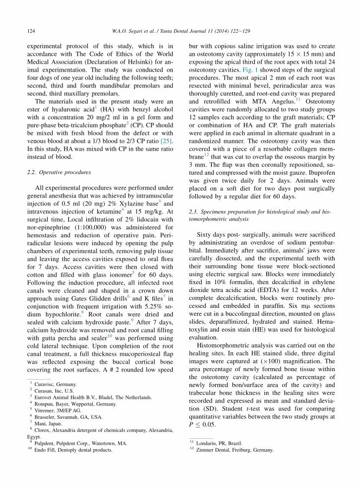
bur with copious saline irrigation was used to createan osteotomy cavity (approximately 15 � 15 mm) andexposing the apical third of the root apex with total 24osteotomy cavities. Fig. 1 showed steps of the surgicalprocedures. The most apical 2 mm of each root wasresected with minimal bevel, periradicular area wasthoroughly curetted, and root-end cavity was preparedand retrofilled with MTA Angelus.11 Osteotomycavities were randomly allocated to two study groups12 samples each according to the graft materials; CPor combination of HA and CP. The graft materialswere applied in each animal in alternate quadrant in arandomized manner. The osteotomy cavity was thencovered with a piece of a resorbable collagen mem-brane12 that was cut to overlap the osseous margin by3 mm. The flap was then coronally repositioned, su-tured and compressed with the moist gauze. Ibuprofenwas given twice daily for 2 days. Animals wereplaced on a soft diet for two days post surgicallyfollowed by a regular diet for 60 days.
2.3. Specimens preparation for histological study and his-tomorphometric analysis
Sixty days post- surgically, animals were sacrificedby administrating an overdose of sodium pentobar-bital. Immediately after sacrifice, animals' jaws werecarefully dissected, and the experimental teeth withtheir surrounding bone tissue were block-sectionedusing electric surgical saw. Blocks were immediatelyfixed in 10% formalin, then decalcified in ethylenedioxide tetra acidic acid (EDTA) for 12 weeks. Aftercomplete decalcification, blocks were routinely pro-cessed and embedded in paraffin. Six mm sectionswere cut in a buccolingual direction, mounted on glassslides, deparaffinized, hydrated and stained. Hema-toxylin and eosin stain (HE) was used for histologicalevaluation.
Histomorphometric analysis was carried out on thehealing sites. In each HE stained slide, three digitalimages were captured at (�100) magnification. Thearea percentage of newly formed bone tissue withinthe osteotomy cavity (calculated as percentage ofnewly formed bon/surface area of the cavity) andtrabecular bone thickness in the healing sites wererecorded and expressed as mean and standard devia-tion (SD). Student t-test was used for comparingquantitative variables between the two study groups atP � 0.05.
11 Londarin, PR, Brazil.12 Zimmer Dental, Freiburg, Germany.

Fig. 1. Photographs showing the steps of surgical procedure: (A) Osteotomy cavity was created with an approximately 15 � 15 mm. (B)
Osteotomy cavity exposed the apical one third of the root apex, (C) was filled with graft material and then (D) was covered with the collagen
membrane. (E) Closure of the surgical site.
125W.A.O. Segari et al. / Tanta Dental Journal 11 (2014) 122e129
3. Results
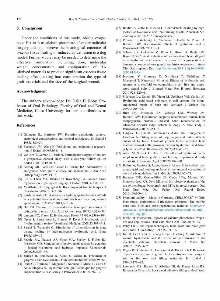
Histological examination of all treated cavitiessites demonstrated newly formed bone tissue withcomplete resorption of implemented materials (Figs. 2and 3). The newly formed bone consisted mainly ofosteoid bone trabeculae with some more maturelamellar bone present at the periphery of cavity (Figs.2 and 3).
Cavities treated with CP showed more regularlyarranged osteoid bone trabeculae separated by marrowspaces (Fig. 2). Bone trabeculae with embeded osteo-cyt cells and lined with osteoblat cells were seen(Fig. 2A and B). Dens bone formation with the
Fig. 2. Histological sections of CP treated group (A, B & C). (A & B) Secti
by marrow spaces (MS)). Note, bone trabeculae lined with osteoblast cells
Section showing coalescence of bone trabeculae together in the osteotomy c
(For interpretation of the references to color in this figure legend, the read
appearance of cortical bone was observed at the pe-riphery of cavity site (Fig. 2C). Bone trabeculae wereseen coalescing together in certain areas and separatedby marrow spaces in other sites (Fig. 2C).
HA and CP treated cavities revealed nearly similarhistological picture to CP treated cavity; osteoid bonetrabeculae dispersed within the bony defect and sepa-rated by marrow spaces (Fig. 3). However, marrowspaces separating the bone trabeculae were wider thanCP treated group and with more vascular fibrous con-nective tissue (Fig. 3A and B). The newly formed bonetrabeculae contain osteocytes and lined with cemen-toblast cells. Coalescence of thick bone trabeculaecontaining variable size marrow spaces was recorded
ons showing regularly arranged osteoid bone trabecula (BT) separated
(blue arrows) and contains osteocyt cells (small black arrows). (C)
avity (CBT) with the dense bone at the periphery (DB). (HE � 100).
er is referred to the web version of this article.)

Fig. 3. Histological sections of HA and CP treated group (A, B & C). (A) Section showing regularly arranged osteoid bone trabecula (BT)
dispersed by wide marrow space (MS) with fibrous connective tissue (F) and blood vessels (BV). Note, bone trabeculae with embedded osteocyt
cells (black arrows) and lined with osteoblast cells (blue arrows). (B & C) Sections showing coalescence thick bone trabeculae separated (CBT) by
marrow space with variable sizes (MS). Note, dense bone (DB) formation at the periphery of bone cavity. (HE � 100). (For interpretation of the
references to color in this figure legend, the reader is referred to the web version of this article.)
126 W.A.O. Segari et al. / Tanta Dental Journal 11 (2014) 122e129
(Fig. 3B and C). At the defect periphery, dense bonewith embedded osteocytes was seen (Fig. 3C).
The Mean and SD of the area percentage for newbone tissue and trabecular bone thickness were recor-ded in Table 1. HA and CP treated cavities demon-strated lower percentage of new bone tissue and lowermean of bone trabeculaue thickness within the formedtissue components compared to CP treated cavities.However, Student t-test didn't show any significancedifference between the two study groups (P > 0.05).
4. Discussion
The mechanisms by which HA promote bone for-mation were attributed to some actions. HA acceler-ated bone regeneration by means of chemotaxis,proliferation and successive differentiation of mesen-chymal cells [11]. It significantly increased alkalinephosphatase and hence stimulate cell mineralization[20]. HA allowed the early deposition of osteoid tissueby providing a scaffold on which osteoprogenitor cellattached and so stimulated osteoblastic differentiation[21]. This action is supported by Aslan et al. [22] whohave noted that HA stimulated bone healing throughaccelerating the three phases of healing; inflammation,proliferation and migration of mesenchymal cells with
Table 1
Bone formation analysis for each study group.
Mean SD P value*
Newly formed
bone tissue
CP group 56.78 11.23 0.8
CP and HA group 55.44 11.13
Bone trabecular
thickness
CP group 564.64 121.05 0.6
CP and HA group 536.33 108.469
CP: Calcium phosphate, HA: Hyaluronic acid
P* value � 0.05 was considered significant.
the production of bone matrix. Additionally, hyal-uronic acid holds the advantage of complete degrada-tion that allows its complete replacement with newlyformed bone tissue [19]. Furthermore, the anti-inflammatory and antibacterial ability of exogenousHA may reduce the postsurgical inflammation andbacterial contamination when added to the surgical siteand finally improve the surgical outcomes [12e14].
Calcium phosphate has been selected for this studyas a one of the most commonly used alloplasts andbecause of its biocompatibility, handling characteris-tics, porosity, chemical and physical resemblance tobone minerals and potentially unlimited supply at alow price [26,27]. Additionally, previous investigationfound that, addition different concentrations of HAsolution to CP had a little effect on its porous structureand enhanced its bioactivity demonstrated by theapatite crystal formation on its surface [28].
The dog model has been used previously to evaluatethe efficiency of bone graft materials [20,29]. Yet,variations in root canal morphology, bone anatomy andhealing process could present difficulties that may bearupon the final results if not properly explored prior toany experiment [29,30]. Other difficulties thatencountered during this study and recorded previouslywere shallow mucobuccal fold, extensive muscleattachment and thick cortical bone of posterior area[29]. These difficulties represented obstacles to themucoperiosteal flap reflection process and accessibilityto root apex as well as root end preparation. Further-more, excessive bleeding during surgical proceduresaffected root end visibility that added another difficultyduring per-radicular surgery.
Several animal studies have investigated the ca-pacity of HA to augment bone healing based oncreating a small bony defect in sterile environments

127W.A.O. Segari et al. / Tanta Dental Journal 11 (2014) 122e129
[11,20,22]. Such cavities may underestimate the clin-ical conditions that may affect the surgical result.Studies recorded that, presence and size of peri-radicular radiolucency adversely affected the outcomeof periradicular surgery [31,32]. Therefore, inductionof periapical lesion can sidestep this restriction.Experimentally, periapical lesion was induced previ-ously in a dog model by allowing root canal microbialcontamination from the oral cavity [24,33e37].Following contamination, a microbial shift towardspredominance of obligate gram negative anaerobes,which are essential with endotoxins for the establish-ment of periapical inflammation will occur. This wasdone by opening the access cavity of dog teeth andleaving the root canal open to oral flora contaminationfor different time periods [35]. For accelerating theprocess of induction, some authors closed the accesscavity following canal contamination demonstratingthat close environment modifies the oxygen level andnutrient supply and hence favor development of se-lective anaerobic gram negative bacteria [33,35,37].Previous studies tested this methodology andconfirmed through histological and radiographic ex-amination its ability to produce periapical lesions inpredictable away [27,33,35,37]. In the present study,periapical lesion was induced by leaving the accesscavity open for seven days, then sealing it for 60 days[27,33,35]. Following periapical lesion induction, nonsurgical, surgical and histological procedures in thepresent study were followed Regan et al. [29].
In the present study, histological examination oftreated bone cavities of both study groups demon-strated new bone trabeculae formation separated bymarrow spaces filled with vascular fibrous tissue. Thenewly formed bone trabeculae contains osteocytesand lined with cementoblast cells denoting enhancedbone apposition. Additionally, the presence of bonetrabeculae coalesence and dense bone formation at thetreated cavity is indicative of tissue maturationprocess.
Histomorphometric measurements carried out onthe healing sites recorded that, there was no influenceof adding HA to CP as adjunctive to osseous tissuehealing. This finding isn't in line with other previousreports that recorded significant benefits of the com-bined use of HA with other graft materials in sup-porting bone induction [20e22].
The absence of the observed difference, contrary toother published studies may be explained by the vari-ations in the experimental models and methodology.Previous studies investigated the osteoinduction of HAin different animal models through creating a small
bony cavity and in conjunction with different bonesubstitutes. The capacity of bone healing and rat ofremodeling among different experimental animal spe-cies are variable [38]. Rodents animals used in previ-ous investigations such as rat [11,21] and rabbit [22]have faster bone healing capacity [39] that may causemore rapid bone growth and remodeling. Furthermore,different criteria for histological evaluations of bonerepair have been utilized. In the present study, thepercentage of new bone formation [21] and trabecularebone thickness was used. In the other study, scoringsystem was used for histological evaluation [22].Sensitivities of these different criteria may be variedand might affect the histological outcome.
Another explanation for our finding could be thevariations in the formulation, dose and configuration ofused HA. It was suggested that HA has a molecularweight-specific and dose-specific mode of action thatmay enhance the osteogenic and osteoinductive prop-erties of bone graft materials [19]. It significantlyincreased alkaline phosphatase activity, and hencestimulate cell mineralization in a dose-dependentmanner [40]. HA configuration is another factor thatcan affect its action through allowing more area andvolume for more cell attachment, thereby more newlyformed tissue [19]. A variety of commercially availablepreparations of HA derivatives and cross-linked HAmaterials have been developed in forms such as films,microspheres, liposomes, fibers and hydrogels [9]. Inthe present study, high molecular weight HA in a gelform was used; in the other studies different configu-rations have been used such as Autocross linked HAsponge [20] and a biodegradable polymer configuredas a non woven mesh [21]. High molecular weight HAin a gel form was used because it recorded promisingresult of bone fracture healing when combined withfibroblast growth factor-2 [41]. In addition, the gelform allowed easily mixing of HA with CP and facil-itated handling the mix into bony cavity.
Regarding the dose, earlier authors presented nodetails regarding the used dose of HA and specificallythe ratio of HA to graft materials [20e22]. So, the doseof HA applied to CP in the present study might be notthe optimum level required to achieve significant re-sults especially in a big cavity size. This might explainthe presence of more fibrous tissue and large marrowspaces in the CP and HA treated cavities.
Based on the previous, there is a demand for moreknowledge concerning the effective formulation,configuration and dose of HA when used in conjunc-tion with graft materials to augment the bone healingprocess.
128 W.A.O. Segari et al. / Tanta Dental Journal 11 (2014) 122e129
5. Conclusions
Under the conditions of this study, adding exoge-nous HA to b-tricalcium phosphate after periradicularsurgery did not improve the histological outcome ofosseous tissue healing of induced apical lesion in a dogmodel. Further studies may be needed to determine theeffective formulation including; dose, molecularweight, concentration and configuration of HA-derived materials to produce significant osseous tissuehealing effect, taking into consideration the type ofgraft materials and the size of the surgical wound.
Acknowledgment
The authors acknowledge Dr. Dalia El Roby, Pro-fessor of Oral Pathology, Faculty of Oral and DentalMedicine, Cairo University, for her contributions tothis work.
References
[1] Gutmann JL, Harrison JW. Posterior endodontic surgery:
anatomical considerations and clinical techniques. Int Endod J
1985;18:8e34.
[2] Bashutski JM, Wang H. Periodontal and endodontic regenera-
tion. J Endod 2009;35:321e8.
[3] von Arx T, Gerber C, Hardt N. Periradicular surgery of molars:
a prospective clinical study with a one-year follow-up. Int
Endod J 2001;34:520e5.
[4] Gazdag AR, Lane JM, Glaser D, Forster RA. Alternatives to
autogenous bone graft: efficacy and indications. J Am Acad
Orthop Surg 1995;3:1e8.
[5] Lin L, Chen MY, Ricucci D, Rosenberg PA. Guided tissue
regeneration in periapical surgery. J Endod 2010;36:618e25.[6] McAllister BS, Haghighat K. Bone augmentation techniques. J
Periodontol 2007;78:377e96.
[7] Krishnamurithy G. A review on hydroxyapatite-based scaffolds
as a potential bone graft substitute for bone tissue engineering
applications. JUMMEC 2013;16:1e6.
[8] Hak DJ. The use of osteoconductive bone graft substitutes in
orthopedic trauma. J Am Acad Orthop Surg 2007;15:525e36.
[9] Laurent TC, Fraser R. Hyaluronan. Faseb J 1992;6:2398e404.[10] Necas J, Bartosikova L, Brauner P, Kolar J. Hyaluronic acid
(hyaluronan): a review. Veterinarni Medicina 2008;53:397e411.
[11] Sasaki T, Watanabe C. Stimulation of osteoinduction in bone
wound healing by high-molecular hyaluronic acid. Bone
1995;16:9e15.
[12] Peattle RA, Nayate AP, Firpo MA, Shelby J, Fisher RJ,
Prestwich GD. Stimulation of in vivo angiogenesis by cytokine
e loaded hyaluronic acid hydrogel implants. Biomaterials
2004;25:2789e98.
[13] Jentsch H, Pomowski R, Kundt G, G€ocke R. Treatment of
gingivitis with hyaluronan. J Clin Periodontol 2003;30:159e64.[14] Prato GP, Rotundo R, Magnani C, Soranzo C, Muzzi L, Cairo F.
An autologous cell hyaluronic acid graft technique for gingival
augmentation: a case series. J Periodontol 2003;74:262e7.
[15] Baldini A, Zaffe D, Nicolin G. Bone-defects healing by high-
molecular hyaluronic acid: preliminary results. Annali di Sto-
matologia 2010;I:2e7. mucoperiosteal.
[16] Pirnazar P, Wolinsky L, Nachnani S, Haake S, Pilloni A,
Bernard GW. Bacteriostatic effects of hyaluronic acid. J
Periodontol 1999;70:370e4.
[17] Schwartz Z, Goldstein M, Raviv E, Hirsch A, Ranly DM,
Boyan BD. Clinical evaluation of demineralized bone allograft
in a hyaluronic acid carrier for sinus lift augmentation in
humans: a computed tomography and histomorphometric study.
Clin Oral Implants Res. http://dx.doi.org/10.1111/j.1600-0501.
2006.01303.x.
[18] Inuyama Y, Kitamura C, Nishihara T, Nishihara T,
Morotomi T, Nagayoshi M, et al. Effects of hyaluronic acid
sponge as a scaffold on odontoblastic cell line and ampu-
tated dental pulp. J Biomed Mater Res B Appl Biomater
2010;92B:120e8.
[19] Solchaga LA, Dennis JE, Victor M, Goldberg VM, Caplan AI.
Hyaluronic acid-based polymers as cell carriers for tissue-
engineered repair of bone and cartilage. J Orthop Res
1999;3:205e13.
[20] Hunt DR, Jovanovic SA, Wikesjo UM, Wozney JM,
Bernard GW. Hyaluronan supports recombinant human bone
morphgenetic protein-2 induced bone reconstruction of
advanced alveolar ridge defects in dogs. A pilot study. J
Periodontol 2001;72:651e8.
[21] Lisignoli G, Fini M, Giavaresi G, Aldini NN, Toneguzzi S,
Facchini A. Osteogenesis of large segmental radius defects
enhanced by basic fibroblast growth factor activated bone
marrow stromal cells grown on-woven hyaluronic acid-based
polymer scaffold. Biomaterials 2002;23:1043e51.
[22] Aslan M, Simsek G, Day E. The effect of hyaluronic acid-
supplemented bone graft in bon healing: experimental study
in rabbits. J Biomater Appl 2006;20:209e20.
[23] Ballini A, Cantore S, Capodiferro S, Grassi F. Esterified hyal-
uronic acid and autologous bone in the surgical correction of
the infra-bone defects. Int J Med Sci 2009;6:65e71.
[24] Bernab�e PFE, Gomes-Filho JE, Cintra LTA, Moretto MJ,
Simonetti Lodi CS, Nery MJ, et al. Histologic evaluation of the
use of membrane, bone graft, and MTA in apical surgery. Oral
Surg Oral Med Oral Pathol Oral Radiol Endod
2010;109:309e14.
[25] Premium quality e Made in Germany. CERASORB® M Orth.
Pure-phase, multiporous ß-tricalcium phospate. The perfect
bone void filler and bone regeneration material. http://www.
curasan.de/_downloads/fachkreise/cerasorb/cerasorb_m_ortho_
brochure_engl.pdf.
[26] Jarcho M. Biomaterial aspects of calcium phosphates. Proper-
ties and applications. Dent Clin North Am 1986;30:25e47.
[27] Perry CR. Bone repair techniques, bone graft, and bone graft
substitutes. Clin Orthop 1999;252:71e86.
[28] Kai D, Li D, Zhu X, Zhang I, Fan H, Zhang X. Addition of
sodium hyaluronate and the effect on performance of the
injectable calcium phosphate cement. J Mater Sci
2009;20:1595e602.
[29] Regan JD, Gutmann JL, Lacopino AM, Diekwisch T. Response
of periradicular tissue to growth factors introduced into surgical
site in the root end filling materials. Int Endod J
1999;3:171e82.[30] Leonardo MR, Barnett F, Debelian GJ, de Pontes Lima RK,
Bezerra da Silva LA. Root canal adhesive filling in dogs' teeth

129W.A.O. Segari et al. / Tanta Dental Journal 11 (2014) 122e129
with or without coronal restoration: a histopathological evalu-
ation. J Endod 2007;33:1299e303.
[31] Rahbran S, Gilthorpe MS, Harison SD, Gulabivala K. Com-
parison of the clinical outcome of periradicular surgery in
endodontic and oral surgery units of teaching dental hospital: a
retrospective study. Oral Surg Oral Med Oral Pathol Oral
Radiol Endod 2001;91:700e9.[32] Molven O, Halse A, Grung B. Surgical management of end-
odontic failure; indications and treatment results. Int Dent J
1991;41:33e42.
[33] Leonardo MR, Almeida WA, Ito IY, Silva LAB. Radiographic
and microbiologic evaluation of post-treatment apical and
periapical repair of root canals of dogs’ teeth with experimen-
tally induced chronic lesion. Oral Surg Oral Med Oral Pathol
Oral Radiol Endod 1994;78:232e8.
[34] Grecca FS, Leonardo MR, Silva LAB, Tanomaru Filho M,
Borges MA. Radiographic evaluation of periradicular repair
after endodontic treatment of dog’s teeth with induced peri-
radicular periodontitis. J Endod 2001;27:610e2.
[35] Tanomaru-Filho M, Poliseli-Neto A, Leonardo MR, Silva LA,
Tanomaru JM, Ito IY. Methods of experimental induction of
periapical inflammation. Microbiological and radiographic
evaluation. Int Endod J 2005;38:477e82.
[36] Leonardo MR, Hernandez MEFT, Silva LAB, Tanomaru-
Filho M. Effect of a calcium hydroxide-based root canal dres-
sing on periapical repair in dogs: a histological study. Oral Surg
Oral Med Oral Pathol Oral Radiol Endod 2006;102:680e5.
[37] De Rossi A, De Rossi M, Rocha LB, da Silva LAB, Rossi MA.
Morphometric analysis of experimentally induced periapical
lesions: radiographic vs histopathological findings. Dentomax-
illofac Radiol 2007;36:211e7.
[38] Petite H, Quarto R. Experimental animal models for tissue
engineered bone regeneration. In: Engineered bon. Georgetown,
Texas. U.S: Lanes Bioscience: Tissue engineering intelligence
unit; 2005. p. 90.
[39] Schmitz JP, Hollinger JO. The critical size defect as an exper-
imental model for craniomandibulofacial nonunion. Clin
Orthop Relat Res 1985;205:299e308.
[40] Huang L, Cheng YY, Koo PL, Kee KM, Qin L, Cheng JC, et al.
The effect of hyaluronan on osteoblast proliferation and dif-
ferentiation in rat calvarial-derived cell cultures. J Biomed
Mater Res 2003;66A:880e4.
[41] Radomsky ML, Aufdemorte TB, Swain LD, Fox WC, Spiro
RC, Poser JW. Novel formulation of fibroblast growth-2 in a
hyaluronan gel accelerates fracture healing in non human pri-
mates. J Orthop Res;17:607e14.