Embed Size (px)
Citation preview

Aus der Abteilung für Klinische Pharmakologie
Leiter: Prof. Dr. med. Stefan Endres
Arbeitsgruppe Tumorimmunologie
Leitung: Prof. Dr. med. Dr. rer. nat. Carole Bourquin
Ludwig-Maximilians-Universität München
The effect of innate immune activation on maturation of
myeloid-derived suppressor cells
Dissertation
zum Erwerb des Doktorgrades der Medizin
an der Medizinischen Fakultät der
Ludwig-Maximilians-Universität zu München
vorgelegt von
Daniel Nörenberg
aus Datteln
2016

II
Mit Genehmigung der Medizinischen Fakultät
der Universität München
Berichterstatterin: Prof. Dr. med. Dr. rer. nat. Carole Bourquin
Mitberichterstatter: Prof. Dr. rer. nat. Thomas Brocker
Prof. Dr. med. Dr. sci. nat. Christoph Klein
Prof. Dr. rer. nat. Helga Schmetzer
Mitbetreuung durch die
promovierten Mitarbeiter: Prof. Dr. med. Stefan Endres
Dr. rer. nat. Christine Zoglmeier
Dekan: Prof. Dr. med. dent. Reinhard Hickel
Tag der mündlichen Prüfung: 28.07.2016

III
Dedicated to my parents Marion and Markus

IV

V
INDEX
1 INTRODUCTION ........................................................................................................... 1
1.1 The human immune system ................................................................................... 1
1.2 Pattern recognition receptor families ................................................................... 2
1.2.1 Toll-like receptors and their ligands ................................................................ 2
1.2.2 RIG-I-like receptors .......................................................................................... 4
1.2.3 Signaling of pattern recognition receptors....................................................... 6
1.2.3.1 Signal transduction via Toll-like receptors ................................................... 7
1.2.3.2 Signal transduction via RIG-I-like receptors ................................................. 9
1.3 The human immune system and cancer ................................................................ 9
1.3.1 Cancer Immunology: Immunosurveillance and Immunoediting ....................... 9
1.3.2 Tumor-escape mechanisms ............................................................................10
1.3.2.1 Tumor cell-intrinsic traits ............................................................................10
1.3.2.2 Tumor cell-extrinsic traits ...........................................................................12
1.4 Myeloid-derived suppressor cells: Innate regulators of the immune system .....14
1.4.1 Origin of MDSC ...............................................................................................15
1.4.2 Phenotype and MDSC heterogeneity ..............................................................15
1.4.3 MDSC expansion and activation in cancer ......................................................17
1.4.4 Mechanisms of immunosuppression exerted by MDSC ..................................19
1.5 Immunotherapy of cancer .....................................................................................21
1.5.1 Overview .........................................................................................................21
1.5.2 Pattern recognition receptor ligands: a double-edged sword in cancer ..........23
1.5.2.1 PRR signaling and tumor progression .........................................................23
1.5.2.2 Use of PRR ligands in the immunotherapy of cancer ..................................24
1.6 Objectives ..............................................................................................................26
2 MATERIALS AND METHODS .....................................................................................28
2.1 Materials ................................................................................................................28
2.1.1 Technical equipment .......................................................................................28
2.1.2 Chemicals, reagents and buffers .....................................................................28
2.1.3 Cell culture materials, reagents and media .....................................................29
2.1.4 PRR ligands, cytokines and growth factors .....................................................30
2.1.5 Tumor cell lines ..............................................................................................32
2.1.6 Kits ..................................................................................................................32
2.1.7 Antibodies: FACS and Immunofluorescence ...................................................33
2.1.8 Software ..........................................................................................................34

VI
2.2 Animal experiments ..............................................................................................34
2.2.1 Animals ...........................................................................................................34
2.2.2 Tumor induction and measurement ................................................................35
2.2.3 Therapy with PRR ligands ...............................................................................36
2.2.4 Organ and single cell preparation ...................................................................37
2.2.4.1 Blood collection and serum analysis ...........................................................37
2.2.4.2 Tumor removal and digestion ......................................................................37
2.2.4.3 Isolation of splenocytes ...............................................................................38
2.2.4.4 Preparation of lungs and lymph nodes ........................................................38
2.2.4.5 Bone marrow isolation ................................................................................38
2.3 Cell culture experiments.......................................................................................39
2.3.1 General culture conditions and cell viability ...................................................39
2.3.2 Tumor cell cultures .........................................................................................39
2.3.3 Generation of bone marrow-derived macrophages .........................................40
2.3.4 In vitro stimulation with PRR-ligands ..............................................................40
2.3.5 MDSC isolation via magnetic-activated cell sorting ........................................41
2.3.5.1 Principle of magnetic-activated cell sorting.................................................41
2.3.5.2 Miltenyi protocol for isolation of MDSC subpopulations .............................42
2.3.5.3 Stem cell protocol for isolation of Gr1+CD11b+ MDSC.................................43
2.4 Immunological methods .......................................................................................44
2.4.1 Fluorescent-activated cell sorting (FACS) .......................................................44
2.4.1.1 Principle ......................................................................................................44
2.4.1.2 Analysis of cell surface antigens ..................................................................45
2.4.1.3 Flow cytometry and gating of MDSC ...........................................................45
2.4.1.4 Detection of apoptosis .................................................................................46
2.4.2 Enzyme-linked immunosorbent assay (ELISA) ................................................47
2.4.3 Immunofluorescence of tumor slices ..............................................................48
2.4.4 BrdU suppression assay ..................................................................................48
2.5 Generation of ppp-RNA .........................................................................................49
2.6 Statistical Analysis ................................................................................................50
3 RESULTS ......................................................................................................................51
3.1 Effect of systemic CpG-DNA administration on Gr1+CD11b+ immature myeloid cells (iMC) in tumor-free mice ................................................................51
3.1.1 iMC numbers in different organs following systemic TLR stimulation ............51
3.1.2 Effect of CpG treatment on iMC-mediated suppressivity.................................52
3.1.3 Shifting of myeloid cell subset composition upon CpG-DNA therapy .............53

VII
3.1.4 Alteration of iMC phenotype through systemic TLR9 activation .....................54
3.2 Myeloid-derived suppressor cells (MDSC) in tumor-bearing mice .....................55
3.2.1 Comparison of MDSC in different mouse tumor models .................................55
3.2.2 Surface marker analysis of MDSC in tumor-bearing mice versus iMC in naïve mice .......................................................................................................57
3.2.3 Different immunosuppressive activity of MDSC subpopulations .....................58
3.3 Effect of immunotherapy with pattern recognition receptor ligands on MDSC in tumor-bearing mice ...............................................................................59
3.3.1 MDSC numbers upon TLR ligand therapy in tumor-bearing mice ..................59
3.3.2 MDSC subset composition and phenotype following systemic PRR ligation ...61
3.3.3 Impact of poly(I:C) therapy on intratumoral MDSC .........................................65
3.3.4 Phenotypical changes on MDSC upon systemic TLR stimulation are IFNα-dependent .......................................................................................................66
3.3.5 Role of ppp-RNA and TGFβ-silencing on MDSC in tumor-bearing mice .........67
3.4 Characterization of stem cell antigen-1 (Sca-1) on MDSC ..................................70
3.4.1 Expression of Sca-1 in different tissues and MDSC subpopulations ...............71
3.4.2 Role of Sca-1 in tumor development and MDSC-mediated immunosuppression ........................................................................................72
3.4.3 Induction of Sca-1 on MDSC via TLR ligands in vivo and in vitro ...................73
3.4.4 Analysis of cytokine induction in Sca-1-/- mice upon PRR ligation ..................76
4 DISCUSSION ................................................................................................................79
4.1 MDSC subpopulations ...........................................................................................79
4.1.1 Differentiated view on MDSC subpopulations in tumor-bearing hosts ............79
4.1.2 Shift in subset composition upon TLR activation ............................................81
4.2 Myeloid-derived suppressor cells and pattern recognition receptor ligands .....82
4.2.1 Expansion and activation of MDSC by Toll-like receptor ligands ....................82
4.2.2 Pattern recognition receptor expression on MDSC .........................................83
4.3 MDSC in the context of cancer immunotherapy with PRR ligands ....................84
4.3.1 MDSC as a target of cancer immunotherapy with CpG ...................................85
4.3.2 MDSC as a target of cancer immunotherapy with poly(I:C) ............................87
4.3.3 MDSC as a target of cancer immunotherapy with 5’-triphosphate-RNA .........87
4.4 Maturation of myeloid-derived suppressor cells .................................................88
4.4.1 Interferon-α as key effector molecule in differentiation and maturation of MDSC upon stimulation of pattern recoginition receptor ligands ...................91
4.4.2 MDSC maturation is not due only to IFN-α .....................................................93
4.5 Stem cell antigen-1 (Sca-1) as a possible marker for MDSC maturation ............94
4.5.1 General aspects of Sca-1 .................................................................................94

VIII
4.5.2 Sca-1 induction via innate immune receptor activation ..................................95
4.5.3 Sca-1 as a possible differentiation marker on myeloid-derived suppressor cells .................................................................................................................95
4.6 MDSC as therapeutic targets in cancer................................................................97
4.7 Summary ............................................................................................................. 101
4.8 Zusammenfassung .............................................................................................. 102
5 REFERENCE LIST ...................................................................................................... 103
6 APPENDIX ................................................................................................................. 133
6.1 Abbreviations ...................................................................................................... 133
6.2 Publications ......................................................................................................... 139
6.2.1 Original publications ..................................................................................... 139
6.2.2 Book chapter ................................................................................................. 139
6.2.3 Oral presentations ......................................................................................... 139
6.2.4 Poster presentations ..................................................................................... 140
6.3 Curriculum vitae .................................................................................................. 141
6.4 Acknowledgements ............................................................................................. 142

IX


1 Introduction
1 INTRODUCTION
1.1 The human immune system
Our body’s integrity is constantly jeopardized by extrinsic and intrinsic threats such as
microbiota and incipient cancer cells. As a result of evolutionary pressure, the human
immune system has evolved into a complex network composed of different cell types,
humoral factors and signaling pathways involved in the prevention and clearance of infective
pathogens and in control of constant malignant transformation. A hallmark feature is the
capability of discriminating “self” from “non-self” or “altered-self”: foreign pathogens or
cancer cells have to be recognized and eliminated while remaining tolerant to the body’s own
tissue and commensal microflora. Shifting this delicate balance in one direction or the other
can lead to severe consequential damage such as neoplastic transformation, sepsis or
autoimmune disease.
In all jawed vertebrates, the immune system is classically comprised of two branches: innate
and adaptive immunity. The innate immune system, as a phylogenetically ancious and
evolutionary conserved system, serves as a first-line host defense of every multicellular
organism. In mammals, its cellular basis is largely reflected by phagocytes such as
neutrophils, monocytes/macrophages and dendritic cells that sense microbial infection or
engulf pathogens and induce a subsequent immune answer. Furthermore, innate immunity
is indispensable as driver for acquired immunity. In contrast, the adaptive (or acquired)
immune system shows delayed responses to pathogens but allows for immunological memory
and tailored response. Whereas innate immunity is not a single entity, the adaptive immune
system is mainly reflected by highly specialized T and B cells. This specificity is ensured by
antigen-specific receptors expressed on the cell surface as the result of somatic
hypermutation and individual recombination of receptor-encoding genes during lymphocyte
maturation. As a result, this assembly process generates an enormous diversity of receptors
each specific to a single antigen and expressed on the surface of its corresponding
lymphocyte. Only receptors that bind their matching antigen induce clonal expansion of the
corresponding lymphocyte.
In contrast to adaptive immunity, the molecular detection mechanisms of the innate immune
system are mediated via germline-encoded and evolutionally highly conserved pattern
recognition-receptors (PRR) [Zhang et al., 2010]. Thus, the specificity of any given PRR is
genetically determined. Whereas the hereditability of such receptors is a decisive advantage,
as no prior antigen exposure is necessary, the disadvantage is their limited diversity in
comparison to the manifold repertoire of rearranged receptors used by the acquired system.

Introduction 2
1.2 Pattern recognition receptor families
Pattern recognition receptors (PRRs) are responsible for sensing the presence of
microorganisms by recognizing invariant microbial structures termed pathogen-associated
molecular patterns (PAMPs). Even before they have been discovered, Charles Janeway
postulated the presence of such receptors [Janeway, 1992]. PRRs have common
characteristics: First, they are germ-line encoded, non-clonal and constitutively expressed on
all cells of a given type. Second, they sense specific molecular patterns that share a common
motif. In microorganisms, those structures are essential for survival thus highly conserved
and difficult to alter during evolution. Third, receptor-binding elicits a rapid reaction and
upregulation of genes involved in inflammatory responses. In 1996, Matzinger already
hypothesized that the activation of PRRs is not limited to exogenous PAMPs but that they also
sense endogenous molecules released after cell stress and tissue damage, termed damage-
associated molecular patterns (DAMPs) (reviewed [Matzinger, 2002]). A concept that is
nowadays well proven and clinically relevant in the pathogenesis of malignant, cardiovascular
and autoimmune disease [Rakoff-Nahoum and Medzhitov, 2009; Frantz et al., 2007; Takeuchi
and Akira, 2010].
Until now, five different classes of PRR families have been identified. These families include
transmembrane proteins such as Toll-like receptors (TLRs) [Kawai and Akira, 2010], C-type-
lectin receptors (CLRs) [Geijtenbeek and Gringhuis, 2009], scavenger receptors [Peiser et al.,
2002], cytosolic helicases such as (RIG)-I-like receptors (RLRs) [Yoneyama and Fujita, 2007]
and NOD-like receptors (NLRs) [Chen et al., 2009]. With regard to the enormous spectrum of
abovementioned PRRs, the following sections are limited to the detailed description of TLRs
and RLRs.
1.2.1 Toll-like receptors and their ligands
The best characterized innate immune receptors are Toll-like receptors. TLRs are responsible
for sensing pathogens on the cell surface and in intracellular endosomes and lysosomes
[Akira et al., 2006]. They are evolutionally conserved from the worm Caenorhabditis elegans
to mammals [Beutler and Rehli, 2002; Hoffmann 2003}. Toll, the eponymous member of the
TLR family was initially described in Drosophila melanogaster as a gene product regulating
dorsoventral polarity in Drosophila [Nusslein-Volhard and Wieschaus, 1980; Hashimoto et
al., 1988]. Later, Hoffmann and colleagues showed that Toll is also essential for antifungal
response in flies [Lemaitre et al., 1996]. The first human homologue of the Drosophila toll
protein - Toll-like receptor 4 - was shown to induce the expression of proinflammatory
cytokines and co-stimulatory molecules [Medzhitov et al., 1997].

3 Introduction
To date, fourteen TLRs have been identified among different species [Kawai and Akira, 2010].
Ten TLRs have been found in human and twelve in mice. TLR1-9 are conserved among mice
and human, although TLR8 is non-functional in mice and human TLR10 lacks subsequent
signaling due to gene disruption by insertion of an endogenous retrovirus. TLR10-13 are not
expressed in humans. Recently TLR14 has been described in fish [Palti, 2011].
The cellular localization seems to play important roles in downstream signaling and
maintaining tolerance to self-molecules such as nucleic acids. Some TLRs (TLR3, 7, 8 and 9)
are exclusively located in intracellular compartments such as the endoplasmatic reticulum,
endosomes and lysosomes where they sense nucleic acids. Another group of TLRs (TLR1, 2,
4, 5, 6, 11) is expressed on cell surfaces and mainly recognizes microbial membrane
components such as lipoproteins and carbohydrates. While TLR1, 2 and 6 recognize ligands
by forming heterodimers, the remaining receptors seem to function as homodimers.
Importantly, some TLRs have been shown to monitor the host’s internal environment to detect
endogenous abnormal self-antigens. For instance, numerous endogenous TLR ligands have
been identified so far such as heat-shock proteins, high-mobility group box-1 protein
(HMGB1), extracellular matrix components as well as endogenous nucleic acids [Asea et al.,
2002; Park et al., 2004; Midwood et al., 2009; Kariko et al., 2004]. Endogenous TLR agonists
are now thought to play an important role in regulating inflammation and seem to be involved
in the pathogenesis of certain non-infectious disease such as autoimmune disorders, cancer
and atherosclerosis [Piccinini and Midwood, 2010]. Table 1 gives an overview of different
TLRs with their respective exogenous and endogenous ligands.
TLR PAMP/DAMP Origin Reference
TLR1+2 Triacetylated lipopeptide
β-defensin-3
(Myco-)bacteria
Self
[Takeuchi et al., 1999]
[Funderburg et al., 2007]
TLR2+6
Diacetylated lipopeptide
Zymosan
HMGB-1
Heat-shock proteins
Mycoplasma
Fungus
Self
Self
[Takeuchi et al., 2001]
[Ozinsky et al., 2000]
[Park et al., 2004]
[Asea et al., 2002]
TLR3 dsRNA
mRNA
Virus
Self
[Alexopoulou et al., 2001]
[Kariko et al., 2004]
TLR4
Lipopolysaccharide
Envelope proteins
Mannan
Taxol
Heat-shock proteins
HMGB-1
Fibrinogen
Bacteria
Virus
Fungus
Plants
Self
Self
Self
[Poltorak et al., 1998]
[Kurt-Jones et al., 2000]
[Tada et al., 2002]
[Byrd-Leifer et al., 2001]
[Asea et al., 2002]
[Park et al., 2004]
[Smiley et al., 2001]
TLR5 Flagellin Bacteria [Hayashi et al., 2001]

Introduction 4
Table 1.1. Overview of Toll-like receptors and their main endogenous and exogenous ligands
(modified from Ishii et al., 2006; Piccinini and Midwood, 2010; Takeuchi and Akira, 2010).
TLRs are differentially expressed depending on tissue and cell type. Moreover, most tissues
express at least one TLR with predominance in cells associated with immune function. As an
example, splenic cells and peripheral blood leukocytes express almost all TLRs as well as
immune-associated tissue such as epithelial cells and fibroblasts [Zarember and Godowski,
2002]. Focusing on expression patterns in immune cells, myeloid cells constitutively express
TLR1 and 6, whereas macrophages preferentially express TLR2, 3, 4 and 8. Cells of acquired
immunity have been shown to express different TLRs, namely TLR2, 3, 5 and 9 on T cells
[Kabelitz, 2007] and TLR1, 7, 9 and 10 on B cells [Bourke et al., 2003; Dasari et al., 2005].
Notably, the expression pattern of TLRs in dendritic cells significantly differs between mice
and human: human myeloid DC express all TLRs with the exception of TLR9 while
plasmacytoid DC express TLR1, 6, 7 and 9. In contrast, murine plasmacytoid DC express
almost all TLR but TLR3 and 4. In addition, the expression level of TLR7 and 9 in murine DC
is generally higher than in human DC [Muzio et al., 2000; Hornung et al., 2002]. These
differences in expression patterns might explain why several studies using TLR7 and TLR9
agonists as adjuvants in cancer immunotherapy did not produce the expected results
generated from preclinical studies performed in mice.
1.2.2 RIG-I-like receptors
Retinoic acid-inducible gene I (RIG-I)-Iike receptors (RLRs) are a family of cytoplasmic
DExD/H box RNA helicases that play a major role in sensing of RNA viruses to initiate and
modulate antiviral immunity. The downstream signaling cumulates in the induction of a type
I interferon response and antiviral gene expression. To date, three RIG-I-like helicases have
TLR7
ssRNA
Imiquimod/Resiquimod
Antiphospholipid antibody
RNA virus/self
Synthetic
Self
[Vollmer et al., 2005]
[Hemmi et al., 2002]
[Hurst et al., 2009]
hTLR8 ssRNA
Antiphospholipid antibody
RNA virus/self
Self
[Heil et al., 2004]
[Doring et al., 2010]
TLR9
CpG DNA
DNA
Malaria hemozoin
IgG-chromatin complexes
Bacteria
DNA virus
Plasmodia
self
[Hemmi et al., 2000]
[Lund et al., 2003]
[Coban et al., 2005]
[Leadbetter et al., 2002]
hTLR10 Unknown Unknown
mTLR11 Unknown
Profilin-like molecule
Uropath. bacteria
Toxoplasma
[Zhang et al., 2004]
[Yarovinsky et al., 2005]
mTLR12 Unknown Unknown
mTLR13 Unknown Unknown

5 Introduction
been discovered: RIG-I (retinoic acid-inducible gene I), which is the first and therefore best
characterized RLR, MDA5 (melanoma differentiation associated factor 5) and LGP2
(laboratory of genetics and physiology 2) [Kang et al., 2002; Yoneyama et al., 2005]. RLRs
are broadly expressed in most tissues and their expression is greatly enhanced with IFN
exposure and after viral infection. Whereas in many cell types they play a dominant role in
triggering antiviral immune defenses, plasmacytoid dendritic cells mainly use other RNA-
sensors such as TLR3, 7 and for IFN production [Kato et al., 2005].
RIG-I and MDA-5 preferentially sense dsRNA either from dsRNA viruses or as an intermediate
from ssRNA viruses. In addition, both are able to induce a potent IFN response following
stimulation with synthetic polyinosinic:polycytidylic acid poly(I:C), especially when
transfected intracellularily. In its low molecular form (0.2-1.0 kb) it mainly induces RIG-I
activation, whereas high molecular weight poly(I:C) (1.5-8.0 kb) has been shown to be
preferentially sensed by MDA-5 [Kato et al., 2008]. Furthermore, RIG-I detects RNA
sequences marked with a 5’triphosphorylated (5’ppp) moiety which defines a non-self PAMP
[Hornung et al., 2006]. Endogenous RNA also contains 5’ppp structures but is either capped
or modified in the nucleus before it reaches the cytosol. Recent discoveries extended the
spectrum of possible RIG-I substrates. As an example, Ablasser and colleagues showed a
potent RIG-I response to dsDNA poly(dA:dT) from intracellular pathogens through
recognition of a non-self product of polymerase III transcription [Kumar et al., 2006; Ablasser
et al., 2009]. LGP2 is implicated in regulating the function of both of its family members
depending on the type of RNA virus [Rothenfusser et al., 2005; Venkataraman et al., 2007].
Structurally, LGP2 is a homolog of RIG-I and MDA-5 but lacks the CARD domain and thus
has no signaling ability but likewise has been shown to detect dsRNA [Li et al., 2009].
Notably, it is important to recognize that pathogens mostly do not activate one single PRR.
The beginning of an innate immune answer and subsequent shaping of acquired immunity
reflects an interplay between various PRRs in order to orchestrate a coordinated
inflammatory response (reviewed in [Mogensen, 2009; Broz and Monack, 2013]. Accordingly,
the current view on pathogen recognition has been shaped during the last two decades,
initiated by Janeway’s hypothesis and stimulated by the identification of over 100 PRRs to
date. Unraveling the complex interplay between different PRRs and the network of innate and
adaptive immunity is subject of intensive and ongoing research activity thus our
understanding of this interplay might improve substantially in the next years.
Introduction 6
1.2.3 Signaling of pattern recognition receptors
Downstream signaling of RIG-I-like helicases, TLRs, NOD-like receptors and C–type lectin
receptors is mediated via some central proteins and transcription factors, the most popular
being nuclear factor (NF-)κB, activated protein 1 (AP-1) and mitogen-activated protein
kinases (MAPK) - together dozens of transcription factors are cooperating in the upregulation
of inflammatory genes. More specifically, TLRs initiate common NFκB/AP-1 whereas others
have shown to initiate the expression of type I IFN via interferon regulatory factor (IRF)3 and
IRF7 [Kawai and Akira, 2006; Thompson et al., 2011]. CARD-domain containing helicases
such as RIG-I and MDA-5 trigger the induction of interferons via the adaptor protein
interferon promoter stimulator 1 (IPS-1). Nucleotide oligomerization domain (NOD)-like
receptors (NLRs) also signal via NFκB but share a second innate immune pathway by
activating inflammasomes [Martinon et al., 2002]. Dectin-1 as a prototype for C-type lectins
is important in triggering antifungal immunity and activates NFκB. Figure 1.1 summarizes
main signaling pathways of PRRs targeted in this work; details are given in the text.
Figure 1.1. Major signaling pathways of TLRs and RLRs with main target transcription factors relevant
in this work (own drawing, adapted from Kawai and Akira, 2010; Geijtenbeek and Gringhuis, 2009;
Yoneyama and Fujita, 2007).

7 Introduction
1.2.3.1 Signal transduction via Toll-like receptors
All TLRs are integral transmembrane glycoproteins that belong to a superfamily called the
Toll/IL-1 receptor (TIR) family. The cytoplasmic TIR-domain shares considerable homology
with Interleukin-1 receptors (IL-1Rs) and is required for mediating downstream signaling.
The extracellular domain of TLRs contains variable leucine-rich repeats (LRR) motifs
responsible for ligand recognition [Akira and Takeda, 2004]. TLRs share common and distinct
signaling pathways: following LRR domain ligand recognition, TLRs dimerize and undergo
conformational changes which is an essential step in the recruitment of cytoplasmic TIR-
domain-containing adaptor molecules to the intracellular TIR domain of the activated TLR.
Five adaptor molecules have been described yet: the myeloid differentiation primary
response gene 88 (MyD88), TIR-containing adapter inducing IFN-β (TRIF), TIR-associated
protein (TIRAP)/MyD88-adaptor-like (MAL), TRIF-related adapter molecule (TRAM) and
Sterile-alpha and Armadillo motif-containing protein (SARM) [O'Neill et al., 2003; Oshiumi et
al., 2003; Kawai and Akira, 2006]. Together these signaling pathways activate the
transcription factors NFκB and AP-1, leading to the production of various pro-inflammatory
cytokines and chemokines as well as the upregulation of co-stimulatory molecules in order
to facilitate an adaptive immune response [Iwasaki and Medzhitov, 2004]. In addition, the
inflammatory response is further amplified by recruiting innate immune cells such as
monocytes, neutrophils and natural killer (NK) cells to the site of inflammation. They also
lead to production of type I interferons via IRF3/7 upon activation of TLRs 3, 4, 7, 8, 9 and
RLRs.
TLR signaling is divided into two pathways depending on the adaptor molecule used, MyD88
or TRIF. The MyD88-dependent pathway is used by all TLRs with the exception of TLR3. In
detail, upon PAMP recognition, MyD88 associates with the cytoplasmic portion of TLRs
though homophilic TIR-TIR domain interaction. TLR2 and TLR4 signaling additionally
requires the adaptor protein TIRAP/MAL for bridging between the TLR and MyD88 [Horng
et al., 2002]. Once activated, MyD88 recruits IL-1R associated kinases (IRAK)-1 and IRAK 4.
Subsequent signaling eventually leads to the phosphorylation of the IκB kinase (IKK)-β and
MAP kinase 6. The IKK complex (composed of IKKα, IKKβ and NFκB essential modulator
[NEMO]), phosphorylates IκB, an inhibitory protein NFκB. IκB then undergoes degradation
and releases NFκB for translocation into the nucleus. In turn, activation of the MAP kinase
cascade activates another major transcription factor complex, AP-1. Both transcription
factors elicit an upregulation of inflammatory genes leading to the induction of a variety of
chemokines and cytokines such IL-1β, IL-6, IL-10, IL-12, IP-10, TNFα and IFNγ [Barr et al.,
2007; Makela et al., 2009].

Introduction 8
As mentioned before, TLR2 and 4 require the presence of MAL for MyD88-dependent
downstream signaling. In addition, Kawai and colleagues have shown that MyD88-deficient
mice do not suffer from septic shock following treatment with high doses of LPS [Kawai et
al., 1999]. However, neither NFκB nor MAPK showed abrogated activity which suggests a
second signaling pathway independent of MyD88. This MyD88-independent signaling
pathway has been well described during the last decade and termed TRIF-dependent
pathway. It gets activated via TLR3 as well as TLR4. In TLR4 signaling requires the
recruitment of TRAM as a bridging adaptor to the TIR domain of TLR4 [Fitzgerald et al., 2003;
Yamamoto et al. 2003]. In addition to the activation of the canonical NFκB pathway and the
activation of MAP kinases, downstream signaling of TRIF induces phosphorylation of IRF3
and IRF7 which form homodimers, translocate into the nucleus and induce the transcription
of type I interferons [Fitzgerald et al., 2003].
TLR-dependent type I IFN induction uses different signaling pathways and key molecules
depending on the stimulated TLR, co-stimulatory effects of other PRRs and the cell-type
stimulated. TLR4 as LPS-sensing molecule and TLR3 as sensor for dsRNA mainly activate
IRF3 and 7 via the adaptor proteins TRAM/TRIF [Doyle et al., 2002]. TLR7, 8 and 9 in turn
activate IRF5 and 7 in a TRIF-independent and MyD88-dependent manner with a central role
for IRAK-1 downstream of MyD88 and IRAK-4 [Schoenemeyer et al., 2005; Uematsu et al.,
2005]. IRF7 plays an essential role as transcription factor in the IFN induction following
stimulation with CpG-DNA as TLR9 agonist and R848 as TLR7 agonist [Honda et al., 2005].
Notably, IFN induction upon TLR stimulation considerably differs between
myeloid/conventional dendritic cells (mDCs) and plasmacytoid dendritic cells (pDCs).
Conventional DCs mainly use the TLR3/4-TRIF dependent pathway for the induction of IFNβ
that leads to upregulation of IRF-7 in an autocrine manner [Au et al., 1998]. In addition, TLR7
and 9 expression levels as well as that of IRF7 as key transcription factor for IFNα production
are significantly lower in cDCs. Honda and colleagues could unravel an additional
mechanism, namely that CpG-DNA rapidly lysosomally degrades in cDCs, but is retained
much longer in the endosome of pDCs thus facilitating the encounter between ligand and
receptor-complex [Honda et al., 2005]. Interestingly, in human pDCs it has been shown that
TLR9 exhibits a unique feature: knowing that TLR9 activation can result in a potent IFNα
response and/or in triggering an adaptive immune response via IL-6 and TNFα secretion, this
dual function has been attributed to the intracellular location where TLR9 encounters its
ligand. Whereas in early endosomes TLR9 signaling primarily elicits the production of type I
IFN, triggering TLR9 in late endosomes has been shown to mainly result in the production of
inflammatory cytokines [Gilliet et al., 2008].

9 Introduction
1.2.3.2 Signal transduction via RIG-I-like receptors
The RIG-I-like helicases MDA-5 and RIG-I recognize long dsRNA such as poly(I:C) and short
dsRNA with 5’ppp moiety [Seth et al., 2005]. Once activated, both signal through homophilic
interaction via CARD domains with a mitochondrial associated protein named IFNβ promotor
stimulator 1 (IPS-1). IPS-1 has also been designated as mitochondrial anti-viral signaling
(MAVS), CARD adapter inducing IFN-β (Cardif) or virus-induced signaling adapter (VISA)
]Loo and Gale, 2011]. Downstream signaling results in a potent type I IFN response via
TRAF3, TBK 1 and the already mentioned IRF3 and 7. Simultaneously, IPS-1-dependent
signaling also mediates the nuclear translocation of NFκB via its non-CARD region which
involves the activation of caspase-8. Nevertheless, the biological significance of that
alternative pathway needs to be determined [Seth et al., 2005].
1.3 The human immune system and cancer
1.3.1 Cancer Immunology: Immunosurveillance and Immunoediting
Burnet and Thomas hypothesized 1957 that our immune system is capable of recognizing
and eliminating nascent and continuously arising transformed cells; a concept which has
been termed immunosurveillance [Burnet, 1957]. Already in 1909, Paul Ehrlich envisioned
that our immune system is able to suppress and monitor neoplastic transformation. Notably,
at that time Ehrlich already worked on sarcoma vaccination in rodents. Burnet’s and Thomas’
hypothesis gained further interest when the first tumor-associated antigens were discovered
[Feldman, 1963; Old and Boyse, 1964; Rosenberg, 1999]. The concept of immunosurveillance
ultimately gained acceptance in the 1990s with rising technological advances when DC
vaccination with tumor-antigens and adoptive T-cell transfer showed significant immune
responses against malignant cells and pivotal studies identified key effector molecules such
as IFNγ [Kaplan et al., 1998; Shankaran et al., 2001] and perforin [van der Bruggen et al.,
1991; Smyth et al., 2001] in protecting the host in both chemically induced and spontaneous
tumors. Furthermore, extensive research of the last two decades revealed that
immunosurveillance seems to be only one dimension in the complex relationship of cancer
and our immune system [Smyth et al., 2001; Dunn et al., 2002; Dunn et al., 2004]. Tumor-
immunologists learned that immunity not only protects the development of neoplastic lesions
in terms of immunosurveillance, but also sculpts tumor immunogenicity and eventually can
support tumor growth by selecting for more aggressive tumor escape variants with reduced
immunogenicity - a concept that has been termed immunoediting [Dunn et al., 2004]. Cancer
immunoediting resembles a three-step process: The first step is ̀ the elimination phase’ which
resembles above described immunosurveillance by a cooperative action of innate and
acquired immunity. The second phase has been named ‘equilibrium phase’; genetically

Introduction 10
unstable and rapidly mutating tumor cells that withstood the elimination’s phase selection
pressure enter this latency period as an intermediate between elimination phase to the
emergence of clinically detectable malignant disease which defines the ‘escape phase’ as last
phase (see below). Particularly, it is important to recognize that the process of immunoediting
is not an inevitable linear process. Incipient malignant transformation can be cleared in the
elimination phase and cancer cells that have entered the equilibrium phase can eventually
still get eliminated by the immune system.
1.3.2 Tumor-escape mechanisms
To become clinically relevant in immunocompetent hosts, tumor cells must overcome innate
and adaptive detection mechanisms. The failure of this immune recognition is arguably due
to its inability to recognize cancer cells in an immunologic context which can be due to
induction of immunogenic tolerance [Willimsky and Blankenstein, 2005] or by avoiding
immune recognition; the concept of avoiding immune destruction has even pronounced as
one of the emerging hallmarks of cancer [Hanahan and Weinberg, 2000; Hanahan and
Weinberg, 2011].
First evidence for the role of defective immunological monitoring in the context of tumor
formation was made by clinical epidemiologists in the late 1980s to 1990s when a striking
increase of certain cancers such as Kaposi’s sarcoma, Hodgkin’s and non-Hodgkin’s
lymphoma in HIV/AIDS patients and immunocompromised patients following solid organ or
bone marrow transplantation was observed [Farge, 1993; Lai et al., 1997; Vajdic and van
Leeuwen, 2009]. However, the majority of these tumor entities were viral-induced cancers
and given that over 80% of human malignancies are of non-viral etiology the conclusion of a
general role of an over-alert immune system limiting the formation of nascent cancer cells
into clinically evident tumors is not justified. Nevertheless, further research with genetically
engineered mouse models as well as clinical evidence has warranted that our immune system
indeed acts in limiting tumor formation and that neoplastic growth, in terms of a Darwinian
fashion, triggers a variety of immunosuppressive features to avoid detection and to foster its
own survival [Rabinovich et al., 2007; Hanahan and Weinberg, 2011].
1.3.2.1 Tumor cell-intrinsic traits
Tumor cells can alter distinct genes and signaling pathways to effectively avoid immune
recognition. Several groups have shown that human cancer cells either downregulate or loose
their HLA class I expression thus limiting the activation and cytotoxic effect of tumor-specific
CD8+ T cells [Ferrone and Marincola, 1995; Algarra et al., 2000]. Likewise, mutations in the
11 Introduction
β2-microglobulin locus and loss-of-function mutations in the TAP1 locus lead to defects in
signaling pathways involved in processing and presentation of antigens on tumor cells
[Maeurer et al., 1996; Marincola et al., 2000; Rivoltini et al., 2002]. Other genetic lesions
include the absence of IFNγ receptors on the surface which has been demonstrated for lung
carcinoma cell lines [Kaplan et al., 1998] and melanoma cells [Wong et al., 1997] rendering
them unresponsive to IFNγ-induced upregulation of HLA molecules.
Another striking mechanism is the expression of factors involved in modulating antitumor
responses by negative co-stimulatory pathways with the programmed cell death protein 1
(PD-1) / programmed cell death protein 1 ligand (PD-L1) system as a prime example. PD-1
and PD-L1 are both membrane-bound proteins with PD-1 expressed on T cells and known
for its regulatory effect on T cell receptor signaling. In turn, PD-L1 as its ligand has been
shown to be expressed by almost all murine and a variety of human cancer cells playing a
pivotal role in the escape from host immune response by blocking the effector phase of
specific T cell antigen receptor mediated lysis of tumor cells [Blank et al., 2004]. Anti-PD-L1
antibodies were already evaluated in phase-I-trial for patients with selected advanced stage
cancer [Brahmer et al., 2012].
Another mechanism contributing to immune evasion is the microenvironmental influence on
amino acid metabolism. Uyttenhove and colleagues identified the enzyme indoleamine 2,3-
dioxygenase (IDO) to induce T cell tolerance by oxidative breakdown of tryptophan
[Uyttenhove et al., 2003]. Apart from its production in tumor cells, IDO has been found to be
expressed by tolerogenic antigen-presenting cells (APCs) thus amplifying the suppression of
T cell immunity and the recruitment of regulatory T cells (Treg) [Prendergast, 2008].
A counterintuitive feature of cancer cells is using the immune system’s own antitumoral
defense mechanisms to evade immune destruction. Fas ligand or CD95L as a type II
transmembrane protein is expressed by T cells and NK cells. By binding to its receptor
FasR/CD95 it triggers an intracellular cascade leading to apoptotic cell death; a mechanism
essential in mounting an effective T cell response in antitumor immunity [Siegel et al., 2000;
Shanker et al., 2009]. Cancer cells twist around this feature by acquiring FasL on their cell
surface making them capable of delivering death signals to Fas-positive cytotoxic T
lymphocytes and NK cells. [Hahne et al., 1996; Andreola et al., 2002]. In accordance with
this, the secretion of soluble CD95 in tumor patients abolishes antitumor response and
significantly correlates with a poorer outcome [Ugurel et al., 2001; Igney and Krammer,
2002].

Introduction 12
Finally, even highly immunogenic cancer cells may avoid immune destruction by
overproduction of immunosuppressive factors such as transforming growth factor-β, IL-10,
galectin-1 and PGE2 [Yang et al., 2010]. IL-10 can impair adequate DC responses by impairing
TAA cross-presentation [Gerlini et al., 2004]. TGFβ in turn has been shown to exert
pleiotropic effects on tumor formation and tumor-associated immunosuppression. In early
phases of tumor formation, TGFβ functions as a regulator of tissue homeostasis and acts as
an inhibitor of tumor-progression via TGFβ Receptor II (TGFβ RII) and SMAD-dependent
induction of apoptosis [Arteaga et al., 1990; Edlund et al., 2003]. That has been demonstrated
in multiple mouse models and supported by clinical studies showing mutations of TGFβ RII
and SMAD proteins in human lung, prostate, colon and breast cancer [Markowitz et al., 1995;
Jakowlew, 2006]. On the other hand, numerous other studies outlined the detrimental effect
of TGFβ in cancer biology. Alongside non-immunological and pro-tumoral mechanisms like
activation of epithelial-to-mesenchymal transition [Ellenrieder et al., 2001; Drabsch and ten
Dijke; 2012] and fostering tumor angiogenesis [Roberts et al., 1986], TGFβ is commonly
known as a potent and naturally occurring suppressor of the immune system - a function
generously used by tumor cells to facilitate their immune escape. Secreted by tumor or
bystander cells of the microenvironment, TGFβ blocks the production of perforin, granzymes
and IFNγ thus inducing T cell anergy of CTLs [Fukunaga et al., 2004; Thomas and Massague,
2005]. In addition, TGFβ shifts a TH1-skewed immune response to a tumor-promoting TH2
phenotype and participates in the induction of Tregs and recruitment and activation of
myeloid-derived suppressor cells into the tumor-microenvironment [Chen et al., 2003; Li et
al., 2012].
1.3.2.2 Tumor cell-extrinsic traits
In addition to intrinsic immune evasive mechanisms, more subtle mechanisms of
immunosuppression in cancer operate together in order to recruit immunosuppressive
bystander cells to the tumors immediate microenvironment and draining lymph nodes where
tumor neoantigens scavenged by antigen-presenting cells are cross-presented to the adaptive
immune system.
An increasing body of evidence shows that neoplastic immune escape is also mediated by
regulatory T cells (TRegs). Regulatory T cells have initially been described by Sakaguchi and
colleagues as naturally occurring CD4+CD25+ forkhead box p3 (Foxp3) expressing cells in the
context of immune homeostasis and in preventing autoimmunity by suppressing autoreactive
T cells [Sakaguchi et al., 1995; Vieweg et al., 2007]. The proof-of-principle on the role of TRegs
in tumor immunity was made by Curiel in 2004 when he and colleagues found that tumor-
associated TRegs were recruited to tumor sites in a CCL22 dependent fashion and specifically

13 Introduction
inhibited T cell-mediated antitumor immunity in patients with advanced stage ovarian cancer.
In addition, on an individual base, they demonstrated that an increase in the amount of tumor-
infiltrating TRegs predicts for poorer survival [Curiel et al., 2004]. Since then, multiple other
studies in mice and human cancer have provided mechanistic insights into the
immunosuppressive capabilities of TRegs. On a cellular basis they inhibit antigen-presenting
cells, NK cells and T cells in the tumor microenvironment or systemically. They do so via the
expression of TGFβ, IL-10, IL-35 [O'Garra et al., 2004; Collison et al., 2007], induction of IDO-
expression in APCs [Fallarino et al., 2003; Vignali et al., 2008], or via the expression of
cytotoxic T lymphocyte antigen 4 (CTLA4) thus shutting down the costimulatory pathway in
APCs by inhibitory binding to B7-H1 or B7-H2 [Waterhouse et al., 1995].
Dendritic cells (DCs) are critically important in mounting an effective antitumoral response
via uptake and presentation of tumor-associated antigens to effector cells [Guermonprez et
al., 2002]. Cancer patients, however, show decreased numbers of functionally active DCs in
lymph nodes, spleen and peripheral blood [Almand et al., 2000; Hoffmann et al., 2002]. DCs
loose their functionality during tumor progression as evidenced by studies in patients with
prostate cancer, malignant glioma and breast cancer showing marked reduction in antigen-
presentation and induction of IFNγ secretion by T cells [Pinzon-Charry et al., 2005].
Additionally, cancer cells induce expansion and accumulation of tolerogenic dendritic cells
(tDCs). In 2003, Steinmann and colleagues described a physiologically occurring population
of dendritic cells that bear an antigen-specific tolerogenic role in limiting autoimmunity and
overwhelming immune responses by presenting antigens without concurrent co-stimulatory
signals and simultaneous paracrine secretion of immunosuppressive molecules [Steinman et
al., 2003]. Tolerogenic DCs, phenotypically matching pDCs, have been found in a variety of
human malignancies and have been well studied in murine tumors models [Hartmann et al.,
2003; Vermi et al., 2003]. By mechanisms depending on a tumor-induced differentiation
blockage and increase in STAT3 expression, tDCs are kept immature and acquire
immunosuppressive features such as IL-10, TGFβ and IDO-expression [Gabrilovich et al.,
1996; Geissmann et al., 1999; Gabrilovich, 2004; Lob and Konigsrainer, 2008]. When T cells
encounter tDCs that lack co-stimulatory receptors, surrounded by a suppressing cytokine
milieu, they are either rendered anergic or even differentiate into regulatory T cells.
In addition to the above-mentioned immunosuppressive cells, other regulatory cell
populations also contribute to impaired tumor surveillance including regulatory B cells (Bregs)
[Mauri and Bosma, 2012], regulatory NK cells [Deniz et al., 2008], tumor-associated
macrophages (TAMs) [Mantovani et al., 2002] and tumor-associated neutrophils (TANs)
[Fridlender and Albelda, 2012]. Recently a population of immature myeloid cells termed

Introduction 14
MDSC (myeloid derived suppressor cells) has been described which will be further discussed
in detail below.
1.4 Myeloid-derived suppressor cells: Innate regulators of the immune
system
Cancer-associated immunosuppressive myeloid cells were already described over 30 years
ago [Duwe and Singhal, 1979; Young et al., 1987]. The appreciation of their functional
importance only recently became apparent when researchers demonstrated that the
administration of anti-Gr1 antibodies could significantly slow tumor growth by eliminating
myeloid cells circulating in the blood of tumor-bearing mice [Seung et al., 1995] and when
clinicians discovered CD34+ cells in tumors and lymph nodes of patients with head and neck
cancer [Pak et al., 1995]. Recently, these cells were generally termed myeloid-derived
suppressor cells (MDSC) to reflect the abnormal nature of myelopoiesis during tumor
formation [Gabrilovich and Nagaraj, 2009].
MDSC resemble a heterogeneous group of myeloid cells comprised of macrophage
precursors, immature granulocytes and dendritic cells as well as myeloid cells at earlier
stages of differentiation, all of which have been prevented from fully differentiating into
myeloid cells by tumor-derived factors [Gabrilovich and Nagaraj, 2009]. By definition, this
group shares two distinct features: their myeloid origin and potent immunosuppressive
function. In addition, MDSC have been found in numerous human malignancies and in almost
all murine cancer models tested [Nagaraj and Gabrilovich, 2010; Rodrigues et al., 2010; Sun
et al., 2012]. MDSC have been detected in bone marrow, blood, spleen, tumor-draining lymph
nodes as well as the tumor microenvironment of tumor-bearing mice and human cancer
patients [Sinha et al., 2005; Serafini et al., 2006; Vincent et al., 2010]. Notably, a recent study
by Jordan and colleagues showed MDSC accumulation in peripheral blood as an individual
prognostic factor of poor outcome in patients with melanoma [Jordan et al., 2013].
MDSC employ multiple immunosuppressive features including their remarkable ability to
suppress T cell responses as well as non-immunological functions such as promotion of
angiogenesis, tumor invasion and metastasis. During the last decade, tumor immunologists
started to further characterize MDSCs in the context of human and murine malignancies and
begin to dissect molecular pathways regulating their expansion and activation thereby
opening the doors for a new potential target in immunotherapy of malignant disease
[Talmadge and Gabrilovich, 2013].

15 Introduction
1.4.1 Origin of MDSC
Immature myeloid cells (iMCs) are an integral part of the normal process of myelopoiesis.
Myelopoiesis in the bone marrow is a tightly regulated process and controlled by a complex
network of soluble factors including cytokines such as IL-3, stem-cell derived factor (SDF),
granulocyte/macrophage colony-stimulating factor (GM-CSF), FMS-related tyrosine kinase
and a myriad of growth-factor receptors [Moore, 1979, Bender et al., 1987; Friedman, 2002].
Hematopoietic stem cells differentiate into common myeloid progenitor cells and then into
immature myeloid cells. Normally iMCs constitute about 20 to 30% bone marrow cells, 2 to
4% splenic cells and approximately 0.5% of peripheral blood mononuclear cells, quickly
migrating into peripheral organs and differentiating into macrophages, dendritic cells and
mature granulocytes [Almand et al., 2001; Movahedi et al., 2008]. However, during trauma,
inflammation and tumor formation they accumulate, become activated and migrate to sites
of inflammation, alongside with further blockage of their differentiation. When these cells
have finally acquired immunosuppressive properties they are further termed myeloid-derived
suppressor cells (MDSC).
Notably, the importance of MDSC accumulation has also been described in a variety of other
disease as cancer: bacterial, viral and parasite infections [Brys et al., 2005; Delano et al.,
2007; Dietlin et al., 2007; De Santo et al., 2008], sepsis [Cuenca et al., 2011], chemotherapy
[Angulo et al., 2000], traumatic stress [Makarenkova et al., 2006], autoimmunity [Zhu et al.
2007; Kerr et al., 2008] and in the setting of bone marrow transplantation [Highfill et al.,
2010]. In cancer, MDSC play a detrimental role by immunosuppression of the host and
simultaneously represent major obstacles for effective immunotherapy approaches. However,
in the setting of hyperinflammation or autoimmunity such as autoimmune encephalitis,
fulminant sepsis or graft-versus-host disease their presence might be ultimately beneficial by
limiting immune-mediated damage to the host.
1.4.2 Phenotype and MDSC heterogeneity
In mice, MDSC are phenotypically characterized by the co-expression of the granulocyte
differentiation antigen Gr-1 (constituted by the 2 epitopes Ly6G and Ly6C) and the αM-
integrin CD11b or macrophage-1 antigen (Mac-1) [Kusmartsev and Gabrilovich, 2002]. MDSC
are further divided into two major subpopulations with respect to their morphologic
appearance and their differential expression of the Ly6G and Ly6C antigen:
polymorphonuclear Gr1+CD11b+Ly6GhighLy6Clow MDSC (PMN-MDSC) and monocytic
Gr1+CD11b+Ly6GnegLy6Chigh MDSC (MO-MDSC). Both subsets were found to potently inhibit
antigen-specific T-cell responses in vitro and in vivo. Notably, PMN- and MO-MDSC further
differ in their expression of specific enzymes involved in their immunosuppressive function:

Introduction 16
While PMN-MDSC express high levels of arginase-1, MO-MDSC contain both arginase and
inducible nitric oxide synthase (iNOS) (Figure 2.2).
DAPI Arginase-1 iNOS Merged
Figure 2.2. Immunofluorescence of Arg1 and iNOS of magnetically purified murine MDSC-
subpopulations.
Other phenotypical markers have been described differing on murine MDSC-subpopulations
such as CD71 (transferrin receptor), CD115 (macrophage colony-stimulating factor
receptor/M-CSFR), CD31 (PECAM-1), CD1d which are more prominent or exclusively
expressed on MO-MDSC [Movahedi et al., 2008].
Human MDSC were initially identified as HLA-DR-CD33+ or CD14-CD11b+ populations, both
of which were able to suppress T cell activity [Almand et al., 2001]. Currently, the human
equivalents of MDSC are commonly defined as CD34+CD11b+LIN-HLA-DR-CD14- cells, or
more narrowly as cells expressing either one or both of the common myeloid markers CD33
or CD11b, lack lineage markers of mature myeloid or lymphoid cells (LIN-) such as CD3,
CD19, CD56 and CD13 and do not express the MHC class II molecule HLA-DR [Talmadge
and Gabrilovich, 2013]. However, the expression of CD14 in human MDSC is still a matter of
debate, as different groups reported the expression of CD14 on MO-MDSC [Zea et al., 2005;
Filipazzi et al., 2007]. As human MDSC lack a phenotypical equivalent to the murine Gr1-
antgen and likewise lack Ly6G/C equivalents, efforts have been made to differentiate between
MO-MDSC and PMN-MDSC in the human setting either on the basis of their maturity or by
phenotypical means [Dumitru et al., 2012]. So far, MO-MDSC have been found to have a
CD14high phenotype whereas PMN-MDSC are CD14neg/low. In addition, some groups used
CD15 to discriminate between granulocytic CD15+ and monocytic myeloid cells [Rodriguez
et al., 2009]. Additional markers are under investigation such as differential expression of
CD66b which seems to be more prominent on PMN-MDSC. Phenotypes of human and mouse
MDSC and as well as variable mechanisms of immunosuppression of MDSC subpopulations
are further reviewed here [Serafini et al., 2006; Gabrilovich and Nagaraj, 2009].
17 Introduction
1.4.3 MDSC expansion and activation in cancer
Cancer patients show a four- to tenfold increase in peripheral MDSC numbers [Filipazzi et
al., 2007; Hoechst et al., 2008; Diaz-Montero et al., 2009]. Likewise, MDSC levels in murine
tumor models are significantly increased up to 70% of bone marrow cells, 10 to 20%
peripheral blood mononuclear cells (PBMCs) and 5 to 40% of splenic leukocytes depending
on the respective tumor model [Gabrilovich and Nagaraj, 2009; Ostrand-Rosenberg and
Sinha, 2009]. During tumor formation they also home to tumor-draining lymph nodes and
represent a major cell type within the tumor microenvironment [Meyer et al., 2011].
In order to completely fulfill their immunosuppressive function, MDSC first need to be
expanded and then activated [Gabrilovich and Nagaraj, 2009]. The past decade of research
has failed to identify a single factor or signaling pathway that simultaneously mediates both
necessary steps. Moreover, their accumulation is rather seen as a result of an interaction
between different soluble molecules and receptors that are hypersecreted under pathological
conditions, the lion’s share being inflammatory mediators thereby linking MDSC with the old
concept of chronic inflammation and cancer [Coussens and Werb, 2002]. Not surprisingly,
the list of driver molecules for MDSC expansion seen in acute and chronic inflammatory
conditions significantly overlaps with those identified in serum and tumor microenvironment
of cancer patients and murine tumor models. This concept is supported by the fact that
pharmacological inhibition of inflammatory mediators that drive and support tumor formation
such as cyclooxygenase-2 (COX-2), prostaglandin E2 (PGE2), vascular endothelial growth
factor (VEGF), IL-6, IL-1β and others, has concurrently shown to inhibit MDSC expansion
and/or their activation [De Santo et al., 2005; Kusmartsev et al., 2008; Sumida et al., 2012;
Zitvogel et al., 2012].
Several tumor-derived factors either directly secreted by tumor cells or released by
microenvironmental bystander cells can induce a marrow expansion of MDSC and
subsequently track them to the tumor site or other lymphoid organs such as tumor-draining
lymph nodes and spleen [Gabrilovich, 2004]. The biological impact of these tumor-derived
factors on myeloid cells was already shown in 1987, when researchers described that normal
bone marrow cells gather an immunosuppressive phenotype and properties in vitro when
cultured together with tumor-conditioned medium [Young et al., 1987]. Additional support
came from studies in cancer patients revealing a significant decline in peripheral MDSC
frequencies following cytoreductive surgery [Diaz-Montero et al., 2009]. More narrowly, GM-
CSF has been shown to be a major promotor of MDSC accumulation in human as well as in
murine cancer models [Pak et al., 1995]. In addition, complement factor 5a (C5a) contributes
to tumor growth by inducing MDSC. C5aR-deficient mice showed impairment of T cell

Introduction 18
suppression by MDSC accompanied by a greater influx of intratumoral CTLs. Myeloid
progenitors express receptors for S100 calcium-binding protein family members. S100A8/9
proteins are released during inflammation and signal via TLR4 and the receptor for advanced
glycation endproducts (RAGE) [Ehrchen et al., 2009; Leclerc et al., 2009] and have been
linked to tumorigenesis [Ichikawa et al., 2011]. Notably, S100A9-deficient mice fail to
increase MDSC numbers after challenging them with complete Freud’s adjuvant [Cheng et
al., 2008]. Complementarily, IL-1β as the first cytokine to be described, has been shown to
mobilize and activate MDSC in a mouse model of gastric cancer [Tu et al., 2008]. Table 1.2.
depicts soluble factors known to drive accumulation or activation of MDSC.
Tumor-derived factor Reference
Granulocyte-colony stimulating factor (G-CSF) [Sawanobori al., 2008]
Granulocyte/macrophage-colony stimulating factor (GM-CSF)
[Bronte et al., 1999; Filipazzi et al., 2007]
Vascular endothelial growth factor (VEGF) [Kusmartsev et al., 2008]
Transforming growth factor beta (TGFβ) [Terabe et al., 2003; Li et al., 2012]
Interleukin-1β (IL-1β) [Tu et al., 2008]
Interleukin-6 (IL-6) [Bunt et al., 2007]
Interleukin-10 (IL-10) [Huang et al., 2006]
Interleukin-12 (IL-12) [Li et al., 2004]
Interleukin-13 (IL-13) [Terabe et al., 2003]
Interferon-γ (IFNγ) [Gallina et al., 2006]
Tumor necrosis factor α (TNF-α) [Zhao et al., 2012]
Stem cell factor (SCF) [Pan et al., 2008]
Prostaglandins [Sinha et al., 2007]
Complement factor 5a (C5a) [Markiewski et al., 2008]
Matrix metalloproteinase 9 (MMP9) [Melani et al., 2007]
S100A8 and S100 A9 [Cheng et al., 2008]
Heat-shock protein 72 (Hsp72) [Chalmin et al., 2010]
CC-chemokine ligand 2 (CCL2) [Fridlender et al., 2010]
CXC-chemokine ligand 5 (CXCL5) [Toh et al., 2011]
CXC-chemokine ligand 15 (CXCL12) [Obermajer et al., 2011]
Table 1. 2. Factors implicated in the expansion or activation of MDSC in cancer.
Upon differential activation, MDSC express a number of cytokines and proinflammatory
molecules producing an autocrine feedback loop that triggers their own activation or sustains
MDSC within the tumor microenvironment.

19 Introduction
MDSC secreted cytokines Reference
Transforming growth factor beta (TGFβ) [Terabe et al., 2003]
Interleukin-1β (IL-1β) [Bruchard et al., 2013]
Interleukin-10 (IL-10) [Chen et al., 2001]
Interleukin-13 (IL-13) [Gallina et al., 2006]
Tumor necrosis factor α (TNFα) [Umemura et al., 2008]
Table 1. 3. Cytokines released by MDSC.
Most of the factors described above converge in common signaling pathways, some of which
have been implicated in regulating MDSC expansion such as Janus kinase 2 (JAK2) and signal
transducer and activator of transcription 3 (STAT3). Engagement of cytokine receptors
activates JAKs that subsequently induce phosphorylation of STAT protein family members
[Rawlings et al., 2004]. Whereas constitutive activation of STAT3 in cancer cells and other
tumor-infiltrating cells is well documented [Catlett-Falcone et al., 1999; Hu et al., 2014],
Kusmartsev and colleagues could demonstrate a pivotal role for STAT3 in the expansion of
MDSC by showing that STAT3 is persistently activated in MDSC and prevents myeloid cells
from further differentiating [Kortylewski et al., 2005]. Blockage of STAT3 with selective
inhibitors resulted in stronger antitumoral T cell responses and a significant decrease in
MDSC frequency [Nefedova et al., 2007].
1.4.4 Mechanisms of immunosuppression exerted by MDSC
Myeloid derived suppressor cells use multiple mechanisms to suppress antitumor immunity.
Their main feature is preventing the host from mounting an effective T cell response by
inhibiting the proliferation of CD8+ cytotoxic T lymphocytes, inducing T cell anergy or
apoptosis, perturbing T cell activation or downregulation of IFNγ secretion by T cells. Most
studies showed the necessity of direct cell-cell interaction with target cells implicating that
MDSC function either via cell surface receptors and/or via the release of soluble short-lived
mediators.
MDSC effectively suppress antigen-specific T cell responses by amino acid metabolism. In
particular, both MDSC subsets express high levels of arginase 1 (Arg1) which allows them to
metabolize L-arginine to L-ornithine and urea resulting in depletion of L-arginine from the
microenvironment [Bronte and Zanovello, 2005; Rodriguez et al., 2009]. T cells in turn lack
L-arginine and then fail to express CD3ζ-chain, which keeps them in the G0-G1 cell cycle
phase thus repressing protein synthesis [Baniyash, 2004; Rodriguez and Ochoa, 2008].
Simultaneously, L-arginine is the substrate for another enzyme: inducible nitric oxide
synthase (iNOS) that is mainly expressed by MO-MDSC and catalyzes the conversion of L-

Introduction 20
arginine to L-citrulline and nitric oxide (NO). NO operates through various mechanism to
suppress T cell function such as interfering with the IL-2 signaling pathway, preventing TCR
activation and eventually leading to apoptosis [Fischer et al., 2001; Ferlito et al., 2006].
Another mechanism that has been recently described is the sequestration of cysteine. T cells
lack the machinery to generate cysteine, thus it represents an essential amino acid for them.
During activation, T cells especially require cysteine for protein synthesis and differentiation.
Under steady-state conditions it is provided by antigen-presenting cells via direct import
during antigen presentation. However, MDSC are unable to export cysteine thus depleting it
from the environment.
Increased production of reactive oxygen species (ROS) has emerged as one of the main
factors by which MDSC contribute to immunosuppression in tumor-bearing mice and patients
with cancer [Schmielau and Finn, 2001; Kusmartsev et al., 2004]. ROS are derived from
metabolism of cellular oxygen and include highly reactive species such as super oxide O2-,
hydrogen peroxide H2O2 and peroxynitrite (ONOO-) as the product of NO and superoxide
O3-. ROS are involved in both cancer initiation as well as progression and directly associated
with T cell unresponsiveness and immunosuppression in patients with advanced disease
[Mantovani et al., 2003]. Peroxynitrite induces nitrosylation of amino acids of the TCR
complex during cell-cell contact which blocks the formation of CD8/MHC-I complexes
rendering T cells unresponsive to antigen-specific stimulation [Nagaraj et al., 2007]. Several
known tumor-derived factors induce ROS generation by MDSC such as TGFβ, IL-10, IL-6 and
GMCSF (Table 1.3).
Other described mechanisms of MDSC mediated immunosuppression include the
downregulation of L-selectin, also known as CD62L, on naïve T cells. CD62L acts as a homing-
receptor by promoting extravasation and tracking of naïve T cells to antigen-containing sites
such as lymph nodes and the tumor mircroenvironment [Tedder et al., 1995]. L-selectin
expression on T lymphocytes is inversely correlated with MDSC frequency in tumor-bearing
mice and cancer patients. Additionally, co-culture of MDSC with CD4+ and CD8+ T cells results
in downregulation of L-selectin expression [Hanson et al., 2009]. The induction of Tregs by
MDSC as a contributing factor for tolerance to tumor-specific antigens in vivo has been
described by several groups [Serafini et al., 2006; Movahedi et al., 2008]. However, due to
conflicting reports, the physiological relevance and presence of this mechanism needs further
scientific proof. Additional mechanisms described are the induction of NK cell anergy via a
mechanism involving MDSC-bound TGFβ [Li et al., 2009]. Interestingly, Park and colleagues
showed that a specific subset of CD11b+ NK cells within the tumor microenvironment can be
converted into GR1+CD11b+ MDSC ex vivo in a GM-CSF-dependent manner.

21 Introduction
MDSCs also exhibit several non-immunological features to promote tumorigenesis. They
foster tumor invasion and metastasis by expression of matrix metalloproteinases (MMPs)
such as MMP9, a process that has shown to depend on the upregulation of hypoxia-inducible
factor 1-α and microRNA-494 [Du et al., 2008; Liu et al., 2012]. Via secretion of VEGF, MDSC
also promote tumor angiogenesis [Lechner et al., 2010]. Furthermore, one study
demonstrates that splenic Gr1+CD11b+ cells can directly differentiate into endothelial
progenitor cells and thereby contribute to de novo vasculogenesis of tumors [Yang et al.,
2004].
Given the complexity of the described mechanism regulating MDSC accumulation, activation
and the variety of immunosuppressive mechanism they exploit, it will be essential to
determine which conditions and factors are dominant in order to specifically target MDSC-
associated immunosuppressive features.
1.5 Immunotherapy of cancer
1.5.1 Overview
Most cancer patients are treated with a conventional combination of surgery, radio- and
chemotherapy. Nevertheless, the primary failure in reducing cancer-related mortality is the
insufficient control of advanced disease, metastatic spread and the presence of
micrometastases or minimal residual disease which are not recognized by diagnostic
imaging. As cancer becomes a chronic disease, another issue obtaining increasing attention
is therapy-related mortality. In the last two decades multiple attempts have been made in
order to eradicate clinically non-detectable disease and reducing therapy-related mortality.
Meanwhile, besides surgery, chemotherapy and radiation, immunotherapy of cancer is
already established as a fourth mainstay in clinical oncology.
Clinically established approaches include the administration of cytokines: a number of
cytokines including GM-CSF, IL-7, IL-12, IL-15, IL-18 and IL-21 have been evaluated in
preclinical studies and are now entering clinical trials for patients with advanced stage
disease. To date, two cytokines have been FDA-approved, namely IL-2 for treatment of
metastatic melanoma and renal cell cancer [Coppin et al., 2005] and IFNα for stage III
melanoma and a number of hematologic neoplasms such as chronic myeloid leukemia
[Talpaz et al., 1986; Golomb et al., 1986].
A second approach involves the use of monoclonal antibodies (mAb) targeting disease-related
proteins for degradation. Monoclonal antibodies have achieved considerable success in
recent years: over 30 mAbs are currently FDA-approved, the most popular in oncology being

Introduction 22
rituximab, bevacizumab and trastuzumab [Scott et al., 2012]. Future trends facilitate the
development of bifunctional antibodies such as bispecific T cell engagers (BiTEs) or
trifunctional bispecific antibodies (trAb) aiming to bring cytotoxic T-cells, tumor cells and
eventually innate immune effector cells into close proximity. Catumaxomab was the first trAb
receiving FDA-approval in 2009 for treatment of malignant ascites [Heiss et al., 2005].
Considerable progress has been made in the last decade by using oncolytic viruses, adoptive
T cell transfer or using cancer vaccines for cancer immunotherapy (reviewed by [Parato et
al., 2005; Rosenberg et al., 2008]). Adoptive T cell transfer has being successfully studied in
mice [Bourquin et al., 2010] and is now evaluated in various clinical trials in patients with
advanced stage disease with our without dendritic cell vaccination (cp.
http://www.clinicaltrials.gov/ct2/results?term=T+cell+transfer). Cancer vaccines are one of
the latest strategies in immunotherapy. They include the use of tumor-associated antigens in
combination with adjuvants for induction of a long-lasting and antigen specific immune
response [Baxevanis et al., 2009]. Dendritic cell-based vaccines are currently under
evaluation. Autologous DC are stimulated ex vivo and provided with the tumor-antigen via
peptide-uptake, mRNA or cDNA. Autologous DC are a then reinjected into the patient with or
without an additional adjuvant or disease specific therapy. Those approaches show promising
results both in preclinical studies in mice as well as in clinical trials mice [Wurzenberger et
al., 2009].
A novel therapeutic approach with remarkable antitumor potential that has been already
evaluated in large scale clinical trials during the last years is targeting immune checkpoints.
Tumors co-opt signaling pathways that normally avoid overactivation of T cells and maintain
self-tolerance in healthy individuals. Many of these “immunological breaks” are initiated by
distinct ligand-receptor interactions and can be targeted by so-called immune checkpoint
inhibitors (ICIs). Thus far, these antibody-based treatment strategies target programmed cell
death ligand 1 (PD-L1), programmed cell death 1 (PD-1) and cytotoxic T-lymphocyte antigen
4 (CTLA4). Importantly, ICIs have shown significant and durable responses not only in highly
immunogenic malignancies such as malignant melanoma and renal cell carcinoma but also
in a number of solid cancer entities that were previously not believed to be accessible to
immune-based therapies [Brahmer et al, 2012; Brahmer and Pardoll, 2013].
The increasing knowledge in tumor immunology, namely the strong immunosuppressive
paramalignant environment leading to inability of the immune system to recognize cancer
cells, has led to the concept of activating the immune system in order to restore its
functionality. Concurrent growing inside into the signaling and biological function of pattern
recognition receptors and their central involvement in the initiation of innate and adaptive

23 Introduction
immunity, has moved PRRs into the field of immunotherapy. However, there are conflicting
reports questioning the use of PRR ligands, bearing in mind that TLRs and other PRRs are
also expressed in tumor cells which might limit their clinical use in anticancer treatment
[Huang et al., 2008].
1.5.2 Pattern recognition receptor ligands: a double-edged sword in cancer
Tumor cell signaling pathways that trigger essential malignant features such as uncontrolled
proliferation, resistance to apoptosis, induction of angiogenesis and escape from immune
evasion are partially understood. Recent work revealed that some of the signaling pathways
coincide with those initiated by PRR ligands. Despite the use of PRR ligands in anti-cancer
immunotherapy, various PRR receptors, especially TLRs are also expressed on a variety of
tumors suggesting that TLR signaling may play important roles in tumor biology and thus
limiting the effect of PRR-based immunotherapy.
1.5.2.1 PRR signaling and tumor progression
The initial idea that TLR stimulation may drive tumorigenesis came from reports
demonstrating that systemic TLR4 and TLR5 activation unexpectedly augmented tumor
growth in adoptively transferred murine tumor models [Pidgeon et al., 1999; Sfondrini et al.,
2006]. Researchers from Mount Sinai Hospital and others have shown that human and murine
cancer cell lines express multiple functionally active TLRs compared to the respective non-
neoplastic tissue [Huang et al., 2005]. For instance, human lung, cervical and prostate cancer
cells have been found to express high levels of TLR9 [Droemann et al., 2005; Ilvesaro et al.,
2007; Lee et al., 2007], whereas TLR2, 3 and 4 expression has been found on laryngeal
carcinoma, melanoma, multiple myeloma and leukemic cells [Bohnhorst et al., 2006; Molteni
et al., 2006; Szczepanski et al., 2007]. Accumulating evidence now suggests that neoplastic
transformation may usurp the signaling pathways used by TLRs and other pattern recognition
receptors to favor cancer progression and facilitate the evasion of immune surveillance. In
line with this, key transcription factors in TLR-dependent signaling pathways such as NFκB
proteins, MyD88 and PI3K/AKT have been found to directly mediate survival, proliferation
and increased angiogenesis during tumor progression [Karin and Greten, 2005; Rakoff-
Nahoum and Medzhitov, 2007]. Notably, TLR signaling and NFκB activation may even play a
more important role on the tumor microenvironment. By secreting inflammatory cytokines
and chemokines, sometimes elicited by TLR activation, tumor cells attract non-malignant
cells which foster local immunosuppression by upregulation of crucial factors like vascular-
endothelial growth factor (VEGF) and immunosuppressive cytokines like IL-10 or TGFβ [Sato
et al., 2009]. Given that tumor growth can mimic tissue damage, the studies discussed above

Introduction 24
indicate TLRs might serve as a link between tissue repair, inflammation and tumorigenesis.
Further dissection of associated signaling pathways and delineation of differences between
microenvironmental versus systemic PRR stimulation should provide interesting insights into
cancer development and the use of PRR ligands in immunotherapy.
1.5.2.2 Use of PRR ligands in the immunotherapy of cancer
Over a century ago, the New York surgeon William Coley showed that intratumoral injection
of streptococcus pyogenes and later a mixture of heat-killed streptococcus pyogenes and
serratia marcescens (Coley’s toxin) can induce significant regression of soft tissue sarcoma
[Coley, 1991]. Without understanding the underlying biologic mechanisms, he has set the
cornerstone for modern immunotherapy. Today we know that such approaches function by
stimulating PRR signaling and activate both innate and adaptive immune responses.
The idea of PRR agonists to provide a “danger signal” and break tolerance to tumor antigens
has been well embraced by tumor immunologists. They make up attractive targets in cancer
immunotherapy because of different reasons: in contrast to the application of distinct
cytokines alone, PRR signaling induces a coordinated immune response, more resembling
the natural situation. Furthermore, several PRR ligands have shown to induce co-stimulatory
molecules (CD80, CD86, CD40) on dendritic cells and inflammatory cytokines such as TNFα
and IL-12 both of which polarize a TH1-skewed immune response. Finally, a number of PRR
ligands generate a potent type I IFN response which is essential against intracellular
pathogens and immunological defense against host tumor cells.
Because TLRs were the first PRRs to be described and their ligands and signaling pathways
have been well elucidated, they were the first to be studied in anti-cancer immunotherapy
and to enter clinical trials. Our group is especially interested in using different PRR ligands
in murine tumors models. During the last years, we could show in vivo tumoricidal efficacy
of different TLR and RLR ligands such as poly(I:C), R848, CpG-ODN and ppp-RNA that signal
via TLR3/MDA-5, TLR7, TLR9 and RIG-I, respectively [Bourquin et al., 2006; Bourquin et al.,
2009; Bourquin et al., 2011; Zoglmeier et al., 2011]. Whitmore and colleagues showed that
administration of CpG-DNA complexed with cationic liposome-based lipoplexes elicits a
potent induction of TH1 cytokines (TNFα, IFNγ and IL-12) and showed tumoricidal NK cell
activity in a murine model of pulmonary metastasis [Whitmore et al., 2001]. The same group
reported in 2004 that dsRNA and CpG can synergistically enhance antitumor activity
[Whitmore et al., 2004]. Multiple other studies during the last decade have proven that TLR
ligands can have potent antitumor activity in murine tumor models [Okamoto et al., 2006;
Sfondrini et al., 2006; Favaro et al., 2012; Stier et al., 2013]. Current efforts are mainly focused
25 Introduction
on TLR7 and TLR9 [Lee et al., 2003; Pashenkov et al., 2006; Krieg, 2007]. However, the
promise of TLR agonist-based immunotherapy remains to be realized in clinical practice. To
date, there are only a few FDA-approved TLR agonists. As an example, bacillus Calmette-
Guerin (BCG) and imiquimod are approved as monotherapy for urothelial carcinoma and
basal cell carcinoma. Clinical trials have shown promising results for CpG-DNA in
combination therapies for B-cell lymphoma [Brody et al., 2011], non-Hodgkin’s lymphoma
[Friedberg et al., 2005] and non-small cell lung cancer [Manegold et al., 2008]. Finally, recent
reports also underline the role of Pattern recognition receptors in a process called
immunogenic cell death (ICD). ICD is described as a cell death modality that stimulates an
immune response against dead-cell antigens that function as damage-associated molecular
patterns (DAMPs). Via TLR4, DAMPs induce a tumor-antigen specific immune response via
TLR4 which requires cross-presentation of tumor antigens to DCs on MHC class I to generate
CTLs [Apetoh et al., 2007]. Two years later the same group showed that NLRP3-dependent
release of IL-1β links innate and adaptive immune responses against dying tumor cells
[Ghiringhelli et al., 2009]. A current search on ClinicalTrials.gov using the key words “TLR”
and “cancer” returned 35 listings. Despite promising results in some studies to date, some of
which are still recruiting, quite a few have been withdrawn showing no benefit of
conventional therapy versus conventional therapy plus the use of TLR agonists.
Other PRRs that gained interest as a target for tumor immunotherapy are the RLRs RIG-I and
MDA-5. A prior study by Besch and colleagues showed potent induction of apoptosis when
human melanoma cells were transfected with HMW-poly(I:C) as ligand for MDA-5 and
pppRNA as ligand for RIG-I in vivo. Those results were confirmed in a humanized mouse
model of lung metastasis in NOD/SCID mice showing potent antitumoral activity of both
ligands by induction of apoptosis in line with a pronounced type I IFN response [Besch et al.,
2009]. Researchers, including our own group now started to combine the effect of the ppp-
moiety on RIG-I activation with siRNAs against known tumor-promoting factors such as B-
cell lymphoma 2 protein (Bcl-2) or transforming growth factor beta (TGFβ) [Petrocca and
Lieberman, 2008; Poeck et al., 2008].
These opposing roles of TLRs in immunotherapy delineate the need of research in order to
elucidate how to overcome potentially harmful effects on PRR-expressing tumor cells while
maintaining beneficial antitumoral activity and to minimize the therapy-limiting effect of a
strong immunosuppressive environment.

Introduction 26
1.6 Objectives
Overcoming the ability of malignant tumors to evade and suppress host immune responses
is vital for successful immunotherapy approaches. Various immune suppressor cells have
been identified as key mediators of such immune dysfunction in cancer patients. In particular,
myeloid-derived suppressor cells (MDSC) have emerged as major regulators of immune
tolerance, because they do not only enable and sustain malignant growth and metastatic
disease, but also represent a major obstacle for effective immunotherapies. MDSC are a
heterogenous population accumulating in cancer patients defined by their immature state
due to a partial differentiation block. Little is known about the phenotypical difference of non-
immunosuppressive immature myeloid cells in tumor-free hosts versus myeloid-derived
suppressor cells in tumor-bearing hosts. Furthermore, it remains largely obscure if reversing
this paramalignant differentiation block is feasible in order to enable a more vigorous anti-
tumor immune response.
Activation of the innate immune system by stimulation of pattern recognition receptors has
been evaluated as a potent approach in cancer immunotherapy protocols, thereby minimizing
toxic side effects of chemotherapy alone. Specifically therapeutic oligonucleotides, such as
CpG-ODN, have been intensively studied as immune adjuvants for cancer therapy. Previous
studies, including work from our own group, have shown that innate immune activation with
the TLR9 agonist CpG-ODN evokes efficient antitumor immune responses in mice with
significant decrease in tumor load and prolonged survival [Heckelsmiller et al., 2002;
Bourquin et al., 2008]. However, some studies suggest a pivotal role of Toll-like receptor
activation in the expansion and/or activation of MDSC, calling the clinical utility of such
approaches into question.
By using Toll-like receptor ligands to induce innate immune activation in tumor-free and
tumor-bearing mice, this work was designed to answer the following questions:
(a) Does stimulation of TLR9 and other innate immune receptors in vivo affect phenotype
and function of Gr1+CD11b+ immature myeloid cells?
(b) If so, does the same holds true for Gr1+CD11b+ myeloid-derived suppressor cells in
tumor-bearing hosts?
(c) Which cellular or soluble factors following in vivo stimulation of pattern recognition
receptors might be crucial for the observed effects?


Materials and Methods 28
2 MATERIALS AND METHODS
2.1 Materials
2.1.1 Technical equipment
Balance (LP 6209) Sartorius, Göttingen, Germany
Confocal fluorescence microscope Leica TCS SP5, Wetzlar, Germany
Cell culture CO2 incubator (BD 6220) Heraeus, Hanau, Germany
Cell culture laminar flow Heraeus, Hanau, Germany
Centrifuge 5417 R Eppendorf, Hamburg, Germany
Centrifuge 5424 Eppendorf, Hamburg, Germany
DynaMag 15/50 magnet Invitrogen Dynal, Carlsbad, USA
EasySep©Magnet StemCell Technologies, Grenoble, France
FACS Canto II BD Biosciences, San Diego, USA
Lab Water Purification Direct-Q 3 UV Millipore, Billerica, Massachusetts, USA
Microscope Axiovert 25 Zeiss, Jena, Germany
MiniMACS, QuadroMACS, LS columns Miltenyi, Bergisch Gladbach, Germany
Mithras LB940 multilabel plate reader Berthold, Bad Wildbad, Germany
Multifuge 3L-R Heraeus (Hanau, D)
Multifuge 4KR Heraeus (Hanau, D)
Nanodrop ND-1000 NanoDrop, Wilmington, USA
Neubauer hemocytometer Optik Labor Frischknecht, Balgach, Germany
pH meter WTW, Weilheim, Germany
Refrigerators (4°C, -20°C) Bosch, Gerlingen, Germany
Refrigerators (-80°C) Thermo Scientific, Waltham, USA
Shaker NeoLab, Heidelberg, Germany
Thermocycler T3 Biometra, Göttingen, Germany
Thermomixer Eppendorf, Hamburg, Germany
Vortex VF2 Janke & Kunkel, Staufen, Germany
2.1.2 Chemicals, reagents and buffers
Agarose LE Biozym, Hess. Oldendorf, Germany
Aqua ad injectabilia Braun AG, Melsungen, Germany
Bovine serum albumine (BSA) Sigma Aldrich, Steinheim, Germany
Collagenase D Sigma Aldrich, Steinheim, Germany
Chloroform Sigma Aldrich, Steinheim, Germany
DNAse I Sigma Aldrich, Steinheim, Germany
Dimethyl sulfoxide (DMSO) Sigma Aldrich, Steinheim, Germany
Dulbecco’s PBS (1x) PAA, Pasching, Germany
Dynabeads® T-Activator Invitrogen Dynal AS, Oslo, Norway
Ethylenediaminetetraacetic acid (EDTA) Sigma Aldrich, Steinheim, Germany
FACSFlow, FACSClean Becton Dickinson, San Jose, USA
H2SO4 Sigma Aldrich, Steinheim, Germany
Heparin-Natrium 25000 I.E./5 ml Braun AG, Melsungen, Germany
in vivo-JetPEI Peqlab, Erlangen, Germany

29 Materials and Methods
Isoflurane (Forene®) Abbott, Zug, Switzerland
Isopropanol (70 Vol%) Apotheke Innenstadt, LMU Munich
PharmLyse (10x) RBS lysis Becton Dickinson, Heidelberg, Germany
Paraformaldehyde (PFA) Sigma Aldrich, Steinheim, Germany
Sodium azide (NaN3) Sigma Aldrich, Steinheim, Germany
Sodium chloride (NaCl 0.9%) Apotheke Innenstadt, LMU Munich
Sulfuric acid H2SO4, 2N) Apotheke Innenstadt, LMU Munich
Trypan blue Sigma Aldrich, Steinheim, Germany
Tris-HCL (pH 7,4) Sigma-Aldrich, Steinheim, Germany
Trypan blue Sigma-Aldruch, Steinheim, Germany
Trypsin (10x) PAA, Pasching, Austria
Tween 20 (pH 7,0) Sigma-Aldrich, Steinheim, Germany
Erythrocyte lysis buffer Dissection buffer (tumors)
10% PharmLyse 1 mg/ml Collagenase D
in PBS 0.05 mg/ml DNAse I
10% FCS
In RPMI
MACS buffer StemCell buffer Gr1-PE positive sort 10% FCS or 0.5 Vol% BSA 2% FCS 2 mM EDTA 1 mM EDTA in PBS in PBS
FACS buffer Cell fixation buffer 5% FCS 2% PFA in PBS in PBS
ELISA coating buffer 1 ELISA coatin buffer 2 0.2 M sodium phosphate 0.1 mM sodium carbonate in water in water pH 6.5 pH 9.5 (variations according to manufacturer) (variations according to manufacturer)
ELISA assay diluents ELISA washing buffer
10% FCS 0.05% Tween 20
in PBS in PBS
pH 7.0
(variations according to manufacturer)
2.1.3 Cell culture materials, reagents and media
β-Mercaptoethanol Sigma-Aldrich, Steinheim, Germany DOTAP (N-[1-(2,3-Dioleoyloxy)propyl]-
3N-trimethylammonium-methylsulfate Roche, Mannheim, Germany
Dulbeccos’s modified Eagle’s medium (DMEM), high glucose PAA, Pasching, Austria
Dynabeads® Mouse T-Activator CD3/CD28 GibcoBRL, Karlsruhe, Germany
Fetal Calf Serum (FCS) GibcoBRL, Karlsruhe, Germany
HEPES Buffer Sigma Aldrich, Steinheim, Germany

Materials and Methods 30
Iscove’s modified Dulbecco’s medium PAA, Pasching, Austria
L-glutamine 200 mM PAA, Pasching, Austria
LipofectamineTM 2000 Invitrogen, Karlsruhe, Germany
Minimum essential medium with non-essential amino acids (MEM-NEAA) GibcoBRL, Karlsruhe, Germany
Opti-MEM® Reduced Serum Medium Gibco, Life Technologies, Frankfurt
Phosphate buffered saline (PBS) PAA, Pasching, Austria
Penicillin, Streptomycin (100x) PAA, Pasching, Austria
Ciprofloxacin (2 mg/ml) Bayer Pharma AG, Berlin, Germany
Rat serum StemCell Technologies, Grenoble, France
Roswell Park Memorial Institute (RPMI) 1640 medium PAA, Pasching, Austria
RPMI VLE PAA, Pasching, Austria
Sodium pyruvate Biochrom, Berlin, Germany
RPMI complete medium DMEM complete medium 10% FCS 10% FCS 2 mM L-glutamine 2 mM L-glutamine 100 IU/ml penicillin 100 IU/ml penicillin, 100 µg/ml streptomycin 100 µg/ml streptomycin 1 mM sodium pyruvate Cryo medium 1% MEM-NEAA 40% DMEM complete medium 3.75 x 10⁻⁴ β-mercaptoethanol 50% FCS In RPMI 1640 10% DMSO
T-cell medium (TCM) Bone marrow-derived macrophage 10 % FCS medium (BMDM medium) 2 mM L-glutamine 10% FCS 100 IU/ml penicillin 30% L929-cell-conditioned medium (LCCM) 100 μg/ml streptomycin 0.5% Ciprofloxacin 100x 1 mM sodium pyruvate in RPMI 1% MEM-NEAA 1 x 10⁻⁴ % β-mercaptoethanol in RPMI 1640 VLE
Disposable plastic materials for cell culture experiments were purchased from Becton
Dickinson (Heidelberg, Germany), Bibby Sterrilin (Stone, Staffordshire, Great Britain),
Corning (Corning, USA), Eppendorf (Hamburg, Germany), Falcon (Heidelberg, Germany),
Greiner (Frickenhausen, Germany), Henke-Sass Wolf (Tuttlingen, Germany), Nunc
(Rochester, USA) or Sarstedt (Nümbrecht, Germany).
2.1.4 PRR ligands, cytokines and growth factors
All following stimuli are displayed with used concentration in vitro or in vivo unless indicated
otherwise. Special methodical remarks (e.g. complexation of RNA) are displayed below the
respective list.
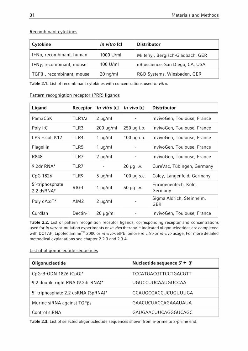
31 Materials and Methods
Recombinant cytokines
Cytokine In vitro [c] Distributor
IFNα, recombinant, human 1000 U/ml Miltenyi, Bergisch-Gladbach, GER
IFNγ, recombinant, mouse 100 U/ml eBioscience, San Diego, CA, USA
TGFβ1, recombinant, mouse 20 ng/ml R&D Systems, Wiesbaden, GER
Table 2.1. List of recombinant cytokines with concentrations used in vitro.
Pattern recognigtion receptor (PRR) ligands
Ligand Receptor In vitro [c] In vivo [c] Distributor
Pam3CSK TLR1/2 2 µg/ml - InvivoGen, Toulouse, France
Poly I:C TLR3 200 µg/ml 250 µg i.p. InvivoGen, Toulouse, France
LPS E.coli K12 TLR4 1 µg/ml 100 µg i.p. InvivoGen, Toulouse, France
Flagellin TLR5 1 µg/ml - InvivoGen, Toulouse, France
R848 TLR7 2 µg/ml - InvivoGen, Toulouse, France
9.2dr RNA* TLR7 - 20 µg i.v. CureVac, Tübingen, Germany
CpG 1826 TLR9 5 µg/ml 100 µg s.c. Coley, Langenfeld, Germany
5‘-triphosphate
2.2 dsRNA* RIG-I 1 µg/ml 50 µg i.v.
Eurogenentech, Köln,
Germany
Poly dA:dT* AIM2 2 µg/ml - Sigma Aldrich, Steinheim,
GER
Curdlan Dectin-1 20 µg/ml - InvivoGen, Toulouse, France
Table 2.2. List of pattern recognition receptor ligands, corresponding receptor and concentrations
used for in vitro stimulation experiments or in vivo therapy. * indicated oligonucleotides are complexed
with DOTAP, LipofectamineTM 2000 or in vivo-JetPEI before in vitro or in vivo usage. For more detailed
methodical explanations see chapter 2.2.3 and 2.3.4.
List of oligonucleotide sequences
Oligonucleotide Nucleotide sequence 5’ 3’
CpG-B-ODN 1826 (CpG)* TCCATGACGTTCCTGACGTT
9.2 double right RNA (9.2dr RNA)* UGUCCUUCAAUGUCCAA
5‘-triphosphate 2.2 dsRNA (3pRNA)* GCAUGCGACCUCUGUUUGA
Murine siRNA against TGFβ1 GAACUCUACCAGAAAUAUA
Control siRNA GAUGAACUUCAGGGUCAGC
Table 2.3. List of selected oligonucleotide sequences shown from 5-prime to 3-prime end.

Materials and Methods 32
Indicated oligonucleotides (*) carry a PTO-modification. PTOs contain one sulfur atom
instead of an oxygen atom in the internucleotidic linkage resulting in higher stability and less
enzymatic degradation. siRNAs against TGFβ1 were designed according to published
guidelines including a 3´-dTdT overhang and were purchased from Eurofins MWG Operon,
Ebersberg, Germany.
2.1.5 Tumor cell lines
Cell line Background Disease Origin Reference
B16-F10 C57Bl/6 Melanoma Prof. T. Brocker, Institut für
Immunologie, LMU Munich (Fidler 1975)
C26 Balb/c Colon cancer
CLS, Eppelheim, Germany (Corbett, Griswold
et al. 1975)
E.G7 C57Bl/6 Thymoma ATCC, Manassas, USA (Moore, Carbone et
al. 1988)
EL-4 C57Bl/6 Thymoma ATCC, Manassas, USA (Ralph and
Nakoinz 1973)
mGC8 C57Bl/6 Gastric cancer
Prof. W. Zimmermann, LIFE
Center, LMU Munich
(Nockel, van den
Engel et al. 2006)
RMA-S C57Bl/6 Lymphoma Dr. J. Charo, Max-
Dellbrück- Center, Berlin
(Karre, Ljunggren
et al. 1986)
Panc02 C57Bl/6 Pancreatic cancer
National Cancer Institute
(NCI)
(Corbett, Griswold
et al. 1975)
Table 2.4. List of tumor cell lines used in this work with listed mouse background, origin and reference.
2.1.6 Kits
Magnetic Cell Sorting
EasySep© Gr1-PE positive sort Gr-1-PE-conjugated antibody, murine (StemCell Technologies, Grenoble, France) PE selection cocktail Magnetic NanoParticles
MDSC Isolation Kit, murine Anti-Ly-6G- Biotin, murine
(Miltenyi Biotec, Bergisch Gladbach, GER) Anti-Gr-1-Biotin, murine
Anti-Biotin and Streptavidin MicroBeads
FcR Blocking Reagent, murine
Anti-CD19 Microbeads, murine Miltenyi Biotec, Bergisch Gladbach, GER
Anti-CD3-Biotin, murine Miltenyi Biotec, Bergisch Gladbach, GER
Anti-Biotin-MicroBeads Miltenyi Biotec, Bergisch Gladbach, GER
CD8 T-Cell Isolation Kit, murine Miltenyi Biotec, Bergisch Gladbach, GER

33 Materials and Methods
Cytokine ELISA sets
IL-1β, murine BD Biosciences, San Diego, USA
IL-6, murine BD Biosciences, San Diego, USA
IL-10, murine BD Biosciences, San Diego, USA
IL-12p40, murine BD Biosciences, San Diego, USA
CXCL10 / IP-10, murine R&D Systems, Wiesbaden, Germany
IFNγ, murine R&D Systems, Wiesbaden, Germany
Cytokine ELISA antibodies
Detection of murine IFNα: Capture-Ab: Anti-IFNα (RMMA-1) PBL, New Brunswick, USA
Detection-Ab: Anti-IFNα (polyclonal, rabbit anti-mouse) PBL, New Brunswick, USA
HRP-conjugated F(ab´)2 fragments (donkey anti-rabbit) Biomeda, Foster City, USA
MDSC Suppression Assay Cell Proliferation ELISA, BrdU (chemiluminescent) Roche Diagnostics, Mannheim, Germany
Apoptosis Assay FAM Flica TM Caspase 3 & 7 Assay Kit ImmunoChemistry, Bloomington, USA
3p-RNA Transcription Kit
MEGAshortscriptTM T7 Kit Ambion, Austin, Texas, USA
2.1.7 Antibodies: FACS and Immunofluorescence
Flow cytometry antibodies
All antibodies used in this work were either purchased from BD/Pharmingen (Heidelberg,
GER) or BioLegend (San Diego, CA, USA). Following fluorochromes were used depending on
the experimental setup: FITC, PE, PeCy7, PerCP, APC, APC-Cy7, PacificBlue, PacificOrange.
Description Isotype Clone
Anti-CD3 Rat IgG2b 17A2
Anti-CD4 Rat IgG2b GK 1.5
Anti-CD8a Rat (LOU/Ws1/M) IgG2a, κ 53-6.7
Anti-CD11b Rat (DA) IgG2b, κ M1/70
Anti-CD11c Armenian Hamster IgG1a N418
Anti-CD19 Rat, IgG2a, κ 1D3
Anti-CD45/B220 Rat IgG2a, κ RA3-6B2
Anti-CD45 Rat IgG2b 30-F11
Anti-CD80 (B7-1) Armenian Hamster IgG2, κ 16-10A1
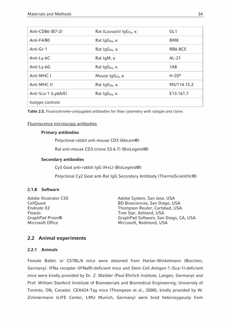
Materials and Methods 34
Anti-CD86 (B7-2) Rat (Louvain) IgG2a, κ GL1
Anti-F4/80 Rat IgG2a, κ BM8
Anti-Gr-1 Rat IgG2b, κ RB6-8C5
Anti-Ly-6C Rat IgM, κ AL-21
Anti-Ly-6G Rat IgG2a, κ 1A8
Anti-MHC I Mouse IgG2a, κ H-2Db
Anti-MHC II Rat IgG2b, κ M5/114.15.2
Anti-Sca-1 (Ly6A/E) Rat IgG2a, κ E13.161.7
Isotype controls
Table 2.5. Fluorochrome-conjugated antibodies for flow cytometry with isotype and clone.
Fluorescence microscopy antibodies
Primary antibodies
Polyclonal rabbit anti-mouse CD3 (Abcam®)
Rat anti-mouse CD3 (clone 53-6.7) (BioLegend®)
Secondary antibodies
Cy3 Goat anti-rabbit IgG (H+L) (BioLegend®)
Polyclonal Cy2 Goat anti-Rat IgG Secondary Antibody (ThermoScientific®)
2.1.8 Software
Adobe Illustrator CS5 Adobe System, San Jose, USA CellQuest BD Biosciences, San Diego, USA Endnote X2 Thompson Reuter, Carlsbad, USA FlowJo Tree Star, Ashland, USA GraphPad Prism® GraphPad Software, San Diego, CA, USA Microsoft Office Mircosoft, Redmond, USA
2.2 Animal experiments
2.2.1 Animals
Female Balb/c or C57BL/6 mice were obtained from Harlan-Winkelmann (Borchen,
Germany). IFNα receptor (IFNαR)-deficient mice and Stem Cell Antigen-1 (Sca-1)-deficient
mice were kindly provided by Dr. Z. Waibler (Paul-Ehrlich Institute, Langen, Germany) and
Prof. William Stanford (Institute of Biomaterials and Biomedical Engineering, University of
Toronto, ON, Canada). CEA424-Tag mice [Thompson et al., 2000], kindly provided by W.
Zimmermann (LIFE Center, LMU Munich, Germany) were bred heterozygously from
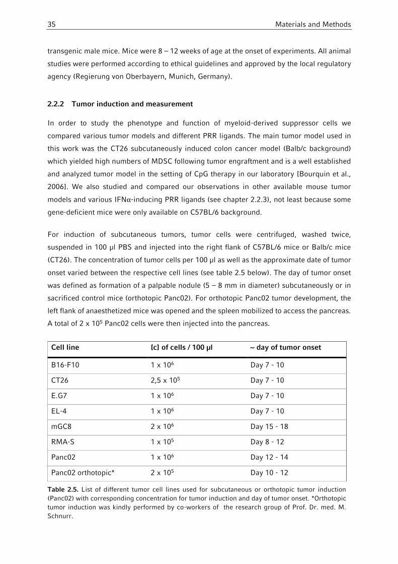
35 Materials and Methods
transgenic male mice. Mice were 8 – 12 weeks of age at the onset of experiments. All animal
studies were performed according to ethical guidelines and approved by the local regulatory
agency (Regierung von Oberbayern, Munich, Germany).
2.2.2 Tumor induction and measurement
In order to study the phenotype and function of myeloid-derived suppressor cells we
compared various tumor models and different PRR ligands. The main tumor model used in
this work was the CT26 subcutaneously induced colon cancer model (Balb/c background)
which yielded high numbers of MDSC following tumor engraftment and is a well established
and analyzed tumor model in the setting of CpG therapy in our laboratory [Bourquin et al.,
2006]. We also studied and compared our observations in other available mouse tumor
models and various IFNα-inducing PRR ligands (see chapter 2.2.3), not least because some
gene-deficient mice were only available on C57BL/6 background.
For induction of subcutaneous tumors, tumor cells were centrifuged, washed twice,
suspended in 100 µl PBS and injected into the right flank of C57BL/6 mice or Balb/c mice
(CT26). The concentration of tumor cells per 100 µl as well as the approximate date of tumor
onset varied between the respective cell lines (see table 2.5 below). The day of tumor onset
was defined as formation of a palpable nodule (5 – 8 mm in diameter) subcutaneously or in
sacrificed control mice (orthotopic Panc02). For orthotopic Panc02 tumor development, the
left flank of anaesthetized mice was opened and the spleen mobilized to access the pancreas.
A total of 2 x 10⁵ Panc02 cells were then injected into the pancreas.
Cell line [c] of cells / 100 µl ~ day of tumor onset
B16-F10 1 x 10⁶ Day 7 - 10
CT26 2,5 x 10⁵ Day 7 - 10
E.G7 1 x 10⁶ Day 7 - 10
EL-4 1 x 10⁶ Day 7 - 10
mGC8 2 x 10⁶ Day 15 - 18
RMA-S 1 x 10⁵ Day 8 - 12
Panc02 1 x 10⁶ Day 12 - 14
Panc02 orthotopic* 2 x 10⁵ Day 10 - 12
Table 2.5. List of different tumor cell lines used for subcutaneous or orthotopic tumor induction
(Panc02) with corresponding concentration for tumor induction and day of tumor onset. *Orthotopic
tumor induction was kindly performed by co-workers of the research group of Prof. Dr. med. M.
Schnurr.

Materials and Methods 36
CEA424-Tag mice [Thompson et al., 2000] bearing autochthonous gastric tumors were used
at the age of 85 – 100 days for analysis of myeloid-derived suppressor cells.
For subcutaneous tumors, tumor area is expressed as the product of perpendicular diameters
of individual tumors. The tumor volume was measured thrice per week unless indicated
otherwise. The volume of CEA424-Tag-derived gastric tumors (SV40) was calculated as the
product of 3 perpendicular diameters. When tumor volume exceeded 225 mm2 or when signs
of animal distress were observed twice in 48 hours, mice were anaesthetized and sacrificed
by cervical dislocation.
2.2.3 Therapy with PRR ligands
In order to determine the role of systemic Toll-like receptor and RIG-I activation on phenotype
and function of myeloid-derived suppressor cells as well as on Stem Cell Antigen-1
expression, mice were treated with different immunostimulants. Tumor-free and tumor-
bearing mice underwent the same treatment protocol unless indicated otherwise. The
different treatment protocols used in this work as well as sites of injection are depicted in
Table 2.6. Treatment of tumor-bearing mice was initiated following establishment of tumors
(tumor area > 25 mm2 or age of CEA424-TAg mice > 85 days) unless indicated otherwise.
Therapy of orthotopic Panc02 tumor-bearing mice started on day 12, when sacrificed control
mice showed visible tumor nodules (5 – 8 mm diameter). In general, 100 µg LPS, 100 µg CpG-
oligodesoxyribonucleotides (ODN) and 250 µg poly(I:C) were dissolved in 100 µl PBS and
injected subcutaneously or intraperitoneally as indicated in table 2.6. Immunostimulatory
RNA was injected intravenously after complexation with DOTAP - a liposomal transfection
reagent - or in vivo-jetPEI in order to prevent degradation and to improve rate of transfection.
In detail, for treatment with 3pRNA or siRNA, 8 µl of in vivo-jetPEI were mixed with 50 µg of
nucleic acid at an N:P ratio of 8:1 (8 nitrogen residues of jetPEI per RNA phosphate) in a
volume of 200 µl 5% glucose solution, incubated 15 minutes at room temperature and
injected intravenously into the retro-bulbar venous plexus or tail vein [Poeck et al., 2008;
Ellermeier et al., 2013]. For in vivo administration of 9.2dr RNA, 20 µg of RNA were incubated
with 100 µg DOTAP and 40 µl PBS for 20 minutes and injected into the retro-bulbar venous
plexus. Depending on the experimental setup, 24 – 48 hours after the last treatment, mice
were sacrificed, organs removed and prepared for further analysis as described below
(chapter 2.2.4).

37 Materials and Methods
Table 2.6. Treatment protocols and injections sites for in vivo administration with PRR ligands unless
indicated otherwise. * Day 1 is defined as first day of treatment depending on tumor volume in mm².
Day 0 or 12 are defined as days after tumor inoculation.
2.2.4 Organ and single cell preparation
2.2.4.1 Blood collection and serum analysis
For analysis of MDSC phenotype with or without PRR ligand therapy, blood cells were
analyzed via flow cytometry. Mice were anesthetized with isoflurane and sacrificed via
cervical dislocation. In order to receive a maximum of blood, the left chamber of the heart
was punctured with a heparin coated syringe right after cervical dislocation. The blood was
then transferred into 15 ml Falcon tubes, washed with 13 ml PBS, centrifuged (400 x G, 7
minutes) and resuspended in 3 ml erythrocyte lysis buffer. After incubation for 5 min at room
temperature, reaction was stopped with 14 ml PBS. Red blood cell debris was removed by a
second centrifugation step. The resulting cell pellet was then resuspended in 1 ml FACS
buffer for further analysis.
To determine serum levels of interleukins, 24 hours after PRR ligand therapy, the retro-bulbar
venous plexus of mice was punctured with heparinized capillary tubes until a minimum of
500 µl blood was obtained. Blood was allowed to coagulate for 20 minutes and then
centrifuged at 13.000 x G for another 20 minutes. The resulting serum in the supernatant was
collected and stored at -20°C for subsequent evaluation of interleukin serum levels via ELISA.
2.2.4.2 Tumor removal and digestion
Mice bearing established subcutaneous tumors (tumor volume at least > 100 mm²) were
anesthetized with isoflurane and sacrificed via cervical dislocation. Tumors were dissected
and the skin was carefully removed. The resulting tumor tissue was dissected into small
PRR ligand Injection site Days of treatment
Tumor-free Tumor-bearing*
organ removal after last course
LPS intraperitoneally 0 – 3 – 7 - 24 hours
CpG-ODN subcutaneously 0 – 3 – 6 1 – 4 – 7 24 hours
Poly I:C intraperitoneally 0 – 3 – 6 1 – 4 – 7 24 hours
3p-RNA intravenously 0 – 3 – 6 1 – 4 – 7 24 hours
9.2dr RNA intravenously - 0 – 2 – 4 – 6 – 8 – 12 48 hours
(3p-)siTGFβ intravenously - 12 – 14 24 hours

Materials and Methods 38
pieces and incubated in dissection buffer at moderate stirring for 30 – 45 minutes at 37°C.
The digested tumor tissue was then passed through a 100 µm-pore cell strainer, followed by
a second passage through a 40 µm-pore mesh. Single cell suspensions were then washed
twice, resuspended in FACS buffer and analyzed via flow cytometry.
2.2.4.3 Isolation of splenocytes
Mice were anesthetized with isoflurane and sacrificed via cervical dislocation. Spleens were
resected and passed with PBS through a 40 µm-pore cell strainer for mechanical tissue
disintegration. The resulting single cell suspension was centrifuged (400 x G, 7 minutes) and
resuspended in erythrocyte lysis buffer. After incubation for 3 minutes at room temperature,
reaction was stopped by adding PBS and red cell debris was removed by a second
centrifugation step. Splenocytes were then resuspended in either RPMI complete medium or
FACS buffer for further analysis.
2.2.4.4 Preparation of lungs and lymph nodes
Mice were anesthetized with isoflurane and sacrificed via cervical dislocation. Lungs,
peripheral lymph nodes (axillary or inguinal) or tumor-draining lymph nodes were removed.
Single cell suspensions of lungs were prepared as described above (chapter 2.2.4.2) and
analyzed via flow cytometry. Lymph nodes of individual mice were pooled and directly passed
through a 40 µm-pore cell strainer. After washing twice with PBS, single cell suspensions
were resuspended in FACS buffer and analyzed via FACS.
2.2.4.5 Bone marrow isolation
Mice were anesthetized with isoflurane and sacrificed via cervical dislocation. Femur and
tibia were dissected bilaterally. The remaining muscle tissue was carefully removed with
small scissors and scalpel followed by external cleaning with 70% isopropanol and complete
RPMI medium. Each end of bone was then cut off and bone marrow was expelled using a
27G needle / 1 ml syringe filled with complete RPMI medium. After passage through a 40
µm-pore cell strainer, single cell suspensions were washed once with complete RPMI medium
and resuspended in erythrocyte lysis buffer. After lysis, cells were washed again and
resuspended in RPMI complete medium for later experiments.

39 Materials and Methods
2.3 Cell culture experiments
2.3.1 General culture conditions and cell viability
All cell culture experiments were performed with sterile technique and under sterile
conditions using a laminar air flow hood. Culture of cell lines or primary cells was performed
in incubators under 37°C, 5% CO2/air mixture and 95% air humidity. Cell viability was tested
using trypan blue exclusion test: whereas the outer membrane of dying cells loses its integrity
and absorbs trypan blue, it cannot enter the cell through intact membranes. Thus, the
cytoplasm of dead or dying cells stains dark blue, whereas viable cells remain golden under
light microscopy. Thus, after appropriate dilution, cell numbers of viable cells could be
determined by using a Neubauer hemocytometer.
2.3.2 Tumor cell cultures
Tumor cell lines were obtained as described above (chapter 2.1.5). In general, cell lines were
cultured in T75-T175 culture flasks in cell culture medium according to the respective cell
line (see table 2.7). Cells were continuously checked via light microscopy for level of cell
growth, signs of cell death and bacterial contamination. Medium was replaced regularly every
two to three days depending on cell growth. In addition, adherent cell lines were splitted at
a ratio of 1:2 – 1:10 at least twice a week and transferred to new culture flasks according to
level of cell confluence and rate of cell growth. Therefore, medium was discarded and cells
were gently detached either with a cell-scraper or by using 1 % trypsin-EDTA solution. Cells
were then washed twice with PBS and re-disseminated in appropriate medium at lower
concentrations. Non-adherent cell lines were splitted continuously two to three times a week.
For in vivo tumor engraftment, tumor cells were splitted one day before injection to ensure
optimal growth conditions. On day of tumor inoculation, tumor cells were harvested as
described, re-suspended in PBS and separated by flushing through a 27G needle for three
times.
Cell line Cell growth Culture medium Split frequency
B16-F10 adherent 10% FCS, 1% P/S, 1% L-glu in DMEM 2-3 x per week, 1:10
CT26 adherent 10% FCS, 1% P/S, 1% L-glu in DMEM 2-3 x per week, 1:2
E.G7 suspension 10% FCS, 1% P/S, 1% L-glu, 1% So-
Pyr, 1% NEAA in DMEM 3 x per week, 1:3
EL-4 suspension 10% FCS, 1% P/S, 1% L-glu in DMEM 3 x per week, 1:3
mGC8 adherent 10% FCS (Gold), 1% P/S, 1% L-glu,
1% So-Pyr , 1% NEAA in DMEM 1 x per week, 1:2

Materials and Methods 40
Table 2.7. List of used tumor cell lines in this work and appropriate medium and split frequency for in
vitro culture. P/S = penicillin/streptomycin, L-glu = L-glutamine, So-Pyr = sodium pyruvate.
2.3.3 Generation of bone marrow-derived macrophages
Bone marrow-derived macrophages (BMDM) were generated as described previously with
some modifications [Zanoni et al., 2009; Marim et al., 2010]. M-CSF is a lineage-specific
growth factor that is responsible for the differentiation from myeloid progenitors into
macrophages. We used M-CSF transfected L929 cells which secret high amounts of M-CSF
into the supernatant in order to obtain enough M-CSF for BMDM generation ex vivo [Englen
et al., 1995]. L929 cells were kindly provided by Prof. Veit Hornung (University of Bonn,
Germany). For generation of L929-conditioned medium (LCM), 2 x 10⁷ L929 cells were
seeded in T175 tissue flasks in 20 ml L929 culture medium. After one week of culture,
supernatant was collected, sterile filtered and stored at -20°C until usage. For generation of
murine BMDM, bone marrow was prepared as described above (chapter 2.2.4.5). Following
washing with PBS, bone marrow cells of individual mice were splitted in half, suspended in
25 ml of BMDM medium containing 30% LCM and seeded onto two T175 culture flasks,
resulting in a concentration of approximately 1 x 10⁶ cells/ml. After one week of culture,
BMDM were harvested, washed twice in PBS and further used for in vitro stimulation with
PRR ligands. Purity of BMDM was analyzed with flow cytometry via F4/80 expression and
was typically over 90%.
2.3.4 In vitro stimulation with PRR-ligands
For in vitro stimulation of immature myeloid cells (iMC), myeloid-derived suppressor cells
(MDSC) and bone marrow-derived macrophages (BMDM), single cells suspensions were
prepared as described. Cells were then resuspended in RPMI complete medium and seeded
on 96-well plates at a concentration of 2 x 10⁵ MDSC/iMC per well or 1.5 x 10⁵ BMDM per
well in 200 µl medium. Experimental conditions were always performed in triplicates. Cells
were allowed to attach to the well bottom for 1 hour before immunostimulants and cytokines
were added to the culture. The respective cytokines and PRR ligands are described in chapter
2.1.4 with their respective concentrations unless indicated otherwise.
RMA-S suspension 10% FCS, 1% P/S, 1% L-glu, 1% So-
Pyr, 1% NEAA in RPMI 2-3 x per week, 1:5
Panc02 adherent 10% FCS, 1% P/S, 1% L-glu in DMEM 2-3 x per week, 1:10
L929 adherent 10% FCS, 0.5 % Cipro, 1% So-Pyr in
DMEM 1 x per week, 1:5

41 Materials and Methods
As mentioned in chapter 2.2.3, RNA has to be complexed before in vivo and in vitro usage in
order to prevent degradation and ensure optimal transfection rate. For in vitro experiments,
LipofectamineTM 2000 - a polycationic lipid - was used for transfection of RNA. In this work,
poly(dA:dT) and ppp-RNA had to be transfected for in vitro stimulation. Both, the appropriate
amount of RNA as well as Lipofectamine were incubated in two separated tubes in the same
volume of OptiMem at room temperature. After 5 minutes, both tubes were added together
and incubated for another 20 minutes, before adding the desired amount into culture.
After addition of cytokines and PRR ligands, cells were regularly cultured at 37°C for 24 hours
unless indicated otherwise. For measurement of cytokine induction, 96-well plates were
centrifuged (400 x G, 7 minutes), supernatant was collected and stored at -20°C for later
analysis via ELISA. In some experiments, the remaining cell pellet was washed twice with
PBS and resuspended in FACS buffer for flow cytometric analysis.
2.3.5 MDSC isolation via magnetic-activated cell sorting
2.3.5.1 Principle of magnetic-activated cell sorting
Magnetic-activated cell sorting (MACS) is used for isolation of viable and functionally active
cells via labeling with magnetic particles. The technique of magnetic cell separation was first
described in 1990 by Miltenyi Biotec. A mixture of cells is incubated with supraparamagnetic
particles (MicroBeads) of approximately 50 nanometers in diameter, which are composed of
a biodegradable matrix. Thus, it is not necessary to detach them after the purification process
resulting in minimal interference with subsequent experiments. These MicroBeads are
coated with antibodies against a particular cell surface antigen that attach them to cells
expressing the desired antigen. After incubation, the labeled cell solution is transferred in a
column and placed in a strong static magnetic field induced by a permanent magnet. Antigen-
expressing and bead-bound cells are retained in the column, whereas cells not expressing
the particular antigen pass through the column and can be collected. The retaining cell
population is eluted from the column after removal of the magnetic field. With this method,
cell can be positively and negatively separated depending on the particular antigen. Positive
selection means, that a target population of cells expressing the desired antigen is retained
in the column, eluted and then used for further experiments. Negatively selected cells are
isolated by depletion of undesired non-target cells, often via incubation with multiple non-
target antibodies. Hence, only the non-magnetic and untouched target population rinses
through and can be used for subsequent experiments.
In this work, two different purification methods were used for isolation of Gr1+CD11b+ MDSC
and their respective subpopulations. Unless stated otherwise, materials and reagents from

Materials and Methods 42
StemCell Technologies were used to isolate the total population of Gr1+CD11b+ MDSC. In
order to separate MDSC subpopulations from each other, the MDSC Isolation Kit from
Miltenyi Biotec was used with some modification. For some proliferation assays, CD8+ T Cells
were purified with a CD8 T-Cell Isolation Kit (Miltenyi Biotec) and used as responder cells
instead of splenocytes (see chapter 2.2.4). In general, magnetic cell separation technologies
required fast working on ice and the use of pre-cooled solutions. Incubation periods were
always performed at 4°C under gentle tilting. Rotation and centrifugation was done at 300 x
G for 10 minutes, instead of the usual 400 x G for 7 minutes, all to ensure optimal cell viability.
2.3.5.2 Miltenyi protocol for isolation of MDSC subpopulations
As mentioned above (chapter 1.4.2), myeloid-derived suppressor cell subpopulations can be
phenotypically distinguished via their expression of the Ly-6G antigen (Figure 2.1). Whereas
polymorphonuclear MDSC (PMN-MDSC) are Gr1+CD11b+Ly6Ghigh, monocytic MDSC (MO-
MDSC) are Gr1+CD11b+Ly6Gneg. In order to analyze and compare the function of MDSC
subsets, we used a mixture of positive and negative selection methods for magnetic
enrichment based on their differential expression of Ly-6G.
Therefore, we used the Miltenyi MDSC Isolation Kit with previous depletion of CD19+ and
CD3+ cells (mainly B and T cells) to improve cell purity. After preparation of single-cell
suspensions cells were adjusted to 108 per 350 µl MACS buffer. To prevent unspecific binding
sites during the following incubation periods, cells were first incubated with 50 µl FcR
Blocking Reagent for 10 minutes. After incubation with 50 µl anti-CD3-Biotin for another 10
minutes, cells were washed and readjusted to 350 µl starting volume. Then the cells were
incubated simultaneously with 100 µl Anti-CD19- and 100 µl Anti-Biotin-Microbeads for 10
minutes and washed afterwards. LS columns were rinsed with 3 ml MACS buffer and loaded
with labeled cells diluted in 500 µl MACS buffer. Columns were washed three times and the
flow-through was gathered as CD3/CD19-depleted cell suspension.
For magnetic separation of PMN-MDSC, these cells were first diluted in 400 µl MACS buffer
and incubated with 100 µl of Anti-Ly6G-Biotin for 10 minutes. After a washing step, cells
were resuspended in 800 µl buffer and 200 µl Anti-Biotin-Microbeads were added. Following
incubation for 15 minutes, cells were washed, diluted in 500 µl MACS buffer and added onto
pre-rinsed LS columns. Columns were washed three times with 3 ml MACS buffer. The flow
through was gathered as Ly-6G negative fraction. After removal of the magnetic field,
Ly-6Gpos PMN-MDSC were eluted by flushing the column with 2 ml MACS buffer by using
the provided plunger. PMN-MDSC were centrifuged and kept in complete RPMI medium until
further usage.

43 Materials and Methods
To purify MO-MDSC, the Ly-6G depleted flow through was centrifuged and diluted in 400 µl
MACS buffer per total 108 cells. Cells were incubated with 100 µl Anti-Gr1-Biotin for 10
minutes, washed and further incubated with 100 µl Streptavidin MicroBeads in 900 µl MACS
buffer for another 10 minutes. According to remaining cell numbers, either LS or MS-columns
were pre-rinsed again with buffer. In most experiments, MS columns were rinsed, loaded,
washed thrice and after removal of the magnetic field, the column was flushed with 1 ml of
buffer. To increase the purity of the eluted Gr1+Ly6Gneg MO-MDSC, they were rinsed over a
second MS column. MO-MDSC were centrifuged and kept in complete RPMI medium until
further usage.
Cell purity was checked regularly via flow cytometry and always between 85 to 95% for PMN-
MDSC and 75 to 85% for MO-MDSC (Figure 2.1 B). The purity of each subpopulation varied
depending on MDSC numbers in the starting population, e.g. purity was always slightly
higher in tumor-bearing mice with respect to higher amounts of MDSC in spleens or bone
marrow.
2.3.5.3 Stem cell protocol for isolation of Gr1+CD11b+ MDSC
For isolation of the whole population of splenic or bone marrow MDSC, we used a novel
separation method developed by StemCell Technologies. The difference between Miltenyi
and StemCell protocols was that the latter did not require passage through magnetic columns.
Instead, a FACS tube was placed in a strong magnet (EasySep®) with accumulation of
magnetically labeled cells at the surrounding inner surface. According to the manufacturer,
this techniques’ advantages are higher cell viability and function due to less mechanical cell
stress. In detail, single-cell suspensions of splenocytes or bone marrow cells were washed
once with StemCell buffer and adjusted to a concentration of 108 per ml. After incubation
with 25 µl/ml of Anti-Gr1-PE-conjugated antibody for 7 minutes at 4°C, cells were washed
with StemCell buffer and readjusted to the starting volume. The Gr-1-PE labeled cell
suspension was then incubated with 50 µl/ml PE-selection-cocktail for another 7 – 10 minutes
at 4°C followed by direct addition of 25 µl NanoParticles. Following incubation for another 7-
10 minutes at 4°C and volume adjustment up to 2.5 ml, the tube was placed into the EasySep®
magnet for 5 minutes resulting in accumulation of Gr-1+, NanoParticle-bound cells at the
inner surface of the tube. For cell separation, the supernatant was discarded containing non-
labeled, non-magnetic cells while the tube was still placed in the magnetic field. For further
enrichment, previous steps were repeated two times with already purified target cells.
Purified MDSC were centrifuged and kept in complete RPMI medium for subsequent
experiments. The purity of Gr1+CD11+ cells was verified via flow cytometry and was typically
over 90% (Figure 2.1 A).

Materials and Methods 44
2.4 Immunological methods
2.4.1 Fluorescent-activated cell sorting (FACS)
2.4.1.1 Principle
Flow cytometry or fluorescence-activated cell sorting (FACS) is a laser based method that
allows the analysis of properties of single cell suspensions or particles suspended in a liquid
phase. A suspension of cells is surrounded by a sheath fluid stream and aspirated with high
velocity through a capillary into a flow chamber. By forcing the cells to enter a small nozzle
one at a time, they are hydrodynamically focused and pass through a focused laser one by
one. At the point where the laser is hitting the cell stream, the illuminating light is either
scattered or absorbed. Simultaneously, cells can be stained with an antibody linked
fluorescent dye, which absorbs light of a particular wavelength and emits light at a longer
wavelength. The scattered light and emitted fluorescence are measured by a number of
detectors. The detector in line with the light beam is called Forward Scatter (FSC). Several
other detectors are arranged perpendicular to it - one that measures a parameter which is
logged as Side Scatter (SSC) and multiple fluorescence detectors. FSC estimates the cell
volume by analyzing the angle light that is scattered in straight direction. SSC correlates with
inner cell complexity such as membrane structure and the amount of cytoplasmatic granules.
Thus, SSC roughly serves as a proxy for cell granularity. The detected fluorescence and light
scatter signals are amplified by multiple photodiodes and converted into electrical signals
that are electronically processed. Unwanted light emission is blocked by several optical
filters.
Each fluorochrome has to be excited by a laser of particular wavelength and measured by
detectors appropriate for the emitted spectrum of light. All flow cytometric analyses were
performed with a FACSCanto II (BD Biosciences) which allowed simultaneous examination
of up to eight different antigens.

45 Materials and Methods
A B
Table 2.9. Dyes (A) and laser of FACSCanto II with corresponding excited dyes (B).
2.4.1.2 Analysis of cell surface antigens
For analysis of cell surface antigens, single cell suspensions were prepared as described
previously (chapter 2.2.4). Depending on the experiment, cells were diluted at 2 x 10⁵ up to
1 x 10⁶ in 200 µl ice-cold FACS buffer or cell culture medium. Single cell suspensions were
then incubated with the desired fluorochrome-conjugated antibodies for 30 minutes at 4°C
with minimum light exposure (for overview of used FACS antibodies see Table 2.5). Cells
were washed twice with PBS and re-suspended in 200 µl FACS buffer. If not analyzed
immediately, cells were fixed in 200 µl fixation buffer and kept cold at 4°C until data
acquisition. Each experimental condition was at least analyzed in triplets.
2.4.1.3 Flow cytometry and gating of MDSC
MDSC were analyzed from primary organs or following in vitro culture of purified MDSC or
iMC, respectively. Given that MDSC subpopulations differ in their expression of the Ly-6G
antigen, each staining contained at least the phenotypical marker Gr-1, CD11b and Ly-6G
with additional maturation and differentiation markers (see chapter 1.4.2). The main gating
strategy of MDSC subsets is illustrated below.
Laser Wave length Dye
Blue 488 nm
FITC
PE
PerCP
Pe-Cy7
Red 633 nm APC
APC-Cy7
Violet 405 nm PacBlue
PacOrange
Dye Exmax Emmax
FITC 495 nm 519 nm
PE 565 nm 575 nm
PerCP 490 nm 675 nm
Pe-Cy7
480 nm
565 nm
743 nm
767 nm
APC 650 nm 660 nm
APC-Cy7 650 nm 785 nm
PacBlue 410 nm 455 nm
PacOrange 410 nm 551 nm

Materials and Methods 46
Figure 2.1. Representative flow cytometry data of MDSC with corresponding subpopulations.
Dead splenocytes were excluded via scatter discrimination (not shown) and MDSC were gated on their
status as Gr1+CD11b+ (left panel, A). MDSC were further divided into the major subpopulations of MO-
MDSC and PMN-MDSC with respect to their differential expression of the Ly-6G antigen (right panel,
A). B shows backgating of MDSC subsets and illustrates subset-related differences in cell size and
granularity by variable appearance on forward (FSC) and sideward scatter (SSC) analysis.
2.4.1.4 Detection of apoptosis
For evaluation of apoptosis in myeloid-derived suppressor cells, the FAM FLICA TM Caspase
3&7 Assay Kit (Immunochemistry) was used. Therefore, after staining of surface antigens,
primary cells were diluted in 290 µl complete RPMI with addition of 10 µl FLICA (Fluorescent
Labeled Inhibitors of Caspases) that covalently bind with active caspase enzymes. Cells were
than incubated at 37°C for 45 minutes and washed twice with 2 ml 1 X Apoptosis Wash Buffer.
After resuspension in 300 µl Apoptosis Wash Buffer, cells were ready for colorimetric
analysis. In some experiments, propidium iodine (PI) was used to discriminate between early
47 Materials and Methods
(FLICAposPIneg) and late apoptotic cells (FLICAposPIpos) as well as exclusion of necrotic cells
which than appear as FLICAnegPIpos).
2.4.2 Enzyme-linked immunosorbent assay (ELISA)
The enzyme-linked immunosorbent assay, or ELISA, is a technique used to analyze a specific
antigen in a liquid sample. The method is based on binding of enzyme-linked antibodies
against the desired antigen leading to subsequent conversion of a colorless substrate to a
colored reagent. The color change is detected and converted into a quantifiable electrical
signal that correlates to the initial amount of antigen.
Different cytokine levels in serum of mice as well as from murine cell supernatants were
measured in this work. The particular ELISA kits are listed in chapter 2.1.6. For cytokine
detection, plates are coated with a capture antibody specific for the target cytokine. After
addition of the sample, any given cytokine present is bound by the capture antibody. Plates
are incubated again with biotinylated detection antibody which in turn is bound by
streptavidin coupled to a peroxidase. By adding 3,3’,5,5’ – tetramethylbenzidine (TMB) as a
substrate, this enzyme catalyzes a reaction that entails a blue color change. The reaction is
stopped by addition of H2SO4 and read in a multiplate reader at 450 nm wavelength with a
wavelength correction subtraction at 590 nm. All cytokines in this work except IFNα were
measured by using commercially available kits according to the manufacturer’s protocol.
Depending on the expected amount of cytokines present in supernatant or serum, the
samples were generally diluted 1:2; for the detection of IL-12p40 and IFN, samples were
diluted 1:5 or 1:10.
Detection of IFNα was performed by using a set of antibodies listed in chapter 2.1.6: Plates
were coated overnight at 4°C with 50 µl/well coating buffer containing 1 µg/ml of capture
antibody. In order to block unspecific binding sites, plates were then blocked for 3 hours with
150 µl of assay diluent per well at room temperature. After intensive washing for several times
with washing buffer, plates were incubated overnight with sample and standard protein at
4°C. The highest level of standard was generally set at 105 IU/ml, samples were loaded
undiluted. After several washing steps, 50 µl/well of detection antibody at a concentration of
625 ng/ml was added and incubated again for 3 hours at room temperature. Following
extensive washing, horseradish peroxidase (HRP)-conjugated F(ab’)2 fragments were used
at a concentration of 15 µg/ml and incubated for 3 hours in order to detect antigen-bound
detection antibody. After extensive washing 50 µl of substrate solution were added to each
well to induce a colored reaction. The reaction was stopped by addition of 2N H2SO4 and read
in a multiplate reader at 450 nm with correctional subtraction at 590 nm.

Materials and Methods 48
2.4.3 Immunofluorescence of tumor slices
Immunofluorescence of tumor slices was performed together with Tina Adunka and Jonathan
Ellermeier (AG Prof. Dr. med. M. Schnurr). Briefly, for evaluating influx of tumor-infiltrating
cytotoxic T-lymphocytes (CTLs), immunofluorescence was performed on frozen tissue
sections. Following removal of Panc02 tumors, tissue was immediately stored in cryotubes,
shock frozen in liquid nitrogen and stored at -80°C. Consecutive cryostat tissue sections (5
μm) were mounted on glass slides and fixed in cold acetone (-20°C) for 10 minutes. Sections
were blocked with 10% goat serum in PBS for 20 minutes followed by labeling with primary
antibody overnight at 4°C (for antibodies see section 2.1.7). After washing with PBS, tumor
slices were stained with the respective secondary antibodies diluted in 10% donkey serum
for another 45 minutes and washed again with PBS. Slices were mounted with Aqua-
PolyMount (Polysciences) and then stored at -20°C for analysis of tumor-infiltrating CTLs.
2.4.4 BrdU suppression assay
To access the immunoregulatory capacity of myeloid-derived suppressor cells on antigen-
specific and -unspecific proliferation of T cells, we used a 5’Bromo-2’deoxy-uridine (BrdU)
incorporation assay. The principle of measuring T cell proliferation in the presence of MDSC
in vitro, as a surrogate for MDSC-mediated suppressivity, has been well described in
literature (Dolcetti, Peranzoni et al. 2010). In detail, 1 x 10⁵ splenocytes or 7.5 x 10⁴ splenic
T cells (Dynal Mouse Negative T Cell Isolation Kit, Invitrogen, Carlsbad, CA) from naïve mice
were cultured with different ratios of MDSC in complete RPMI medium. Cultures were
incubated in triplicate at 37°C and 5% CO2 in flat-bottom 96-well plates. T cell proliferation
was stimulated by the addition of anti-CD3/anti-CD28 coated microbeads (Invitrogen) at a
bead-to-cell ratio of 1:75. Cells were co-cultured for 60 hours before 7.5 µM BrdU was added
for another 12 hours. Cell culture plates were then centrifuged (300 x G, 10 minutes), medium
was discarded and stored for further analysis before plates were dried with a hairdryer. Cell
fixation, incubation with anti-BrdU-antibody and substrate reaction were done according to
the manufacturer’s protocol. BrdU incorporation was detected by chemiluminescence-based
assay and was measured in relative light units (rlu) with a multiple plate reader.
Suppression of T cell proliferation in the presence of MDSC in increasing concentration was
calculated relative to stimulated responder cells alone (MDSC-splenocyte-ratio 1:16, 1:8, 1:4,
1:2 with anti-CD3/CD28-stimulated SC or T cells as positive control). Percentage suppression
of proliferation was calculated as
(1 −𝑝𝑟𝑜𝑙𝑖𝑓𝑒𝑟𝑎𝑡𝑖𝑜𝑛𝑤𝑖𝑡ℎ𝑀𝐷𝑆𝐶
𝑝𝑟𝑜𝑙𝑖𝑓𝑒𝑟𝑎𝑡𝑖𝑜𝑛𝑤𝑖𝑡ℎ𝑜𝑢𝑡𝑀𝐷𝑆𝐶
) 𝑥 100.

49 Materials and Methods
Figure 2.2 shows a typical bar graph used in this work to display the results of a T cell
proliferation assay with or without co-culture of T cell suppressing immature myeloid cells
(iMC) or MDSC. As increasing concentration of iMC/MDSC in the culture are depressing T
cell proliferation, the relative proliferation rate decreases. Negative controls are exemplary
illustrated once and left out in other figures.
Figure 2.2. Example of a standard BrdU incorporation assay for measuring MDSC-mediated
suppression of T cell proliferation ex vivo. The y-axis shows the relative percentage of T cell or
splenocyte (SC) proliferation with or without co-culture of immature myeloid cells (iMC). Therefore, a
100% proliferation rate as positive control is defined as proliferation of anti-CD3/CD28-stimulated
splenocytes alone. X-axis shows the different experimental conditions with internal controls (left side,
white bars) and with co-culture of T cell suppressing immature myeloid cells or MDSC in ascending
concentration (iMC-splenocyte-ratio 1:16, 1:8, 1:4, 1:2; black bars). SC = splenocytes; stim = anti-
CD3/CD28-stimulation. * Relative light units (rlu) ** depending on the experimental setup MDSC-like
immature myeloid cells or myeloid-derived suppressor cells were used *** iMC-splenocyte-ratio 1:16,
1:8, 1:4, 1:2 unless indicated otherwise.
2.5 Generation of ppp-RNA
In vitro transcription of ppp-RNA was performed using the MEGAshortscriptTM T7 Kit
(Ambion, Austin, Texas) according to the manufacturer’s protocol. siRNAs against TGFβ1
including a 3´-dTdT overhang and matching 5’-triphosphate-modified siRNA were kindly
provided by Prof. Max Schnurr (Division of Clinical Pharmacology, LMU Munich). Respective
oligonucleotide sequences are depicted in section 2.1.4.

Materials and Methods 50
2.6 Statistical Analysis
All data in this work is given as arithmetic mean values. Variance of mean values is expressed
as standard error of the mean (SEM). Statistical significance in most experimental finding
was calculated by independent two-tailed student’s t-test. Comparison of multiple groups
were performed using 1-way ANOVA with pair-wise Bonferroni posttest. Tumor sizes were
analyzed using Mann-Whitney U test. Significance was considered at p levels < 0.05, p < 0.01
and p < 0.001 and were then indicated with *, ** and ***. Statistical analyses and design were
performed using GraphPad Prism 5 (GraphPad Software), Microsoft Office and Adobe
Illustrator CS5 (Adobe System).

51 Results
3 RESULTS
3.1 Effect of systemic CpG-DNA administration on Gr1+CD11b+ immature
myeloid cells (iMC) in tumor-free mice
3.1.1 iMC numbers in different organs following systemic TLR stimulation
In our group, we previously demonstrated that activation of the innate immune system with
the Toll-like receptor (TLR) 9 ligand CpG evokes efficient antitumor immune responses in
mice bearing subcutaneous CT26 tumors [Heckelsmiller et al., 2002; Bourquin et al., 2008],
despite a large number of highly immunosuppressive MDSC in this model [Gallina et al.,
2006; Youn et al., 2008]. However, Vaknin and colleagues recently reported that chronic
activation of innate immunity via TLR2, 4, 7 and 9 using a slow-release delivery system leads
to immunosuppression in tumor-free mice attributed to the induction of Gr1+CD11b+
immature myeloid cells (iMC) or MDSC [Vaknin et al., 2008].
To address this issue, in a first experiment the capacity of CpG-DNA - our most intensively
studied TLR ligand - was investigated for its ability to induce Gr1+CD11b+ iMCs in vivo.
Therefore, mice were challenged with three repeated doses of 100 µg subcutaneous CpG at
a 3-day interval; the dosage and application resembled typical anti-tumor treatment
regimens. Indeed, when spleens were analyzed via flow cytometry for the number of
Gr1+CD11b+ iMC, we saw a two-fold increase in iMC numbers compared to untreated mice
(Figure 3.1 A,C). This effect was not restricted to splenic Gr1+CD11b+ cells, because the same
percentage increase of iMCs was observed in other compartments such as blood and bone
marrow indicating a systemic effect of TLR9 stimulation on iMC accumulation (Figure 3.1 B).
Lymph nodes did only show minor iMC infiltration in untreated mice or in mice treated with
CpG (data not shown). In a second step, the effect of other PRR ligands on iMC expansion
was assessed by in vivo administration of poly(I:C), LPS and complexed triphosphate-RNA
(3pRNA) using the same treatment protocol. TLR3 and TLR4 stimulation by poly(I:C) and LPS,
respectively, induced splenic iMC accumulation comparable to CpG. However, systemic
activation of the pattern recognition receptor RIG-I by 3pRNA did not increase the percentage
of Gr1+CD11b+ iMCs in tumor free mice (Figure 3.1 C).

Results 52
Figure 3.1. TLR therapy in tumor-free mice leads to expansion of MDSC-like cells. Naïve Balb/c
mice received either 3 s.c. injections of CpG or PBS (no therapy) as a control at 3-day intervals (n = 5
per group). 2 days after the last injection mice were sacrificed and the number of Gr1+CD11b+
immature myeloid cells (iMC) was determined using flow cytometry. A Representative plot gated on
viable splenocytes showing the total number of splenic iMC (Gr1+CD11b+) before and after CpG
treatment. B Histograms showing total numbers of iMC in spleen, blood and bone marrow. C shows
the percentage of Gr1+CD11b+ iMC in spleens of mice after therapy with different TLR- or RIG-I ligands
(3p RNA). Treatment protocol was done as mentioned above. Data are the mean + SEM from at least
three independent experiments. *, p < 0.05; ** p < 0.01; paired Student’s t test.
3.1.2 Effect of CpG treatment on iMC-mediated suppressivity
Immune suppression by MDSC is not only a result of their expansion, but in particular due to
their activation [Gabrilovich et al., 2009]. A hallmark ability of MDSC is inhibiting T-cell
proliferation by various mechanism (see chapter 1.4.4). Therefore, in a second step the ability
of splenic Gr1+CD11b+ iMC to suppress the proliferation of T cells ex vivo was examined via
a BrdU proliferation assay, which is a standard experiment for evaluating MDSC-mediated
immunosuppression. Naïve mice were treated with CpG again as described above. One day
after the last injection, spleens were removed and Gr1+CD11b+ cells were separated by
magnetic sorting. Purified iMC from both untreated and CpG-treated mice showed a

53 Results
moderately suppressive effect on CD3/CD28-stimulated T cells (Figure 3.2), but there was no
difference when both groups were compared, which was validated in two other independent
experiments. Thus, in vivo immune stimulation with TLR9 does not result in activation of
MDSC-like Gr1+CD11b+, which contrasts the results observed by Vaknin and co-workers
[Vaknin et al., 2008].
Figure 3.2. CpG treatment has no effect on suppressive function of iMC in tumor-free mice. Naïve
Balb/c mice received either 3 s.c. injections of CpG or PBS (no therapy) as a control at 3-day intervals
(n = 5 per group). 2 days after the last injection mice were sacrificed and iMCs were purified from
spleens of untreated or CPG-treated mice by sequential MACS separation (magnetic activated cell
sorting). Purity was verified via flow cytometry. Degree of suppressivity was assessed with a T-cell
proliferation assay using CD3/CD28-stimulated splenocytes (SC) as responder cells co-cultured with
MDSC-like immature myeloid cells for 48 hours (MDSC-splenocyte-ratio 1:8, 1:4, 1:2 with anti-
CD3/CD28-stimulated SC as positive control). p > 0.05 was considered as not significant (n.s.).
3.1.3 Shifting of myeloid cell subset composition upon CpG-DNA therapy
As MDSC are comprised of two distinct subpopulations, the effect of TLR9 activation on the
subset composition was analyzed. The most prominent subset in Balb/c as well as in C57BL/6
mice are polymorphonuclear iMC or Ly6Ghigh iMC, respectively. When mice were challenged
with CpG, we observed a strinking change on MDSC-defining markers on the surface: Ly6G,
the PMN-MDSC defining marker, gets strongly downregulated which is accompanied by a
significant upregulation of Ly6C (Figure 1 B). More narrowly, the relation of about 70% PMN-
iMC to 30% MO-iMC is reversed when mice were challenged with CpG. This findings
conclude that the induction of myeloid precursors by innate immune activation via TLR9 is
mainly due to an increase in monocytic myeloid cells. In addition, this shift was not exclusive
for splenic iMC, but was again seen in other organs (Figure 1 C).

Results 54
Figure 3.3. Treatment with CpG-ODN results in a shift of splenic iMC subpopulations. Naïve Balb/c
mice received either 3 s.c. injections of CpG or PBS (no therapy) as a control at 3-day intervals (n = 5
per group). 2 days after the last injection mice were sacrificed and iMC in different organs were
analyzed by flow cytometry. A Pre-gating of splenic myeloid cells was performed via Side-Scatter and
CD11bhigh inclusion. As an example, the amount of CD11bhigh-myeloid cells is depicted in the right
upper corner. B CD11bhigh myeloid cells were further divided into two major subpopulations by using
Ly6G and Ly6C marker expression: Ly6GlowLy6Chigh monocytic iMC and Ly6GhighLy6Clow granulocytic
iMC, respectively. A representative dot plot shows the shift of those two subpopulations before and
after in vivo CpG-ODN treatment. C Bar graphs showing the effect of CpG therapy on iMC
subpopulations in different organs. Data are the mean from at least three independent experiments.
3.1.4 Alteration of iMC phenotype through systemic TLR9 activation
One of the defining traits of MDSC compared to their physiologically occurring counterparts
of immature myeloid cells is that MDSC are prevented from further differentiating whereas
iMC rapidly mature and home to their target tissue (Kusmartsev and Gabrilovich 2006). To
address the maturation status of naïve iMC versus iMC following in vivo treatment with CpG-
ODN, splenic Gr1+CD11b+ cells from both groups were analyzed for maturation and
differentiation marker expression by flow cytometry (Figure 3.4). The macrophage
differentiation marker F4/80 as well as the DC differentiation marker CD11c and MHCII were
significantly upregulated on iMC following TLR9 activation (p < 0.05). Additionally, there was

55 Results
a trend towards higher expression of the co-stimulatory molecules CD80 and CD86, even
though it did not reach statistical significance. Most of the markers analyzed were
upregulated on both Ly6Glow and Ly6Ghigh subsets (data not shown). Unexpectedly, we
observed a highly significant upregulation of stem cell antigen-1 (Sca-1) which is also known
as Ly6A/E and typically expressed by murine hematopoietic stem cells. Moreover, when
maturation marker expression was compared between in Sca-1+Gr1+CD11b+ iMCs versus
Sca-1-Gr1+CD11b+ iMCs, Sca-1+ iMCs expressed significantly higher maturation and
differentiation markers (data not shown). All together, these results show that CpG-ODN
promotes maturation and differentiation of immature myeloid cells in vivo.
Figure 3.4. CpG therapy induces maturation and differentiation of iMC in tumor free hosts. Naïve
Balb/c mice received either 3 s.c. injections of CpG or PBS (no therapy) as a control at 3-day intervals
(n = 5 per group). 2 days after the last injection mice were sacrificed, spleens were harvested and
examined by flow cytometry. Representative histograms show the expression of different surface and
maturation markers on pre-gated splenic Gr1+CD11b+ iMC with and without in vivo CpG
administration. Data analysis was performed using paired Student’s t test comparing mean fluorescent
intensity (MFI) of each surface marker. p < 0.05 was considered as significant.
3.2 Myeloid-derived suppressor cells (MDSC) in tumor-bearing mice
3.2.1 Comparison of MDSC in different mouse tumor models
Before starting to investigate the effect of innate immune stimulation on MDSC in tumor-
bearing mice, we examined different subcutaneous murine tumor models on their ability to
drive MDSC accumulation in vivo. Therefore, Balb/c or C57BL/6 mice were inoculated with
respective tumor cell lines by subcutaneous injection into the left flank and spleens were
analyzed for the presence of Gr1+CD11b+ MDSC. To exclude an effect of tumor load on the
number of peripheral Gr1+CD11b+ cells [Pan et al., 2008], mice were sacrificed when tumor
size reached 150 – 200 mm2 or were 90 – 100 days of age in case of SV40-tumors in the

Results 56
autochtonous CEA424-Tag-model. As expected, in all tumor models examined, the number
of MDSC was significantly increased compared to naïve mice (Figure 3.5 A). More narrowly,
in C57BL/6 or Balb/c mice about 3% of splenocytes were Gr1+CD11b+, which is consistent
with current literature [Gabrilovich and Nagaraj, 2009]. Whereas comparatively low numbers
of splenic MDSC were observed in mice bearing mGC8, RMA-S and PancO2 tumors ranging
from 5.0 to 6.5%, MDSC accumulation was more pronounced in the autochtonous SV40 as
well as EL4 and B16 tumor model (9.8 to 14.6%). Balb/c mice inoculated with CT26 colon
carcinoma cells, exhibited the highest number of MDSC with around 21% of splenocytes
(Balb/c background).
In a second step, Gr1+CD11b+ cells were further investigated for the composition of
subpopulations by discriminating between Ly6G+ PMN-MDSC and Ly6G- MO-MDSC. PMN-
MDSC were the predominant subpopulation ranging from 40 to 70% in naïve mice and all
tumor models examined, except for mice bearing subcutaneous B16 melanoma (Figure 3.5
B). Moreover, tumor induction resulted in significant increase of Ly6G+ PMN-MDSC
compared to naïve mice (p < 0.05). Notably, in CT26 tumor-bearing mice more than 90% of
MDSC were granulocytic PMN-MDSC suggesting they play a major role in myeloid-cell
associated immune suppression in this model.
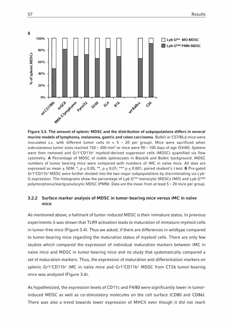
57 Results
Figure 3.5. The amount of splenic MDSC and the distribution of subpopulations differs in several
murine models of lymphoma, melanoma, gastric and colon carcinoma. Balb/c or C57/BL6 mice were
inoculated s.c. with different tumor cells (n = 5 – 20 per group). Mice were sacrificed when
subcutaneous tumor sizes reached 150 – 200 mm2 or mice were 90 – 100 days of age (SV40). Spleens
were then removed and Gr1+CD11b+ myeloid-derived suppressor cells (MDSC) quantified via flow
cytometry. A Percentage of MDSC of viable splenocytes in Black/6 and Balb/c background. MDSC
numbers of tumor bearing mice were compared with numbers of iMC in naïve mice. All data are
expressed as mean + SEM. *, p < 0.05; **, p < 0.01; *** p < 0.001, paired student’s t test. B Pre-gated
Gr1+CD11b+ MDSC were further divided into the two major subpopulations by discriminating via Ly6-
G expression. The histograms show the percentage of Ly6-Glow monocytic MDSCs (MO) and Ly6-Ghigh
polymorphonuclear/granulocytic MDSC (PMN). Data are the mean from at least 5 – 20 mice per group.
3.2.2 Surface marker analysis of MDSC in tumor-bearing mice versus iMC in naïve
mice
As mentioned above, a hallmark of tumor-induced MDSC is their immature status. In previous
experiments it was shown that TLR9 activation leads to maturation of immature myeloid cells
in tumor-free mice (Figure 3.4). Thus we asked, if there are differences in wildtype compared
to tumor-bearing mice regarding the maturation status of myeloid cells. There are only few
studies which compared the expression of individual maturation markers between iMC in
naïve mice and MDSC in tumor-bearing mice and no study that systematically compared a
set of maturation markers. Thus, the expression of maturation and differentiation markers on
splenic Gr1+CD11b+ iMC in naïve mice and Gr1+CD11b+ MDSC from CT26 tumor-bearing
mice was analyzed (Figure 3.6).
As hypothesized, the expression levels of CD11c and F4/80 were significantly lower in tumor-
induced MDSC as well as co-stimulatory molecules on the cell surface (CD80 and CD86).
There was also a trend towards lower expression of MHCII even though it did not reach

Results 58
statistical significance. In addition, the expression of Ly6G and Ly6C as subset-defining
markers was changed contrary to the changes seen after TLR9 activation with upregulation
of the Ly6G- and downregulation of the Ly6C antigen. That in turn reflects the substantial
increase in PMN-MDSC and the relative decrease in MO-MDSC during tumorigenesis (Figure
3.5 B). Summarized, it is shown that MDSC are in fact less matured than their physiological
counterparts which can objectified by analyzing a set of maturation markers. The same results
were shown for MDSC and iMC in bone marrow, blood and lymph nodes, even though the
downregulation of maturation markers in tumor-bearing mice was less pronounced in the
bone marrow compared to peripheral compartments (data not shown).
Figure 3.6. Tumor-induced MDSC exhibit less maturation markers than naïve iMC. Balb/c mice
were inoculated subcutaneously with CT-26 colon carcinoma cells. When tumor size reached at least
150 mm², mice were sacrificed, spleens removed and splenocytes were analyzed by flow cytometry.
The histograms show representatives of maturation marker expression (MFI) on pre-gated splenic
Gr1+CD11b+ iMC in tumor free mice (grey curve) vs. Gr1+CD11b+ MDSC in CT26 colon cancer bearing
mice (black curve). Statistics are calculated from at least 2 – 3 independent experiments (n = 5 mice
per group in each experiment). Data analysis was performed using paired Student’s t test comparing
mean fluorescent intensity (MFI) of each surface marker. p < 0.05 was considered as significant.
3.2.3 Different immunosuppressive activity of MDSC subpopulations
With previous experiments showing that PMN-MDSC were the main population expanding
during tumor growth, we decided to investigate the immunosuppressive effect of individual
subpopulations. Therefore a magnetic-based technique to isolate Ly6Glow MO-MDSC and
Ly6Ghigh MDSC was developed (see chapter 2.5.3.2). Mice bearing subcutaneous RMA-S
lymphoma were sacrificed and subpopulations were isolated and separately co-cultured with
CD3/CD28-stimulated splenocytes for 72 hours. T cell proliferation was analyzed using a
BrdU proliferation ELISA.

59 Results
Figure 3.7 shows that PMN-MDSC more potently suppress T cell proliferation than MO-
MDSC. That was consistently observed throughout all experiments and was not restricted to
the RMA-S lymphoma model but also shown for CT26 [Zoglmeier et al., 2011] and B16 tumor-
bearing mice (data not shown). In addition, both tumor-derived MDSC subpopulations were
significantly more suppressive than their respective counterparts from tumor-free mice (data
not shown).
Figure 3.7. PMN-MDSC bear higher immunosuppressive capacities than MO-MDSC in tumor-
bearing mice. Mice bearing subcutaneous RMA-S lymphoma (at least 150 mm² size) were sacrificed,
spleens removed and MDSC subpopulations were purified by sequential MACS separation [PMN =
polymorphonuclear MDSC; MO = monocytic MDSC]. Purity was verified via flow cytometry and was at
least > 85% in each group. Degree of suppressivity was assessed with a T-cell proliferation assay using
CD3/CD28-stimulated splenocytes (SC) as responder cells cocultured with either PMN-MDSC or MO-
MDSC for 72 hours (MDSC-splenocyte-ratio 1:8, 1:4, 1:2 with anti-CD3/CD28-stimulated SC as positive
control). Data are expressed as mean + SEM. *, p < 0.05; **, p < 0.01, paired Student’s t test.
3.3 Effect of immunotherapy with pattern recognition receptor ligands
on MDSC in tumor-bearing mice
3.3.1 MDSC numbers upon TLR ligand therapy in tumor-bearing mice
In previous experiments, we showed that innate immune activation with CpG-ODN and other
TLR ligands increases the number of Gr1+CD11b+ immature myeloid cells and concurrently
induces their maturation without further augmenting their immunosuppressive properties.
Likewise, it was shown that tumor growth inhibits their differentiation and induces MDSC,
especially the polymorphonuclear subset which is highly suppressive.

Results 60
In a next step, the effect of innate immune activation on the number, phenotype and function
of MDSC in tumor-bearing mice was examined. Based on prior experiments, the CT26 colon
carcinoma model was selected due to high numbers of MDSC and rapid tumor development.
Therefore, mice bearing subcutaneous CT26 were treated on day 14 and 17 with either
subcutaneous poly(I:C), CpG or sodium chloride (NaCl) as control. Therapy with TLR ligands
resulted in a potent tumor reduction compared to control mice (Figure 3.8 A). 48 hours after
the last therapy, mice were sacrificed and organs were further analyzed by flow cytometry.
As expected, tumor growth expanded the number of splenic Gr1+CD11b+ MDSC compared
to non-tumor bearing mice (Figure 3.8 D). Unexpectedly, mice treated with CpG-ODN
showed no further expansion of splenic Gr1+CD1b+ cells compared to control mice with 12 to
14% splenic MDSC in both groups (Figure 3.8 C,D). Same results were obtained when mice
were treated with poly(I:C). Likewise, no changes in absolute MDSC numbers exceeding the
percentage seen in untreated mice could be observed in peripheral blood or bone marrow
(data not shown).
Despite that IFNγ has been associated with MDSC expansion and activation in tumor-free
mice [Greifenberg et al., 2009], a common surrogate marker for evaluating MDSC-mediated
immunosuppression is the measurement of IFNγ serum levels, given the fact that MDSC
directly inhibit IFNγ production by T cells. Additionally, IFNγ is crucial for mounting an
effective immune response against malignant tumors [Shankaran et al., 2001] and reverses
the immunosuppressive properties of tumor-associated macrophages [Duluc et al., 2009].
Consequently, beside a potent IFNα induction by CpG-ODN and poly(I:C) [Krug et al., 2001;
Essers et al., 2009], measurement of serum cytokine levels showed that TLR9 activation with
CpG-ODN restored the CT26-induced IFNγ-depletion (Figure 3.8 B). Because the number of
MDSC was not altered by innate immune activation, we assessed the function of MDSC by ex
vivo proliferation assay as described before. It showed that suppressivity of isolated Ly6Ghigh
MDSC on T-cell proliferation and IFNγ production was significantly inhibited by CpG therapy.
For the Ly6Glow subpopulation, suppressivity was also reduced by CpG treatment, although
the effect was not significant [Zoglmeier et al., 2011].
Thus, we demonstrated that CpG-ODN do not further increase MDSC as key mediators of
immunosuppression in tumor bearing hosts and, moreover, reverse their immunosuppressive
capacities they exert on T cells. Moreover, TLR9 stimulation in vivo restores the tumor-
mediated IFNγ-depletion.

61 Results
Figure 3.8. Treatment of established CT26 tumor-bearing mice with TLR ligands does not further
induce of Gr1+CD11b+ MDSC. Balb/c mice were inoculated subcutaneously with CT-26 colon
carcinoma cells. Mice bearing established tumors (area at least > 25 mm²) were treated with CpG or
poly(I:C) on day 14 and 17 after tumor induction (n = 5 per group). A Tumors were continuously
measured with calipers every day in two dimensions. Mice were sacrificed two days after second
therapy (day 19) and used for further analysis. B Serum levels of IFNγ in CT26 tumor-bearing mice
before and after CpG therapy. Naïve Balb/c mice are shown as controls (wt, white bar). Mice were bled
24 hours after last therapy and serum levels of IFNγ levels were measured via ELISA. C,D
Representative FACS plot and histograms showing the total number of splenic Gr1+CD11b+ MDSCs in
untreated (PBS) and CpG-treated mice. The number of MDSC in non tumor-bearing mice is depicted
as a control (white bar, C). p < 0.05 was considered as significant (paired Student’s t test).
3.3.2 MDSC subset composition and phenotype following systemic PRR ligation
Previous studies showed contradicting results regarding the role of different MDSC subsets
in tumor-mediated immunosuppression. Thus far, we showed that PMN- or Ly6Ghigh - MDSC
are the most prominent subset induced during tumor growth in various murine tumor models.
Compatibly, they seem to be the more potent suppressors of T cell proliferation (see chapter

Results 62
3.2). Observing that treatment of tumor bearing mice with CpG-ODN not further induces
myeloid-derived suppressor cells in total accompanied by a marked reduction in tumor size,
in a next step we asked whether systemic Pattern recognition receptor ligation might
influence the subset composition of tumor-induced MDSC.
Therefore, mice with established CT26 colon carcinoma, B16 melanoma or RMA-S lymphoma
were challenged with 9.2RNA (TLR7), poly(I:C) (TLR3), CpG-ODN (TLR9) and ppp-RNA (RIG-
I). Mice were than sacrificed and analyzed for MDSC subsets (Figure 3.9 A). As expected, in
all three tumor models, systemic MDSC expansion was markedly due to a high number of
Ly6Ghigh PMN-MDSC. This was most prominent in spleens of CT26 colon carcinoma bearing
mice, but also visible in RMA-S lymphoma and B16 melanoma. Furthermore, when mice were
challenged with the respective Pattern recognition receptor ligand, we saw a strong and
significant reduction of PMN-MDSC with augmentation of the Ly6Glow MO-MDSC subset. The
effect was mostly prominent for CpG but to a lesser extent also seen upon therapy with other
PRR ligands. However, as described previously, the total number of MDSC remained
unchanged. Again, the effect was not restricted to spleens alone but was likewise confirmed
in bone marrow, blood and tumor-draining lymph nodes, even though less pronounced in
bone marrow (data not shown).
Splenic Ly6Ghigh PMN-MDSC and Ly6Glow MO-MDSC subsets were than further analyzed for
maturation and differentiation marker expression by flow cytometry. Given that one of the
defining traits of MDSC is their immature status and that we could previously show that
MDSC of tumor-bearing mice challenged with CpG exhibit less immunosuppressive
capacities, we thus hypothesized that systemic TLR ligation alters their phenotype towards
matured myeloid cells. Indeed, in vivo treatment with CpG led to an increase in expression of
the macrophage differentiation marker F4/80, the DC differentiation marker CD11c and the
co-stimulatory molecule CD80. As shown in wild-type mice before, we observed a highly
significant upregulation of Sca-1 on both MDSC subsets again. Table 3 summarizes and
compares different maturation, differentiation and activation marker in naïve and CT26
tumor-bearing mice with or without CpG therapy.

63 Results
Naïve vs. CT26 Naïve vs naïve + CpG CT26 vs CT26 + CpG
Marker Change (p) Change (p) Change (p)
Ly6C - < 0.001 + < 0.001 + < 0.001
Ly6G + < 0.01
- < 0.05 - < 0.01
CD11b + < 0.05 + < 0.01 + < 0.05
Sca-1 - < 0.05 + < 0.001 + < 0.001
CD11c - < 0.05
+ < 0.01 + < 0.05
MHCII - < 0.05 + < 0.05 + < 0.05
F4/80 - < 0.05
+ < 0.05 (+) 0.08
CD80 - < 0.01
(+) 0.07 + < 0.01
CD86 - < 0.05 (+) 0.09 (+) 0.059
CD88 (+) 0.088 n.t. n.t. - < 0.01
CD124 (+) 0.088
- < 0.05
= n.s.
CD62L = n.s.
= n.s.
= n.s.
CD31 (+) 0.16 n.t. n.t. (-) 0.09
Table 3. Comparison of maturation, differentiation and activation marker on Gr1+CD11b+ myeloid-
derived suppressor cells in naïve and tumor-bearing with or without treatment with CpG. n.t. : not
tested, = : not significant (n.s.), - : significant downregulation, + : significant upregulation, (+) : trend
towards upregulation, (-) : trend towards downregulation.
Thus, we demonstrated that challenging tumor-bearing mice with CpG-ODN and other TLR
ligands, is able to overcome the tumor-induced differentiation block of myeloid cell
differentiation [Gabrilovich and Nagaraj, 2009], restores expression levels of myeloid cell
markers to those expressed in Gr1+CD11b+ cells in tumor-free mice and reverses the
imbalance of Ly6Ghigh/Ly6Glow MDSC to a proportion seen in naïve mice.
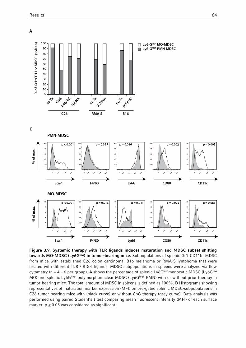
Results 64
Figure 3.9. Systemic therapy with TLR ligands induces maturation and MDSC subset shifting
towards MO-MDSC (Ly6Glow) in tumor-bearing mice. Subpopulations of splenic Gr1+CD11b+ MDSC
from mice with established C26 colon carcinoma, B16 melanoma or RMA-S lymphoma that were
treated with different TLR / RIG-I ligands. MDSC subpopulations in spleens were analyzed via flow
cytometry (n = 4 – 6 per group). A shows the percentage of splenic Ly6Glow monocytic MDSC (Ly6Glow
MO) and splenic Ly6Ghigh polymorphonuclear MDSC (Ly6Ghigh PMN) with or without prior therapy in
tumor-bearing mice. The total amount of MDSC in spleens is defined as 100%. B Histograms showing
representatives of maturation marker expression (MFI) on pre-gated splenic MDSC-subpopulations in
C26 tumor-bearing mice with (black curve) or without CpG therapy (grey curve). Data analysis was
performed using paired Student’s t test comparing mean fluorescent intensity (MFI) of each surface
marker. p < 0.05 was considered as significant.
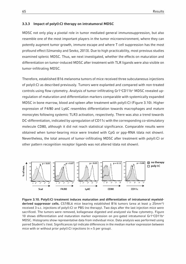
65 Results
3.3.3 Impact of poly(I:C) therapy on intratumoral MDSC
MDSC not only play a pivotal role in tumor mediated general immunosuppression, but also
resemble one of the most important players in the tumor microenvironment, where they can
potently augment tumor growth, immune escape and where T cell suppression has the most
profound effect [Umansky and Sevko, 2013]. Due to high practicability, most previous studies
examined splenic MDSC. Thus, we next investigated, whether the effects on maturation and
differentiation on tumor-induced MDSC after treatment with TLR ligands were also visible on
tumor-infiltrating MDSC.
Therefore, established B16 melanoma tumors of mice received three subcutaneous injections
of poly(I:C) as described previously. Tumors were explanted and compared with non-treated
controls using flow cytometry. Analysis of tumor-infiltrating Gr1+CD11b+ MDSC revealed up-
regulation of maturation and differentiation markers comparable with systemically expanded
MDSC in bone marrow, blood and spleen after treatment with poly(I:C) (Figure 3.10). Higher
expression of F4/80 and Ly6C resembles differentiation towards macrophages and mature
monocytes following systemic TLR3 activation, respectively. There was also a trend towards
DC-differentiation, indicated by upregulation of CD11c with the corresponding co-stimulatory
molecule CD80, although it did not reach statistical significance. Comparable results were
obtained when tumor-bearing mice were treated with CpG or ppp-RNA (data not shown).
Nevertheless, the total amount of tumor-infiltrating MDSC after treatment with poly(I:C) or
other pattern recognition receptor ligands was not altered (data not shown).
Figure 3.10. Poly(I:C) treatment induces maturation and differentiation of intratumoral myeloid-
derived suppressor cells. C57/BL6 mice bearing established B16 tumors (area at least > 25mm²)
received 3 s.c. injections of poly(I:C) or PBS (no therapy). Two days after the last injection mice were
sacrificed. The tumors were removed, kollagenase digested and analyzed via flow cytometry. Figure
10 shows differentiation and maturation marker expression on pre-gated intratumoral Gr1+CD11b+
MDSC. Histograms show representative data from individual mice. Data analysis was performed using
paired Student’s t test. Significances (p) indicate differences in the median marker expression between
mice with or without prior poly(I:C) injections (n = 5 per group).

Results 66
3.3.4 Phenotypical changes on MDSC upon systemic TLR stimulation are IFNα-
dependent
After establishing that therapy with different TLR agonists and the RIG-I agonist 5’-
triphosphate-RNA is able to induce maturation, differentiation and a population shift of
myeloid-derived suppressor cell subsets, accompanied by a marked reduction in tumor size,
we then asked what factors might contribute to the observed effects.
Maturation of MDSC as well as blocking their immunosuppressive properties was less
prominent for the TLR7/8 agonist 9.2dr-RNA, R848 and the TLR4 agonist LPS (data not
shown) and was most prominent for CpG-ODN (TLR9), poly(I:C) (TLR3) and 3p-RNA (RIG-I).
When comparing differences and similarities of the systemically induced cytokine profile by
these PRR ligands, a major difference of the latter is their greater ability to induce high
amounts of IFNα [de Clercq, 1980; Krug et al., 2001; Hornung et al., 2006], suggesting IFNα
as a major contributor for MDSC maturation and differentiation in vivo.
To test this hypothesis, we used the immunostimulatory RNA ligand poly(I:C) as one the
strongest IFNα inducers throughout our experiments. Wildtype and type I IFN-receptor
deficient mice (IFNαR-/-) were subcutaneously inoculated with B16 melanoma cells and
treated with one dose of poly(I:C) or PBS as a control. Notably, the sole deficiency of type I
interferon signaling in IFNαR-/- mice was sufficient to induce a population shift towards
higher numbers of monocytic Ly6Glow MDSC, accompanied by a decrease in
polymorphonuclear MDSC (Figure 3.11 A). There were no differences seen when comparing
MDSC subset composition in wildtype mice bearing B16 tumors after one dose of poly(I:C),
indicating that the effect of subset recomposition may take longer than the 24 hours. When
performing maturation marker analysis, one dose of poly(I:C) resulted in phenotypic and
functional changes in MDSC similar to those induced by CpG. Treatment with poly(I:C) was
sufficient to significantly upregulate Sca-1 and other maturation markers (Figure 3.11 B).
Strikingly, this upregulation was completely abolished in IFNαR-/- mice, indicating that type
I IFN is critically involved in MDSC maturation following innate immune activation.
Performing flow cytometry analysis of collagenase digested B16 tumors showed comparable
results with intratumoral maturation and differentiation of tumor-infiltrating MDSC following
IFNα-induction with poly(I:C), yet without altering the total number of MDSC infiltrating the
tumor tissue (data not shown).

67 Results
Figure 3.11. Maturation, differentiation and shifting of MDSC subpopulations after treatment with
TLR ligands is IFNα-dependent. Wild type or type I IFN-receptor deficient (IFNαR-/-) mice bearing
s.c. B16 tumors (tumor area approximately 50 mm²) were treated once with poly(I:C) or PBS (no
therapy). One day after injection mice were sacrificed. Organs were removed, tumors collagenase
digested and analyzed via flow cytometry. A shows the percentage of splenic Ly6Glow monocytic MDSC
(Ly6Glow MO) and splenic Ly6Ghigh polymorphonuclear MDSC (Ly6Ghigh PMN) with or without prior
poly(I:C) therapy. The whole amount of MDSC (not shown) is defined as 100%. B shows differentiation
and maturation marker expression on pre-gated splenic Gr1+CD11b+ MDSC. Surface markers are
demonstrated and compared in bar graphs as MFI (mean fluorescent intensity).
3.3.5 Role of ppp-RNA and TGFβ-silencing on MDSC in tumor-bearing mice
In preceding experiments we have observed that treatment of tumor-bearing mice with
distinct pattern recognition receptor ligands, especially those that induce high amounts of
IFNα such as CpG, poly(I:C) and 5’triphosphate-RNA, cause maturation and differentiation of
systemic and intratumoral myeloid-derived suppressor cells. We showed that even though
PRR ligation has pleiotropic downstream signaling effects, the effect of abolishing the
maturation block, changing MDSC subset composition and attenuating their

Results 68
immunosuppressive properties is largely dependent on type I IFN signaling [Zoglmeier et al.,
2011].
However, numerous other tumor-derived factors and cytokines may lessen the beneficial
effect of an IFNα-mediated MDSC maturation following innate immune stimulation. TGFβ has
been emerged as one of the most crucial cytokines involved in MDSC accumulation in line
with prevention of DC maturation and polarization of myeloid cells towards immune
suppressive cells in the tumor microenvironment [Flavell et al., 2010]. In addition, Fridlender
and colleagues showed that TGFβ-blockage results in polarization of tumor-associated
neutrophils or Ly6Ghigh MDSC towards an antitumoral neutrophil population.
Hence, we wanted to investigate whether the maturation and differentiation of MDSC by
systemic IFNα-induction following innate immune stimulation can be further augmented by
concurrent blockage of TGFβ. Therefore a bifunctional ppp-siRNA was used that combines
RIG-I activation for IFNα induction with gene silencing of TGFβ1 and its therapeutic efficacy
was studied in the orthotopic Panc02 mouse model of pancreatic cancer [Ellermeier et al.,
2013].
Treatment of PancO2-bearing mice with si-TGFβ or ppp-TGFβ led to a significant decrease
of circulating myeloid-derived suppressor cells, illustrated by the percentage of Gr1+CD11b+
MDSC found in spleen (Figure 3.12 A). This effect was partially depending on TGFβ-blockage,
although there was a trend towards further decrease in circulating MDSC with the
triphosphate moiety and subsequent RIG-I activation. As hypothesized, downregulation of
TGFβ by siRNA resulted in a highly significant change in subset composition with a decrease
of polymorphonuclear Ly6Ghigh MDSC and a relative increase of the monocytic subset. A trend
towards this change in subset composition could also be demonstrated for 3p-RNA alone,
albeit it lacked significance (data not shown, also see Figure 3.9 A). Again, by combining RIG-
I activation and TGF-blockage with bifunctional ppp-TGFβ-siRNA, the shift in myeloid cell
subset composition could not be further augmented. When analyzing apoptosis on an
individual cell basis via measurement of Caspase-9 activity (FLICA), splenic Gr1+CD11b+
MDSC showed a significantly higher rate of apoptotic cell death compared to untreated mice.
A more detailed analysis of MDSC subpopulations revealed, that the induction of apoptosis
in this experimental setting on PMN-MDSC is largely dependent on TGFβ-blockage, whereas
apoptosis in the monocytic subset is primarily induced by RIG-I activation via the 5’-
triphosphate moiety (Figure 3.12 B).
To examine whether TGFβ-blockage alone or in combination with RIG-I activation can also
promote MDSC differentiation in tumor bearing mice, Gr-1+CD11b+ MDSC were assessed on

69 Results
their expression of CD80, CD11c, Ly6C and Sca-1 (Figure 3.12 C). Knockdown of TGFβ via
siRNA was sufficient to induce monocytic cell differentiation in MDSC illustrated by
upregulation of Sca-1 and Ly6C [Serbina et al., 2009]. However, DC differentiation with
higher expression levels of CD11c and the co-stimulatory molecule CD80 was solely induced
via the accessory triphosphate moiety with subsequent RIG-I activation.
MDSC are major inhibitors of cytotoxic T lymphocytes, inducing T cell anergy and negatively
correlate with the influx of CD8+ tumor-infiltrating cytotoxic T lymphocytes [Gabrilovich and
Nagaraj, 2009; Curran et al., 2010]. By analyzing tumor slices via fluorescence microscopy
we examined the influx of cytotoxic CD3+CD8+ T lymphocytes (CTLs) following therapy with
bifunctional ppp-siRNA against TGFβ (Figure 3.12). We saw a significant increase in CTL-
infiltration following TGF-beta blockage with either siTGFβ or ppp-TGFβ compared to control
OH-RNA (Figure 3.12 D,E). Systemic RIG-I activation via a –ppp moiety was not able to further
increase CTL influx, although a trend was visible. This might serve as a proxy of the observed
pro-apoptotic and differentiating effects of TGFβ blockage and RIG-I activation on MDSC in
this setting. Nevertheless, a casual link between both has yet to be determined.

Results 70
Figure 3.12. Increased apoptosis and maturation of MDSC by TGFβ blockage and IFNα induction
via bifunctional 5’triphosphate-siTGFβ RNA. Mice bearing orthotopic PancO2 tumors were
subsequently treated intravenously with 50 µg RNA on days 12 and 14 with unspecific RNA (control-
OH; white bar), small-interfering RNA directed against TGFβ1 (siTGFβ; grey bar) or bifunctional
5’triphophosphate-siTGFβ RNA (ppp-TGFβ; black bar). Spleens and tumors were removed on day 15
and further analyzed by flow cytometry and immunofluorescence. A Percentage of Gr-1+CD11b+ MDSC
(gated on live cells) and respective subpopulations (gated on MDSC). B Degree of apoptosis in MDSC
was assessed by measuring Caspase-9 (FLICA) activity via flow cytometry in PMN-MDSC or MO-
MDSC. C Maturation and differentiation of Gr-1+CD11b+ MDSC as assessed by mean fluorescence
intensity (MFI) of CD80, CD11c, Ly6C and Sca-1, respectively. D Tumor slices were stained with Cy2
anti-CD8 (green) and Cy3 anti-CD3 (red). Representative pictures of CD3+CD8+ tumor-infiltrating
cytotoxic T-lymphocytes (CTLs) into tumor tissue. E shows the average number of CTLs per visual
field. Cell infiltration was averaged measuring at least 15 visual fields per sample out of 3-5 different
tumor samples. Pooled data of 4 – 6 mice per group are shown as means + SEM. * indicates p < 0.05.
** indicates p < 0.01. *** indicates p < 0.001. All data are expressed as mean ± SEM. A two-tailed p-
value of < 0.05 was considered significant.
3.4 Characterization of stem cell antigen-1 (Sca-1) on MDSC
Following innate immune stimulation and therapy of tumor-bearing mice with different PRR
ligands, the Ly6 protein family member Sca-1 (Ly6A/E) has been shown to be one of the most
significantly upregulated proteins on MDSC among the surface proteins throughout our

71 Results
experiments. Stem cell antigen-1 (Sca-1) or lymphocyte activation protein-6A (Ly6A) is a GPI-
anchored protein and is localized to lipid rafts of the plasma membrane [Curran et al., 2010].
It has been originally identified as an antigen upregulated on activated lymphocytes more
than 30 years ago [Yutoku et al., 1974].
In prior experiments we had shown for the first time that Sca-1 is strongly upregulated by
MDSC in response to CpG-ODN, poly(I:C) or 5’triphosphate-RNA and that it represents a
highly sensitive activation/differentiation markers for this cell population. Furthermore, its
upregulation inversely correlates with the suppressive activity of MDSC. Therefore, we aimed
to further dissect the role of Sca-1 on MDSC mediated immunosuppression and tumor
development.
3.4.1 Expression of Sca-1 in different tissues and MDSC subpopulations
Using flow cytometry, we first started to analyze the expression of Sca-1 on Gr1+CD11b+
immature myeloid cells (iMC) in non-tumor-bearing mice. Mean fluorescence intensity of
Sca-1 on iMC was lowest in bone marrow with induction of its expression after peripheral
homing of immature myeloid cells into spleen, lung and peripheral lymph nodes with the
highest measurable Sca-1 expression in lymphoid tissue (Figure 3.13 A). Surprisingly, when
comparing tumor-induced splenic MDSC and tumor-associated MDSC in CEA424-TAg mice
bearing spontaneous gastric tumors, tumor-associated MDSC showed three-fold higher
expression levels of Sca-1 in comparison to splenic MDSC (Figure 3.13 C). In a next step, we
compared Sca-1 expression on splenic immature myeloid cells in tumor-free mice versus
tumor-induced myeloid derived suppressor cells showing that MDSC express significantly
lower levels of Sca-1 than their naïve counterparts. In addition, when comparing MDSC
subpopulations on their differential expression of Sca-1, monocytic Ly6Chigh MDSC express
higher amounts of Sca-1 on their cell surface than granulocytic PMN-MDSC (Figure 3.13 B).
We thus show that Sca-1 is upregulated during homing of Gr1+CD11b+ myeloid cells into
peripheral organs and even tumor tissue. Furthermore, Sca-1 seems to be downregulated on
MDSC via tumor-derived factors, which complements previous results showing Sca-1
upregulation via TGFβ-blockage (Figure 3.12 C). In addition, when performing detailed flow
cytometry analysis of Sca-1+Gr1+CD11b+ cells compared to Sca-1-Gr1+CD11b+ cells, the
expression of Sca-1 strongly correlated with a more mature and differentiated MDSC
phenotype in both naïve and tumor-bearing mice (data not shown).

Results 72
Figure 3.13. Sca-1 expression on MDSC is tissue-dependent and differs between naïve iMC,
MDSC-homing site and MDSC subpopulations. A Naïve Balb/c mice (n = 3) were sacrificed and Sca-
1 expression on Gr1+CD11b+ iMCs was analyzed via flow cytometry in indicated tissues (pLN:
peripheral lymph nodes). Histograms display an example of representative individual mice. B Balb/c
mice were inoculated subcutaneously with CT26 colon carcinoma cells. Mice bearing established
tumors (area at least > 50 mm²) are compared with non-tumor bearing mice (n = 5 per group). Bar
graphs show the amount of Sca-1+ MDSC in spleen (left) and Sca-1 expression on MDSC
subpopulations (right) displayed as Mean Fluorescent intensity (MFI). C MDSC in CEA424-TAg mice
bearing spontaneous gastric tumors (SV40; mice aged 90 – 100 days; n = 3 per group) were analyzed
via flow cytometry. Bar graphs show the mean fluorescent intensity (MFI) of Sca-1 on splenic and
intratumoral Gr1+CD11b+ MDSC. Analysis was performed using paired Student’s t test. Significances
are depicted in the left upper corners.
3.4.2 Role of Sca-1 in tumor development and MDSC-mediated immunosuppression
To further evaluate the role of Sca-1 in tumor formation and to evaluate a functional role in
MDSC-mediated immune escape, wild type C57/BL6 mice and mice deficient in stem cell
antigen-1 (Sca-1-/-) were subcutaneously inoculated with PancO2 cells. Tumor formation was
then continuously measured. When mice were sacrificed, we purified Gr1+CD11b+ MDSC
from spleens as described previously and performed a T cell suppression assay. Based on

73 Results
prior experiments, we hypothesized that Sca-1 may act as a negative regulator of MDSC-
mediated tumor tolerance, thereby promoting a more efficient immune answer.
To our surprise, when we compared Sca-1-/- MDSC with wild type MDSC on their capability
to suppress CD3/CD28-activated splenocyte proliferation ex vivo, MDSC lacking Sca-1 were
significantly less suppressive than their naïve counterparts (Figure 3.14 B). In addition, Sca-
1-/- mice showed a trend towards slower tumor growth, even it did not reach statistical
significance (p = 0.11). In contrast to what we had expected, we thus show for the first time
that Sca-1 functionally fosters the capability of myeloid-derived suppressor cells to suppress
T cell proliferation and thereby might attenuate immune surveillance of malignant tumors.
Figure 3.14. Sca-1-/- mice have less immunosuppressive MDSC without significant alteration of
tumor growth. A C57/BL6 and Sca-1-/- mice were inoculated subcutaneously with PancO2 pancreatic
carcinoma cells (n = 5 – 8 per group). Tumors were continuously measured with calipers in two
dimensions. Mice were sacrificed on day 32 after tumor inoculation when tumor size reached at least
> 75 mm2 in each group. B MDSC were purified from spleens of wildtype and Sca-1-/- PancO2-bearing
mice by sequential MACS separation. Purity was verified via flow cytometry and was > 80% in each
group. Degree of MDSC-mediated suppressivity was assessed with a T-cell proliferation assay using
CD3/CD28-stimulated splenocytes (SC) as responder cells cocultured with MDSC for 48 hours [MDSC-
splenocyte-ratio 1:16, 1:8, 1:4, 1:2 with anti-CD3/CD28-stimulated SC as positive control (SC, act)]. p
< 0.05 was considered as significant.
3.4.3 Induction of Sca-1 on MDSC via TLR ligands in vivo and in vitro
Preceding experiments using immunotherapy with different TLR ligands in vivo showed a
significant upregulation of Sca-1 on MDSC in tumor bearing mice, concurrent with an inverse
relation of Sca-1 expression with immunosuppressive properties of Gr1+CD11+ MDSC.
Throughout our experiments, especially PRR ligands known for their potential to induce high
amounts of type I interferons after receptor ligation such as CpG-ODN (TLR9), poly(I:C)
(TLR3) and 3p-RNA (RIG-I) were the most efficient inducers of Sca-1 on MDSC in vivo.

Results 74
To validate this finding, CT26 tumor-bearing mice received three repeated injections of CpG-
ODN and poly(I:C) again, as we and others have shown a major type I interferon response in
vivo following treatment with these immunostimulatory oligonucleotides [de Clercq, 1980;
Krug et al., 2001]. Indeed, Sca-1 expression on myeloid derived suppressor cells was
upregulated four- to tenfold as compared to untreated mice (Figure 3.15 A).
In a next step, we used B16 melanoma-bearing IFNαR-/- mice and injected poly(I:C) as it
appeared to be the strongest inducer of IFNα with subsequent upregulation of Sca-1 on
Gr1+CD11b+ cells (Figure 3.15 B). Whereas poly(I:C) treatment of wild type mice led to Sca-
1 upregulation on splenic as well as intratumoral MDSC, its expression was completely
abrogated in IFNαR-/- mice. This is supported by recent literature by Essers and colleagues,
showing that Sca-1 is inducible via IFNα in a STAT1 dependent fashion in vivo [Essers et al.,
2009].
In parallel experiments we found that abrogation of MDSC suppressivity, their maturation
and Sca-1 induction following CpG treatment, is mainly mediated by type I IFN induction via
plasmacytoid dendritic cells, rather than a direct effect of TLR9 activation on MDSC
[Zoglmeier et al., 2011]. To test whether direct stimulation of Gr1+CD11b+ immature myeloid
cells with different Toll-like receptor ligands is able to induce Sca-1 expression, we purified
Gr1+CD11b+ immature myeloid cells from bone marrow via magnetic sorting. Cell purity was
greater than 98% which was validated via flow cytometry. As hypothesized, IFNα induces
the highest Sca-1 expression on immature myeloid cells, at the same time proving that
iMC/MDSC express a functional type I interferon receptor (Figure 3.15 C). Neither CpG-ODN
or poly(I:C) were able to induce Sca-1 expression in vitro. In contrast, LPS (TLR4) and R848
(TLR7) as well as the combination of Pam3CysK (TLR2) and Curdlan, a beta-1,3-glucan-
polymer binding to Dectin-1, significantly induced Sca-1 expression in immature myeloid
cells. Neither Pam3CysK nor Curdlan alone were able to upregulate Sca-1 alone, which
supports findings of Ferwerda and colleagues in 2008, showing that dectin-1 amplifies TLR2-
dependent induction of cytokines [Ferwerda et al., 2008].
Thus, we show that upregulation of Sca-1 on Gr1+CD11b+ immature myeloid cells upon
stimulation with CpG and poly(I:C) in vivo is mainly mediated via indirect induction of IFNα
as direct stimulation of their corresponding Toll-like receptor on iMC did not result in a
significant upregulation of Sca-1. Concurrently, we show that immature myeloid cells express
functional TLR2, TLR4, TLR 7 and Dectin-1 and to a certain extent, Sca-1 expression is also
inducible via their direct activation.

75 Results
Figure 3.15. TLR ligands induce Sca-1 on MDSC in vivo and in vitro with an essential role for IFNα.
A CT26 tumor-bearing mice received 3 s.c. injections of CpG, poly(I:C) or PBS (no therapy). Two days
after the last injection mice were sacrificed and Sca-1 expression on splenic myeloid-derived

Results 76
suppressor cells was analyzed via flow cytometry (MFI = mean fluorescent intensity). Pooled data of 4
– 6 mice per group are shown as means + SEM. **** indicates p < 0.0001. ***** indicates p < 0.00001.
B Wildtype or IFN-I receptor deficient (IFNαR-/-) mice bearing s.c. B16 tumors (tumor area
approximately 50 mm²) were treated once with poly(I:C) or PBS (no therapy). Organs were removed,
tumors kollagenase digested and analyzed via flow cytometry. Histograms show the changes in the
expression of Sca-1 on splenic and intratumoral MDSC two days after treatment; an example of
representative individual mice is depicted [gray, no therapy; black line, poly(I:C)]. Significances (p)
indicate differences in median expression (n = 5 per group). C Gr1+CD11b+ iMC from bone marrow of
naïve C57/BL6 mice were purified, plated in 96-well and cultured in vitro with indicated stimuli. Purity
of Gr1+CD11b+ was greater than 98% (validated via FACS). Sca-1 expression (MFI) on iMC was
evaluated after 24 hours of stimulation via flow cytometry. [CpG:TLR9; R848:TLR7; IFNα:IFNαR;
Flagellin:TLR5; Curdlan:Dectin-1; Pam3CysK:TLR2; TGFβ:TGFβR; LPS:TLR4; poly(I:C):TLR3].
Significances (p) indicate differences in the median marker expression (n = 3 per group). A two-tailed
p-value < 0.05 was considered significant (Student’s t test).
3.4.4 Analysis of cytokine induction in Sca-1-/- mice upon PRR ligation
On the one hand we found out that the induction of IFNα via innate immune receptors is
critically involved in maturation, differentiation and attenuation of MDSC suppressivity, along
with a highly significant increase in the expression of stem cell antigen-1. In addition, we
unexpectedly found that myeloid-derived suppressor cells deficient for Sca-1 exhibit less
immunosuppressive properties in tumor-bearing mice in conjunction with a trend towards
slower tumor growth in vivo. On the other hand, increasing evidence in the literature
implicated that Sca-1 might not only be a bystander protein indicating T cell activation or
serving as a phenotypical marker for lineage determination in haematopoietic stem cells [Ma
et al., 2002; Holmes and Stanford, 2007], but may also play a functional role in distinct
signaling pathways, e.g. type I interferon and TGFβ signaling [Essers et al., 2009, Upadhyay
et al., 2011].
Based on that findings and in order to dissect functional differences between Sca-1-/- and
wildtype mice, C57/BL6 and Sca-1-/- mice received one injection of poly(I:C). Twenty hours
later mice were bled and serum was tested for cytokine levels. Based on findings of other
groups that Sca-1 may prohibit downstream type I interferon signaling, we hypothesized
lower levels of IFNα upon systemic TLR3 activation via poly(I:C). To our surprise, serum levels
of IFNα were comparable in wild type and Sca-1-/- mice (Figure 3.16 A, left column). When
we further screened for serum levels of other cytokines, no differences in the induction of IL-
6, IL12p70, TNFα or IL1β could be detected (data not shown). Incidentally, we detected high
levels of IP-10/CXCL-10 in Sca-1 deficient mice upon therapy with poly(I:C) (Figure 3.19 A,
right column). IFNγ-inducible protein of 10 kDa or IP-10/CXCL10 is an early response gene
mainly induced by Interferon-γ but also type I interferons, LPS or CpG-ODN [Neville et al.,
1997; Imaizumi et al., 2013] and is critically involved into effector T cell trafficking to sites of

77 Results
inflammation [Dufour et al., 2002]. Experimental models also demonstrated that CXCL10/IP-
10 has antitumorigenic properties and reduces angiogenesis [Narvaiza et al., 2000].
Next, we wanted to evaluate if this pronounced induction of IP-10 in vivo is either an indirect
effect of TLR3 activation via other soluble mediators/cytokines and likewise, if stimulation of
other pattern recognition receptor pathways also triggers a more pronounced IP-10
induction. Therefore, bone marrow-derived macrophages (BMDM) from wild type and Sca-
1-/- mice were generated as previously described [Manzanero, 2012], cultivated, plated in 96-
well plates and stimulated with various pattern recognition receptor ligands available in our
laboratory. BMDM were used because they resemble a common tool when analyzing innate
immune receptor signaling and cytokine production [Kobayashi et al., 2002].
Direct stimulation of wild type macrophages with poly(I:C), LPS, poly(dA:dT), 5’ppp-RNA and
IFNα/γ induced moderate levels of IP-10 secretion into the supernatant (Figure 3.16 B). In
this experimental setting, IP-10 induction via IFNγ at 100 U/ml was lower as expected after
24 hours of stimulation. When we repeated the same experimental setting with higher
concentrations of IFNγ (up to 500 U/ml), IP-10 induction was markedly increased in a dose
dependent manner (data not shown). When we measured IP-10 levels in the supernatant of
pattern recoginiton receptor stimulated Sca-1-/- BMDM, a highly significant increase in its
production compared to wildtype mice was observed. Especially stimulation of TLR3, TLR4,
the AIM2-inflammasome and RIG-I resulted in a three- to sixfold increase in IP-10 production
as compared to macrophages posessing the Sca-1 antigen. Comparable results (only less
pronounced) were obtained when bone marrow, splenocytes and magnetically sorted
Gr1+CD11b+ immature myeloid cells from wild type and Sca-1-/- mice were used in the same
experimental setting (data not shown).
Thus, we show for the first time that mice deficient for Stem cell antigen-1 show a pronounced
IP-10 production following innate immune stimulation with various PRR ligands in vivo and
in vitro. Whether that observation has a biological significance, or might even explain
reduced MDSC suppressivity of Sca-1-/- mice, needs to be elucidated in further studies.

Results 78
Figure 3.16. Pronounced IP-10 induction in vivo and in vitro following PRR ligation in Sca-1-/- mice.
A Naive C57/BL6 and Sca-1-/- mice (n = 5 per group) received one s.c. injection of 250 µg poly(I:C). 20
hours later mice were bled and serum was analyzed for the amount of IFNα, IL-6 and IP-10 via ELISA.
B Bone marrow-derived macrophages (BMDM) from wildtype (wt) and Sca1-/- mice were cultured,
plated in 96-well and stimulated for 24 hours with indicated PRR ligands. Supernatant was measured
for the amount of IP-10 via ELISA. ** indicates p < 0.01. *** indicates p < 0.001. **** indicates p <
0.0001. All data are pooled of w wells per group and expressed as mean ± SEM. A two-tailed p-value
of < 0.05 was considered significant.
79 Discussion
4 DISCUSSION
4.1 MDSC subpopulations
4.1.1 Differentiated view on MDSC subpopulations in tumor-bearing hosts
In recent years, the role of MDSC in tumor-associated immune suppression has been
established in a large number of clinical and preclinical studies. It quickly became apparent
that MDSC do not represent a single cell population, but are rather comprised of myeloid
cells at different stages of cell differentiation [Almand et al., 2001: Bronte, 2009]. Tumor-
induced MDSC are further dichotomized into two major subpopulations, in mice based on
their differential expression of Ly6G and Ly6C on the cell surface: polymorphonuclear
Gr1+CD11b+Ly6GhighLy6Clow MDSC and monocytic Gr1+CD11b+Ly6GnegLy6Chigh MDSC (MO-
MDSC) [Movahedi et al., 2008]. However, with more studies on MDSC biology, it becomes
more and more apparent that this classical dichotomy is more complex than originally
thought. Based on their differential expression of Gr-1, CD11b, scatter-appearance, other
surface expression markers such as CD115 and morphologic appearance, Greifenberg and
colleagues could identify six different populations in the Gr1+CD11b+ compartment with
varying immunosuppressive properties [Greifenberg et al., 2009]. Other groups made
attempts to further subdivide polymorphonuclear and monocytic MDSC in more or less
immunosuppressive phenotypes by using additional surface markers such as CD49d, CD115
or CD124/IL-4Rα [Mandruzzato et al., 2009; Haile et al., 2010].
Due to feasibility and the lack of clearly defined additional subpopulations of myeloid-derived
suppressor cells other than Ly6Ghigh PMN and Ly6Glow MO-MDSC, we have adopted the
classical division of MDSC in this work. All tumor models examined in this study showed a
variable but significant increase in the whole population of MDSC. Intriguingly, PMN-MDSC
were the most abundant cell type induced by tumor growth in vivo. This finding is supported
by the literature showing the granulocytic subset as a predominant population of MDSC in
tumor-bearing mice [Youn et al., 2008; Gabrilovich and Nagaraj, 2009]. However, in other
pathologic conditions, such as experimental autoimmune encephalitis, a mouse model of
multiple sclerosis, a significant increase in the number of CD11b+Ly6G-Ly6Chigh cells
resembling monocytic MDSC was observed that limited the course of disease. At the bottom
line, the basis for this subset dichotomy remains elusive as the exact nature of tumor-derived
factors that drive MDSC accumulation and define the expansion of a specific subset.
Likewise, in the context of tumor development, there is still ongoing debate whether
granulocytic or monocytic MDSC exploit more immunosuppressive features. In this work, we
consistently observed that the Ly6Ghigh fraction (when purified and compared to Ly6Glow

Discussion 80
MDSC) was substantially more suppressive. This was validated in the murine B16 melanoma,
CT26 colon carcinoma, RMA-S lymphoma and orthotopic SV40 gastric carcinoma model. In
contrast, a study using EL-4 lymphoblastic lymphoma, MO-MDSC have shown to inhibit T
cell proliferation more potently than PMN-MDSC [Movahedi et al., 2008]. Likewise, another
study by Dolcetti and colleagues proposed an inverse correlation of Gr1-brightness to the
immunosuppressive power of MDSC on Ag-stimulated CD8+ T cells in a C26-GM and 4T1
tumor model [Dolcetti et al., 2010]. On the contrary, our data is strengthened by a recent
study analyzing MDSC subpopulations from mice bearing 3LL, MCA-38 and EL-4 tumors that
shows that PMN-MDSC had higher ability to impair proliferation and expression of effector
molecules in activated T cells, thus inducing T cell anergy [Raber et al., 2014]. Regarding
human MDSC, a recent NIH study by Tim Greten and colleagues analyzed monocytic (CD14+)
and granulocytic (CD15+) MDSC in patients with gastrointestinal malignancies and found the
granulocytic fraction to be more suppressive than their counterparts [Duffy et al., 2013].
However, defining the exact differences between MDSC subsets in mice and particularly in
human has not reached a consensus yet. For the future studies of MDSC biology it will be
crucial to further dissect molecular pathways and mechanisms of immunosuppression which
are partially substantially different between MDSC subsets. Even though, these variations are
not overly surprising since the factors driving MDSC accumulation vary between different
tumor models and human cancers: If a tumor of a given type will produce a particular set of
MDSC driver molecules that induce a MDSC phenotype of a given type, another tumor entity
will produce other driver molecules that induce a different MDSC phenotype.
In addition, variations between different study groups regarding MDSC suppressivity and
their behavior following pharmacologic therapy may be explained by different isolation
techniques used for MDSC purification as well as the functional readout used for estimation
of their immunsosuppressive properties. In this study we used different magnetic-bead based
technologies to sort MDSC subpopulations with high purity. Nevertheless, by histologic
evaluation, purified Ly6Ghigh MDSC were exclusively polymorphonuclear, whereas the
nuclear morphology of Ly6Glow MDSC was more heterogeneous, comprising both monocyte-
like and polymorphonuclear cells [Zoglmeier et al., 2011]. That may limit the direct transfer
of our data when compared to studies in which other isolation techniques have been utilized.
Furthermore it may clarify that these cells do not represent a defined subset of cells but a
group of phenotypically heterogenous myeloid cells sharing common biological activity
rather than specific phenotypical characteristics.

81 Discussion
4.1.2 Shift in subset composition upon TLR activation
In non-tumor bearing mice, the proportion of polymorphonuclear Ly6Ghigh to monocytic
Ly6Glow immature myeloid cells is relatively stable varying from 1:1 up to 3:2 in favor of
granulocytic myeloid cells with minor distinctions between different mouse strains. In vivo
treatment with the TLR ligands CpG and poly(I:C) mainly increases the frequency of
monocytic immature myeloid cells in naive mice, whereas the total number of PMN-MDSC
remains relatively stable, ultimately resulting in a so-called “shift” in subset composition with
the majority of Gr1+CD11b+ cells being monocytic in their nature. As discussed before,
granulocytic MDSC outnumber monocytic MDSC in numerous mouse tumor models and our
data show them to be the major players of immunosuppression in tumor-bearing mice. In
addition, when tumor-bearing mice were treated with CpG, poly(I:C) and to a lesser extent
with 3pRNA, a strong reduction of PMN-MDSC accompanied by a significant increase of MO-
MDSC could be observed, resulting in a proportion of MDSC subsets observed in naïve mice.
This shift was almost completely abrogated in IFNα-deficient mice and nor could we observe
it in tumor-bearing mice treated with LPS as it lacks potent type I IFN induction in vivo
without prior priming [Richez et al., 2009].
As hypothesis, this may be a reflection of the effects of IFNα on (1) the induction of monocyte
differentiation and proliferation [Paquette et al., 1998; Svane et al., 2006] and (2) the negative
influence of type I interferons on myelo-/granulopoiesis [Koren and Fleischmann, 1993;
Paquette et al., 1998] thus shutting down the pool of subsequent granulocytic MDSC from
the bone marrow. That hypothesis is further strengthened by our observation that the
decrease of PMN-MDSC in tumor bearing mice is equally observed in bone marrow. In
addition, PMN-MDSC are not capable of proliferating and have a substantial shorter lifespan
than MO-MDSC [Condamine et al., 2014].
Interestingly, work by the group of Dimitry Gabrilovich recently assessed one of the
underlying mechanisms of PMN-MDSC abundance in tumor-bearing hosts [Youn et al.,
2013]. By performing detailed fate-mapping experiments in tumor-bearing mice, their results
suggest that in cancer, a large proportion of MO-MDSC can convert into PMN-MDSC instead
of differentiating into macrophages and dendritic cells. This process was tightly regulated by
epigenetic transcriptional silencing of the retinoblastoma gene (RB). Our data, illustrating a
decrease in PMN-MDSC with a relative increase in more mature and differentiated MO-
MDSC, suggests that systemic activation of distinct TLRs might overcome this fate decision
of pathologic myeloid differentiation in cancer. Of note, induction of the RB protein by
Interferon-α has been described more than 20 years ago, although it was shown in human
Burkitt lymphoma cells rather than in the myeloid compartment [Kumar and Atlas, 1992].

Discussion 82
If the individual proportion of PMN-MDSC to MO-MDSC displays a prognostic or even
predictive value upon therapy remains elusive as well as a potential interplay within MDSC
subsets as no studies exist that have investigated the effect of granulocytic MDSC on
monocytic MDSC and vice versa. If at all, the significance of our finding may only be conferred
to tumor models in which PMN-MDSC display the major immunosuppressive subset as well.
4.2 Myeloid-derived suppressor cells and pattern recognition receptor
ligands
4.2.1 Expansion and activation of MDSC by Toll-like receptor ligands
Myeloid-derived suppressor cells accumulate under certain pathological conditions in bone
marrow, blood, peripheral lymphoid organs, in case of malignancy at the tumor site itself or
even in premalignant lesions [Tu et al., 2008]. Whether the stimulus for MDSC expansion
concurrently triggers their activation to gain fully immunosuppressive MDSC remains
questionable. In fact, research in the last years suggests a so-called “two-signal-model”
proposing that MDSC expansion and activation is mediated by several different factors with
molecules driving MDSC accumulation and other factors involved in direct activation of
MDSC [Gabrilovich and Nagaraj, 2009; Condamine and Gabrilovich, 2011].
The significance of Toll-like receptor activation and other pattern recognition receptors in
MDSC activation has not been fully elucidated yet. If MDSC expanded by innate immune
stimulation acquire immunosuppressive properties remains largely elusive. In a model of
polymicrobial sepsis, Delano and colleagues demonstrated that MDSC expansion is
dependent on the TLR adaptor protein MyD88 but does not require TLR4, suggesting other
PRRs/TLRs to be responsible for MDSC accumulation during systemic inflammation [Delano
et al., 2007]. The above described “two-signal-model” with LPS as TLR4 agonist involved in
the expansion of MDSC, has been nicely demonstrated by Greifenberg and colleagues. They
showed that the combined treatment of mice with LPS and IFNγ activating the JAK/STAT axis
enhanced immunosuppressive MDSC functions and impaired the capacity of Gr1+CD11b+ to
mature into dendritic cells. Recently, membrane bound heat-shock protein 72 (Hsp72)
expressed on tumor-derived exosomes has shown to be involved in triggering MDSC
expansion in a STAT-3 and TLR2-dependent manner [Chalmin et al., 2010]. In fact, evidence
for an activation of MDSC suppressivity has so far only been provided for TLR2 and TLR4,
which may therefore hold a unique position among TLRs.
In 2008, with the use of a slow-delivery system, Vaknin and colleagues showed that chronic
immune stimulation with TLR2, 3, 4, and TLR 9 ligands leads to systemic expansion of MDSC-
like cells [Vaknin et al., 2008]. They proposed that TLR stimulation in this setting also delivers
83 Discussion
the second signal required to activate MDSC suppressivity. Because TLR9 ligands are used
in cancer vaccines, where MDSC activation would be deleterious for the clinical outcome, we
investigated the effect of systemic TLR stimulation on accumulation and suppressive function
of MDSC in naïve mice.
Indeed, our own data support the expansion of Gr1+CD11b+ immature myeloid cells via
systemic activation of Toll-like receptors in vivo. More specific, TLR9 activation with CpG-
ODN, TLR3 activation with poly(I:C), TLR7 activation with R848 (data not shown) and TLR4
activation with LPS resulted in a significant increase of iMC in naïve mice. This induction was
systemic as it has been shown in different organs. We propose it is probably due to a higher
proliferation rate and systemic induction of myelopoiesis, given that we also found a two- to
threefold increase of iMC numbers in the bone marrow. This consequence upon TLR-
stimulation is not overly surprising, as short term TLR stimulation mimics an acute infection
of the host and triggers the mobilization of myeloid cells, in particular neutrophils and
monocytes, from the bone marrow to infected tissue. This process in particular has been
termed “emergency myelopoiesis” [Takizawa et al., 2012]. In addition, we could not see
induction of myelopoiesis and immature myeloid cells upon RIG-I stimulation via 5’ppp-RNA
in vivo. Early studies on the suppression of myelopoiesis by type I IFNs and work published
in 2008 demonstrate a critical role of RIG-I by negatively regulating granulopoiesis, support
this finding [Platanias et al., 1999; Zhang et al., 2008].
It is important to note that in the context of acute infection or a short immunogenic stimulus,
an increase in the generation or recruitment of immature myeloid cells does not necessarily
reflects an expansion of an immunosuppressive MDSC population. Albeit we observed a
reproducible and significant increase in immature myeloid cells upon TLR stimulation, CpG-
and poly(I:C)- expanded cells were not activated towards higher suppressivity. Thus, whereas
expansion of immature myeloid cells in tumor-free mice can be triggered by several TLR
ligands, activation of suppressivity may be limited to selected TLR ligands or the duration of
immune stimulation. Especially in the case of endosomal TLRs such as TLR3, 7 and TLR9, no
convincing study has yet shown the accumulation of immunosuppressive MDSC via induction
and concurrent activation by the same TLR ligand.
4.2.2 Pattern recognition receptor expression on MDSC
The fact that myeloid-derived suppressor cells and immature myeloid cells can be induced
by systemic activation of Toll-like receptors does not necessarily mean that MDSC are directly
expanded by ligand-receptor activation. Several known factors and cytokines which have
been shown to be involved in MDSC accumulation such as IL-1β, IL-6, IL-10, IL-12, IFNγ,
TNFα and others are also effector molecules of innate immune stimulation by PRR ligands.

Discussion 84
Besides a number of studies analyzing TLRs in the context of MDSC biology, Dectin-1 and
the NLRP3 inflammasome as other innate immune receptors have been attributed to play a
role in regulating the immunosuppressive function of MDSC [Li et al., 2010; Bruchard et al.,
2013]. Regarding the function of Toll-like receptors on MDSC, by using transcriptomic
analysis, Fridlender and colleagues could show an upregulation of Toll-like receptor signaling
genes in granulocytic MDSC when compared to neutrophils in naïve mice [Fridlender et al.,
2012]. Nevertheless, the expression Toll-like and other innate immune receptors on myeloid-
derived suppressor cells has not been systematically assessed yet. In fact, only TLR2, TLR4
and TLR5 have been described to be expressed in MDSC [Bunt et al., 2006; Ostrand-
Rosenberg and Sinha, 2009; Chalmin et al., 2010; Rieber et al., 2013].
By using rtPCR, the work of former doctoral students in our laboratory confirmed that MDSC
express high levels of TLR4 [Zoglmeier et al., 2011]. On the contrary, when we stimulated
isolated MDSC with CpG, we did not observe changes in their ability to suppress T cell
proliferation nor a change in maturation marker expression, suggesting that CpG cannot
directly activate MDSC. Likewise, TLR9 on MDSC was only expressed at minor levels when
compared to plasmacytoid dendritic cells. This contrasts a recent work by Shirota and
colleagues; they showed that monocytic MDSC express TLR9 and can be actively maturated
into tumoricidal macrophages by direct TLR9 activation with CpG in vitro. Of interest, they
also described TLR2, 3, 4, 7 and 8 expression by monocytic MDSC [Shirota et al., 2012]. By
measuring Sca-1 expression levels on Gr1+CD11b+ cells following stimulation with a set of
pattern recognition receptor ligands in vitro, we could show significant upregulation of Sca-
1 by Pam3CysK, LPS, R848 and Dectin-1. Of course, that finding does not necessarily exclude
the presence of other TLRs and PRRs on immature myeloid cells nor it addresses the issue of
up- or downregulation of pattern recognition receptor ligands on MDSC in the course of
disease, though it demonstrates that immature myeloid cells at least express functional TLR2,
TLR4, TLR 7 and Dectin-1.
Taken together at least for the majority of TLRs it appears probable that MDSC induction is
rather mediated by secondary effector molecules than a consequence of direct TLR
stimulation on MDSC in an in vivo experimental setup. Nevertheless, more systematic studies
are needed to clarify direct and indirect effects of pattern recognition receptor activation and
their relevance for MDSC function in the context of disease.
4.3 MDSC in the context of cancer immunotherapy with PRR ligands
Current efforts bringing TLR agonists in the focus of cancer immunotherapy are mainly
focused on TLR3, TLR7 and TLR9 [Krieg, 2007; Nicodemus et al., 2010, Hotz et al., 2012].

85 Discussion
During the last years, our own group could demonstrate potent antitumoral activity for the
TLR3/MDA-5 ligand poly(I:C), TLR7 ligand R848 and the TLR9 ligand CpG in murine tumor
models [Bourquin et al., 2006; Bourquin et al., 2009; Bourquin et al., 2011].
MDSC strongly rely on the local NF-κB driven inflammatory milieu for their suppressive
capabilities [Karin and Greten, 2005]. In addition, proinflammatory cytokines, in particular
TNFα, IL-1 and IL-6, drive MDSC accumulation and further lead to NF-κB activation
[Gabrilovich and Nagaraj, 2009]. On the other hand, NF-κB as a center molecule in TLR-
signaling cascades and its activation by TLR ligands in vivo leads to the induction of many
proinflammatory cytokines, most of which are also known for MDSC induction [Medzhitov,
2001; Gabrilovich and Nagaraj, 2009]. Thus, it may seem counterintuitive to use TLR and
other innate immune receptor ligands to break MDSC induced tumor tolerance and
immunosuppression. Indeed, we demonstrated that in vivo activation of TLRs with poly(I:C),
R848 and CpG resulted in a potent induction of MDSC-like cells. But in contrast to recent
findings by Vaknin and colleagues they lacked immunosuppressive properties [Vaknin et al.,
2008].
Nevertheless, tumor formation alters the local and systemic cytokine-milieu, enabling MDSC
accumulation as well as their attraction to the tumor-site. For a number of murine tumor
models, we and others could demonstrate that MDSC numbers are already substantially
higher than in naïve mice. For example, the use of a monoclonal antibody binding IL-6 has
been shown to eliminate MDSC in mice and ovarian cancer patients [Coward et al., 2011;
Sumida et al., 2012]. We hypothesize that innate immune sensing in tumor-bearing mice
induces additional antitumoral cytokines (such as IFNα and IL-12 which will further discussed
below) that were not present in the tumor-induced local and systemic cytokine profile and
thus break MDSC-mediated immunosuppression. In that case, the additional induction of
“bystander” cytokines such as IL6 or TNFα might be dispensable.
4.3.1 MDSC as a target of cancer immunotherapy with CpG
Specifically in mice bearing subcutaneous CT26 tumors we could previously demonstrate that
activation of the innate immune system with the TLR9 ligand CpG evokes efficient antitumor
immune responses, despite the presence of large numbers of highly immunosuppressive
MDSC in this model [Gallina et al., 2006; Youn et al., 2008]. By showing that systemic therapy
with CpG, and to a lesser extent with poly(I:C), cannot further increase immunosuppressive
MDSC populations and mitigates their T-cell suppressive activity in line with initiating their
maturation, we demonstrate that immune modulators such as CpG and poly(I:C) are capable

Discussion 86
of converting the immunosuppressive tumor microenvironment and are able to overcome
MDSC-mediated immunosuppression.
Our findings are complemented by a recent study of Shirota and co-workers, demonstrating
that intratumoral injection of CpG oligonucleotides results in differentiation of monocytic
MDSC in line with a decrease of their immunosuppressive properties [Shirota et al., 2012].
We show that CpG induced maturation and especially the loss of suppressivity is most
pronounced on the Ly6Ghigh polymorphonuclear subset of MDSC, that displays the dominant
population associated with immunosuppression in our murine tumor models. Instead, Shirota
and colleagues show monocytic MDSC as the main target population of TLR9 activation. One
explanation for these divergent results may be different isolation techniques as well as
differences in the experimental setup regarding the analysis of MDSC subsets versus the
entire MDSC population. For in vitro experiments we used the whole population of
Gr1+CD11b+ magnetically sorted MDSC, whereas Shirota and colleagues used FACS-sorted
monocytic MDSC. Of note, by analyzing PMN-MDSC they could only detect minor expression
of TLR9 and failed to elicit further maturation of PMN-MDSC by direct stimulation with CpG
in vitro. Given that we assessed the maturation potential and suppressive activity in the total
population of MDSC in vitro, a possible effect on monocytic MDSC may be superimposed by
PMN-MDSC that make up to 90% of Gr1+CD11b+ cells in CT26 tumor-bearing mice.
Although we did not find direct effects of CpG on tumor-associated MDSC [Zoglmeier et al.,
2011], another group described a direct effect of TLR agonists affecting influenza-associated
MDSC differentiation [De Santo et al., 2008]. In their work, the TLR3 agonist poly(I:C), TLR7/8
agonist R848 and the TLR9 agonist CpG, were all individually able to attenuate the
suppressive capacity of isolated MDSC on T cells. Interestingly, this could be further
augmented by co-incubation with invariant NKT (iNKT) cells, thereby maximizing IL-12
production. Notably, whereas we used splenic MDSC, De Santo and colleagues used bone
marrow-derived MDSC by culturing bone marrow with GM-CSF followed by depletion of
CD11c+ cells and positive selection via Gr1+ purification. If these cells represent tumor-
associated MDSC, that have been “in vivo – generated” by a variety of tumor-derived factors,
remains questionable. Besides, an oncolytic adenovirus engineered for increased TLR9
stimulation in a xenograft model of lung cancer has recently achieved similar TLR9-
dependent, MDSC-modulating and T-cell immunity-stimulating results. Furthermore, in mice
bearing orthotopic renal cell carcinomas, CpG was used as an adjuvant for a TRAIL-encoding
recombinant adenovirus, CpG modulated phenotype and function of MDSC and enabled a
more vigorous antitumoral response [Cerullo et al., 2012; James et al., 2014].

87 Discussion
Despite ambiguities regarding a direct or indirect effect of TLR9 activation on MDSC in
tumor-bearing mice, our data as well as work by others clearly support the role of systemic
TLR9 activation in order to overcome MDSC mediated immunosuppression.
4.3.2 MDSC as a target of cancer immunotherapy with poly(I:C)
Our data also demonstrate comparable results for the TLR3/MDA5 ligand poly(I:C) in
maturation, differentiation and attenuation of MDSC-mediated immune suppression upon in
vivo therapy of tumor-bearing mice. In addition, poly(I:C) treatment induced maturation of
intratumoral MDSC. Due to methodical difficulties regarding the purification of intratumoral
MDSC we could not confirm a lack of suppressivity as it has been demonstrated for splenic
MDSC upon poly(I:C) treatment. Data on the effect of TLR3 activation on myeloid-derived
suppressor cells are even more seldom than with TLR9 activation. A study by Liu and
colleagues in 2011 showed that in addition to GM-CSF and IL-4, poly(I:C) can induce bone
marrow precursors to differentiate into cells with MDSC-like properties [Liu et al., 2011].
However, the acquisition of these results is restricted to in vitro studies and lacks validation
in a clinically relevant setting in vivo. In contrast, our data are further strengthened by
unpublished data presented by Forghani and Waller at the annual ASH meeting in 2014; in a
4T1 murine breast cancer model they show a poly(I:C)-induced polarization of MDSC into M1
macrophages. As proof of principle, by performing in vitro studies they demonstrate antigen-
presenting properties of splenic MDSC when stimulated with poly(I:C). In addition, they also
observe a significant reduction in tumor-infiltrating MDSC upon TLR3 activation in vivo. This
could not be validated by our data – despite intratumoral MDSC differentiation we did not
see a reduction in B16 melanoma-infiltrating MDSC upon poly(I:C) treatment which might be
explained by different murine tumor models used.
4.3.3 MDSC as a target of cancer immunotherapy with 5’-triphosphate-RNA
The innate cytosolic immune receptor RIG-I has recently gained interest as a target for tumor
immunotherapy. Importantly, in contrast to TLRs, RIG-I is expressed in all nucleated cells in
the body, including tumor cells. By sensing double-stranded RNA structures it serves as a
sensor of viral infection of the host cell, inducing apoptosis accompanied by a pronounced
type I IFN induction in immune, non-immune and tumor cells [Yoneyama et al., 2004; Kawai
et al., 2005]. The hallmark activating structure of RIG-I, a short and blunt-ended 5’-
triphosphate dsRNA (3pRNA), has been used in vivo in a humanized mouse model of
melanoma where it induced high amounts of type I interferons and IL-12, thereby mediating
an effective antitumor immunity [Poeck et al., 2008].

Discussion 88
Previous work in our laboratory showed that in vivo treatment of tumor-bearing mice with
3pRNA is able to overcome MDSC-mediated immunosuppression on T cells (data
unpublished). When we identified IFNα as key player for Toll-like receptor induced
maturation and differentiation of MDSC, we were interested whether 3pRNA may be equally
able to induce maturation and differentiation of MDSC in vivo. By using bifunctional siRNA
with RIG-I triggering and silencing of TGFβ (which will further be discussed below)
(Ellermeier et al., 2013], we could indeed see maturation of MDSC by introducing a
triphosphate group at the 5’ end of siRNA. In addition, we observed that ppp-TGFβ-siRNA
reduced the frequency of Gr1+CD11b+ MDSC more significantly that TGF-blockage alone.
That finding could not be demonstrated for other IFN-inducing TLR ligands in vivo.
Surprisingly, we also found that the RIG-I triggering part of the bifunctional siRNA, was able
to induce apoptosis in PMN- and MO-MDSC. Interestingly, by downregulating the Fas-
signaling pathway, MDSC have been shown to have a higher apoptotic resistance than their
non-inflammatory counterparts [Ostrand-Rosenberg et al., 2012]. Only few studies addressed
the induction of apoptosis in myeloid-derived suppressor cells. Cimetidine has been shown
to suppress lung tumor growth in mice through the induction of apoptosis in MDSC and the
anthracycline doxorubicine has been shown to deplete MDSC by mediating their apoptosis
in a ROS-dependent fashion [Zheng et al., 2013].
We thus demonstrate a potential novel antitumoral mechanism of 3pRNA by inducing
maturation as well as apoptosis in tumor-associated myeloid-derived suppressor cells. Our
data on the IFNα-dependent differentiation of MDSC upon TLR activation support the
hypothesis that RIG-I induced maturation of MDSC is as well caused by the strong type I IFN
induction upon 3pRNA therapy in vivo. Whether the induction of apoptosis is caused by direct
RIG-I activation in Gr1+CD11b+ cells or a secondary result via Interferon-dependent apoptotic
pathways remains to be determined in future studies.
4.4 Maturation of myeloid-derived suppressor cells
Myelopoiesis is a tightly regulated hierarchical process of cell lineage commitment. This
process is altered in cancer, skewing myeloid differentiation to the expansion and activation
of myeloid-derived suppressor cells. Most importantly, a hallmark feature of MDSC is their
immaturity as they lack specific markers expressed by mature monocytes, macrophages or
dendritic cells. Early studies showed that only a minor portion of tumor-induced MDSC have
the ability to form myeloid-cell colonies in vitro and about one third can differentiate into
major macrophages and dendritic cells upon appropriate cytokine stimulation in vitro and in
vivo [Bronte et al., 2000; Kusmartsev and Gabrilovich, 2002; Li et al., 2004]. It is assumed
that MO-MDSC and PMN-MDSC develop along different pathways involving

89 Discussion
monocyte/macrophage and granulocyte progenitors, respectively. Increasing evidence
additionally suggests, that even this process of tightly controlled myelopoiesis in naïve mice,
is altered and disturbed in tumor-bearing hosts with MO-MDSC preferentially differentiating
into PMN-MDSC, thus resembling an even more complex system that might be targeted for
therapeutical benefit [Youn et al., 2013].
Maturation of myeloid cells involves the upregulation of known differentiation markers for
macrophages and dendritic cells. F4/80 (with its human homologue EMR1) is a typical
macrophage differentiation marker in mice [Austyn and Gordon, 1981]. CD11c serves as a
marker for myeloid differentiation towards dendritic cells in mice and human, although it has
been shown to be expressed by neutrophils, macrophages and some B cells as well [Corbi
and Lopez-Rodriguez, 1997]. Ly6C is differentially expressed on activated circulating
monocytes [Gordon and Taylor, 2005]. Some other maturation markers used to
phenotypically assess the maturation status of MDSC in this study were the co-stimulatory
molecules CD80 and CD86, that are both indispensable for an effective T cell priming [Acuto
and Michel, 2003], MHC II as an antigen-presenting molecule and indicator of immature to
mature dendritic cell conversion [Shin et al., 2006] and Stem cell antigen-1 which will further
be discussed below.
Indeed, when we compared immature myeloid cells and tumor-induced myeloid-derived
suppressor cells, these and other markers assessed for evaluating the maturation status were
significantly downregulated on tumor-associated myeloid cells, indicating a tumor-induced
differentiation block and its measurability by these surface molecules. In contrast, here we
show for the first time, that systemic activation of distinct Toll-like receptors (TLR3/9) and
RIG-I in tumor-bearing mice is able to overcome altered features of myelopoiesis by reversing
the population shift as well as promoting differentiation and maturation of MDSC into
antigen-presenting and functionally non-immunosuppressive cells. Notably, we observed a
comparable upregulation of CD11c, CD80, Ly6C and to a lesser extent F4/80 on intratumoral
MDSC following systemic TLR activation. This may seem counterproductive as Ly6Chigh
monocytes have been suggested to represent tumor-associated macrophage precursors may
tipping the balance back towards tumor promotion [Movahedi et al., 2010]. However, our
data regarding loss of suppressivity and the practical outcome of efficient tumor regression
demonstrate that microenvironmental changes are maintained in favor of antitumor
immunity. Supporting evidence comes from more recent studies, showing that the
emergence of Ly6Chigh inflammatory myeloid cells in a murine model of colon cancer, using
cyclophosphamide/anti-CD25-mAb and IL-12 as chemoimmunotherapy, coincides with the
emergence of effector T cells into the tumor microenvironment [Medina-Echeverz et al.,
2011]. In addition, the group around Laurence Zitvogel discovered intratumoral

Discussion 90
CD11b+CD11c+Ly6Ghigh myeloid cells, which displayed dendritic cells and granulomonocytic
precursors as crucial mediators in anthracycline-induced immunogenic cell death and
priming of antitumoral T cells [Ma et al., 2013].
Not only MO-MDSC, with further capabilities of differentiating into macrophages and
dendritic cells, but also PMN-MDSC express co-stimulatory molecules such as CD80 and
CD86 [Movahedi et al., 2008; Talmadge et al., 2013]. Interestingly, we observed an
upregulation of the dendritic cell differentiation marker CD11c as well as the co-stimulatory
molecules CD80, CD86 and MHC class II on PMN-MDSC following TLR stimulation in vivo.
Especially maturation of PMN-MDSC was associated with decreased immunosuppressive
properties, as Ly6Ghigh cells from non-treated tumor-bearing mice represented the major
suppressors of T cell stimulation in our tumor models. This may seem counterintuitive,
because PMN-MDSC as granulocytic cells are principally thought to be terminally
differentiated [Gabrilovich and Nagaraj, 2009; Condamine et al., 2014]. Accordingly, F4/80
expression was not altered indicating that redifferentiation into monocytic cells, as it has
been demonstrated vice versa [Youn et al., 2013], was not occurring following systemic TLR
stimulation. Based on phenotypical assessment of PMN-MDSC, alongside with abolishment
of their immunosuppressive function exerted on T cells, our results reinforce the notion that
neutrophils (and in that case granulocytic MDSC) can acquire antigen-presenting cell (APC)
- like properties. In accordance with this, neutrophils have shown to act as antigen-presenting
cells via co-expression of MHC II and upregulate CD80 and CD86 in the presence of
appropriate cytokines and bystander cells [Radsak et al., 2000; Ashtekar and Saha, 2003]. In
the presence of GM-CSF, immature and mature neutrophils can differentiate into a “hybrid”
population exhibiting phenotypical und functional features of both neutrophils and dendritic
cells [Matsushima et al., 2013]. Thus, upregulation of co-stimulatory molecules on PMN-
MDSC and their interaction with corresponding ligands on T cells may impart a loss of
immunosuppressive properties.
Of course, more work is needed to distinguish between “immature” and “mature” PMN-
MDSC in tumor-bearing hosts and to further dissect antigen-presenting properties of IFNα-
or TLR ligand-stimulated PMN-MDSC in an appropriate in vivo setting. Currently there is no
reliable marker such as morphologic appearance or flow cytometry analysis to distinguish
PMN-MDSC from their natural counterparts. As naïve neutrophils and PMN-MDSC share
distinct morphological features, PMN-MDSC might just represent a different functional state,
rather than a separately occurring cell subset. Here we show, besides significant maturation
of MO-MDSC, that at least a part of tumor-associated PMN-MDSC further differentiates into
granulocytes with an antigen-presenting phenotype following in vivo stimulation with type I

91 Discussion
IFN inducing pattern recognition receptor ligands. Thus, we propose a new mechanism by
which CpG, and other type I IFN-inducing TLR ligands, exert their anti-tumoral effect in vivo.
4.4.1 Interferon-α as key effector molecule in differentiation and maturation of MDSC
upon stimulation of pattern recoginition receptor ligands
As inhibition of MDSC suppressivity and maturation by stimulation with CpG could not be
observed in vitro [Zoglmeier et al., 2011], we tried to identify factors that might be crucial for
the seen effects. A hallmark of stimulation through TLR3 and TLR9 is, however, the induction
of high levels of IFNα [Uematsu and Akira, 2007; Matsumoto and Seya, 2008]. In addition,
3pRNA that is known for its potent induction of type I Interferons results in differentiation
and maturation of MDSC when administered to tumor-bearing mice as well. Indeed, the
positive effects on MDSC maturation and subset recomposition by poly(I:C) could not be
observed in IFNαR-/- mice. When we compared B16-melanoma induced MDSC in wild type
and IFNαR-/- mice, MDSC already expressed lower baseline levels of common maturation
markers such as CD80, CD86 and MHC II, thus indicating that endogenous Interferon-α may
serve as a baseline stimulus for MDSC maturation and differentiation. Indeed, one of the
myriad of immunological functions of IFNα is the activation of dendritic cells with
upregulation of MHC proteins and co-stimulatory molecules such as CD80 and CD86 as well
as their maturation from monocytes [Santini et al., 2000; Baccala et al., 2005]. Interestingly,
IFNα has been reported to synergistically cooperate with all-trans retinoic acid to induce
maturation and differentiation of promyelocytic blasts in acute promyelocytic leukemia
[Gallagher et al. 1987; Nason-Burchenal et al., 1996]. Our observations are complementary
to findings by Jablonska and colleagues that showed endogenous Interferon-β as major
negative regulator of GR1+CD11b+ granulocytic MDSC driven tumor angiogenesis.
Considering that Interferon-β and Interferon-α as type I Interferons bind to the very same
receptor (IFNαR), we would expect comparable results following a potent Interferon-α
induction. In adoptive transfer experiments, granulocytic MDSC deficient for the IFNα-
receptor dramatically increased tumor growth in contrast to PMN-MDSC from wild type mice.
Simultaneously, this proves the expression of a functional IFNαR on Gr1+CD11b+ and thus
matches our results showing that co-incubation of purified MDSC with IFNα significantly
upregulates Sca-1 as an interferon-response gene [Bamezai et al., 1995; Pietras et al., 2014].
Although Jablonska and colleagues did not analyze the maturation status of L6Ghigh MDSC,
nor did we examine tumor angiogenesis, their results strengthen our data as a proof of
principle [Jablonska et al., 2010].
Further validation of interferon-induced maturation of MDSC was gained by other students
in our laboratory demonstrating that stimulation of isolated MDSC with recombinant IFNα

Discussion 92
induces a significant upregulation of F4/80. Accordingly, short-term treatment of tumor-
bearing mice with IFNα significantly reduced MDSC-mediated immune suppression
accompanied by their differentiation [Zoglmeier et al., 2011]. We could further identify
plasmacytoid dendritic cells as the major source of Interferon-α upon stimulation of TLR9
with CpG [Siegal et al., 1999], closing the gap between TLR9, type I interferons and
maturation of myeloid-derived suppressor cells in vivo.
Several biological functions of type I IFNs, including their modulating function of innate and
adaptive immunity as well as their proapoptotic and antiangiogenic effects, make them an
attractive molecule in anti-cancer immunity. Indeed, type I IFNs have been used with success
in a number of human cancers, including hematological malignancies and solid tumors
[Ferrantini et al., 2007; Moschos and Kirkwood, 2007]. However, the mechanism of its
antitumor effect remains largely elusive. Interesting results come from the group of Robert
Schreiber, identifying IFNα/β as critical components in cancer immunoediting [Dunn et al.,
2005]. Surprisingly, not tumor cells, but hematopoietic and in particular IFNα/β-responsive
NK cells were the main targets of type I IFNs and sufficient for mediating tumor rejection.
A recent study revealed interferon-regulatory-factor-8 (IRF-8) as a critical transcriptional
component in the control of tumor-induced MDSC development [Waight et al., 2013]. IRF-8
enhancement ameliorated granulocytic MDSC accumulation and slightly increased
monocytic MDSC in IRF8-transgenic mice which was accompanied by a significant reduction
of tumor-size. Notably, the same effects were notified when tumor-bearing mice were
injected with CpG, poly(I:C) or Interferon-α. In addition, several tumor-induced factors
inhibited IRF-8 expression leading to expansion of granulocytic and, to a lesser extent,
monocytic MDSC. IRF-8 also seems to act as a positive regulator of IFN signaling in the
murine system. In addition, IRF8-/- mice display a general defect in pro-inflammatory cytokine
induction and upregulation of co-stimulatory molecules such as MHCII and CD40 in splenic
DCs stimulated with CpG [Tsujimura et al., 2002; Tsujimura et al., 2003]. Since IRF-8 is tightly
involved in the TLR- and interferon response of the host, these findings provide an interesting
cross-point between our results and possible mechanistic actions of TLR stimulation and
IFNα treatment on MDSC maturation and subset shifting in vivo.
STAT3 has been generally accepted as one of the main transcription factors that regulate
expansion and activation of MDSC [Gabrilovich and Nagaraj, 2009; Rebe et al., 2013;
Vasquez-Dunddel et al., 2013] and has been recognized as a negative regulator of type I IFN
responses in vivo [Kirkwood et al., 1999; Vasquez-Dunddel et al., 2013]. In turn, systemic
Interferon-α treatment leads to STAT3-inactivation, which has been demonstrated in
melanoma precursor lesions. Interestingly, a number of studies has shown that MDSC perturb

93 Discussion
endogenous anti-tumor immunity effects of IFNα by downregulation of IFNα-induced STAT1
phosphorylation in effector immune cells. This has been demonstrated in mice as well as in
human malignancies [Mundy-Bosse et al., 2011; Mundy-Bosse et al., 2011]. Notably, the
ability of MDSC to interfere with IFN responses, suggests the importance that IFN-α might
have in converting their immunosuppressive phenotype and function.
Taken together, our findings suggest that IFNα plays a major role in mediating the functional
and phenotypic changes in MDSC following TLR activation. Here we describe a new potential
antitumor-mechanism of type I IFNs and TLR3/9-mediated IFNα-induction, namely by
maturating and differentiating myeloid-derived suppressor cells hence mitigating their
immunosuppressive properties. Regarding the underlying mechanism, definitely further
studies are needed to dissect associated signaling pathways that may be involved.
4.4.2 MDSC maturation is not due only to IFN-α
Like IFNα, the cytokine IL-12 can potently induce MDSC differentiation and promote tumor
immunity. IL-12 triggers a programmatic change in MDSC maturing them into antigen-
presenting cells as displayed by upregulation of costimulatory markers such as CD80, CD86
and differentiation markers F4/80 and MHC-II [Steding et al., 2011] and forces them to
support rather than suppress proliferation of CD8+ cytotoxic T lymphocytes [Kerkar et al.,
2011]. The immunostimulatory effect of IL-12 is further enhanced by a MDSC-reducing
combinatory treatment with oxaliplatin in a murine model of liver metastasis [Hernandez-
Alcoceba and Berraondo, 2012]. When used together with cyclophosphamide, IL-12 even
shows the ability of converting MDSC into immunostimulating myeloid cells that facilitate
tumor rejection [Medina-Echeverz et al., 2011].
TGFβ is an essential mediator of immunosuppressive effects within the tumor
microenvironment, allowing tumors to usurp its homeostatic effect for promoting tumor
growth, invasion and metastasis [Massague, 2008]. As a naturally occurring suppressor of
the immune system, tumors alienate its function to facilitate their immune escape. A number
of studies has tightly linked TGFβ as a crucial cytokine involved in MDSC-mediated
immunosuppression. For instance, depletion of GR1+CD11b+ cells in a murine mammary
model of breast cancer, diminishes the antitumor effect of TGFβ neutralization [Li et al.,
2012]. Fridlender and colleagues showed that tumor-associated inflammation polarized
granulocytic MDSC from an anti-tumoral G1 to a pro-tumoral G2 phenotype. By inhibiting
the TGFβ-receptor with a kinase inhibitor, SM16, they could reprogram PMN-MDSC from G2
to G1 [Fridlender et al., 2009]. Accordingly, anti-TGFβ antibody can drive a shift from the M2
to M1 monocytic/macrophage phenotype in myeloid cells [Gong et al., 2012]. Moreover, in

Discussion 94
murine colon adenocarcinoma and murine glioma, tumor-infiltrating MDSC are the main
producers of TGFβ within the tumor microenvironment [Umemura et al., 2008].
When we found IFNα as the main driver of MDSC maturation in TLR3 and TLR9 stimulated
hosts, we were interested whether this is an exclusive mechanism for IFNα or might also be
achieved by targeting factors detrimentally involved in MDSC-mediated immune evasion of
tumors, as it has been shown for TGFβ. Indeed, siRNA-mediated knockdown of TGFβ resulted
in differentiation of GR1+CD11b+ MDSC in vivo, though the effect was not as significant as
seen with additional RIG-I activation and subsequent type I IFN induction [Ellermeier et al.,
2013]. Interestingly, TGFβ gene silencing led to a significant decrease in circulating MDSC
and resulted in a comparable shift in MDSC subset composition as seen after treatment of
tumor-bearing mice with CpG or poly(I:C), respectively. In accordance with our findings,
TGFβ partially exerts its pro-tumoral activity by preventing DC maturation within the tumor-
microenvironment, thus limiting an efficient CD8+ T cell response [Flavell et al., 2010].
Although Interferon-α plays a crucial role in mediating maturation of MDSC upon
pharmacological therapy with distinct TLR ligands, overcoming the tumor-induced
differentiation block of myeloid cells can also be achieved by targeting other cytokines such
as TGFβ.
4.5 Stem cell antigen-1 (Sca-1) as a possible marker for MDSC maturation
4.5.1 General aspects of Sca-1
The Ly6 protein family member Sca-1 (Ly6A/E), has been originally identified on activated
lymphocytes [Yutoku et al., 1974]. Since then, for more than 20 years it has been widely used
as a marker for hematopoietic stem cells (HSCs) and their enrichment ex vivo [Spangrude et
al., 1988]. Its expression during hematologic maturation is regulated in a complex fashion:
When HSCs further differentiate into myeloid/lymphoid progenitors, Sca-1 expression is
downregulated [Akashi et al., 2000], becoming upregulated in colony-forming unit
progenitors, mature thymocytes and peripheral T cells again [Bamezai et al., 1995]. Outside
the hematopoietic system, Sca-1 is similarly expressed by stem- and progenitor cells as well
as differentiated cell types in a variety of tissues and organs as well as on some murine tumors
including retinoblastoma, mammary gland tumors and prostate cancer [Seigel et al., 2005;
Xin et al., 2005; Batts et al., 2011].
Nonetheless, little is known about its biological function. Recent studies suggest that it might
be more than just a convenient marker for enrichment of adult murine HSCs [Holmes and
Stanford, 2007]. Interestingly, Sca-1 is a glycosyl phosphatidylinositol–anchored protein
(GPI-AP) and thus lacks an individual cell signaling pathway upon ligand-receptor interaction.

95 Discussion
In addition, no specific Sca-1 ligand could be identified yet. As GPI-APs are localized to lipid
rafts of the plasma membrane, Sca-1 rather seems to affect signaling pathways of other cell
surface receptors [Stefanova et al., 1991; Lingwood and Simons; 2010]. Indeed, Upadhyay
and colleagues could demonstrate that Sca-1 attenuates GDF10-dependent TGFβ signaling
by disrupting TGFβ-receptor heterodimerization [Upadhyay et al., 2011]. In addition, Sca-1
seems to be involved in myeloblast/cardiomyogenic differentiation as mice lacking Stem cell
antigen-1 display mild defects in self-renewal of hematopoietic and stem cell progenitor cells
[Ito et al., 2003; Mitchell et al., 2005]. Notably, Sca-1 has been linked to tumorigenesis; in the
prostate, selective AKT overexpression in Sca-1+ cells initiates tumorigenesis and cancer
progression correlated with increased Sca-1-positivity. Similarly, repression of Sca-1 in
mammary gland tumor affected proliferation and migration of malignant cells and high Sca-
1 levels in tumor cells were significantly associated with a more aggressive phenotype [Katz
et al., 1994; Xin et al., 2005; Batts et al., 2011]. However, none of the studies depicted above
has evaluated the role of Sca-1 on tumor-associated bystander cells or systematically
analyzed its function on tumorigenesis in an appropriate in vivo model.
4.5.2 Sca-1 induction via innate immune receptor activation
We demonstrated that Sca-1 is highly upregulated on Gr1+CD11b+ myeloid-derived
suppressor cells following innate immune stimulation with different Toll-like receptor
ligands. However, whereas LPS, R848, Dectin-1, Curdlan as well as IFNα significantly
induced Sca-1 expression in vitro, CpG and poly(I:C) did not. In contrast, the most significant
Sca-1 induction in vivo was seen following treatment with poly(I:C), CpG and to a lesser extent
3pRNA. Notably, upregulation of Sca-1 upon TLR3 activation with poly(I:C) in vivo was
abolished in IFNαR-/- mice, suggesting a pivotal role for type I Interferons in Sca-1 induction.
Indeed, early work and some newer studies suggested Sca-1 as an Interferon-response gene
[Ma et al., 2001; Pietras et al., 2014]. Moreover, in a widely acclaimed work by Essers and
colleagues in 2009, they not only confirm our results of impaired Sca-1 induction following
poly(I:C) treatment in IFNαR-/- mice, but also demonstrate that IFNα-induced proliferation of
HSCs is also dependent on Sca-1 itself [Essers et al., 2009]. However, our results and work
by other groups support the notion that Sca-1 can also be induced by other cytokines and
innate immune receptors as shown for LPS and E. coli bacteremia [Shi et al., 2013].
4.5.3 Sca-1 as a possible differentiation marker on myeloid-derived suppressor cells
Throughout our experiments, we observed a highly significant upregulation of Stem cell
antigen-1 (Sca-1) on myeloid-derived suppressor cells following innate immune activation
with TLR and RIG-I ligands in naïve as well as tumor bearing mice. Sca-1 is known to be

Discussion 96
highly expressed on HSCs as highly undifferentiated cells and immaturity is a hallmark of
MDSCs, this may seem counterintuitive. But given the complexity of Sca-1 up- and
downregulation during hematopoietic ontology, branding Sca-1 as an overall marker for
undifferentiated cells may be oversimplified. One could also hypothesize, that this
upregulation may just be a result of IFN-induced gene expression on target cells, including
Sca-1 with no further use in drawing conclusions from phenotype to function.
Nevertheless, we hypothesize Sca-1 can serve as a highly sensitive marker for MDSC
maturation. First and foremost, tumor-associated MDSC express significantly lower levels of
Sca-1 than their naïve, non-immunosuppressive counterparts. This finding is further
supported by Movahedi and colleagues that could not detect measurable Sca-1 expression
on tumor-induced untreated mice [Movahedi et al., 2008]. Second, upon TLR stimulation, the
strength of Sca-1 expression was inversely correlated with their immunosuppressive activity
on T cells. Third, Sca-1 expression also correlated to the specific “homing-site” - whereas
immature myeloid cells in bone marrow and blood showed minor levels of Sca-1, its
expression was significantly upregulated during homing into peripheral organs and lymph
nodes, where undifferentiated myeloid cells will be without use. In line with this, recent work
by a German group showed that Sca-1 expression on plasmacytoid dendritic cells defines
their developmental stage as Sca-1- pDC are mainly found in the bone marrow and represent
an early developmental stage, whereas Sca-1+ pDC are mainly located in secondary lymphoid
organs and show higher MHC class II expression [Niederquell et al., 2013]. Accordingly,
when we performed detailed flow cytometry analysis, Sca-1+ MDSC had significantly higher
expression levels of other maturation/differentiation marker such as CD11c, F4/80, CD80 and
CD86 when compared to Sca-1- MDSC. Finally, knock-down of TGFβ in PancO2 tumor-
bearing mice by siRNA, also led to upregulation of maturation and differentiation marker,
including Sca-1. Although we did not test for MDSC suppressive activity in this experimental
setting, given the crucial role for TGFβ in mediating immune suppression via MDSC (as
discussed previously), we would expect a loss of their immunosuppressive potential as it has
been demonstrated previously [Fridlender et al., 2009].
Although we propose Sca-1 as a new marker for less immunosuppressive and more maturated
MDSC, it does not necessarily reflect an anti-tumoral role of Sca-1 in tumor-bearing mice.
Moreover, when we challenged Sca-1-/- and wildtype mice with syngenic PancO2 tumor cells,
we unexpectedly found that myeloid-derived suppressor cells deficient for Sca-1 exhibit less
immunosuppressive properties in conjunction with a trend towards slower tumor growth in
vivo. Thus, in addition to prior studies that have demonstrated a detrimental role for Sca-1
expression on tumor cells, we find that Sca-1 and seem to foster the capability of myeloid-
derived suppressor cells to suppress T cell proliferation and might attenuate immune

97 Discussion
surveillance of malignant tumors. Unravelling the underlying mechanism will be work of
future projects and should include bone-marrow chimeras in order elucidate relevant in vivo
effects of tumor-associated and tumor-infiltrating myeloid cells.
When increasing evidence also suggested a role for the involvement of Sca-1 into distinct
signaling processes, as it has been suggested for TGFβ and IFNα, respectively [Essers et al.,
2009; Fridlender et al., 2009; Upadhyay et al., 2011] we aimed to further dissect functional
differences between Sca-1-/- and wildtype mice treated with the TLR3 ligand poly(I:C).
Incidentally, we detected high levels of IP-10/CXCL10 in Sca-1-/- mice upon therapy with
poly(I:C), whereas other cytokine levels, especially Interferon α, remained stable. Comparable
results were observed in vitro with a pronounced IP-10 induction following stimulation of
bone marrow-derived macrophages with various PRR ligands. IFNγ-inducible protein of 10
kDa or IP-10/CXCL10 is an early response gene, induced by type I and type II interferons,
LPS or CpG-ODN, attracts effector T lymphocytes to infectious sites and serologically serves
as a proxy for a TH1 skewed immune response [Neville et al., 1997; Dufour et al., 2002]. Sca-
1 obviously dampens IP-10 induction in vivo. Our in vitro studies suggests that this effect is
mediated on cellular basis. These results are hard to interpret as there is scarce literature on
how Sca-1 might be involved in cell signaling pathways and nothing is yet published about a
relationship of Sca-1 and IP-10/CXCL10 [Holmes and Stanford, 2007]. Given the anitumoral
properties of IP-10 [Narvaiza et al., 2000], this pronounced IP-10 response might explain why
tumor formation in Sca-1-/- might be slower than in wild type mice. Interestingly, MDSC have
shown to significantly affect IP-10/CXCL10 mediated tumor infiltration of CTLs [Fujita et al.,
2011]. However, these results have to be carefully interpreted and validated (1) for their
reproducibility in other tumor models and mouse strains other than C57/BL6 and (2) for their
biological significance.
4.6 MDSC as therapeutic targets in cancer
Since MDSC are still poorly defined and the knowledge of their molecular mechanisms of
action is still in its infancy, they are difficult to target. Significantly, only two clinical trial have
launched yet that selectively target myeloid-derived suppressor cells in cancer patients, albeit
various approaches that have undertaken in murine tumor models and cancer patients aiming
to boost the host’s immune response by targeting MDSC [Greten et al., 2011]. Our findings
in this work provide further evidence for the concept that overcoming the maturity block of
MDSC is a highly promising approach to abrogate MDSC-related immunosuppression and to
improve the success of cancer vaccines.

Discussion 98
Apart from multiple strategies that target MDSC-associated immunosuppression in cancer
including depletion, inactivation and inhibition of their development, a major strategy
pursued by cancer immunologists is initiating MDSC differentiation. Besides individual
molecules/cytokines that have been studied in preclinical murine studies, a number of
commercially available drugs including all-trans retinoic acid (ATRA), 25-hydroxy-vitamin D
(D3) and 5-Aza-2'-deoxycytidin (Vidaza®) lead to maturation and hence less-
immunosuppressive MDSC in human [Lathers et al., 2004; Mirza et al., 2006; Daurkin et al.,
2010]. ATRA, that showed groundbreaking results in facilitating the differentiation of
leukemic promyelocytes in acute promyelocytic leukemia, has already entered randomized
phase II trials that employ ATRA to modulate MDSC (NCT0060179, NCT00618891).
Interestingly, in some studies murine as well as human MDSC morphologically matched
promyelocytes [Greten et al., 2011; Youn et al., 2013]. Vitamin D3 has also shown efficacy in
human studies by increasing HLA-DR expression on PBMC and decreased CD34+ circulating
myeloid cells in patients with HNSCC [Wiers et al., 2000]. Tyrosine-kinase inhibitors used for
the therapy of a variety of human cancers have also shown to decrease the number of
circulating MDSC and to block their T cell suppressive activity. Sunitinib, used for the
treatment of renal cell carcinoma, leads to a decline of CD33+HLA-DR- and CD15+CD14-
MDSC paralleled by an increase in IFNγ-release by T cells stimulated with anti-CD3
antibodies ex vivo [Rodriguez et al., 2009]. In addition, Vemurafenib, a selective blocker of
the serine/threonine-protein kinase B-Raf decreases both PMN- and MO-MDSC as well as
reverses their suppressing effect on autologous T cell proliferation in patients with malignant
melanoma [Schilling et al., 2013]. Even conventional chemotherapy drugs such as
doxorubicin, gemcitabine and 5-FU reduced the number of circulating MDSC in both murine
tumor models and cancer patients [Le et al., 2009; Vincent et al., 2010; Alizadeh et al., 2014].
Some compounds that are already FDA approved (e.g. ATRA, PDE5 inhibitors, COX-2
inhibitors and bisphosphonates) will likely be the first to enter late phase clinical trials to test
their ability to suppress MDSC and improve the efficacy of other immunactivating agents.
However, the precise mechanisms leading to abnormal differentiation of myeloid progenitor
cells into MDSC remain largely elusive and will provide a goal and more tailored
pharmacological starting point for future studies. Table 4.1 features some selected available
strategies and interventions targeting MDSC in murine and human cancer.
Therapeutic agent Effect on MDSCs (mouse / human) Reference
ATRA Differentiation of immature myeloid cells to mature leukocytes, partially via ROS neutralization (m/h)
[Mirza et al., 2006; Nefedova et al., 2007]
5-Aza-2'-deoxycytidin (Vidaza®)
Differentiation into APCs (m) [Daurkin et al., 2010]
Vitamin D3 Differentiation and inhibition of expansion (m/h)
[Lathers et al., 2004; Wiers et al., 2000]

99 Discussion
Interleukin-12 Differentiation and reduction of tumor-infiltration (m)
[Steding et al., 2011; Kerkar et al., 2011]
Paclitaxel Differentiation in small doses, inhibition of immunosuppression in higher doses (m/h)
[Naiditch et al., 2011; Sevko et al., 2013]
Sunitinib
Inhibition of proliferation, STAT-3 inhibition, reduction of tumor-infiltration, induction of apoptosis? (m/h)
[Rodriguez et al., 2009; Ko et al., 2009; Xin et al., 2009]
N-Bisphosphonates Inhibition of expansion, decreased immunosuppression via prenylation of MMP9 (m)
[Heissig et al., 2002; Melani et al., 2007]
STAT-3 inhibitors
Differentiation, reduction of MDSC numbers, inhibition of apoptosis, proliferative and pro-angiogenic gene expression (m/h)
[Nefedova et al., 2005; Lu et al., 2012]
Doxorubicin Reduction of MDSC numbers (m/h) [Alizadeh et al., 2014; Diaz-Montero et al., 2009]
Gemcitabine Reduction of MDSC numbers by apoptosis, direct mitigation of T cell suppression (m/h)
[Le et al., 2009; Suzuki et al., 2005]
5-Fluoruracil Reduction of MDSC numbers by apoptosis (m)
[Vincent et al., 2010]
PDE-5 inhibitors Downregulation of IL4Rα and reduced immunosuppressive function (m)
[Serafini et al., 2008]
COX-inhibitors Inhibition of iNOS and Arg2 (m) [Sinha et al., 2007; Rodriguez et al., 2005]
CXCR2/4 antagonists Altered recruitment to the tumor site (m/h)
[Porvasnik et al., 2009]
PPARγ inhibitor Induces proliferation of MDSC (m) [Wu et al., 2012]
Very small size proteo-liposomes
Differentiation of MDSC into mature APCs, changes in MDSC subset distribution (m)
[Fernandez et al., 2014]
Table 4.1. Selected pharmacological agents targeting MDSC in murine and human tumors.
Adapted from [Gabrilovich et al., 2007; Greten et al., 2011; Talmadge et al., 2013; Wesolowski et al.,
2013].
Although MDSC have gained increasing interest by cancer immunologists and oncologists,
their biological significance in other disease has also been recognized. They are intimately
involved in regulating immune responses during viral, bacterial and parasitic infections,
sepsis and trauma. Notably, given their immunosuppressive nature, they do not exclusively
exert detrimental effects on the host but possibly bear a distinct therapeutic potential under
certain pathological conditions for which dampening the immune response will be of benefit.
Indeed, a number of studies implicated beneficial effects of MDSC in preclinical models of
multiple sclerosis, inflammatory bowel disease, systemic lupus erythematosus and described
their occurrence and prognostic impact in human autoimmune disease [Drujont et al., 2014].
Furthermore, recent work using therapeutic administration of MDSC in mouse models of
multiple sclerosis, rheumatoid arthritis and type I diabetes has shown promising results in
delayed onset or reduction of disease severity [Chou et al., 2012; Ioannou et al., 2012; Fujii
et al., 2013].

Discussion 100
Interestingly, endosomal TLRs such as TLR3, 7 and 9 as well as type I IFNs have been closely
connected to the pathogenesis of both systemic and organ-specific autoimmune disease.
[Baccala et al., 2007; Theofilopoulos, 2012]. Congruent findings implicate the innate immune
system as culprit for subsequent auto-antibody and T cell-mediated tissue damage of the
host. Via recognition of self-antigens, especially when sensed by nucleic acid-sensing TLRs,
elicit a strong production of IFNα. Moreover, IFNα in lupus patients promoted the maturation
of monocytes to efficient antigen-presenting cells [Blanco et al., 2001]. Although previous
studies did not analyze the occurrence and significance of MDSC in these patients and
experimental models of autoimmunity, based on our data it is tempting to speculate that TLR-
and type I IFN-elicited maturation of MDSC may play a disease-perpetuating and -promoting
role. Future studies will provide additional insights regarding a potential interplay between
MDSC, Toll-like receptors and type I IFNs in autoimmune diseases. Nevertheless, our
findings emphasize the role of MDSC as crucial disease modulators and the promotion of
their maturation as a promising goal in order to restore immunosurveillance of malignant
tumors. Likewise, our work underlines the potential of distinct Toll-like receptor ligands such
as CpG and poly(I:C) in cancer immunotherapy.

101 Discussion
4.7 Summary
Neoplastic growth triggers the induction of highly immunosuppressive myeloid cells that
have been termed myeloid-derived suppressor cells (MDSC). MDSC disrupt local and
systemic immunosurveillance, enable and sustain malignant growth and represent a major
obstacle for effective cancer immunotherapy. A hallmark of MDSC is their immaturity due to
a tumor-induced differentiation block reflecting the abnormal nature of myelopoiesis in
cancer. Restoring a disrupted myelopoiesis and blocking MDSC-related immunosuppression
in cancer patients might attenuate disease progression and open the path for more effective
immunotherapy protocols. Existing immunotherapy protocols use Toll-like receptor ligands
such as the TLR3 ligand poly(I:C) or the TLR9 ligand CpG-DNA. However, some studies
suggest a pivotal role of Toll-like receptor activation in the expansion and activation of MDSC,
calling the clinical utility of such approaches into question.
Using flow cytometry and ex vivo T-cell suppression assays, in the first part of this study we
could demonstrate that in vivo treatment of tumor-free mice with CpG or poly(I:C) indeed
causes systemic accumulation of immature myeloid cells, but these cells lack
immunosuppressive features. In tumor-bearing mice, we show that PMN-MDSC are the most
abundant MDSC subset in nearly all tumor models examined and exhibit significantly
stronger immunosuppressive properties than MO-MDSC. Moreover, in vivo treatment with
CpG and poly(I:C) leads to a significant reduction of PMN-MDSC, reverses the paramalignant
differentiation block and reestablishes the subset composition to a proportion seen in naïve
mice, thus enabling a more vigorous anti-tumor immune response. Using IFNαR-/- mice, we
could further identify IFNα as a key effector molecule upon stimulation with TLR ligands in
vivo. In addition, by performing siRNA experiments in vivo, we could demonstrate that TGFβ
might be another detrimental cytokine rendering MDSC in an immature immunosuppressive
state. Finally, we show that stem cell antigen-1 (Sca-1) serves as a highly sensitive maturation
marker on MDSC, its upregulation significantly correlating with maturation and
differentiation status and inversely correlating with their suppressive activity as measured by
their capacity to suppress T cell proliferation ex vivo.
Taken together, our findings provide further evidence for the concept that targeting the
disrupted myelopoiesis during tumorigenesis and overcoming the maturation block of MDSC
is a highly promising approach in cancer immunotherapy. Here we describe a new potential
antitumor-mechanism of type I IFN and of TLR3- and TLR9-mediated IFNα-induction, namely
by maturating and differentiating myeloid-derived suppressor cells hence mitigating their
immunosuppressive properties. Thus, our work further underlines the potential of distinct
Toll-like receptor ligands such as CpG and poly(I:C) in cancer immunotherapy.

Discussion 102
4.8 Zusammenfassung
Die Unterdrückung der immunologischen Tumorabwehr wird wesentlich durch die Induktion
sogenannter myeloider Suppressorzellen (MDSC) bedingt. Durch Umgehung der
Immunüberwachung des Wirtes fördern MDSC nicht nur progredientes malignes Wachstum
und die Entstehung einer metastasierenden Erkrankung, sondern stellen auch ein
wesentliches Hindernis für effektive Immuntherapien dar. Bedingt durch einen tumor-
induzierten Differenzierungsblock, ist die hämatologische Unreife ein zentrales Merkmal
myeloider Suppressorzellen. Die Wiederherstellung einer gestörten Myelopoese mit
konsekutiver Aufhebung der MDSC-assoziierten Immunsuppression bietet daher einen
attraktiven Ansatz für effektivere Immuntherapien. Ein Element experimenteller
Tumorimmuntherapien ist der Einsatz von Toll-like Rezeptor-Liganden wie dem TLR3-Ligand
poly(I:C) oder dem TLR9-Ligand CpG-DNA. Einige Studien schreiben Toll-like Rezeptoren
jedoch eine zentrale Rolle in der Aktivierung und Expansion myeloider Suppressorzellen zu,
was deren klinischen Nutzen in Frage stellt.
Im ersten Teil dieser Arbeit konnten wir mittels Durchflusszytometrie und ex vivo T-Zell
Suppressions-Assays zeigen, dass eine CpG- und poly(I:C)-vermittelte Toll-like-Rezeptor-
Aktivierung in tumorfreien Mäusen zu einer Akkumulation von myeloiden Vorläuferzellen
führt, diesen jedoch immunsuppressive Eigenschaften fehlen. Weiterhin stellten tumor-
assoziierte PMN-MDSC nicht nur zahlenmäßig die dominierende Subpopulation dar, sondern
wiesen auch eine signifikant höhere antiproliferative Wirkung auf T-Zellen als MO-MDSC auf.
Die in vivo Therapie tumortragender Mäuse mit CpG oder poly(I:C) führte sowohl zur
signifikanten Reduktion von PMN-MDSC, als auch zur Aufhebung des paramalignen
Differenzierungsblockes. Mittels IFNαR-/- Mäusen, ließ sich Interferon-α als ein
Schlüsselmolekül der Toll-like Rezeptor-induzierten Maturierung von MDSC identifizieren.
Eine entgegengesetzte Rolle konnten wir durch siRNA-basierte in vivo Experimente TGF-β
als differenzierungsblockierendem Molekül myeloider Zellen zuschreiben. Zuletzt konnten
wir zeigen, dass stem cell antigen-1 (Sca-1) einen hochsensitiven Differenzierungsmarker für
die Ausdifferenzierung myeloider Suppressorzellen darstellt, dessen
durchflusszytometrisches Expressionslevel hochsignifikant mit dem Maturierungsstatus von
MDSC, sowie invers mit deren antiproliferativer Wirkung auf T-Zellen korreliert.
Zusammengefasst unterstützen die Ergebnisse dieser Arbeit, dass die Wiederherstellung
einer normalen Hämatopoese und das Überwinden des Differenzierungsblockes myeloider
Suppressorzellen einen vielversprechenden Ansatz in der Tumorimmuntherapie darstellen.
Weiterhin unterstreichen unsere präklinischen Daten den potentiellen Nutzen einer TLR3-
und TLR9-Liganden vermittelten IFNα-Induktion zur Überwindung einer tumorassoziierten
Immunsuppression sowie deren klinisches Potenzial.

103 Reference List
5 REFERENCE LIST
Ablasser, A., F. Bauernfeind, G. Hartmann, E. Latz, K. A. Fitzgerald and V. Hornung (2009). "RIG-I-dependent sensing of poly(dA:dT) through the induction of an RNA polymerase III-transcribed RNA intermediate." Nat Immunol 10(10): 1065-1072.
Acuto, O. and F. Michel (2003). "CD28-mediated co-stimulation: a quantitative support for TCR signalling." Nat Rev Immunol 3(12): 939-951.
Akashi, K., D. Traver, T. Miyamoto and I. L. Weissman (2000). "A clonogenic common myeloid progenitor that gives rise to all myeloid lineages." Nature 404(6774): 193-197.
Akira, S. and K. Takeda (2004). "Toll-like receptor signalling." Nat Rev Immunol 4(7): 499-511.
Akira, S., S. Uematsu and O. Takeuchi (2006). "Pathogen recognition and innate immunity." Cell 124(4): 783-801.
Alexopoulou, L., A. C. Holt, R. Medzhitov and R. A. Flavell (2001). "Recognition of double-stranded RNA and activation of NF-kappaB by Toll-like receptor 3." Nature 413(6857): 732-738.
Algarra, I., T. Cabrera and F. Garrido (2000). "The HLA crossroad in tumor immunology." Hum Immunol 61(1): 65-73.
Alizadeh, D., M. Trad, N. T. Hanke, C. B. Larmonier, N. Janikashvili, B. Bonnotte, E. Katsanis and N. Larmonier (2014). "Doxorubicin eliminates myeloid-derived suppressor cells and enhances the efficacy of adoptive T-cell transfer in breast cancer." Cancer Res 74(1): 104-118.
Almand, B., J. I. Clark, E. Nikitina, J. van Beynen, N. R. English, S. C. Knight, D. P. Carbone and D. I. Gabrilovich (2001). "Increased production of immature myeloid cells in cancer patients: a mechanism of immunosuppression in cancer." J Immunol 166(1): 678-689.
Almand, B., J. R. Resser, B. Lindman, S. Nadaf, J. I. Clark, E. D. Kwon, D. P. Carbone and D. I. Gabrilovich (2000). "Clinical significance of defective dendritic cell differentiation in cancer." Clin Cancer Res 6(5): 1755-1766.
Andreola, G., L. Rivoltini, C. Castelli, V. Huber, P. Perego, P. Deho, P. Squarcina, P. Accornero, F. Lozupone, L. Lugini, A. Stringaro, A. Molinari, G. Arancia, M. Gentile, G. Parmiani and S. Fais (2002). "Induction of lymphocyte apoptosis by tumor cell secretion of FasL-bearing microvesicles." J Exp Med 195(10): 1303-1316.
Angulo, I., F. G. de las Heras, J. F. Garcia-Bustos, D. Gargallo, M. A. Munoz-Fernandez and M. Fresno (2000). "Nitric oxide-producing CD11b(+)Ly-6G(Gr-1)(+)CD31(ER-MP12)(+) cells in the spleen of cyclophosphamide-treated mice: implications for T-cell responses in immunosuppressed mice." Blood 95(1): 212-220.
Apetoh, L., F. Ghiringhelli, A. Tesniere, M. Obeid, C. Ortiz, A. Criollo, G. Mignot, M. C. Maiuri, E. Ullrich, P. Saulnier, H. Yang, S. Amigorena, B. Ryffel, F. J. Barrat, P. Saftig, F. Levi, R. Lidereau, C. Nogues, J. P. Mira, A. Chompret, V. Joulin, F. Clavel-Chapelon, J. Bourhis, F. Andre, S. Delaloge, T. Tursz, G. Kroemer and L. Zitvogel (2007). "Toll-like receptor 4-dependent contribution of the immune system to anticancer chemotherapy and radiotherapy." Nat Med 13(9): 1050-1059.
Arteaga, C. L., R. J. Coffey, Jr., T. C. Dugger, C. M. McCutchen, H. L. Moses and R. M. Lyons (1990). "Growth stimulation of human breast cancer cells with anti-transforming

Reference List 104
growth factor beta antibodies: evidence for negative autocrine regulation by transforming growth factor beta." Cell Growth Differ 1(8): 367-374.
Asea, A., M. Rehli, E. Kabingu, J. A. Boch, O. Bare, P. E. Auron, M. A. Stevenson and S. K. Calderwood (2002). "Novel signal transduction pathway utilized by extracellular HSP70: role of toll-like receptor (TLR) 2 and TLR4." J Biol Chem 277(17): 15028-15034.
Ashtekar, A. R. and B. Saha (2003). "Poly's plea: membership to the club of APCs." Trends Immunol 24(9): 485-490.
Au, W. C., P. A. Moore, D. W. LaFleur, B. Tombal and P. M. Pitha (1998). "Characterization of the interferon regulatory factor-7 and its potential role in the transcription activation of interferon A genes." J Biol Chem 273(44): 29210-29217.
Austyn, J. M. and S. Gordon (1981). "F4/80, a monoclonal antibody directed specifically against the mouse macrophage." Eur J Immunol 11(10): 805-815.
Baccala, R., K. Hoebe, D. H. Kono, B. Beutler and A. N. Theofilopoulos (2007). "TLR-dependent and TLR-independent pathways of type I interferon induction in systemic autoimmunity." Nat Med 13(5): 543-551.
Baccala, R., D. H. Kono and A. N. Theofilopoulos (2005). "Interferons as pathogenic effectors in autoimmunity." Immunol Rev 204: 9-26.
Bamezai, A., D. Palliser, A. Berezovskaya, J. McGrew, K. Higgins, E. Lacy and K. L. Rock (1995). "Regulated expression of Ly-6A.2 is important for T cell development." J Immunol 154(9): 4233-4239.
Baniyash, M. (2004). "TCR zeta-chain downregulation: curtailing an excessive inflammatory immune response." Nat Rev Immunol 4(9): 675-687.
Barr, T. A., S. Brown, G. Ryan, J. Zhao and D. Gray (2007). "TLR-mediated stimulation of APC: Distinct cytokine responses of B cells and dendritic cells." Eur J Immunol 37(11): 3040-3053.
Batts, T. D., H. L. Machado, Y. Zhang, C. J. Creighton, Y. Li and J. M. Rosen (2011). "Stem cell antigen-1 (sca-1) regulates mammary tumor development and cell migration." PLoS One 6(11): e27841.
Baxevanis, C. N., S. A. Perez and M. Papamichail (2009). "Combinatorial treatments including vaccines, chemotherapy and monoclonal antibodies for cancer therapy." Cancer Immunol Immunother 58(3): 317-324.
Bender, J. G., D. E. Van Epps and C. C. Stewart (1987). "Regulation of myelopoiesis." Comp Immunol Microbiol Infect Dis 10(2): 79-91.
Berendt, M. J. and R. J. North (1980). "T-cell-mediated suppression of anti-tumor immunity. An explanation for progressive growth of an immunogenic tumor." J Exp Med 151(1): 69-80.
Besch, R., H. Poeck, T. Hohenauer, D. Senft, G. Hacker, C. Berking, V. Hornung, S. Endres, T. Ruzicka, S. Rothenfusser and G. Hartmann (2009). "Proapoptotic signaling induced by RIG-I and MDA-5 results in type I interferon-independent apoptosis in human melanoma cells." J Clin Invest 119(8): 2399-2411.
Beutler, B. and M. Rehli (2002). "Evolution of the TIR, tolls and TLRs: functional inferences from computational biology." Curr Top Microbiol Immunol 270: 1-21.
Blanco, P., A. K. Palucka, M. Gill, V. Pascual and J. Banchereau (2001). "Induction of dendritic cell differentiation by IFN-alpha in systemic lupus erythematosus." Science 294(5546): 1540-1543.

105 Reference List
Blank, C., I. Brown, A. C. Peterson, M. Spiotto, Y. Iwai, T. Honjo and T. F. Gajewski (2004). "PD-L1/B7H-1 inhibits the effector phase of tumor rejection by T cell receptor (TCR) transgenic CD8+ T cells." Cancer Res 64(3): 1140-1145.
Bohnhorst, J., T. Rasmussen, S. H. Moen, M. Flottum, L. Knudsen, M. Borset, T. Espevik and A. Sundan (2006). "Toll-like receptors mediate proliferation and survival of multiple myeloma cells." Leukemia 20(6): 1138-1144.
Bourke, E., D. Bosisio, J. Golay, N. Polentarutti and A. Mantovani (2003). "The toll-like receptor repertoire of human B lymphocytes: inducible and selective expression of TLR9 and TLR10 in normal and transformed cells." Blood 102(3): 956-963.
Bourquin, C., D. Anz, K. Zwiorek, A. L. Lanz, S. Fuchs, S. Weigel, C. Wurzenberger, P. von der Borch, M. Golic, S. Moder, G. Winter, C. Coester and S. Endres (2008). "Targeting CpG oligonucleotides to the lymph node by nanoparticles elicits efficient antitumoral immunity." J Immunol 181(5): 2990-2998.
Bourquin, C., C. Hotz, D. Noerenberg, A. Voelkl, S. Heidegger, L. C. Roetzer, B. Storch, N. Sandholzer, C. Wurzenberger, D. Anz and S. Endres (2011). "Systemic cancer therapy with a small molecule agonist of toll-like receptor 7 can be improved by circumventing TLR tolerance." Cancer Res 71(15): 5123-5133.
Bourquin, C., L. Schmidt, A. L. Lanz, B. Storch, C. Wurzenberger, D. Anz, N. Sandholzer, R. Mocikat, M. Berger, H. Poeck, G. Hartmann, V. Hornung and S. Endres (2009). "Immunostimulatory RNA oligonucleotides induce an effective antitumoral NK cell response through the TLR7." J Immunol 183(10): 6078-6086.
Bourquin, C., S. Schreiber, S. Beck, G. Hartmann and S. Endres (2006). "Immunotherapy with dendritic cells and CpG oligonucleotides can be combined with chemotherapy without loss of efficacy in a mouse model of colon cancer." Int J Cancer 118(11): 2790-2795.
Bourquin, C., P. von der Borch, C. Zoglmeier, D. Anz, N. Sandholzer, N. Suhartha, C. Wurzenberger, A. Denzel, R. Kammerer, W. Zimmermann and S. Endres (2010). "Efficient eradication of subcutaneous but not of autochthonous gastric tumors by adoptive T cell transfer in an SV40 T antigen mouse model." J Immunol 185(4): 2580-2588.
Brahmer, J. R., S. S. Tykodi, L. Q. Chow, W. J. Hwu, S. L. Topalian, P. Hwu, C. G. Drake, L. H. Camacho, J. Kauh, K. Odunsi, H. C. Pitot, O. Hamid, S. Bhatia, R. Martins, K. Eaton, S. Chen, T. M. Salay, S. Alaparthy, J. F. Grosso, A. J. Korman, S. M. Parker, S. Agrawal, S. M. Goldberg, D. M. Pardoll, A. Gupta and J. M. Wigginton (2012). "Safety and activity of anti-PD-L1 antibody in patients with advanced cancer." N Engl J Med 366(26): 2455-2465.
Brahmer, J. R. and Pardoll D. M. (2013). "Immune Checkpoint Inhibitors: Making Immunotherapy a Reality for the Treatment of Lung Cancer." Cancer Immunol Res 1(2):85-91.
Brody, J., H. Kohrt, A. Marabelle and R. Levy (2011). "Active and passive immunotherapy for lymphoma: proving principles and improving results." J Clin Oncol 29(14): 1864-1875.
Bronte, V. (2009). "Myeloid-derived suppressor cells in inflammation: uncovering cell subsets with enhanced immunosuppressive functions." Eur J Immunol 39(10): 2670-2672.
Bronte, V., E. Apolloni, A. Cabrelle, R. Ronca, P. Serafini, P. Zamboni, N. P. Restifo and P. Zanovello (2000). "Identification of a CD11b(+)/Gr-1(+)/CD31(+) myeloid progenitor capable of activating or suppressing CD8(+) T cells." Blood 96(12): 3838-3846.
Bronte, V., D. B. Chappell, E. Apolloni, A. Cabrelle, M. Wang, P. Hwu and N. P. Restifo (1999). "Unopposed production of granulocyte-macrophage colony-stimulating factor by tumors inhibits CD8+ T cell responses by dysregulating antigen-presenting cell maturation." J Immunol 162(10): 5728-5737.

Reference List 106
Bronte, V. and P. Zanovello (2005). "Regulation of immune responses by L-arginine metabolism." Nat Rev Immunol 5(8): 641-654.
Broz, P. and D. M. Monack (2013). "Newly described pattern recognition receptors team up against intracellular pathogens." Nat Rev Immunol 13(8): 551-565.
Bruchard, M., G. Mignot, V. Derangere, F. Chalmin, A. Chevriaux, F. Vegran, W. Boireau, B. Simon, B. Ryffel, J. L. Connat, J. Kanellopoulos, F. Martin, C. Rebe, L. Apetoh and F. Ghiringhelli (2013). "Chemotherapy-triggered cathepsin B release in myeloid-derived suppressor cells activates the Nlrp3 inflammasome and promotes tumor growth." Nat Med 19(1): 57-64.
Brys, L., A. Beschin, G. Raes, G. H. Ghassabeh, W. Noel, J. Brandt, F. Brombacher and P. De Baetselier (2005). "Reactive oxygen species and 12/15-lipoxygenase contribute to the antiproliferative capacity of alternatively activated myeloid cells elicited during helminth infection." J Immunol 174(10): 6095-6104.
Bunt, S. K., P. Sinha, V. K. Clements, J. Leips and S. Ostrand-Rosenberg (2006). "Inflammation induces myeloid-derived suppressor cells that facilitate tumor progression." J Immunol 176(1): 284-290.
Bunt, S. K., L. Yang, P. Sinha, V. K. Clements, J. Leips and S. Ostrand-Rosenberg (2007). "Reduced inflammation in the tumor microenvironment delays the accumulation of myeloid-derived suppressor cells and limits tumor progression." Cancer Res 67(20): 10019-10026.
Burnet, M. (1957). "Cancer; a biological approach. I. The processes of control." Br Med J 1(5022): 779-786.
Byrd-Leifer, C. A., E. F. Block, K. Takeda, S. Akira and A. Ding (2001). "The role of MyD88 and TLR4 in the LPS-mimetic activity of Taxol." Eur J Immunol 31(8): 2448-2457.
Catlett-Falcone, R., T. H. Landowski, M. M. Oshiro, J. Turkson, A. Levitzki, R. Savino, G. Ciliberto, L. Moscinski, J. L. Fernandez-Luna, G. Nunez, W. S. Dalton and R. Jove (1999). "Constitutive activation of Stat3 signaling confers resistance to apoptosis in human U266 myeloma cells." Immunity 10(1): 105-115.
Cerullo, V., I. Diaconu, V. Romano, M. Hirvinen, M. Ugolini, S. Escutenaire, S. L. Holm, A. Kipar, A. Kanerva and A. Hemminki (2012). "An oncolytic adenovirus enhanced for toll-like receptor 9 stimulation increases antitumor immune responses and tumor clearance." Mol Ther 20(11): 2076-2086.
Chalmin, F., S. Ladoire, G. Mignot, J. Vincent, M. Bruchard, J. P. Remy-Martin, W. Boireau, A. Rouleau, B. Simon, D. Lanneau, A. De Thonel, G. Multhoff, A. Hamman, F. Martin, B. Chauffert, E. Solary, L. Zitvogel, C. Garrido, B. Ryffel, C. Borg, L. Apetoh, C. Rebe and F. Ghiringhelli (2010). "Membrane-associated Hsp72 from tumor-derived exosomes mediates STAT3-dependent immunosuppressive function of mouse and human myeloid-derived suppressor cells." J Clin Invest 120(2): 457-471.
Chen, G., M. H. Shaw, Y. G. Kim and G. Nunez (2009). "NOD-like receptors: role in innate immunity and inflammatory disease." Annu Rev Pathol 4: 365-398.
Chen, M. L., F. H. Wang, P. K. Lee and C. M. Lin (2001). "Interleukin-10-induced T cell unresponsiveness can be reversed by dendritic cell stimulation." Immunol Lett 75(2): 91-96.
Chen, W., W. Jin, N. Hardegen, K. J. Lei, L. Li, N. Marinos, G. McGrady and S. M. Wahl (2003). "Conversion of peripheral CD4+CD25- naive T cells to CD4+CD25+ regulatory T cells by TGF-beta induction of transcription factor Foxp3." J Exp Med 198(12): 1875-1886.
Cheng, P., C. A. Corzo, N. Luetteke, B. Yu, S. Nagaraj, M. M. Bui, M. Ortiz, W. Nacken, C. Sorg, T. Vogl, J. Roth and D. I. Gabrilovich (2008). "Inhibition of dendritic cell

107 Reference List
differentiation and accumulation of myeloid-derived suppressor cells in cancer is regulated by S100A9 protein." J Exp Med 205(10): 2235-2249.
Chou, H. S., C. C. Hsieh, R. Charles, L. Wang, T. Wagner, J. J. Fung, S. Qian and L. L. Lu (2012). "Myeloid-derived suppressor cells protect islet transplants by B7-H1 mediated enhancement of T regulatory cells." Transplantation 93(3): 272-282.
Coban, C., K. J. Ishii, T. Kawai, H. Hemmi, S. Sato, S. Uematsu, M. Yamamoto, O. Takeuchi, S. Itagaki, N. Kumar, T. Horii and S. Akira (2005). "Toll-like receptor 9 mediates innate immune activation by the malaria pigment hemozoin." J Exp Med 201(1): 19-25.
Coley, W. B. (1991). "The treatment of malignant tumors by repeated inoculations of erysipelas. With a report of ten original cases. 1893." Clin Orthop Relat Res(262): 3-11.
Collison, L. W., C. J. Workman, T. T. Kuo, K. Boyd, Y. Wang, K. M. Vignali, R. Cross, D. Sehy, R. S. Blumberg and D. A. Vignali (2007). "The inhibitory cytokine IL-35 contributes to regulatory T-cell function." Nature 450(7169): 566-569.
Condamine, T. and D. I. Gabrilovich (2011). "Molecular mechanisms regulating myeloid-derived suppressor cell differentiation and function." Trends Immunol 32(1): 19-25.
Condamine, T., V. Kumar, I. R. Ramachandran, J. I. Youn, E. Celis, N. Finnberg, W. S. El-Deiry, R. Winograd, R. H. Vonderheide, N. R. English, S. C. Knight, H. Yagita, J. C. McCaffrey, S. Antonia, N. Hockstein, R. Witt, G. Masters, T. Bauer and D. I. Gabrilovich (2014). "ER stress regulates myeloid-derived suppressor cell fate through TRAIL-R-mediated apoptosis." J Clin Invest 124(6): 2626-2639.
Coppin, C., F. Porzsolt, A. Awa, J. Kumpf, A. Coldman and T. Wilt (2005). "Immunotherapy for advanced renal cell cancer." Cochrane Database Syst Rev(1): CD001425.
Corbett, T. H., D. P. Griswold, Jr., B. J. Roberts, J. C. Peckham and F. M. Schabel, Jr. (1975). "Tumor induction relationships in development of transplantable cancers of the colon in mice for chemotherapy assays, with a note on carcinogen structure." Cancer Res 35(9): 2434-2439.
Corbi, A. L. and C. Lopez-Rodriguez (1997). "CD11c integrin gene promoter activity during myeloid differentiation." Leuk Lymphoma 25(5-6): 415-425.
Coussens, L. M. and Z. Werb (2002). "Inflammation and cancer." Nature 420(6917): 860-867.
Coward, J., H. Kulbe, P. Chakravarty, D. Leader, V. Vassileva, D. A. Leinster, R. Thompson, T. Schioppa, J. Nemeth, J. Vermeulen, N. Singh, N. Avril, J. Cummings, E. Rexhepaj, K. Jirstrom, W. M. Gallagher, D. J. Brennan, I. A. McNeish and F. R. Balkwill (2011). "Interleukin-6 as a therapeutic target in human ovarian cancer." Clin Cancer Res 17(18): 6083-6096.
Cuenca, A. G., M. J. Delano, K. M. Kelly-Scumpia, C. Moreno, P. O. Scumpia, D. M. Laface, P. G. Heyworth, P. A. Efron and L. L. Moldawer (2011). "A paradoxical role for myeloid-derived suppressor cells in sepsis and trauma." Mol Med 17(3-4): 281-292.
Curiel, T. J., G. Coukos, L. Zou, X. Alvarez, P. Cheng, P. Mottram, M. Evdemon-Hogan, J. R. Conejo-Garcia, L. Zhang, M. Burow, Y. Zhu, S. Wei, I. Kryczek, B. Daniel, A. Gordon, L. Myers, A. Lackner, M. L. Disis, K. L. Knutson, L. Chen and W. Zou (2004). "Specific recruitment of regulatory T cells in ovarian carcinoma fosters immune privilege and predicts reduced survival." Nat Med 10(9): 942-949.
Curran, M. A., W. Montalvo, H. Yagita and J. P. Allison (2010). "PD-1 and CTLA-4 combination blockade expands infiltrating T cells and reduces regulatory T and myeloid cells within B16 melanoma tumors." Proc Natl Acad Sci U S A 107(9): 4275-4280.

Reference List 108
Dasari, P., I. C. Nicholson, G. Hodge, G. W. Dandie and H. Zola (2005). "Expression of toll-like receptors on B lymphocytes." Cell Immunol 236(1-2): 140-145.
Daurkin, I., E. Eruslanov, J. Vieweg and S. Kusmartsev (2010). "Generation of antigen-presenting cells from tumor-infiltrated CD11b myeloid cells with DNA demethylating agent 5-aza-2'-deoxycytidine." Cancer Immunol Immunother 59(5): 697-706.
De Clercq, E. (1980). "Interferon inducers." Antibiot Chemother (1971) 27: 251-287.
De Santo, C., M. Salio, S. H. Masri, L. Y. Lee, T. Dong, A. O. Speak, S. Porubsky, S. Booth, N. Veerapen, G. S. Besra, H. J. Grone, F. M. Platt, M. Zambon and V. Cerundolo (2008). "Invariant NKT cells reduce the immunosuppressive activity of influenza A virus-induced myeloid-derived suppressor cells in mice and humans." J Clin Invest 118(12): 4036-4048.
De Santo, C., P. Serafini, I. Marigo, L. Dolcetti, M. Bolla, P. Del Soldato, C. Melani, C. Guiducci, M. P. Colombo, M. Iezzi, P. Musiani, P. Zanovello and V. Bronte (2005). "Nitroaspirin corrects immune dysfunction in tumor-bearing hosts and promotes tumor eradication by cancer vaccination." Proc Natl Acad Sci U S A 102(11): 4185-4190.
Delano, M. J., P. O. Scumpia, J. S. Weinstein, D. Coco, S. Nagaraj, K. M. Kelly-Scumpia, K. A. O'Malley, J. L. Wynn, S. Antonenko, S. Z. Al-Quran, R. Swan, C. S. Chung, M. A. Atkinson, R. Ramphal, D. I. Gabrilovich, W. H. Reeves, A. Ayala, J. Phillips, D. Laface, P. G. Heyworth, M. Clare-Salzler and L. L. Moldawer (2007). "MyD88-dependent expansion of an immature GR-1(+)CD11b(+) population induces T cell suppression and Th2 polarization in sepsis." J Exp Med 204(6): 1463-1474.
Deniz, G., G. Erten, U. C. Kucuksezer, D. Kocacik, C. Karagiannidis, E. Aktas, C. A. Akdis and M. Akdis (2008). "Regulatory NK cells suppress antigen-specific T cell responses." J Immunol 180(2): 850-857.
Diaz-Montero, C. M., M. L. Salem, M. I. Nishimura, E. Garrett-Mayer, D. J. Cole and A. J. Montero (2009). "Increased circulating myeloid-derived suppressor cells correlate with clinical cancer stage, metastatic tumor burden, and doxorubicin-cyclophosphamide chemotherapy." Cancer Immunol Immunother 58(1): 49-59.
Dietlin, T. A., F. M. Hofman, B. T. Lund, W. Gilmore, S. A. Stohlman and R. C. van der Veen (2007). "Mycobacteria-induced Gr-1+ subsets from distinct myeloid lineages have opposite effects on T cell expansion." J Leukoc Biol 81(5): 1205-1212.
Dolcetti, L., E. Peranzoni and V. Bronte (2010). "Measurement of myeloid cell immune suppressive activity." Curr Protoc Immunol Chapter 14: Unit 14 17.
Dolcetti, L., E. Peranzoni, S. Ugel, I. Marigo, A. Fernandez Gomez, C. Mesa, M. Geilich, G. Winkels, E. Traggiai, A. Casati, F. Grassi and V. Bronte (2010). "Hierarchy of immunosuppressive strength among myeloid-derived suppressor cell subsets is determined by GM-CSF." Eur J Immunol 40(1): 22-35.
Doring, Y., J. Hurst, M. Lorenz, N. Prinz, N. Clemens, M. D. Drechsler, S. Bauer, J. Chapman, Y. Shoenfeld, M. Blank, K. J. Lackner and P. von Landenberg (2010). "Human antiphospholipid antibodies induce TNFalpha in monocytes via Toll-like receptor 8." Immunobiology 215(3): 230-241.
Doyle, S., S. Vaidya, R. O'Connell, H. Dadgostar, P. Dempsey, T. Wu, G. Rao, R. Sun, M. Haberland, R. Modlin and G. Cheng (2002). "IRF3 mediates a TLR3/TLR4-specific antiviral gene program." Immunity 17(3): 251-263.
Drabsch, Y. and P. ten Dijke (2012). "TGF-beta signalling and its role in cancer progression and metastasis." Cancer Metastasis Rev 31(3-4): 553-568.
109 Reference List
Droemann, D., D. Albrecht, J. Gerdes, A. J. Ulmer, D. Branscheid, E. Vollmer, K. Dalhoff, P. Zabel and T. Goldmann (2005). "Human lung cancer cells express functionally active Toll-like receptor 9." Respir Res 6: 1.
Drujont, L., L. Carretero-Iglesia, L. Bouchet-Delbos, G. Beriou, E. Merieau, M. Hill, Y. Delneste, M. C. Cuturi and C. Louvet (2014). "Evaluation of the therapeutic potential of bone marrow-derived myeloid suppressor cell (MDSC) adoptive transfer in mouse models of autoimmunity and allograft rejection." PLoS One 9(6): e100013.
Du, R., K. V. Lu, C. Petritsch, P. Liu, R. Ganss, E. Passegue, H. Song, S. Vandenberg, R. S. Johnson, Z. Werb and G. Bergers (2008). "HIF1alpha induces the recruitment of bone marrow-derived vascular modulatory cells to regulate tumor angiogenesis and invasion." Cancer Cell 13(3): 206-220.
Duffy, A., F. Zhao, L. Haile, J. Gamrekelashvili, S. Fioravanti, C. Ma, T. Kapanadze, K. Compton, W. D. Figg and T. F. Greten (2013). "Comparative analysis of monocytic and granulocytic myeloid-derived suppressor cell subsets in patients with gastrointestinal malignancies." Cancer Immunol Immunother 62(2): 299-307.
Dufour, J. H., M. Dziejman, M. T. Liu, J. H. Leung, T. E. Lane and A. D. Luster (2002). "IFN-gamma-inducible protein 10 (IP-10; CXCL10)-deficient mice reveal a role for IP-10 in effector T cell generation and trafficking." J Immunol 168(7): 3195-3204.
Duluc, D., M. Corvaisier, S. Blanchard, L. Catala, P. Descamps, E. Gamelin, S. Ponsoda, Y. Delneste, M. Hebbar and P. Jeannin (2009). "Interferon-gamma reverses the immunosuppressive and protumoral properties and prevents the generation of human tumor-associated macrophages." Int J Cancer 125(2): 367-373.
Dumitru, C. A., K. Moses, S. Trellakis, S. Lang and S. Brandau (2012). "Neutrophils and granulocytic myeloid-derived suppressor cells: immunophenotyping, cell biology and clinical relevance in human oncology." Cancer Immunol Immunother 61(8): 1155-1167.
Dunn, G. P., A. T. Bruce, H. Ikeda, L. J. Old and R. D. Schreiber (2002). "Cancer immunoediting: from immunosurveillance to tumor escape." Nat Immunol 3(11): 991-998.
Dunn, G. P., A. T. Bruce, K. C. Sheehan, V. Shankaran, R. Uppaluri, J. D. Bui, M. S. Diamond, C. M. Koebel, C. Arthur, J. M. White and R. D. Schreiber (2005). "A critical function for type I interferons in cancer immunoediting." Nat Immunol 6(7): 722-729.
Dunn, G. P., L. J. Old and R. D. Schreiber (2004). "The immunobiology of cancer immunosurveillance and immunoediting." Immunity 21(2): 137-148.
Dunn, G. P., L. J. Old and R. D. Schreiber (2004). "The three Es of cancer immunoediting." Annu Rev Immunol 22: 329-360.
Duwe, A. K. and S. K. Singhal (1979). "The immunoregulatory role of bone marrow. I. Suppression of the induction of antibody responses to T-dependent and T-independent antigens by cells in the bone marrow." Cell Immunol 43(2): 362-371.
Edlund, S., S. Bu, N. Schuster, P. Aspenstrom, R. Heuchel, N. E. Heldin, P. ten Dijke, C. H. Heldin and M. Landstrom (2003). "Transforming growth factor-beta1 (TGF-beta)-induced apoptosis of prostate cancer cells involves Smad7-dependent activation of p38 by TGF-beta-activated kinase 1 and mitogen-activated protein kinase kinase 3." Mol Biol Cell 14(2): 529-544.
Ehrchen, J. M., C. Sunderkotter, D. Foell, T. Vogl and J. Roth (2009). "The endogenous Toll-like receptor 4 agonist S100A8/S100A9 (calprotectin) as innate amplifier of infection, autoimmunity, and cancer." J Leukoc Biol 86(3): 557-566.

Reference List 110
Ellenrieder, V., S. F. Hendler, C. Ruhland, W. Boeck, G. Adler and T. M. Gress (2001). "TGF-beta-induced invasiveness of pancreatic cancer cells is mediated by matrix metalloproteinase-2 and the urokinase plasminogen activator system." Int J Cancer 93(2): 204-211.
Ellermeier, J., J. Wei, P. Duewell, S. Hoves, M. R. Stieg, T. Adunka, D. Noerenberg, H. J. Anders, D. Mayr, H. Poeck, G. Hartmann, S. Endres and M. Schnurr (2013). "Therapeutic efficacy of bifunctional siRNA combining TGF-beta1 silencing with RIG-I activation in pancreatic cancer." Cancer Res 73(6): 1709-1720.
Englen, M. D., Y. E. Valdez, N. M. Lehnert and B. E. Lehnert (1995). "Granulocyte/Macrophage Colony-Stimulating Factor Is Expressed and Secreted in Cultures of Murine L929 Cells." Journal of Immunological Methods 184(2): 281-283.
Essers, M. A., S. Offner, W. E. Blanco-Bose, Z. Waibler, U. Kalinke, M. A. Duchosal and A. Trumpp (2009). "IFNalpha activates dormant haematopoietic stem cells in vivo." Nature 458(7240): 904-908.
Fallarino, F., U. Grohmann, K. W. Hwang, C. Orabona, C. Vacca, R. Bianchi, M. L. Belladonna, M. C. Fioretti, M. L. Alegre and P. Puccetti (2003). "Modulation of tryptophan catabolism by regulatory T cells." Nat Immunol 4(12): 1206-1212.
Farge, D. (1993). "Kaposi's sarcoma in organ transplant recipients. The Collaborative Transplantation Research Group of Ile de France." Eur J Med 2(6): 339-343.
Favaro, W. J., O. S. Nunes, F. R. Seiva, I. S. Nunes, L. K. Woolhiser, N. Duran and A. J. Lenaerts (2012). "Effects of P-MAPA Immunomodulator on Toll-Like Receptors and p53: Potential Therapeutic Strategies for Infectious Diseases and Cancer." Infect Agent Cancer 7(1): 14.
Feldman, D. G. (1963). "Origin and distribution of virus-like particles associated with mammary tumors in DBA strain mice. II. Virus-like particles in the blood and organs." J Natl Cancer Inst 30: 503-515.
Ferlito, M., K. Irani, N. Faraday and C. J. Lowenstein (2006). "Nitric oxide inhibits exocytosis of cytolytic granules from lymphokine-activated killer cells." Proc Natl Acad Sci U S A 103(31): 11689-11694.
Ferrantini, M., I. Capone and F. Belardelli (2007). "Interferon-alpha and cancer: mechanisms of action and new perspectives of clinical use." Biochimie 89(6-7): 884-893.
Ferrone, S. and F. M. Marincola (1995). "Loss of HLA class I antigens by melanoma cells: molecular mechanisms, functional significance and clinical relevance." Immunol Today 16(10): 487-494.
Ferwerda, G., F. Meyer-Wentrup, B. J. Kullberg, M. G. Netea and G. J. Adema (2008). "Dectin-1 synergizes with TLR2 and TLR4 for cytokine production in human primary monocytes and macrophages." Cell Microbiol 10(10): 2058-2066.
Fidler, I. J. (1975). "Biological behavior of malignant melanoma cells correlated to their survival in vivo." Cancer Res 35(1): 218-224.
Filipazzi, P., R. Valenti, V. Huber, L. Pilla, P. Canese, M. Iero, C. Castelli, L. Mariani, G. Parmiani and L. Rivoltini (2007). "Identification of a new subset of myeloid suppressor cells in peripheral blood of melanoma patients with modulation by a granulocyte-macrophage colony-stimulation factor-based antitumor vaccine." J Clin Oncol 25(18): 2546-2553.
Fischer, T. A., A. Palmetshofer, S. Gambaryan, E. Butt, C. Jassoy, U. Walter, S. Sopper and S. M. Lohmann (2001). "Activation of cGMP-dependent protein kinase Ibeta inhibits interleukin 2 release and proliferation of T cell receptor-stimulated human peripheral T cells." J Biol Chem 276(8): 5967-5974.

111 Reference List
Fitzgerald, K. A., D. C. Rowe, B. J. Barnes, D. R. Caffrey, A. Visintin, E. Latz, B. Monks, P. M. Pitha and D. T. Golenbock (2003). "LPS-TLR4 signaling to IRF-3/7 and NF-kappaB involves the toll adapters TRAM and TRIF." J Exp Med 198(7): 1043-1055.
Flavell, R. A., S. Sanjabi, S. H. Wrzesinski and P. Licona-Limon (2010). "The polarization of immune cells in the tumour environment by TGFbeta." Nat Rev Immunol 10(8): 554-567.
Frantz, S., G. Ertl and J. Bauersachs (2007). "Mechanisms of disease: Toll-like receptors in cardiovascular disease." Nat Clin Pract Cardiovasc Med 4(8): 444-454.
Fridlender, Z. G. and S. M. Albelda (2012). "Tumor-associated neutrophils: friend or foe?" Carcinogenesis 33(5): 949-955.
Fridlender, Z. G., G. Buchlis, V. Kapoor, G. Cheng, J. Sun, S. Singhal, M. C. Crisanti, L. C. Wang, D. Heitjan, L. A. Snyder and S. M. Albelda (2010). "CCL2 blockade augments cancer immunotherapy." Cancer Res 70(1): 109-118.
Fridlender, Z. G., J. Sun, S. Kim, V. Kapoor, G. Cheng, L. Ling, G. S. Worthen and S. M. Albelda (2009). "Polarization of tumor-associated neutrophil phenotype by TGF-beta: "N1" versus "N2" TAN." Cancer Cell 16(3): 183-194.
Fridlender, Z. G., J. Sun, I. Mishalian, S. Singhal, G. Cheng, V. Kapoor, W. Horng, G. Fridlender, R. Bayuh, G. S. Worthen and S. M. Albelda (2012). "Transcriptomic analysis comparing tumor-associated neutrophils with granulocytic myeloid-derived suppressor cells and normal neutrophils." PLoS One 7(2): e31524.
Friedberg, J. W., H. Kim, M. McCauley, E. M. Hessel, P. Sims, D. C. Fisher, L. M. Nadler, R. L. Coffman and A. S. Freedman (2005). "Combination immunotherapy with a CpG oligonucleotide (1018 ISS) and rituximab in patients with non-Hodgkin lymphoma: increased interferon-alpha/beta-inducible gene expression, without significant toxicity." Blood 105(2): 489-495.
Friedman, A. D. (2002). "Transcriptional regulation of myelopoiesis." Int J Hematol 75(5): 466-472.
Fujii, W., E. Ashihara, H. Hirai, H. Nagahara, N. Kajitani, K. Fujioka, K. Murakami, T. Seno, A. Yamamoto, H. Ishino, M. Kohno, T. Maekawa and Y. Kawahito (2013). "Myeloid-derived suppressor cells play crucial roles in the regulation of mouse collagen-induced arthritis." J Immunol 191(3): 1073-1081.
Fujita, M., G. Kohanbash, W. Fellows-Mayle, R. L. Hamilton, Y. Komohara, S. A. Decker, J. R. Ohlfest and H. Okada (2011). "COX-2 blockade suppresses gliomagenesis by inhibiting myeloid-derived suppressor cells." Cancer Res 71(7): 2664-2674.
Fukunaga, A., M. Miyamoto, Y. Cho, S. Murakami, Y. Kawarada, T. Oshikiri, K. Kato, T. Kurokawa, M. Suzuoki, Y. Nakakubo, K. Hiraoka, T. Itoh, T. Morikawa, S. Okushiba, S. Kondo and H. Katoh (2004). "CD8+ tumor-infiltrating lymphocytes together with CD4+ tumor-infiltrating lymphocytes and dendritic cells improve the prognosis of patients with pancreatic adenocarcinoma." Pancreas 28(1): e26-31.
Funderburg, N., M. M. Lederman, Z. Feng, M. G. Drage, J. Jadlowsky, C. V. Harding, A. Weinberg and S. F. Sieg (2007). "Human -defensin-3 activates professional antigen-presenting cells via Toll-like receptors 1 and 2." Proc Natl Acad Sci U S A 104(47): 18631-18635.
Gabrilovich, D. (2004). "Mechanisms and functional significance of tumour-induced dendritic-cell defects." Nat Rev Immunol 4(12): 941-952.
Gabrilovich, D. I., H. L. Chen, K. R. Girgis, H. T. Cunningham, G. M. Meny, S. Nadaf, D. Kavanaugh and D. P. Carbone (1996). "Production of vascular endothelial growth

Reference List 112
factor by human tumors inhibits the functional maturation of dendritic cells." Nat Med 2(10): 1096-1103.
Gabrilovich, D. I. and S. Nagaraj (2009). "Myeloid-derived suppressor cells as regulators of the immune system." Nat Rev Immunol 9(3): 162-174.
Gallagher, R. E., K. J. Lurie, R. D. Leavitt and P. H. Wiernik (1987). "Effects of interferon and retinoic acid on the growth and differentiation of clonogenic leukemic cells from acute myelogenous leukemia patients treated with recombinant leukocyte-alpha A interferon." Leuk Res 11(7): 609-619.
Gallina, G., L. Dolcetti, P. Serafini, C. De Santo, I. Marigo, M. P. Colombo, G. Basso, F. Brombacher, I. Borrello, P. Zanovello, S. Bicciato and V. Bronte (2006). "Tumors induce a subset of inflammatory monocytes with immunosuppressive activity on CD8+ T cells." J Clin Invest 116(10): 2777-2790.
Geijtenbeek, T. B. and S. I. Gringhuis (2009). "Signalling through C-type lectin receptors: shaping immune responses." Nat Rev Immunol 9(7): 465-479.
Geissmann, F., P. Revy, A. Regnault, Y. Lepelletier, M. Dy, N. Brousse, S. Amigorena, O. Hermine and A. Durandy (1999). "TGF-beta 1 prevents the noncognate maturation of human dendritic Langerhans cells." J Immunol 162(8): 4567-4575.
Gerlini, G., A. Tun-Kyi, C. Dudli, G. Burg, N. Pimpinelli and F. O. Nestle (2004). "Metastatic melanoma secreted IL-10 down-regulates CD1 molecules on dendritic cells in metastatic tumor lesions." Am J Pathol 165(6): 1853-1863.
Gershon, R. K. and K. Kondo (1971). "Infectious immunological tolerance." Immunology 21(6): 903-914.
Ghiringhelli, F., L. Apetoh, A. Tesniere, L. Aymeric, Y. Ma, C. Ortiz, K. Vermaelen, T. Panaretakis, G. Mignot, E. Ullrich, J. L. Perfettini, F. Schlemmer, E. Tasdemir, M. Uhl, P. Genin, A. Civas, B. Ryffel, J. Kanellopoulos, J. Tschopp, F. Andre, R. Lidereau, N. M. McLaughlin, N. M. Haynes, M. J. Smyth, G. Kroemer and L. Zitvogel (2009). "Activation of the NLRP3 inflammasome in dendritic cells induces IL-1beta-dependent adaptive immunity against tumors." Nat Med 15(10): 1170-1178.
Gilliet, M., W. Cao and Y. J. Liu (2008). "Plasmacytoid dendritic cells: sensing nucleic acids in viral infection and autoimmune diseases." Nat Rev Immunol 8(8): 594-606.
Golomb, H. M., A. Jacobs, A. Fefer, H. Ozer, J. Thompson, C. Portlock, M. Ratain, D. Golde, J. Vardiman, J. S. Burke and et al. (1986). "Alpha-2 interferon therapy of hairy-cell leukemia: a multicenter study of 64 patients." J Clin Oncol 4(6): 900-905.
Gong, D., W. Shi, S. J. Yi, H. Chen, J. Groffen and N. Heisterkamp (2012). "TGFbeta signaling plays a critical role in promoting alternative macrophage activation." BMC Immunol 13: 31.
Gordon, S. and P. R. Taylor (2005). "Monocyte and macrophage heterogeneity." Nat Rev Immunol 5(12): 953-964.
Greifenberg, V., E. Ribechini, S. Rossner and M. B. Lutz (2009). "Myeloid-derived suppressor cell activation by combined LPS and IFN-gamma treatment impairs DC development." Eur J Immunol 39(10): 2865-2876.
Greten, T. F., M. P. Manns and F. Korangy (2011). "Myeloid derived suppressor cells in human diseases." Int Immunopharmacol 11(7): 802-807.
Guermonprez, P., J. Valladeau, L. Zitvogel, C. Thery and S. Amigorena (2002). "Antigen presentation and T cell stimulation by dendritic cells." Annu Rev Immunol 20: 621-667.

113 Reference List
Hahne, M., D. Rimoldi, M. Schroter, P. Romero, M. Schreier, L. E. French, P. Schneider, T. Bornand, A. Fontana, D. Lienard, J. Cerottini and J. Tschopp (1996). "Melanoma cell expression of Fas(Apo-1/CD95) ligand: implications for tumor immune escape." Science 274(5291): 1363-1366.
Haile, L. A., J. Gamrekelashvili, M. P. Manns, F. Korangy and T. F. Greten (2010). "CD49d is a new marker for distinct myeloid-derived suppressor cell subpopulations in mice." J Immunol 185(1): 203-210.
Hanahan, D. and R. A. Weinberg (2000). "The hallmarks of cancer." Cell 100(1): 57-70.
Hanahan, D. and R. A. Weinberg (2011). "Hallmarks of cancer: the next generation." Cell 144(5): 646-674.
Hanson, E. M., V. K. Clements, P. Sinha, D. Ilkovitch and S. Ostrand-Rosenberg (2009). "Myeloid-derived suppressor cells down-regulate L-selectin expression on CD4+ and CD8+ T cells." J Immunol 183(2): 937-944.
Hartmann, E., B. Wollenberg, S. Rothenfusser, M. Wagner, D. Wellisch, B. Mack, T. Giese, O. Gires, S. Endres and G. Hartmann (2003). "Identification and functional analysis of tumor-infiltrating plasmacytoid dendritic cells in head and neck cancer." Cancer Res 63(19): 6478-6487.
Hashimoto, C., K. L. Hudson and K. V. Anderson (1988). "The Toll gene of Drosophila, required for dorsal-ventral embryonic polarity, appears to encode a transmembrane protein." Cell 52(2): 269-279.
Hayashi, F., K. D. Smith, A. Ozinsky, T. R. Hawn, E. C. Yi, D. R. Goodlett, J. K. Eng, S. Akira, D. M. Underhill and A. Aderem (2001). "The innate immune response to bacterial flagellin is mediated by Toll-like receptor 5." Nature 410(6832): 1099-1103.
Heckelsmiller, K., K. Rall, S. Beck, A. Schlamp, J. Seiderer, B. Jahrsdorfer, A. Krug, S. Rothenfusser, S. Endres and G. Hartmann (2002). "Peritumoral CpG DNA elicits a coordinated response of CD8 T cells and innate effectors to cure established tumors in a murine colon carcinoma model." J Immunol 169(7): 3892-3899.
Heil, F., H. Hemmi, H. Hochrein, F. Ampenberger, C. Kirschning, S. Akira, G. Lipford, H. Wagner and S. Bauer (2004). "Species-specific recognition of single-stranded RNA via toll-like receptor 7 and 8." Science 303(5663): 1526-1529.
Heiss, M. M., M. A. Strohlein, M. Jager, R. Kimmig, A. Burges, A. Schoberth, K. W. Jauch, F. W. Schildberg and H. Lindhofer (2005). "Immunotherapy of malignant ascites with trifunctional antibodies." Int J Cancer 117(3): 435-443.
Heissig B, Hattori K, Dias S, Friedrich M, Ferris B, Hackett NR, Crystal RG, Besmer P, Lyden D, Moore MA, Werb Z, Rafii S: Recruitment of stem and progenitor cells from the bone marrow niche requires MMP-9 mediated release of kit-ligand. Cell 2002, 109:625–637.
Hemmi, H., T. Kaisho, O. Takeuchi, S. Sato, H. Sanjo, K. Hoshino, T. Horiuchi, H. Tomizawa, K. Takeda and S. Akira (2002). "Small anti-viral compounds activate immune cells via the TLR7 MyD88-dependent signaling pathway." Nat Immunol 3(2): 196-200.
Hemmi, H., O. Takeuchi, T. Kawai, T. Kaisho, S. Sato, H. Sanjo, M. Matsumoto, K. Hoshino, H. Wagner, K. Takeda and S. Akira (2000). "A Toll-like receptor recognizes bacterial DNA." Nature 408(6813): 740-745.
Hernandez-Alcoceba, R. and P. Berraondo (2012). "Immunochemotherapy against colon cancer by gene transfer of interleukin-12 in combination with oxaliplatin." Oncoimmunology 1(1): 97-99.

Reference List 114
Highfill, S. L., P. C. Rodriguez, Q. Zhou, C. A. Goetz, B. H. Koehn, R. Veenstra, P. A. Taylor, A. Panoskaltsis-Mortari, J. S. Serody, D. H. Munn, J. Tolar, A. C. Ochoa and B. R. Blazar (2010). "Bone marrow myeloid-derived suppressor cells (MDSCs) inhibit graft-versus-host disease (GVHD) via an arginase-1-dependent mechanism that is up-regulated by interleukin-13." Blood 116(25): 5738-5747.
Hoechst, B., L. A. Ormandy, M. Ballmaier, F. Lehner, C. Kruger, M. P. Manns, T. F. Greten and F. Korangy (2008). "A new population of myeloid-derived suppressor cells in hepatocellular carcinoma patients induces CD4(+)CD25(+)Foxp3(+) T cells." Gastroenterology 135(1): 234-243.
Hoffmann, J. A. (2003). "The immune response of Drosophila." Nature 426(6962): 33-38.
Hoffmann, T. K., J. Muller-Berghaus, R. L. Ferris, J. T. Johnson, W. J. Storkus and T. L. Whiteside (2002). "Alterations in the frequency of dendritic cell subsets in the peripheral circulation of patients with squamous cell carcinomas of the head and neck." Clin Cancer Res 8(6): 1787-1793.
Holmes, C. and W. L. Stanford (2007). "Concise review: stem cell antigen-1: expression, function, and enigma." Stem Cells 25(6): 1339-1347.
Honda, K., Y. Ohba, H. Yanai, H. Negishi, T. Mizutani, A. Takaoka, C. Taya and T. Taniguchi (2005). "Spatiotemporal regulation of MyD88-IRF-7 signalling for robust type-I interferon induction." Nature 434(7036): 1035-1040.
Honda, K., H. Yanai, H. Negishi, M. Asagiri, M. Sato, T. Mizutani, N. Shimada, Y. Ohba, A. Takaoka, N. Yoshida and T. Taniguchi (2005). "IRF-7 is the master regulator of type-I interferon-dependent immune responses." Nature 434(7034): 772-777.
Horng, T., G. M. Barton, R. A. Flavell and R. Medzhitov (2002). "The adaptor molecule TIRAP provides signalling specificity for Toll-like receptors." Nature 420(6913): 329-333.
Hornung, V., J. Ellegast, S. Kim, K. Brzozka, A. Jung, H. Kato, H. Poeck, S. Akira, K. K. Conzelmann, M. Schlee, S. Endres and G. Hartmann (2006). "5'-Triphosphate RNA is the ligand for RIG-I." Science 314(5801): 994-997.
Hornung, V., S. Rothenfusser, S. Britsch, A. Krug, B. Jahrsdorfer, T. Giese, S. Endres and G. Hartmann (2002). "Quantitative expression of toll-like receptor 1-10 mRNA in cellular subsets of human peripheral blood mononuclear cells and sensitivity to CpG oligodeoxynucleotides." J Immunol 168(9): 4531-4537.
Hotz, C. and C. Bourquin (2012). "Systemic cancer immunotherapy with Toll-like receptor 7 agonists: Timing is everything." Oncoimmunology 1(2): 227-228.
Hu, X., B. Li, X. Li, X. Zhao, L. Wan, G. Lin, M. Yu, J. Wang, X. Jiang, W. Feng, Z. Qin, B. Yin and Z. Li (2014). "Transmembrane TNF-alpha promotes suppressive activities of myeloid-derived suppressor cells via TNFR2." J Immunol 192(3): 1320-1331.
Huang, B., P. Y. Pan, Q. Li, A. I. Sato, D. E. Levy, J. Bromberg, C. M. Divino and S. H. Chen (2006). "Gr-1+CD115+ immature myeloid suppressor cells mediate the development of tumor-induced T regulatory cells and T-cell anergy in tumor-bearing host." Cancer Res 66(2): 1123-1131.
Huang, B., J. Zhao, H. Li, K. L. He, Y. Chen, S. H. Chen, L. Mayer, J. C. Unkeless and H. Xiong (2005). "Toll-like receptors on tumor cells facilitate evasion of immune surveillance." Cancer Res 65(12): 5009-5014.
Huang, B., J. Zhao, J. C. Unkeless, Z. H. Feng and H. Xiong (2008). "TLR signaling by tumor and immune cells: a double-edged sword." Oncogene 27(2): 218-224.
Hurst, J., N. Prinz, M. Lorenz, S. Bauer, J. Chapman, K. J. Lackner and P. von Landenberg (2009). "TLR7 and TLR8 ligands and antiphospholipid antibodies show synergistic

115 Reference List
effects on the induction of IL-1beta and caspase-1 in monocytes and dendritic cells." Immunobiology 214(8): 683-691.
Ichikawa, M., R. Williams, L. Wang, T. Vogl and G. Srikrishna (2011). "S100A8/A9 activate key genes and pathways in colon tumor progression." Mol Cancer Res 9(2): 133-148.
Igney, F. H. and P. H. Krammer (2002). "Death and anti-death: tumour resistance to apoptosis." Nat Rev Cancer 2(4): 277-288.
Ilvesaro, J. M., M. A. Merrell, T. M. Swain, J. Davidson, M. Zayzafoon, K. W. Harris and K. S. Selander (2007). "Toll like receptor-9 agonists stimulate prostate cancer invasion in vitro." Prostate 67(7): 774-781.
Imaizumi, T., K. Murakami, K. Ohta, H. Seki, T. Matsumiya, P. Meng, R. Hayakari, F. Xing, T. Aizawa-Yashiro, T. Tatsuta, H. Yoshida and H. Kijima (2013). "MDA5 and ISG56 mediate CXCL10 expression induced by toll-like receptor 4 activation in U373MG human astrocytoma cells." Neurosci Res 76(4): 195-206.
Ioannou, M., T. Alissafi, I. Lazaridis, G. Deraos, J. Matsoukas, A. Gravanis, V. Mastorodemos, A. Plaitakis, A. Sharpe, D. Boumpas and P. Verginis (2012). "Crucial role of granulocytic myeloid-derived suppressor cells in the regulation of central nervous system autoimmune disease." J Immunol 188(3): 1136-1146.
Ishii, K. J., S. Uematsu and S. Akira (2006). "'Toll' gates for future immunotherapy." Curr Pharm Des 12(32): 4135-4142.
Ito, C. Y., C. Y. Li, A. Bernstein, J. E. Dick and W. L. Stanford (2003). "Hematopoietic stem cell and progenitor defects in Sca-1/Ly-6A-null mice." Blood 101(2): 517-523.
Iwasaki, A. and R. Medzhitov (2004). "Toll-like receptor control of the adaptive immune responses." Nat Immunol 5(10): 987-995.
Jablonska, J., S. Leschner, K. Westphal, S. Lienenklaus and S. Weiss (2010). "Neutrophils responsive to endogenous IFN-beta regulate tumor angiogenesis and growth in a mouse tumor model." J Clin Invest 120(4): 1151-1164.
Jakowlew, S. B. (2006). "Transforming growth factor-beta in cancer and metastasis." Cancer Metastasis Rev 25(3): 435-457.
James, B. R., K. G. Anderson, E. L. Brincks, T. A. Kucaba, L. A. Norian, D. Masopust and T. S. Griffith (2014). "CpG-mediated modulation of MDSC contributes to the efficacy of Ad5-TRAIL therapy against renal cell carcinoma." Cancer Immunol Immunother 63(11): 1213-1227.
Janeway, C. A., Jr. (1992). "The immune system evolved to discriminate infectious nonself from noninfectious self." Immunol Today 13(1): 11-16.
Jordan, K. R., R. N. Amaria, O. Ramirez, E. B. Callihan, D. Gao, M. Borakove, E. Manthey, V. F. Borges and M. D. McCarter (2013). "Myeloid-derived suppressor cells are associated with disease progression and decreased overall survival in advanced-stage melanoma patients." Cancer Immunol Immunother 62(11): 1711-1722.
Kabelitz, D. (2007). "Expression and function of Toll-like receptors in T lymphocytes." Curr Opin Immunol 19(1): 39-45.
Kang, D. C., R. V. Gopalkrishnan, Q. Wu, E. Jankowsky, A. M. Pyle and P. B. Fisher (2002). "mda-5: An interferon-inducible putative RNA helicase with double-stranded RNA-dependent ATPase activity and melanoma growth-suppressive properties." Proc Natl Acad Sci U S A 99(2): 637-642.
Kaplan, D. H., V. Shankaran, A. S. Dighe, E. Stockert, M. Aguet, L. J. Old and R. D. Schreiber (1998). "Demonstration of an interferon gamma-dependent tumor surveillance system in immunocompetent mice." Proc Natl Acad Sci U S A 95(13): 7556-7561.
Reference List 116
Kariko, K., H. Ni, J. Capodici, M. Lamphier and D. Weissman (2004). "mRNA is an endogenous ligand for Toll-like receptor 3." J Biol Chem 279(13): 12542-12550.
Karin, M. and F. R. Greten (2005). "NF-kappaB: linking inflammation and immunity to cancer development and progression." Nat Rev Immunol 5(10): 749-759.
Karre, K., H. G. Ljunggren, G. Piontek and R. Kiessling (1986). "Selective rejection of H-2-deficient lymphoma variants suggests alternative immune defence strategy." Nature 319(6055): 675-678.
Kato, H., S. Sato, M. Yoneyama, M. Yamamoto, S. Uematsu, K. Matsui, T. Tsujimura, K. Takeda, T. Fujita, O. Takeuchi and S. Akira (2005). "Cell type-specific involvement of RIG-I in antiviral response." Immunity 23(1): 19-28.
Kato, H., O. Takeuchi, E. Mikamo-Satoh, R. Hirai, T. Kawai, K. Matsushita, A. Hiiragi, T. S. Dermody, T. Fujita and S. Akira (2008). "Length-dependent recognition of double-stranded ribonucleic acids by retinoic acid-inducible gene-I and melanoma differentiation-associated gene 5." J Exp Med 205(7): 1601-1610.
Katz, B. Z., R. Eshel, O. Sagi-Assif and I. P. Witz (1994). "An association between high Ly-6A/E expression on tumor cells and a highly malignant phenotype." Int J Cancer 59(5): 684-691.
Kawai, T., O. Adachi, T. Ogawa, K. Takeda and S. Akira (1999). "Unresponsiveness of MyD88-deficient mice to endotoxin." Immunity 11(1): 115-122.
Kawai, T. and S. Akira (2006). "Innate immune recognition of viral infection." Nat Immunol 7(2): 131-137.
Kawai, T. and S. Akira (2010). "The role of pattern recognition receptors in innate immunity: update on Toll-like receptors." Nat Immunol 11(5): 373-384.
Kawai, T., K. Takahashi, S. Sato, C. Coban, H. Kumar, H. Kato, K. J. Ishii, O. Takeuchi and S. Akira (2005). "IPS-1, an adaptor triggering RIG-I- and Mda5-mediated type I interferon induction." Nat Immunol 6(10): 981-988.
Kerkar, S. P., R. S. Goldszmid, P. Muranski, D. Chinnasamy, Z. Yu, R. N. Reger, A. J. Leonardi, R. A. Morgan, E. Wang, F. M. Marincola, G. Trinchieri, S. A. Rosenberg and N. P. Restifo (2011). "IL-12 triggers a programmatic change in dysfunctional myeloid-derived cells within mouse tumors." J Clin Invest 121(12): 4746-4757.
Kerr, E. C., B. J. Raveney, D. A. Copland, A. D. Dick and L. B. Nicholson (2008). "Analysis of retinal cellular infiltrate in experimental autoimmune uveoretinitis reveals multiple regulatory cell populations." J Autoimmun 31(4): 354-361.
Kirkwood, J. M., D. L. Farkas, A. Chakraborty, K. F. Dyer, D. J. Tweardy, J. L. Abernethy, H. D. Edington, S. S. Donnelly and D. Becker (1999). "Systemic interferon-alpha (IFN-alpha) treatment leads to Stat3 inactivation in melanoma precursor lesions." Mol Med 5(1): 11-20.
Ko, J.S., Zea, A.H., Rini, B.I., Ireland, J.L., Elson, P., Cohen, P., Golshayan, A., Rayman, P.A., Wood, L., Garcia, J., et al. (2009). Sunitinib mediates reversal of myeloid-derived suppressor cell accumulation in renal cell carcinoma patients. Clin Cancer Res 15, 2148-2157.
Kobayashi, K., L. D. Hernandez, J. E. Galan, C. A. Janeway, Jr., R. Medzhitov and R. A. Flavell (2002). "IRAK-M is a negative regulator of Toll-like receptor signaling." Cell 110(2): 191-202.
Koren, S. and W. R. Fleischmann, Jr. (1993). "Circadian variations in myelosuppressive activity of interferon-alpha in mice: identification of an optimal treatment time associated with reduced myelosuppressive activity." Exp Hematol 21(4): 552-559.

117 Reference List
Kortylewski, M., M. Kujawski, T. Wang, S. Wei, S. Zhang, S. Pilon-Thomas, G. Niu, H. Kay, J. Mule, W. G. Kerr, R. Jove, D. Pardoll and H. Yu (2005). "Inhibiting Stat3 signaling in the hematopoietic system elicits multicomponent antitumor immunity." Nat Med 11(12): 1314-1321.
Krieg, A. M. (2007). "Development of TLR9 agonists for cancer therapy." J Clin Invest 117(5): 1184-1194.
Krug, A., S. Rothenfusser, V. Hornung, B. Jahrsdorfer, S. Blackwell, Z. K. Ballas, S. Endres, A. M. Krieg and G. Hartmann (2001). "Identification of CpG oligonucleotide sequences with high induction of IFN-alpha/beta in plasmacytoid dendritic cells." Eur J Immunol 31(7): 2154-2163.
Kumar, H., T. Kawai, H. Kato, S. Sato, K. Takahashi, C. Coban, M. Yamamoto, S. Uematsu, K. J. Ishii, O. Takeuchi and S. Akira (2006). "Essential role of IPS-1 in innate immune responses against RNA viruses." J Exp Med 203(7): 1795-1803.
Kumar, R. and I. Atlas (1992). "Interferon alpha induces the expression of retinoblastoma gene product in human Burkitt lymphoma Daudi cells: role in growth regulation." Proc Natl Acad Sci U S A 89(14): 6599-6603.
Kurt-Jones, E. A., L. Popova, L. Kwinn, L. M. Haynes, L. P. Jones, R. A. Tripp, E. E. Walsh, M. W. Freeman, D. T. Golenbock, L. J. Anderson and R. W. Finberg (2000). "Pattern recognition receptors TLR4 and CD14 mediate response to respiratory syncytial virus." Nat Immunol 1(5): 398-401.
Kusmartsev, S., E. Eruslanov, H. Kubler, T. Tseng, Y. Sakai, Z. Su, S. Kaliberov, A. Heiser, C. Rosser, P. Dahm, D. Siemann and J. Vieweg (2008). "Oxidative stress regulates expression of VEGFR1 in myeloid cells: link to tumor-induced immune suppression in renal cell carcinoma." J Immunol 181(1): 346-353.
Kusmartsev, S. and D. I. Gabrilovich (2002). "Immature myeloid cells and cancer-associated immune suppression." Cancer Immunol Immunother 51(6): 293-298.
Kusmartsev, S. and D. I. Gabrilovich (2006). "Role of immature myeloid cells in mechanisms of immune evasion in cancer." Cancer Immunol Immunother 55(3): 237-245.
Kusmartsev, S., Y. Nefedova, D. Yoder and D. I. Gabrilovich (2004). "Antigen-specific inhibition of CD8+ T cell response by immature myeloid cells in cancer is mediated by reactive oxygen species." J Immunol 172(2): 989-999.
Lai, S., J. B. Page and H. Lai (1997). "Solid cancers after bone marrow transplantation." N Engl J Med 337(5): 345-346.
Lathers, D. M., J. I. Clark, N. J. Achille and M. R. Young (2004). "Phase 1B study to improve immune responses in head and neck cancer patients using escalating doses of 25-hydroxyvitamin D3." Cancer Immunol Immunother 53(5): 422-430.
Le, H. K., L. Graham, E. Cha, J. K. Morales, M. H. Manjili and H. D. Bear (2009). "Gemcitabine directly inhibits myeloid derived suppressor cells in BALB/c mice bearing 4T1 mammary carcinoma and augments expansion of T cells from tumor-bearing mice." Int Immunopharmacol 9(7-8): 900-909.
Leadbetter, E. A., I. R. Rifkin, A. M. Hohlbaum, B. C. Beaudette, M. J. Shlomchik and A. Marshak-Rothstein (2002). "Chromatin-IgG complexes activate B cells by dual engagement of IgM and Toll-like receptors." Nature 416(6881): 603-607.
Lechner, M. G., D. J. Liebertz and A. L. Epstein (2010). "Characterization of cytokine-induced myeloid-derived suppressor cells from normal human peripheral blood mononuclear cells." J Immunol 185(4): 2273-2284.

Reference List 118
Leclerc, E., G. Fritz, S. W. Vetter and C. W. Heizmann (2009). "Binding of S100 proteins to RAGE: an update." Biochim Biophys Acta 1793(6): 993-1007.
Lee, J., T. H. Chuang, V. Redecke, L. She, P. M. Pitha, D. A. Carson, E. Raz and H. B. Cottam (2003). "Molecular basis for the immunostimulatory activity of guanine nucleoside analogs: activation of Toll-like receptor 7." Proc Natl Acad Sci U S A 100(11): 6646-6651.
Lee, J. W., J. J. Choi, E. S. Seo, M. J. Kim, W. Y. Kim, C. H. Choi, T. J. Kim, B. G. Kim, S. Y. Song and D. S. Bae (2007). "Increased toll-like receptor 9 expression in cervical neoplasia." Mol Carcinog 46(11): 941-947.
Lemaitre, B., E. Nicolas, L. Michaut, J. M. Reichhart and J. A. Hoffmann (1996). "The dorsoventral regulatory gene cassette spatzle/Toll/cactus controls the potent antifungal response in Drosophila adults." Cell 86(6): 973-983.
Li, B., Y. Cai, C. Qi, R. Hansen, C. Ding, T. C. Mitchell and J. Yan (2010). "Orally administered particulate beta-glucan modulates tumor-capturing dendritic cells and improves antitumor T-cell responses in cancer." Clin Cancer Res 16(21): 5153-5164.
Li, H., Y. Han, Q. Guo, M. Zhang and X. Cao (2009). "Cancer-expanded myeloid-derived suppressor cells induce anergy of NK cells through membrane-bound TGF-beta 1." J Immunol 182(1): 240-249.
Li, Q., P. Y. Pan, P. Gu, D. Xu and S. H. Chen (2004). "Role of immature myeloid Gr-1+ cells in the development of antitumor immunity." Cancer Res 64(3): 1130-1139.
Li, X., C. T. Ranjith-Kumar, M. T. Brooks, S. Dharmaiah, A. B. Herr, C. Kao and P. Li (2009). "The RIG-I-like receptor LGP2 recognizes the termini of double-stranded RNA." J Biol Chem 284(20): 13881-13891.
Li, Z., Y. Pang, S. K. Gara, B. R. Achyut, C. Heger, P. K. Goldsmith, S. Lonning and L. Yang (2012). "Gr-1+CD11b+ cells are responsible for tumor promoting effect of TGF-beta in breast cancer progression." Int J Cancer 131(11): 2584-2595.
Lingwood, D. and K. Simons (2010). "Lipid rafts as a membrane-organizing principle." Science 327(5961): 46-50.
Liu, C., C. Zhang, H. Lu, J. Cai, Z. Wang, J. Chen, F. Liu, Z. Wu, X. Liu and W. Sun (2011). "Poly(I:C) induce bone marrow precursor cells into myeloid-derived suppressor cells." Mol Cell Biochem 358(1-2): 317-323.
Liu, Y., L. Lai, Q. Chen, Y. Song, S. Xu, F. Ma, X. Wang, J. Wang, H. Yu, X. Cao and Q. Wang (2012). "MicroRNA-494 is required for the accumulation and functions of tumor-expanded myeloid-derived suppressor cells via targeting of PTEN." J Immunol 188(11): 5500-5510.
Lob, S. and A. Konigsrainer (2008). "Is IDO a key enzyme bridging the gap between tumor escape and tolerance induction?" Langenbecks Arch Surg 393(6): 995-1003.
Loo, Y. M. and M. Gale, Jr. (2011). "Immune signaling by RIG-I-like receptors." Immunity 34(5): 680-692.
Lu P, Yu B, Xu J: Cucurbitacin B regulates immature myeloid cell differentiation and enhances antitumor immunity in patients with lung cancer. Cancer Biother Radiopharm 2012, 27:495–503.
Lund, J., A. Sato, S. Akira, R. Medzhitov and A. Iwasaki (2003). "Toll-like receptor 9-mediated recognition of Herpes simplex virus-2 by plasmacytoid dendritic cells." J Exp Med 198(3): 513-520.

119 Reference List
Ma, X., K. W. Ling and E. Dzierzak (2001). "Cloning of the Ly-6A (Sca-1) gene locus and identification of a 3' distal fragment responsible for high-level gamma-interferon-induced expression in vitro." Br J Haematol 114(3): 724-730.
Ma, X., C. Robin, K. Ottersbach and E. Dzierzak (2002). "The Ly-6A (Sca-1) GFP transgene is expressed in all adult mouse hematopoietic stem cells." Stem Cells 20(6): 514-521.
Ma, Y., S. Adjemian, S. R. Mattarollo, T. Yamazaki, L. Aymeric, H. Yang, J. P. Portela Catani, D. Hannani, H. Duret, K. Steegh, I. Martins, F. Schlemmer, M. Michaud, O. Kepp, A. Q. Sukkurwala, L. Menger, E. Vacchelli, N. Droin, L. Galluzzi, R. Krzysiek, S. Gordon, P. R. Taylor, P. Van Endert, E. Solary, M. J. Smyth, L. Zitvogel and G. Kroemer (2013). "Anticancer chemotherapy-induced intratumoral recruitment and differentiation of antigen-presenting cells." Immunity 38(4): 729-741.
Maeurer, M. J., S. M. Gollin, D. Martin, W. Swaney, J. Bryant, C. Castelli, P. Robbins, G. Parmiani, W. J. Storkus and M. T. Lotze (1996). "Tumor escape from immune recognition: lethal recurrent melanoma in a patient associated with downregulation of the peptide transporter protein TAP-1 and loss of expression of the immunodominant MART-1/Melan-A antigen." J Clin Invest 98(7): 1633-1641.
Makarenkova, V. P., V. Bansal, B. M. Matta, L. A. Perez and J. B. Ochoa (2006). "CD11b+/Gr-1+ myeloid suppressor cells cause T cell dysfunction after traumatic stress." J Immunol 176(4): 2085-2094.
Makela, S. M., M. Strengell, T. E. Pietila, P. Osterlund and I. Julkunen (2009). "Multiple signaling pathways contribute to synergistic TLR ligand-dependent cytokine gene expression in human monocyte-derived macrophages and dendritic cells." J Leukoc Biol 85(4): 664-672.
Mandruzzato, S., S. Solito, E. Falisi, S. Francescato, V. Chiarion-Sileni, S. Mocellin, A. Zanon, C. R. Rossi, D. Nitti, V. Bronte and P. Zanovello (2009). "IL4Ralpha+ myeloid-derived suppressor cell expansion in cancer patients." J Immunol 182(10): 6562-6568.
Manegold, C., D. Gravenor, D. Woytowitz, J. Mezger, V. Hirsh, G. Albert, M. Al-Adhami, D. Readett, A. M. Krieg and C. G. Leichman (2008). "Randomized phase II trial of a toll-like receptor 9 agonist oligodeoxynucleotide, PF-3512676, in combination with first-line taxane plus platinum chemotherapy for advanced-stage non-small-cell lung cancer." J Clin Oncol 26(24): 3979-3986.
Mantovani, A., S. Sozzani, M. Locati, P. Allavena and A. Sica (2002). "Macrophage polarization: tumor-associated macrophages as a paradigm for polarized M2 mononuclear phagocytes." Trends Immunol 23(11): 549-555.
Mantovani, G., A. Maccio, C. Madeddu, L. Mura, G. Gramignano, M. R. Lusso, E. Massa, M. Mocci and R. Serpe (2003). "Antioxidant agents are effective in inducing lymphocyte progression through cell cycle in advanced cancer patients: assessment of the most important laboratory indexes of cachexia and oxidative stress." J Mol Med (Berl) 81(10): 664-673.
Manzanero, S. (2012). "Generation of mouse bone marrow-derived macrophages." Methods Mol Biol 844: 177-181.
Marim, F. M., T. N. Silveira, D. S. Lima, Jr. and D. S. Zamboni (2010). "A method for generation of bone marrow-derived macrophages from cryopreserved mouse bone marrow cells." PLoS One 5(12): e15263.
Marincola, F. M., E. M. Jaffee, D. J. Hicklin and S. Ferrone (2000). "Escape of human solid tumors from T-cell recognition: molecular mechanisms and functional significance." Adv Immunol 74: 181-273.

Reference List 120
Markiewski, M. M., R. A. DeAngelis, F. Benencia, S. K. Ricklin-Lichtsteiner, A. Koutoulaki, C. Gerard, G. Coukos and J. D. Lambris (2008). "Modulation of the antitumor immune response by complement." Nat Immunol 9(11): 1225-1235.
Markowitz, S., J. Wang, L. Myeroff, R. Parsons, L. Sun, J. Lutterbaugh, R. S. Fan, E. Zborowska, K. W. Kinzler, B. Vogelstein and et al. (1995). "Inactivation of the type II TGF-beta receptor in colon cancer cells with microsatellite instability." Science 268(5215): 1336-1338.
Martinon, F., K. Burns and J. Tschopp (2002). "The inflammasome: a molecular platform triggering activation of inflammatory caspases and processing of proIL-beta." Mol Cell 10(2): 417-426.
Massague, J. (2008). "TGFbeta in Cancer." Cell 134(2): 215-230.
Matsumoto, M. and T. Seya (2008). "TLR3: interferon induction by double-stranded RNA including poly(I:C)." Adv Drug Deliv Rev 60(7): 805-812.
Matsushima, H., S. Geng, R. Lu, T. Okamoto, Y. Yao, N. Mayuzumi, P. F. Kotol, B. J. Chojnacki, T. Miyazaki, R. L. Gallo and A. Takashima (2013). "Neutrophil differentiation into a unique hybrid population exhibiting dual phenotype and functionality of neutrophils and dendritic cells." Blood 121(10): 1677-1689.
Matzinger, P. (2002). "The danger model: a renewed sense of self." Science 296(5566): 301-305.
Mauri, C. and A. Bosma (2012). "Immune regulatory function of B cells." Annu Rev Immunol 30: 221-241.
Medina-Echeverz, J., J. Fioravanti, M. Zabala, N. Ardaiz, J. Prieto and P. Berraondo (2011). "Successful colon cancer eradication after chemoimmunotherapy is associated with profound phenotypic change of intratumoral myeloid cells." J Immunol 186(2): 807-815.
Medzhitov, R. (2001). "Toll-like receptors and innate immunity." Nat Rev Immunol 1(2): 135-145.
Medzhitov, R., P. Preston-Hurlburt and C. A. Janeway, Jr. (1997). "A human homologue of the Drosophila Toll protein signals activation of adaptive immunity." Nature 388(6640): 394-397.
Melani, C., Sangaletti, S., Barazzetta, F.M., Werb, Z., and Colombo, M.P. (2007). Amino-biphosphonate-mediated MMP-9 inhibition breaks the tumor-bone marrow axis responsible for myeloid-derived suppressor cell expansion and macrophage infiltration in tumor stroma. Cancer Res 67, 11438-11446.
Meyer, C., A. Sevko, M. Ramacher, A. V. Bazhin, C. S. Falk, W. Osen, I. Borrello, M. Kato, D. Schadendorf, M. Baniyash and V. Umansky (2011). "Chronic inflammation promotes myeloid-derived suppressor cell activation blocking antitumor immunity in transgenic mouse melanoma model." Proc Natl Acad Sci U S A 108(41): 17111-17116.
Midwood, K., S. Sacre, A. M. Piccinini, J. Inglis, A. Trebaul, E. Chan, S. Drexler, N. Sofat, M. Kashiwagi, G. Orend, F. Brennan and B. Foxwell (2009). "Tenascin-C is an endogenous activator of Toll-like receptor 4 that is essential for maintaining inflammation in arthritic joint disease." Nat Med 15(7): 774-780.
Mirza, N., M. Fishman, I. Fricke, M. Dunn, A. M. Neuger, T. J. Frost, R. M. Lush, S. Antonia and D. I. Gabrilovich (2006). "All-trans-retinoic acid improves differentiation of myeloid cells and immune response in cancer patients." Cancer Res 66(18): 9299-9307.
121 Reference List
Mitchell, P. O., T. Mills, R. S. O'Connor, E. R. Kline, T. Graubert, E. Dzierzak and G. K. Pavlath (2005). "Sca-1 negatively regulates proliferation and differentiation of muscle cells." Dev Biol 283(1): 240-252.
Mogensen, T. H. (2009). "Pathogen recognition and inflammatory signaling in innate immune defenses." Clin Microbiol Rev 22(2): 240-273, Table of Contents.
Molteni, M., D. Marabella, C. Orlandi and C. Rossetti (2006). "Melanoma cell lines are responsive in vitro to lipopolysaccharide and express TLR-4." Cancer Lett 235(1): 75-83.
Moore, M. A. (1979). "Humoral regulation of granulopoiesis." Clin Haematol 8(2): 287-309.
Moore, M. W., F. R. Carbone and M. J. Bevan (1988). "Introduction of soluble protein into the class I pathway of antigen processing and presentation." Cell 54(6): 777-785.
Moschos, S. and J. M. Kirkwood (2007). "Present role and future potential of type I interferons in adjuvant therapy of high-risk operable melanoma." Cytokine Growth Factor Rev 18(5-6): 451-458.
Movahedi, K., M. Guilliams, J. Van den Bossche, R. Van den Bergh, C. Gysemans, A. Beschin, P. De Baetselier and J. A. Van Ginderachter (2008). "Identification of discrete tumor-induced myeloid-derived suppressor cell subpopulations with distinct T cell-suppressive activity." Blood 111(8): 4233-4244.
Movahedi, K., D. Laoui, C. Gysemans, M. Baeten, G. Stange, J. Van den Bossche, M. Mack, D. Pipeleers, P. In't Veld, P. De Baetselier and J. A. Van Ginderachter (2010). "Different tumor microenvironments contain functionally distinct subsets of macrophages derived from Ly6C(high) monocytes." Cancer Res 70(14): 5728-5739.
Mundy-Bosse, B. L., G. B. Lesinski, A. C. Jaime-Ramirez, K. Benninger, M. Khan, P. Kuppusamy, K. Guenterberg, S. V. Kondadasula, A. R. Chaudhury, K. M. La Perle, M. Kreiner, G. Young, D. C. Guttridge and W. E. Carson, 3rd (2011). "Myeloid-derived suppressor cell inhibition of the IFN response in tumor-bearing mice." Cancer Res 71(15): 5101-5110.
Mundy-Bosse, B. L., G. S. Young, T. Bauer, E. Binkley, M. Bloomston, M. A. Bill, T. Bekaii-Saab, W. E. Carson, 3rd and G. B. Lesinski (2011). "Distinct myeloid suppressor cell subsets correlate with plasma IL-6 and IL-10 and reduced interferon-alpha signaling in CD4(+) T cells from patients with GI malignancy." Cancer Immunol Immunother 60(9): 1269-1279.
Muzio, M., D. Bosisio, N. Polentarutti, G. D'Amico, A. Stoppacciaro, R. Mancinelli, C. van't Veer, G. Penton-Rol, L. P. Ruco, P. Allavena and A. Mantovani (2000). "Differential expression and regulation of toll-like receptors (TLR) in human leukocytes: selective expression of TLR3 in dendritic cells." J Immunol 164(11): 5998-6004.
Nagaraj, S. and D. I. Gabrilovich (2010). "Myeloid-derived suppressor cells in human cancer." Cancer J 16(4): 348-353.
Nagaraj, S., K. Gupta, V. Pisarev, L. Kinarsky, S. Sherman, L. Kang, D. L. Herber, J. Schneck and D. I. Gabrilovich (2007). "Altered recognition of antigen is a mechanism of CD8+ T cell tolerance in cancer." Nat Med 13(7): 828-835.
Naiditch H, Shurin MR, Shurin GV. Targeting myeloid regulatory cells in cancer by chemotherapeutic agents. Immunol Res 2011, 50:276–285.
Narvaiza, I., G. Mazzolini, M. Barajas, M. Duarte, M. Zaratiegui, C. Qian, I. Melero and J. Prieto (2000). "Intratumoral coinjection of two adenoviruses, one encoding the chemokine IFN-gamma-inducible protein-10 and another encoding IL-12, results in marked antitumoral synergy." J Immunol 164(6): 3112-3122.

Reference List 122
Nason-Burchenal, K., D. Gandini, M. Botto, J. Allopenna, J. R. Seale, N. C. Cross, J. M. Goldman, E. Dmitrovsky and P. P. Pandolfi (1996). "Interferon augments PML and PML/RAR alpha expression in normal myeloid and acute promyelocytic cells and cooperates with all-trans retinoic acid to induce maturation of a retinoid-resistant promyelocytic cell line." Blood 88(10): 3926-3936.
Nefedova, Y., M. Fishman, S. Sherman, X. Wang, A. A. Beg and D. I. Gabrilovich (2007). "Mechanism of all-trans retinoic acid effect on tumor-associated myeloid-derived suppressor cells." Cancer Res 67(22): 11021-11028.
Nefedova, Y., Nagaraj, S., Rosenbauer, A., Muro-Cacho, C., Sebti, S.M., and Gabrilovich, D.I. (2005). Regulation of dendritic cell differentiation and antitumor immune response in cancer by pharmacologic-selective inhibition of the janus-activated kinase 2/signal transducers and activators of transcription 3 pathway. Cancer Res 65, 9525-9535.
Neville, L. F., G. Mathiak and O. Bagasra (1997). "The immunobiology of interferon-gamma inducible protein 10 kD (IP-10): a novel, pleiotropic member of the C-X-C chemokine superfamily." Cytokine Growth Factor Rev 8(3): 207-219.
Nicodemus, C. F. and J. S. Berek (2010). "TLR3 agonists as immunotherapeutic agents." Immunotherapy 2(2): 137-140.
Niederquell, M., S. Kurig, J. A. Fischer, S. Tomiuk, M. Swiecki, M. Colonna, I. C. Johnston and A. Dzionek (2013). "Sca-1 expression defines developmental stages of mouse pDCs that show functional heterogeneity in the endosomal but not lysosomal TLR9 response." Eur J Immunol 43(11): 2993-3005.
Nockel, J., N. K. van den Engel, H. Winter, R. A. Hatz, W. Zimmermann and R. Kammerer (2006). "Characterization of gastric adenocarcinoma cell lines established from CEA424/SV40 T antigen-transgenic mice with or without a human CEA transgene." BMC Cancer 6: 57.
Nusslein-Volhard, C. and E. Wieschaus (1980). "Mutations affecting segment number and polarity in Drosophila." Nature 287(5785): 795-801.
O'Garra, A., P. L. Vieira, P. Vieira and A. E. Goldfeld (2004). "IL-10-producing and naturally occurring CD4+ Tregs: limiting collateral damage." J Clin Invest 114(10): 1372-1378.
O'Neill, L. A., K. A. Fitzgerald and A. G. Bowie (2003). "The Toll-IL-1 receptor adaptor family grows to five members." Trends Immunol 24(6): 286-290.
Obermajer, N., R. Muthuswamy, K. Odunsi, R. P. Edwards and P. Kalinski (2011). "PGE(2)-induced CXCL12 production and CXCR4 expression controls the accumulation of human MDSCs in ovarian cancer environment." Cancer Res 71(24): 7463-7470.
Okamoto, M., T. Oshikawa, T. Tano, S. U. Ahmed, S. Kan, A. Sasai, S. Akashi, K. Miyake, Y. Moriya, Y. Ryoma, M. Saito and M. Sato (2006). "Mechanism of anticancer host response induced by OK-432, a streptococcal preparation, mediated by phagocytosis and Toll-like receptor 4 signaling." J Immunother 29(1): 78-86.
Old, L. J. and E. A. Boyse (1964). "Immunology of Experimental Tumors." Annu Rev Med 15: 167-186.
Oshiumi, H., M. Matsumoto, K. Funami, T. Akazawa and T. Seya (2003). "TICAM-1, an adaptor molecule that participates in Toll-like receptor 3-mediated interferon-beta induction." Nat Immunol 4(2): 161-167.
Ostrand-Rosenberg, S. and P. Sinha (2009). "Myeloid-derived suppressor cells: linking inflammation and cancer." J Immunol 182(8): 4499-4506.
Ostrand-Rosenberg, S., P. Sinha, O. Chornoguz and C. Ecker (2012). "Regulating the suppressors: apoptosis and inflammation govern the survival of tumor-induced

123 Reference List
myeloid-derived suppressor cells (MDSC)." Cancer Immunol Immunother 61(8): 1319-1325.
Ozinsky, A., D. M. Underhill, J. D. Fontenot, A. M. Hajjar, K. D. Smith, C. B. Wilson, L. Schroeder and A. Aderem (2000). "The repertoire for pattern recognition of pathogens by the innate immune system is defined by cooperation between toll-like receptors." Proc Natl Acad Sci U S A 97(25): 13766-13771.
Pak, A. S., M. A. Wright, J. P. Matthews, S. L. Collins, G. J. Petruzzelli and M. R. Young (1995). "Mechanisms of immune suppression in patients with head and neck cancer: presence of CD34(+) cells which suppress immune functions within cancers that secrete granulocyte-macrophage colony-stimulating factor." Clin Cancer Res 1(1): 95-103.
Palti, Y. (2011). "Toll-like receptors in bony fish: from genomics to function." Dev Comp Immunol 35(12): 1263-1272.
Pan, P. Y., G. X. Wang, B. Yin, J. Ozao, T. Ku, C. M. Divino and S. H. Chen (2008). "Reversion of immune tolerance in advanced malignancy: modulation of myeloid-derived suppressor cell development by blockade of stem-cell factor function." Blood 111(1): 219-228.
Paquette, R. L., N. C. Hsu, S. M. Kiertscher, A. N. Park, L. Tran, M. D. Roth and J. A. Glaspy (1998). "Interferon-alpha and granulocyte-macrophage colony-stimulating factor differentiate peripheral blood monocytes into potent antigen-presenting cells." J Leukoc Biol 64(3): 358-367.
Parato, K. A., D. Senger, P. A. Forsyth and J. C. Bell (2005). "Recent progress in the battle between oncolytic viruses and tumours." Nat Rev Cancer 5(12): 965-976.
Park, J. S., D. Svetkauskaite, Q. He, J. Y. Kim, D. Strassheim, A. Ishizaka and E. Abraham (2004). "Involvement of toll-like receptors 2 and 4 in cellular activation by high mobility group box 1 protein." J Biol Chem 279(9): 7370-7377.
Pashenkov, M., G. Goess, C. Wagner, M. Hormann, T. Jandl, A. Moser, C. M. Britten, J. Smolle, S. Koller, C. Mauch, I. Tantcheva-Poor, S. Grabbe, C. Loquai, S. Esser, T. Franckson, A. Schneeberger, C. Haarmann, A. M. Krieg, G. Stingl and S. N. Wagner (2006). "Phase II trial of a toll-like receptor 9-activating oligonucleotide in patients with metastatic melanoma." J Clin Oncol 24(36): 5716-5724.
Peiser, L., S. Mukhopadhyay and S. Gordon (2002). "Scavenger receptors in innate immunity." Curr Opin Immunol 14(1): 123-128.
Petrocca, F. and J. Lieberman (2008). "RIG-ing an antitumor response." Nat Med 14(11): 1152-1153.
Piccinini, A. M. and K. S. Midwood (2010). "DAMPening inflammation by modulating TLR signalling." Mediators Inflamm 2010.
Pidgeon, G. P., J. H. Harmey, E. Kay, M. Da Costa, H. P. Redmond and D. J. Bouchier-Hayes (1999). "The role of endotoxin/lipopolysaccharide in surgically induced tumour growth in a murine model of metastatic disease." Br J Cancer 81(8): 1311-1317.
Pietras, E. M., R. Lakshminarasimhan, J. M. Techner, S. Fong, J. Flach, M. Binnewies and E. Passegue (2014). "Re-entry into quiescence protects hematopoietic stem cells from the killing effect of chronic exposure to type I interferons." J Exp Med 211(2): 245-262.
Pinzon-Charry, A., C. S. Ho, R. Laherty, T. Maxwell, D. Walker, R. A. Gardiner, L. O'Connor, C. Pyke, C. Schmidt, C. Furnival and J. A. Lopez (2005). "A population of HLA-DR+ immature cells accumulates in the blood dendritic cell compartment of patients with different types of cancer." Neoplasia 7(12): 1112-1122.
Reference List 124
Platanias, L. C., S. Uddin, E. Bruno, M. Korkmaz, S. Ahmad, Y. Alsayed, D. Van Den Berg, B. J. Druker, A. Wickrema and R. Hoffman (1999). "CrkL and CrkII participate in the generation of the growth inhibitory effects of interferons on primary hematopoietic progenitors." Exp Hematol 27(8): 1315-1321.
Poeck, H., R. Besch, C. Maihoefer, M. Renn, D. Tormo, S. S. Morskaya, S. Kirschnek, E. Gaffal, J. Landsberg, J. Hellmuth, A. Schmidt, D. Anz, M. Bscheider, T. Schwerd, C. Berking, C. Bourquin, U. Kalinke, E. Kremmer, H. Kato, S. Akira, R. Meyers, G. Hacker, M. Neuenhahn, D. Busch, J. Ruland, S. Rothenfusser, M. Prinz, V. Hornung, S. Endres, T. Tuting and G. Hartmann (2008). "5'-Triphosphate-siRNA: turning gene silencing and Rig-I activation against melanoma." Nat Med 14(11): 1256-1263.
Poltorak, A., X. He, I. Smirnova, M. Y. Liu, C. Van Huffel, X. Du, D. Birdwell, E. Alejos, M. Silva, C. Galanos, M. Freudenberg, P. Ricciardi-Castagnoli, B. Layton and B. Beutler (1998). "Defective LPS signaling in C3H/HeJ and C57BL/10ScCr mice: mutations in Tlr4 gene." Science 282(5396): 2085-2088.
Porvasnik S, Sakamoto N, Kusmartsev S, Eruslanov E, Kim WJ, Cao W, Urbanek C, Wong D, Goodison S, Rosser CJ (2009). "Effects of CXCR4 antagonist CTCE-9908 on prostate tumor growth." Prostate 69(13):1460-9.
Prendergast, G. C. (2008). "Immune escape as a fundamental trait of cancer: focus on IDO." Oncogene 27(28): 3889-3900.
Raber, P. L., P. Thevenot, R. Sierra, D. Wyczechowska, D. Halle, M. E. Ramirez, A. C. Ochoa, M. Fletcher, C. Velasco, A. Wilk, K. Reiss and P. C. Rodriguez (2014). "Subpopulations of myeloid-derived suppressor cells impair T cell responses through independent nitric oxide-related pathways." Int J Cancer 134(12): 2853-2864.
Rabinovich, G. A., D. Gabrilovich and E. M. Sotomayor (2007). "Immunosuppressive strategies that are mediated by tumor cells." Annu Rev Immunol 25: 267-296.
Radsak, M., C. Iking-Konert, S. Stegmaier, K. Andrassy and G. M. Hansch (2000). "Polymorphonuclear neutrophils as accessory cells for T-cell activation: major histocompatibility complex class II restricted antigen-dependent induction of T-cell proliferation." Immunology 101(4): 521-530.
Rakoff-Nahoum, S. and R. Medzhitov (2007). "Regulation of spontaneous intestinal tumorigenesis through the adaptor protein MyD88." Science 317(5834): 124-127.
Rakoff-Nahoum, S. and R. Medzhitov (2009). "Toll-like receptors and cancer." Nat Rev Cancer 9(1): 57-63.
Ralph, P. and I. Nakoinz (1973). "Inhibitory effects of lectins and lymphocyte mitogens on murine lymphomas and myelomas." J Natl Cancer Inst 51(3): 883-890.
Rawlings, J. S., K. M. Rosler and D. A. Harrison (2004). "The JAK/STAT signaling pathway." J Cell Sci 117(Pt 8): 1281-1283.
Rebe, C., F. Vegran, H. Berger and F. Ghiringhelli (2013). "STAT3 activation: A key factor in tumor immunoescape." JAKSTAT 2(1): e23010.
Richez, C., K. Yasuda, A. A. Watkins, S. Akira, R. Lafyatis, J. M. van Seventer and I. R. Rifkin (2009). "TLR4 ligands induce IFN-alpha production by mouse conventional dendritic cells and human monocytes after IFN-beta priming." J Immunol 182(2): 820-828.
Rieber, N., A. Brand, A. Hector, U. Graepler-Mainka, M. Ost, I. Schafer, I. Wecker, D. Neri, A. Wirth, L. Mays, S. Zundel, J. Fuchs, R. Handgretinger, M. Stern, M. Hogardt, G. Doring, J. Riethmuller, M. Kormann and D. Hartl (2013). "Flagellin induces myeloid-derived suppressor cells: implications for Pseudomonas aeruginosa infection in cystic fibrosis lung disease." J Immunol 190(3): 1276-1284.

125 Reference List
Rivoltini, L., M. Carrabba, V. Huber, C. Castelli, L. Novellino, P. Dalerba, R. Mortarini, G. Arancia, A. Anichini, S. Fais and G. Parmiani (2002). "Immunity to cancer: attack and escape in T lymphocyte-tumor cell interaction." Immunol Rev 188: 97-113.
Roberts, A. B., M. B. Sporn, R. K. Assoian, J. M. Smith, N. S. Roche, L. M. Wakefield, U. I. Heine, L. A. Liotta, V. Falanga, J. H. Kehrl and et al. (1986). "Transforming growth factor type beta: rapid induction of fibrosis and angiogenesis in vivo and stimulation of collagen formation in vitro." Proc Natl Acad Sci U S A 83(12): 4167-4171.
Rodrigues, J. C., G. C. Gonzalez, L. Zhang, G. Ibrahim, J. J. Kelly, M. P. Gustafson, Y. Lin, A. B. Dietz, P. A. Forsyth, V. W. Yong and I. F. Parney (2010). "Normal human monocytes exposed to glioma cells acquire myeloid-derived suppressor cell-like properties." Neuro Oncol 12(4): 351-365.
Rodriguez, P.C., Hernandez, C.P., Quiceno, D., Dubinett, S.M., Zabaleta, J., Ochoa, J.B., Gilbert, J., and Ochoa, A.C. (2005). Arginase I in myeloid suppressor cells is induced by COX-2 in lung carcinoma. J Exp Med 202, 931-939.
Rodriguez, P. C., M. S. Ernstoff, C. Hernandez, M. Atkins, J. Zabaleta, R. Sierra and A. C. Ochoa (2009). "Arginase I-producing myeloid-derived suppressor cells in renal cell carcinoma are a subpopulation of activated granulocytes." Cancer Res 69(4): 1553-1560.
Rodriguez, P. C. and A. C. Ochoa (2008). "Arginine regulation by myeloid derived suppressor cells and tolerance in cancer: mechanisms and therapeutic perspectives." Immunol Rev 222: 180-191.
Rosenberg, S. A. (1999). "A new era for cancer immunotherapy based on the genes that encode cancer antigens." Immunity 10(3): 281-287.
Rosenberg, S. A., N. P. Restifo, J. C. Yang, R. A. Morgan and M. E. Dudley (2008). "Adoptive cell transfer: a clinical path to effective cancer immunotherapy." Nat Rev Cancer 8(4): 299-308.
Rothenfusser, S., N. Goutagny, G. DiPerna, M. Gong, B. G. Monks, A. Schoenemeyer, M. Yamamoto, S. Akira and K. A. Fitzgerald (2005). "The RNA helicase Lgp2 inhibits TLR-independent sensing of viral replication by retinoic acid-inducible gene-I." J Immunol 175(8): 5260-5268.
Sakaguchi, S., N. Sakaguchi, M. Asano, M. Itoh and M. Toda (1995). "Immunologic self-tolerance maintained by activated T cells expressing IL-2 receptor alpha-chains (CD25). Breakdown of a single mechanism of self-tolerance causes various autoimmune diseases." J Immunol 155(3): 1151-1164.
Santini, S. M., C. Lapenta, M. Logozzi, S. Parlato, M. Spada, T. Di Pucchio and F. Belardelli (2000). "Type I interferon as a powerful adjuvant for monocyte-derived dendritic cell development and activity in vitro and in Hu-PBL-SCID mice." J Exp Med 191(10): 1777-1788.
Sato, Y., Y. Goto, N. Narita and D. S. Hoon (2009). "Cancer Cells Expressing Toll-like Receptors and the Tumor Microenvironment." Cancer Microenviron 2 Suppl 1: 205-214.
Sawanobori, Y., S. Ueha, M. Kurachi, T. Shimaoka, J. E. Talmadge, J. Abe, Y. Shono, M. Kitabatake, K. Kakimi, N. Mukaida and K. Matsushima (2008). "Chemokine-mediated rapid turnover of myeloid-derived suppressor cells in tumor-bearing mice." Blood 111(12): 5457-5466.
Schilling, B., A. Sucker, K. Griewank, F. Zhao, B. Weide, A. Gorgens, B. Giebel, D. Schadendorf and A. Paschen (2013). "Vemurafenib reverses immunosuppression by myeloid derived suppressor cells." Int J Cancer 133(7): 1653-1663.

Reference List 126
Schmielau, J. and O. J. Finn (2001). "Activated granulocytes and granulocyte-derived hydrogen peroxide are the underlying mechanism of suppression of t-cell function in advanced cancer patients." Cancer Res 61(12): 4756-4760.
Schoenemeyer, A., B. J. Barnes, M. E. Mancl, E. Latz, N. Goutagny, P. M. Pitha, K. A. Fitzgerald and D. T. Golenbock (2005). "The interferon regulatory factor, IRF5, is a central mediator of toll-like receptor 7 signaling." J Biol Chem 280(17): 17005-17012.
Scott, A. M., J. D. Wolchok and L. J. Old (2012). "Antibody therapy of cancer." Nat Rev Cancer 12(4): 278-287.
Seigel, G. M., L. M. Campbell, M. Narayan and F. Gonzalez-Fernandez (2005). "Cancer stem cell characteristics in retinoblastoma." Mol Vis 11: 729-737.
Serafini, P., I. Borrello and V. Bronte (2006). "Myeloid suppressor cells in cancer: recruitment, phenotype, properties, and mechanisms of immune suppression." Semin Cancer Biol 16(1): 53-65.
Serafini P, Mgebroff S, Noonan K, Borrello I (2008). Myeloid-derived suppressor cells promote cross-tolerance in B-cell lymphoma by expanding regulatory T cells. Cancer Res. 68:5439–49.
Serbina, N. V., T. M. Hohl, M. Cherny and E. G. Pamer (2009). "Selective expansion of the monocytic lineage directed by bacterial infection." J Immunol 183(3): 1900-1910.
Seth, R. B., L. Sun, C. K. Ea and Z. J. Chen (2005). "Identification and characterization of MAVS, a mitochondrial antiviral signaling protein that activates NF-kappaB and IRF 3." Cell 122(5): 669-682.
Seung, L. P., D. A. Rowley, P. Dubey and H. Schreiber (1995). "Synergy between T-cell immunity and inhibition of paracrine stimulation causes tumor rejection." Proc Natl Acad Sci U S A 92(14): 6254-6258.
Sevko A, Michels T, Vrohlings M, Umansky L, Beckhove P, Kato M, Shurin GV, Shurin MR, Umansky V. Antitumor effect of paclitaxel is mediated by inhibition of myeloid-derived suppressor cells and chronic inflammation in the spontaneous melanoma model. J Immunol. 2013 Mar 1;190(5):2464-71.
Sfondrini, L., A. Rossini, D. Besusso, A. Merlo, E. Tagliabue, S. Menard and A. Balsari (2006). "Antitumor activity of the TLR-5 ligand flagellin in mouse models of cancer." J Immunol 176(11): 6624-6630.
Shankaran, V., H. Ikeda, A. T. Bruce, J. M. White, P. E. Swanson, L. J. Old and R. D. Schreiber (2001). "IFNgamma and lymphocytes prevent primary tumour development and shape tumour immunogenicity." Nature 410(6832): 1107-1111.
Shanker, A., A. D. Brooks, K. M. Jacobsen, J. W. Wine, R. H. Wiltrout, H. Yagita and T. J. Sayers (2009). "Antigen presented by tumors in vivo determines the nature of CD8+ T-cell cytotoxicity." Cancer Res 69(16): 6615-6623.
Shi, X., R. W. Siggins, W. L. Stanford, J. N. Melvan, M. D. Basson and P. Zhang (2013). "Toll-like receptor 4/stem cell antigen 1 signaling promotes hematopoietic precursor cell commitment to granulocyte development during the granulopoietic response to Escherichia coli bacteremia." Infect Immun 81(6): 2197-2205.
Shin, J. S., M. Ebersold, M. Pypaert, L. Delamarre, A. Hartley and I. Mellman (2006). "Surface expression of MHC class II in dendritic cells is controlled by regulated ubiquitination." Nature 444(7115): 115-118.
Shirota, Y., H. Shirota and D. M. Klinman (2012). "Intratumoral injection of CpG oligonucleotides induces the differentiation and reduces the immunosuppressive activity of myeloid-derived suppressor cells." J Immunol 188(4): 1592-1599.

127 Reference List
Siegal, F. P., N. Kadowaki, M. Shodell, P. A. Fitzgerald-Bocarsly, K. Shah, S. Ho, S. Antonenko and Y. J. Liu (1999). "The nature of the principal type 1 interferon-producing cells in human blood." Science 284(5421): 1835-1837.
Siegel, R. M., F. K. Chan, H. J. Chun and M. J. Lenardo (2000). "The multifaceted role of Fas signaling in immune cell homeostasis and autoimmunity." Nat Immunol 1(6): 469-474.
Sinha, P., V. K. Clements, A. M. Fulton and S. Ostrand-Rosenberg (2007). "Prostaglandin E2 promotes tumor progression by inducing myeloid-derived suppressor cells." Cancer Res 67(9): 4507-4513.
Sinha, P., V. K. Clements and S. Ostrand-Rosenberg (2005). "Reduction of myeloid-derived suppressor cells and induction of M1 macrophages facilitate the rejection of established metastatic disease." J Immunol 174(2): 636-645.
Smiley, S. T., J. A. King and W. W. Hancock (2001). "Fibrinogen stimulates macrophage chemokine secretion through toll-like receptor 4." J Immunol 167(5): 2887-2894.
Smyth, M. J., N. Y. Crowe and D. I. Godfrey (2001). "NK cells and NKT cells collaborate in host protection from methylcholanthrene-induced fibrosarcoma." Int Immunol 13(4): 459-463.
Smyth, M. J., D. I. Godfrey and J. A. Trapani (2001). "A fresh look at tumor immunosurveillance and immunotherapy." Nat Immunol 2(4): 293-299.
Spangrude, G. J., S. Heimfeld and I. L. Weissman (1988). "Purification and characterization of mouse hematopoietic stem cells." Science 241(4861): 58-62.
Steding, C. E., S. T. Wu, Y. Zhang, M. H. Jeng, B. D. Elzey and C. Kao (2011). "The role of interleukin-12 on modulating myeloid-derived suppressor cells, increasing overall survival and reducing metastasis." Immunology 133(2): 221-238.
Stefanova, I., V. Horejsi, I. J. Ansotegui, W. Knapp and H. Stockinger (1991). "GPI-anchored cell-surface molecules complexed to protein tyrosine kinases." Science 254(5034): 1016-1019.
Steinman, R. M., D. Hawiger and M. C. Nussenzweig (2003). "Tolerogenic dendritic cells." Annu Rev Immunol 21: 685-711.
Stier, S., C. Maletzki, U. Klier and M. Linnebacher (2013). "Combinations of TLR ligands: a promising approach in cancer immunotherapy." Clin Dev Immunol 2013: 271246.
Sumida, K., D. Wakita, Y. Narita, K. Masuko, S. Terada, K. Watanabe, T. Satoh, H. Kitamura and T. Nishimura (2012). "Anti-IL-6 receptor mAb eliminates myeloid-derived suppressor cells and inhibits tumor growth by enhancing T-cell responses." Eur J Immunol 42(8): 2060-2072.
Sun, H. L., X. Zhou, Y. F. Xue, K. Wang, Y. F. Shen, J. J. Mao, H. F. Guo and Z. N. Miao (2012). "Increased frequency and clinical significance of myeloid-derived suppressor cells in human colorectal carcinoma." World J Gastroenterol 18(25): 3303-3309.
Suzuki E, Kapoor V, Jassar AS, Kaiser LR, Albelda SM: Gemcitabine selectively eliminates splenic Gr-1+/CD11b + myeloid suppressor cells in tumor-bearing animals and enhances antitumor immune activity. Clin Cancer Res 2005, 11:6713–6721.
Svane, I. M., K. Nikolajsen, M. R. Walter, S. Buus, M. Gad, M. H. Claesson and A. E. Pedersen (2006). "Characterization of monocyte-derived dendritic cells maturated with IFN-alpha." Scand J Immunol 63(3): 217-222.
Szczepanski, M., M. Stelmachowska, L. Stryczynski, W. Golusinski, H. Samara, I. Mozer-Lisewska and J. Zeromski (2007). "Assessment of expression of toll-like receptors 2, 3 and 4 in laryngeal carcinoma." Eur Arch Otorhinolaryngol 264(5): 525-530.

Reference List 128
Tada, H., E. Nemoto, H. Shimauchi, T. Watanabe, T. Mikami, T. Matsumoto, N. Ohno, H. Tamura, K. Shibata, S. Akashi, K. Miyake, S. Sugawara and H. Takada (2002). "Saccharomyces cerevisiae- and Candida albicans-derived mannan induced production of tumor necrosis factor alpha by human monocytes in a CD14- and Toll-like receptor 4-dependent manner." Microbiol Immunol 46(7): 503-512.
Tailor, P., T. Tamura, H. J. Kong, T. Kubota, M. Kubota, P. Borghi, L. Gabriele and K. Ozato (2007). "The feedback phase of type I interferon induction in dendritic cells requires interferon regulatory factor 8." Immunity 27(2): 228-239.
Takeuchi, O. and S. Akira (2010). "Pattern recognition receptors and inflammation." Cell 140(6): 805-820.
Takeuchi, O., K. Hoshino, T. Kawai, H. Sanjo, H. Takada, T. Ogawa, K. Takeda and S. Akira (1999). "Differential roles of TLR2 and TLR4 in recognition of gram-negative and gram-positive bacterial cell wall components." Immunity 11(4): 443-451.
Takeuchi, O., T. Kawai, P. F. Muhlradt, M. Morr, J. D. Radolf, A. Zychlinsky, K. Takeda and S. Akira (2001). "Discrimination of bacterial lipoproteins by Toll-like receptor 6." Int Immunol 13(7): 933-940.
Takizawa, H., S. Boettcher and M. G. Manz (2012). "Demand-adapted regulation of early hematopoiesis in infection and inflammation." Blood 119(13): 2991-3002.
Talmadge, J. E. and D. I. Gabrilovich (2013). "History of myeloid-derived suppressor cells." Nat Rev Cancer 13(10): 739-752.
Talpaz, M., H. M. Kantarjian, K. McCredie, J. M. Trujillo, M. J. Keating and J. U. Gutterman (1986). "Hematologic remission and cytogenetic improvement induced by recombinant human interferon alpha A in chronic myelogenous leukemia." N Engl J Med 314(17): 1065-1069.
Tedder, T. F., D. A. Steeber and P. Pizcueta (1995). "L-selectin-deficient mice have impaired leukocyte recruitment into inflammatory sites." J Exp Med 181(6): 2259-2264.
Terabe, M., S. Matsui, J. M. Park, M. Mamura, N. Noben-Trauth, D. D. Donaldson, W. Chen, S. M. Wahl, S. Ledbetter, B. Pratt, J. J. Letterio, W. E. Paul and J. A. Berzofsky (2003). "Transforming growth factor-beta production and myeloid cells are an effector mechanism through which CD1d-restricted T cells block cytotoxic T lymphocyte-mediated tumor immunosurveillance: abrogation prevents tumor recurrence." J Exp Med 198(11): 1741-1752.
Theofilopoulos, A. N. (2012). "TLRs and IFNs: critical pieces of the autoimmunity puzzle." J Clin Invest 122(10): 3464-3466.
Thomas, D. A. and J. Massague (2005). "TGF-beta directly targets cytotoxic T cell functions during tumor evasion of immune surveillance." Cancer Cell 8(5): 369-380.
Thompson, J., T. Epting, G. Schwarzkopf, A. Singhofen, A. M. Eades-Perner, H. van der Putten and W. Zimmermann (2000). "A transgenic mouse line that develops early-onset invasive gastric carcinoma provides a model for carcinoembryonic antigen-targeted tumor therapy." International Journal of Cancer 86(6): 863-869.
Thompson, M. R., J. J. Kaminski, E. A. Kurt-Jones and K. A. Fitzgerald (2011). "Pattern recognition receptors and the innate immune response to viral infection." Viruses 3(6): 920-940.
Toh, B., X. Wang, J. Keeble, W. J. Sim, K. Khoo, W. C. Wong, M. Kato, A. Prevost-Blondel, J. P. Thiery and J. P. Abastado (2011). "Mesenchymal transition and dissemination of cancer cells is driven by myeloid-derived suppressor cells infiltrating the primary tumor." PLoS Biol 9(9): e1001162.

129 Reference List
Tsujimura, H., T. Nagamura-Inoue, T. Tamura and K. Ozato (2002). "IFN consensus sequence binding protein/IFN regulatory factor-8 guides bone marrow progenitor cells toward the macrophage lineage." J Immunol 169(3): 1261-1269.
Tsujimura, H., T. Tamura and K. Ozato (2003). "Cutting edge: IFN consensus sequence binding protein/IFN regulatory factor 8 drives the development of type I IFN-producing plasmacytoid dendritic cells." J Immunol 170(3): 1131-1135.
Tu, S., G. Bhagat, G. Cui, S. Takaishi, E. A. Kurt-Jones, B. Rickman, K. S. Betz, M. Penz-Oesterreicher, O. Bjorkdahl, J. G. Fox and T. C. Wang (2008). "Overexpression of interleukin-1beta induces gastric inflammation and cancer and mobilizes myeloid-derived suppressor cells in mice." Cancer Cell 14(5): 408-419.
Uematsu, S. and S. Akira (2007). "Toll-like receptors and Type I interferons." J Biol Chem 282(21): 15319-15323.
Uematsu, S., S. Sato, M. Yamamoto, T. Hirotani, H. Kato, F. Takeshita, M. Matsuda, C. Coban, K. J. Ishii, T. Kawai, O. Takeuchi and S. Akira (2005). "Interleukin-1 receptor-associated kinase-1 plays an essential role for Toll-like receptor (TLR)7- and TLR9-mediated interferon-{alpha} induction." J Exp Med 201(6): 915-923.
Ugurel, S., G. Rappl, W. Tilgen and U. Reinhold (2001). "Increased soluble CD95 (sFas/CD95) serum level correlates with poor prognosis in melanoma patients." Clin Cancer Res 7(5): 1282-1286.
Umansky, V. and A. Sevko (2013). "Tumor microenvironment and myeloid-derived suppressor cells." Cancer Microenviron 6(2): 169-177.
Umemura, N., M. Saio, T. Suwa, Y. Kitoh, J. Bai, K. Nonaka, G. F. Ouyang, M. Okada, M. Balazs, R. Adany, T. Shibata and T. Takami (2008). "Tumor-infiltrating myeloid-derived suppressor cells are pleiotropic-inflamed monocytes/macrophages that bear M1- and M2-type characteristics." J Leukoc Biol 83(5): 1136-1144.
Upadhyay, G., Y. Yin, H. Yuan, X. Li, R. Derynck and R. I. Glazer (2011). "Stem cell antigen-1 enhances tumorigenicity by disruption of growth differentiation factor-10 (GDF10)-dependent TGF-beta signaling." Proc Natl Acad Sci U S A 108(19): 7820-7825.
Uyttenhove, C., L. Pilotte, I. Theate, V. Stroobant, D. Colau, N. Parmentier, T. Boon and B. J. Van den Eynde (2003). "Evidence for a tumoral immune resistance mechanism based on tryptophan degradation by indoleamine 2,3-dioxygenase." Nat Med 9(10): 1269-1274.
Vajdic, C. M. and M. T. van Leeuwen (2009). "Cancer incidence and risk factors after solid organ transplantation." Int J Cancer 125(8): 1747-1754.
Vaknin, I., L. Blinder, L. Wang, R. Gazit, E. Shapira, O. Genina, M. Pines, E. Pikarsky and M. Baniyash (2008). "A common pathway mediated through Toll-like receptors leads to T- and natural killer-cell immunosuppression." Blood 111(3): 1437-1447.
van der Bruggen, P., C. Traversari, P. Chomez, C. Lurquin, E. De Plaen, B. Van den Eynde, A. Knuth and T. Boon (1991). "A gene encoding an antigen recognized by cytolytic T lymphocytes on a human melanoma." Science 254(5038): 1643-1647.
Vasquez-Dunddel, D., F. Pan, Q. Zeng, M. Gorbounov, E. Albesiano, J. Fu, R. L. Blosser, A. J. Tam, T. Bruno, H. Zhang, D. Pardoll and Y. Kim (2013). "STAT3 regulates arginase-I in myeloid-derived suppressor cells from cancer patients." J Clin Invest 123(4): 1580-1589.
Venkataraman, T., M. Valdes, R. Elsby, S. Kakuta, G. Caceres, S. Saijo, Y. Iwakura and G. N. Barber (2007). "Loss of DExD/H box RNA helicase LGP2 manifests disparate antiviral responses." J Immunol 178(10): 6444-6455.

Reference List 130
Vermi, W., R. Bonecchi, F. Facchetti, D. Bianchi, S. Sozzani, S. Festa, A. Berenzi, M. Cella and M. Colonna (2003). "Recruitment of immature plasmacytoid dendritic cells (plasmacytoid monocytes) and myeloid dendritic cells in primary cutaneous melanomas." J Pathol 200(2): 255-268.
Vieweg, J., Z. Su, P. Dahm and S. Kusmartsev (2007). "Reversal of tumor-mediated immunosuppression." Clin Cancer Res 13(2 Pt 2): 727s-732s.
Vignali, D. A., L. W. Collison and C. J. Workman (2008). "How regulatory T cells work." Nat Rev Immunol 8(7): 523-532.
Vincent, J., G. Mignot, F. Chalmin, S. Ladoire, M. Bruchard, A. Chevriaux, F. Martin, L. Apetoh, C. Rebe and F. Ghiringhelli (2010). "5-Fluorouracil selectively kills tumor-associated myeloid-derived suppressor cells resulting in enhanced T cell-dependent antitumor immunity." Cancer Res 70(8): 3052-3061.
Vollmer, J., S. Tluk, C. Schmitz, S. Hamm, M. Jurk, A. Forsbach, S. Akira, K. M. Kelly, W. H. Reeves, S. Bauer and A. M. Krieg (2005). "Immune stimulation mediated by autoantigen binding sites within small nuclear RNAs involves Toll-like receptors 7 and 8." J Exp Med 202(11): 1575-1585.
Waight, J. D., C. Netherby, M. L. Hensen, A. Miller, Q. Hu, S. Liu, P. N. Bogner, M. R. Farren, K. P. Lee, K. Liu and S. I. Abrams (2013). "Myeloid-derived suppressor cell development is regulated by a STAT/IRF-8 axis." J Clin Invest 123(10): 4464-4478.
Waterhouse, P., J. M. Penninger, E. Timms, A. Wakeham, A. Shahinian, K. P. Lee, C. B. Thompson, H. Griesser and T. W. Mak (1995). "Lymphoproliferative disorders with early lethality in mice deficient in Ctla-4." Science 270(5238): 985-988.
Whitmore, M. M., M. J. DeVeer, A. Edling, R. K. Oates, B. Simons, D. Lindner and B. R. Williams (2004). "Synergistic activation of innate immunity by double-stranded RNA and CpG DNA promotes enhanced antitumor activity." Cancer Res 64(16): 5850-5860.
Whitmore, M. M., S. Li, L. Falo, Jr. and L. Huang (2001). "Systemic administration of LPD prepared with CpG oligonucleotides inhibits the growth of established pulmonary metastases by stimulating innate and acquired antitumor immune responses." Cancer Immunol Immunother 50(10): 503-514.
Wiers, K. M., D. M. Lathers, M. A. Wright and M. R. Young (2000). "Vitamin D3 treatment to diminish the levels of immune suppressive CD34+ cells increases the effectiveness of adoptive immunotherapy." J Immunother 23(1): 115-124.
Willimsky, G. and T. Blankenstein (2005). "Sporadic immunogenic tumours avoid destruction by inducing T-cell tolerance." Nature 437(7055): 141-146.
Wong, L. H., K. G. Krauer, I. Hatzinisiriou, M. J. Estcourt, P. Hersey, N. D. Tam, S. Edmondson, R. J. Devenish and S. J. Ralph (1997). "Interferon-resistant human melanoma cells are deficient in ISGF3 components, STAT1, STAT2, and p48-ISGF3gamma." J Biol Chem 272(45): 28779-28785.
Wurzenberger, C., V. H. Koelzer, S. Schreiber, D. Anz, A. M. Vollmar, M. Schnurr, S. Endres and C. Bourquin (2009). "Short-term activation induces multifunctional dendritic cells that generate potent antitumor T-cell responses in vivo." Cancer Immunol Immunother 58(6): 901-913.
Xin, H., Zhang, C., Herrmann, A., Du, Y., Figlin, R., and Yu, H. (2009). Sunitinib inhibition of Stat3 induces renal cell carcinoma tumor cell apoptosis and reduces immunosuppressive cells. Cancer Res 69, 2506-2513.
Xin, L., D. A. Lawson and O. N. Witte (2005). "The Sca-1 cell surface marker enriches for a prostate-regenerating cell subpopulation that can initiate prostate tumorigenesis." Proc Natl Acad Sci U S A 102(19): 6942-6947.
131 Reference List
Yamamoto, M., S. Sato, H. Hemmi, K. Hoshino, T. Kaisho, H. Sanjo, O. Takeuchi, M. Sugiyama, M. Okabe, K. Takeda and S. Akira (2003). "Role of adaptor TRIF in the MyD88-independent toll-like receptor signaling pathway." Science 301(5633): 640-643.
Yamamoto, M., S. Sato, H. Hemmi, S. Uematsu, K. Hoshino, T. Kaisho, O. Takeuchi, K. Takeda and S. Akira (2003). "TRAM is specifically involved in the Toll-like receptor 4-mediated MyD88-independent signaling pathway." Nat Immunol 4(11): 1144-1150.
Yang, L., L. M. DeBusk, K. Fukuda, B. Fingleton, B. Green-Jarvis, Y. Shyr, L. M. Matrisian, D. P. Carbone and P. C. Lin (2004). "Expansion of myeloid immune suppressor Gr+CD11b+ cells in tumor-bearing host directly promotes tumor angiogenesis." Cancer Cell 6(4): 409-421.
Yang, L., Y. Pang and H. L. Moses (2010). "TGF-beta and immune cells: an important regulatory axis in the tumor microenvironment and progression." Trends Immunol 31(6): 220-227.
Yarovinsky, F., D. Zhang, J. F. Andersen, G. L. Bannenberg, C. N. Serhan, M. S. Hayden, S. Hieny, F. S. Sutterwala, R. A. Flavell, S. Ghosh and A. Sher (2005). "TLR11 activation of dendritic cells by a protozoan profilin-like protein." Science 308(5728): 1626-1629.
Yoneyama, M. and T. Fujita (2007). "Function of RIG-I-like receptors in antiviral innate immunity." J Biol Chem 282(21): 15315-15318.
Yoneyama, M., M. Kikuchi, K. Matsumoto, T. Imaizumi, M. Miyagishi, K. Taira, E. Foy, Y. M. Loo, M. Gale, Jr., S. Akira, S. Yonehara, A. Kato and T. Fujita (2005). "Shared and unique functions of the DExD/H-box helicases RIG-I, MDA5, and LGP2 in antiviral innate immunity." J Immunol 175(5): 2851-2858.
Yoneyama, M., M. Kikuchi, T. Natsukawa, N. Shinobu, T. Imaizumi, M. Miyagishi, K. Taira, S. Akira and T. Fujita (2004). "The RNA helicase RIG-I has an essential function in double-stranded RNA-induced innate antiviral responses." Nat Immunol 5(7): 730-737.
Youn, J. I., V. Kumar, M. Collazo, Y. Nefedova, T. Condamine, P. Cheng, A. Villagra, S. Antonia, J. C. McCaffrey, M. Fishman, A. Sarnaik, P. Horna, E. Sotomayor and D. I. Gabrilovich (2013). "Epigenetic silencing of retinoblastoma gene regulates pathologic differentiation of myeloid cells in cancer." Nat Immunol 14(3): 211-220.
Youn, J. I., S. Nagaraj, M. Collazo and D. I. Gabrilovich (2008). "Subsets of myeloid-derived suppressor cells in tumor-bearing mice." J Immunol 181(8): 5791-5802.
Young, M. R., M. Newby and H. T. Wepsic (1987). "Hematopoiesis and suppressor bone marrow cells in mice bearing large metastatic Lewis lung carcinoma tumors." Cancer Res 47(1): 100-105.
Yutoku, M., A. L. Grossberg and D. Pressman (1974). "A cell surface antigenic determinant present on mouse plasmacytes and only about half of mouse thymocytes." J Immunol 112(5): 1774-1781.
Zanoni, I., R. Ostuni, G. Capuano, M. Collini, M. Caccia, A. E. Ronchi, M. Rocchetti, F. Mingozzi, M. Foti, G. Chirico, B. Costa, A. Zaza, P. Ricciardi-Castagnoli and F. Granucci (2009). "CD14 regulates the dendritic cell life cycle after LPS exposure through NFAT activation." Nature 460(7252): 264-268.
Zarember, K. A. and P. J. Godowski (2002). "Tissue expression of human Toll-like receptors and differential regulation of Toll-like receptor mRNAs in leukocytes in response to microbes, their products, and cytokines." J Immunol 168(2): 554-561.
Zea, A. H., P. C. Rodriguez, M. B. Atkins, C. Hernandez, S. Signoretti, J. Zabaleta, D. McDermott, D. Quiceno, A. Youmans, A. O'Neill, J. Mier and A. C. Ochoa (2005).

Reference List 132
"Arginase-producing myeloid suppressor cells in renal cell carcinoma patients: a mechanism of tumor evasion." Cancer Res 65(8): 3044-3048.
Zhang, D., G. Zhang, M. S. Hayden, M. B. Greenblatt, C. Bussey, R. A. Flavell and S. Ghosh (2004). "A toll-like receptor that prevents infection by uropathogenic bacteria." Science 303(5663): 1522-1526.
Zhang, N. N., S. H. Shen, L. J. Jiang, W. Zhang, H. X. Zhang, Y. P. Sun, X. Y. Li, Q. H. Huang, B. X. Ge, S. J. Chen, Z. G. Wang, Z. Chen and J. Zhu (2008). "RIG-I plays a critical role in negatively regulating granulocytic proliferation." Proc Natl Acad Sci U S A 105(30): 10553-10558.
Zhang, Q., C. M. Zmasek and A. Godzik (2010). "Domain architecture evolution of pattern recognition receptors." Immunogenetics 62(5): 263-272.
Zhao, X., L. Rong, X. Zhao, X. Li, X. Liu, J. Deng, H. Wu, X. Xu, U. Erben, P. Wu, U. Syrbe, J. Sieper and Z. Qin (2012). "TNF signaling drives myeloid-derived suppressor cell accumulation." J Clin Invest 122(11): 4094-4104.
Zheng, Y., M. Xu, X. Li, J. Jia, K. Fan and G. Lai (2013). "Cimetidine suppresses lung tumor growth in mice through proapoptosis of myeloid-derived suppressor cells." Mol Immunol 54(1): 74-83.
Zhu, B., Y. Bando, S. Xiao, K. Yang, A. C. Anderson, V. K. Kuchroo and S. J. Khoury (2007). "CD11b+Ly-6C(hi) suppressive monocytes in experimental autoimmune encephalomyelitis." J Immunol 179(8): 5228-5237.
Zitvogel, L., O. Kepp, L. Galluzzi and G. Kroemer (2012). "Inflammasomes in carcinogenesis and anticancer immune responses." Nat Immunol 13(4): 343-351.
Zoglmeier, C., H. Bauer, D. Norenberg, G. Wedekind, P. Bittner, N. Sandholzer, M. Rapp, D. Anz, S. Endres and C. Bourquin (2011). "CpG blocks immunosuppression by myeloid-derived suppressor cells in tumor-bearing mice." Clin Cancer Res 17(7): 1765-1775.

133 Appendix
6 APPENDIX
6.1 Abbreviations
3p Triphosphate
3pRNA 5‘-triphosphate 2.2 dsRNA
5-FU 5-Fluoruracil
A
AIM2 Absent in melanoma 2
AKT Protein kinase B
AP-1 Activated protein 1
APC Allophycocyanin
APC Antigen-presenting cell
Arg1 Arginase 1
ASH American society of hematology
ATRA All-trans retinoic acid
B
B7-H1 B7 homolog 1
BCG Bacillus Calmette-Guerin
Bcl-2 B-cell lymphoma 2 protein
BiTE Bi-specific T-cell engager
BMDM Bone marrow-derived macrophages
BrdU 5-bromo-2’-deoxyuridine
Breg Regulatory B cell
C
C5a Complement factor 5a
CARD Caspase recruitment domain
Cardif CARD adapter inducing IFN-β
CCL Chemokine C-C motif ligand
CD Cluster of differentiation
cDC Conventional dendritic cell
cDNA Copy-desoxyribonucleic acid
CLR C-type lectin receptor
COX-2 Cyclooxygenase-2
CpG Oligonucleotide with cytosine-(phosphate)-guanine motifs
CTL Cytotoxic T lymphocyte

Appendix 134
CTLA4 Cytotoxic T-lymphocyte antigen 4
CXCL10 C-X-C motif chemokine 10
D
DAMP Damage-associated molecular pattern
DMEM Dulbecco's modified Eagle´s medium
DNA Desoxyribonucleid acid
DOTAP Dioleoyl trimethylammonium propane
dsRNA Double-stranded RNA
dsRNA Double-stranded RNA
ELISA Enzyme-linked immunosorbent assay
F
F4/80 Macrophage-differentiation marker
FACS Fluorescence-activated cell sorting
FCS Fetal calf serum
FITC Fluorescein isocyanate
FLICA Fluorochrome-labeled inhibitor of caspases
Foxp3 Forkhead box p3
FSC Forward scatter
G
GM-SCF Granulocyte/macrophage colony-stimulating factor
GPI-AP Glycosyl phosphatidylinositol–anchored protein
Gr-1 Granulocyte-differentiation antigen-1
H
H2SO4 Sulfuric acid
HIF Hypoxia inducible factor
HLA Human leukocyte antigen
HMGB1 High-Mobility-Group-Protein B1
HMW High molecular weight
HNSCC Head and neck squamous cell carcinoma
HRP Horseradish peroxidase
HSC Hematopoietic stem cell
HSP72 Heat-shock protein 72
I
ICD Immunogenic cell death

135 Appendix
ICI Immune checkpoint inhibitor
IDO Indoleamine 2,3-dioxygenase
IFN Interferon
IFNαR Interferon α receptor
IL Interleukin
IL-1R Interleukin-1 receptor
IL-1β Interleukin-1β
iMC Immature myeloid cell
iNOS Inducible nitric oxide synthase
IP10 Interferon gamma-induced protein 10
IPS-1 Interferon promoter stimulator 1
IRAK Interleukin-1 receptor associated kinase
IRF Interferon regulatory factor
IRF-8 Interferon regulatory factor 8
IκB NFκB inhibitor
J
JAK2 Janus kinase 2
K
k.o. Knockout
kDa Kilodalton
L
LCM L929-conditioned medium
LGP2 Laboratory of genetics and physiology 2
LMW Low molecular weight
LPS Lipopolysaccharide
LRR Leucin-rich repeat
Ly6G/C Lymphocyte antigen 6G/C
M
mAb Monoclonal antibody
MACS Magnetic Activated Cell Sorting
MAL MyD88-adaptor-like
MAPK Mitogen-activated protein kinase
MAVS Mitochondrial anti-viral signaling
MDA5 Melanoma differentiation-associated factor 5
MDSC Myeloid-derived suppressor cell

Appendix 136
MHC Major histocompatibility complex
MMP9 Matrix metalloproteinase 9
MO-MDSC Monocytic myeloid-derived suppressor cell
mRNA Messenger RNA
MyD88 Myeloid differentiation primary response gene 88
N
NEMO NFκB essential modulator
NFκB Nuclear factor of kappa light polypeptide gene enhancer in B-cells
NK cell Natural killer cell
NLR NOD-like receptor
NLRP3 NOD-like receptor family, pyrin domain containing 3
NOD Nucleotide-binding oligomerization domain
O
ODN Oligodesoxyribonucleotide
ONOO- Hydrogen peroxide H2O2 and peroxynitrite
P
PacBlue Pacific blue
PacOrange Pacific orange
PAM3CSK4 Synthetic triacylated lipopetide
PAMP Pathogen-associated molecular pattern
PBMC Peripheral blood monocytes
PBS Phosphate buffered saline
PD-1 Programmed cell death protein 1
pDC Plasmacytoid dendritic cell
PD-L1 Programmed cell death protein 1 ligand
PE Phycoerythrin
PE-Cy7 Phycoerythrin-cyanine dye 7
PerCP Peridinin chlorophyll protein
PGE Prostaglandine E2
PI Propidium iodine
PI3K Phosphoinositide 3-kinase
PMN-MDSC Polymorphonulcear myeloid-derived suppressor cell
poly dA:dT Poly(deoxyadenylic-deoxythymidylic) acid
Poly(I:C) Polyinosinic:polycytidylic acid
PPARγ Peroxisome proliferator-activated receptor gamma
PRR Pattern recognition receptor

137 Appendix
PTO Phosphorothioate
R
R848 Resiquimod
RAGE Receptor for advanced glycation endproducts
Rb Retinoblastoma
RIG-I Retinoic acid inducible gene
RLR RIG-I like receptor
rlu relative light units
RNA Ribonucleic acid
ROS reactive oxygen species
rtPCR Real-time PCR
S
s.c. Subcutaneous
SARM Sterile-alpha and Armadillo motif-containing protein
Sca-1 Stem cell antigen-1
SDF Stem-cell derived factor
SEM Standard error of the mean
siRNA Small-interfering RNA
SSC Sideward scatter
ssRNA Single-strand RNA
STAT Signal transducer and activator of transcription
SV40 Simian vacuolating virus 40
T
TAA Tumor-associated antigen
TAM Tumor-associated macrophage
TAN Tumor-associated neutrophil
TAP-1 Transporter associated antigen 1
TBK 1 TANK-binding kinase 1
TCR T cell receptor
tDC Tolerogenic dendritic cell
TGF-β Transforming growth factor beta
TH1/2 T helper cell 1/2
TIR Toll/IL-1 receptor
TIRAP TIR-associated protein
TLR Toll-like receptor

Appendix 138
TMB 3,3’,5,5’ – tetramethylbenzidine
TNFα Tumor necrosis factor α
trAb Trifunctional bispecific antibody
TRAF TNF receptor-associated factor
TRAM TRIF-related adapter molecule
Treg Regulatory T cell
TRIF TIR-containing adapter inducing IFN-β
V
VEGF Vascular endothelial growth factor
VISA Virus-induced signaling adapter

139 Appendix
6.2 Publications
6.2.1 Original publications
1. Zoglmeier, C., H. Bauer, D. Noerenberg, G. Wedekind, P. Bittner, N. Sandholzer, M. Rapp,
D. Anz, S. Endres and C. Bourquin (2011). "CpG blocks immunosuppression by myeloid-
derived suppressor cells in tumor-bearing mice." Clin Cancer Res 17(7): 1765-1775.
Commented by: Lechner M, Epstein A. A new mechanism for blocking myeloid-derived
suppressor cells by CpG. Clinical Cancer Research 2011 Apr 1;17(7):1645-8.
2. Bourquin, C., C. Hotz, D. Noerenberg, A. Voelkl, S. Heidegger, L. C. Roetzer, B. Storch, N.
Sandholzer, C. Wurzenberger, D. Anz and S. Endres (2011). "Systemic cancer therapy with a
small molecule agonist of toll-like receptor 7 can be improved by circumventing TLR
tolerance." Cancer Res 71(15): 5123-5133.
3. Pinilla S, Weckbach LT, Alig SK, Noerenberg D, Singer K, Tiedt S (2013). „Blogging
medical students: a qualitative analysis.“ GMS Z Med Ausbild 30(1):Doc9.
4. Ellermeier, J., J. Wei, P. Duewell, S. Hoves, M. R. Stieg, T. Adunka, D. Noerenberg, H. J.
Anders, D. Mayr, H. Poeck, G. Hartmann, S. Endres and M. Schnurr (2013). "Therapeutic
efficacy of bifunctional siRNA combining TGF-beta1 silencing with RIG-I activation in
pancreatic cancer." Cancer Res 73(6): 1709-1720.
6.2.2 Book chapter
1. Trenkwalder C, Nörenberg D, Trenkwalder T. Aus- und Weiterbildung in der Neurologie.
Aus- und Weiterbildung in der klinischen Medizin. Didaktik und Ausbildungskonzepte. M.G.
Krukemeyer. Schattauer Verlag 2011.
6.2.3 Oral presentations
Nörenberg, D. CpG effectively blocks immunosuppressive activity of myeloid-derived
suppressor cells. Förderprogramm für Forschung und Lehre (FöFoLe) Retreat, 2010,
Herrsching, Germany.
Nörenberg, D. The impact of therapeutic oligonucleotides on myeloid-derived suppressor
cells. 1st Autumn Schoool Current Concepts in Immunology, 2009, Bad Schandau, Germany.

Appendix 140
6.2.4 Poster presentations
1. Nörenberg, D., Wedekind G., Wurzenberger C., Sandholzer N., Endres S., Bourquin C.,
Zoglmeier C. The impact of innate immune activation on the distribution and phenotype of
myeloid-derived suppressor cells. 3rd Tegernsee Conference on immunotherapy of Cancer,
2009, Starnberg, Germany.
2. Nörenberg D., Bauer H., Wedekind G., Bittner P., Sandholzer N., Zimmermann W., Endres
S., Bourquin C, Zoglmeier C. The impact of TLR ligands on the function and phenotype of
myeloid-derived suppressor cell subpopulations. 8th World Congress on Trauma, Schock,
Inflammation and Sepsis, TSIS 2010, Munich, Germany.
3. Nörenberg D, Zoglmeier C, Bauer H, Wedekind G, Bittner P, Sandholzer N, Endres S,
Bourquin C. In vivo treatment of tumor-bearing mice with CpG reduces suppressivity and
promotes maturation of myeloid-derived suppressor cells. World Immune Regulation
Meeting IV, 2010, Davos, Switzerland.
4. Nörenberg D, Frick M, Cuoronné L, Lenze D, Baldus CD, Bastard C, Okosun J, Fitzgibbon
J, Dörken B, Schmitt CA, Zenz T, Hummel M, Bernard OA, Damm F. NFKBIE mutations occur
in 15% of GCB DLBCL and in various other lymphoid malignancies. 20th Congress European
Hematology Association (EHA), 2015, Vienna, Austria.

141 Appendix
6.3 Curriculum vitae
Persönliche Daten
Name Daniel Nörenberg
Geburtsdatum/-ort 2. September 1988 in Datteln (NRW)
Nationalität Deutsch
Ausbildung und Studium
2006 Abitur Ernst-Barlach Gymnasium, Unna (1.0)
09/2006 – 09/2008 Vorklinischer Studienabschnitt Humanmedizin, LMU München
09/2008 1. Abschnitt der ärztlichen Prüfung (2.0)
seit 11/2008 Promotion in der Abteilung für Klinische Pharmakologie, Klinikum der
Universität München (Leiter: Prof. Dr. Stefan Endres), AG
Tumorimmunologie
10/2008 – 07/2013 Klinischer Studienabschnitt Humanmedizin, LMU München
19.07.13 2. Abschnitt der ärztlichen Prüfung (1.0)
seit 08/2013 Assistenzarzt Hämatologie/Onkologie, Campus Virchow Klinikum,
Charité Berlin, CC-14
09/2013 Prüfarzt nach AMG
Praktisches Jahr
02/2012 – 04/2012 Abteilung für Viszeralchirurgie, Chirurgische Klinik Innenstadt, LMU
München, Prof. Dr. W. Mutschler
04/2012 – 08/2012 Innere Medizin, Harvard Medical School, Boston
Onkologie (MGH), Kardiointensiv (BIDMC), Gastroenterologie (BWH),
Notfallmedizin (BWH)
08/2012 – 10/2012 Neurochirurgische Klinik und Poliklinik, Klinikum Großhadern, LMU
München, Prof. Dr. J.-C. Tonn
10/2012 – 01/2013 Institut für Klinische Radiologie, Klinikum Großhadern, LMU
München, Prof. Dr. h.c. M. Reiser
Stipendien und Preise
2006 Stiftung Zukunft, Auszeichnung für das beste regionale Abitur
2007 - 2013 Förderung durch die Studienstiftung des deutschen Volkes
2009 – 2010 Promotionsstipendium Förderprogramm für Forschung und Lehre der
LMU München
2010 – 2011 Stipendiat des DFG-Graduiertenkollegs 1202
06/2010 60. Lindau Nobel Laureate Meeting 2010
02/2011 Reisestipendium LMU-PROSA, Harvard-Medical-School, Boston
2012 PJ-Stipendium im Rahmen der München-Harvard-Allianz, Innere
Medizin, Harvard Medical School, Boston
2015 Reisestipendium Berliner Krebsgesellschaft, 20. EHA Kongress, Wien

Appendix 142
6.4 Acknowledgements
First and foremost, I want to thank Professor Stefan Endres for giving me the opportunity to
work on my thesis in his Division of Clinical Pharmacology. He does not only provide valuable
project-related support, but also supports students and doctoral candidates in an outstanding
way and continuously drives young students to broaden their horizon. In this regard, I would
like to thank him for supporting my selection and participation in the 60th Lindau Nobel
Laureate Meeting.
I would also like to express my special gratitude to my academic supervisor, Professor Carole
Bourquin, who constantly offered technical and professional advice over the years and gave
me the opportunity to participate in implementing new projects. She also taught me to move
projects in the right direction, something which I am very grateful for. I wish her all the best
and continued success at the University of Fribourg.
In particular, I want to thank Dr. Christine Zoglmeier, first of all for creating a constructive
and motivating working atmosphere. She continuously developed the project as post-doc of
our small “MDSC-group” and always provided a solution not only for technical but also
personal issues.
Last but not least, I would like to thank all of my fellow students and co-workers for the cordial
atmosphere and for my time in the “Klin-Pharm”. Days and nights would have been very
lonely without a stimulating and human atmosphere.

143 Appendix
Eidesstattliche Versicherung Nörenberg, Daniel Ich erkläre hiermit an Eides statt,
dass ich die vorliegende Dissertation mit dem Thema:
„The effect of innate immune activation on maturation of myeloid-derived suppressor
cells“
selbständig verfasst, mich außer der angegebenen keiner weiteren Hilfsmittel bedient und
alle Erkenntnisse, die aus dem Schrifttum ganz oder annähernd übernommen sind, als solche
kenntlich gemacht und nach ihrer Herkunft unter Bezeichnung der Fundstelle einzeln
nachgewiesen habe.
Ich erkläre des Weiteren, dass die hier vorgelegte Dissertation nicht in gleicher oder in
ähnlicher Form bei einer anderen Stelle zur Erlangung eines akademischen Grades
eingereicht wurde.
Berlin, den 20.08.2016 ______________________ Daniel Nörenberg