Embed Size (px)
Citation preview

Eastern Kentucky University Eastern Kentucky University
Encompass Encompass
Online Theses and Dissertations Student Scholarship
January 2018
The Effects Of Oxidative Stress On P75ntr Signaling In The Effects Of Oxidative Stress On P75ntr Signaling In
Dopaminergic Neurons Dopaminergic Neurons
Cassandra Marie Escobedo Eastern Kentucky University
Follow this and additional works at: https://encompass.eku.edu/etd
Part of the Molecular Biology Commons
Recommended Citation Recommended Citation Escobedo, Cassandra Marie, "The Effects Of Oxidative Stress On P75ntr Signaling In Dopaminergic Neurons" (2018). Online Theses and Dissertations. 565. https://encompass.eku.edu/etd/565
This Open Access Thesis is brought to you for free and open access by the Student Scholarship at Encompass. It has been accepted for inclusion in Online Theses and Dissertations by an authorized administrator of Encompass. For more information, please contact [email protected].

THE EFFECTS OF OXIDATIVE STRESS ON P75NTR SIGNALING IN DOPAMINERGIC NEURONS

STATEMENT OF PERMISSION TO USE
In presenting this thesis in partial fulfillment of the requirements for a Master of Science
degree at Eastern Kentucky University, I agree that the Library shall make it available to
borrowers under rules of the Library. Brief quotations from this document are allowable
without special permission, provided that accurate acknowledgements of the source is
made. Permission for extensive quotation from or reproduction of this document may
be granted by my major professor in [his/her] absence, by the Head of Interlibrary
Services when, in the opinion of either, the proposed use of the material is for scholarly
purposes. Any copying or use of the material in this document for financial gain shall not
be allowed without my written permission.
Signature:
Date: 7/19/2018

THE EFFECTS OF OXIDATIVE STRESS ON P75NTR SIGNALING IN DOPAMINERGIC
NEURONS
BY
CASSANDRA ESCOBEDO
Submitted to the Faculty of the Graduate School of
Eastern Kentucky University
in partial fulfillment of the requirements for the degree of
MASTER OF SCIENCE
2018

ii
© Copyright by CASSANDRA ESCOBEDO, 2018 All Rights Reserved.

iii
DEDICATION
To my wonderful family, for never allowing me to steer myself away from my dreams;
and to my adorable dog, Kobi, whose constant company and unconditional love kept
me sane as I prepared this thesis.

iv
ACKNOWLEDGEMENTS
This work would not have been possible without the financial support received
from the Kentucky Biomedical Research Infrastructure Network (grant #:
ULRF-13-1493C-04) and the National Institute of General Medical Sciences at the
National Institutes of Health (grant #: 1R25GM102776-01). I am extremely grateful for
these funds, as they allowed me to further my education.
I am sincerely thankful of my research advisor, Dr. Bradley Kraemer, for his
constant time, patience, and effort dedicated to my training over the last two years.
Your persistent guidance empowered me to gain confidence in my academic abilities,
work to my fullest potential, and develop into an independent researcher. For that, I
am forever grateful. I would also like to express gratitude to my other committee
members for their time, suggestions, and assistance. I am thankful to Dr. Lindsay
Calderon who taught me the very basics of research when I first arrived at EKU. I
appreciate her patience, endless and encouragement, and incredible kindness while
serving on my committee. I am thankful to Dr. Tanea Reed for providing an infinite
amount of honesty, guidance, reassurance, despite her very busy schedule.

v
ABSTRACT
The p75 neurotrophin receptor (p75NTR) is responsible for implementing
cellular death during embryonic development and in response to cellular injury. The
receptor has been recognized as a contributor of neurodegeneration in numerous
pathological conditions. Cleavage of p75NTR by Tumor Necrosis Factor converting
enzyme (TACE) and γ-secretase has been observed to be associated with an increase in
neurodegeneration. In a previous study, p75NTR was discovered to become activated in
sympathetic neurons in response to oxidative stress induced by 4-hydroxynonenal
(HNE) in a ligand-independent mechanism. Furthermore, cleavage of the receptor was
demonstrated to contribute to the death of sympathetic neurons following oxidative
insult. This study investigates the effects of oxidative stress on p75NTR signaling in
dopaminergic neurons. Because dopaminergic neurons only compromise a very small
percentage of the ventral midbrain, the Lund Human Mesencephalic cell line
(LUHMES) was used in our study. LUHMES cells can be differentiated into mature
dopaminergic neurons following a two-step differentiation procedure. The p75NTR was
found to be activated and cleaved in response to 6-hydroxydopamine (6-OHDA), a
neurotoxin frequently used to mimic oxidative stress in dopaminergic neurons.
Furthermore, the pretreatment of dopaminergic neurons with a ligand-blocking
antibody specific for the extracellular domain of p75NTR (α- p75 NTR ECD) failed to
protect neurons from 6-OHDA-induced death. Our results suggest that p75NTR may
contribute to the death of dopaminergic neurons exposed to oxidative stress in a
ligand-independent manner.

vi
TABLE OF CONTENTS
CHAPTER PAGE
CHAPTER 1 ........................................................................................................................ 1
INTRODUCTION ................................................................................................................ 1
1.1 Neurotrophins ........................................................................................................ 1
1.2 Neurotrophin Receptors ......................................................................................... 4
1.3 Structure of p75NTR ................................................................................................. 8
1.4 p75NTR Mediates Cell Death .................................................................................. 10
1.5 Apoptotic Signal Transduction by p75NTR ............................................................. 12
1.6 Signaling Pathways of p75NTR ............................................................................... 14
1.7 p75NTR Cytosolic Interactions ................................................................................ 16
1.8 Proteolytic Cleavage of p75NTR ............................................................................. 19
1.9 Proneurotrophins ................................................................................................. 21
1.10 Role of p75NTR in Pathology ................................................................................ 24
1.11 Oxidative Stress .................................................................................................. 27
1.12 Dopaminergic Signaling ...................................................................................... 29
1.13 Dopamine Metabolism ....................................................................................... 30
1.14 Dopamine Oxidation ........................................................................................... 32
1.15 Oxidative Stress Mediates Programmed Cell Death .......................................... 34
vii
1.16 Oxidative Stress and p75NTR ................................................................................ 35
1.17 Thesis Goals: Investigating links between Dopaminergic Neuron Degeneration
and p75NTR .................................................................................................................. 37
CHAPTER 2 ...................................................................................................................... 39
EXPERIMENTAL METHODS ............................................................................................ 39
2.1 Cell Culture ....................................................................................................... 39
2.2 Cell Treatments................................................................................................. 40
2.3 Western Blot Analyses ...................................................................................... 42
2.4 Quantification of Neuronal Cell Death ............................................................. 44
2.5 Statistical Analyses ........................................................................................... 45
CHAPTER 3 ...................................................................................................................... 46
EVALUATING PROTEOLYSIS OF P75NTR WITHIN DOPAMINERGIC NEURONS SUBJECTED
TO OXIDATIVE STRESS .................................................................................................... 46
3.1 Introduction ...................................................................................................... 46
3.2 Experimental Procedures ................................................................................. 49
3.3 Results .............................................................................................................. 49
3.4 Discussion ......................................................................................................... 57
CHAPTER 4 ...................................................................................................................... 68
CONCLUSIONS AND FUTURE DIRECTIONS ...................................................................... 68

viii
4.1 Conclusions ....................................................................................................... 68
4.2 Future Studies ................................................................................................... 68
References ...................................................................................................................... 72
ix
LIST OF FIGURES
FIGURE PAGE
Figure 1: The Dopamine Synthesis Pathway ................................................................... 31
Figure 2: 6-Hydroxydopamine (6-OHDA) Chemical Structure ........................................ 33
Figure 3: Visualization of 6-OHDA treatment in 8-well chamber slide .......................... 40
Figure 4: Visualization of HNE treatment in 8-well chamber slide ................................ 42
Figure 5: Bradford Assay (volume of BSA per well) ........................................................ 43
Figure 6: 6-OHDA Promotes Dose-Dependent Apoptosis of Differentiated LUHMES
Neurons........................................................................................................................... 50
Figure 7: 6-OHDA Promotes Proteolytic Cleavage of p75NTR after 12 hours .................. 52
Figure 8: 6-OHDA Promotes Proteolytic Cleavage of p75NTR after 18 hours. ................. 53
Figure 9: 6-OHDA induces p75NTRproteolytic cleavage in a ligand-independent
mechanism. ..................................................................................................................... 54
Figure 10: Treatment of differentiated LUHMES neurons with 9650 ligand-blocking
α-p75 ECD antibody protects LUHMES cells from 6-OHDA-induced death .................... 56
Figure 11: Oxidative stress does not induce ERK-1/2 Activation in LUHMES neurons ... 57
Figure 12: HNE Promotes Dose-Dependent Apoptosis of Hippocampal Neurons .......... 59

x
LIST OF ABBREVIATIONS
6-OHDA 6-Hydroxydopamine
AD Alzheimer’s disease
APAF-1 Apoptotic Activating Protease Factor-1
BDNF Brain-Derived Neurotrophic Factor
COMT Catechol-O-methyltransferase
CTF C-terminal Fragment
DAPI 4, 6-diamidino-2-phenylindole
DAT Dopamine transporter
EAE Encephalitis
ECD Extracellular Domain
ERK Extracellular signal-related Kinase
GIRK G-protein-coupled inwardly rectifying potassium
HNE 4-hydroxy-2-nonenal
ICD Intracellular Domain
JNK c-jun N-terminal Kinase
LUHMES Lund Human Mesencephalic cells
MAGE Melanoma-associated antigen
MAO Monoamine Oxidase
NADE p75NTR Associated Death Executioner
NF-κB Nuclear factor-κB
NGF Nerve Growth Factor
NRAGE Neurotrophin receptor-interacting MAGE homolog
NRIF Neurotrophin Receptor-Interacting Factor
NT-3 Neurotrophin-3
NT-4 Neurotrophin-4
p75NTR p75 Neurotrophin Receptor
PD Parkinson’s disease

xi
PI3K Phosphatidylinositol-3-kinase
ROS Reactive Oxygen Species
SOD Superoxide Dismutase
TACE Tumor Necrosis Factor Converting Enzyme
TNF Tumor Necrosis Factor
TNFR Tumor Necrosis Factor Receptor
TRAF TNF Receptor Associated Factor
Trk Tropomyosin Related Kinase
VMAT Vesicular monoamine transporter

1
CHAPTER 1
INTRODUCTION
1.1 Neurotrophins
The majority of neurons that compromise both divisions of the nervous system,
the central and peripheral nervous systems, are made up of a cell body and its
appendages called dendrites and axons. Neurons are responsible for receiving
information through their dendrites, which can lead to the stimulation or inhibition of
neuron electrical impulses. In an excited neuron, an action potential is generated,
which is conducted down the axon. Axons are insulated with myelin, a fatty substance
that wraps around the axon and enhances action potential propagations quickly and
sufficiently. At the end of the axon, there are branches called axon terminals, which
are responsible for communicating to specific target cells and tissues. These neuronal
connections are termed synapses, and these are the sites in which information is
carried from the first neuron to the target neuron, also known as the pre- and
postsynaptic neurons, respectively. The overall health of neurons is reliant on proper
growth, differentiation, and function which can be sufficiently provided by specific
growth factors.
Neurotrophins are a family of neuronal growth factors that regulate neuronal
growth, development, differentiation, and plasticity(E. J. Huang & Reichardt, 2001;
Sorkin & von Zastrow, 2002). Their presence has been characterized as a necessity for
the survival of developing neurons in which limited quantity of neurotrophins during
development can severely affect the number of surviving neurons(Skaper, 2018).
Additionally, alterations of neurotrophin levels have been connected to
neurodegenerative diseases such as Alzheimer’s and Huntington’s disease, as well as
mental illnesses like substance abuse and depression(M. V. Chao, Rajagopal, & Lee,
2006). The four members of the neurotrophin family include Nerve Growth Factor
(NGF), Brain-Derived Neurotrophin Factor (BDNF), Neurotrophin-3 (NT-3), and
Neurotrophin-4 (NT-4). Initially, all four neurotrophins are synthesized as precursors

2
until they are enzymatically cleaved to yield the mature neurotrophin form (Ramee
Lee, Kermani, Teng, & Hempstead, 2001). Within the Golgi apparatus and endoplasmic
reticulum, proneurotrophins can be proteolytically cleaved by furin and pro-protein
convertases (Seidah et al., 1996). Alternatively, proneurotrophins can be secreted into
the extracellular cleavage and be cleaved by plasmin and matrix mellatoproteases
thereby releasing mature neurotrophins (R Lee, Kermani, Teng, & Hempstead, 2001).
At this point, mature neurotrophins can then exert their survival-promoting effects by
binding to one or more of the three members of the Tropomyosin Related Kinases
(Trk) receptor family, TrkA, TrkB, or TrkC (M. V. Chao et al., 2006; E. J. Huang &
Reichardt, 2001; Sorkin & Zastrow, 2002).
Nerve Growth Factor (NGF) was the first neurotrophin to be discovered and it is
the most well characterized member of the neurotrophin family (M. V. Chao et al.,
2006). Upon NGF’s discovery, the role of cellular interactions was revealed and the
“neurotrophic hypothesis” was established (Raff et al., 1993). The neurotrophic
hypothesis entails that during nervous system development, a retrograde flow of
neurotrophic factors is formed, in which NGF travels from the post-synaptic neuron or
target tissue to the cell body of the presynaptic neuron. Neurons that successfully
form this retrograde flow survive developmental death while neurons that fail to do so
die. This retrograde flow of NGF must consistently occur throughout life in order for
the neuron to remain functional. Confirmations of the neurotrophic hypothesis have
been investigated, in which removal of post-synaptic neurons results in the death of
the developing pre-synaptic neurons that should innervate the target (Raff et al.,
1993). Additionally, augmenting NGF to the target area of innervation rescued neurons
that would have otherwise resulted in death (Hamburger, Brunso-Bechtold, & Yip,
1981).
NGF serves as a trophic factor that is able to bind and activate TrkA to mediate
the proper development, maintenance, and survival of various types of neurons such
as sympathetic, peripheral sensory and cholinergic neurons(Dreyfus, 1989). For
3
example, NGF has been shown to regulate proper differentiation of neurons during
development (Skaper, 2018). Furthermore, NGF has been characterized as a promoter
of neurite outgrowth in sympathetic and sensory ganglia during development(Viktor &
Rita, 2018). As neurons mature, they begin to lose their dependency on NGF for
survival. However, NGF has also been shown to promote survival effects in adult
neurons. For example, NGF has been suggested to promote the survival and
regeneration of injured neurons (Korsching, 1993). Following peripheral nerve injury,
the release of cytokines has been demonstrated to promote the synthesis of NGF in
Schwann cells, fibroblasts, and mast cells within the injured nerve (Korsching, 1993).
Furthermore, NGF has been shown to induce sensitization of nociceptive neurons
during inflammation by increasing the expression of proinflammatory molecules and
specific ion channels (Vivas, Kruse, & Hille, 2014). NGF has been shown to modulate
synaptic transmission of cholinergic neurons by promoting the synthesis of
Acetylcholine (Hefti, 1986), a neurotransmitter responsible for stimulating muscle
contraction.
Aside from NGF, neurons have also been shown to also express other
neurotrophins such as NT-4 and BDNF(Ibáñez, 1995), which bind and activate TrkB
with similar affinity(Proenca et al., 2017). However, the functions they each mediate
are distinct. For example, BDNF is notoriously recognized as a critical modulator of
synaptic plasticity(Ibáñez, 1995; Proenca et al., 2017) and higher order cognitive
functions(Zhe-Yu Chen et al., 2006) whereas the role of NT-4 in synaptic transmission is
less known. NT-4 has been revealed as a regulator for the proper development and
maintenance of preganglionic sympathetic neurons extending from the spinal cord to
innervate their sympathetic targets(Roosen et al., 2001; Schober et al., 1998). There is
evidence indicating that the reason behind the diverse biological functions of BDNF
and NT-4 is due to the difference in endocytic sorting of the TrkB receptor once bound
and activated(Proenca et al., 2017). BDNF has also been observed to promote stem cell

4
differentiation in rat adipose derived stem cells by exerting a synergistic effect with
NT-3 (Ji, Zhang, Ji, Wang, & Qiu, 2015).
The neurotrophic activity of NT-3 is broad, ranging from involvement in
peripheral nerve repair (Midha, Munro, Dalton, Tator, & Shoichet, 2003; Sterne,
Brown, Green, & Terenghi, 1997), to enforcing the formation of neuronal synapses
(Maisonpierre et al., 2018). NT-3 has been described to have an important role in the
development and differentiation of enteric neurons and glial cells, as well as the
promotion of mitosis in neural crest cells (Gao, Wei, Wang, Wu, & Zhu, 2012). NT-3 is
commonly used to induce the differentiation of stem cells into neurons (Gao et al.,
2012). A partial reason as to why the biological functions of NT-3 are so broad may be
due to its broad specificity for more than one Trk receptor. Typically, members of the
neurotrophin family bind and activate only one of the three members of the Trk
family. NT-3 characteristically binds and activates TrkC. However, NT-3 has been
observed to activate other Trk receptors with less efficiency (Reichardt, 2006). For
example, in a study conducted by Yancopoulous and colleagues, the specificity of NT-3
was assessed by comparing the ability of NT-3 to promote neurite outgrowth with NGF
and BDNF in two different neuronal cell populations (Maisonpierre et al., 2018). BDNF
and NGF were shown to only have the ability to promote neurite outgrowth in one of
the two neuronal cell populations (Maisonpierre et al., 2018). However, NT-3 was able
to promote neurite outgrowth in both cell populations. These results indicate that NT-
3 likely has a broader binding specificity compared to other neurotrophins, meaning
the neurotrophin can bind to more than one type of Trk receptor. Nonetheless, NT-3
has been dubbed as an essential protein for the survival and growth of developing
neurons during the embryonic stages of life(Fox & McAdams, 2010; Sahenk et al.,
2010).
1.2 Neurotrophin Receptors
Trk receptors are a class of receptors that belong to the larger family of
Receptor Tyrosine Kinases (RTKs) (Sorkin & von Zastrow, 2002). Trk receptors exist as

5
dimers and have intrinsic tyrosine kinase activity. The structure of Trk receptors
includes an extracellular domain containing a cysteine rich cluster followed by three
leucine rich repeats, a second cysteine rich cluster, and two immunoglobulin-like
domains (M. V. Chao & Hempstead, 1995; Iacaruso et al., 2011). The cytoplasmic
region of Trk receptors contains a tyrosine kinase domain which can phosphorylate
and dimerize in response to neurotrophin binding (Sorkin & von Zastrow, 2002). The
binding of neurotrophins to Trk receptors has been shown to lead to the activation of
the proteins Ras, phosphatidylinositol 3-kinase (PI3-K), and phospholipase C-γl, as well
as signaling pathways that include these proteins (Skaper, 2018).
The mechanics of Trk signaling have been widely established (Zweifel, Kuruvilla,
& Ginty, 2005), which involves the retrograde transport of neurotrophins from
postsynaptic neurons to the cell bodies of presynaptic neurons by way of the axon
(Vivas et al., 2014). Experiments utilizing compartmentalized neuronal cultures
demonstrated that the direct application of NGF to distal axons supported the
extension of axons to their final targets (Zweifel et al., 2005). In contrast, the direct
application of NGF to cell bodies prevented axons from being able to innervate their
targets leading neurons to death (Zweifel et al., 2005). The removal of NGF from distal
axons caused axons to retract and degenerate, ultimately causing neuronal death.
However, the direct application of NGF to cell bodies allowed neurons to survive.
Therefore, it was established that in order for neurons to survive and successfully
innervate their specific targets, neurotrophins must be retrogradely transported from
distal axons to neuronal cell bodies (Zweifel et al., 2005). The mechanism at which this
retrograde signaling occurs has been demonstrated to occur by way of the endocytic
pathway. A significant amount of evidence indicates that the binding of neurotrophins
to a Trk receptor causes the ligand-bound receptor to become internalized and
retrogradely transported to the neuronal cell body through the axon via a ‘signaling
endosome’ (Zweifel et al., 2005). However, the exact consensus of the signaling
endosome remains vague.

6
Nevertheless, Trk receptor activation has been shown to stimulate endocytosis
leading to the engulfment of activated Trk receptors by clathrin-coated pits (Saxena et
al., 2005; Sorkin & von Zastrow, 2009). The internalized Trk receptors are then passed
on to sorting endosomes where they meet their fate via a recycling or degradative
pathway (Saxena et al., 2005; Sorkin & von Zastrow, 2009). If Trk receptors are
recycled, they are returned to the plasma membrane of the cell where they can then
undergo ligand induced activation once again. The degradative pathway leads Trk
receptors to multivesicular bodies, also known as late endosomes, which are believed
to terminate the functions of the receptor by blocking any potential neurotrophin
binding (Saxena et al., 2005). The catalytic activity of Trk receptors has been observed
to correlate with endocytosis in which this correlation has been described as a
“homeostatic regulatory loop”(Sorkin & von Zastrow, 2009). Endocytosis is said to
control cell signaling by regulating the number of available Trk receptors within the
plasma membrane for activation. Comparatively, activated Trk receptors trigger
endocytosis to occur to help prevent excessive cell signaling activation (Sorkin & von
Zastrow, 2009). Additionally, endocytosis is believed to alter the concentration of
ligand required for Trk receptor activation in order to control the strength or duration
of many cell signaling pathways (Sorkin & von Zastrow, 2009).
The signaling activities of Trk receptors, particularly TrkA, can be enhanced
under the presence of a second receptor known as the p75 neurotrophin receptor
(p75NTR) (Iacaruso et al., 2011). The initial discovery of NGF led to the encounter of
p75NTR due to the observation that NGF could bind two distinct sites, one of high
affinity and the other of low affinity (Sutter, Riopelle, Harris-Warrick, & Shooter, 1979).
After numerous investigations, scientists began to question if the two receptors
worked together to mediate the survival effects of NGF since both receptors are co-
expressed in many cell types. Interestingly enough, co-expression of p75NTR and TrkA
was identified to produce a high affinity binding for NGF which led to the
enhancement of Trk signaling (M. V. Chao & Hempstead, 1995). Remarkably, the
7
differential expression of p75NTR was shown to alter Trk receptor signaling. For
example, the reduction of p75NTR expression (Benedetti, Levi, & Chao, 1993) or the
inhibition of p75NTR with functional-blocking antibodies (Barker & Shooter, 1994;
Philippe P Roux & Barker, 2002) led to a reduction in Trk receptor expression as well as
in a reduction of Trk receptor responsiveness to NGF. Furthermore, the coexpression
of p75NTR and TrkA has been suggested to contribute the massive loss of peripheral
neurons and decreased skin innervation observed in adult p75NTR knockout mice (K.
Lee et al., 1992).
Although there is an extensive amount of research stating that the interactions
between p75NTR and TrkA result in increased responsiveness for NGF, the exact
mechanism through which these two receptors interact with each other has not been
completely illuminated. However, there are two proposed mechanisms for the two-
receptor system. The first, referred to as the ‘ligand passing mechanism,’ suggests that
the expression of p75NTR with TrkA allows the two receptors to interact with each
other, leading to an increase in the local concentration of NGF. NGF will initially bind to
p75NTR, but then bind to TrkA with greater stability (Barker & Shooter, 1994). The
second mechanism, termed the ‘heterodimer mechanism,’ hypothesizes that p75NTR
and TrkA form a heterodimer that leads to an induction of NGF. The presence of NGF
causes the two receptors to dissociate, further leading to TrkA to undergo a
conformational change that is highly favored by NGF. Consequently, this allosteric
regulation allows NGF to only bind to TrkA (Esposito et al., 2001).
A variety of characteristics about p75NTR led investigators to further explore the
receptor’s individual role in neurotrophin signaling. For example, the level of co-
expression of TrkA has a major impact on the survival signals p75NTR mediates. In the
absence of TrkA, the binding of NGF to p75NTR initiates apoptosis (María Frade,
Rodríguez-Tébar, & Barde, 1996). However, co-expression allows p75NTR to mediate
cell differentiation and neuronal cell survival (María Frade et al., 1996). Additionally,
p75NTR is widely distributed among numerous cell types, such as fibroblasts

8
(Dobrowsky, Jenkins, & Hannun, 1995), oligodendrocytes (Casaccia-Bonnefil, Carter,
Dobrowsky, & Chao, 1996), Schwann cells (Carter et al., 1996), as well as throughout
both divisions of the developing nervous system (Ernfors et al., 1991). In contrast, TrkA
expression is limited to cholinergic neurons of the basal forebrain (Holtzman et al.,
1995). Although, p75NTR is recognized to lack a signaling domain (Dobrowsky et al.,
1995), the receptor has been shown to mediate several cellular responses such as
myelination(Chan et al., 2004), axonal outgrowth (Marushige, Raju, Marushige, &
Koestner, 1987), cell cycle regulation (Verbeke et al., 2010), and Schwann cell
migration (Anton, Weskamp, Reichardt, & Matthew, 1994), as well as initiating the
activation of several intracellular signaling pathways such as NF-κB (Carter et al., 1996),
c-jun terminal kinase (JNK) (Casaccia-Bonnefil et al., 1996), and caspases (Gu, Casaccia-
bonnefil, Srinivasan, & Chao, 1999).
1.3 Structure of p75NTR
The p75NTR is a Type I transmembrane protein that belongs to the tumor
necrosis factor receptor (TNFR) superfamily (Rodriguez-Tébar, Dechant, & Barde, 1990;
Squinto et al., 1991) and it has the ability to bind all four members of the neurotrophin
family (Rodriguez-Tébar et al., 1990). The extracellular domain (ECD) of p75NTR is made
up of four cysteine rich domains, which serves as the ligand-binding domain of p75NTR
and is a distinguished feature of all TNFR family members (Barker, 1998). Similar to Trk
receptors, p75NTR undergoes ligand-induced dimerization which can lead to the
activation of various intracellular signaling pathways (Yano & Chao, 2000). This
mechanism through which p75NTR becomes activated in response to neurotrophin
binding has been proposed as “the snail-tong model” (Marçal Vilar et al., 2009). Within
this model, the activation of p75 is suggested to occur through a ligand-induced
rearrangement of disulfide-linked receptor subunits in a scissors-like movement. The
proposed mechanism states that when the neurotrophin binds to the ECD of p75, the
two ECD subunits move closer together while the two ICD subunits separate. The
disulfide-linked subunits of the transmembrane domain are thought to act as a

9
fulcrum. The separation of the two ICD subunits is suggested to facilitate the activation
of various downstream signaling events (Marçal Vilar et al., 2009).
Although p75NTR has been recognized to lack intrinsic catalytic activity, the
intracellular domain of the receptor (ICD) has been suggested to contain regions that
can interact with various adaptor proteins to mediate several down-stream signaling
events leading to cell death (Philippe P Roux & Barker, 2002). For example, the ICD of
p75NTR contains an additional notable feature of the TNFR family known as the ‘death
domain’ (Barker, 1998; Philippe P Roux & Barker, 2002) The death domains of the
related TNFR family members Fas and TNFR1 have been identified to interact with
adaptor proteins that link the receptors to caspase activation, ultimately resulting in
programmed cell death (Hofmann & Tschopp, 1995). However, the death domain of
p75NTR has been shown to function differently. For example, the death domains of
TNF1 and Fas are able to interact with death domain effector proteins such as TRADD
and FADD, leading to the activation of the extrinsic apoptotic signaling cascade.
Analysis of the p75NTR death domain using NMR spectroscopy revealed that the
receptor’s death domain is unable to interact with death domain effector proteins
(Liepinsh, Ilag, Otting, & Ibáñez, 1997). Furthermore, p75NTR has been shown that it is
actually unable to cause activation of the extrinsic apoptotic signaling cascade (Gu et
al., 1999). Despite functional differences between the death domain of p75NTR and the
death domains of other TNF receptors, the p75NTR death domain has been shown to be
necessary for the induction of apoptosis. A significant reduction of neuronal death in
the hippocampus, somatosensory, piriform, and entorhinal cortices was observed in
mutant mice lacking the p75NTR death domain compared to wild-type controls (Tanaka,
Kelly, Goh, Lim, & Ibáñez, 2016).
The death domain of p75NTR is connected to the transmembrane domain of the
receptor by a flexible juxtamembrane region termed the ‘chopper domain.’ This region
of p75NTR has also been implicated in promotion of cell death through caspase
activation in a variety of neuronal and non-neuronal cell types such as dorsal root
10
ganglion, Schwann cells, and fibroblasts (E J Coulson et al., 2000). The chopper domain
has been suggested to be necessary for the initiation of p75NTR-mediated cell death
due to the inability of peripheral neurons transfected with mutant constructs lacking
the chopper domain to undergo apoptosis (E J Coulson et al., 2000). However, there
still remains much controversy regarding the juxtamembrane of the receptor and the
structural component of p75NTR responsible for mediating cell death since a recent
investigation conducted by Ibanez and colleagues revealed that a p75NTR construct
deficient in the chopper domain but abundant in the death domain resulted in the
inability of p75NTR to induce cell death (Tanaka et al., 2016).
1.4 p75NTR Mediates Cell Death
There are numerous in vivo and in vitro studies showing that p75NTR has a role
in promoting apoptosis, but the precise mechanism underlying this function remains
unclear. Due to the ability of p75NTR to attenuate Trk receptor function, it has been
often difficult to interpret whether the receptor can actually facilitate pro-apoptotic
effects on its own. However, the pro-apoptotic functions of p75NTR have been better
understood by the initiation of neurotrophin withdrawal (Rabizadeh et al., 2017). The
expression of p75NTR in immortalized neural cells cultured in medium containing serum
had no effect on death. However, when serum was withdrawn, the expression of
p75NTR in these cells resulted in death (Rabizadeh et al., 2017). Remarkably, the
addition of NGF to the cells following serum withdrawal lead to suppressed cell death
(Rabizadeh et al., 2017). Similar results were seen in sympathetic neurons (Rukenstein,
Rydel, & Greene, 1991) and differentiated PC12 cells (Rabizadeh et al., 2017). The
withdrawal of NGF from culture medium led to the death of PC12 cells and
sympathetic neurons. However, the addition of NGF following withdrawal resulted in
suppressed cell death (Rabizadeh et al., 2017; Rukenstein et al., 1991).
The ability of p75NTR to induce cell death has been further exemplified in
knockout mouse models (Miller & Kaplan, 2001). The four cysteine rich domains
localized on ECD of p75NTR serve as the ligand binding domain for the receptor and are

11
encoded by exonIII. The first p75NTR knockout mouse model was generated by
abolishing the exonIII gene (K. Lee et al., 1992). As a result, newborn p75NTR/exonIII pups
exhibited a greater number of sympathetic neurons, providing the first line of evidence
that p75NTR plays a role in apoptosis during development (K. Lee et al., 1992). Naturally
occurring cell death is a significant feature of nervous system development. The
process ensures appropriate neuronal connections are made and that the appropriate
cell number is generated among neural tissues in order to cultivate a properly
functioning nervous system. Further investigations utilizing p75NTR knockout mice
established the receptor as a critical regulator of neurodevelopment through the
promotion of naturally occurring cell death within the basal forebrain (Naumann et al.,
2002), superior cervical ganglion (Bamji et al., 1998), spinal cord (Frade & Barde, 1999),
and retina(María Frade et al., 1996) during embryonic development. Deletion of the
receptor within the basal forebrain resulted in an increased number of cholinergic
neurons (Naumann et al., 2002), thereby suggesting that p75NTR helps mediate normal
developmental death within this tissue region.
The pro-apoptotic function of p75NTR has also been identified as a response to
cellular injury. During development, the expression of p75NTR is elevated but once
adulthood is reached, levels of the receptor decrease. This observation initially led to
the idea that the receptor had limited function in the adult nervous system. However,
the expression of p75NTR has been shown to increase in response to various forms of
cellular injury such as spinal cord injury (Chu, Yu, & Fehlings, 2007), seizures (Özbas-
Gerçeker et al., 2004; P P Roux, Colicos, Barker, & Kennedy, 1999) and neuronal
axotomy (Johnson, Hökfelt, & Ulfhake, 1999; Zhou, Rush, & McLachlan, 1996).
Furthermore, reexpression of the receptor correlated with the induction of apoptosis.
For example, following spinal cord injury, a higher percentage of cell death was
observed in rat oligodendrocytes expressing p75 compared to oligodendrocytes not
expressing the receptor (Chu et al., 2007). Additionally, rats subjected to pilocarpine-
induced seizures resulted in increased expression of p75NTR within the hippocampus,

12
entorhinal cortex, and the piriform cortex which was accompanied with apoptosis (P P
Roux et al., 1999).
1.5 Apoptotic Signaling of p75NTR
Caspases are inactive proenzymes known as cysteine proteases. In order for
caspases to become activated and induce apoptosis, they must first be cleaved at
specific aspartic acid residues (Aggarwal, 2000). There are currently 14 known
members of the caspase family (Gu et al., 1999), which are divided into two main
groups known as upstream initiators (caspase-2, -8, -9, and -10) and downstream
executioners (caspase-3, -6, and 7) (Aggarwal, 2000). Upstream initiator caspases
undergo auto-cleavage to induce their own activation, but they are also responsible
for cleaving and activating the downstream executioner caspases which ultimately
trigger the execution phase of apoptosis (Boatright et al., 2003).
There are two major apoptotic signaling pathways, the extrinsic and intrinsic
pathways. The two pathways are distinguished by the presence or absence of
ligand-induced activation of the apoptotic signaling cascade(Elmore, 2007). For
example, TNF-induced apoptosis has been implicated to be associated with the
extrinsic apoptotic pathway, which involves the binding of a lethal ligand such as TNF-α
or Fas, to a TNFR related receptor (Hongmei & Vogt, 2012). Once a death signal has
bound the receptor, various cytoplasmic adaptor proteins, such as TRADD or FADD
(Aggarwal, 2000), are recruited to the receptor’s death domain resulting in the
formation of a death-inducing signaling complex (DISC) (Hongmei & Vogt, 2012).
Consequently, caspase-8 activation occurs, followed by apoptotic cell death.
The intrinsic pathway triggers apoptosis through a series of mitochondria
related events initiated by a wide range of non-receptor mediated stimuli including
radiation, toxins, hypoxia, and free radicals (Elmore, 2007). Each of these stimuli can
produce intracellular signals that permeabilize the mitochondrial membrane causing
the release of the small hemeprotein, cytochrome-c (Elmore, 2007). After its release,
cytochrome-c joins procaspase-9 to bind to the apoptosis protease activation factor-1
13
(Apaf-1) to produce a wheel-shaped protein complex called the apoptosome (Boatright
et al., 2003). The apoptosome serves as a large caspase activating complex which first
cleaves and activates caspase-9 followed by caspase-3 cleavage and activation (Li et al.,
1997). Cytochrome-c release has also been shown to be regulated by the Bcl-2 protein
family (Cory & Adams, 2002). The protein family consists of members that either
promote cell survival or promote cell death (Shamas-Din, Brahmbhatt, Leber, &
Andrews, 2011). For example, the pro-apoptotic Bcl-2 family members Bax and Bak can
reduce mitochondrial integrity and cause cytochrome-c release (Gross, McDonnell, &
Korsmeyer, 1999).
The cellular mechanisms responsible for initiating the p75NTR-mediated
apoptotic death cascade remains poorly understood. However, it is almost certain the
cascade involves caspase activation since caspase inhibitors targeted against
caspases-1, -3, -6, -7, and -9 prevented p75NTR-mediated cell death from occurring (Gu
et al., 1999; Troy, Friedman, & Friedman, 2002). The apoptotic pathway induced by
p75NTR is unique compared to its TNFR relatives. The death domain of p75NTR is unable
to bind TRADD or FADD, and caspase-8 activation is not apparent in the p75NTR-
mediated apoptotic cascade (Becker, Howell, Kodama, Barker, & Bonni, 2004; Gu et al.,
1999). Furthermore, p75NTR activation was shown to cause mitochondrial release of
cytochrome-c as well as caspase-9 activation within oligodendrocytes and striatal
neurons (Becker et al., 2004; Gu et al., 1999). Dominant negative forms of caspase-9
were shown to attenuate p75NTR-induced death, further indicating that p75NTR initiates
cell death differently than other TNFR family members (Becker et al., 2004).
Intriguingly, the p75NTR-apoptotic cascade of oligodendrocytes was shown to consist of
the same pattern of caspase activation as radiation-induced apoptosis, which requires
p53, a tumor suppressor protein (Liebermann, Hoffman, & Steinman, 1995).
The p53 tumor suppressor protein has been indicated as an essential
downstream component of p75NTR-mediated cell death in sympathetic neurons (Aloyz
et al., 1998). Typically, p53 initiates apoptosis in response to DNA damage or cellular
14
stress in order to regulate the cell cycle and ultimately prevent tumor formation.
However, levels of p53 have been shown to increase in response to p75NTR activation
during neuronal apoptosis (Aloyz et al., 1998). Upregulation of p53 has been implicated
as a necessity for the occurrence of apoptosis in sympathetic neurons, in which
inhibition of p53 expression results in the prevention of apoptosis (Aloyz et al., 1998).
Elevated levels of p53 have been shown to correlate with increased levels of the Bcl-2
pro-apoptotic protein, Bax, as well as with the hyperphosphorylation of c-jun kinase
(Aloyz et al., 1998). Consequently, p75NTR is suggested to initiate cell death via the JNK-
p53-Bax pathway even though phosphorylation of c-jun is not required for the
induction of cell death in sympathetic neurons (Palmada et al., 2002).
1.6 Signaling Pathways of p75NTR
The ability of p75NTR to activate sphingomyelin hydrolysis and promote the
production of ceramide, a second messenger molecule (Dobrowsky et al., 1995), was
the first piece of evidence demonstrating the receptor could facilitate cellular
signaling. Before this, p75NTR was thought to only serve as a binding partner for Trk
receptors that helped lead to Trk receptor activation (M. V. Chao & Hempstead, 1995).
However, in response to neurotrophin binding, p75NTR activation can lead to the
production of a ceramide, a bioactive sphingolipid metabolite (Dobrowsky et al., 1995).
Ceramide has been demonstrated to regulate antimitogenic pathways that result in
cell growth inhibition, cell differentiation, and apoptosis (Hannun & Linardic, 1993).
Additionally, ceramide production has been found to result in activation of c-jun N-
terminal kinase (JNK) (Verheij et al., 1996), a stress-activated protein that plays a role
in triggering apoptosis (Xia Z, Dickens M, Raingeaud J, Davis Rj, & Greenberg Me,
1995). In response to neurotrophin binding, JNK activation has been observed in
p75NTR-mediated apoptosis within a variety of cell types including oligodendrocytes
(Casaccia-Bonnefil et al., 1996), PC12 cells (Philippe P Roux, Bhakar, Kennedy, & Barker,
2001), sympathetic neurons (Aloyz et al., 1998), and hippocampal neurons (Friedman,
2000). The use of JNK inhibitors further confirmed the role of JNK in p75NTR-mediated

15
cell death, in which inhibition of upstream JNK activity protected hippocampal neurons
from p75NTR-mediated cell death (Friedman, 2000).
Intriguingly, the activation of JNK could only be observed in cells that solely
expressed p75NTR whereas cells coexpressing TrkA and p75NTR had suppressed JNK
activity in response to NGF binding (Yoon, Casaccia-Bonnefil, Carter, & Chao, 1998). A
similar effect was observed in the case of sphingomyelin hydrolysis, in which cells only
expressing p75NTR were able to produce ceramide (Dobrowsky et al., 1995).
Furthermore, cells expressing both TrkA and p75NTR failed to undergo sphingomyelin
hydrolysis (Dobrowsky et al., 1995) and there was an increase in mitogen-activated
protein kinase (MAPK) activity (Yoon et al., 1998), which is a notable cellular effect
induced by Trk receptors. These observations led to the speculation that TrkA may
modulate the signaling activities of p75NTR.
Consistent with other TNFR family members, p75NTR has the ability to activate
the Nuclear Factor kappa B (NF-κB) pathway (Carter et al., 1996). Once activated, the
NF-κB transcription factor has the ability to translocate to the nucleus and promote
the transcription of various anti-apoptotic genes (Serasanambati & Chilakapati, 2016).
In Schwann cells, the binding of NGF to p75NTR caused the activation of the NF-κB
(Carter et al., 1996). Fascinatingly, ceramide treatment was also able to activate the
NF-κB pathway in p75NTR-expressing Schwann cells (Carter et al., 1996), further
suggesting the role of NF-κB as an anti-apoptotic signal under stressful cellular
conditions. A study conducted by Chao and colleagues (Yoon et al., 1998) proposed
TrkA is able to modulate the survival pathways activated by p75NTR. For example, the
lone expression of p75NTR in oligodendrocytes led to an increase in JNK activity
following NGF binding. However, when p75NTR was coexpressed with TrkA, JNK activity
was suppressed (Yoon et al., 1998). This result corresponds with the ability of TrkA to
produce sphingosine-1-phosphate, a sphingolipid that has been shown to support cell
survival as well as inhibit JNK activity (X. Huang, Withers, & Dickson, 2014). In contrast,
p75NTR was shown to activate the NF-κB pathway in response to ceramide synthesis in

16
the presence or absence of TrkA expression in oligodendrocytes (Yoon et al., 1998).
These results suggested that p75NTR survival signals can be controlled by TrkA.
Ultimately, these results suggested that p75NTR is able to signal through two different
pathways, JNK and NF-κB, which is characteristic of other TNFR family members. For
example, the tumor necrosis factor-1 (TNF-1) receptor is able to activate both JNK and
NF-κB and promote various cellular effects such as cell proliferation, differentiation,
and apoptosis (Smith, Farrah, & Goodwin, 1994).
1.7 p75NTR Cytosolic Interactions
The death domain of p75NTR is structurally homologous to the death domains of
the TNFR family members, TNFR-1 and Fas (Chapman, 1995). In order to induce cell
death, most other TNF receptor family members must first dimerize with themselves
via their death domains. This self-association forms a receptor complex that enables
the recruitment of death-domain containing adaptor proteins such as TRADD, TRAF,
RAIDD, FADD, or RIPP (Dempsey, Doyle, He, & Cheng, 2003). The recruitment of these
adaptor proteins leads to caspase activation and ultimately cell death. Despite
structural similarities, p75NTR was observed to not induce cell death through the same
mechanism. Using a yeast-two hybrid assay, p75NTR failed to undergo
homodimerization as well as failed to interact with FADD, TRADD, RIPP, and RAIDD
(Nichols, Martinou, Maundrell, & Martinou, 1998). However, p75NTR has been shown
to successfully bind all six members of the TNFR associated factor (TRAF) protein
family, leading to either NF-κB activation or cell death induction (Ye et al., 1999).
The TRAF protein family was the first group of cytosolic interactors identified
for the p75 neurotrophin receptor (Rothe, Wong, & Goeddel, 1994). The TRAF
proteins were isolated based on their ability to interact with the cytoplasmic domains
of specific TNFR family members (Rothe et al., 1994). TRAF proteins are characterized
as signal transducers, but not every member of the TRAF protein family mediates the
same signaling effects. For example, TRAF2, TRAF4, and TRAF6 each modulate NF-κB
activation. However, the individual coexpression of each of the three TRAF members

17
with p75NTR leads to discrete NF-κB activity. The coexpression of p75NTR with TRAF2 or
TRAF6 in HEK 293T induced a synergistic effect that results in a significant amount of
upregulation of NF-κB activation (Ye et al., 1999). Surprisingly, the individual
coexpression of TRAF6 with p75NTR resulted in a greater synergistic effect on NF-κB
activation compared to TRAF2 coexpression. In contrast, individual coexpression of
TRAF4 with p75NTR induced a complete inhibitory effect on p75NTR-mediated activation
of NF-κB (Ye et al., 1999).
The interactions of TRAF proteins with p75NTR remain elusive. However, TRAF6
has gained much recognition as a requirement for the activation of both the NF-κB and
JNK pathways as well as the activation of p75NTR-mediated apoptosis. The role of
TRAF6 became more evident when Carter and colleagues showed p75NTR signaling
deficiencies in in TRAF6 knockout mice (Yeiser, 2004). Wild-type and heterozygous
TRAF6 mice treated with NGF led to the activation of NF-κB and phosphorylation of
JNK whereas TRAF6 null mice did not experience the same effects. To ensure that the
lack of NF-κB and JNK activation were mediated by p75NTR, Carter and colleagues
treated Schwann cells with TNF, a bioactive cytokine known to activate the NF-κB and
JNK pathways independent of p75NTR and TRAF6. Remarkably, TRAF6 deficient
Schwann cells experienced the same amount of NF-κB activation and JNK
phosphorylation as wild-type Schwann cells (Yeiser, 2004). To further characterize the
role of TRAF6 in p75NTR-mediated apoptosis, the amount of developmental death that
occurred within the sympathetic cervical ganglion of TRAF6 knockout mice and wild-
type mice was analyzed. TRAF6 deficient mice experienced a significantly lower
amount of naturally occurring death compared to TRAF6+/+ counterparts. TRAF6
knockout mice were observed to have 125% more neurons per ganglia compared to
wild-type mice (Yeiser, 2004). Astonishingly, these results were similar to those
observed in p75NTR knockout mice in which there were 130% more neurons per ganglia
observed in p75NTR null mice compared to their wild-type littermates (Yeiser, 2004).
18
Another adaptor protein found to interact with the intracellular domain of
p75NTR is the neurotrophin receptor interacting factor (NRIF). NRIF was first identified
as a necessity for programmed cell death due to the observation of mice lacking the
NRIF gene experienced a significant reduction in apoptosis within developing retinal
cells (Casademunt et al., 1999). This result was noticeably similar to p75NTR null mice
(Frade & Barde, 1999), thus implicating a role for NRIF in developmental programmed
cell death. NRIF has also been recognized to possibly play a role in initiating death
through p75NTR in response to injury (Volosin et al., 2008). Interestingly, NRIF knockout
mice displayed an increase in p75NTR expression in response to seizures, but the
neurons failed to undergo apoptosis compared to wild-type mice. NRIF was also found
to participate in p75NTR-mediated cell death in Human embryonic kidney (HEK) 293
cells by binding to the receptor’s intracellular domain in response to neurotrophin. The
relevance of this physiological interaction was confirmed by deleting the binding
region of NRIF to p75NTR which resulted in a reduction of cell death within embryonic
retina (Casademunt et al., 1999).
The exact functions of NRIF in p75NTR signaling are not well understood, but it is
believed that NRIF functions as a transcription factor that translocates to the nucleus
in order to initiate cell death. An investigation conducted by Carter and colleagues
displayed a functional interaction between TRAF6 and the NRIF that influenced the
translocation of NRIF to nucleus as well p75NTR signaling (Gentry, Rutkoski, Burke, &
Carter, 2004).The direct association between TRAF6 and NRIF was shown to
dramatically increase the nuclear localization of NRIF as well enhance TRAF6-mediated
activation of JNK (Gentry et al., 2004). Additionally, p75NTR was only able to induce the
activation of JNK in response to NGF binding under the presence of both TRAF6 and
NRIF (Gentry et al., 2004). Thus, the interactions between NRIF and TRAF6 are
suggested to play an important role in p75NTR-mediated activation of JNK. It has been
suggested that in response to p75NTR activation, NRIF must first undergo

19
polyubiquination in order to associate with TRAF6 and then translocate to the nucleus
(Geetha, Kenchappa, Wooten, & Carter, 2005).
1.8 Proteolytic Cleavage of p75NTR
One of the most well-established signaling mechanisms of p75NTR is its
proteolytic cleavage. The receptor undergoes regulated intramembrane proteolysis
(RIP), which is carried out by two enzymes, TNFα-converting enzyme (TACE) and
γ-secretase (K. Jung et al., 2003). The extracellular domain of p75NTR is first cleaved by
TACE, thereby causing the release of the receptor’s ectodomain (ECD) as well as
producing a 25 kDa C-terminal fragment (CTF). Next, the transmembrane region of
p75NTR is cleaved by γ-secretase, causing the release of a 20 kDa intracellular domain
fragment (ICD) (K. Jung et al., 2003). The order that mellatoproteinases cleave p75NTR
is consistent, with TACE-mediated cleavage occurring prior to γ-secretase-mediated
cleavage (Zampieri, Xu, Neubert, & Chao, 2005). These sequences of events were
established with the use of inhibitors, in which cleavage of p75NTR by γ-secretase was
prevented when TACE activity was blocked. In contrast, the inhibition of γ-secretase
did not affect TACE function (Zampieri et al., 2005).
Cleavage of p75NTR has been demonstrated to contribute to the induction of
p75NTR-mediated cell death during development as well as in response to cellular
injuries. For example, p75NTR cleavage has been shown to contribute to developmental
apoptosis that occurs within the superior cervical ganglion. In rats, superior cervical
ganglion experience on-going apoptosis at postnatal day 4, but apoptosis subsides at
postnatal day 24. Remarkably, the CTF and ICD fragments of p75NTR were observed in
superior cervical ganglia of postnatal day 4 rats but not postnatal day 24 (Kenchappa
et al., 2006). Proteolytic cleavage of p75NTR has also been observed in neurons
subjected to injuries such as pilocarpine-seizures (Volosin et al., 2008) and oxidative
stress(Kraemer et al., 2014). Pilocarpine-seizures have been demonstrated to cause
the induction of proNGF, a pro-apoptotic ligand and precursor of mature NGF, in
hippocampal neurons (Volosin et al., 2008). The treatment of hippocampal neurons

20
with proNGF or NGF resulted in cleavage of p75NTR followed by cell death. Remarkably,
the application of TACE or γ-secretase inhibitors led to an attenuation of apoptosis in
hippocampal neurons (Volosin et al., 2008). Similar results were observed in
sympathetic neurons exposed to oxidative stress (Kraemer et al., 2014), a common
feature of pathological conditions. These results indicate p75NTR proteolytic cleavage
must occur in order for p75NTR-mediated cell death to be facilitated.
The death-inducing ability of the CTF of p75NTR has not been completely
elucidated, but it has been demonstrated that the juxtamembrane region of the CTF,
termed the “chopper domain,” activates an apoptotic protease activating factor
(APAF-1)-dependent death pathway via potassium ionic flux (Elizabeth J Coulson et al.,
2008). This potassium ionic flux has been observed to occur through G-protein-coupled
inwardly rectifying potassium (GIRK) channels (Elizabeth J Coulson et al., 2008). The
GIRK channels are potassium-selective ion channels that are widely expressed within
the developing and adult nervous systems and have been shown to cause neuron
excitability when activated (S. C. Chen, Ehrhard, Goldowitz, & Smeyne, 1997; Karschin
& Karschin, 1997; Signorini, Liao, Duncan, Jan, & Stoffel, 1997; Wickman, Karschin,
Karschin, Picciotto, & Clapham, 2000). Upon neurotrophin binding to p75NTR, GIRK
channel activation occurs, followed by potassium efflux through the channels. A
reduction in cytosolic potassium transpires, ultimately promoting the formation of the
apoptosome and caspase activation (Elizabeth J Coulson et al., 2008).
The cleavage fragments produced by regulated intramembrane proteolysis of
p75NTR have been suggested to serve multiple functions. Firstly, the ECD of p75NTR has
been suggested to have a neuroprotective role against amyloid-beta (Aβ) toxicity (Yao
et al., 2015). The restoration of p75NTR-ECD in Alzheimer’s disease mouse models
resulted in the attenuation of neurite degeneration, neuronal death, and Tau
phosphorylation (Yao et al., 2015). Secondly, the CTF of p75NTR has been shown to
promote cell death when it is over-expressed in sensory neurons (E J Coulson et al.,
2000) and PC12 cells (Matusica et al., 2013). For example, PC12 cells overexpressing a

21
protein mimicking the CTF of p75NTR experienced an increased amount of cell death
compared to several other p75NTR variant constructs (Matusica et al., 2013). Likewise,
Aβ protein-mediated cell death was observed to correlate with an increased
production of the p75NTR C-terminal fragment (Sotthibundhu et al., 2008).
Proteolytic cleavage of p75NTR has been suggested to regulate a variety of cell
signaling events following generation of the ICD. The ICD of p75NTR has been suggested
to bind various intracellular proteins and then translocate to the nucleus to initiate
many downstream signaling events. For example, the ICD has been suggested to
initiate JNK activation, NRIF translocation (Volosin et al., 2008), and cell cycle arrest
(Ceni et al., 2010). The inhibition of TACE by TAPI or γ-secretase by DAPT was shown to
block nuclear translocation of NRIF (Volosin et al., 2008). In addition, the ICD has been
suggested to play an important role in facilitating Trk signaling events. For instance,
the overexpression of the p75NTR ICD in PC12 cells was observed to cause Akt
phosphorylation and PI3K activation (Ceni et al., 2010). When p75NTR expression was
depleted, there was a significant reduction in Akt phosphorylation. However, the
restoration of p75NTR ICD expression was able to rescue the signaling defect (Ceni et al.,
2010). Barker and colleagues have proposed a mechanism at which these events occur
(Kommaddi, Thomas, Ceni, Daigneault, & Barker, 2011a). The proposed model
suggests that Trk receptor activation causes the phosphorylation of TACE in order to
initiate p75NTR cleavage. Consequently, p75NTR ICD accumulation occurs, enabling the
small fragment to facilitate Trk signaling and neurotrophin survival (Kommaddi et al.,
2011a).
1.9 Proneurotrophins
All neurotrophins are initially synthesized as larger precursors of approximately
30 to 34 kDa (Suter, Heymach, & Shooter, 1991). These larger precursors, known as
proneurotrophins, can be cleaved by furin and convertases within the endoplasmic
reticulum or the Golgi apparatus (Heymach & Shooter, 1995) to yield mature
neurotrophins of approximately 13 kDa (Suter et al., 1991). In addition,

22
proneurotrophins are able to be secreted into the extracellular matrix and then
cleaved by plasmin and matrix mellatoproteases to yield mature neurotrophins (R Lee
et al., 2001). Proneurotrophins were initially believed to be biologically inactive.
Neurotrophin prodomains were initially recognized to only ensure proper protein
folding and neurotrophin secretion, whereas the mature domain was commonly
viewed as the secreted ligand responsible for carrying out neurotrophic effects (Suter
et al., 1991). However, the perplexing ability of neurotrophins to initiate pro-survival
or pro-survival effects led to the hypothesis that p75NTR could be activated by ligands
other than neurotrophins (Friedman, 2000). The discovery of proNGF (Ramee Lee et al.,
2001) and proBDNF (H. K. Teng et al., 2005) to selectively bind and activate p75NTR to
elicit death provided better insight that various neurotrophin isoforms are able to
mediate a diverse variety of functions.
Proneurotrophins are unable to bind and activate Trk receptors, but they have
been demonstrated to have a high-affinity for p75NTR to induce apoptosis (K. K. Teng,
Felice, Kim, & Hempstead, 2010). Thus, the biological functions of neurotrophins are
said to be regulated by their proteolytic cleavage, with immature forms preferentially
binding to p75NTR to induce death and mature forms binding Trk receptors to induce
cell survival. Proneurotrophins are able to induce cell death by binding to the high
affinity receptor complex consisting of p75NTR and Sortilin, a member of the Vps10p-
domain receptor family (Nykjaer, Lee, Teng, & Jansen, 2004). Sortilin recognizes and
binds the prodomains of proneurotrophins whereas the mature domains of
proneurotrophins bind to p75NTR. Consequently, the high affinity receptor complex is
formed and is then able to convey apoptotic signaling through the intrinsic death
pathway (Nykjaer et al., 2004).
ProNGF is widely recognized as a pro-apoptotic ligand of p75NTR. The ligand is
able to mediate its pro-apoptotic signals by binding to the p75NTR-Sortillin complex
(Nykjaer et al., 2004). In Schwann cells, it has been observed that if one of the
receptors is absent, then cells are resistant to proNGF-mediated apoptosis (Nykjaer et

23
al., 2004). The death inducing abilities of proNGF have linked to cellular injury and
disease progression. For example, a study conducted by the Friedman lab
demonstrated that proNGF is secreted in response to pilocarpine-induced seizure
(Volosin et al., 2008). Similarly, proNGF secretion could be detected after corticospinal
injury (Harrington et al., 2004). The induction of proNGF after injury was found to
cause neuronal death within the hippocampus (Volosin et al., 2008) and the spinal cord
(Harrington et al., 2004) in which blockage of the ligand’s prodomain was able to
significantly protect neurons from death (Harrington et al., 2004; Volosin et al., 2008).
Intriguingly, anti-proNGF treatment was able to prevent p75NTR upregulation from
occurring within the hippocampus after seizure (Volosin et al., 2008), suggesting the
pro-apoptotic ligand is able to regulate the induction of the receptor in order to elicit
apoptosis. Further characterization of the role of proNGF in p75NTR-mediated cell
death involved p75NTR knockout mice. ProNGF treatment of oligodendrocytes deficient
in the receptor resulted in a significant amount of protection from proNGF-mediated
cell death (Beattie et al., 2002).These results indicate that in order for proNGF to
execute its death signal, it must bind to p75NTR.
Similar to proNGF, proBDNF also exhibits high affinity binding to the p75NTR-
Sortilin complex via its prodomain to elicit cell death (H. K. Teng et al., 2005). However,
proBDNF has also been categorized as a modulator of synaptic plasticity. The
conversion of proBDNF to mature BDNF has been shown to promote TrkB-dependent
long term potentiation (LTP) (Pang et al., 2004). LTP is defined as the strengthening of
neuronal synaptic connections, and it has been established as the cellular basis of
memory in the mammalian forebrain (Adams & Dudek, 2005). In contrast, proBDNF
has been observed to regulate NMDA-dependent long term depression through p75NTR
(Woo et al., 2005). LTD is the opposing process of LTP, due to it selective weakening of
neuronal synaptic connections. The application of proBDNF to hippocampal slices
expressing p75NTR enhanced LTD, therefore suggesting that proBDNF could act as an
endogenous ligand to directly regulate LTD (Woo et al., 2005). Due to the expression of
24
p75NTR being developmentally regulated, it is proposed that different forms of synaptic
plasticity may be controlled by various BDNF isoforms. Thus, proBDNF induces LTD in
the developing hippocampus and mature BDNF induces LTP in the adult brain (K. K.
Teng et al., 2010).
Although the majority of studies have focused on the biological functions of
proNGF and proBDNF, there has been speculation that the precursors of NT-3 and
NT-4 exhibit biological actions as well. ProNT3 has been shown to exhibit
pro-apoptotic effects within sympathetic neurons (Yano, Torkin, Andres-Martin, V
Chao, & K Teng, 2009) and developing spiral ganglion neurons (Jacob et al., 2011) by
binding to the p75NTR-Sortilin high affinity complex. Additionally, proNT3 may play a
role in responding to cellular injuries. Following barotrauma injury, proNT3 expression
could be detected alongside p75NTR and Sortilin in spiral ganglion neurons (Jacob et al.,
2011). Furthermore, upregulation of proNT3 and p75NTR could be observed in
photoreceptors after selective Müeller cell ablation whereas NT3 expression was
significantly reduced (Shen, Zhu, Lee, Chung, & Gillies, 2013). Müeller cells are the
principal glial cells of the retina and their loss or dysfunction has been linked to
multiple retinal diseases such as macular telangiectasia type 2 and diabetic retinopathy
(Baumann et al., 2017). The intravitreal injection of mature NT3 and p75NTR rabbit
polyclonal antibody was able to significantly protect photoreceptors from
degeneration (Baumann et al., 2017). Currently, there are not any reports indicating if
proNT4 demonstrates pro-apoptotic activity or any other biological actions. However,
the prodomain of NT4 is smaller than other neurotrophin prodomains, and it is unable
to bind to Sortilin (Z.-Y. Chen, 2005). Therefore, NT-4 is suggested to only exist as a
ligand for TrkB.
1.10 Role of p75NTR in Pathology
As the nervous system matures, the expression of p75NTR is highly down
regulated, with the exception of sympathetic and basal forebrain cholinergic neurons
(Meeker & Williams, 2014). Due to this change, p75NTR was initially believed to have
25
limited function in the adult nervous system. However, p75NTR has been shown to
modulate hippocampal synapse modifications (Woo et al., 2005) and neurogenesis
(Bernabeu & Longo, 2010) within the adult brain. Additionally, numerous studies have
displayed that p75NTR expression can rapidly upregulate in response to a variety of
pathological conditions in both the central and peripheral nervous systems. For
example, increased p75NTR expression has been observed following neuronal axotomy
(Harrington et al., 2004), seizure (P P Roux et al., 1999; Volosin et al., 2008), ischemia
(Kokaia, Andsberg, Martinez-Serrano, & Lindvall, 1998), and elevated intraocular
pressure (Wei et al., 2007).
The induction of p75NTR reexpression during the adult life is thought to be a
part of a process that promotes apoptosis of damaged tissues. Following seizure, the
expression of p75NTR dramatically increased within cortical neurons subjected to
pilocarpine-induced seizure, in which the reexpression of p75NTR correlated with
cortical neuron cell death (P P Roux et al., 1999). Similarly, p75NTR expression was
induced and associated with apoptosis of cholinergic neurons exposed to the
excitotoxin, kainic acid (Oh, Chartisathian, Chase, & Butcher, 2000). Death of
cholinergic neurons could be prevented by the function-blocking antibody, Rex,
thereby signifying the contribution of p75NTR signaling in excitotoxin-induced death of
within this neuronal subpopulation.
In addition to neurons, upregulation of p75NTR has been observed in Schwann
cells (Taniuchi, Clark, & Johnson, 1986), the cells of the peripheral nervous system
responsible for the regeneration of damaged nerve fibers. Schwann cell migration has
been suggested to be regulated by p75NTR due to the cells’ migration being prevented
by the Rex antibody (Anton et al., 1994). This effect could only be observed in
damaged sciatic nerves rather than healthy sciatic nerves, thus p75NTR may help
regulate the movement of Schwann cells to injured areas. Intriguingly, p75NTR
expression has also been found to increase in immune cells of the monocyctic lineage

26
(Dowling et al., 1999). However, little is known about the precise effects of p75NTR on
cells of the immune system.
Increased p75NTR expression has been suggested to contribute several
neurodegenerative diseases. Over the last few decades, there have been several
studies suggesting a link between p75NTR and Alzheimer’s disease (AD). Principally,
p75NTR is vulnerable to the same proteolytic cleavage as amyloid precursor protein
(APP) (K. M. Jung et al., 2003; Kanning et al., 2003), the protein from which Aβ derives.
Both APP and p75NTR are proteolytic substrates for presenilin-1, γ-secretase, and α-
secretase. The highest levels of p75NTR expression in the adult brain are found in
cholinergic neurons of the basal forebrain (Rabizadeh et al., 2017), which are cells
severely damaged by AD. In AD animal models, Aβ, the main component of plaques
found within AD patients, serves as a ligand of p75NTR capable of initiating apoptosis
(Yaar et al., 1997). Furthermore, the brains of AD patients have been shown to express
increased levels of proNGF (Peng, Wuu, Mufson, & Fahnestock, 2004). Remarkably,
proNGF isolated from these brain samples induced p75NTR-mediated cell death in
sympathetic neurons (Pedraza et al., 2005). Thus, upregulation of proNGF in AD may
lead to p75NTR apoptotic signaling which could contribute to the neurodegeneration
within the AD brain.
The p75NTR has also been claimed to cause the degeneration of motor neurons
during the progression of amyotrophic lateral sclerosis (ALS). Reexpression of the
receptor has been detected in spinal motor neurons of an ALS mouse model as well as
in spinal cord samples from human ALS patients. Knockdown of the receptor in ALS
animal models has been shown to reduce neural damage as well as delay the
progression of the disease. For example, in the superoxide dismutase 1 (SOD1) mutant
mouse, the antisense knockdown of p75NTR resulted in a delay of ALS onset and
progression (Turner et al., 2003). Furthermore, extended survival of SOD1 double
transgenic mice genetically deficient in p75NTR was observed. Although these results
indicate p75NTR contributes to the progression of ALS, further studies are needed to

27
determine the role of p75NTR in ALS since the SOD mutation only represents a small
population of ALS patients.
1.11 Oxidative Stress
Among all of the pathological conditions in which p75NTR-mediated
degeneration has been observed, nearly all are associated with elevated reactive
oxygen species (ROS) (Jenner Peter, 2003). The production of ROS is a natural
incidence during mitochondrial electron transport. When produced in excess, the
highly reactive molecules can cause a range of deleterious events. Detoxification of
ROS is paramount to the survival of all aerobic life forms. All cells obtain a variety of
antioxidant enzymes that are able to eradicate the harmful effects of ROS. However, if
an imbalance occurs between excessive ROS production and the inability of
antioxidant enzymes to detoxify the reactive species, then oxidative stress occurs. This
imbalance could result from limited antioxidant production or limited antioxidant
activity.
The term ROS collectively refers to both free radical and non-radical derivatives
of molecular oxygen (Halliwell, 2006). The superoxide anion serves as a precursor for
other ROS species including hydrogen peroxide, hydroxyl radical, hydroperoxyl radical,
and nitric oxide. Superoxide anion is a product of a one-electron reduction of
molecular oxygen and can be further reduced by one electron to yield hydrogen
peroxide (Halliwell, 2006). Hydrogen peroxide is very unstable and it can easily diffuse
through the plasma membrane of cells to cause cellular destruction and death (Fisher,
2009). The mitochondrial electron transport chain is a major source of ROS production
due to the numerous redox reactions that occur during the process. Normally,
electrons pass through a series of electron carrier proteins to ultimately reduce
molecular oxygen to water. However, if an electron leakage transpires, molecular
oxygen can instead be reduced to form superoxide anion. Nevertheless, superoxide
anion and other ROS are produced as necessary intermediates during enzymatic
reactions (Jastroch, Divakaruni, Mookerjee, Treberg, & Brand, 2010)

28
There are various antioxidant defense mechanisms cells can utilize in order to
control and counterattack the toxic effects of ROS accumulation. Superoxide
dismutase (SOD) is able to catalyze the breakdown of superoxide anion to oxygen and
hydrogen peroxide (Dasuri, Zhang, & Keller, 2013). Peroxide is then targeted and
eliminated by peroxidase enzymes. Additionally, hydrogen peroxide can be eradicated
by catalase, an enzyme that converts the reactive molecule to water and oxygen by
using manganese or iron as a cofactor (Dasuri et al., 2013). In the brain, catalase is
localized in peroxisomes (Dasuri et al., 2013). In vivo models of Alzheimer’s disease
have shown that catalase is neuroprotective, in which impairments of the enzyme
resulted in increased susceptibility to neuronal injury by Aβ production (Clausen, Bi, &
Baudry, 2012; Mao et al., 2012).
In addition, glutathione (GSH) and glutathione peroxidase (GPX) have been
implicated to play important roles in modulating levels of oxidative stress within the
brain (Peter Klivenyi et al., 2000; M. Lee et al., 2010). Both enzymes are part of the
selenium-containing enzyme family, and they each mediate the removal of hydrogen
peroxide by ultimately catalyzing the oxidant’s reduction to water (Halliwell, 2006).
The two enzymes work together to remove hydrogen peroxide by coupling the
reduction of hydrogen peroxide with the oxidation of GSH. This reaction forms
oxidized glutathione (GSSG), which can be converted back to GSH by glutathione
reductase enzymes (Halliwell, 2006). The inhibition of GSH synthesis in human glial
cells by buthionine sulfoximine resulted in a neuroinflammatory response (M. Lee et
al., 2010). Furthermore, when oxidative stress was induced in wild-type and GPX
knockout mice by malonate injection, there was a significant amount of hydroxyl
radical generation in GPX knockout mice compared to wild-type control mice (P
Klivenyi et al., 2000). Due to these observations, GSH and GPX have been implicated as
important hydrogen peroxide eliminators.
The brain is highly susceptible to oxidative stress for several reasons. Firstly,
the brain constitutes 2% of human body weight, but the organ utilizes approximately

29
20% of the body’s oxygen consumption (Dasuri et al., 2013). The primary reason for
the high consumption of oxygen is due to the massive amounts of ATP needed to
maintain neuronal intracellular ion homeostasis for proper action potential
propagation and neurotransmitter secretion (Dasuri et al., 2013). Secondly, the brain is
rich in iron due to the presence of important iron-containing proteins such as
cytochromes, ferritin, and tyrosine hydroxylases (Dasuri et al., 2013). Therefore, ROS
can easily be generated by way of the Fenton reaction (Kanti Das, Wati, & Fatima-Shad,
2014). The Fenton reaction is defined as the reaction of ferrous iron (Fe2+) with
hydrogen peroxide to produce ferric iron (Fe3+) and the highly reactive hydroxyl radical
(Kanti Das et al., 2014).
1.12 Dopaminergic Signaling
Dopamine serves as an additional source of ROS production within the brain
due to the natural production of ROS during dopamine degradation (Muñoz,
Huenchuguala, Paris, & Segura-Aguilar, 2012). Dopamine is classified as a
catecholamine neurotransmitter that is secreted by dopaminergic neurons and it has
been shown to regulate a variety of functions including voluntary movement,
addiction, memory, learning, and attention (Vogt Weisenhorn, Giesert, & Wurst,
2016).
Within the brain, dopaminergic neurons can be found within the
mesencephalon (also known as the midbrain), diencephalon, and the olfactory bulbs
(Arias-Carrián, Stamelou, Murillo-Rodríguez, Menéndez-Gonzlez, & Pöppel, 2010).
However, the majority of all dopaminergic neurons within the central nervous system
are located in the ventral part of the mesencephalon (ventral midbrain) (Arias-Carrián
et al., 2010). Dopaminergic neurons within in the ventral midbrain, also known as
mesencephalic dopaminergic neurons, can be found in three midbrain structures
known as the substantia nigra pars compacta (SNc), the ventral tegmental area (VTA),
and the retrorubral field (RRF) (Arias-Carrián et al., 2010). The integrity of these
30
structures is critical for proper dopaminergic signaling. For example, the degeneration
of the substantia nigra is the ultimate cause of Parkinson’s disease.
There are four main pathways in which dopamine transmission can undergo.
The four pathways include the mesolimbic pathway, the mesocortical pathway, the
tuberoinfundibular pathway, and the nigrostriatal pathway (Agid, 1991). The cell
bodies of dopaminergic neurons of the mesolimbic pathway originate in the VTA and
their axons project and terminate in the ventral striatum (Adinoff, 2004). This pathway
modulates reward and pleasure. The mesocortical pathway, which regulates emotion-
related behavior, contains dopaminergic neurons that originate in the VTA and project
to the prefrontal cortex (Arias-Carrián et al., 2010). Dopaminergic neurons of the
tuberoinfundibular pathway originate in the arcuate nucleus of the hypothalamus and
project to the median eminence (Weiner & Ganong, 1978). This pathway controls the
secretion of prolactin from the anterior pituitary gland. The nigrostriatal pathway
consists of dopaminergic neurons whose cell bodies originate in the substantia nigra
and terminate in the dorsal striatum (Arias-Carrián et al., 2010). This pathway is
responsible for regulating voluntary movement. The degeneration of neuronal
projections within the nigrostriatal pathway has been shown to cause the
characteristic tremors seen in Parkinson’s disease patients (Barbeau, 1962).
1.13 Dopamine Metabolism
Dopamine is synthesized in a sequential reaction within the cytosol of
catecholaminergic neurons (Meiser, Weindl, & Hiller, 2013). First, L-tyrosine is
hydroxylated by tyrosine hydroxylase (TH) to yield dihydroxyphenylalanine (DOPA).
DOPA is then decarboxylated by L-amino acid decarboxylase to yield dopamine (Meiser
et al., 2013) (Fig. 1). Upon excitation of presynaptic dopaminergic neurons; dopamine
is released from synaptic vesicles and released into the synaptic cleft to bind to
postsynaptic dopaminergic receptors. The physiological actions of dopamine are
mediated by five G protein-coupled receptors (GPCRs) named D1, D2, D3, D4, and D5
(Beaulieu & Gainetdinov, 2011).

31
Figure 1: The Dopamine Synthesis Pathway (Meiser et al., 2013)
Dopaminergic signaling is terminated by two pathways, reuptake or
degradation. These pathways help prevent the oxidation of dopamine to
aminochrome, the precursor of neuromelanin and a molecule known to be neurotoxic
(Graumann et al., 2005). Dopamine reuptake is mediated by the dopamine transporter
(DAT), a transmembrane protein located on the presynaptic dopaminergic neuron
(Vaughan & Foster, 2013). Molecules of dopamine are translocated from the synaptic
cleft into the presynaptic neuron and then repackaged into synaptic vesicles by
vesicular monoamine transporters (VMATs) (Beaulieu & Gainetdinov, 2011). VMATs
mediate dopamine translocation into vesicles by generating a proton gradient (Muñoz
et al., 2012). For every one molecule of dopamine taken up by VMAT, two protons are
released. The degradation of dopamine is mediated by two enzymes, monoamine
oxidase (MAO) and catechol-o-methyltransferase (COMT). MAO degrades excess
dopamine by catalyzing the oxidative deamination of dopamine to 3, 4-
dihydroxyphenylacetaldehyde (DOPAL) and hydrogen peroxide (Meiser et al., 2013;
Muñoz et al., 2012). DOPAL is then converted to 3, 4-dihydroxyphenylacetic acid
(DOPAC), which can then be converted to homovanilic acid by COMT (Muñoz et al.,
2012).
32
Interestingly, both enzymes are expressed in all neurons of the central nervous
system (Segura-Aguilar et al., 2014). MAO enzymes are localized within the outer
membranes of mitochondria in neurons and glial cells. The two isoforms of COMT,
soluble-COMT and membrane-bound-COMT, are found in the cell body, axons, and
dendrites of neurons (Segura-Aguilar et al., 2014). Despite the two enzymes’
ubiquitous expression within various types of neurons, MAO is less prevalent in
substantia nigral neurons and COMT is not detected at all in this neuronal
subpopulation (Meiser et al., 2013).
1.14 Dopamine Oxidation
Dopamine is vulnerable to oxidation due to its electron-rich catechol moiety
(Meiser et al., 2013). Under aerobic conditions, free cytosolic dopamine is able to
spontaneously oxidize to dopamine o-quinones in the absence of metal catalysts
(Meiser et al., 2013; Muñoz et al., 2012; Segura-Aguilar et al., 2014). The one-electron
oxidation of dopamine generates dopamine o-semiquionone and one molecule of
superoxide. Subsequently, dopamine o-semiquionone is oxidized to dopamine
o-quionone due to the reduction of two oxygen molecules to superoxide radicals
(Segura-Aguilar et al., 2014). Dopamine o-quinone is unstable in the cytosol at
physiological pH, therefore the radical’s amino chain cyclizes to form aminochrome
(Meiser et al., 2013; Muñoz et al., 2012; Segura-Aguilar et al., 2014).
Aminochrome serves as the precursor to neuromelanin, a pigment mostly
found within the substantia nigra (Meiser et al., 2013). Similar to melanin,
neuromelanin is accountable for the dark color of the substantia nigra. Studies of post
mortem brain tissue of healthy subjects has recognized neuromelanin formation as a
normal process that occurs over time and accumulates with age (Muñoz et al., 2012).
Despite the pigment’s supportive role in dopamine oxidation, neuromelanin has been
indicated to have a neuroprotective role as a metal chelator (Gerlach et al., 2003).
However, the presence of neuromelanin supports the occurrence of dopamine
oxidation due to the initial presence of its precursor, aminochrome.

33
Aminochrome has been reported to induce mitochondrial dysfunction by
forming protein adducts with protein complexes I and III of the electron transport
chain (Van Laar, Mishizen, Cascio, & Hastings, 2009). Additionally, aminochrome has
been found to be disruptive to cytoskeleton networks in a substantia nigra-derived cell
line by forming aggregates with α- and β-tubulin (Paris et al., 2010). Interestingly, its
precursor, dopamine o-quinone has been reported to form adducts with a variety of
proteins such as tyrosine hydroxylase, glutathione peroxidase, dopamine transporter,
superoxide dismutase, and mitochondrial complexes I, III, and V. Increased levels of
dopamine and glutathione peroxidase adducts have been detected in Parkinson’s
disease (Spencer et al., 1998). However, due to the rapid cyclization of dopamine
o-quinone, it is unclear if the damaging effects resulting from dopamine oxidation are
caused by dopamine o-quinone or aminochrome (Segura-Aguilar et al., 2014).
The oxidation of dopamine can also be catalyzed by enzymes with peroxidase
activity such as cyclooxygenases and cytochrome p450 isoforms (Muñoz et al., 2012).
Additionally, a variety of transition metals such as copper, iron, and manganese can
catalyze the oxidation of dopamine (Muñoz et al., 2012). For example, the
autooxidation and MAO-mediated degradation of dopamine generates the production
of hydrogen peroxide, which can easily be reduced to the hydroxyl radical by way of
the Fenton reaction (Hermida-Ameijeiras, ndez- lvarez, nchez-Iglesias,
anmart n-Suárez, & Soto-Otero, 2004). In addition, the reaction of dopamine o-
quinone with iron can lead to the production of the neurotoxin 6-hydroxydopamine
(6-OHDA; Fig. 2) (Napolitano, Pezzella, & Prota, 1999).
6-OHDA is a hydroxylated analogue of dopamine (Blum et al., 2001) and is
frequently used to model Parkinson’s disease in vitro and in vivo (Ungerstedt, 1968).
Figure 2: 6-Hydroxydopamine (6-OHDA) Chemical Structure (Napolitano et al., 1999)
34
Due to its structural similarities to dopamine, 6-OHDA is able to travel from the cytosol
and into the presynaptic terminal of dopaminergic neurons by way of the dopamine
transporter (Luthman, Fredriksson, Sundström, Jonsson, & Archer, 1989).
Consequently, the toxicity of 6-OHDA is selective for catecholaminergic neurons of
both the central and peripheral nervous systems. In addition, the contribution of 6-
OHDA to oxidative stress is widely established. Firstly, 6-OHDA is able to auto-oxidize
to generate hydrogen peroxide, superoxide anion, and catecholamine quinones
(Padiglia et al., 1997). Similarly, the oxidation of 6-OHDA by MAO produces hydrogen
peroxide, which stimulates the production of additional reactive species (Cohen, 1984).
Furthermore, 6-OHDA has been shown to reduce the enzymatic activity of glutathione
peroxidase and superoxide dismutase, thereby promoting neuronal damage induced
by ROS (Perumal, Gopal, Tordzro, Cooper, & Cadet, 1992). 6-OHDA is also able to
impair neuronal structure and metabolism by interfering with the normal functions of
complex I of the electron transport chain (Cleeter, Cooper, & Schapira, 1992). As a
result, the formation of superoxide radicals occurs, causing additional neuronal
damage.
1.15 Oxidative Stress Mediates Programmed Cell Death
The uncontrollable accumulation of ROS within a variety of cell types has been
found to play a vital role in mediating programmed cell death at high concentrations
(Sinha, Das, Pal, & Sil, 2013). ROS are able to promote the release of cytochrome c by
catalyzing the oxidation of cardiolipin, an anionic phospholipid that functions to attach
cytochrome c to the inner mitochondrial membrane (Hirsch et al., 1997). After its
release, cytochrome c can then interact with Apaf-1 and procaspase-9 to form the
apoptosome, which ultimately leads to caspase activation, followed by apoptosis (Ott,
Robertson, Gogvadze, Zhivotovsky, & Orrenius, 2002). The JNK signaling pathway is
also regulated by oxidative stress (M. C. Wang, Bohmann, & Jasper, 2003). When
activated, JNK signaling is able to promote programmed cell death. JNK is able to
promote apoptosis by way of the intrinsic pathway by causing the release of

35
cytochrome c from the mitochondria (Sinha et al., 2013). The inhibition of JNK activity
in fibroblast cells prevented cytochrome c release in response to TNF-α treatment
(Kamata et al., 2005), thereby preventing caspase-3 cleavage and ultimately apoptosis
from occurring. JNK is also able to indirectly activate apoptosis by activating a variety
of transcription factors, such as c-jun, which in turn initiate the upregulation of various
pro-apoptotic genes such as Bak, TNFα, and Fas-L (Sinha et al., 2013).
Surprisingly, JNK has also been described as protective response that works to
alleviate the toxic effects of ROS (M. C. Wang et al., 2003). For example, the
introduction of a mutation in Drosophila melanogaster that augmented JNK signaling
resulted in less oxidative damage and increase lifespan compared to wild-type flies (M.
C. Wang et al., 2003). Additionally, the inhibition of JNK activity in human fibroblasts
cells exposed to ultraviolet light resulted in a 30% increase in cell death compared to
controls (Courtial et al., 2017). These results suggest that JNK activity is necessary for
the repression of ROS accumulation. However, the association between oxidative
stress and JNK signaling remains elusive.
1.16 Oxidative Stress and p75NTR
It is widely established that Parkinson’s disease (PD) results from the
degeneration of ventral midbrain dopaminergic neurons, also known as mesencephalic
dopaminergic (mesDA) neurons (Vogt Weisenhorn et al., 2016). Consequently, a
diminished amount of dopamine is released, leading to motor deficits. The underlying
molecular mechanisms that cause the degeneration of dopaminergic neurons are
unknown, but the progression of PD has been highly suggested to be associated with
oxidative stress. The presence of oxidative stress in PD is supported by analyses of
postmortem brain tissues of PD patients which have shown increased levels of protein
carbonylation (Floor & Wetzel, 2002) as well as the DNA and RNA oxidation products 8-
hydroxy-deoxyguanosine (Alam et al., 2002) and 8-hydroxy-guanosine (J. Zhang et al.,
1999), respectively. Additionally, postmortem brain tissue from PD patients has shown
a decreased amount of glutathione (Sian et al., 1994), a compound that functions as an

36
antioxidant molecule within the mitochondria. The link between oxidative stress and
Parkinson’s disease is further supported by the observation that toxins such as
1-methyl-4-phenyl-1,2,3,6-tetrahydropyridine (MPTP) (Sedelis, Schwarting, & Huston,
2001), rotenone (Y. Wang et al., 2017), and 6-hydroxydopamine (6-OHDA) (Tieu, 2011)
are capable of causing PD-like motor symptoms seen in rodents.
Intriguingly, p75NTR has been implicated to play a role in the degeneration of
mesencephalic dopaminergic neurons in vitro. A variety of studies conducted by
Simon and colleagues have indicated the homeodomain transcription factors,
Engrailed-1 and Engrailed-2, are critical for the survival and maintenance of mesDA
neurons during PD (Alberi, 2004; Simon, Bhatt, Gherbassi, Sgadó, & Alberí, 2006;
imon, aueressig, Wurst, Goulding, & O’Leary, 2001). In fact, Engrailed double
mutant mice (En1-/-; En2-/-) experienced a progressive loss in mesDA neurons as well as
PD-like motor deficiencies (Sgadò et al., 2006). Additionally, Engrailed double mutant
mice exhibited increased expression of p75NTR within the ventral midbrain (Alavian,
Sgadò, Alberi, Subramaniam, & Simon, 2009). The knockdown of p75NTR expression or
the application of a p75NTR-inhibiting antibody significantly protected Engrailed double
mutant mesDA neurons from apoptosis (Alavian et al., 2009). These results were the
first line of evidence suggesting p75NTR contributes to mesDA degeneration during PD.
However, it is still unknown if p75NTR is directly responsible for causing PD-associated
nigral neurodegeneration since double mutant Engrailed neurons are vulnerable to
death regardless. Nonetheless, p75NTR appears to function as a stress-activated
receptor that promotes neuronal degeneration in response to injury.
A previous report using PC12 cells discovered that p75NTR is protective against
oxidative stress, thereby characterizing the receptor as anti-apoptotic under oxidative
conditions (Tyurina et al., 2005). The antioxidant capability of p75NTR was
demonstrated to be associated with glutathione oxidation due to the increased
amount of oxidized glutathione observed following exposure to oxidative stress
(Tyurina et al., 2005). These results were each observed in the presence of serum, a

37
protein-rich liquid that contains several neurotrophins that are able to bind to p75NTR.
Therefore, each of these results was replicated in the absence of serum to determine if
p75NTR mediates its cellular response to oxidative stress in PC12 cells in the presence
or absence of a ligand. Remarkably, the survival rates and the amount of oxidized
glutathione were unchanged (Tyurina et al., 2005). Thus, it was determined that p75NTR
is able to execute its protective activity against oxidative stress in ligand-independent
manner.
Investigations of sympathetic neurons have found that p75NTR is activated and
cleaved in response to ROS (Kraemer et al., 2014). Using the lipid peroxidation product
4-hydroxynonenal to model oxidative stress, Kraemer et al. discovered the ability of
p75NTR to induce neuronal death and axonal fragmentation under these conditions.
The neurodegenerative effects were attenuated in p75NTR deficient mice, thereby
confirming the receptor’s destructive role during oxidative stress (Lotharius, 2005).
Additionally, cleavage of p75NTR was observed to be associated with p75NTR-mediated
neurodegeneration due to the significant protection of sympathetic neurons from
apoptosis after TACE or DAPT pre-treatment. Surprisingly, the neurodegenerative
effects mediated by p75NTR were not associated with the induction of neurotrophins or
neurotrophin binding (Kraemer et al., 2014). Blockade of the extracellular domain of
p75NTR had no significant effect on HNE-induced neurite degeneration and apoptosis.
Thus, p75NTR exhibited the ability to become activated and cleaved in a ligand-
independent manner under conditions of oxidative stress (Kraemer et al., 2014).
1.17 Thesis Goals: Investigating links between Dopaminergic Neuron Degeneration
and p75NTR
Although there is evidence showing oxidative stress can cause the activation
and cleavage of p75NTR in sympathetic neurons (Kraemer et al., 2014), it is currently
unknown if similar events occur within brain regions vulnerable to oxidative stress. The
p75NTR has been demonstrated to contribute to cell death caused by a variety of
cellular injuries and in neurodegenerative disease models associated with oxidative
38
stress, but the deteriorating effects of the receptor have not been clearly defined
within the dopaminergic neuron population. Expression of p75NTR has been suggested
to contribute to the degeneration of dopaminergic neurons of the substantia nigra
(Alavian et al., 2009), a region of the brain that is highly degenerated by Parkinson’s
disease. However, the direct contribution of p75NTR to dopaminergic neuron
degeneration has not been investigated. A previous report utilizing sympathetic
neurons demonstrated that p75NTR signaling is able to become activated by the
catecholaminergic specific neurotoxin, 6-OHDA (Kraemer et al., 2014). This neurotoxin
is frequently used to model oxidative stress in dopaminergic neurons to investigate the
biochemical dysfunctions that occur in Parkinson’s disease. Therefore, my thesis
project involved the exploration of how oxidative stress affects p75NTR signaling in
dopaminergic neurons to better understand the underlying mechanisms that lead to
dopaminergic neuron degeneration.

39
CHAPTER 2
EXPERIMENTAL METHODS
2.1 Cell Culture
2.1.1 LUHMES Cells
Lund Human Mesencephalic (LUHMES) cells were purchased from ATCC
(CRL-2927). For all experimental procedures using LUHMES cells, all cell media was
placed in the incubator for at least 15 minutes in order for media to reach proper pH.
T175 cell culture flasks (VWR) were pre-coated with 7 mL of poly-ornithine (50 μg/mL)
in water at room temperature overnight. The poly-ornithine coating solution was
removed and the flask was washed two times with water and air-dried. Next, the cell
culture flask was coated with 5 mL of fibronectin (1 μg/mL) in PB (1X) for 3 hours at
37℃ in a humidified 95% air, 5% CO2 atmosphere. The fibronectin coating solution was
removed and the flask was washed three times with water and air-dried before
plating. Complete growth medium (19 mL) was used to allow proliferation of the
LUH E cells after plating. Complete growth medium consisted of Dulbecco’s
modified Eagle’s medium/F12, N2 supplement (100x), glutamine (100x), and 40 ng/mL
recombinant human fibroblast growth factor (R&D Systems). LUHMES cells were
grown at 37℃ in a humidified 95% air, 5% CO2 atmosphere.
Proliferating LUHMES cells received a half-half exchange of complete growth
medium every two days until they were ready for passaging. When proliferating cells
reached 80% confluency, they were dissociated with 0.25% trypsin and then subjected
to centrifugation (1150 RPM for 4 minutes at 23℃). Cells were then passaged onto
poly-ornithine/fibronectin coated cell culture plates and 8-well chamber slides.
LUHMES cells were differentiated into mature dopaminergic neurons with
differentiating medium, which consisted of Dulbecco’s modified Eagle’s edium/F12,
N2 supplement (100x), glutamine (100x), dibutyryl-cAMP (50X), human recombinant-
glial derived neurotrophic factor (2 ng/mL), and tetracycline (1 μg/mL). Differentiating

40
LUHMES cells received a half-half exchange of differentiating medium every two days
until treatment.
2.1.2 Primary Hippocampal Culture
For primary hippocampal neuronal cultures, 8-well chamber slides were coated
with poly-d-lysine/laminin solution (300 μL per well) overnight at room temperature.
The next day, wells were washed with sterile water three times and then allowed to air
dry before use. Primary hippocampal neurons were isolated from postnatal day 0
wild-type C57BL6 pups and plated into 8-well chamber slides using Neurobasal
medium supplemented with B-27 and glutamine. Neurons received a half-half media
exchange every two days. On their 5th day in culture, hippocampal neurons were ready
for cell treatment.
2.2 Cell Treatments
2.2.1 6-Hydroxydopamine (6-OHDA)
6-OHDA was dissolved in phosphate-buffered saline (1X PBS) containing 0.02%
ascorbate. Due to the high reactivity of 6-OHDA with oxygen, aliquots of 6-OHDA were
stored at -80℃ under inert gas. Differentiated LUHMES cells were treated on their 5th
day in culture.
For dose-response assays, LUHMES cells were plated in 8-well chamber slides
with complete growth medium and then differentiated with differentiating medium
when they reached 50 to 60% confluency. For treatment, 3.5 mL of differentiating
medium was used to treat cells with various concentrations of 6-OHDA (2.5, 5, 7.5, and
10 μ ) for 24 hours (Figure 3). First, 600 μL of differentiating media was reserved for
Figure 3: Visualization of 6-OHDA treatment in 8-well chamber slide

41
untreated cells. Next, 2 μL of 10 m 6-OHDA was added to 2 mL of differentiating
medium to make 10 μ 6-OHDA. To make 5 μ 6-OHDA, 500 μL of 10 μ 6-OHDA
was added to 500 μL of plain differentiating media. The 7.5 μ 6-OHDA solution was
made by taking 300 μL of 10 μ 6-OHDA and combining it with 300 μL of 5 μ 6-
OHDA. To make the 2.5 μ 6-OHDA solution, 300 μL of 5μ 6-OHDA was combined
with 300 μL of plain differentiating media. Each well contained 300 μL of its indicated
concentrated solution. After treatment, cells were fixed with 4% paraformaldehyde
(PFA) for 25 minutes at room temperature. Following PFA fixation, cells were rinsed
with PBS (1X) two times at room temperature and then stored at 4℃ in 200 μL of PB
(1X).
For western blot analyses of p75NTR cleavage fragments, LUHMES cells were
plated in 6-well plates with complete growth medium and then differentiated with
differentiating medium when they reached 50 to 60% confluency. Cells were then
treated with 6-OHDA on their 5th day of treatment. To prepare contents for 6-OHDA
treatment, 6 mL of this media was reserved for cells that would be untreated and the
other 6 mL of media was reserved for 6-OHDA treatment. To prepare the 10 μ
6-OHDA solution, 6 μL of 10 m 6-OHDA was placed in 6 mL of differentiating media.
Cells were then treated with 10 μ 6-OHDA for 12 or 18 hours. Following treatment,
cells were lysed using a cell scraper and then triturated in 50 μL of Nonidet P-40 (NP-
40) lysis buffer supplemented with a Complete Mini EDTA-free protease inhibitor
tablet (Roche) and PHOS-STOP (Roche). To extract protein from the cell lysates,
sonication was conducted, followed by protein clarification using a centrifuge (12,000
RPM for 10 minutes at 4℃). Lysates were then stored at -80℃.
For assessment of neurotrophin involvement in 6-OHDA induced apoptosis,
neurons were pretreated with 9650 immune serum containing ligand-blocking
antibody specific for the p75NTR extracellular domain (α-p75NTR ECD, 1:500) for 45
minutes. Neurons were then exposed to 5 or 10 μ 6-OHDA for 24 hours followed by
PFA fixation. For western blot analyses of neurotrophin involvement in the induction
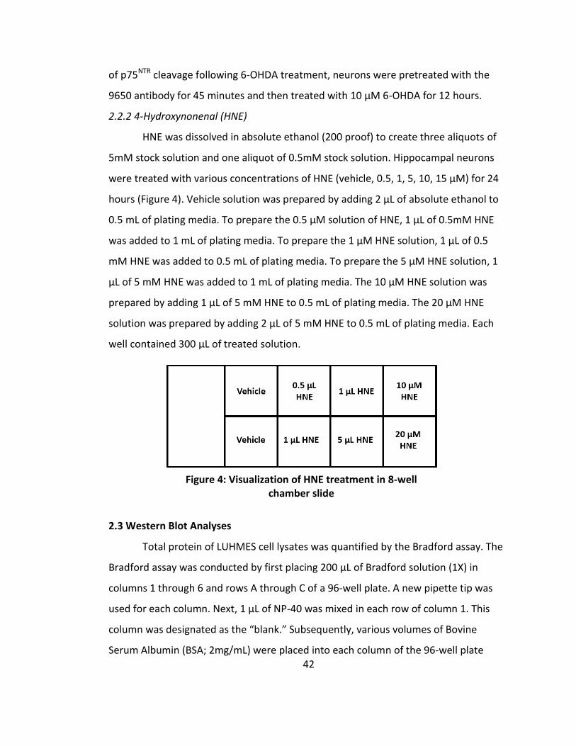
42
of p75NTR cleavage following 6-OHDA treatment, neurons were pretreated with the
9650 antibody for 45 minutes and then treated with 10 μ 6-OHDA for 12 hours.
2.2.2 4-Hydroxynonenal (HNE)
HNE was dissolved in absolute ethanol (200 proof) to create three aliquots of
5mM stock solution and one aliquot of 0.5mM stock solution. Hippocampal neurons
were treated with various concentrations of HNE (vehicle, 0.5, 1, 5, 10, 15 μ ) for 24
hours (Figure 4). Vehicle solution was prepared by adding 2 μL of absolute ethanol to
0.5 mL of plating media. To prepare the 0.5 μ solution of HNE, 1 μL of 0.5m HNE
was added to 1 mL of plating media. To prepare the 1 μ HNE solution, 1 μL of 0.5
m HNE was added to 0.5 mL of plating media. To prepare the 5 μ HNE solution, 1
μL of 5 m HNE was added to 1 mL of plating media. The 10 μ HNE solution was
prepared by adding 1 μL of 5 m HNE to 0.5 mL of plating media. The 20 μ HNE
solution was prepared by adding 2 μL of 5 m HNE to 0.5 mL of plating media. Each
well contained 300 μL of treated solution.
2.3 Western Blot Analyses
Total protein of LUHMES cell lysates was quantified by the Bradford assay. The
Bradford assay was conducted by first placing 200 μL of Bradford solution (1X) in
columns 1 through 6 and rows A through C of a 96-well plate. A new pipette tip was
used for each column. Next, 1 μL of NP-40 was mixed in each row of column 1. This
column was designated as the “blank.” ubsequently, various volumes of Bovine
Serum Albumin (BSA; 2mg/mL) were placed into each column of the 96-well plate
Figure 4: Visualization of HNE treatment in 8-well chamber slide

43
(Figure 5). Column 7 was used to mix 1 μL of the ‘Untreated’ cell lysate. Column 8 was
used to mix 1 μL of ‘10 μ 6-OHDA’. The Bradford assay was then placed in a
microplate reader to calculate the absorbance of each sample at 595 nm. The
absorbance values of each column were averaged together and then used to generate
a standard curve with the dependent variable (BSA mg/mL) on the X-axis and the
independent variable (absorbance) on the Y-axis. A linear equation was then
generated using Excel and the protein concentration for each cell lysate was calculated
by solving for X.
LUH E protein samples (40 μg) were prepared in Laemmli sample buffer (6X)
and boiled for 10 minutes at 98℃. Samples were then loaded into a 15% acrylamide
gradient gel and separated by SDS PAGE using 5X running buffer at 150 millivolts. After
successful protein separation, protein was then transferred to a PVDF membrane using
the standard wet transfer method for 18 hours at 111 milliamps. After transfer
completion, membranes were blocked in 5% dry milk dissolved in 0.1% Tween-PBS
(PBS-T), followed by overnight incubation with primary p75NTR ICD anti-serum (1:3000)
at 4℃. Subsequently, membranes were washed of primary antibody multiple times
Figure 5: Bradford Assay (volume of BSA per well)
44
with PBS-T, and then incubated with secondary anti-rabbit horseradish peroxidase
(HRP)-conjugated antibody (1:10,000) at room temperature for 2 hours. Once
secondary incubation was complete, membranes were washed and developed using
ECL for visualization of the p75NTR full length receptor FEMTO was used to visualize the
p75NTR C-terminal fragment and the p75NTR intracellular domain. Image acquisition was
conducted using the Bio-Rad ChemiDoc MP System. To assess equal gel loading among
protein samples, Actin was used as a loading control. Blots were stripped for 12
minutes using stripping buffer and then washed 8 times with PBS-T. Blots were then
blocked in 5% milk for 1 hour and then incubated with Actin (1:1000) overnight at 4℃.
Following primary antibody incubation, blots were then washed several times with
PBS-T and then incubated with secondary anti-mouse HRP antibody (1:5000) for 2
hours. ECL was used to for Actin visualization. For assessment of ERK proteins (1:1000),
protein samples were loaded into 10% acrylamide gradient gels and then incubated in
secondary anti-rabbit HRP antibody (1:10,000) for visualization with ECL. Actin was
also used as a loading control for ERK blots.
2.4 Quantification of Neuronal Cell Death
Following 6-OHDA treatment, LUHMES cells in 8-well chamber slides were fixed
with 4% paraformaldehyde for 25 minutes at room temperature. Cells were then
washed of PFA two times with PBS (1X). In order for immunostaining antibodies to
successfully bind to their targets, cells were permeabilized with 0.1% Triton-PCS for 30
minutes and then washed three times with PBs (1X). Next, cells were incubated in
blocking solution (10% normal goat serum diluted in 0.1% Triton-PBS) for one hour at
room temperature. Subsequently, cells were incubated with the neuron-specific β-III
tubulin antibody, TUJ1 (1:1000), overnight at 4℃. After primary incubation, slides were
washed three times with 0.1% Triton-PBS and then two times with PBS (1X). Cells were
then incubated with the rabbit anti-mouse Alexa Fluor 568 secondary antibody
(1:1000) for 2 hours at room temperature. The secondary antibody was washed 7
times with PBS (1X) followed by nuclei labeling with the nucleic acid stain

45
4’,6-Diamidino-2-Phenlindole (DAPI; 5 μg/μL) for 5 minutes. DAPI was then washed off
with PBS (1X) three times. The 8-well chamber slides were then prepared for coverslip
mounting by removing the chambers of the 8-well using a Chamber removal tool. Once
the chambers were removed, slides were then mounted with a coverslip using
Permount. Permount was allowed to dry overnight to ensure the coverslip was
strongly adhered to the slide. Nuclei were blindly scored as apoptotic or non-apoptotic
based on the appearance of nuclei being condensed or fragmented. Confocal images
were collected on a ZEISS LSM 800 laser scanning confocal microscope using a 40X
objective.
2.5 Statistical Analyses
All data were expressed as the mean ± standard error means (SEM). Statistical
comparisons between the p75NTR cleavage fragments of 6-OHDA treated and
untreated LUH E neurons were performed using the tudent’s t-test. Statistical
significance was set at p < 0.05.

46
CHAPTER 3
EVALUATING PROTEOLYSIS OF P75NTR WITHIN DOPAMINERGIC NEURONS SUBJECTED
TO OXIDATIVE STRESS
3.1 Introduction
Despite the variety of ligands, co-receptors, and adaptor proteins that have
been reported to activate p75NTR-mediated signaling pathways, the precise molecular
mechanisms to which p75NTR signaling becomes activated remains poorly understood.
However, the regulated intramembrane proteolysis of p75NTR has been firmly
established among all p75NTR signaling mechanisms. During this process, the
extracellular domain of p75NTR is cleaved by the mellatoprotease TNF-α converting
enzyme (TACE), leaving the remaining portion of the membrane-bound receptor, the
C-terminal fragment (CTF), vulnerable to a second proteolytic cleavage. The
γ-secretase complex further cleaves the CTF within its transmembrane region, thereby
releasing the intracellular domain (ICD) of p75NTR. These cleavage events have been
demonstrated to promote the activation of various signaling pathways and cellular
physiological responses such as c-jun N-terminal kinase (JNK) activation (Kenchappa et
al., 2010), nuclear translocation of neurotrophin receptor interacting-factor (NRIF)
(Volosin et al., 2008), apoptosis (Friedman, 2000; Kenchappa et al., 2006; Kraemer et
al., 2014), cell cycle arrest (Ceni et al., 2010), and Trk-mediated activation of survival
signaling pathways (Ceni et al., 2010; Kommaddi, Thomas, Ceni, Daigneault, & Barker,
2011b). The proteolytic cleavage of p75NTR has been demonstrated to occur in
sympathetic neurons exposed to the lipid peroxidation product, 4-hydroxynonenal
(HNE). These events were shown to occur in a ligand-independent manner, indicating
the role of p75NTR in sympathetic neuron degeneration is novel. However, it is
unknown if similar events occur in dopaminergic neurons.
Primary cultures of midbrain neurons comprise a mixture of different cell
populations, in which only 1% of those populations are dopaminergic neurons. This
small population percentage is unsuitable for analysis of proteins by western blot

47
analysis. Therefore, our studies utilized the Lund Human Mesencephalic (LUHMES) cell
line, a conditionally-immortalized human midbrain cell line, attributable to their
expression of a tetracycline-responsive v-myc gene that can be differentiated into
mature dopaminergic neurons (Lotharius, 2005). Within several days of incubation in
cell culture medium containing tetracycline, cyclic-AMP (cAMP), and glial derived
neurotrophic factor (GDNF), LUHMES cells can be fully differentiated into mature
dopaminergic-like neurons. The dopaminergic neuron-related features of
differentiated LUHMES cells have been confirmed by their expression of dopaminergic
biomarkers including tyrosine hydroxylase and dopamine transporter (X. M. Zhang, Yin,
& Zhang, 2014). Cytotoxicity has been observed within the LUHMES cell culture system
after treatment with MPP+, a dopaminergic neuron specific toxin known to be uptaken
by the dopamine transporter to induce its toxic effects (X. M. Zhang et al., 2014).
Similar to human dopaminergic neurons, LUHMES cells are able to release and uptake
dopamine as well as conduct action potentials (Scholz et al., 2011).
The physiological relevance, large culture scale, and homogenous population of
LUHMES cells make them an efficient model for studying the effects of degenerating
dopaminergic neurons. Furthermore, the cell line is human-derived, thereby making
degenerative effects more relevant to human neurodegenerative diseases. Therefore,
our investigations have utilized the LUHMES cell line. In this study, we exposed
LUHMES cells to 6-OHDA, a neurotoxin used to model oxidative stress, in order to
assess if p75NTR proteolysis occurs under these conditions. 6-OHDA is dopaminergic
specific due to its ability to enter dopaminergic neurons via the dopamine transporter
(Luthman et al., 1989). Once inside, 6-OHDA has been shown to deplete proteasome
activity (Elkon, Melamed, & Offen, 2004), activate cell death pathways by causing
caspase-3 elevation (Hanrott, Gudmunsen, O’Neill, & Wonnacott, 2006), and interfere
with normal mitochondrial function by impairing protein complexes of the electron
transport chain (Cleeter et al., 1992). The auto-oxidation of 6-OHDA has been shown to
promote the generation of various reactive oxygen species such as hydrogen peroxide,

48
superoxide anion, hydroxyl radicals, and quinones (Hanrott et al., 2006). Furthermore,
hydrogen peroxide has been implicated as a key mediator of 6-OHDA-induced cell
death due to the attenuation of catalase activity which led to a significant amount of
protection of PC12 cells from 6-OHDA-induced cell death (Hanrott et al., 2006).
In order to understand the pathogenicity of neurodegenerative conditions such
as Parkinson’s disease, scientific investigations must identify the triggers and
mechanisms that are involved in the progression of the disease. By understanding how
Parkinson’s disease begins, therapeutic treatments can successfully slow or stop
neurodegeneration from occurring. There are a wide range of cellular models that
have been used to study Parkinson’s disease such as toxin-based models and genetic
models (Potashkin, Blume, & Runkle, 2011). Ideally, a model of Parkinson’s disease
should allow the characteristic of Parkinson’s disease progression to occur in order to
assess the onset of neurodegeneration. This is one reason why genetic models of
Parkinson’s disease, such as the Engrailed deficient model, are useful because they
provide the opportunity to intervene with the progression of neurodegeneration
(Potashkin et al., 2011). In contrast, toxin-based models, such as 6-OHDA, induce
immediate neurodegeneration, thereby preventing assessment of neurodegeneration
progression (Potashkin et al., 2011). However, numerous reports have showed that 6-
OHDA toxicity is able to induce molecular alterations that are similar to those seen in
Parkinson’s disease (Potashkin et al., 2011). Therefore the 6-OHDA-induced model of
Parkinson’s disease is beneficial to explore the mechanisms of neurodegeneration in
Parkinson’s disease.
Due to a previous report showing that p75NTR is able to become activated and
cleaved in response to oxidative stress, as well as mediate the degeneration of
sympathetic neurons exposed to oxidative stress, these results provide evidence that
there may be a link between p75NTR signaling and oxidative stress. Furthermore, these
results suggest that p75NTR signaling could be one mechanism by which
neurodegeneration occurs in sympathetic neurons exposed to oxidative stress. These

49
findings discovered by Kraemer et al. enhance the understanding of how p75NTR
signaling can be activated as well as how the receptor may contribute to the etiology
of neurodegenerative conditions. Whether p75NTR can become activated by oxidative
stress in dopaminergic neurons is unknown. By using 6-OHDA to induce a
well-established model of oxidative stress in dopaminergic neurons, a better
understanding of p75NTR signaling in the brain will be achieved.
3.2 Experimental Procedures
3.2.1 Cell Culture
Please refer to Chapter 2, section 2.1, for information regarding cell culture
procedures.
3.2.2 Cell Treatments
Please refer to Chapter 2, section 2.2, for information regarding cell treatment
procedures.
3.2.3 Western Blot Analyses
Please refer to Chapter 2, section 2.3, for information regarding Western blot
analyses.
3.2.4 Quantification of Neuronal Death
Please refer to Chapter 2, section 2.4, for information regarding quantification
of neuronal death.
3.3 Results
3.3.1 Cleavage of p75NTR is stimulated by 6-OHDA
The 6-OHDA neurotoxin is commonly used to induce degeneration of
dopaminergic neurons in order to investigate motor and biochemical dysfunctions that
occur in Parkinson’s disease. To examine the effect of 6-OHDA on the viability of our
culture system, LUHMES neurons were treated with a range of 6-OHDA concentrations
for 24 hours and then blindly scored for death based on nuclear morphology. 6-OHDA
induced death of differentiated LUHMES cells in a dose-dependent manner, with 10
μ 6-OHDA being the maximally effective dose (Figure 6).

50
Figure 6: 6-OHDA Promotes Dose-Dependent Apoptosis of Differentiated LUHMES Neurons. A; Confocal images of differentiated LUHMES cells treated with the indicated concentrations of 6-OHDA for 24 hours. Neurons were stained for the neuronal marker ß-III tubulin (displayed in green). Nuclei were labeled with the nucleic acid stain 4', 6-Diamidino-2-Phenylindole (DAPI) (displayed in blue) and scored as apoptotic or non-apoptotic. B; Quantification of apoptosis induced by various concentrations of 6-OHDA (n=6 for each treatment group). An increased percentage of apoptosis was observed as the concentration of 6-OHDA increased. Results are expressed as the mean ± S.E.M.
A
B

51
Using the maximally effective dose of 6-OHDA (10 μ ), we investigated
whether 6-OHDA is able to stimulate proteolytic cleavage of p75NTR. LUHMES cells
were treated with 10 μ 6-OHDA for 12 or 18 hours and then subjected to Western
blot analysis using an antibody specific for the intracellular domain of p75NTR (1:3000).
Cleavage of p75NTR could be observed after 12 hours (Figure 7) in which there was a
significant difference between the treated and untreated p75NTR full-length receptor
and the p75NTR ICD.
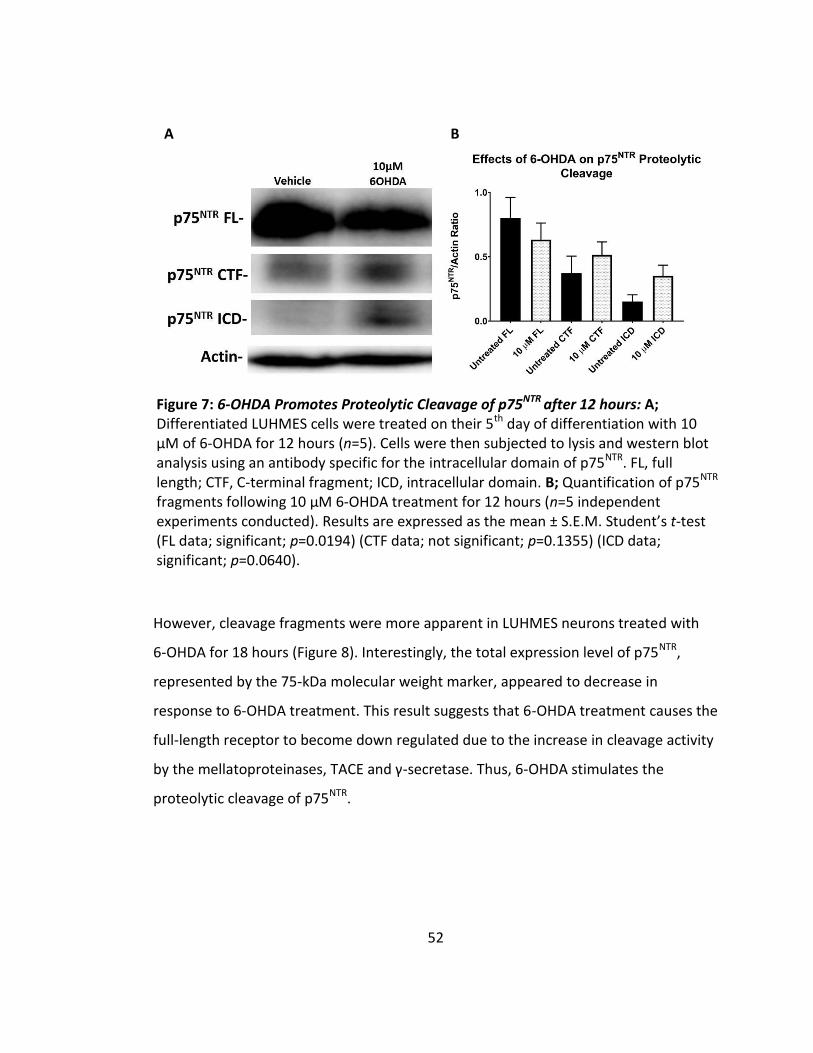
52
However, cleavage fragments were more apparent in LUHMES neurons treated with
6-OHDA for 18 hours (Figure 8). Interestingly, the total expression level of p75NTR,
represented by the 75-kDa molecular weight marker, appeared to decrease in
response to 6-OHDA treatment. This result suggests that 6-OHDA treatment causes the
full-length receptor to become down regulated due to the increase in cleavage activity
by the mellatoproteinases, TACE and γ-secretase. Thus, 6-OHDA stimulates the
proteolytic cleavage of p75NTR.
Figure 7: 6-OHDA Promotes Proteolytic Cleavage of p75NTR after 12 hours: A; Differentiated LUHMES cells were treated on their 5th day of differentiation with 10 μM of 6-OHDA for 12 hours (n=5). Cells were then subjected to lysis and western blot analysis using an antibody specific for the intracellular domain of p75NTR. FL, full length; CTF, C-terminal fragment; ICD, intracellular domain. B; Quantification of p75NTR fragments following 10 μM 6-OHDA treatment for 12 hours (n=5 independent experiments conducted). Results are expressed as the mean ± S.E.M. tudent’s t-test (FL data; significant; p=0.0194) (CTF data; not significant; p=0.1355) (ICD data; significant; p=0.0640).
A B

53
Figure 8: 6-OHDA Promotes Proteolytic Cleavage of p75NTR after 18 hours: A; Differentiated LUHMES cells were treated with 10 μM 6-OHDA for 18 hours (n=5 independent experiments conducted). Cells were then subjected to lysis and western blot analysis using an antibody specific for the intracellular domain of p75NTR. FL, full length; CTF, C-terminal fragment; ICD, intracellular domain. B; Quantification of p75NTR fragments following 10 μM 6-OHDA treatment for 12 hours (n=5). Results are expressed as the mean ± S.E.M. (FL data; not significant; p=0.0588) (CTF data; significant; p=0.0330) (ICD data; significant; p=0.0066).
A
B

54
3.3.2 Ligand-Mediated Activation of P75NTR is not Required for Proteolytic Cleavage or
Apoptosis Induction
Due to the ligand-independent mechanism at which p75NTR becomes cleaved in
sympathetic neurons subjected to oxidative stress, we investigated whether similar
events occurred in dopaminergic neurons exposed to 6-OHDA. Neurons were
pre-treated with the 9650 immune serum containing ligand-blocking antibody specific
for the extracellular domain of p75NTR (α-p75NTR ECD) for 45 minutes, followed by
exposure to 6-OHDA (10 μ ) for 12 hours. Cells were then subjected to Western blot
analyses using an antibody specific for the p75NTR ICD. Surprisingly, proteolytic
cleavage of p75NTR was able to occur regardless of ligand binding (Figure 9).
Proteolytic cleavage of p75NTR has been implicated to contribute to cell death in
sympathetic(Kenchappa et al., 2006; Kraemer et al., 2014) and hippocampal neurons
(Friedman, 2000). Within these cell types, inhibition of p75NTR cleavage using TACE and
γ-secretase inhibitors significantly protected neurons from apoptosis. Due to the
ability of p75NTR still being able to undergo proteolytic cleavage in the absence of
ligand binding, we next wanted to see if blocking the ligand-binding domain of p75NTR
Figure 9: 6-OHDA induces p75NTRproteolytic cleavage in a ligand-independent mechanism: LUHMES neurons were pre-treated with ligand-blocking α-p75NTR ECD antibody for 45 minutes and then exposed to 6-OHDA (10 μM) for 12 hours (n=1 independent experiment conducted). Neurons were then subjected to Western blot analysis using an antibody specific for the p75NTR ICD.

55
could protect cells from the toxic effects of 6-OHDA. Neurons plated into 8-well
chamber slides were pre-treated with α-p75NTR ECD for 45 minutes, followed by 5 μ
6-OHDA treatment for 24 hours. Remarkably, inhibition of the extracellular domain of
p75NTR failed to significantly protect neurons from 6-OHDA-induced death (Figure 10).
Collectively, these data suggest that oxidative stress promotes p75NTR-mediated
cleavage and induction of apoptosis in dopaminergic neurons in a ligand-independent
manner.

56
Figure 10: Treatment of differentiated LUHMES neurons with 9650 ligand-blocking α-p75 ECD antibody protects LUHMES cells from 6-OHDA-induced death: A; Confocal images of LUHMES cells exposed to 5μM 6-OHDA for 24 hours following a 45 minute pretreatment with immune serum containing a ligand blocking antibody specific for the extracellular domain of p75NTR. Neurons were stained for the neuronal marker, ßIII-tubulin (red). Nuclei were labeled with DAPI stain and scored as apoptotic (indicated by arrowheads) or non-apoptotic. A reduction in apoptosis was observed in cells pre-treated with the ligand-blocking antibody compared to cells lacking pretreatment. B; Quantification of apoptosis induced by 6-OHDA after a 45 minute pretreatment with immune serum containing a ligand blocking antibody specific for the extracellular domain of p75NTR (n=4 for each treatment group). Pretreatment with the ligand-blocking antibody caused a substantial reduction in apoptosis induced by 6-OHDA.
A
B
A

57
3.3.3 6-OHDA Suppresses ERK Activity
An investigation conducted by Alvian and Simon et al. showed that ERK
phosphorylation could be detected within wild-type mesDA neurons but not in double
mutant Engrailed neurons, which contain elevated levels of p75NTR expression175.
These results led our lab to investigate whether oxidative stress is able to suppress the
phosphorylation of ERK. LUHMES cells were treated with 6-OHDA (10 μ ) for 18 hours
and then subjected to cell lysis for western blot analyses. Remarkably, there was a
suppression of ERK phosphorylation following 6-OHDA treatment despite equal levels
of total ERK protein (Figure 11).
Figure 11: Oxidative stress does not induce ERK-1/2 Activation in LUHMES neurons. Representative Western blot of ERK-1/2 phosphorylation in untreated and 6-OHDA (10 μ for 18 hours) treated LUHMES neurons.

58
3.3.4 HNE Causes Dose-Dependent Death of Hippocampal Neurons
In addition to our experiments utilizing dopaminergic neurons, our lab also
began exploring the effect of oxidative stress on hippocampal neurons, another
population of cells that are located within a region of the brain that is highly
degenerated by Alzheimer’s disease. To mimic oxidative stress in hippocampal
neurons, our lab used the lipid peroxidation product 4-hydroxynonenal (HNE) due to
its wide recognition as a key mediator of neuronal apoptosis induced by oxidative
stress. We observed that HNE was able to cause death of hippocampal neurons in a
dose-dependent manner (Figure 12). Furthermore, we noticed that the 20 μM
concentration of HNE was able to cause almost 100 percent cell death of hippocampal
neurons.
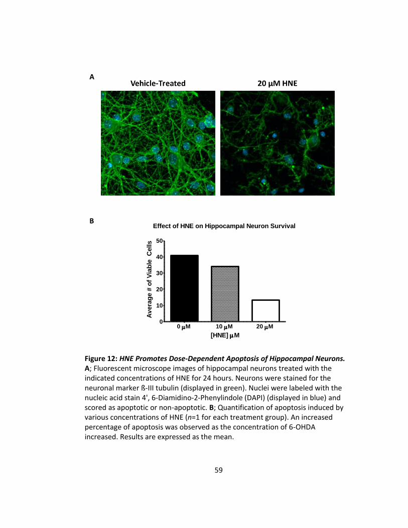
59
A
0 M 10 M 20 M0
10
20
30
40
50
Effect of HNE on Hippocampal Neuron Survival
[HNE] M
Av
era
ge #
of
Via
ble
C
ells
B
Figure 12: HNE Promotes Dose-Dependent Apoptosis of Hippocampal Neurons. A; Fluorescent microscope images of hippocampal neurons treated with the indicated concentrations of HNE for 24 hours. Neurons were stained for the neuronal marker ß-III tubulin (displayed in green). Nuclei were labeled with the nucleic acid stain 4', 6-Diamidino-2-Phenylindole (DAPI) (displayed in blue) and scored as apoptotic or non-apoptotic. B; Quantification of apoptosis induced by various concentrations of HNE (n=1 for each treatment group). An increased percentage of apoptosis was observed as the concentration of 6-OHDA increased. Results are expressed as the mean.
60
3.4 Discussion
The role of p75NTR as an apoptotic mediator in response to oxidative stress has
been established in sympathetic neurons (Kraemer et al., 2014). The lipid peroxidation
product, HNE, has been demonstrated to induce p75NTR-mediated apoptosis and
axonal degeneration of sympathetic neurons. Neurons lacking the receptor were
significantly protected from HNE-induced axonal degeneration and apoptosis, thereby
suggesting p75NTR as a regulator of neuronal responses to oxidative stress. Here, we
demonstrate that p75NTR is expressed in fully differentiated LUHMES cells exposed to
6-OHDA neurotoxicity. Therefore, p75NTR may regulate responses to oxidative stress in
dopaminergic neurons as well.
The expression of p75NTR is ubiquitous during embryonic development;
however, numerous investigations have demonstrated that reexpression of the
receptor during adulthood is associated with various cellular injuries and
neurodegenerative conditions, leading to cell death. Reexpression of p75NTR has been
observed to occur in response to seizures (P P Roux et al., 1999; Volosin et al., 2008),
ischemia (Kokaia et al., 1998), neuronal axotomy (Harrington et al., 2004), as well as
within models of Alzheimer’s disease (Murphy et al., 2015) and Amyotrophic sclerosis
(Lowry et al., 2001; Turner et al., 2004), conditions highly associated with oxidative
stress. It is currently unknown why reexpression of p75NTR expression occurs in
response to various injuries, but a few hypotheses have been made suggesting the
receptor may promote positive effects following injuries. For instance, the expression
of p75NTR has been previously demonstrated to increase following axonal injury and
induce apoptosis(Harrington et al., 2004). However, there have been reports that
p75NTR signaling is able to promote remyelination of injured sciatic nerves. Adult mice
deficient in p75NTR experienced a reduction in the number of myelinated axons
following sciatic nerve lesion compared to wild-type controls (Song, Zhou, Zhong, Wu,
& Zhou, 2006). Furthermore, the transplantation of p75NTR deficient Schwann cells in
mice resulted in a significant impairment in motor neuron recovery following sciatic

61
nerve injury (Tomita et al., 2007). The p75NTR deficient Schwann cells failed to promote
remyelination, axonal regrowth, and radial growth in axon calibers compared to
wild-type controls (Tomita et al., 2007). Therefore, the receptor might initiate
apoptosis in injured sciatic nerves in order to provide a more suitable environment for
remyelination.
The pro-apoptotic role of p75NTR has also been suggested to control
inflammation by encouraging the elimination of damaged cells. For example, the
induction of encephalomyelitis (EAE) in has been demonstrated to cause an
upregulation of p75NTR in endothelial cells, leading to cell death (Copray et al., 2004).
However, when EAE was induced in mice deficient for p75NTR, the mice developed
increased levels of inflammation within the central nervous system compared to
wild-type controls (Copray et al., 2004). It is widely established that without
inflammatory control, widespread tissue damage can contribute to the pathogenesis
of numerous diseases (Rock & Kono, 2008). Therefore the detrimental effects of p75NTR
may be a part of a homeostatic mechanism that goes out of control. Supportive
evidence of this theory includes a study conducted by Miller and colleagues (Park,
Grosso, Aubert, Kaplan, & Miller, 2010) demonstrating that p75NTR regulates the
specificity of neural connectivity in basal forebrain axons by inducing axonal
degeneration.
The mechanism of p75NTR proteolytic cleavage is well-established, but its
functional significance has not been fully elucidated. Pharmacological investigations
have demonstrated that p75NTR proteolytic cleavage relates to the induction of cell
death in hippocampal (Kenchappa et al., 2006) and sympathetic neurons (Volosin et al.,
2008) after seizure and proBDNF treatment, respectively. Likewise, p75NTR cleavage
was detected in sympathetic neurons subjected to oxidative stress, which
corresponded with neuronal apoptosis and degeneration. The induction of p75NTR
cleavage has been observed in sympathetic neurons following HNE and 6-OHDA
treatment, indicating multiple oxidants are able to initiate p75NTR signaling

62
mechanisms within these cells. Although p75NTR activation could be observed in
response to oxidative stress in sympathetic neurons, further investigations are needed
to determine if p75NTR can be similarly activated by oxidative stress in neurons of the
brain, especially in brain regions vulnerable to oxidative stress such as the ventral
midbrain. Furthermore, investigating whether p75NTR contributes to the degeneration
of neurons in the ventral midbrain could provide insight that the receptor is linked to
oxidative stress-associated disorders such as Parkinson’s disease. Due to the
abundance of studies reporting 6-OHDA induces oxidative stress and apoptosis in
dopaminergic neurons, we wanted to investigate if 6-OHDA is able to stimulate
proteolysis of p75NTR within the dopaminergic neuron population.
The ventral midbrain contains a basal ganglia structure known as the substantia
nigra which is highly degenerated by Parkinson’s disease, a neurodegenerative
condition associated with oxidative stress. The utilization of the LUHMES culture
system, an immortalized human mesencephalic cell line that can be differentiated to
mature dopaminergic neurons, allowed us to investigate the effects of oxidative stress
on p75NTR proteolytic cleavage and dopaminergic neuron viability. Our results
demonstrated p75NTR undergoes proteolysis in response to 6-OHDA treatment within
the LUHMES cell culture system. The cleavage fragments of 6-OHDA treated LUHMES
cells were noticeably prominent compared to untreated cells, which was similar to the
results of sympathetic neurons treated with HNE. Our assessment of p75NTR proteolysis
was conducted by exposing dopaminergic neurons to 6-OHDA treatment for 12 hours,
which was consistent with previous treatment times utilizing sympathetic neurons
treated with HNE. Proteolytic cleavage of p75NTR in sympathetic neurons was
characterized as an event that needed to occur prior to the induction of programmed
cell death. Inhibition of p75NTR cleavage by TACE inhibitors significantly protected
sympathetic neurons from HNE-induced apoptosis (Kraemer et al., 2014). Due to the
similarities in CTF and ICD in dopaminergic neurons and sympathetic neurons following
63
12 hour exposure to oxidative stress, we hypothesize that p75NTR proteolysis will be
the initiating event of 6-OHDA-induced cell death in dopaminergic neurons.
Both p75NTR cleavage fragments have been described as necessary components
for p75NTR-mediated cell death, but there is much controversy on which fragment is
actually the main regulatory component of p75NTR-mediated cell death. Coulson and
colleagues propose the CTF is primarily responsible for the death inducing abilities of
p75NTR due to the presence of a 29 amino acid sequence in the p75NTR CTF
juxtamembrane membrane region, known as the “chopper domain”(E J Coulson et al.,
2000). On the other hand, the p75NTR ICD has been heavily suspected as a critical
regulator of p75NTR-mediated apoptosis due to its expression of a “death domain,” as
well as its ability to interact with adaptor proteins that regulate apoptotic signaling,
such as TRAF6, NRIF, NRAGE, NADE, and MAGE homologs. Our dose response assays
showed that the maximally effective dose of 6-OHDA (10 μ ) caused almost 100
percent cell death. In analyses of p75NTR cleavage, the maximally effective dose of 6-
OHDA induced an abundant expression of p75NTR ICD compared to p75NTR CTF in
dopaminergic neurons. Furthermore, a reasonable amount of CTF expression could still
be detected in untreated cells, thereby suggesting p75NTR undergoes low basal rates of
TACE cleavage. We speculate 6-OHDA has a marked effect on γ-secretase cleavage
compared to TACE cleavage. Inhibition of γ-secretase cleavage has previously been
shown to prevent p75NTR-mediated apoptosis (Kenchappa et al., 2006). ince γ-
secretase cleavage leads the generation of the p75NTR ICD, we also speculate that the
p75NTR ICD may have a critical role in mediating dopaminergic neuron death in
response to oxidative insult compared to the p75NTR CTF. However, further
investigations are needed to examine the downstream effects of p75NTR cleavage in
dopaminergic neurons.
Interestingly, total expression of p75NTR full length (represented by band at the
75 kDa weight marker) decreased in 6-OHDA treated cells compared to untreated cells.
Our results contrasted with those of numerous investigations indicating that

64
expression of p75NTR is upregulated following several injuries. The expression of p75NTR
has been shown to upregulate in a time-dependent manner following axotomy
(Harrington et al., 2004), spinal cord injury (Beattie et al., 2002), seizure (P P Roux et
al., 1999), and kainic acid toxicity (Y.-Q. Wang, Bian, Bai, Cao, & Chen, 2008). However,
the expression of p75NTR peaked several days after injury (Beattie et al., 2002;
Harrington et al., 2004; P P Roux et al., 1999) or until neurons could no longer undergo
cell death (Harrington et al., 2004). In our experiments, downregulation of p75NTR full
length receptor could be observed following 12 hours of treatment with the 6-OHDA
maximally effective dose. Relative to prior research (Harrington et al., 2004), these
results may indicate that after 12 hours of 6-OHDA treatment, LUHMES neurons are
unable to undergo cell death any further, thereby initiating downregulation of p75NTR.
We currently have not assessed expression of the receptor prior to 12 hours of
6-OHDA treatment, therefore it is possible p75NTR expression could be upregulated
before then. Increased expression of p75NTR could be detected in dopaminergic
neurons after 30 minutes of exposure to kainic acid, so it is likely p75NTR initiates an
immediate and rapid response in these cells following injury. However, further
investigations are needed to understand the contribution of p75NTR signaling to the
degeneration of dopaminergic neurons.
Transgenic mouse models have revealed that the transcription factors
Engrailed-1/Engrailed-2 (collectively called Engrailed or En1/2), are critical for the
survival and maintenance of mesDA neurons during late embryonic life (Rekaik,
Blaudin de Thé, Prochiantz, Fuchs, & Joshi, 2015). Engrailed double mutant (En1-/-;
En2-/-) mesDA neurons have been observed to die by apoptosis during the stage when
the induction of Engrailed expression begins (Alberi, 2004; Simon et al., 2001).
Remarkably, immunohistochemistry experiments revealed Engrailed deficient mesDA
neurons exhibit elevated levels of p75NTR along with caspase-3 and caspase-9
activation (Alavian et al., 2009). These results suggest if p75NTR contributes to the
death of Engrailed deficient neurons then the receptor must do so by way of the

65
intrinsic apoptotic signaling cascade. Similarly, 6-OHDA treatment of Engrailed
heterozygous mice induced caspase-9 activation (Alavian et al., 2009). Relative to our
investigations, these results suggest that the intrinsic apoptotic signaling cascade is
responsible for killing LUHMES neurons exposed to oxidative stress. Furthermore,
previous investigations have shown that the intrinsic apoptotic signaling cascade is a
downstream signaling pathway of p75NTR due to the significant protection of
hippocampal neurons from programmed cell death following Apaf-1 and caspase-9
downregulation (Troy et al., 2002). Therefore, it is possible p75NTR mediates the death
of LUHMES neurons through the intrinsic death pathway following 6-OHDA treatment.
However, further research is needed to clarify the apoptotic signaling cascade that is
mediated by p75NTR in these cells.
Previous investigations have shown that p75NTR is able to augment Trk
signaling107, 192 thereby promoting cellular survival. The coexpression of p75NTR with
TrkA in PC12 cells was shown to delay the internalization and degradation of TrkA
thereby prolonging TrkA signaling events due to TrkA being able to remain on the
cellular surface following neurotrophin binding (Makkerh et al., 2005). Furthermore,
Barker and colleagues have demonstrated that Trk receptors are able to promote
proteolytic cleavage of p75NTR following neurotrophin binding, further leading to the
phosphorylation of the Akt and ERK proteins following generation of the p75NTR ICD by
γ-secretase (Ceni et al., 2010). However, high expression of p75NTR has been shown to
suppress the phosphorylation of ERK in double mutant Engrailed mesDA neurons
(Alavian et al., 2009). Double mutant Engrailed neurons have been shown to express
high levels of p75NTR that correlate with the induction of cell death. Additionally, these
neurons exhibited a suppression of ERK activity compared to wild-type mesDA neurons
(Alavian et al., 2009). These results suggest that the suppression of ERK activity could
promote p75NTR-mediated cell death. Similarly, our experiments revealed the
treatment of LUHMES neurons with 6-OHDA resulted in a suppression of ERK activity.
However, due to the downregulation of the p75NTR full-length receptor following 6-

66
OHDA treatment, it cannot be determined whether ERK suppression is mediated by
p75NTR. The knockdown of p75NTR expression in double mutant Engrailed neurons
allowed ERK activation to occur, thereby suggesting a relationship between the loss of
ERK activity and upregulation of p75NTR expression (Alavian et al., 2009). Therefore,
further investigations utilizing p75NTR knockout neurons are needed in order to
determine if p75NTR suppresses ERK activity in dopaminergic neurons exposed to
oxidative stress.
The regulation of p75NTR-mediated cell death has been heavily indicated to
occur in response to neurotrophin or proneurotrophin binding. However,
overexpression of p75NTR cleavage fragments or p75NTR recombinant mutants have
displayed the ability to induce apoptotic signaling pathways such as JNK
phosphorylation and caspase-3 cleavage in a ligand independent manner (M. Vilar et
al., 2009). Relative to these results, sympathetic neurons pre-treated with a ligand-
blocking antibody specific for the p75NTR extracellular domain (α-p75NTR ECD) failed to
protect neurons from HNE-induced apoptosis, thereby revealing p75NTR mediates
these events in a ligand-independent mechanism. In our investigations, we observed
similar results when LUHMES neurons were exposed to 6-OHDA toxicity. Blockage of
the p75NTR ligand-binding domain using α-p75NTR ECD failed to protect LUHMES
neurons from 6-OHDA-induced death. Thus, our results suggest if p75NTR is responsible
for promoting apoptosis of dopaminergic neurons in response to oxidative stress, then
the mechanism at which this occurs is ligand-independent. Previous investigations
have demonstrated p75NTR activation can occur in the absence of ligands due to the
expression of a highly conserved cysteine residue 257 (Cys257) in the transmembrane
domain of the receptor (M. Vilar et al., 2009). It was initially believed p75NTR could only
dimerize in response to neurotrophin binding by way of the proposed ‘snail-tong’
model (Marçal Vilar et al., 2009). This model suggests that when a neurotrophin binds
to p75NTR, the receptor dimerizes, causing the intracellular domains to separate in a
scissors-like movement. However, the Cys257 residue has been shown to enable p75NTR
67
to dimerize at the plasma membrane independent of ligand-binding due to disulfide
bonding of the juxtamembrane of the receptor. The precise reason as to why this
occurs is unknown, but it is believed that various cysteine residues, such as Cys257 may
induce different degrees of separation between the p75NTR intracellular domains.
Additionally, it is suggested that p75NTR may undergo an array of conformational
changes that each have its own particular function (M. Vilar et al., 2009). Therefore, it
is possible that oxidative stress stimulates p75NTR to undergo a conformational change
that highly favors the induction of cell death.

68
CHAPTER 4
CONCLUSIONS AND FUTURE DIRECTIONS
4.1 Conclusions
This thesis project aimed to investigate the activation of p75NTR in
dopaminergic neurons subjected to oxidative stress. Our findings exhibit a novel
mechanism of p75NTR activation in dopaminergic neurons exposed to oxidative stress.
The induction of p75NTR cleavage occurred following 6-OHDA toxicity along with
dopaminergic neuronal cell death. Furthermore, inhibition of the p75NTR extracellular
domain failed to protect neurons from 6-OHDA induced death. These results provide
evidence that p75NTR could be a potential contributor of the degeneration of
dopaminergic neurons in Parkinson’s disease. However, further investigations are
needed to fully clarify the pro-apoptotic role of p75NTR in this neuronal population
under stressful conditions, determine if p75NTR proteolysis contributes to oxidative
stress-induced death, and to identify activation of signaling cascades downstream of
p75NTR.
4.2 Future Studies
Our results provide a novel mechanism of p75NTR activation in dopaminergic
neurons exposed to oxidative stress, which is relative to sympathetic neurons exposed
to the same conditions. However, the functional role of p75NTR in sympathetic neurons
subjected to HNE-induced oxidative stress is more pronounced due to the utilization of
cleavage inhibitors and p75NTR knockout mice (Kraemer et al., 2014). These
investigations discovered p75NTR proteolytic cleavage contributed to the induction of
apoptosis and axonal degeneration in sympathetic neurons by HNE due to the
significant amount of protection sympathetic neurons experienced from HNE-induced
degeneration following TACE cleavage (Kraemer et al., 2014). Several studies have
indicated the functions of p75NTR are cell-type-specific, meaning that the receptor is
able to elicit distinct responses in different cell types. For example, hippocampal
neurons and cerebral granule neurons exhibit different patterns of p75NTR cleavage,

69
which is suggested to underlie the different p75NTR signaling activities in the two
neuronal cell types (Vicario, Kisiswa, Tann, Kelly, & Ibanez, 2015). Given the discovery
that p75NTR cleavage contributes to the induction of HNE-induced death in sympathetic
neurons, pharmacological investigations using TACE and γ-secretase inhibitors need to
be conducted using dopaminergic neurons in order to understand the functional
significance of p75NTR proteolysis in oxidative-stress induced death.
Several investigations have demonstrated that the death signal initiated by
p75NTR occurs through the downstream induction of the stress-activated kinase c-Jun
N-terminal kinase (JNK) pathway. JNK has been shown to promote pro-apoptotic
p75NTR signaling in various cell types such as sympathetic neurons (Palmada et al.,
2002), cerebral granule neurons (Vicario et al., 2015), oligodendrocytes (Casaccia-
Bonnefil et al., 1996), Schwann cells (Yeiser, 2004), and hippocampal neurons
(Friedman, 2000). Therefore, it would beneficial to assess if JNK activation occurs in
dopaminergic neurons exposed to oxidative stress. These results could provide
evidence that JNK contributes to the degeneration of dopaminergic neurons.
The utilization of p75NTR knockout neurons further confirmed the contribution
of p75NTR in the death of sympathetic neurons due to the significant protection of
knockout neurons from HNE-induced apoptosis (Kraemer et al., 2014). Therefore, the
assessment of death in p75NTR knockout dopaminergic neurons subjected to oxidative
stress should be performed. These experiments would further define the contributions
of p75NTR signaling in the degeneration of brain regions vulnerable to oxidative stress
as well as provide insight on the possible contribution of p75NTR in oxidative stress-
associated disorders such as Parkinson’s disease. Furthermore, p75NTR knockout
neurons would also determine if p75NTR is directly responsible for activating JNK in
dopaminergic neurons subjected to oxidative stress.
The ability of p75NTR to induce apoptosis is the most studied function of the
receptor. However, p75NTR has also been shown to promote axonal degeneration
during the development of sympathetic (Singh et al., 2008) and cholinergic neurons
70
(Park et al., 2010). Furthermore, p75NTR has been established as a promoter of axonal
degeneration in sympathetic neurons exposed to oxidative stress due to the
remarkable protection that sympathetic neurons lacking p75NTR experienced following
HNE treatment (Kraemer et al., 2014). It has not been determined if p75NTR functions
similarly in brain neurons. Axonal degeneration has been characterized as an
important step in the pathomechanism of neurodegenerative conditions (Lingor, Koch,
Tönges, & Bähr, 2012). The process has been suggested to occur early in the course of
neurodegenerative diseases, providing a potential target for therapeutic strategies.
Parkinson’s disease, a neurological condition related to oxidative stress, has been
shown to be associated with axonal degeneration (Lingor et al., 2012). Additionally,
axonal degeneration in Parkinson’s disease has been suggested to precede neuronal
death (Lingor et al., 2012). Therefore it would be essential to investigate if p75NTR
contributes to the axonal degeneration of dopaminergic neurons by exposing wild-
type and p75NTR knockout dopaminergic neurons to 6-OHDA and then analyzing the
amount of axonal degeneration that occurred following treatment.
Trk receptors induce the activation of several cell survival pathways such as
PI3K/Akt, Ras/ERK, and phospholipase-C. A previous investigation has exhibited a
correlation between high expression of p75NTR and the suppression of ERK activity in
mesencephalic dopaminergic neurons deficient in the Engrailed genes (Alavian et al.,
2009). However, there was not a significant difference in the activation of the Akt
pathway in the deficient neurons compared to wild-type neurons. Furthermore, there
were not any signs of differential activation in the JNK pathway between Engrailed
deficient neurons compared to wild-type neurons (Alavian et al., 2009). Our lab has
begun investigating the suppression of Trk signaling in dopaminergic neurons exposed
to oxidative stress and so far we have observed that 6-OHDA causes a suppression of
ERK activity. However, future investigations should include analysis of the Akt and
phospholipase-C pathways to further characterize if all Trk signaling pathways are
suppressed under conditions of oxidative stress. Furthermore, future investigations

71
should also utilize dopaminergic neurons deficient in p75NTR to determine if p75NTR
plays a role in the suppression of Trk signaling in dopaminergic neurons under
conditions of oxidative stress.
Numerous reports have shown Alzheimer’s disease is associated with oxidative
stress. The hippocampus, a brain region known to play a critical role in learning and
memory (Sakaguchi, Kadoshima, Soen, Narii, & Ishida, 2015), is highly degenerated by
Alzheimer’s disease. Analyses of postmortem brain tissue of Alzheimer’s disease
patients have shown increased levels of p75NTR within the hippocampus (Ito et al.,
2016). Furthermore, the loss of p75NTR expression in a murine model of Alzheimer’s
disease was shown to reduce Alzheimer’s-associated behavioral symptoms (Murphy et
al., 2015). Our lab recently began evaluating the effects of HNE on hippocampal
neuron viability. HNE is a product of lipid peroxidation and it is commonly used to
mimic oxidative stress in neurons due to its reputation as a key mediator of neuronal
apoptosis during oxidative stress. Our preliminary results exhibited a dose-dependent
effect of HNE on hippocampal neuron survival. Furthermore, we observed the 20 μ
concentration of HNE was able to cause almost 100 percent cell death. However,
additional experimental trials are needed to confirm 20 μ HNE is the maximally
effective dose. This finding will then allow our lab to evaluate the effect of oxidative
stress on p75NTR signaling in hippocampal neurons.

72
References
Adams, J. P., & Dudek, S. M. (2005). Late-phase long-term potentiation: getting to the
nucleus. Nature Reviews Neuroscience, 6, 737. Retrieved from
http://dx.doi.org/10.1038/nrn1749
Adinoff, B. (2004). Neurobiologic processes in drug reward and addiction. Harvard
Review of Psychiatry, 12(6), 305–320.
http://doi.org/10.1080/10673220490910844
Aggarwal, B. B. (2000). Tumour necrosis factors receptor associated signalling
molecules and their role in activation of, 59(suppl I), 6–16.
Agid, Y. (1991). NEUROANATOMY OF DOPAMINERGIC SYSTEM IN THE HUMAN BRAIN
Yves AGID, 1–2.
Alam, Z. I., Jenner, A., Daniel, . E., Lees, A. J., Cairns, N., arsden, C. D., … Halliwell, B.
(2002). Oxidative DNA Damage in the Parkinsonian Brain: An Apparent Selective
Increase in 8-Hydroxyguanine Levels in Substantia Nigra. Journal of
Neurochemistry, 69(3), 1196–1203. http://doi.org/10.1046/j.1471-
4159.1997.69031196.x
Alavian, K. N., Sgadò, P., Alberi, L., Subramaniam, S., & Simon, H. H. (2009). Elevated
P75NTR expression causes death of engrailed-deficient midbrain dopaminergic
neurons by Erk1/2 suppression. Neural Development, 4, 11.
http://doi.org/10.1186/1749-8104-4-11
Alberi, L. (2004). Engrailed genes are cell-autonomously required to prevent apoptosis
in mesencephalic dopaminergic neurons. Development, 131(13), 3229–3236.
http://doi.org/10.1242/dev.01128
Aloyz, R. S., Bamji, S. X., Pozniak, C. D., Toma, J. G., Atwal, J., Kaplan, D. R., & Miller, F.
D. (1998). P53 Is Essential for Developmental Neuron Death as Regulated by the
TrkA and p75 Neurotrophin Receptors, 143(6), 1691–1703.

73
Anton, E. S., Weskamp, G., Reichardt, L. F., & Matthew, W. D. (1994). Nerve growth
factor and its low-affinity receptor promote Schwann cell migration. Proceedings
of the National Academy of Sciences of the United States of America, 91(7), 2795–
2799. Retrieved from http://www.ncbi.nlm.nih.gov/pmc/articles/PMC43457/
Arias-Carrián, O., Stamelou, M., Murillo-Rodríguez, E., Menéndez-Gonzlez, M., &
Pöppel, E. (2010). Dopaminergic reward system: A short integrative review.
International Archives of Medicine, 3(1), 1–6. http://doi.org/10.1186/1755-7682-
3-24
Bamji, . X., ajdan, ., Pozniak, C. D., Belliveau, D. J., Aloyz, R., Kohn, J., … iller, F.
D. (1998). The p75 neurotrophin receptor mediates neuronal apoptosis and is
essential for naturally occurring sympathetic neuron death. The Journal of Cell
Biology, 140(4), 911–23. http://doi.org/10.1083/jcb.140.4.911
Barbeau, A. (1962). The Pathogenesis of Parkinson’s Disease: A New Hypothesis.
Canadian Medical Association Journal, 87(15), 802–807. Retrieved from
http://www.ncbi.nlm.nih.gov/pmc/articles/PMC1849683/
Barker, P. A. (1998). p75 NTR : A study in contrasts, 346–356.
Barker, P. A., & Shooter, E. M. (1994). Disruption of NGF binding to the low affinity
neurotrophin receptor p75LNTR reduces NGF binding to TrkA on PC12 cells.
Neuron, 13(1), 203–215. http://doi.org/https://doi.org/10.1016/0896-
6273(94)90470-7
Baumann, B., terling, J., ong, Y., ong, D., Fruttiger, ., Gillies, ., … Dunaief, J. L.
(2017). Conditional Müller Cell Ablation Leads to Retinal Iron Accumulation.
Investigative Ophthalmology & Visual Science, 58(10), 4223–4234. Retrieved from
http://dx.doi.org/10.1167/iovs.17-21743
Beattie, . ., Harrington, A. W., Lee, R., Kim, J. Y., Boyce, . L., Longo, F. ., … Yoon, .
O. (2002). ProNGF Induces p75-Mediated Death of Oligodendrocytes following
Spinal Cord Injury. Neuron, 36(3), 375–386. Retrieved from
http://www.ncbi.nlm.nih.gov/pmc/articles/PMC2681189/

74
Beaulieu, J., & Gainetdinov, R. R. (2011). The Physiology , Signaling , and Pharmacology
of Dopamine Receptors, 63(1), 182–217.
http://doi.org/10.1124/pr.110.002642.182
Becker, E. B. E., Howell, J., Kodama, Y., Barker, P. A., & Bonni, A. (2004).
Characterization of the c-Jun N-Terminal Kinase-BimEL Signaling Pathway in
Neuronal Apoptosis. Journal of Neuroscience, 24(40), 8762–8770.
http://doi.org/10.1523/JNEUROSCI.2953-04.2004
Benedetti, M., Levi, A., & Chao, M. V. (1993). Differential expression of nerve growth
factor receptors leads to altered binding affinity and neurotrophin
responsiveness. Proceedings of the National Academy of Sciences of the United
States of America, 90(16), 7859–7863. Retrieved from
http://www.ncbi.nlm.nih.gov/pmc/articles/PMC47242/
Bernabeu, R. O., & Longo, F. M. (2010). The p75 neurotrophin receptor is expressed by
adult mouse dentate progenitor cells and regulates neuronal and non-neuronal
cell genesis. BMC Neuroscience, 11(1), 136. http://doi.org/10.1186/1471-2202-
11-136
Blum, D., Torch, S., Lambeng, N., Nissou, M.-F., Benabid, A.-L., Sadoul, R., & Verna, J.-
M. (2001). Molecular pathways involved in the neurotoxicity of 6-OHDA,
dopamine and PTP: contribution to the apoptotic theory in Parkinson’s disease.
Progress in Neurobiology, 65(2), 135–172.
http://doi.org/https://doi.org/10.1016/S0301-0082(01)00003-X
Boatright, K. ., Renatus, ., cott, F. L., perandio, ., hin, H., Pedersen, I. ., …
Francisco, S. S. (2003). A Unified Model for Apical Caspase Activation, 11, 529–
541.

75
Carter, B. D., Kaltschmidt, C., Kaltschmidt, B., Offenhäuser, N., Böhm-Matthaei, R.,
Baeuerle, P. A., & Barde, Y.-A. (1996). Selective Activation of NF-κB by Nerve
Growth Factor Through the Neurotrophin Receptor p75. Science, 272(5261), 542
LP-545. Retrieved from
http://science.sciencemag.org/content/272/5261/542.abstract
Casaccia-Bonnefil, P., Carter, B. D., Dobrowsky, R. T., & Chao, M. V. (1996). Death of
oligodendrocytes mediated by the interaction of nerve growth factor with its
receptor p75. Nature. http://doi.org/10.1038/383716a0
Casademunt, E., Carter, B. D., Benzel, I., Frade, J. M., Dechant, G., & Barde, Y. A. (1999).
The zinc finger protein NRIF interacts with the neurotrophin receptor p75(NTR)
and participates in programmed cell death. EMBO Journal, 18(21), 6050–6061.
http://doi.org/10.1093/emboj/18.21.6050
Ceni, C., Kommaddi, R. P., Thomas, R., Vereker, E., Liu, X., cPherson, P. ., … Barker,
P. A. (2010). The p75NTR intracellular domain generated by neurotrophin-induced
receptor cleavage potentiates Trk signaling. Journal of Cell Science, 123(13),
2299–2307. http://doi.org/10.1242/jcs.062612
Chan, J. R., Watkins, T. A., Cosgaya, J. ., Zhang, C., Chen, L., Reichardt, L. F., … Barres,
B. A. (2004). NGF Controls Axonal Receptivity to Myelination by Schwann Cells or
Oligodendrocytes. Neuron, 43(2), 183–191.
http://doi.org/10.1016/j.neuron.2004.06.024
Chao, M. V., Rajagopal, R., & Lee, F. S. (2006). Neurotrophin signalling in health and
disease. Clinical Science, 110(2), 167 LP-173. Retrieved from
http://www.clinsci.org/content/110/2/167.abstract
Chao, M. V., & Hempstead, B. L. (1995). p75 and Trk: A two-receptor system. Trends in
Neurosciences, 18(7), 321–326. http://doi.org/10.1016/0166-2236(95)93922-K
Chapman, B. S. (1995). A region of the 75 kDa neurotrophin receptor homologous to
the death domains of TNFR-I and Fas. FEBS Letters, 374(2), 216–220.
http://doi.org/10.1016/0014-5793(95)01113-S

76
Chen, S. C., Ehrhard, P., Goldowitz, D., & Smeyne, R. J. (1997). Developmental
expression of the GIRK family of inward rectifying potassium channels:
implications for abnormalities in the weaver mutant mouse. Brain Research,
778(2), 251–264. http://doi.org/10.1016/s0006-8993(97)00896-2
Chen, Z.-Y. (2005). Sortilin Controls Intracellular Sorting of Brain-Derived Neurotrophic
Factor to the Regulated Secretory Pathway. Journal of Neuroscience, 25(26),
6156–6166. http://doi.org/10.1523/JNEUROSCI.1017-05.2005
Chen, Z.-Y., Jing, D., Bath, K. G., Ieraci, A., Khan, T., Siao, C.-J., … Lee, F. . (2006).
Genetic Variant BDNF (Val66Met) Polymorphism Alters Anxiety-Related Behavior.
Science (New York, N.Y.), 314(5796), 140–143.
http://doi.org/10.1126/science.1129663
Chu, G. K., Yu, W., & Fehlings, M. G. (2007). The p75 neurotrophin receptor is essential
for neuronal cell survival and improvement of functional recovery after spinal
cord injury. Neuroscience, 148(3), 668–682.
http://doi.org/10.1016/j.neuroscience.2007.05.028
Clausen, A., Bi, X., & Baudry, M. (2012). Effects of the superoxide dismutase/catalase
mimetic EUK-207 in a mouse model of alzheimer’s disease: Protection against and
interruption of progression of amyloid and tau pathology and cognitive decline.
Journal of Alzheimer’s Disease, 30(1), 183–208. http://doi.org/10.3233/JAD-2012-
111298
Cleeter, M. W. J., Cooper, J. M., & Schapira, A. H. V. (1992). Irreversible Inhibition of
itochondrial Complex I by 1‐ ethyl‐4‐Phenylpyridinium: Evidence for Free
Radical Involvement. Journal of Neurochemistry, 58(2), 786–789.
http://doi.org/10.1111/j.1471-4159.1992.tb09789.x
Cohen, G. (1984). Oxy-radical toxicity in catecholamine neurons. Neurotoxicology, 5(1),
77–82. Retrieved from http://www.ncbi.nlm.nih.gov/pubmed/6326007
77
Copray, ., Küst, B., Emmer, B., Lin, . Y., Liem, R., Amor, ., … Boddeke, E. (2004).
Deficient p75 low-affinity neurotrophin receptor expression exacerbates
experimental allergic encephalomyelitis in C57/BL6 mice. Journal of
Neuroimmunology, 148(1–2), 41–53.
http://doi.org/10.1016/j.jneuroim.2003.11.008
Cory, S., & Adams, J. M. (2002). The Bcl2 family: regulators of the cellular life-or-death
switch. Nature Reviews Cancer, 2, 647. Retrieved from
http://dx.doi.org/10.1038/nrc883
Coulson, E. J., May, L. ., Osborne, . L., Reid, K., Underwood, C. K., eunier, F. A., …
Sah, P. (2008). p75 neurotrophin receptor mediates neuronal cell death by
activating GIRK channels through phosphatidylinositol 4,5-bisphosphate. The
Journal of Neuroscience : The Official Journal of the Society for Neuroscience,
28(1), 315–24. http://doi.org/10.1523/JNEUROSCI.2699-07.2008
Coulson, E. J., Reid, K., Baca, M., Shipham, K. a, Hulett, S. M., Kilpatrick, T. J., & Bartlett,
P. F. (2000). Chopper, a new death domain of the p75 neurotrophin receptor that
mediates rapid neuronal cell death. The Journal of Biological Chemistry, 275(39),
30537–30545. http://doi.org/10.1074/jbc.M005214200
Courtial, L., Picco, V., Grover, R., Cormerais, Y., Rottier, C., Labbe, A., … Ferrier-Pagès,
C. (2017). The c-Jun N-Terminal kinase prevents oxidative stress induced by UV
and thermal stresses in corals and human cells. Scientific Reports, 7(March), 1–10.
http://doi.org/10.1038/srep45713
Dasuri, K., Zhang, L., & Keller, J. N. (2013). Oxidative stress, neurodegeneration, and
the balance of protein degradation and protein synthesis. Free Radical Biology
and Medicine, 62, 170–185. http://doi.org/10.1016/j.freeradbiomed.2012.09.016
Dempsey, P. W., Doyle, S. E., He, J. Q., & Cheng, G. (2003). The signaling adaptors and
pathways activated by TNF superfamily. Cytokine and Growth Factor Reviews,
14(3–4), 193–209. http://doi.org/10.1016/S1359-6101(03)00021-2

78
Dobrowsky, R. T., Jenkins, G. M., & Hannun, Y. A. (1995). Neurotrophins Induce
Sphingomyelin Hydrolysis, 270(38), 22135–22142.
Dowling, P., Ming, X., Raval, S., Husar, W., Casaccia-Bonnefil, P., Chao, ., … Blumberg,
B. (1999). Up-regulated p75NTR neurotrophin receptor on glial cells in MS
plaques. Neurology, 53(8), 1676–82. http://doi.org/10.1212/WNL.53.8.1676
Dreyfus, C. F. (1989). Effects of nerve growth factor on cholinergic brain neurons.
Trends in Pharmacological Sciences, 10(4), 145–149.
http://doi.org/https://doi.org/10.1016/0165-6147(89)90166-1
Elkon, H., Melamed, E., & Offen, D. (2004). Oxidative stress, induced by 6-
hydroxydopamine, reduces proteasome activities in PC12 cells: implications for
the pathogenesis of Parkinson’s disease. Journal of Molecular Neuroscience : MN,
24(3), 387–400. http://doi.org/10.1385/JMN:24:3:387
Elmore, S. (2007). Apoptosis: A Review of Programmed Cell Death. Toxicologic
Pathology, 35(4), 495–516. http://doi.org/10.1080/01926230701320337
Ernfors, P., Wetmore, C., Eriksdotter-Nilsson, M., Bygdeman, M., Strömberg, I., Olson,
L., & Persson, H. (1991). The nerve growth factor receptor gene is expressed in
both neuronal and non-neuronal tissues in the human fetus. International Journal
of Developmental Neuroscience : The Official Journal of the International Society
for Developmental Neuroscience, 9(1), 57–66. Retrieved from
http://www.ncbi.nlm.nih.gov/pubmed/1849697
Esposito, D., Patel, P., Stephens, R. M., Perez, P., Chao, M. V, Kaplan, D. R., &
Hempstead, B. L. (2001). The Cytoplasmic and Transmembrane Domains of the
p75 and Trk A Receptors Regulate High Affinity Binding to Nerve Growth Factor *,
276(35), 32687–32695. http://doi.org/10.1074/jbc.M011674200
Fisher, A. B. (2009). Redox signaling across cell membranes. Antioxidants & Redox
Signaling, 11(6), 1349–56. http://doi.org/10.1089/ARS.2008.2378

79
Floor, E., & Wetzel, M. G. (2002). Increased Protein Oxidation in Human Substantia
Nigra Pars Compacta in Comparison with Basal Ganglia and Prefrontal Cortex
Measured with an Improved Dinitrophenylhydrazine Assay. Journal of
Neurochemistry, 70(1), 268–275. http://doi.org/10.1046/j.1471-
4159.1998.70010268.x
Fox, E. A., & McAdams, J. (2010). Smooth-muscle-specific expression of neurotrophin-3
in mouse embryonic and neonatal gastrointestinal tract. Cell and Tissue Research,
340(2), 267–286. http://doi.org/10.1007/s00441-010-0959-7
Frade, J. M., & Barde, Y. a. (1999). Genetic evidence for cell death mediated by nerve
growth factor and the neurotrophin receptor p75 in the developing mouse retina
and spinal cord. Development (Cambridge, England), 126(4), 683–90. Retrieved
from http://www.ncbi.nlm.nih.gov/pubmed/9895316
Friedman, W. J. (2000). Neurotrophins induce death of hippocampal neurons via the
p75 receptor. The Journal of Neuroscience : The Official Journal of the Society for
Neuroscience, 20(17), 6340–6. http://doi.org/20/17/6340 [pii]
Gao, H., Wei, M., Wang, Y., Wu, X., & Zhu, T. (2012). Differentiation of GDNF and NT-3
dual gene-modified rat bone marrow mesenchymal stem cells into enteric
neuron-like cells. Journal of Huazhong University of Science and Technology
[Medical Sciences], 32(1), 87–91. http://doi.org/10.1007/s11596-012-0015-9
Geetha, T., Kenchappa, R. S., Wooten, M. W., & Carter, B. D. (2005). TRAF6-mediated
ubiquitination regulates nuclear translocation of NRIF, the p75 receptor
interactor. The EMBO Journal, 24(22), 3859–3868.
http://doi.org/10.1038/sj.emboj.7600845
Gentry, J. J., Rutkoski, N. J., Burke, T. L., & Carter, B. D. (2004). A Functional Interaction
between the p75 Neurotrophin Receptor Interacting Factors, TRAF6 and NRIF.
Journal of Biological Chemistry , 279(16), 16646–16656.
http://doi.org/10.1074/jbc.M309209200

80
Gerlach, M., Double, K. L., Ben-Shachar, D., Zecca, L., Youdim, M. B. H., & Riederer, P.
(2003). Neuromelanin and its interaction with iron as a potential risk factor for
dopaminergic neurodegeneration underlying Parkinson’s disease. Neurotoxicity
Research, 5(1), 35–43. http://doi.org/10.1007/BF03033371
Graumann, R., Paris, I., Martinez-Alvarado, P., Rumanque, P., Perez-Pastene, C.,
Cardenas, . P., … egura-Aguilar, J. (2005). Erratum: Oxidation of dopamine to
aminochrome as a mechanism for neurodegeneration of dopaminergic systems in
Parkinson’s disease. Possible neuroprotective role of DT-diaphorase (Polish
Journal of Pharmacology (2002) vol. 54 (6) (573-579)). Pharmacological Reports,
57(4), 556.
Gross, A., McDonnell, J. M., & Korsmeyer, S. J. (1999). BCL-2 family members and the
mitochondria in apoptosis. Genes & Development , 13(15), 1899–1911. Retrieved
from http://genesdev.cshlp.org/content/13/15/1899.short
Gu, C., Casaccia-bonnefil, P., Srinivasan, A., & Chao, M. V. (1999). Oligodendrocyte
Apoptosis Mediated by Caspase Activation, 19(8), 3043–3049.
Halliwell, B. (2006). Oxidative stress and neurodegeneration: Where are we now?
Journal of Neurochemistry, 97(6), 1634–1658. http://doi.org/10.1111/j.1471-
4159.2006.03907.x
Hamburger, V., Brunso-Bechtold, J. K., & Yip, J. W. (1981). Neuronal death in the spinal
ganglia of the chick embryo and its reduction by nerve growth factor. The Journal
of Neuroscience, 1(1), 60 LP-71. Retrieved from
http://www.jneurosci.org/content/1/1/60.abstract
Hannun, Y. A., & Linardic, C. M. (1993). Sphingolipid breakdown products: anti-
proliferative and tumor- suppressor lipids. Biochim.Biophys.Acta, 1154(3–4), 223–
236.

81
Hanrott, K., Gudmunsen, L., O’Neill, . J., & Wonnacott, S. (2006). 6-
Hydroxydopamine-induced apoptosis is mediated via extracellular auto-oxidation
and caspase 3-dependent activation of protein kinase C?? Journal of Biological
Chemistry, 281(9), 5373–5382. http://doi.org/10.1074/jbc.M511560200
Harrington, A. W., Leiner, B., Blechschmitt, C., Arevalo, J. C., Lee, R., örl, K., … Giehl,
K. M. (2004). Secreted proNGF is a pathophysiological death-inducing ligand after
adult CNS injury. Proceedings of the National Academy of Sciences of the United
States of America, 101(16), 6226–6230. http://doi.org/10.1073/pnas.0305755101
Hefti, F. (1986). Nerve growth factor promotes survival of septal cholinergic neurons
after fimbrial transections. The Journal of Neuroscience : The Official Journal of
the Society for Neuroscience, 6(8), 2155–62. Retrieved from
http://www.ncbi.nlm.nih.gov/pubmed/3746405
Hermida-Ameijeiras, ., ndez- lvarez, E., nchez-Iglesias, ., anmart n-Suárez, C.,
& Soto-Otero, R. (2004). Autoxidation and MAO-mediated metabolism of
dopamine as a potential cause of oxidative stress: role of ferrous and ferric ions.
Neurochemistry International, 45(1), 103–116.
http://doi.org/10.1016/J.NEUINT.2003.11.018
Heymach, J. V., & Shooter, E. M. (1995). The biosynthesis of neurotrophin
heterodimers by transfected mammalian cells. Journal of Biological Chemistry.
http://doi.org/10.1074/jbc.270.20.12297
Hirsch, T., Marchetti, P., Susin, S. A., Dallaporta, B., Zamzami, N., Marzo, I., … Kroemer,
G. (1997). The apoptosis-necrosis paradox. Apoptogenic proteases activated after
mitochondrial permeability transition determine the mode of cell death.
Oncogene, 15, 1573. Retrieved from http://dx.doi.org/10.1038/sj.onc.1201324
Hofmann, K., & Tschopp, J. (1995). The death domain motif found in Fas (Apo-1) and
TNF receptor is present in proteins involved in apoptosis and axonal guidance.
FEBS Letters, 371(3), 321–323. http://doi.org/10.1016/0014-5793(95)00931-X

82
Holtzman, D. M., Kilbridge, J., Li, Y., Cunningham, E. T., Lenn, N. J., Clary, D. O., …
Mobley, W. C. (1995). TrkA Expression in the CNS: Evidence for the Existence of
Several Novel NGF-Responsive CNS Neurons. The Journal of Neuroscience : The
Official Journal of the Society for Neuroscience, 15(2), 1567–1576. Retrieved from
http://www.ncbi.nlm.nih.gov/pmc/articles/PMC2710116/
Hongmei, Z., & Vogt, C. (2012). Extrinsic and Intrinsic Apoptosis Signal Pathway Review
Scientist, 3–22.
Huang, E. J., & Reichardt, L. F. (2001). Neurotrophins: Roles in Neuronal Development
and Function. Annual Review of Neuroscience, 24, 677–736.
http://doi.org/10.1146/annurev.neuro.24.1.677
Huang, X., Withers, B. R., & Dickson, R. C. (2014). Sphingolipids and lifespan regulation.
Biochimica et Biophysica Acta, 1841(5), 657–664.
http://doi.org/10.1016/j.bbalip.2013.08.006
Iacaruso, M. F., Galli, S., Martí, M., Villalta, J. I., Estrin, D. A., Jares-Erijman, E. A., &
Pietrasanta, L. I. (2011). Structural Model for p75NTR–TrkA Intracellular Domain
Interaction: A Combined FRET and Bioinformatics Study. Journal of Molecular
Biology, 414(5), 681–698.
http://doi.org/https://doi.org/10.1016/j.jmb.2011.09.022
Ibáñez, C. F. (1995). Neurotrophic factors: From structure-function studies to designing
effective therapeutics. Trends in Biotechnology, 13(6), 217–227.
http://doi.org/https://doi.org/10.1016/S0167-7799(00)88949-0
Ito, S., Ménard, M., Atkinson, T., Brown, L., Whitfield, J., & Chakravarthy, B. (2016).
Relative expression of the p75 neurotrophin receptor, tyrosine receptor kinase A,
and insulin receptor in SH-SY5Y neuroblastoma cells and hippocampi from
Alzheimer’s disease patients. Neurochemistry International, 101, 22–29.
http://doi.org/https://doi.org/10.1016/j.neuint.2016.09.015

83
Jacob, T., Camilla, G., Ilsø, C. E., Pernille, J., Anders, N., R., N. J., … unck, P. C. (2011).
Proneurotrophin-3 may induce Sortilin-dependent death in inner ear neurons.
European Journal of Neuroscience, 33(4), 622–631. http://doi.org/10.1111/j.1460-
9568.2010.07556.x
Jastroch, M., Divakaruni, A. S., Mookerjee, S., Treberg, J. R., & Brand, M. D. (2010).
Mitochondrial proton and electron leaks. Essays in Biochemistry, 47, 53–67.
http://doi.org/10.1042/bse0470053
Jenner Peter. (2003). Oxidative stress in Parkinson’s disease. Anals of Neurology, 53,
S26-38. http://doi.org/10.1002/ana.10483.uitination
JI, W., ZHANG, X., JI, L. E., WANG, K., & QIU, Y. (2015). Effects of brain-derived
neurotrophic factor and neurotrophin-3 on the neuronal differentiation of rat
adipose-derived stem cells. Molecular Medicine Reports, 12(4), 4981–4988.
http://doi.org/10.3892/mmr.2015.4099
Johnson, H., Hökfelt, T., & Ulfhake, B. (1999). Expression of p75NTR, trkB and trkC in
nonmanipulated and axotomized motoneurons of aged rats. Molecular Brain
Research, 69(1), 21–34. http://doi.org/https://doi.org/10.1016/S0169-
328X(99)00068-6
Jung, K. ., Tan, ., Landman, N., Petrova, K., urray, ., Lewis, R., … Kim, T. W. (2003).
Regulated Intramembrane Proteolysis of the p75 Neurotrophin Receptor
Modulates Its Association with the TrkA Receptor. Journal of Biological Chemistry,
278(43), 42161–42169. http://doi.org/10.1074/jbc.M306028200
Jung, K., Tan, ., Landman, N., Petrova, K., urray, ., Lewis, R., … Kim, T. (2003).
Regulated Intramembrane Proteolysis of the p75 Neurotrophin Receptor
Modulates Its Association with the TrkA Receptor *, 278(43), 42161–42169.
http://doi.org/10.1074/jbc.M306028200
84
Kamata, H., Honda, S. I., Maeda, S., Chang, L., Hirata, H., & Karin, M. (2005). Reactive
oxygen species promote TNFα-induced death and sustained JNK activation by
inhibiting MAP kinase phosphatases. Cell, 120(5), 649–661.
http://doi.org/10.1016/j.cell.2004.12.041
Kanning, K. C., Hudson, M., Amieux, P. S., Wiley, J. C., Bothwell, M., & Schecterson, L.
C. (2003). Proteolytic processing of the p75 neurotrophin receptor and two
homologs generates C-terminal fragments with signaling capability. Journal of
Neuroscience, 23(13), 5425–5436. http://doi.org/23/13/5425 [pii]
Kanti Das, T., Wati, M. R., & Fatima-Shad, K. (2014). Oxidative Stress Gated by Fenton
and Haber Weiss Reactions and Its Association With Alzheimer’s Disease. Archives
of Neuroscience, 2(3). http://doi.org/10.5812/archneurosci.20078
Karschin, C., & Karschin, A. (1997). Ontogeny of Gene Expression of Kir Channel
Subunits in the Rat. Molecular and Cellular Neuroscience, 10(3), 131–148.
http://doi.org/https://doi.org/10.1006/mcne.1997.0655
Kenchappa, R. S., Tep, C., Korade, Z., Urra, S., Bronfman, F. C., Yoon, S. O., & Carter, B.
D. (2010). P75 Neurotrophin receptor-mediated apoptosis in sympathetic neurons
involves a biphasic activation of JNK and up-regulation of tumor necrosis factor-
??-converting enzyme/ADAM17. Journal of Biological Chemistry, 285(26), 20358–
20368. http://doi.org/10.1074/jbc.M109.082834
Kenchappa, R. S., Zampieri, N., Chao, M. V., Barker, P. A., Teng, H. K., Hempstead, B. L.,
& Carter, B. D. (2006). Ligand-Dependent Cleavage of the P75 Neurotrophin
Receptor Is Necessary for NRIF Nuclear Translocation and Apoptosis in
Sympathetic Neurons. Neuron, 50(2), 219–232.
http://doi.org/10.1016/j.neuron.2006.03.011
Klivenyi, P., Andreassen, O. A., Ferrante, R. J., Dedeoglu, A., ueller, G., Lancelot, E., …
Beal, M. F. (2000). Mice deficient in cellular glutathione peroxidase show
increased vulnerability to malonate, 3-nitropropionic acid, and 1-methyl-4-
phenyl-1,2,5,6-tetrahydropyridine. Journal of Neuroscience, 20(1), 1–7.

85
Klivenyi, P., Andreassen, O. A., Ferrante, R. J., Dedeoglu, A., Mueller, G., Lancelot, E., …
Beal, M. F. (2000). Mice Deficient in Cellular Glutathione Peroxidase Show
Increased Vulnerability to Malonate, 3-Nitropropionic Acid, and 1-Methyl-4-
Phenyl-1,2,5,6-Tetrahydropyridine. The Journal of Neuroscience, 20(1), 1 LP-7.
Retrieved from http://www.jneurosci.org/content/20/1/1.abstract
Kokaia, Z., Andsberg, G., Martinez-Serrano, A., & Lindvall, O. (1998). Focal cerebral
ischemia in rats induces expression of p75 neurotrophin receptor in resistant
striatal cholinergic neurons. Neuroscience, 84(4), 1113–1125.
http://doi.org/https://doi.org/10.1016/S0306-4522(97)00579-4
Kommaddi, R. P., Thomas, R., Ceni, C., Daigneault, K., & Barker, P. a. (2011a). Trk-
dependent ADAM17 activation facilitates neurotrophin survival signaling. The
FASEB Journal : Official Publication of the Federation of American Societies for
Experimental Biology, 25(6), 2061–2070. http://doi.org/10.1096/fj.10-173740
Kommaddi, R. P., Thomas, R., Ceni, C., Daigneault, K., & Barker, P. A. (2011b). Trk-
dependent ADAM17 activation facilitates neurotrophin survival signaling. The
FASEB Journal , 25(6), 2061–2070. http://doi.org/10.1096/fj.10-173740
Korsching, S. (1993). The neurotrophic factor concept: a reexamination. The Journal of
Neuroscience, 13(7), 2739 LP-2748. Retrieved from
http://www.jneurosci.org/content/13/7/2739.abstract
Kraemer, B. R., Snow, J. P., Vollbrecht, P., Pathak, A., Valentine, W. M., Deutch, A. Y., &
Carter, B. D. (2014). A role for the p75 neurotrophin receptor in axonal
degeneration and apoptosis induced by oxidative stress. Journal of Biological
Chemistry, 289(31), 21205–21216. http://doi.org/10.1074/jbc.M114.563403
Lee, K., Li, E., Huber, L. J., Landis, S. C., Sharpe, A. H., Chao, M. V, & Jaenisch, R. (1992).
Targeted Mutation of the Gene Encoding the Low Affinity NGF Receptor ~ 75
Leads to Deficits in the Peripheral Sensory Nervous System, 69, 737–749.
86
Lee, M., Cho, T., Jantaratnotai, N., Wang, Y. T., McGeer, E., & McGeer, P. L. (2010).
Depletion of GSH in glial cells induces neurotoxicity: relevance to aging and
degenerative neurological diseases. The FASEB Journal, 24(7), 2533–2545.
http://doi.org/10.1096/fj.09-149997
Lee, R., Kermani, P., Teng, K. K., & Hempstead, B. L. (2001). Regulation of cell survival
by secreted proneurotrophins. Science (New York, N.Y.), 294(5548), 1945–1948.
http://doi.org/10.1126/science.1065057
Lee, R., Kermani, P., Teng, K. K., & Hempstead, B. L. (2001). Regulation of Cell Survival
by Secreted Proneurotrophins Linked references are available on JSTOR for this
article : Regulation of Cell Survival by Secreted Proneurotrophins, 294(5548),
1945–1948.
Li, P., Nijhawan, D., Budihardjo, I., Srinivasula, S. M., Ahmad, M., Alnemri, E. S., &
Wang, X. (1997). Cytochrome c and dATP-Dependent Formation of Apaf-
1/Caspase-9 Complex Initiates an Apoptotic Protease Cascade. Cell, 91(4), 479–
489. http://doi.org/https://doi.org/10.1016/S0092-8674(00)80434-1
Liebermann, D. A., Hoffman, B., & Steinman, R. A. (1995). Molecular controls of growth
arrest and apoptosis: p53-dependent and independent pathways. Oncogene,
11(1), 199–210. Retrieved from http://europepmc.org/abstract/MED/7624128
Liepinsh, E., Ilag, L. L., Otting, G., & Ibáñez, C. F. (1997). NMR structure of the death
domain of the p75 neurotrophin receptor. The EMBO Journal, 16(16), 4999–5005.
http://doi.org/10.1093/emboj/16.16.4999
Lingor, P., Koch, J. C., Tönges, L., & Bähr, M. (2012). Axonal degeneration as a
therapeutic target in the CNS. Cell and Tissue Research, 349(1), 289–311.
http://doi.org/10.1007/s00441-012-1362-3
Lotharius, J. (2005). Progressive Degeneration of Human Mesencephalic Neuron-
Derived Cells Triggered by Dopamine-Dependent Oxidative Stress Is Dependent
on the Mixed-Lineage Kinase Pathway. Journal of Neuroscience, 25(27), 6329–
6342. http://doi.org/10.1523/JNEUROSCI.1746-05.2005

87
Lowry, K., Murray, S., McLean, C., Talman, P., Mathers, S., Lopes, E., & Cheema, S.
(2001). A potential role for the p75 low-affinity neurotrophin receptor in spinal
motor neuron degeneration in murine and human amyotrophic lateral sclerosis.
Amyotrophic Lateral Sclerosis and Other Motor Neuron Disorders, 2(3), 127–134.
http://doi.org/10.1080/146608201753275463
Luthman, J., Fredriksson, A., Sundström, E., Jonsson, G., & Archer, T. (1989). Selective
lesion of central dopamine or noradrenaline neuron systems in the neonatal rat:
motor behavior and monoamine alterations at adult stage. Behavioural Brain
Research, 33(3), 267–277. http://doi.org/https://doi.org/10.1016/S0166-
4328(89)80121-4
aisonpierre, P. C., Belluscio, L., quinto, ., Ip, N. Y., ark, E., Lindsay, R. ., …
Yancopoul, G. D. (2018). Neurotrophin-3 : A Neurotrophic Factor Related to NGF
and BDNF, 247(4949), 1446–1451.
Makkerh, J. P. S., Ceni, C., Auld, D. S., Vaillancourt, F., Dorval, G., & Barker, P. A. (2005).
p75 neurotrophin receptor reduces ligand-induced Trk receptor ubiquitination
and delays Trk receptor internalization and degradation. EMBO Reports, 6(10),
936–941. http://doi.org/10.1038/sj.embor.7400503
ao, P., anczak, ., Calkins, . J., Truong, Q., Reddy, T. P., Reddy, A. P., … Reddy, P.
H. (2012). Mitochondria-targeted catalase reduces abnormal APP processing,
amyloid β production and BACE1 in a mouse model of Alzheimer’s disease:
implications for neuroprotection and lifespan extension. Human Molecular
Genetics, 21(13), 2973–2990. http://doi.org/10.1093/hmg/dds128
María Frade, J., Rodríguez-Tébar, A., & Barde, Y.-A. (1996). Induction of cell death by
endogenous nerve growth factor through its p75 receptor. Nature, 383, 166.
Retrieved from http://dx.doi.org/10.1038/383166a0

88
Marushige, Y., Raju, N. R., Marushige, K., & Koestner, A. (1987). Modulation of Growth
and of Morphological Characteristics in Glioma Cells by Nerve Growth Factor and
Glia Maturation Factor. Cancer Research, 47(15), 4109 LP-4115. Retrieved from
http://cancerres.aacrjournals.org/content/47/15/4109.abstract
Matusica, D., Skeldal, S., Sykes, A. M., Palstra, N., Sharma, A., & Coulson, E. J. (2013).
An Intracellular Domain Fragment of the p75 Neurotrophin Receptor ( p75 NTR )
Enhances Tropomyosin Receptor Kinase A ( TrkA ) Receptor Function * □, 288(16),
11144–11154. http://doi.org/10.1074/jbc.M112.436469
Meeker, R., & Williams, K. (2014). Dynamic nature of the p75 neurotrophin receptor in
response to injury and disease. Journal of Neuroimmune Pharmacology : The
Official Journal of the Society on NeuroImmune Pharmacology, 9(5), 615–628.
http://doi.org/10.1007/s11481-014-9566-9
Meiser, J., Weindl, D., & Hiller, K. (2013). Complexity of dopamine metabolism. Cell
Communication and Signaling, 11(1), 1–18. http://doi.org/10.1186/1478-811X-11-
34
Midha, R., Munro, C. A., Dalton, P. D., Tator, C. H., & Shoichet, M. S. (2003). Growth
factor enhancement of peripheral nerve regeneration through a novel synthetic
hydrogel tube. Journal of Neurosurgery, 99(3), 555–565.
http://doi.org/10.3171/jns.2003.99.3.0555
Miller, F. D., & Kaplan, D. R. (2001). Neurotrophin signalling pathways regulating
neuronal apoptosis. Cellular and Molecular Life Sciences : CMLS, 58(8), 1045–53.
http://doi.org/10.1007/PL00000919
Muñoz, P., Huenchuguala, S., Paris, I., & Segura-Aguilar, J. (2012). Dopamine oxidation
and autophagy. Parkinson’s Disease, 2012. http://doi.org/10.1155/2012/920953
urphy, ., Wilson, Y. ., Vargas, E., unro, K. ., mith, B., Huang, A., … Barrett, G.
L. (2015). Reduction of p75 neurotrophin receptor ameliorates the cognitive
deficits in a model of Alzheimer’s disease. Neurobiology of Aging, 36(2), 740–752.
http://doi.org/https://doi.org/10.1016/j.neurobiolaging.2014.09.014

89
Napolitano, A., Pezzella, A., & Prota, G. (1999). New Reaction Pathways of Dopamine
under Oxidative tress Conditions: Nonenzymatic Iron-Assisted Conversion to
Norepinephrine and the Neurotoxins 6-Hydroxydopamine and 6,7-
Dihydroxytetrahydroisoquinoline. Chemical Research in Toxicology, 12(11), 1090–
1097. http://doi.org/10.1021/tx990079p
Naumann, T., Casademunt, E., Hollerbach, E., Hofmann, J., Dechant, G., Frotscher, M.,
& Barde, Y.-A. (2002). Complete deletion of the neurotrophin receptor p75NTR
leads to long-lasting increases in the number of basal forebrain cholinergic
neurons. The Journal of Neuroscience : The Official Journal of the Society for
Neuroscience, 22(7), 2409–2418. http://doi.org/20026171
Nichols, A., Martinou, I., Maundrell, K., & Martinou, J. (1998). The p75 neurotrophin
receptor : effects on neuron survival in vitro and interaction with death domain-
containing adaptor proteins, 3(4).
Nykjaer, A., Lee, R., Teng, K. K., & Jansen, P. (2004). Sortilin is essential for proNGF-
induced neuronal cell death. Nature, 427(February), 15–20.
http://doi.org/10.1038/nature02289.
Oh, J. D., Chartisathian, K., Chase, T. N., & Butcher, L. L. (2000). Overexpression of
neurotrophin receptor p75 contributes to the excitotoxin-induced cholinergic
neuronal death in rat basal forebrain. Brain Research, 853(2), 174–185.
http://doi.org/10.1016/s0006-8993(99)02054-5
Ott, M., Robertson, J. D., Gogvadze, V., Zhivotovsky, B., & Orrenius, S. (2002).
Cytochrome c release from mitochondria proceeds by a two-step process.
Proceedings of the National Academy of Sciences of the United States of America,
99(3), 1259–1263. http://doi.org/10.1073/pnas.241655498

90
Özbas-Gerçeker, F., Gorter, J. A., Redeker, S., Ramkema, M., Van Der Valk, P., Baayen,
J. C., … Aronica, E. (2004). Neurotrophin receptor immunoreactivity in the
hippocampus of patients with mesial temporal lobe epilepsy. Neuropathology and
Applied Neurobiology, 30(6), 651–664. http://doi.org/10.1111/j.1365-
2990.2004.00582.x
Padiglia, A., Medda, R., Lorrai, A., Biggio, G., Sanna, E., & Floris, G. (1997). Modulation
of 6-hydroxydopamine oxidation by various proteins. Biochemical Pharmacology,
53(8), 1065–1068. http://doi.org/https://doi.org/10.1016/S0006-2952(96)00716-2
Palmada, M., Kanwal, S., Rutkoski, N. J., Gufstafson-Brown, C., Johnson, R. S., Wisdom,
R., & Carter, B. D. (2002). c-jun is essential for sympathetic neuronal death
induced by NGF withdrawal but not by p75 activation. Journal of Cell Biology,
158(3), 453–461. http://doi.org/10.1083/jcb.200112129
Pang, P. T., Teng, H. K., Zaitsev, E., Woo, N. T., akata, K., Zhen, ., … Lu, B. (2004).
Cleavage of proBDNF by tPA/Plasmin Is Essential for Long-Term Hippocampal
Plasticity. Science, 306(5695), 487 LP-491. Retrieved from
http://science.sciencemag.org/content/306/5695/487.abstract
Paris, I., Perez-Pastene, C., C rdenas, ., V squez, P., Iturra, P., uñoz, P., … egura-
Aguilar, J. (2010). Aminochrome Induces Disruption of Actin, Alpha-, and Beta-
Tubulin Cytoskeleton Networks in Substantia-Nigra-Derived Cell Line.
Neurotoxicity research (Vol. 18). http://doi.org/10.1007/s12640-009-9148-4
Park, K. J., Grosso, C. A., Aubert, I., Kaplan, D. R., & Miller, F. D. (2010). P75NTR-
dependent, myelin-mediated axonal degeneration regulates neural connectivity
in the adult brain. Nature Neuroscience, 13(5), 559–566.
http://doi.org/10.1038/nn.2513

91
Pedraza, C. E., Podlesniy, P., Vidal, N., Arévalo, J. C., Lee, R., Hempstead, B., … Espinet,
C. (2005). Pro-NGF Isolated from the Human Brain Affected by Alzheimer’s
Disease Induces Neuronal Apoptosis Mediated by p75NTR. The American Journal
of Pathology, 166(2), 533–543. Retrieved from
http://www.ncbi.nlm.nih.gov/pmc/articles/PMC1602327/
Peng, S., Wuu, J., Mufson, E. J., & Fahnestock, M. (2004). Increased proNGF levels in
subjects with mild cognitive impairment and mild Alzheimer disease. Journal of
Neuropathology and Experimental Neurology, 63(6), 641–649.
Perumal, A. S., Gopal, V. B., Tordzro, W. K., Cooper, T. B., & Cadet, J. L. (1992). Vitamin
E attenuates the toxic effects of 6-hydroxydopamine on free radical scavenging
systems in rat brain. Brain Research Bulletin, 29(5), 699–701.
http://doi.org/https://doi.org/10.1016/0361-9230(92)90142-K
Potashkin, J. A., Blume, S. R., & Runkle, N. K. (2011). Limitations of animal models of
Parkinson’s disease. Parkinson’s Disease, 2011.
http://doi.org/10.4061/2011/658083
Proenca, C. C., Song, M., Lee, F. S., Pathways, M., Circuit, S., & Network, N. (2017). HHS
Public Access, 138(3), 397–406. http://doi.org/10.1111/jnc.13676.Differential
Rabizadeh, ., Oh, J., Zhong, L., Yang, J., Bitler, C. ., Butcher, L. L., … Bredesen, D. E.
(2017). Induction of Apoptosis by the Low-Affinity NGF Receptor Published by :
American Association for the Advancement of cience table URL :
http://www.jstor.org/stable/2881570 JSTOR is a not-for-profit service that helps
scholars , researchers , and students , 261(5119), 345–348.
Raff, M. C., Barres, B. A., Burne, J. F., Coles, H. S., Ishizaki, Y., & Jacobson, M. D. (1993).
Programmed cell death and the control of cell survival: lessons from the nervous
system. Science, 262(5134), 695 LP-700. Retrieved from
http://science.sciencemag.org/content/262/5134/695.abstract

92
Reichardt, L. F. (2006). Neurotrophin-regulated signalling pathways. Philosophical
Transactions of the Royal Society B: Biological Sciences, 361(1473), 1545–1564.
http://doi.org/10.1098/rstb.2006.1894
Rekaik, H., Blaudin de Thé, F.-X., Prochiantz, A., Fuchs, J., & Joshi, R. L. (2015).
Dissecting the role of Engrailed in adult dopaminergic neurons – Insights into
Parkinson disease pathogenesis. FEBS Letters, 589(24), 3786–3794.
http://doi.org/10.1016/J.FEBSLET.2015.10.002
Rock, K. L., & Kono, H. (2008). The Inflammatory Response to Cell Death. Annual
Review of Pathology: Mechanisms of Disease, 3(1), 99–126.
http://doi.org/10.1146/annurev.pathmechdis.3.121806.151456
Rodriguez-Tébar, A., Dechant, G., & Barde, Y. A. (1990). Binding of brain-derived
neurotrophic factor to the nerve growth factor receptor. Neuron, 4(4), 487–492.
http://doi.org/10.1016/0896-6273(90)90107-Q
Roosen, A., chober, A., trelau, J., Böttner, ., Faulhaber, J., Bendner, G., … Unsicker,
K. (2001). Lack of Neurotrophin-4 Causes Selective Structural and Chemical
Deficits in Sympathetic Ganglia and Their Preganglionic Innervation. The Journal
of Neuroscience, 21(9), 3073 LP-3084. Retrieved from
http://www.jneurosci.org/content/21/9/3073.abstract
Rothe, M., Wong, S. C., & Goeddel, D. V. (1994). A Novel Family of Putative Signal
Transducers Associated with the Cytoplasmic Domain of the 75 kDa Tumor
Necrosis Factor Receptor, 76.
Roux, P. P., & Barker, P. A. (2002). Neurotrophin signaling through the p75
neurotrophin receptor. Progress in Neurobiology, 67(3), 203–233.
http://doi.org/https://doi.org/10.1016/S0301-0082(02)00016-3
Roux, P. P., Bhakar, A. L., Kennedy, T. E., & Barker, P. A. (2001). The p75 Neurotrophin
Receptor Activates Akt (Protein Kinase B) through a Phosphatidylinositol 3-Kinase-
dependent Pathway. Journal of Biological Chemistry , 276(25), 23097–23104.
http://doi.org/10.1074/jbc.M011520200
93
Roux, P. P., Colicos, M. a, Barker, P. a, & Kennedy, T. E. (1999). P75 Neurotrophin
Receptor Expression Is Induced in Apoptotic Neurons After Seizure. The Journal of
Neuroscience : The Official Journal of the Society for Neuroscience, 19(16), 6887–
6896.
Rukenstein, A., Rydel, R. E., & Greene, L. A. (1991). Multiple agents rescue PC12 cells
from serum-free cell death by translation- and transcription-independent
mechanisms. The Journal of Neuroscience, 11(8), 2552 LP-2563. Retrieved from
http://www.jneurosci.org/content/11/8/2552.abstract
ahenk, Z., Galloway, G., Edwards, C., alik, V., Kaspar, B. K., Eagle, A., … Lin, J. C.
(2010). TrkB and TrkC agonist antibodies improve function, electrophysiologic and
pathologic features in Trembler J mice. Experimental Neurology, 224(2), 495–506.
http://doi.org/10.1016/j.expneurol.2010.05.013
Sakaguchi, H., Kadoshima, T., Soen, M., Narii, N., & Ishida, Y. (2015). Generation of
functional hippocampal neurons from self-organizing human embryonic stem cell-
derived dorsomedial telencephalic tissue. Nature Communications, 6, 1–11.
http://doi.org/10.1038/ncomms9896
Saxena, S., Howe, C. L., Cosgaya, J. ., teiner, P., Hirling, H., Chan, J. R., … Krüttgen, A.
(2005). Differential endocytic sorting of p75NTR and TrkA in response to NGF: a
role for late endosomes in TrkA trafficking. Molecular and Cellular Neurosciences,
28(3), 571–587. http://doi.org/10.1016/j.mcn.2004.11.011
chober, A., Wolf, N., Huber, K., Hertel, R., Krieglstein, K., inichiello, L., … Unsicker, K.
(1998). TrkB and Neurotrophin-4 Are Important for Development and
Maintenance of Sympathetic Preganglionic Neurons Innervating the Adrenal
Medulla. The Journal of Neuroscience, 18(18), 7272 LP-7284. Retrieved from
http://www.jneurosci.org/content/18/18/7272.abstract

94
Scholz, D., Pöltl, D., Genewsky, A., Weng, M., Waldmann, T., Schildknecht, S., & Leist,
M. (2011). Rapid, complete and large-scale generation of post-mitotic neurons
from the human LUHMES cell line. Journal of Neurochemistry, 119(5), 957–971.
http://doi.org/10.1111/j.1471-4159.2011.07255.x
Sedelis, M., Schwarting, R. K. W., & Huston, J. P. (2001). Behavioral phenotyping of the
PTP mouse model of Parkinson’s disease. Behavioural Brain Research, 125(1),
109–125. http://doi.org/https://doi.org/10.1016/S0166-4328(01)00309-6
Segura-Aguilar, J., Paris, I., Muñoz, P., Ferrari, E., Zecca, L., & Zucca, F. A. (2014).
Protective and toxic roles of dopamine in Parkinson’s disease. Journal of
Neurochemistry, 129(6), 898–915. http://doi.org/10.1111/jnc.12686
eidah, N. G., Benjannet, ., Pareek, ., avaria, D., Hamelin, J., Goulet, B., … urphy,
R. A. (1996). Cellular processing of the nerve growth factor precursor by the
mammalian pro-protein convertases. Biochemical Journal, 314(Pt 3), 951–960.
Retrieved from http://www.ncbi.nlm.nih.gov/pmc/articles/PMC1217149/
Serasanambati, M. R., & Chilakapati, S. R. (2016). Function of Nuclear Factor kappa B (
NF-kB ) in human diseases-A Review, (October).
http://doi.org/10.22205/sijbs/2016/v2/i4/103443
gadò, P., Alb ri, L., Gherbassi, D., Galasso, . L., Ramakers, G. . J., Alavian, K. N., …
Sgado, P. (2006). Slow progressive degeneration of nigral dopaminergic neurons
in postnatal Engrailed mutant mice. Proceedings of the National Academy of
Sciences of the United States of America, 103(41), 15242–15247.
http://doi.org/10.1073/pnas.0602116103
Shamas-Din, A., Brahmbhatt, H., Leber, B., & Andrews, D. W. (2011). BH3-only
proteins: Orchestrators of apoptosis. Biochimica et Biophysica Acta (BBA) -
Molecular Cell Research, 1813(4), 508–520.
http://doi.org/https://doi.org/10.1016/j.bbamcr.2010.11.024

95
Shen, W., Zhu, L., Lee, S.-R., Chung, S. H., & Gillies, M. C. (2013). Involvement of NT3
and P75NTRin photoreceptor degeneration following selective Müller cell
ablation. Journal of Neuroinflammation, 10(1), 137. http://doi.org/10.1186/1742-
2094-10-137
Sian, J., Dexter, D. T., Lees, A. J., Daniel, S., Agid, Y., Javoy-Agid, F., … arsden, C. D.
(1994). Alterations in glutathione levels in Parkinson’s disease and other
neurodegenerative disorders affecting basal ganglia [see comments]. Annals of
Neurology, 36(3), 348–355.
Signorini, S., Liao, Y. J., Duncan, S. A., Jan, L. Y., & Stoffel, M. (1997). Normal cerebellar
development but susceptibility to seizures in mice lacking G protein-coupled,
inwardly rectifying K(+) channel GIRK2. Proceedings of the National Academy of
Sciences of the United States of America, 94(3), 923–927. Retrieved from
http://www.ncbi.nlm.nih.gov/pmc/articles/PMC19615/
SIMON, H. H., BHATT, L., GHERBASSI, D., SGADÓ, P., & ALBERÍ, L. (2006). Midbrain
Dopaminergic Neurons. Annals of the New York Academy of Sciences, 991(1), 36–
47. http://doi.org/10.1111/j.1749-6632.2003.tb07461.x
imon, H. H., aueressig, H., Wurst, W., Goulding, . D., & O’Leary, D. D. (2001). Fate
of midbrain dopaminergic neurons controlled by the engrailed genes. The Journal
of Neuroscience : The Official Journal of the Society for Neuroscience, 21(9), 3126–
34. http://doi.org/21/9/3126 [pii]
Singh, K. K., Park, K. J., Hong, E. J., Kramer, B. M., Greenberg, M. E., Kaplan, D. R., &
Miller, F. D. (2008). Developmental axon pruning mediated by BDNF-p75NTR-
dependent axon degeneration. Nature Neuroscience, 11(6), 649–658.
http://doi.org/10.1038/nn.2114
Sinha, K., Das, J., Pal, P. B., & Sil, P. C. (2013). Oxidative stress: The mitochondria-
dependent and mitochondria-independent pathways of apoptosis. Archives of
Toxicology, 87(7), 1157–1180. http://doi.org/10.1007/s00204-013-1034-4

96
Skaper, S. D. (2018). Neurotrophic Factors: An Overview BT - Neurotrophic Factors:
Methods and Protocols. In S. D. Skaper (Ed.) (pp. 1–17). New York, NY: Springer
New York. http://doi.org/10.1007/978-1-4939-7571-6_1
Smith, C. A., Farrah, T., & Goodwin, R. G. (1994). The TNF receptor superfamily of
cellular and viral proteins: Activation, costimulation, and death. Cell, 76(6), 959–
962. http://doi.org/https://doi.org/10.1016/0092-8674(94)90372-7
Song, X. Y., Zhou, F. H. H., Zhong, J. H., Wu, L. L. Y., & Zhou, X. F. (2006). Knockout of
p75NTR impairs re-myelination of injured sciatic nerve in mice. Journal of
Neurochemistry, 96(3), 833–842. http://doi.org/10.1111/j.1471-
4159.2005.03564.x
Sorkin, A., & von Zastrow, M. (2002). Signal transduction and endocytosis: close
encounters of many kinds. Nature Reviews Molecular Cell Biology, 3, 600.
Retrieved from http://dx.doi.org/10.1038/nrm883
Sorkin, A., & von Zastrow, M. (2009). Endocytosis and signalling: intertwining
molecular networks. Nature Reviews. Molecular Cell Biology, 10(9), 609–622.
http://doi.org/10.1038/nrm2748
orkin, A., & Zastrow, . Von. (2002). IGNAL TRAN DUCTION AND ENDOCYTO I :
CLOSE, 3(August), 600–614. http://doi.org/10.1038/nrm883
Sotthibundhu, A., Sykes, A. M., Fox, B., Underwood, C. K., Thangnipon, W., & Coulson,
E. J. (2008). -Amyloid1-42 Induces Neuronal Death through the p75 Neurotrophin
Receptor. Journal of Neuroscience, 28(15), 3941–3946.
http://doi.org/10.1523/JNEUROSCI.0350-08.2008
Spencer, J. P. E., Jenner, P., Daniel, S. E., Lees, A. J., Marsden, D. C., & Halliwell, B.
(1998). Conjugates of Catecholamines with Cysteine and G H in Parkinson ’ s
Disease : Possible echanisms of Formation Involving Reactive Oxygen pecies.
Brain, 71(5), 2112–2122. http://doi.org/10.1046/j.1471-4159.1998.71052112.x

97
quinto, . P., titt, T. N., Aldrich, T. H., Davis, ., Blanco, . ., RadzieJewski, C., …
Yancopoulos, G. D. (1991). trkB encodes a functional receptor for brain-derived
neurotrophic factor and neurotrophin-3 but not nerve growth factor. Cell, 65(5),
885–893. http://doi.org/https://doi.org/10.1016/0092-8674(91)90395-F
Sterne, G. D., Brown, R. A., Green, C. J., & Terenghi, G. (1997). Neurotrophin-3
delivered locally via fibronectin mats enhances peripheral nerve regeneration.
European Journal of Neuroscience, 9(7), 1388–1396.
http://doi.org/10.1111/j.1460-9568.1997.tb01493.x
Suter, U., Heymach, J. V, & Shooter, E. M. (1991). Two conserved domains in the NGF
propeptide are necessary and sufficient for the biosynthesis of correctly
processed and biologically active NGF. The EMBO Journal, 10(9), 2395–2400.
Retrieved from http://www.ncbi.nlm.nih.gov/pmc/articles/PMC452934/
Sutter, A., Riopelle, R. J., Harris-Warrick, R. M., & Shooter, E. M. (1979). Nerve growth
factor receptors. Characterization of two distinct classes of binding sites on chick
embryo sensory ganglia cells. Journal of Biological Chemistry , 254(13), 5972–
5982. Retrieved from http://www.jbc.org/content/254/13/5972.abstract
Tanaka, K., Kelly, C. E., Goh, K. Y., Lim, K. B., & Ibáñez, C. F. (2016). Death Domain
Signaling by Disulfide-Linked Dimers of the p75 Neurotrophin Receptor Mediates
Neuronal Death in the CNS. The Journal of Neuroscience , 36(20), 5587–5595.
http://doi.org/10.1523/JNEUROSCI.4536-15.2016
Taniuchi, M., Clark, H. B., & Johnson, E. M. (1986). Induction of nerve growth factor
receptor in Schwann cells after axotomy. Proceedings of the National Academy of
Sciences of the United States of America, 83(11), 4094–4098. Retrieved from
http://www.ncbi.nlm.nih.gov/pmc/articles/PMC323673/
Teng, H. K., Teng, K. K., Lee, R., Wright, ., Tevar, ., Almeida, R. D., … Hempstead, B. L.
(2005). ProBDNF Induces Neuronal Apoptosis via Activation of a Receptor
Complex of p75 NTR and Sortilin. Journal of Neuroscience, 25(22), 5455–5463.
http://doi.org/10.1523/JNEUROSCI.5123-04.2005

98
Teng, K. K., Felice, S., Kim, T., & Hempstead, B. L. (2010). Understanding
Proneurotrophin Actions: Recent Advances and Challenges. Developmental
Neurobiology, 70(5), 350–359. http://doi.org/10.1002/dneu.20768
Tieu, K. (2011). A guide to neurotoxic animal models of Parkinson’s disease. Cold
Spring Harbor Perspectives in Medicine, 1(1), a009316.
http://doi.org/10.1101/cshperspect.a009316
Tomita, K., Kubo, T., atsuda, K., Fujiwara, T., Yano, K., Winograd, J. ., … Hosokawa,
K. O. (2007). The neurotrophin receptor p75NTR in Schwann cells is implicated in
remyelination and motor recovery after peripheral nerve injury. Glia, 55(11),
1199–1208. http://doi.org/10.1002/glia.20533
Troy, C. M., Friedman, J. E., & Friedman, W. J. (2002). Mechanisms of p75-mediated
death of hippocampal neurons: Role of caspases. Journal of Biological Chemistry,
277(37), 34295–34302. http://doi.org/10.1074/jbc.M205167200
Turner, B. J., Cheah, I. K., Macfarlane, K. J., Lopes, E. C., Petratos, S., Langford, S. J., &
Cheema, S. S. (2003). Antisense peptide nucleic acid-mediated knockdown of the
p75 neurotrophin receptor delays motor neuron disease in mutant SOD1
transgenic mice. Journal of Neurochemistry, 87(3), 752–763.
http://doi.org/10.1046/j.1471-4159.2003.02053.x
Turner, B. J., Murray, S. S., Piccenna, L. G., Lopes, E. C., Kilpatrick, T. J., & Cheema, S. S.
(2004). Effect of p75 neurotrophin receptor antagonist on disease progression in
transgenic amyotrophic lateral sclerosis mice. Journal of Neuroscience Research,
78(2), 193–199. http://doi.org/10.1002/jnr.20256
Tyurina, Y. Y., Nylander, K. D., irnics, Z. K., Portugal, C., Yan, C., Zaccaro, C., … chor,
N. F. (2005). The intracellular domain of p75NTR as a determinant of cellular
reducing potential and response to oxidant stress. Aging Cell, 4(4), 187–196.
http://doi.org/10.1111/j.1474-9726.2005.00160.x

99
Ungerstedt, U. (1968). 6-hydroxy-dopamine induced degeneration of central
monoamine neurons. European Journal of Pharmacology, 5(1), 107–110.
http://doi.org/https://doi.org/10.1016/0014-2999(68)90164-7
Van Laar, V. S., Mishizen, A. J., Cascio, M., & Hastings, T. G. (2009). PROTEOMIC
IDENTIFICATION OF DOPAMINE-CONJUGATED PROTEINS FROM ISOLATED RAT
BRAIN MITOCHONDRIA AND SH-SY5Y CELLS. Neurobiology of Disease, 34(3), 487–
500. http://doi.org/10.1016/j.nbd.2009.03.004
Vaughan, R. A., & Foster, J. D. (2013). Mechanisms of dopamine transporter regulation
in normal and disease states. Trends in Pharmacological Sciences, 34(9),
10.1016/j.tips.2013.07.005. http://doi.org/10.1016/j.tips.2013.07.005
Verbeke, S., Meignan, S., Lagadec, C., Germain, E., Hondermarck, H., Adriaenssens, E.,
& Le Bourhis, X. (2010). Overexpression of p75NTR increases survival of breast
cancer cells through p21waf1. Cellular Signalling, 22(12), 1864–1873.
http://doi.org/10.1016/j.cellsig.2010.07.014
Verheij, M., Bose, R., Hua Lin, X., Yao, B., Jarvis, W. D., Grant, ., … Kolesnick, R. N.
(1996). Requirement for ceramide-initiated SAPK/JNK signalling in stress-induced
apoptosis. Nature, 380, 75. Retrieved from http://dx.doi.org/10.1038/380075a0
Vicario, A., Kisiswa, L., Tann, J. Y., Kelly, C. E., & Ibanez, C. F. (2015). Neuron-type-
specific signaling by the p75NTR death receptor is regulated by differential
proteolytic cleavage. Journal of Cell Science, 128(8), 1507–1517.
http://doi.org/10.1242/jcs.161745
Viktor, H., & Rita, L.-M. (2018). Proliferation, differentiation and degeneration in the
spinal ganglia of the chick embryo under normal and experimental conditions.
Journal of Experimental Zoology, 111(3), 457–501.
http://doi.org/10.1002/jez.1401110308

100
Vilar, M., Charalampopoulos, I., Kenchappa, R. S., Reversi, A., Klos-Applequist, J. M.,
Karaca, E., … Ibanez, C. F. (2009). Ligand-independent signaling by disulfide-
crosslinked dimers of the p75 neurotrophin receptor. Journal of Cell Science,
122(18), 3351–3357. http://doi.org/10.1242/jcs.055061
Vilar, ., Charalampopoulos, I., Kenchappa, R. ., imi, A., Karaca, E., Reversi, A., …
Ibáñez, C. F. (2009). Activation of the p75 neurotrophin receptor through
conformational rearrangement of disulphide-linked receptor dimers. Neuron,
62(1), 72–83. http://doi.org/10.1016/j.neuron.2009.02.020
Vivas, O., Kruse, M., & Hille, B. (2014). Nerve Growth Factor Sensitizes Adult
Sympathetic Neurons to the Proinflammatory Peptide Bradykinin. Journal of
Neuroscience, 34(36), 11959–11971. http://doi.org/10.1523/JNEUROSCI.1536-
14.2014
Vogt Weisenhorn, D. M., Giesert, F., & Wurst, W. (2016). Diversity matters –
heterogeneity of dopaminergic neurons in the ventral mesencephalon and its
relation to Parkinson’s Disease. Journal of Neurochemistry, 139, 8–26.
http://doi.org/10.1111/jnc.13670
Volosin, ., Trotter, C., Cragnolini, A., Kenchappa, R. ., Light, ., Hempstead, B. L., …
Friedman, W. J. (2008). Induction of proneurotrophins and activation of p75NTR-
mediated apoptosis via neurotrophin receptor-interacting factor in hippocampal
neurons after seizures. The Journal of Neuroscience : The Official Journal of the
Society for Neuroscience, 28(39), 9870–9.
http://doi.org/10.1523/JNEUROSCI.2841-08.2008
Wang, M. C., Bohmann, D., & Jasper, H. (2003). JNK signaling confers tolerance to
oxidative stress and extends lifespan in Drosophila. Developmental Cell, 5(5), 811–
816. http://doi.org/10.1016/S1534-5807(03)00323-X

101
Wang, Y.-Q., Bian, G.-L., Bai, Y., Cao, R., & Chen, L.-W. (2008). Identification and kainic
acid-induced up-regulation of low-affinity p75 neurotrophin receptor (p75NTR) in
the nigral dopamine neurons of adult rats. Neurochemistry International, 53(3–4),
56–62. http://doi.org/10.1016/j.neuint.2008.06.007
Wang, Y., Liu, W., Yang, J., Wang, F., ima, Y., Zhong, Z., … Liu, C.-F. (2017). Parkinson’s
disease-like motor and non-motor symptoms in rotenone-treated zebrafish.
NeuroToxicology, 58, 103–109.
http://doi.org/https://doi.org/10.1016/j.neuro.2016.11.006
Wei, Y., Wang, N., Lu, Q., Zhang, N., Zheng, D., & Li, J. (2007). Enhanced protein
expressions of sortilin and p75NTR in retina of rat following elevated intraocular
pressure-induced retinal ischemia. Neuroscience Letters, 429(2), 169–174.
http://doi.org/https://doi.org/10.1016/j.neulet.2007.10.012
Weiner, R. I., & Ganong, W. F. (1978). Role of brain monoamines and histamine in
regulation of anterior pituitary secretion. Physiological Reviews, 58(4), 905–976.
http://doi.org/10.1152/physrev.1978.58.4.905
Wickman, K., Karschin, C., Karschin, A., Picciotto, M. R., & Clapham, D. E. (2000). Brain
Localization and Behavioral Impact of the G-Protein-Gated
K<sup>+</sup> Channel Subunit GIRK4. The Journal of Neuroscience,
20(15), 5608 LP-5615. Retrieved from
http://www.jneurosci.org/content/20/15/5608.abstract
Woo, N. H., Teng, H. K., Siao, C.-J., Chiaruttini, C., Pang, P. T., ilner, T. a, … Lu, B.
(2005). Activation of p75NTR by proBDNF facilitates hippocampal long-term
depression. Nature Neuroscience, 8(8), 1069–1077.
http://doi.org/10.1038/nn1510
Xia Z, Dickens M, Raingeaud J, Davis Rj, & Greenberg Me. (1995). Opposing effects of
ERK and JNK p38 MAP kinases on apoptosis. Science, 270(5240), 1326–1331.
http://doi.org/10.1126/science.270.5240.1326

102
Yaar, M., Zhai, S., Pilch, P. F., Doyle, S. M., Eisenhauer, P. B., Fine, R. E., & Gilchrest, B.
A. (1997). Binding of beta-amyloid to the p75 neurotrophin receptor induces
apoptosis. A possible mechanism for Alzheimer’s disease. Journal of Clinical
Investigation, 100(9), 2333–2340. Retrieved from
http://www.ncbi.nlm.nih.gov/pmc/articles/PMC508430/
Yano, H., & Chao, M. V. (2000). Neurotrophin receptor structure and interactions. In U.
Gulini, M. Gianella, W. Quaglia, & G. B. T.-P. L. Marucci (Eds.), Receptor Chemistry
towards the Third Millennium (Vol. 31, pp. 253–260). Elsevier.
http://doi.org/https://doi.org/10.1016/S0165-7208(00)80026-4
Yano, H., Torkin, R., Andres-Martin, L., V Chao, M., & K Teng, K. (2009).
Proneurotrophin-3 Is a Neuronal Apoptotic Ligand: Evidence for Retrograde-
Directed Cell Killing. The Journal of neuroscience : the official journal of the Society
for Neuroscience (Vol. 29). http://doi.org/10.1523/JNEUROSCI.2059-09.2009
Yao, X., Jiao, ., aadipour, K., Zeng, F., Wang, Q., Zhu, C., … Zhou, X. (2015). p75NTR
ectodomain is a physiological neuroprotective molecule against amyloid-beta
toxicity in the brain of Alzheimer ’ s disease, (February), 1301–1310.
http://doi.org/10.1038/mp.2015.49
Ye, X., Mehlen, P., Rabizadeh, S., VanArsdale, T., Zhang, H., hin, H., … Bredesen, D. E.
(1999). TRAF family proteins interact with the common neurotrophin receptor
and modulate apoptosis induction. Journal of Biological Chemistry, 274(42),
30202–30208. http://doi.org/10.1074/jbc.274.42.30202
Yeiser, E. C. (2004). Neurotrophin Signaling through the p75 Receptor Is Deficient in
traf6-/- Mice. Journal of Neuroscience, 24(46), 10521–10529.
http://doi.org/10.1523/JNEUROSCI.1390-04.2004
Yoon, S. O., Casaccia-Bonnefil, P., Carter, B., & Chao, M. V. (1998). Competitive
Signaling Between TrkA and p75 Nerve Growth Factor Receptors Determines Cell
Survival. J. Neurosci., 18(9), 3273–3281. Retrieved from
http://www.jneurosci.org/content/18/9/3273.long

103
Zampieri, N., Xu, C. F., Neubert, T. A., & Chao, M. V. (2005). Cleavage of p75
neurotrophin receptor by α-secretase and γ-secretase requires specific receptor
domains. Journal of Biological Chemistry, 280(15), 14563–14571.
http://doi.org/10.1074/jbc.M412957200
Zhang, J., Perry, G., Smith, M. A., Robertson, D., Olson, S. J., Graham, D. G., & Montine,
T. J. (1999). Parkinson’s Disease Is Associated with Oxidative Damage to
Cytoplasmic DNA and RNA in Substantia Nigra Neurons. The American Journal of
Pathology, 154(5), 1423–1429. Retrieved from
http://www.ncbi.nlm.nih.gov/pmc/articles/PMC1866598/
Zhang, X. M., Yin, M., & Zhang, M. H. (2014). Cell-based assays for Parkinson’s disease
using differentiated human LUHMES cells. Acta Pharmacologica Sinica, 35(7),
945–956. http://doi.org/10.1038/aps.2014.36
Zhou, X.-F., Rush, R. A., & McLachlan, E. M. (1996). Differential Expression of the p75
Nerve Growth Factor Receptor in Glia and Neurons of the Rat Dorsal Root Ganglia
after Peripheral Nerve Transection. The Journal of Neuroscience, 16(9), 2901 LP-
2911. Retrieved from http://www.jneurosci.org/content/16/9/2901.abstract
Zweifel, L. S., Kuruvilla, R., & Ginty, D. D. (2005). Functions and mechanisms of
retrograde neurotrophin signalling. Nature Reviews Neuroscience, 6(8), 615–625.
http://doi.org/10.1038/nrn1727


![University of Groningen Unraveling molecular signaling in ...oxidative phosphorylation, ion gradients, membrane potential, ROS generation and heat dissipation [23,24]. We demonstrated](https://img.pdfslide.net/doc/110x75/612846483b346e59a66ce96d/university-of-groningen-unraveling-molecular-signaling-in-oxidative-phosphorylation.jpg)