Embed Size (px)
Citation preview

1
The Endocrine System
Specialist: Endocrinologist
Hormones • Hormones released in one
part of the body, regulate activity of cells in another part of the body
• A single hormone can initiate many different cellular responses in different cells
• Some act within seconds, most within several minutes, some take hours or days to onset of action.
• Duration of action can be seconds to days
Strictly Endocrine Glands
• Pineal - melatonin • Pituitary – hGH, FSH,
LH, TSH, • Thyroid – T3, T4 • Parathyroid - PTH • Adrenal – Aldosterone,
Cortisol, Androgens, Epinephrine, Norepinephrine
Other Tissues/ organs that secrete hormones
• Some tissues or organs are not strictly endocrine glands and have other functions, in addition to secreting hormones.
• Hypothalamus • Thymus • Pancreas • Ovaries • Testes • Kidneys • Stomach • Liver • Small intestine • Skin • Heart • Adipose • placenta

2
Endocrine Glands are highly vascularized
• Endocrine glands are very vascularized
• Gland cells secrete product into interstitial fluid
• Hormone diffuses into the
blood which carries the hormone all over body
• Only cells with receptors for that hormone will react with an action
Chemical Classes of Hormones Lipid Soluble: bind to receptors inside target cells
– Steroids • Aldosterone, cortisol,
androgens – Adrenal cortex
• Calcitriol - Kidneys • Testosterone - Testes • Estrogen, Progesterone –
Ovaries
– Thyroid • T3 (triiodothyronine) • T4 (thyroxine)
– Gas • Nitric Oxide
Water Soluble: bind to receptors on target cell surface
– Amines • Catecholamines: epinephrine,
norepinephrine, dopamine - adrenal medulla
• Melatonin - pineal gland • Histamine – mast cells in CT • Serotonin – platelets
– Peptides & Proteins • Many, including: all
hypothalamic releasing and inhibiting hormones; all pituitary hormones, such as oxytocin, ADH, FSH, LH, TSH, ACTH, HGH, MSH; insulin, glucagon, somatostatin,
– Eicosanoids • Eg Prostaglandins, Leukotrienes
Peptides, Amines, Steroids
• Water soluble peptides or amines bind to receptors on the surface of cells
• Lipid soluble hormones diffuse into the cell and bind receptors inside the cell
Hormone Receptors
• Lipid soluble hormones, like steroids & thyroid hormone, bind to receptors within the cell. They affect cell functions by entering the nucleus and directly altering gene expression
• Water soluble hormones activate plasma membrane receptors, which elicit second messengers that activate various enzymes inside the cells

3
Water soluble hormones & 2nd messengers: cAMP • Water soluble hormones bind
to surface receptors, that activate a G protein.
• The G protein, then activates an enzyme, Adenylate cyclase.
• Adenylate cyclase catalyzes conversion of ATP into the 2nd messenger, cAMP (cyclic AMP)
• cAMP activates the enzyme, protein kinase, which can phosphorylate (add a phosphate) various proteins to activate them & produce a cellular action eg glycogen breakdown
Water soluble hormones & 2nd messengers: DAG & iP3
• Another example of a 2nd messenger.
• Activated G protein can then activate a different enzyme, Phospholipase C (PLC)
• PLC can activate both 2nd messengers, DAG & iP3…
• Binding of a single epinephrine molecule can activate 100 G proteins
• Each adenyl cyclate can produce 1000 cAMP…
Control of Hormone secretion
Hormone secrection is controlled by • signals from the
nervous system • chemical changes in
blood • Other hormones • Negative feedback
Hypothalamus
• Main link between the nervous and endocrine systems

4
Hypothalamic Neurosecretory cells
• Neurons in the hypothalamus are called Neurosecretory cells. • They secrete releasing hormones, such as PRH, Prolactin Releasing
Hormone, or TRH, Thyroid Releasing Hormone. • They also secrete inhibiting hormones, such as, PIH, Prolactin Inhibiting
Hormone.
Releasing and Inhibiting Hormones enter the Pituitary
• The hypothalamic neurons secrete their releasing and inhibiting hormones into a capillary network on the infundibulum, or pituitary stalk.
• This capillary plexus is connected to a second capillary network on the anterior pituitary by the hypophyseal portal vein.
• The posterior pituitary has a separate capillary network.
• Releasing hormones exert their effects on the cells of the pituitary and cause them to release pituitary hormones into the blood.
Anterior Pituitary Hormones • Releasing hormones are secreted
by neurons of the hypothalamus. They travel through the portal blood to stimulate anterior pituitary cells.
• Anterior pituitary endocrine cells produce the pituitary hormones: – Somatotrophs produce human growth
hormone (hGH) – Lactotrophs produce Prolactin (PRL) – Corticotrophs secrete
adrenocorticotropic hormone (ACTH) & melanocyte stimulating hormone (MSH)
– Thyrotrophs secrete thryroid-stimulating hormone (TSH)
– Gonadotrophs synthesize follicle stimulating hormone (FSH) & luteinizing hormone (LH)
Posterior Pituitary: ADH, Oxytocin
• Hormones of the posterior pituitary are released directly from neurons that originate in the hypothalamus.
• The neurosecretory cells make Anti Diuretic Hormone (ADH) and Oxytocin. They release them into the blood from their axon terminals when stimulated to do so.

5
All Pituitary Hormones
Feedback Control of Pituitary Hormones
• The pituitary and hypothalamus have receptors for each of the hormones produced.
• When blood levels of these hormones rise above a certain amount, the hypothalamus and pituitary will stop producing releasing hormones or pituitary hormones.
• This is called negative feedback.
• hGH is most plentiful anterior pituitary hormone. • When pituitary secretes hGH, it stimulates the liver and other tissues to
synthesize and secrete the protein hormone, IGF, insulin-like growth. • IGF is also called somatomedin
Human Growth
Hormone stimulates
synthesis of Insulin Like
Growth Factor (IGF)
IGF METABOLISM • Stimulates growth by increasing
uptake of amino acids & speeding up protein synthesis
• Decreases breakdown of proteins into AAs for ATP production
• Enhances lipolysis to use FA for ATP synthesis, while decreasing glucose uptake and stimulating liver to release glucose (keeps blood sugar high)
EFFECTS • In children promotes growth of
skeleton and skeletal muscles
• In adults maintains muscle and bone mass, promotes tissue healing and repair

6
Hypoglycemia stimulates GHRH
• Low glucose (hypoglycemia) stimulates hypothalamus to release GHRH
• GHRH stimulates pituitary to secrete hGH
• hGH stimulates IGF
Hyperglycemia stimulates GHRIH • High IGF-1 or high blood
glucose, stimulates the hypothalamus to secrete growth hormone inhibiting hormone (GHRIH), also known as somatostatin (SRIF)
• GHRIH inhibits pituitary secretion of hGH
• Low IGF slows glycogen breakdown in the liver & glucose enters the bloodstream more slowly
THYROID TRH, THS, T3, T4
Thyroid Follicles • Below cricoid cartilage of larynx • Made up of spherical sacs
called thyroid follicles
• Follicular cells change shape from cuboidal when inactive to columnar when actively producing thyroid hormones, T4 (Thyroxine) & T3
• Follicular cells surround a lumen filled with sticky fluid colloid, of thyroglobulin and iodine
• Parafollicular cells or C cells produce calcitonin

7
Synthesis of T4 & T3, Iodide & TGB 1. follicular cells actively trap iodide
ions from blood into cytosol 2. Follicular cells synthesize
thyroglobulin (TGB), which contains tyrosine, in RoughER, modify in golgi, package into secretory vesicles, release into lumen of follicle
3. Iodide must be oxidized (remove electron) to iodine by Thyroid peroxidase, TPO
4. Iodine can bind to tyrosine, making MIT, monoiodotyrosine or DIT, diiodotyrosine
Coupling & Secretion of T3 & T4
• MIT or DIT will couple, joining into T3 or T4 • Droplets of colloid re-enter the follicular cell by pinocytosis. • Lysosomes break down thyroglobulin, cleaving off T3 & T4 • Lipid soluble T3 & T4 diffuse out of follicular cell into blood
Transport proteins: TBG
• More than 99% of T3&T4 combine with TBG, thyroxine binding globulin, a transport protein that carries thyroid hormone
T4 to T4 conversion
• Most body cells have receptors for thyroid hormone
• T4 is converted to the more potent, T3 in the body tissues by deiodinase enzymes
• 60% of this conversion occurs in the liver

8
Thyroid hormone targets & effects
• Increases basal metabolic rate ie rate of ATP production by mitochondria – Uses glucose and fatty acids
for metabolism – Increases lipolysis and
reduces cholesterol – Increases protein synthesis
• Increases synthesis of Na/K/ATPase pumps
• Upregulates beta receptors, thus increases effects of Epi & NE
Thyroid Feedback inhibition
• The hypothalamus and pituitary glands will detect elevated amounts of T3 and T4
• They will reduce production of TRH & TSH
Thyroid detailed summary
CALCITONIN Parafollicular cells

9
C cells/ parafollicular cells • Elevated blood Ca2+
stimulates cells in between the follicles, called C cells or parafollicular cells
• These cells secrete the hormone, calcitonin
• Calcitonin decreases Ca and phosphates in blood by inhibiting osteoclasts
• Accelerates uptake of Ca & phosphate by bone
PARATHYROID PTH Increases blood Ca2+ & Mg2+
Parathyroid
• Decreased Ca2+ levels stimulate parathyroid chief cells to secrete PTH, parathyroid hormone
• Oxyphil cells – function unknown
• Major regulator of Ca, Mg, HPO4
2- (phosphate)
Parathyroid hormone functions
• PTH increases activity of osteoclasts ie breakdown & release of Ca, HPO4
2- from bone
• PTH acts on kidney to – increase reabsorption of
Ca, Mg into blood – excrete HPO42- into urine – Make calcitriol ie active
vitamin D • Vitamin D increases
absorption of Ca, Mg2+, & HPO42- from intestines

10
ADRENALS Cortex = Aldosterone, Cortisol, DHEA; Medulla = Epi, NE
Adrenal Cortex 80-90%
Adrenal Cortex has 3 zones:
• Zona glomerulosa secretes mineralcorticoids which affect mineral homeostasis, mainly aldosterone
• Zona fasiculata secretes glucocorticoids, which affect glucose homeostasis, mainly cortisol
• Zona reticularis, secretes weak androgens, DHEA
Mineralcorticoid: Aldosterone
• Increase in blood K+, or Angiotensin II (from RAA) causes adrenal cortex to release aldosterone
• Aldosterone causes: • Increased Na/K pumps causes:
– Reaborption of Na (thus water if adh is present) – Excretion of K+
• Excretion of H+ prevents acidosis of the blood
Glucocorticoid: Cortisol Zona fasiculata secretes 1. cortisol (95%), also known as
hydrocortisone 2. cortisone, and 3. Corticosterone
• Low cortisol, stimulates hypothalamus to secrete CRH.
• CRH or low glucose, physical trauma, stress, IL1 from macrophages, causes pituitary to secrete ACTH

11
Cortisol Raises Blood Glucose • GLUCONEOGENESIS,
especially by liver
• Protein breakdown of muscle liberates amino acids for making glucose
• Fat breakdown. FA used for energy. Glycerol backbone used for gluconeogenesis
• Inhibition of glucose uptake by muscle and adipose cells
Cortisol effects - Immune & stress
• Cortisol has many mechanisms to survive stress – trauma, blood loss, temp extremes, infection:
• Raises blood sugar to make ATP, Depresses all immune responses • Antiinflammatory – inhibits WBCs to slow wound healing, which involves
inflammation • Sensitizes blood vessels to vasoconstriction - Raises blood pressure
(good if bleeding)
Androgens: DHEA • DHEA is a weak androgen. • In children, DHEA contributes to
pre-pubertal growth spurt, growth of axillary and pubic hair
• In males, such a small amount is secreted, so there are few effects
• In females, promotes libido, & is converted into estrogens by other tissues.
• After menopause, when ovarian secretion of estrogen ceases, all estrogen comes from conversion of adrenal androgens
Adrenal Medulla: Epi, NE • A modified sympathetic
ganglion. • Chromaffin cells make 80%
epinephrine(Epi) and 20% norepinephrine (NE)
• Under stress, hypothalamus stimulates sympathetic preganglionic neurons that innervate chromaffin cells to secrete E, NE
• Flight or Fight – increase HR, contractility, thus CO & BP. Increased blood to muscles & liver Dilate airways, now can run!!

12
PANCREAS Glucagon, Insulin, Somatostatin, Pancreatic Peptide
Endocrine Pancreas = ilslets
Islets of Langerhans
– α-cells (17%) secrete Glucagon
– β-cells (70%) secrete insulin – δ-cells 7% secrete
somatostatin – F cells secrete pancreatic
polypeptide
Glucagon & Insulin Islets of Langerhans
• Glucagon – When blood glucose is low, α-
cells (17%) secrete Glucagon. Makes LIVER break down glycogen into glucose, raising blood sugar.
• Insulin – When blood glucose is high, β-
cells (70%) secrete insulin which inserts GLUT transporter into many cells to be able to absorb glucose.
– Increases: conversion of glucose to glycogen, uptake of amino acids, synthesis of fatty acids
GONADS – ORGANS THAT PRODUCE GAMETES
Ovaries & Testes
13
Ovaries • Produce
estrogen and progesterone
• Produce inhibin – inhibits FSH
• Along with placenta, produces relaxin in pregnancy
Testes
• Testes produce testosterone – Descent of testes before
birth – Regulates production of
sperm – Male secondary sex
characteristics (beard growth, deep voice)
– Inhibin – inhibits FSH
Pineal • The pineal gland
secretes melatonin from its pinealocytes, changing in response to light.
• Melatonin is derived from serotonin, which is derived from the amino acid, tryptophan
Melatonin secretion • Visual input (darkness) from the
retina in eyes stimulates the suprachiasmatic nucleus in the hypothalamus
• The suprachiasmatic nucleus stimulates sympathetic neurons of the superior cervical ganglion.
• Stimulates pinealocytes to secrete melatonin. Melatonin increases tenfold during sleep.
• Melatonin is a potent antioxidant, that counteracts free radicals. Higher levels in children. Causes atrophy of gonads.

14
Thymus • Produces: thymosin, thymic
humoral factor (THF), thymic factor (TF) & thymopoietin.
• Immature T-cells, or lymphoid precursors, do not have a T-cell receptor when then enter the Thymus
• Interaction between lymphoid precursors and thymic cells, cause them to express a T cell receptor and differentiate into either naïve CD4 or naïve CD-8 cells by the time they exit the thymus.
• T cells also develop self-tolerance in Thymus
Eicosanoids (20 carbons) • Cell membranes
contain phospholipids.
• A 20-carbon fatty acid, called Arachidonic Acid can be clipped off from a membrane phospholipid.
• Depending on the enzyme (COX or LOX) that acts on arachidonic acid, it will mainly produce either prostaglandins or leukotrienes.
Eicosanoid Actions
• Leukotrienes stimulate WBC chemotaxis & inflammmation
• Thromboxane(TX), a modified prostaglandin, constricts blood vessels and activate platelets
• Prostaglandins have many actions: smooth muscle contraction, glandular secretion, blood flow, platelet function, respiration, nerve transmission…
• Bind to receptors on target cells and stimulate or inhibit synthesis of second messengers.
• Breakdown quickly so are only in blood a short time and in small quantities
Stress
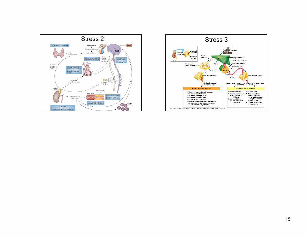
15
Stress 2 Stress 3