Embed Size (px)
Citation preview

By: Thulficar Al-Khafaji
BDS, MSC, PhD
The Endodontics
Introduction
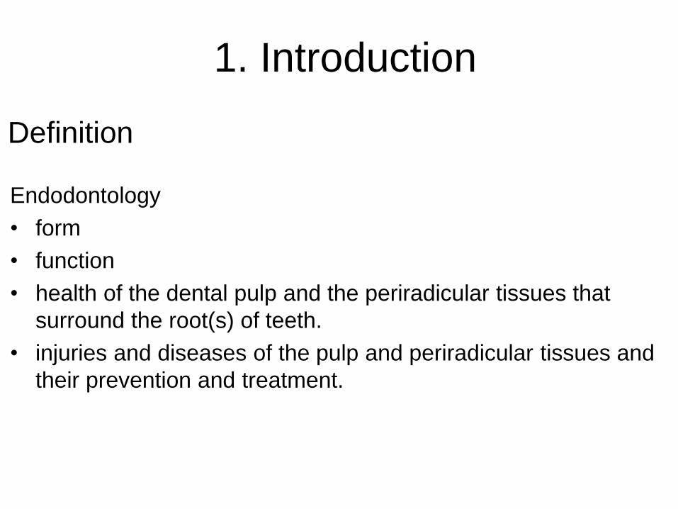
1. Introduction
Endodontology
• form
• function
• health of the dental pulp and the periradicular tissues that
surround the root(s) of teeth.
• injuries and diseases of the pulp and periradicular tissues and
their prevention and treatment.
Definition
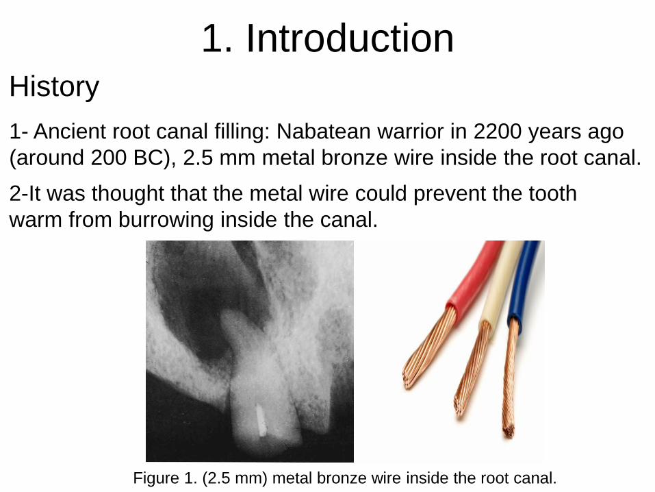
Figure 1. (2.5 mm) metal bronze wire inside the root canal.
1. Introduction
1- Ancient root canal filling: Nabatean warrior in 2200 years ago
(around 200 BC), 2.5 mm metal bronze wire inside the root canal.
2-It was thought that the metal wire could prevent the tooth
warm from burrowing inside the canal.
History
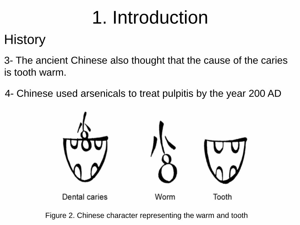
Figure 2. Chinese character representing the warm and tooth
3- The ancient Chinese also thought that the cause of the caries
is tooth warm.
4- Chinese used arsenicals to treat pulpitis by the year 200 AD
1. Introduction History
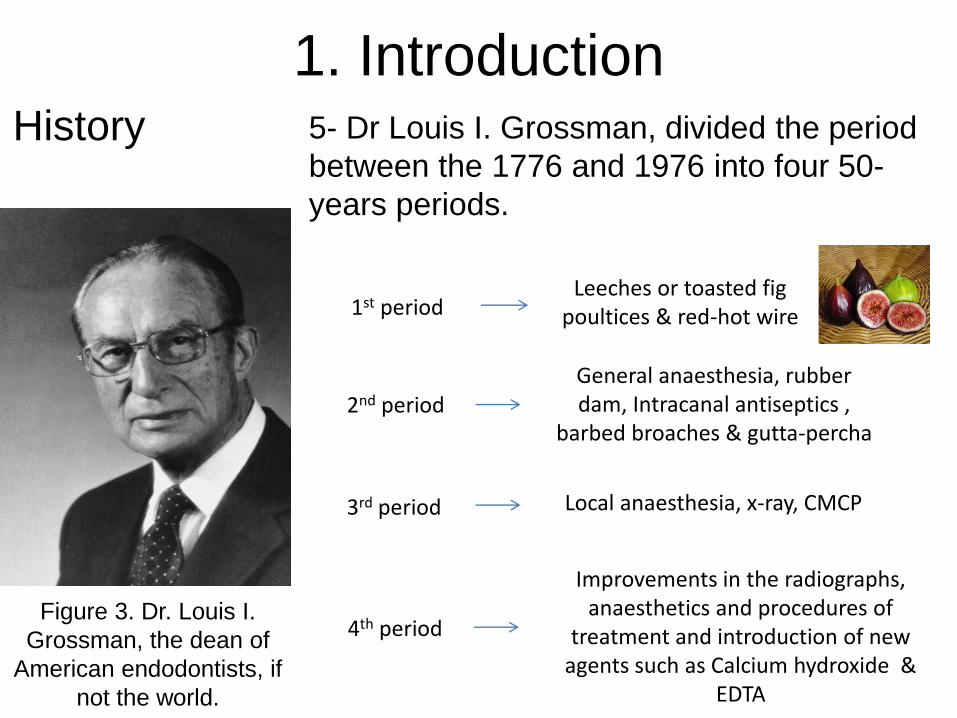
Figure 3. Dr. Louis I.
Grossman, the dean of
American endodontists, if
not the world.
5- Dr Louis I. Grossman, divided the period
between the 1776 and 1976 into four 50-
years periods.
Improvements in the radiographs, anaesthetics and procedures of
treatment and introduction of new agents such as Calcium hydroxide &
EDTA
General anaesthesia, rubber dam, Intracanal antiseptics ,
barbed broaches & gutta-percha
1st period
2nd period
3rd period
4th period
Local anaesthesia, x-ray, CMCP
Leeches or toasted fig poultices & red-hot wire
1. Introduction History
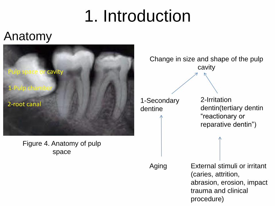
Pulp space or cavity
1-Pulp chamber
2-root canal
Change in size and shape of the pulp
cavity
Aging External stimuli or irritant
(caries, attrition,
abrasion, erosion, impact
trauma and clinical
procedure)
1-Secondary
dentine
2-Irritation
dentin(tertiary dentin
“reactionary or
reparative dentin”)
Figure 4. Anatomy of pulp
space
1. Introduction Anatomy
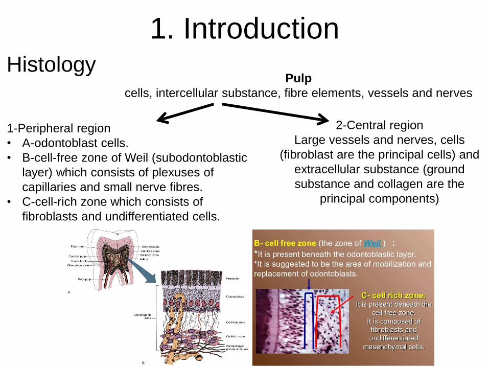
Pulp
cells, intercellular substance, fibre elements, vessels and nerves
2-Central region
Large vessels and nerves, cells
(fibroblast are the principal cells) and
extracellular substance (ground
substance and collagen are the
principal components)
1-Peripheral region
• A-odontoblast cells.
• B-cell-free zone of Weil (subodontoblastic
layer) which consists of plexuses of
capillaries and small nerve fibres.
• C-cell-rich zone which consists of
fibroblasts and undifferentiated cells.
1. Introduction Histology
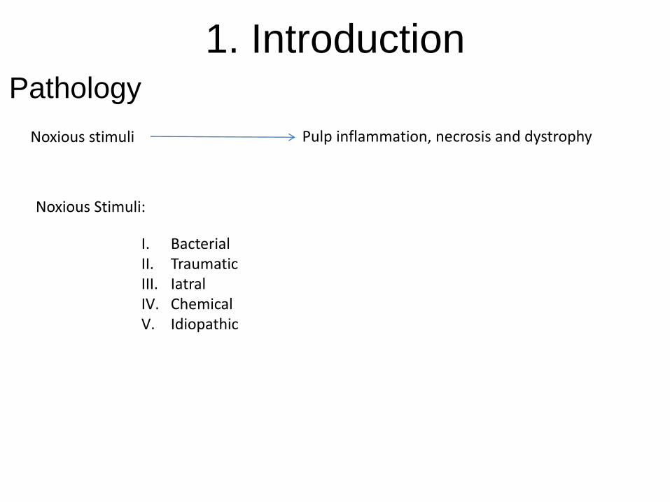
Noxious stimuli Pulp inflammation, necrosis and dystrophy
Noxious Stimuli:
I. Bacterial II. Traumatic III. Iatral IV. Chemical V. Idiopathic
1. Introduction Pathology
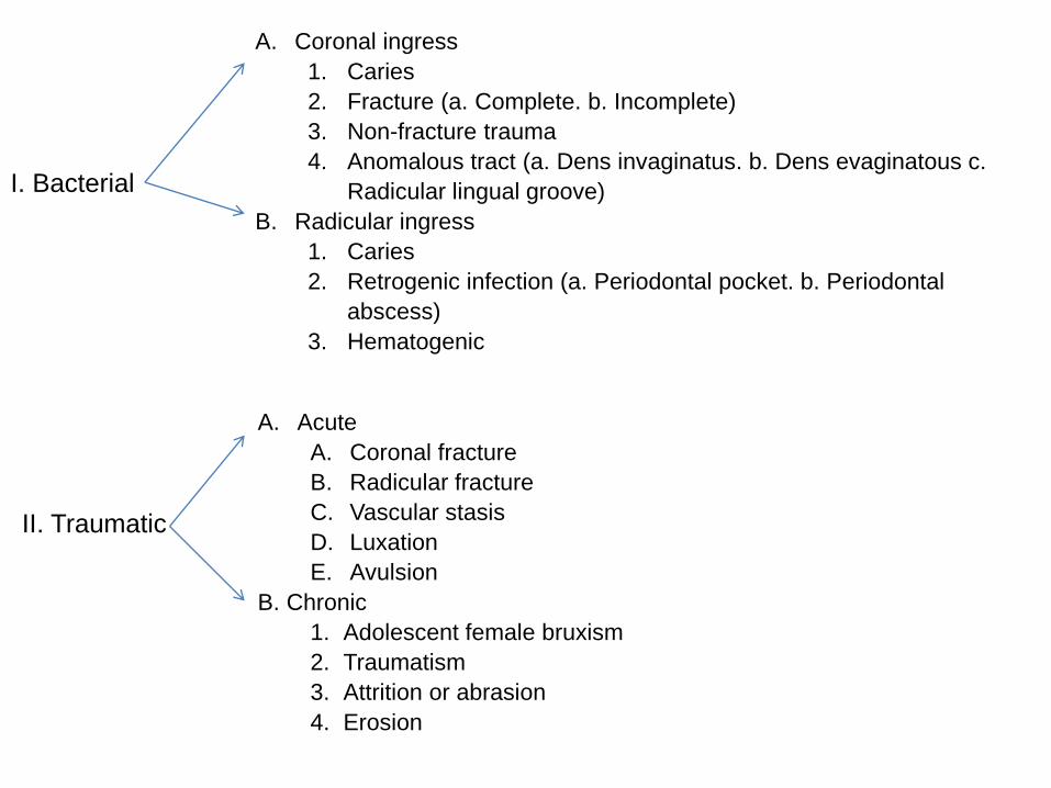
A. Coronal ingress
1. Caries
2. Fracture (a. Complete. b. Incomplete)
3. Non-fracture trauma
4. Anomalous tract (a. Dens invaginatus. b. Dens evaginatous c.
Radicular lingual groove)
B. Radicular ingress
1. Caries
2. Retrogenic infection (a. Periodontal pocket. b. Periodontal
abscess)
3. Hematogenic
A. Acute
A. Coronal fracture
B. Radicular fracture
C. Vascular stasis
D. Luxation
E. Avulsion
B. Chronic
1. Adolescent female bruxism
2. Traumatism
3. Attrition or abrasion
4. Erosion
I. Bacterial
II. Traumatic
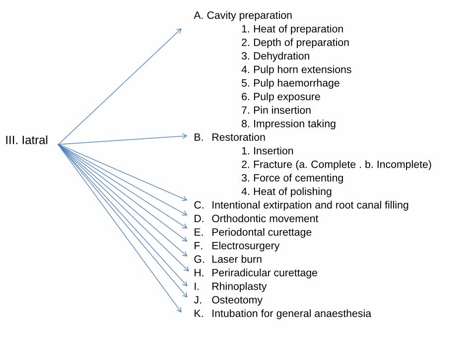
A. Cavity preparation
1. Heat of preparation
2. Depth of preparation
3. Dehydration
4. Pulp horn extensions
5. Pulp haemorrhage
6. Pulp exposure
7. Pin insertion
8. Impression taking
B. Restoration
1. Insertion
2. Fracture (a. Complete . b. Incomplete)
3. Force of cementing
4. Heat of polishing
C. Intentional extirpation and root canal filling
D. Orthodontic movement
E. Periodontal curettage
F. Electrosurgery
G. Laser burn
H. Periradicular curettage
I. Rhinoplasty
J. Osteotomy
K. Intubation for general anaesthesia
III. Iatral
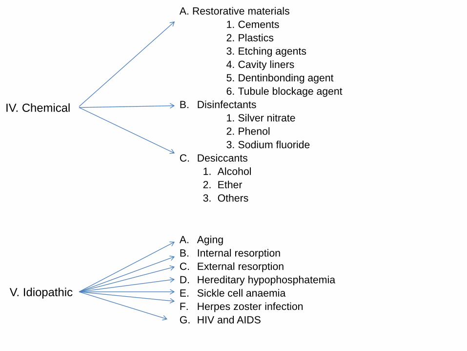
A. Restorative materials
1. Cements
2. Plastics
3. Etching agents
4. Cavity liners
5. Dentinbonding agent
6. Tubule blockage agent
B. Disinfectants
1. Silver nitrate
2. Phenol
3. Sodium fluoride
C. Desiccants
1. Alcohol
2. Ether
3. Others
IV. Chemical
V. Idiopathic
A. Aging
B. Internal resorption
C. External resorption
D. Hereditary hypophosphatemia
E. Sickle cell anaemia
F. Herpes zoster infection
G. HIV and AIDS
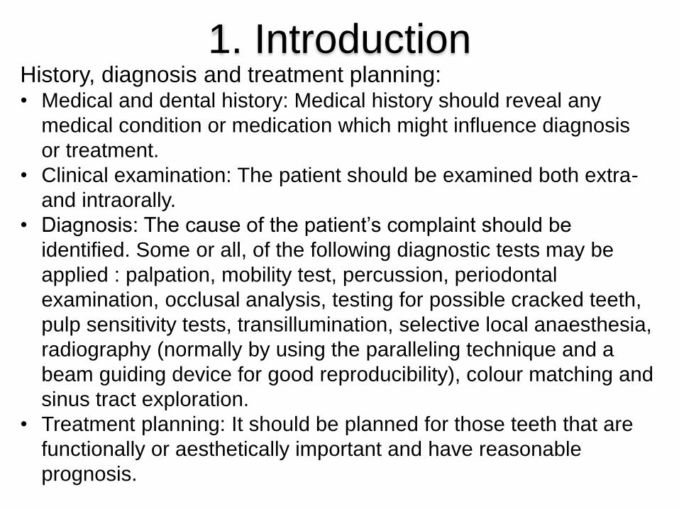
History, diagnosis and treatment planning: • Medical and dental history: Medical history should reveal any
medical condition or medication which might influence diagnosis
or treatment.
• Clinical examination: The patient should be examined both extra-
and intraorally.
• Diagnosis: The cause of the patient’s complaint should be
identified. Some or all, of the following diagnostic tests may be
applied : palpation, mobility test, percussion, periodontal
examination, occlusal analysis, testing for possible cracked teeth,
pulp sensitivity tests, transillumination, selective local anaesthesia,
radiography (normally by using the paralleling technique and a
beam guiding device for good reproducibility), colour matching and
sinus tract exploration.
• Treatment planning: It should be planned for those teeth that are
functionally or aesthetically important and have reasonable
prognosis.
1. Introduction
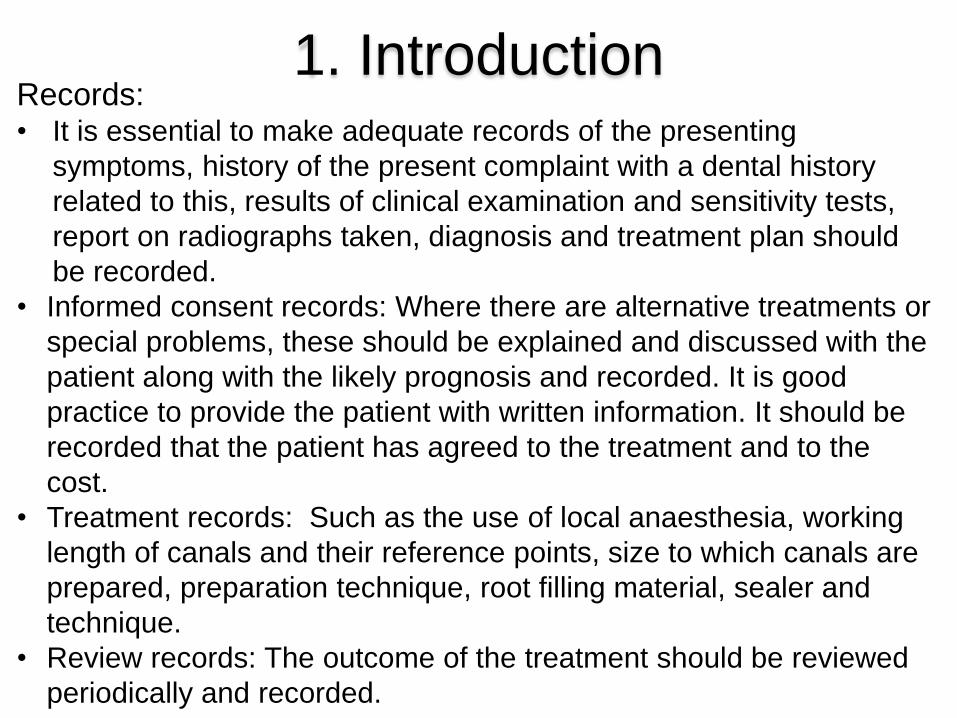
Records: • It is essential to make adequate records of the presenting
symptoms, history of the present complaint with a dental history
related to this, results of clinical examination and sensitivity tests,
report on radiographs taken, diagnosis and treatment plan should
be recorded.
• Informed consent records: Where there are alternative treatments or
special problems, these should be explained and discussed with the
patient along with the likely prognosis and recorded. It is good
practice to provide the patient with written information. It should be
recorded that the patient has agreed to the treatment and to the
cost.
• Treatment records: Such as the use of local anaesthesia, working
length of canals and their reference points, size to which canals are
prepared, preparation technique, root filling material, sealer and
technique.
• Review records: The outcome of the treatment should be reviewed
periodically and recorded.
1. Introduction
Infection control:
• The operator and dental nurse should wear gloves and use
an aseptic technique. All instruments used within the oral
cavity should be sterile, have been decontaminated and
sterilized or disinfected where sterilization is not possible.
1. Introduction
Treatment:
• Early treatment and prevention of pulp damage: Cavity
preparations should be kept as small as possible.
• Treatment for reversible pulp damage:
• 1.Indirect pulp capping (stepwise excavation of caries).
• 2 Direct pulp capping.
• Treatment for irreversible pulp damage:
• 1.Pulp amputation.
• 2.Pulpectomy: Root canal treatment.
1. Introduction
Specific indications for RCT:
Every tooth is indicated for endodontic treatment, from central
incisor to third molar.
The specific indications of endodontic treatment are:
• Teeth with irreversibly damaged or necrotic pulps, with or
without clinical and/or radiographic signs of apical
periodontitis.
• Teeth with no clear evidence of pulp disease, where pulp
space is required for restorative procedure (e.g. post space
preparation, preparation of overdenture abutments, tooth
hemisection, crown preparation on mis-aligned teeth).
So, RCT can be provided for teeth with vital and non-vital pulp
tissues.
1. Introduction
Contra-indications for RCT:
• Teeth that cannot be made functional nor restored.
• Teeth with insufficient periodontal support.
• Teeth with poor prognosis, uncooperative patients or patients
where dental treatment procedures cannot be undertaken.
• Teeth of patients with poor oral condition that cannot be
improved within a reasonable period.
1. Introduction
Indications for RC re-treatment:
• Teeth with inadequate root canal filling with radiological
findings of developing or persisting apical periodontitis and/or
symptoms.
• Teeth with inadequate root canal filling when the coronal
restoration requires replacement or the coronal dental tissue
is to be bleached.
1. Introduction
Indications for surgical endodontics:
• Radiological findings of apical periodontitis and/or symptoms
associated with an obstructed canal (the obstruction proved
not to be removable, displacement did not seem feasible or
the risk of damage was too great).
• Extruded material with clinical or radiological findings of apical
periodontitis and/or symptoms continuing over a prolonged
period.
• Persisting or emerging disease following root canal treatment
when root canal re-treatment is inappropriate.
• Perforation of the root or the floor of the pulp chamber and
where it is impossible to treat from within the pulp cavity.
1. Introduction
Contra-indications for surgical endodontics:
• Local anatomical factors such as an inaccessible root end.
• Tooth with inadequate periodontal support.
• Uncooperative patient.
• Patient with a compromised medical history. See, the contra-
indications for root canal treatment.
1. Introduction
2. Aim
Endodontic or root canal treatment aims to preserve teeth
in a healthy and functional condition by:
• removing diseased pulp tissue
• managing internal infection
• preventing its recurrence.
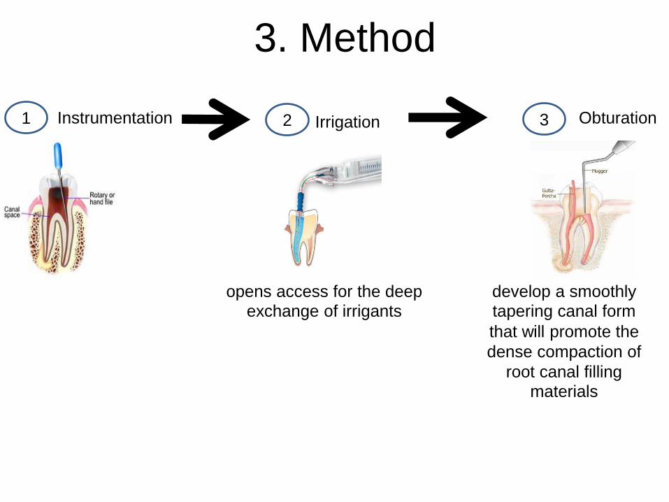
3. Method
opens access for the deep
exchange of irrigants
1 2 3
develop a smoothly
tapering canal form that will promote the
dense compaction of
root canal filling
materials
Instrumentation Irrigation Obturation

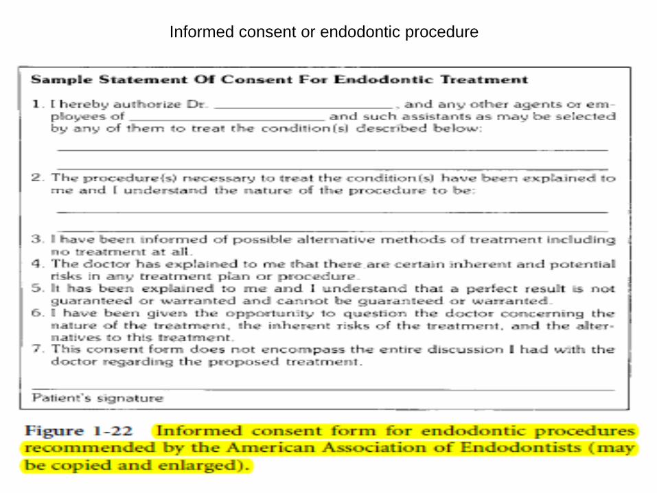
Informed consent or endodontic procedure