Embed Size (px)
Citation preview

RESEARCH ARTICLE Open Access
The etiology of acute meningitis andencephalitis syndromes in a sentinelpediatric hospital, Shenzhen, ChinaHongwei Shen1,2 , Chunqing Zhu3, Xiaorong Liu3, Dongli Ma3, Chunli Song1, Lintao Zhou1, Zuer Wang4,Yongxuan Ou2, Wen Ma1, Xianghui Shi2*, Xuejun Ma5* and Yiwen Zhou1*
Abstract
Background: Acute meningitis and encephalitis syndromes (AMES) is a severe neurological infection which causeshigh case fatality and severe sequelae in children. To determine the etiology of childhood AMES in Shenzhen, ahospital-based study was undertaken.
Methods: A total of 240 cerebrospinal fluid (CSF) samples from 171 children meeting the case definition wereincluded and screened for 12 common causative organisms. The clinical data and conventional testing results werecollected and analyzed. Whole genome sequencing was performed on a Neisseria meningitidis isolate.
Results: A pathogen was found in 85 (49.7%) cases; Group B Streptococcus (GBS) was detected in 17 cases,Escherichia coli in 15, Streptococcus pneumoniae in 14, enterovirus (EV) in 13, herpes simplex virus (HSV) in 3, N.meningitidis in 1, Haemophilus influenzae in 1, and others in 23. Notably, HSV was found after 43 days of treatment.Twelve GBS and 6 E. coli meningitis were found in neonates aged less than 1 month; 13 pneumococcal meningitisin children aged > 3months; and 12 EV infections in children aged > 1 year old. The multilocus sequence typing ofserogroup B N. meningitidis isolate was ST-3200/CC4821. High resistance rate to tetracycline (75%), penicillin (75%),and trimethoprim/sulfamethoxazole (75%) was found in 4 of S. pneumoniae isolates; clindamycin (100%) andtetracycline (100%) in 9 of GBS; and ampicillin (75%) and trimethoprim/sulfamethoxazole (67%) in 12 of E. coli.
Conclusions: The prevalence of N. meningitidis and JEV was very low and the cases of childhood AMES weremainly caused by other pathogens. GBS and E. coli were the main causative organisms in neonates, while S.pneumoniae and EV were mainly found in older children. HSV could be persistently found in the CSF samplesdespite of the treatment. A better prevention strategy for GBS, the introduction of pneumococcal vaccine, andincorporation of PCR methods were recommended.
Keywords: Etiology, Childhood, Meningitis, Encephalitis
BackgroundAcute meningitis and encephalitis syndromes (AMES) isa severe neurological infection which causes high casefatality and severe sequelae including hearing loss and
cognitive deficit [1, 2]. Children under the age of 5 yearsold are the main group at risk and the young infantsbelow 2months old have the highest incidence [2, 3].A broad range of organisms are responsible for bacter-
ial meningitis, including Streptococcus pneumoniae,Haemophilus influenzae, Group B Streptococcus (GBS),Neisseria meningitidis, and Listeria monocytogenes [2].Other causative organisms such as herpes simplex virus(HSV), enterovirus (EV), and Varicella zoster virus(VZV) are associated with aseptic meningitis and en-cephalitis [4].
© The Author(s). 2019 Open Access This article is distributed under the terms of the Creative Commons Attribution 4.0International License (http://creativecommons.org/licenses/by/4.0/), which permits unrestricted use, distribution, andreproduction in any medium, provided you give appropriate credit to the original author(s) and the source, provide a link tothe Creative Commons license, and indicate if changes were made. The Creative Commons Public Domain Dedication waiver(http://creativecommons.org/publicdomain/zero/1.0/) applies to the data made available in this article, unless otherwise stated.
* Correspondence: [email protected]; [email protected];[email protected] District Center for Disease Control and Prevention, Hongli Xi lu 8043,Futian District, Shenzhen 518040, Guangdong, China5Chinese Center for Disease Control and Prevention, National Institute forViral Disease Control and Prevention, Changbai Rd. 155, Changping District,Beijing 102206, China1Shenzhen Hospital of Southern Medical University, Xinhu Rd. 1333, Bao’anDistrict, Shenzhen 518110, Guangdong, ChinaFull list of author information is available at the end of the article
Shen et al. BMC Infectious Diseases (2019) 19:560 https://doi.org/10.1186/s12879-019-4162-5
N. meningitidis and Japanese encephalitis virus (JEV)are the only two causative agents included in the Na-tional Infectious Diseases Surveillance System in China.However, due to the nationwide vaccination program ofserogroup A and serogroup A plus C meningococcal,and JEV vaccines, the incidence of AMES caused by N.meningitidis and JEV has decreased markedly [5]. Theannual incidence of Japanese encephalitis (JE) declinedfrom 0.9489/100,000 in 2000 to 0.12/100,000 in 2011 [6,7]. The annual incidence of meningococcal meningitisdeclined from 0.18/100,000 in 2005 to 0.02/100,000 in2010 and a switch of prevalent serogroups was observedafter integration of meningococcal polysaccharide vaccineinto the national Expanded Program on Immunization(EPI) in 2008 [8, 9]. However, little is known about the fre-quency of other causative pathogens or the disease burdenof AMES in China.A large population-based surveillance for bacterial
meningitis during 2006 and 2009 was undertaken in fourcities and it estimated the annual incidence (per 100,000population) of probable bacterial meningitis in thechildren < 5 years old ranged from 6.95 to 22.30 [3]. Apathogen was identified in a small proportion of testedcases (2.2%) and the cause of other cases remainedundefined [3]. A relative low detection rate (22.6%) wasalso observed in another Chinese study in which sero-logical assays were used for testing viruses [10].PCR had great advantage over conventional methods
and the yield for detection of bacterial pathogens in-creased by 20 to 85% in a study conducted in Brazil [11],and it was widely used for detecting the causative patho-gens of AMES [11, 12].Due to the large geographical and economic variation
in China, the prevalence of vector-borne diseases suchas JE [5], immunization rate, and healthcare qualityvaried in different regions. Apart from the free vaccines,the coverage of others including pneumococcal and H.influenzae type b vaccine would be higher in the devel-oped cities than that in other regions. As a metropoliswith over 12 million population and China’s open doorto the world, it is urgent to understand the etiology ofAMES in Shenzhen, and then provide basis for introdu-cing effective policies for immunization and prevention.Here we undertook a prospective study in a sentinelpediatric hospital which covered 426,000 outpatientsand 77,000 inpatients annually to determine the etiologyof childhood AMES in Shenzhen and extensive testingwas conducted.
MethodsStudy designShenzhen Children’s Hospital (SCH) was chosen as thesentinel hospital. It serves as a referral center in the field
of pediatric diseases and treats over 60% of childhoodAMES cases in Shenzhen each year.Possible AMES were defined based on clinical mani-
festations (altered consciousness that persisted for longerthan 24 h), and with two or more of the following: fever(≥38°C) or the history of fever during the presentingillness; abnormal neuroimaging findings; and CSFpleocytosis (more than four white blood cells per μL).Childhood outpatients and inpatients who met thecriteria, including those transferred from other centersin this region were recruited during June, 2015 andOctober, 2018. Only the possible cases whose guardiansagreed to participate in this study and who had enoughvolume of CSF samples for tests were enrolled in thisstudy. Patients with a different final diagnosis (e.g.,tumor and epilepsy) were excluded (Fig. 1).
Sample collectionOf the enrolled patients, their CSF samples after theroutine tests were collected, stored in − 80 °C and trans-ported to Futian District Center for Disease Control andPrevention for PCR examination once every 5 workdays.The demographic data, blood and CSF test results in-cluding Gram stain and culture results were collected.The use of antimicrobials when collecting CSF samples,route of admission, clinical symptoms, treatment andoutcome were also collected.
Conventional testingThe isolates from blood and CSF samples were collectedand subjected to antimicrobial susceptibility testingusing Vitek 2 Compact system (bioMerieux, France).Reference strains were used as controls according to theguideline. Disc diffusion method was used for testing theantibiotic susceptibility for N. meningitidis according tothe Clinical and Laboratory Standards Institute (CLSI,2016) M100 guideline. S. pneumoniae (ATCC 49619)and E. coli (ATCC 25922) were used as controls for diskdiffusion method. The susceptibilities for the tested anti-microbials were classified as resistant, intermediate, andsusceptible according to the guideline (CLSI, 2016). S.pneumoniae antigen testing was performed on the CSFsamples collected from the suspected pneumococcalmeningitis using lateral flow assay (Alere, USA). The iso-lated N. meningitidis was serotyped with a commerciallykit according to the instruction (Remel, UK).
PCR amplificationThe nucleic acid was extracted using MagNA Pure 96System (Roche) and followed by detection of 12 patho-gens using Real-time PCR assays. The oligonucleotideprimers and probes were listed in Table 1. S. pneumo-niae, H. influenzae, GBS, E. coli, N. meningitidis, and L.monocytogenes were detected using GoTaq Probe qPCR
Shen et al. BMC Infectious Diseases (2019) 19:560 Page 2 of 9

Master Mix (Promega, USA). The final concentration foreach forward primer, reverse primer, and probe was 0.2 μM,0.2 μM, and 0.05 μM, respectively. The cycling conditionswere as follows: 2min at 95°C followed by 40 cycles of 15 sat 95°C, 1min at 60°C. Enterovirus, HSV, JEV, mumps virus(MuV), adenovirus (AdV), and VZV were detected usingQuantiTect Probe RT-PCR Kit (Qiagen, Germany) accord-ing to the instruction. The final concentration for eachforward primer, reverse primer, and probe was 0.4 μM,0.4 μM, and 0.1 μM, respectively. The cycling conditions in-cluded reverse transcription of 30min at 50°C, a initiatingstage of 15min at 95°C, and followed by 45 cycles of 15 s at94°C, 1min at 60°C. The nucleic acids extracted from refer-ence strains, constructed plasmids, or in vitro transcribedRNA templates were used as controls. Amplification, detec-tion, and data analysis were performed in a Bio-Rad CFX96real-time thermal cycler (Bio-Rad, USA).
Next generation sequencingThe nucleic acids of N. meningitidis strains were extractedusing QIAamp DNA Mini Kit (Qiagen, Germany) and
1 μg of DNA was broken using ultrasound, concentrated,and used for preparing DNA library using NEB Next®Ultra™ DNA Library Prep Kit for Illumina (NEB, USA).Whole genome sequencing was performed on IlluminaMiseq (USA). The sequencing results were edited andassembled using SPAdes and PubMLST was used foridentifying alleles for multilocus sequence typing (MLST),antimicrobial resistance related genes including gyrA,penA, and rpoB, and peptide coding gene such as porA,porB, fetA, and fHbp.
ResultsSample informationA total of 634 possible childhood AMES cases wereidentified during June, 2015 and October, 2018 and 240CSF samples collected from 171 patients (male, 116)were included (Fig. 1). Of all the samples, 104 sequentialsamples were collected from 35 patients. Twelve of sam-ples were collected before the use of antibiotics. Patientsranged in age from 0 to 15 years old and 91 of themaged less than 3 months. Of all the patients, 73 were
Fig. 1 Study profile
Shen et al. BMC Infectious Diseases (2019) 19:560 Page 3 of 9

neonates and 27 were preterm; 103 were hospitalized ortransferred from other hospitals. The most commonsymptoms were fever (137, 80.1%), vomiting (73, 42.7%),and seizures (36, 21.1%). Two recurrent cases werefound. Thirteen cases were transferred to other hospitalsand the symptoms of all the others improved whendischarged from SCH. No death was found.
EtiologyOverall, a pathogen was found in 85 (49.7%) cases. Abacterial pathogen was found in 68 (39.8%) of cases(GBS, 17; E. coli, 15; S. pneumoniae, 14; H. influenzae, 1;N. meningitidis, 1; others, 23). Of the other bacteria,Staphylococcus epidermidis was the predominant micro-organism (9 cases) and all were isolated from patientsduring hospitalization and after administration of antibi-otics. EV and HSV were found in 13 and 3 cases of viralinfections, respectively. Candida famata was isolatedfrom the blood sample from a neonate (Table 2). No L.monocytogenes, JEV, MuV, AdV, and VZV were detected.GBS and Pseudomonas aeruginosa was isolated from a
long-hospitalized case in the 1st and 9th month, respect-ively. Of the recurrent cases, S. pneumoniae was foundin 2 episodes of meningitis in case 1. S. pneumoniae andE. coli was found in 1st and 2nd episode of meningitis incase 2, respectively.
Table 1 The oligonucleotide primers and probes for real-time PCR used in this study
Organism Target gene Primer sequence (5′-3′) Probe (5′-3′) Citation
S. pneumoniae lytA F: ACGCAATCTAGCAGATGAAGCR: TGTTTGGTTGGTTATTCGTGC
FAM-TTTGCCGAAAACGCTTGATACAGGG-BHQ [29]
H. influenzae bexA F: GGCGAAATGGTGCTGGTAAR: GGCCAAGAGATACTCATAGAACGTT
HEX-CACCACTCATCAAACGAATGAGCGTGG-BHQ
[29]
GBS cfb F: AGCTCTATTAGAAGTACATGCTR: CATTTGCTGGGCTTGATTATT
ROX-ATCAAGTGACAACTCCACAAGTGGTAA-BHQ2
[30]
E. coli uid F: GTGTGATATCTACCCGCTTCGCR: GAGAACGGTTTGTGGTTAATCAGGA
ROX-TCGGCATCCGGTCAGTGGCAGT-BHQ2 [31]
N. meningitidis ctrA F: GCTGCGGTAGGTGGTTCAAR: TTGTCGCGGATTTGCAACTA
FAM- CATTGCCACGTGTCAGCTGCACAT-BHQ [32]
L. monocytogenes iap F: CTAAAGCGGGAATCTCCCTTR: CCATTGTCTTGCGCGTTAAT
HEX- CTTCTGGCGCACAATACGCTAGCACT-BHQ
[33]
mumps virus Fusion protein F:TCTCACCCATAGCAGGGAGTTATATR: GTTAGACTTCGACAGTTTGCAACAA
HEX-AGGCGATTTGTAGCACTGGATGGAACA-BHQ
[34]
herpes simplex virus polymerase F:CATCACCGACCCGGAGAGGGACR: GGGCCAGGCGCTTGTTGGTGTA
FAM- CCGCCGAACTGAGCAGACACCCGCGC-BHQ
[35]
Japanese encephalitisvirus
polyprotein F: AGAACGGAAGAYAACCATGACTAAAR: CCGCGTTTCAGCATATTGAT
ROX-ACCAGGAGGGCCCGG- BHQ2 [36]
enterovirus polyprotein F: CCGGCCCCTGAATGCR: CACCGGATGGCCAATCCA
ROX-AACCGACTACTTTGGGTGTCCGTGTTTC-BHQ2
[37]
adenovirus hexon F: GCCACGGTGGGGTTTCTAAACTTR: GCCCCAGTGGTCTTACATGCACATC
CY5-TGCACCAGACCCGGGCTCAGGTACTCCGA-BHQ2
[38]
varicella zoster virus DNA polymerase F: CGGCATGGCCCGTCTAT HEX-ATTCAGCAATGGAAACACACGACGCC- [39]
R: TCGCGTGCTGCGGC BHQ1
Table 2 Pathogens detected in the 171 AMES cases by culture/PCR method
Organisma Blood CSF Totalc
culture PCR LFAb
S. pneumoniaed 7 9 3 11 14
E. colie 12 8 2 – 15
GBSf 13 5 4 – 17
H. influenzae 0 0 1 – 1
N. meningitidis 1 0 1 – 1
Othersg 14 9 – – 23
enterovirus – – 13 – 13
HSV h – – 3 – 3
total 47 31 27 11 85i
-, not detecteda GBS, group B Streptococcus; HSV, herpes simplex virus; JEV, Japaneseencephalitis virus; VZV, varicella zoster virusb LFA, lateral flow assayc A causative agent was defined by a positive result tested by any of the methodsd S. pneumoniae was isolated from both blood and CSF samples in 7 cases.Three pneumococcal cases were detected only by LAF due to the use ofantibiotics before samplinge E. coli was isolated from both blood and CSF samples in 5 casesf GBS was isolated from both blood and CSF samples in 4 casesg Others included 6 of Staphylococcus epidermidis, 2 of Pseudomonasaeruginosa, and 1 Staphylococcus capitis isolate from CSF samples; and 3Staphylococcus epidermidis, 4 Klebsiella pneumoniae, 1 Staphylococcus capitis, 2Staphylococcus hominis, 1 Acinetobacter baumannii, 1 Staphylococcus aureus, 1Candida famata, and 1 Enterococcus aerogenes isolate from blood samplesh The 3 isolates were typed using a commercial kit (Daan, China) and all wereidentified as HSV-1i Recurrent and co-infection were excluded
Shen et al. BMC Infectious Diseases (2019) 19:560 Page 4 of 9

Age distribution of the main pathogensGBS and E. coli were the predominant causes of bacter-ial meningitis in neonates. Twelve of 17 GBS and 6 of 15E. coli meningitis were found in neonates aged less than1 month. All the pneumococcal meningitis was detectedin patients more than 3months old and 13 of 14 caseswas found in the age group of 3 months and 5 years old.Of the 13 cases of encephalitis caused by EV, 12 wasfound in children over 1 years old and 8 over 5 years old.Two of 3 HSV encephalitis cases were 2 months old.
CSF examination in AMES caused by common pathogensThe mean white blood cell (WBC) count (106/μl), pro-tein, and glucose in CSF, and hospitalization length inEV encephalitis was 143, 0.40 g/L, 3.23 mmol/L, and 7days, respectively. A higher mean CSF WBC count andprotein, longer hospitalization days, and lower CSF glu-cose were seen in the bacterial meningitis cases. A posi-tive PCR result for HSV could be found in the CSFsample after 43 days of treatment. The longest periodafter treatment to obtain a positive PCR result for E. coli,N. meningitidis, S. pneumoniae, and GBS was 13, 8, 17,and 9 days, respectively.
Antimicrobial susceptibility profiles of the mainmicroorganismsOf the 4 tested isolates of S. pneumoniae, three were re-sistant to tetracycline, penicillin, and trimethoprim/sulfamethoxazole. All the 9 GBS isolates were resistantto clindamycin and tetracycline. Of the 4 GBS strainswhose resistance to roxithromycine, azithromycin, andclarithromycin was tested, they were all resistant to thesedrugs (not shown). The highest resistance rate in E. coliwas found in ampicillin (9/12), followed by trimetho-prim/sulfamethoxazole (8/12). Resistance to penicillin,and oxacillin was found in all the 8 tested S. epidermidisisolates (Table 3).
Characterization of N. meningitidisA serogroup B N. meningitidis was isolated from theblood sample of a 3-month-old meningococcal case. Itwas also detected in the CSF samples by PCR (Table 2).The isolate was sensitive to meropenem, ceftriaxone,chloramphenicol, and cefotaxime, and intermediate sen-sitive to penicillin. The whole genome was 2,305,805 bpand the MLST type was ST-3200/CC4821. The allele forthe antibiotic-associated gene gyrA, penA, and rpoB was116, 1, and 85, respectively. The allele for peptide codinggene porA, porB, fetA, and fHbp was 20,14; 34; F3–9;and 16/A19, respectively.
DiscussionA pathogen was identified in 85 (49.7%) cases and theirage distribution and prevalence of antibiotic resistance
were elucidated. These results could provide basis fordesigning prevention policy, as well as for the diagnosisand empirical treatment of AMES. Therefore, to enablethe timely and appropriate treatment and to improve theoutcome.Compared with 2.2% in a large surveillance study in
which only the laboratory confirmed S. pneumoniae, H.influenzae type b, and N. meningitidis isolates were in-cluded [3], the detection rate in our study was greatlyimproved. In another study where serological tests wereused for detecting viruses, a causative agent was foundin 22.6% (538/2382) of cases [10]. The use of PCRmethod and childhood population might contribute tothe relative higher detection rate in this study. The ab-sence of JEV, MuV, and VZV might due to the differencein sample size, regional or demographic factors. Thechanges of immunization policies might also affect theresults, such as the low detection rate of JEV.Despite of the usefulness of conventional methods, in-
corporation of PCR method could improve the outcomeof surveillance. In the culture negative samples, additional13 EV, 3 HSV, 2 GBS, and 1 H. influenzae were identified.The incidence of H. influenzae meningitis was very lowdue to the use of Hib vaccine in Shenzhen and it wouldnot be considered without a positive result. The positivePCR result could help initiate timely and appropriate ther-apy in the clinical setting. However, due to the long courseof AMES and early initiation of empirical treatment, 228of 240 CSF samples were collected after the use of antibi-otics. The presence of antibiotics in CSF might shortenthe time for the detection of pathogens by PCR. In the se-quential samples, the negative results were obtained afteradministrating antibiotics for a period of time, which wasalso observed in another study [13].Due to the nationwide vaccination of meningococcal
and JEV vaccines, the annual incidence of meningococ-cal disease and JE in Shenzhen decreased to 0.048 and0.664 (per 100,000), respectively [14, 15]. The absence ofJE in this study could be attributed to its low incidence.The detection of serogroup B N. meningitidis was attrib-uted to the group specificity of meningococcal polysac-charide vaccine and its low cross-immunity rate. Theintroduction of meningococcal A vaccine in 2000 andmeningococcal A plus C polysaccharide vaccine in 2008in Guangdong has led to serogroup replacement of N.meningitidis. During 2008 and 2010, serogroup C men-ingococcal diseases (MenC) accounted for majority ofmeningococcal cases, and serogroup B meningococcaldiseases (MenB) occurred relatively infrequently. From2011 onwards, the proportion of MenC cases began todecline and MenB to rise [8, 16, 17]. Other strains withthe same MLST type were isolated in France, UK, andTaiwan, which caused meningococcal disease mainly inyoung children aged less than 1 years old [18].
Shen et al. BMC Infectious Diseases (2019) 19:560 Page 5 of 9
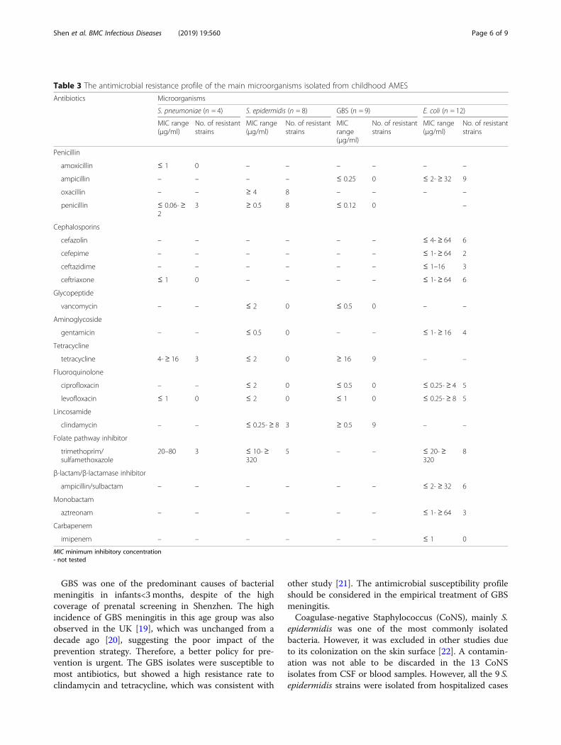
GBS was one of the predominant causes of bacterialmeningitis in infants<3 months, despite of the highcoverage of prenatal screening in Shenzhen. The highincidence of GBS meningitis in this age group was alsoobserved in the UK [19], which was unchanged from adecade ago [20], suggesting the poor impact of theprevention strategy. Therefore, a better policy for pre-vention is urgent. The GBS isolates were susceptible tomost antibiotics, but showed a high resistance rate toclindamycin and tetracycline, which was consistent with
other study [21]. The antimicrobial susceptibility profileshould be considered in the empirical treatment of GBSmeningitis.Coagulase-negative Staphylococcus (CoNS), mainly S.
epidermidis was one of the most commonly isolatedbacteria. However, it was excluded in other studies dueto its colonization on the skin surface [22]. A contamin-ation was not able to be discarded in the 13 CoNSisolates from CSF or blood samples. However, all the 9 S.epidermidis strains were isolated from hospitalized cases
Table 3 The antimicrobial resistance profile of the main microorganisms isolated from childhood AMES
Antibiotics Microorganisms
S. pneumoniae (n = 4) S. epidermidis (n = 8) GBS (n = 9) E. coli (n = 12)
MIC range(μg/ml)
No. of resistantstrains
MIC range(μg/ml)
No. of resistantstrains
MICrange(μg/ml)
No. of resistantstrains
MIC range(μg/ml)
No. of resistantstrains
Penicillin
amoxicillin ≤ 1 0 – – – – – –
ampicillin – – – – ≤ 0.25 0 ≤ 2- ≥ 32 9
oxacillin – – ≥ 4 8 – – – –
penicillin ≤ 0.06-≥2
3 ≥ 0.5 8 ≤ 0.12 0 –
Cephalosporins
cefazolin – – – – – – ≤ 4- ≥ 64 6
cefepime – – – – – – ≤ 1- ≥ 64 2
ceftazidime – – – – – – ≤ 1–16 3
ceftriaxone ≤ 1 0 – – – – ≤ 1- ≥ 64 6
Glycopeptide
vancomycin – – ≤ 2 0 ≤ 0.5 0 – –
Aminoglycoside
gentamicin – – ≤ 0.5 0 – – ≤ 1- ≥ 16 4
Tetracycline
tetracycline 4-≥ 16 3 ≤ 2 0 ≥ 16 9 – –
Fluoroquinolone
ciprofloxacin – – ≤ 2 0 ≤ 0.5 0 ≤ 0.25-≥ 4 5
levofloxacin ≤ 1 0 ≤ 2 0 ≤ 1 0 ≤ 0.25-≥ 8 5
Lincosamide
clindamycin – – ≤ 0.25-≥ 8 3 ≥ 0.5 9 – –
Folate pathway inhibitor
trimethoprim/sulfamethoxazole
20–80 3 ≤ 10-≥320
5 – – ≤ 20-≥320
8
β-lactam/β-lactamase inhibitor
ampicillin/sulbactam – – – – – – ≤ 2- ≥ 32 6
Monobactam
aztreonam – – – – – – ≤ 1- ≥ 64 3
Carbapenem
imipenem – – – – – – ≤ 1 0
MIC minimum inhibitory concentration- not tested
Shen et al. BMC Infectious Diseases (2019) 19:560 Page 6 of 9

after the use of antibiotics, suggesting the possibility ofnosocomial infections. The high incidence and occur-rence of multi-drug resistance strain posed the necessityfor designing effective prevention strategies.S. pneumoniae was the leading cause of bacterial men-
ingitis in the group aged between 3months and 5 years.The estimated annual incidence of pneumococcal men-ingitis was 14 per 100,000 in children between 1monthand 59months in China [23]. According to another re-search, the most common serogroups were 19F, 19A,and 14, which were covered by the 13-vulent pneumo-coccal conjugate vaccine (PCV13) [23, 24]. The intro-duction of PCV13 has resulted in significant reductionsof pneumococcal meningitis caused by new serotypes[25]. But PCV13 was not included in the NationalPlanned Immunization schedule and the vaccination ratewas unknown. To reduce the incidence of pneumococcalmeningitis, the introduction of PCV13 vaccine andfurther long surveillance were needed. The high resist-ance rate to tetracycline, penicillin, and trimethoprim/sulfamethoxazole needed the confirmation with largesample size.In the aseptic meningitis and encephalitis, EV
accounted for the majority of viruses detected and 92%of EV infection was found in children over 1 year of age,in agreement with the previous study [26]. No seriouscomplications or deaths were found. EV PCR was notroutinely performed for diagnosis of central nervoussystem (CNS) infections in the sentinel hospital, andthere was a delay of diagnosis when the samples weretransported to CDC after several days of sampling.Therefore, 11 of 13 EV was found in the samples withunknown cause. As a result, a longer mean duration ofhospitalization stay (7 days) and antibiotic administration(6 days) was observed, compared with a previous study(3 and 2 days, respectively) [26]. It was highly recom-mended to incorporate EV PCR into the routine test fordiagnosis of CNS infection in the group at high risk.Similarly, long mean stay was observed in HSV infec-
tions (25 days) and 2 of 3 HSV-positive cases weretreated as bacterial meningitis initially. The clinical find-ings of Herpes simplex encephalitis (HSE) were notpathognomonic and numerous other CNS infectionscould mimic HSE, so it was important to identify HSVin the high-risk population. All the 3 HSE cases werefound in children below 2 years old, consistent with aprior study which indicated the predisposition to HSEwas tightly age-dependant [27]. Due to the long durationof disease course, efficacy of acyclovir treatment, highmortality and severe sequelae [28], it was important toidentify HSE. Therefore, the appropriate treatment couldbe given and the outcome could be improved.This study had several limitations. Firstly, all the
samples were collected from one sentinel hospital and
the results may not be representative of this region.Secondly, a large proportion of CSF samples subjectedto PCR detection were collected after the use of antibi-otics, which might result in negative results. Thirdly, along storage time of CSF samples has led to a delay ofdiagnosis. Finally, the long-term outcome was notfollowed and the case fatality might be underestimated.Nevertheless, due to the difficulty in collecting CSF sam-ples from children, this was the most comprehensivedata collection on the etiology of AMES in this region.This referral center received childhood AMES casesfrom many other centers in Shenzhen made the resultsreasonably representative. A prospective study involvedmore sentinel hospitals is anticipated to overcome theselimitations.
ConclusionDue to the nationwide use of serogroup A and serogroupA plus C meningococcal, and JEV vaccines, the incidenceof meningococcal disease and JE is very low in Shenzhen.The main causative agents for childhood AMES wereGBS, S. pneumoniae, E. coli, EV, and HSV. A betterprevention strategy for GBS, the introduction of PCV13vaccine, and incorporation of PCR methods were recom-mended to reduce the incidence of AMES and achieveearly diagnosis.
AbbreviationsAdV: Adenovirus; AMES: Acute meningitis and encephalitis syndrome;CDC: Center for Disease Control and Prevention; CLSI: Clinical and LaboratoryStandards Institute; CNS: Central nervous system; CoNS: Coagulase-negativeStaphylococcus; CSF: Cerebrospinal fluid; EPI: Expanded Program onImmunization; EV: Enterovirus; GBS: Group B Streptococcus; HSE: Herpessimplex encephalitis; HSV: Herpes simplex virus; JE: Japanese encephalitis;JEV: Japanese encephalitis virus; LFA: Lateral flow assay; MenB: Serogroup Bmeningococcal diseases; MenC: Serogroup C meningococcal diseases;MIC: Minimum inhibitory concentration; MLST: Multilocus sequence typing;MuV: Mumps virus; PCV13: 13-vulent pneumococcal conjugate vaccine;SCH: Shenzhen Children’s Hospital; VZV: Varicella zoster virus; WBC: Whiteblood cell
AcknowledgmentsWe thank the staff from the clinical laboratory of Shenzhen Children’sHospital who took part in the study. We thank the great help offered by Mr.Muhammad Luqman in manuscript revision.
Authors’ contributionsYZ and XS designed the study. HS, ZW, and YO collected and detectedclinical samples using PCR methods. CZ, XL, and DM undertook theconventional testing. YZ, CS, LZ, and WM analyzed data. HS and XM draftedand revised the manuscript. All authors read and approved the finalmanuscript.
FundingThis study was supported by grants from the China Mega-Project forInfectious Disease (2017ZX10302301–004 and 2017ZX10104001); MedicalScientific Research Foundation of Guangdong Province (A2017143); ShenzhenScience and Technology Innovation Committee (JCYJ20160428152017475);Health and Family Planning Commission of Shenzhen Municipality (201602009,201601040); and Futian District Public Health Research Project (FTWS2016049).These funding supported the collection and detection of clinical samples,analysis of data, and publication.
Shen et al. BMC Infectious Diseases (2019) 19:560 Page 7 of 9

Availability of data and materialsThe datasets used and analyzed during the current study are available fromthe corresponding authors on reasonable request.
Ethics approval and consent to participateAll aspects of the study were performed in accordance with national ethicsregulations and approved by the Ethics Committee of Shenzhen Children’sHospital and Futian District Center for Disease Control and Prevention.Written consent was obtained from children’s parents or guardians.
Consent for publicationNot applicable.
Competing interestsThe authors declare that they have no competing interests.
Author details1Shenzhen Hospital of Southern Medical University, Xinhu Rd. 1333, Bao’anDistrict, Shenzhen 518110, Guangdong, China. 2Futian District Center forDisease Control and Prevention, Hongli Xi lu 8043, Futian District, Shenzhen518040, Guangdong, China. 3Shenzhen Children’s Hospital, Shenzhen, China.4Guangdong Medical College, Dongguan, China. 5Chinese Center for DiseaseControl and Prevention, National Institute for Viral Disease Control andPrevention, Changbai Rd. 155, Changping District, Beijing 102206, China.
Received: 14 February 2019 Accepted: 4 June 2019
References1. Edmond K, Clark A, Korczak VS, Sanderson C, Griffiths UK, Rudan I. Global
and regional risk of disabling sequelae from bacterial meningitis: asystematic review and meta-analysis. Lancet Infect Dis. 2010;10(5):317–28.
2. Thigpen MC, Whitney CG, Messonnier NE, Zell ER, Lynfield R, Hadler JL,Harrison LH, Farley MM, Reingold A, Bennett NM, et al. Bacterial meningitisin the United States, 1998-2007. N Engl J Med. 2011;364(21):2016–25.
3. Li Y, Yin Z, Shao Z, Li M, Liang X, Sandhu HS, Hadler SC, Li J, Sun Y, Li J, etal. Population-based surveillance for bacterial meningitis in China,September 2006-December 2009. Emerg Infect Dis. 2014;20(1):61–9.
4. Granerod J, Ambrose HE, Davies NW, Clewley JP, Walsh AL, Morgan D,Cunningham R, Zuckerman M, Mutton KJ, Solomon T, et al. Causes ofencephalitis and differences in their clinical presentations in England: amulticentre, population-based prospective study. Lancet Infect Dis. 2010;10(12):835–44.
5. Zheng Y, Li M, Wang H, Liang G. Japanese encephalitis and Japaneseencephalitis virus in mainland China. Rev Med Virol. 2012;22(5):301–22.
6. Wang H, Li Y, Liang X, Liang G. Japanese encephalitis in mainland China.Jpn J Infect Dis. 2009;62(5):331–6.
7. Gao X, Li X, Li M, Fu S, Wang H, Lu Z, Cao Y, He Y, Zhu W, Zhang T, et al.Vaccine strategies for the control and prevention of Japanese encephalitisin mainland China, 1951-2011. PLoS Negl Trop Dis. 2014;8(8):e3015.
8. Li J, Shao Z, Liu G, Bai X, Borrow R, Chen M, Guo Q, Han Y, Li Y, Taha MK, et al.Meningococcal disease and control in China: findings and updates from theglobal meningococcal initiative (GMI). J Inf Secur. 2018;76(5):429–37.
9. Li J, Li Y, Shao Z, Li L, Yin Z, Ning G, Xu L, Luo H. Prevalence of meningococcalmeningitis in China from 2005 to 2010. Vaccine. 2015;33(8):1092–7.
10. Xie Y, Tan Y, Chongsuvivatwong V, Wu X, Bi F, Hadler SC, Jiraphongsa C,Sornsrivichai V, Lin M, Quan Y. A population-based acute meningitis andencephalitis syndromes surveillance in Guangxi, China, may 2007-June 2012.PLoS One. 2015;10(12):e0144366.
11. Sacchi CT, Fukasawa LO, Goncalves MG, Salgado MM, Shutt KA, CarvalhanasTR, Ribeiro AF, Kemp B, Gorla MC, Albernaz RK, et al. Incorporation of real-time PCR into routine public health surveillance of culture negativebacterial meningitis in Sao Paulo, Brazil. PLoS One. 2011;6(6):e20675.
12. Chiba N, Murayama SY, Morozumi M, Nakayama E, Okada T, Iwata S,Sunakawa K, Ubukata K. Rapid detection of eight causative pathogens forthe diagnosis of bacterial meningitis by real-time PCR. J Infect Chemother.2009;15(2):92–8.
13. Saha SK, Darmstadt GL, Yamanaka N, Billal DS, Nasreen T, Islam M, HamerDH. Rapid diagnosis of pneumococcal meningitis: implications for treatmentand measuring disease burden. Pediatr Infect Dis J. 2005;24(12):1093–8.
14. Qin Y, Mei S, Xie X, Kong D, Tang X. Analysis of the epidemiologicalcharacteristics of epidemic cerebrospinal meningitis in Shenzhen during2005-2015. J Dis Monitor Control. 2016;10(11):874–5 (in Chinese).
15. Xu L, Xie J, Li Y. Analysis on epidemic characteristics of epidemicencephalitis B in Baoan District of Shenzhen. Pract Prev Med. 2009;16(1):64–5 (in Chinese).
16. Z-j S, Xu L, Gao Y. Epidemiologic trend of serogroup switching of Neisseriameningitidis strains in China [J]. Chin J Vaccines Immunization. 2007;6:012(in Chinese).
17. Zhou H, Liu W, Xu L, Deng L, Deng Q, Zhuo J, Shao Z. Spread of Neisseriameningitidis serogroup W clone, China. Emerg Infect Dis. 2013;19(9):1496–9.
18. Chiou CS, Liao JC, Liao TL, Li CC, Chou CY, Chang HL, Yao SM, Lee YS.Molecular epidemiology and emergence of worldwide epidemic clones ofNeisseria meningitidis in Taiwan. BMC Infect Dis. 2006;6:25.
19. Okike IO, Johnson AP, Henderson KL, Blackburn RM, Muller-Pebody B,Ladhani SN, Anthony M, Ninis N, Heath PT, neoMen Study G. Incidence,etiology, and outcome of bacterial meningitis in infants aged <90 days inthe United Kingdom and Republic of Ireland: prospective, enhanced,national population-based surveillance. Clin Infect Dis. 2014;59(10):e150–7.
20. Heath PT, Balfour G, Weisner AM, Efstratiou A, Lamagni TL, Tighe H,O'Connell LA, Cafferkey M, Verlander NQ, Nicoll A, et al. Group Bstreptococcal disease in UK and Irish infants younger than 90 days. Lancet.2004;363(9405):292–4.
21. Zhang J, Zhao R, Dong Y, Zheng Y. Invasive group B streptococcal infectionin infants in Shenzhen, China. Int J Clin Exp Med. 2015;8(2):2939–43.
22. Cho HK, Lee H, Kang JH, Kim KN, Kim DS, Kim YK, Kim JS, Kim JH, Kim CH,Kim HM, et al. The causative organisms of bacterial meningitis in Koreanchildren in 1996-2005. J Korean Med Sci. 2010;25(6):895–9.
23. Chen Y, Deng W, Wang SM, Mo QM, Jia H, Wang Q, Li SG, Li X, Yao BD, LiuCJ, et al. Burden of pneumonia and meningitis caused by Streptococcuspneumoniae in China among children under 5 years of age: a systematicliterature review. PLoS One. 2011;6(11):e27333.
24. Ma X, Zhao R, Ma Z, Yao K, Yu S, Zheng Y, Yang Y. Serotype distribution andantimicrobial resistance of Streptococcus pneumoniae isolates causing invasivediseases from Shenzhen Children's hospital. PLoS One. 2013;8(6):e67507.
25. Miller E, Andrews NJ, Waight PA, Slack MP, George RC. Effectiveness of thenew serotypes in the 13-valent pneumococcal conjugate vaccine. Vaccine.2011;29(49):9127–31.
26. Michos AG, Syriopoulou VP, Hadjichristodoulou C, Daikos GL, Lagona E,Douridas P, Mostrou G, Theodoridou M. Aseptic meningitis in children:analysis of 506 cases. PLoS One. 2007;2(7):e674.
27. Abel L, Plancoulaine S, Jouanguy E, Zhang SY, Mahfoufi N, Nicolas N,Sancho-Shimizu V, Alcais A, Guo Y, Cardon A et al: Age-dependentMendelian predisposition to herpes simplex virus type 1 encephalitis inchildhood. J Pediatr 2010, 157(4):623–629, 629 e621.
28. Whitley RJ, Gnann JW. Viral encephalitis: familiar infections and emergingpathogens. Lancet. 2002;359(9305):507–13.
29. Azzari C, Moriondo M, Indolfi G, Massai C, Becciolini L, de Martino M, RestiM. Molecular detection methods and serotyping performed directly onclinical samples improve diagnostic sensitivity and reveal increasedincidence of invasive disease by Streptococcus pneumoniae in Italianchildren. J Med Microbiol. 2008;57(Pt 10):1205–12.
30. Gillespie BE, Oliver SP. Simultaneous detection of mastitis pathogens,Staphylococcus aureus, Streptococcus uberis, and Streptococcusagalactiae by multiplex real-time polymerase chain reaction. J Dairy Sci.2005;88(10):3510–8.
31. Maheux AF, Bissonnette L, Boissinot M, Bernier JL, Huppe V, Picard FJ,Berube E, Bergeron MG. Rapid concentration and molecular enrichmentapproach for sensitive detection of Escherichia coli and Shigella species inpotable water samples. Appl Environ Microbiol. 2011;77(17):6199–207.
32. Corless CE, Guiver M, Borrow R, Edwards-Jones V, Fox AJ, Kaczmarski EB.Simultaneous detection of Neisseria meningitidis, Haemophilus influenzae,and Streptococcus pneumoniae in suspected cases of meningitis andsepticemia using real-time PCR. J Clin Microbiol. 2001;39(4):1553–8.
33. Flekna G, Stefanic P, Wagner M, Smulders FJ, Mozina SS, Hein I. Insufficientdifferentiation of live and dead campylobacter jejuni and listeriamonocytogenes cells by ethidium monoazide (EMA) compromises EMA/real-time PCR. Res Microbiol. 2007;158(5):405–12.
34. Uchida K, Shinohara M, Shimada S, Segawa Y, Doi R, Gotoh A, Hondo R.Rapid and sensitive detection of mumps virus RNA directly from clinicalsamples by real-time PCR. J Med Virol. 2005;75(3):470–4.
Shen et al. BMC Infectious Diseases (2019) 19:560 Page 8 of 9

35. Kessler HH, Muhlbauer G, Rinner B, Stelzl E, Berger A, Dorr HW, Santner B,Marth E, Rabenau H. Detection of herpes simplex virus DNA by real-timePCR. J Clin Microbiol. 2000;38(7):2638–42.
36. Shirato K, Miyoshi H, Kariwa H, Takashima I. Detection of West Nile virus andJapanese encephalitis virus using real-time PCR with a probe common toboth viruses. J Virol Methods. 2005;126(1–2):119–25.
37. Piqueur MA, Verstrepen WA, Bruynseels P, Mertens AH. Improvement of a real-time RT-PCR assay for the detection of enterovirus RNA. Virol J. 2009;6:95.
38. Heim A, Ebnet C, Harste G, Pring-Akerblom P. Rapid and quantitativedetection of human adenovirus DNA by real-time PCR. J Med Virol. 2003;70(2):228–39.
39. Weidmann M, Meyer-Konig U, Hufert FT. Rapid detection of herpes simplexvirus and varicella-zoster virus infections by real-time PCR. J Clin Microbiol.2003;41(4):1565–8.
Publisher’s NoteSpringer Nature remains neutral with regard to jurisdictional claims inpublished maps and institutional affiliations.
Shen et al. BMC Infectious Diseases (2019) 19:560 Page 9 of 9


![Meningitis, Encephalitis and Brain Abscess_aaims_HBD_IV-1[1]](https://img.pdfslide.net/doc/110x75/55cf8e97550346703b93b566/meningitis-encephalitis-and-brain-abscessaaimshbdiv-11.jpg)


