Embed Size (px)
Citation preview
ETIOLOGY OF BACTERIAL MENINGITIS IN
ETHIOPIA, 2007 – 2011: A RETROSPECTIVE STUDY
Thesis submitted as a part of the Master of Philosophy Degree in International Community Health.
ARSLAN AHMED
Supervisor: PROF. DOMINIQUE A. CAUGANT, Ph.D
Co-supervisors: DR. ABRAHAM ASEFFA, M.D, Ph.D
DR. GUNNSTEIN NORHEIM, Ph.D
SEPTEMBER, 2012
University of Oslo
Faculty of Medicine
Institute of General Practice and Community Medicine
Section for International Health
ii
Table of contents
Acknowledgements ........................................................................................................................ vi
List of abbreviations: .................................................................................................................... vii
Abstract: .......................................................................................................................................... 1
Chapter One: INTRODUCTION ................................................................................................. 3
1.1. Bacterial meningitis: ........................................................................................................ 3
1.2. Other causes of meningitis: .............................................................................................. 3
1.3. Disease definition: ............................................................................................................ 4
1.4. Epidemiology of bacterial meningitis: ............................................................................. 5
1.4.1. Streptococcus pneumoniae: ...................................................................................... 5
1.4.2. Neisseria meningitidis:.............................................................................................. 6
1.4.3. Haemophilus influenzae type b: ................................................................................ 7
1.4.4. Neonatal meningitis: ................................................................................................. 7
1.4.5. Other organisms of bacterial meningitis: .................................................................. 8
1.5. Pathophysiology of bacterial meningitis: ......................................................................... 8
1.5.1. Bacterial invasion: .................................................................................................... 8
1.5.2. Inflammatory response: ............................................................................................ 9
1.5.3. Neuronal damage: ................................................................................................... 10
1.6. Clinical presentation:...................................................................................................... 10
1.7. Complications of bacterial meningitis: .......................................................................... 11
1.8. Diagnosing bacterial meningitis: .................................................................................... 11
1.9. Treatment: ...................................................................................................................... 13
1.10. Prevention: .................................................................................................................. 13
iii
1.11. Meningitis belt: ........................................................................................................... 15
Chapter two: MENINGITIS IN ETHIOPIA ............................................................................. 18
2.1. Ethiopia: country profile ................................................................................................ 18
2.1.1. Demographics: ............................................................................................................ 18
2.1.2. Climate: ...................................................................................................................... 19
2.1.3. Economy: .................................................................................................................... 19
2.1.4. Health profile: ............................................................................................................. 19
2.2. Meningitis in Ethiopia: ................................................................................................... 22
2.3. Gaps in literature: ........................................................................................................... 23
2.4. Rationale of study: ......................................................................................................... 24
Chapter three: METHODOLOGY AND STUDY DESIGN .................................................... 27
3.1. Brief description of the project: ..................................................................................... 27
3.2. Collaborating institutes: ................................................................................................. 27
3.3. Objectives of study: ........................................................................................................ 28
3.3.1. Primary objective: ................................................................................................... 28
3.3.2. Secondary objectives: ............................................................................................. 28
3.4. Study design: .................................................................................................................. 28
3.5. Study sites: ..................................................................................................................... 30
3.5.1. University of Gondar Medical Hospital, Gondar:................................................... 30
3.5.2. Awassa Referral Hospital, Awassa: ........................................................................ 30
3.6. Study Period: .................................................................................................................. 31
3.7. Inclusion criteria for cases: ............................................................................................ 32
3.8. Exclusion criteria: .......................................................................................................... 32

iv
3.9. Study population: ........................................................................................................... 32
3.10. Target population:....................................................................................................... 33
3.11. Sample size: ................................................................................................................ 33
3.12. Data sources: ............................................................................................................... 33
3.13. Data collection: ........................................................................................................... 33
3.14. Data handling:............................................................................................................. 35
3.14.1. Data entry: ............................................................................................................... 35
3.14.2. Statistical analysis: .................................................................................................. 35
3.15. Ethical considerations: ................................................................................................ 35
3.16. Participant confidentiality: ......................................................................................... 37
3.17. Expected benefits of the study: ................................................................................... 37
3.18. Funding: ...................................................................................................................... 38
3.19. Project timeline: .......................................................................................................... 38
Chapter four: RESULTS ............................................................................................................ 40
4.1. Gondar University Hospital: .......................................................................................... 40
4.1.1. Age-wise and sex-wise distribution of bacterial meningitis: ...................................... 40
4.1.2. Seasonal effect on the incidence of bacterial meningitis: ........................................... 42
4.1.3. Organisms of bacterial meningitis: ............................................................................. 43
4.1.4. Seasonal variations among the various agents of bacterial meningitis: ..................... 49
4.2. Awassa Referral Hospital: .............................................................................................. 52
4.2.1. Age wise and sex wise distribution of bacterial meningitis:................................... 52
4.2.2. Effect of seasonal variation of the incidence of bacterial meningitis: .................... 53
4.2.3. Clinical feature of bacterial meningitis: .................................................................. 54
v
4.2.4. Outcome of bacterial meningitis: ............................................................................ 56
4.2.5. Adverse outcomes of bacterial meningitis: Death .................................................. 57
4.2.6. Adverse outcomes of bacterial meningitis: Sequelae ............................................. 58
Chapter five: DISCUSSION AND RECOMMENDATIONS: ................................................. 59
5.1. Discussion: ..................................................................................................................... 59
5.2. Limitations of the study: ................................................................................................ 64
5.3. Future recommendations: ............................................................................................... 66
Chapter six: REFERENCES ...................................................................................................... 68
Appendix I: Retrospective data collection form: ............................................................................. i
Appendix II: Characterization of etiological agent from Ethiopian CSF samples from 2002-2003
by RT-PCR: .................................................................................................................................... ii

vi
Acknowledgements
This study has been an exciting experience for me. As I conclude with the write-up, I realize that
this work could not have been possible without the help of my supervisor, co-supervisors and the
collaborating staff. As an early stage researcher working on my first research project I was
always in need of guidance. In this regard I am grateful to my supervisor, Prof. Dominique A.
Caugant for being patient with my mistakes and encouraging me and helping me out at each step.
I would extend my deepest gratitude to my co-supervisors Dr. Abraham Aseffa and Gunnstein
Norheim for providing me valuable feedbacks in the light of their experience. I must thank
Gunnstein Norheim for his continuous support during the process of study and also
accompanying me on my initial visit to Ethiopia. I owe my deepest gratitude to the staff at
Norwegian Institute of Public Health particularly Jan Oksnes for the training I received on
laboratory detection techniques and Real-Time PCR.
My sincere appreciation goes to the collaborating staff at Armauer Hansen Research Institute,
Gondar University Hospital and Awassa Referral Hospital in Ethiopia. The help of Mr. Yarid
Merid at Awassa and Mr. Kasim Molla at Gondar made the data collection easier. I am highly
grateful to Mrs. Wude Mihret, Tsehaynesh Lema and Melaku Yedenekachew who were always
very helpful during my visits to Ethiopia. I would thank the wonderful people of Ethiopia
because it was for their love that I never felt away from home.
In the end, I would like to thank all my friends who encouraged me and supported me through
the long hours of thesis writing. Finally, it was the continuous support of my parents, my brother,
my sister and my grandmother that served as the main motivational and driving force. I could not
have done it without your support.
And for all the people of Ethiopia who could not make it in their struggle against meningitis.
May your souls always rest in peace!
Arslan Ahmed
30th
August’ 2012.
vii
List of abbreviations:
AHWO African Health Workforce Observatory
AHRI Armauer Hansen Research Institute
BBB Blood Brain Barrier
CNS Central nervous system
CFR Case fatality rate
CSF Cerebrospinal fluid
EFNS European Federation of Neurological Societies
GDP Gross domestic product
GAVI Global Alliance for Vaccine and Immunization
Hib Haemophilus influenzae type b
HSDP Health Sector Development Program
LPS Lipopolysaccharide
viii
LP Lumbar puncture
MVP Meningitis Vaccine Project
MLST Multilocus sequence typing
NRERC National Health Research Ethics Review Committee
NIPH Norwegian Institute of Public Health
OMV Outer membrane vesicle
PCR polymerase chain reaction
REK Regional committee for medical and healthcare research ethics
SNNPR Southern Nations, Nationalities and Peoples’ Region
TBM Tuberculous meningitis
WHO World health organization
Abstract:
Bacterial meningitis is a serious infection and is associated with considerable mortality and
morbidity in various parts of the world. It has a global epidemiology but sub-Saharan Africa
bears the highest burden of the disease. Located in the eastern horn of Africa, Ethiopia is also
endemic for bacterial meningitis with frequent meningococcal epidemics occurring every few
years particularly in the dry season from December to June. Although it is generally considered a
disease of the childhood, no age group is exempt from the infection. In the developing countries
the fatality rate associated with bacterial meningitis can often be very high. In the absence of
proper treatment, bacterial meningitis is known to cause serious neurological complications
which may persist throughout the life.
Bacterial meningitis has remained a serious health concern for Ethiopia for the past few decades.
Formulation of effective preventive strategies can only be based on the estimates of the recent
epidemiological trends of bacterial meningitis. The study conducted focuses primarily on the
recent trends of the disease in two major cities of Ethiopia i.e. Gondar and Awassa. The data
collected retrospectively from the hospitals’ clinical and laboratory records provide an insight
into the epidemiology, demographical characteristics such as age-wise and sex-wise distribution
of the disease, seasonal variation of the etiological agents and the treatment outcomes of
bacterial meningitis in Ethiopia. The bacterial cultures of the cerebrospinal fluid (CSF) and the
Gram staining results from the past five years were studied to find out the estimated prevalence
of the common agents of bacterial meningitis. The clinical records from the hospital wards
provided insight into the various common clinical signs and symptoms associated with bacterial
meningitis and the treatment outcomes including the various common complications of the
disease.
The study showed a higher prevalence of bacterial meningitis in males with an observed male to
female ratio of 1.7:1 at Gondar and 1.9:1 at Awassa. The disease incidence was highest in small
children and young adults. Infants were the most commonly affected age group at Gondar
University Hospital which formed almost 27% of the cases. Young adults between 15-24 years
of age were among the most effected age groups at Awassa Referral Hospital and also accounted
2
to about 27% of the cases. A marked effect of seasonal variation was observed with more cases
occurring in the summer months. Almost 35% of the cases of bacterial meningitis at Gondar
were recorded in the months of May and June. Culture specific results show that this variation
was most pronounced in meningococcal disease in which almost 2/3 of the cases (67%) occurred
in the dry season during the second quarter of the year i.e. April to June. Among the various
agents of bacterial meningitidis, Streptococcus pneumoniae was the most common organism
which was identified in the CSF cultures of 35 patients (35.3%). This was followed by Neisseria
meningitidis from 27 cases (27.3%) and Haemophilus influenzae from 9 cases (9.1%).
In the absence of laboratory facilities the diagnosis of bacterial meningitis largely depends upon
the clinical signs and symptoms at the time of presentation. The most common clinical
symptoms that were recorded from the clinical records of Awassa Referral Hospital include high
grade fever (88.9% of the cases), neck rigidity (74.8%), headache (69.6%) and nausea and
vomiting (59.3%). Altered mental state was present in more than half of the patients. Various
treatment outcomes were recorded including complete recovery (56.7%), partial recovery with
sequelae (9.2%) and death which was recorded in 23.5% of the cases.
The study had been conducted with the aim to provide data that will be useful for formulation
and implementation of preventive strategies against bacterial meningitis in Ethiopia. The results
represent two major and demographically distinct cities of Ethiopia. These results can be
generalized to give estimate of the recent trends and the current prevalence of bacterial
meningitis in Ethiopia which may provide basis for future research not only in these study sites
but also in other cities of Ethiopia.

3
Chapter One: INTRODUCTION
Meningitis is the inflammation of membranes covering the brain and spinal cord. Meningitis can
be due to both infectious and non-infectious causes. Infectious causes are more common and on
the basis of the causative organism, they can be classified as pyogenic or bacterial meningitis,
viral meningitis, tuberculous and aseptic meningitis. Bacterial meningitis is a serious condition
which demands early diagnosis and prompt treatment.
1.1. Bacterial meningitis:
Bacterial meningitis is the most prevalent type of meningitis. The most common agents of
bacterial meningitis are Haemophilus influenzae type b, Neisseria meningitidis, serogroups A, B,
C, W135 and Y, and Streptococcus pneumoniae. Globally, bacterial meningitis affects
approximately 1.2 million people each year and causes almost 170,000 deaths (1)
. In the absence
of proper treatment, the mortality rate associated with bacterial meningitis can be as high as 50%
(2). For this reason bacterial meningitis is among the 10 leading causes of mortality due to
infections worldwide (3)
. Survivors of bacterial meningitis can suffer from serious neurological
complications such as deafness, blindness, cognitive and intellectual impairment etc which often
persist throughout the life. Although no age group is exempt from acquiring the infection,
bacterial or pyogenic meningitis has the highest incidence in the first year after birth.
Adolescence also shows a higher incidence between 15-24 years of age which accounts for
almost 30% of all the cases of bacterial meningitis (4)
.
1.2. Other causes of meningitis:
Apart from bacterial meningitis many other causes of meningitis exist; these are viral meningitis,
tuberculous meningitis (TBM) and other non-infectious causes of aseptic meningitis. Viral
meningitis, which is usually less severe than bacterial meningitis, is a result of meningeal
infection by various viruses. A virus may only be identified in 50% of the cases; the most
common of those identified are some enteroviruses (5)
. Common childhood infections such as
chicken pox and measles have often been implicated in viral meningitis. TBM is caused by
4
Mycobacterium tuberculosis and is a very severe form of disseminated tuberculosis. Like acute
bacterial meningitis, TBM also results in high rates of neurological complications and often
lifelong sequelae. Without proper treatment the mortality rate with TBM can be very high (6)
.
Tuberculosis is a disease linked to low socio-economic status; therefore TBM is rare in
developed countries. The individuals most at risk of acquiring TBM are the young children
already exposed to primary tuberculosis, immunocompromised such as very old age,
malnourished or patients with concurrent HIV infection (7)
. Aseptic meningitis is a term reserved
for the meningitis for which initial clinical examination and routine laboratory tests (including
Gram staining and CSF culture) fail to reveal a definite cause. The etiology of aseptic meningitis
often includes viral, fungal or TBM. Non-infectious causes of aseptic meningitis such as
malignancies with brain metastasis or some medications notably sulphamethoxazole and non-
steroidal anti-inflammatory drugs have also been identified (8)
.
1.3. Disease definition:
By definition “bacterial meningitis is an inflammatory response to bacterial infection of the
membranes covering the brain and spinal cord” (9)
. In literature various practical definitions
have been used to set up an inclusion criterion for cases of bacterial meningitis. A study
conducted in Mali in 2009 on the persistence and spread of meningococcal meningitis defined a
“suspected case” as the one which is only clinically diagnosed. A “probable case” was defined as
a suspected case with a cloudy CSF sample. A “case” was confirmed only after the etiology was
established biologically (10)
. Some studies defined cases based on clinical signs and symptoms
specific to bacterial meningitis such as neck stiffness, altered consciousness, high grade fever,
seizures etc (11)
. But clinical symptoms are often non-specific and vary from patient to patient.
Other studies in literature have defined a “case” based on WHO recommendations which defines
a case as a patient with purulent CSF and with a cell count of >100 cells/mm3 (12)
. But this
definition requires readily available laboratory assistance which may not be possible in many
hospitals in the developing countries of the world. Due to this reason a more clinical definition of
meningitis is used in many studies to formulate an inclusion or exclusion criteria.
5
1.4. Epidemiology of bacterial meningitis:
The exact incidence of bacterial meningitis worldwide remains difficult to estimate due to the
variation in the surveillance mechanisms present in the different parts of the world. While
surveillance is well established in the industrialized world, the incidence of bacterial meningitis
is underreported in many developing countries. In the past decade a sharp decline in the
incidence of bacterial meningitis in the developed countries has been witnessed, where the
incidence now lies between 1-3 per 100,000 population (13)
. This decrease is attributed to the
introduction of vaccines against common pathogens of bacterial meningitis. Development of
vaccine against H. influenzae type b and its routine use in childhood immunization schedules has
nearly eradicated the corresponding organism from developed countries. Similarly, a substantial
reduction in childhood pneumococcal meningitis has been observed following the introduction of
the conjugate vaccine covering seven different serotypes of S. pneumoniae (14)
. But still in some
developing countries the incidence may be as high as 800 cases per 100,000 population (15)
.
Although most of the environmentally acquired bacteria have the potential to cause meningitis,
the majority of the bacterial meningitis cases are due to S. pneumoniae, N. meningitidis and H.
influenzae type b (Hib). In recent years, due to the decline in the cases of H. influenzae, S.
pneumoniae and N. meningitidis have become the most common causes of bacterial meningitis
(16). The etiology of bacterial meningitis varies with the population under study, the geographical
conditions and the season of the year. This is easily demonstrated by the sharp increase in the
incidence of meningococcal meningitis during the dry season in some parts of the sub-Saharan
Africa. This is particularly attributed to the dry climate and harsh winds, thus causing the “ill
wind bringing meningitis” effect (17)
. Different age groups show high susceptibility to certain
organisms. Gram negative enteric rods such as Escherichia coli, Klebsiella pneumoniae, Proteus
mirabilis and some other organisms such as Listeria monocytogenes are more common during
the neonatal period, while S. pneumoniae and N. meningitidis are more common in the children
and young adults (18)
.
1.4.1. Streptococcus pneumoniae:
S. pneumoniae is one of the most common causes of bacterial meningitis worldwide. It is a
capsulated bacterium which has 93 serotypes based on the different polysaccharide
6
characteristics of the capsule. Most of the serotypes are capable of causing disease but majority
of the infections in the developing countries are caused predominantly by serotypes 1 and 5 (19)
.
Although no age group is exempt from pneumococcal meningitis, it usually affects small
children under the age of 2 years. The other age group with high susceptibility to pneumococcal
infection is the old age. Immunocompromised people are also at a higher risk of acquiring
pneumococcal meningitis (20)
. Like N. meningitidis and H. influenzae, S. pneumoniae spreads as
respiratory droplets. The high rates of pneumococcal infections may partly be due to the high
carriage rates among the general population. Children under 6 years of age have the highest rates
of nasopharyngeal carriage (20)
. Due to this reason pneumococcal disease has become the leading
cause of vaccine preventable deaths in that age group (21)
. The incidence in children under 5
years of age is estimated to be 17 cases per 100,000 population, which is also associated with a
high mortality rate often reaching up to 73% in some parts of the developing world (22)
.
Pneumococcal meningitis incidence may exhibit mild seasonal variations. Although some strains
of S. pneumoniae have been implicated in large outbreaks, causing widespread epidemics is not
considered typical of pneumococcal disease (23)
(24)
.
1.4.2. Neisseria meningitidis:
N. meningitidis is an obligate commensal residing in the human nasopharynx. The highest
incidence of nasopharyngeal carriage of N. meningitidis is in adolescents especially those
residing in overcrowded spaces. Particularly prone are school-going children and college
students, household contacts of meningococcal patients and also military recruits (25)
. Other
factors that may predispose to meningococcal carriage include lower socio-economic status and
concurrent viral or bacterial respiratory tract infection. In such individuals the carriage rates can
be as high as 34% (26)
(27)
. Recent estimates show that the global incidence of meningococcal
disease is 500,000 per annum with a worldwide mortality rate of 10% (28)
. N. meningitidis can
exist with or without a polysaccharide capsule. However, nearly all of the meningococcal
meningitis infections are caused by the capsulated form. Based on the polysaccharide
characteristics N. meningitidis can be divided into at least 12 different serogroups. Serogroups A,
B, C, W135, X and Y are isolated in almost 90% of the infections (15)
. The serogroup distribution
is often related to the age of the patient and more importantly to the geographical location (26)
.
Serogroup A is frequently isolated from CSF samples of meningitis patients in sub-Saharan
7
Africa where it causes epidemics. In these epidemics the incidence is very high, often reaching
up to 1 case per 100 population. The fatality rates even with treatment can be more than 10% (29)
(30). Serogroups B and C are more common as a cause of meningitis in Europe, America and
Australia (31)
. Serogroup C has occasionally been the cause of epidemics and outbreaks in these
countries (32)
. The incidence of serogroup C has been reduced considerably in the recent years
due to the development of effective conjugate vaccines (29)
. Other serogroups such as serogroups
W135, X and Y are prevalent in some parts of Africa and US respectively (2)
. Meningococcal
disease may develop into a widespread blood infection known as meningococcemia, which is a
serious and often fatal form of meningococcal infection.
1.4.3. Haemophilus influenzae type b:
H. influenzae is a common respiratory pathogen which can occur either as capsulated or un-
capsulated form. The difference in structure of the polysaccharide capsule is the basis for the
division of H. influenzae into 6 serotypes; a, b, c, d, e and f (33)
. Out of these 6 serotypes,
serotype b is associated with most of the meningitis infections. Once known as the most common
cause of acute bacterial meningitis, the incidence of Hib has been reduced substantially,
principally due to the introduction of vaccine. A conjugate protein polysaccharide vaccine that
was introduced in early 1990s has been very effective in controlling Hib infections (16)
. The
infection occurs usually in children less than 5 years of age and is rare in adults. The incidence
varies in different parts of the world but it is generally estimated to be higher in Africa where the
incidence is around 46 per 100,000 population. In Europe this incidence is much lower and is
around 16 cases per 100,000 population (34)
. In unimmunized patients, the mortality rate is
estimated to be almost 43% (34)
.
1.4.4. Neonatal meningitis:
Neonates are particularly prone to acquiring bacterial meningitis possibly due to the immaturity
of their immune system. Even in the industrialized countries the incidence of neonatal meningitis
is about 0.3 cases per 1000 live births (35)
. In some parts of Africa and South Asia the incidence
in much higher and is estimated to be around 6.1 per 1000 live birth (36)
. Although the mortality
rates are less than 10% (37)
, the high incidence of long term neurological complications is the
matter of most concern (38)
. The organisms causing neonatal meningitis include group B
8
streptococci which accounts to about half of all the cases of neonatal bacterial meningitis. This is
followed by Gram negative enteric rods particularly E. coli which is isolated in 20% of the cases.
Another 5-10% of the cases are caused by L. monocytogenes (39)
(40)
. In developing countries the
incidence of gram negative rods such as E. coli and K. pneumoniae may be much higher (37)
. A
recent decrease in the incidence of group B streptococci is attributed to the antibiotic prophylaxis
given pre-partum to the neonates at risk (41)
.
1.4.5. Other organisms of bacterial meningitis:
Other uncommon causes of bacterial meningitis include Staphylococcus aureus, Pseudomonas
aeruginosa and some other enterococci. They are usually associated with nosocomial infections
and may be acquired after trauma or some surgical interventions. However, with advancement of
antibiotic therapy, immunization and aseptic techniques during interventions, their incidence is
on a rapid decline (42)
.
1.5. Pathophysiology of bacterial meningitis:
The majority of the symptoms produced as a result of bacterial meningitis are as a result of the
inflammatory response to the invading organism (43)
. This inflammatory response is a step wise
process of acute and chronic humoral immunity directed against the pathogens that produces the
effects of meningitis. The events involved in the development of the disease can be summarized
as follows:
1.5.1. Bacterial invasion:
High grade bacteremia or the invasion of the blood by bacteria having the potential to cause
meningitis is the foremost step in the development of bacterial meningitis. Alternatively,
meningitis can occur as a consequence of direct invasion of the central nervous system (CNS)
which may result from dural defects or local infection of the CNS. Contagious spread of
infection from sinuses and internal ear is also a recognized cause of meningitis in a small portion
of patients (44)
. But usually the infection of the meninges follows a high grade bacteremia (45)
.
The exact site at which the transmission of the bacteria from blood to the CNS occurs is
uncertain. The choroid plexus is believed to be associated with this transmission. This was
demonstrated by Daum et al. in 1978, who observed the transmission of H. influenzae via the
9
choroid plexus (46)
. Recently, with the advancement in imaging and laboratory techniques, certain
other sites that may also serve as a potential point of entry to the CNS have been identified.
Studies have documented the presence of meningococci in the meninges in addition to the
choroid plexus (47)
. Similarly, pneumococcal infiltration of the leptomeningeal vessels has also
been documented (48)
.
The blood brain barrier (BBB) and the sophisticated tight junctions restrict the bacterial entry to
the CNS. The breach of the BBB or the blood-CSF barrier is therefore crucial to the entry of the
bacteria into the CNS. This is achieved by the presence of certain proteins on the surface of the
bacteria which cause a breach in the BBB. The identified proteins include Streptococcal proteins
such as CbpA, meningococcal PilC1 adhesin and outer membrane proteins that assist in bacterial
adhesion and subsequent endocytosis (49)
. Similar adhesive molecules are also identified in GBS
(50) and E. coli, both of which are a common cause of meningitis in newborns. The opacity
proteins expressed on the outer membrane of N. meningitidis (Opa and Opc) serve the purpose of
bacterial adhesion and endocytosis (51)
.
1.5.2. Inflammatory response:
With the bacterial invasion occurs the inflammatory response of the endothelial cells. This
inflammatory response leads to the leukocyte infiltration which is a multi step process involving
the accumulation of leukocytes particularly the granulocytes. The presence of granulocytes in the
CSF is therefore important in the diagnosis of bacterial meningitis. The process of bacterial
invasion and inflammation seem to occur parallel, with the later assisting the former by
increasing the permeability of the BBB. The products of leukocyte activations which include the
matrix metalloproteinases, nitric oxide and others affect the BBB and the blood-CSF barrier by
causing it to break (52)
. This provides bacteria the opportunity to infiltrate the barrier and gain
entry into the CNS. Once inside the sub-arachnoid space, the bacteria replicate. The increase in
number of bacteria along with their autolysis enhances the process of inflammation which is the
basis of pathogenesis of bacterial meningitis.
The inflammatory response to the bacteria is a complex process involving a variety of
inflammatory cells notably endothelial cells, mast cells and perivascular macrophages (53)
.
Bacterial components capable of inducing host inflammatory response include peptidoglycans,
lipoprotein, lipopolysaccharides and lipoteichoic acid. Experiments showed that their potential to
10
trigger the inflammatory mediators remains unaltered even if heat killed bacteria are inoculated
into the host (54)
(55)
.
1.5.3. Neuronal damage:
Bacterial meningitis has a very high incidence of neurological complications with almost 50% of
the patients showing neurological deficits to varying degree (56)
. The neuropathy results from the
inflammation of the subarachnoidal space, vasculitis and edema of the brain tissue. Neuronal
injuries to the cerebral cortex, hippocampus and inner ear are also an important cause for most of
the complications (57)
. The damaged caused to the CNS by the invading bacteria is attributed to
multiple factors such as bacterial toxins, the inflammatory response to the invading organism or
the cytotoxic elements of the complement system. In addition to these factors, the indirect effect
of these intracranial complications on the surrounding structures of the CNS is responsible for
the symptoms due to the “space occupying lesion” effect that accompanies meningitis. The
bacterial toxins for S. pneumoniae include pneumolysin, which is a pore forming cytolysin and
hydrogen peroxide (58)
. The pneumolysin preferentially affects the mitochondrial membrane
causing the damage by virtue of its pore forming activity (59)
. The key virulence factors for N.
meningitidis and other related gram negative bacteria are the lipopolysaccharides and
lipooligosaccharides. These endotoxins trigger the release of interleukin-6 and tumor necrosis
factor alpha (TNF ) along with other cytokines mediators. These mediators are responsible for
the tissue or organ damage and subsequently the symptoms that are characteristic of bacterial
meningitis (60)
.
1.6. Clinical presentation:
Clinical assessment of severity of bacterial meningitis is crucial for identifying the factors that
affect the outcome of bacterial meningitis. Most common signs and symptoms include the
“classic triad” of fever, headache and neck stiffness. However, these classical symptoms occur in
less than half of the cases (61)
. Usually, the symptoms are non-specific early in the course of the
disease with fever, headache and malaise as the main presenting features. Specific symptoms
such as neck stiffness, photophobia and impairment of consciousness represent meningeal
irritation and develop later as the disease progresses (62)
. Signs of meningeal irritation contribute
significantly to the diagnosis of bacterial meningitis, more so in the settings devoid of modern
11
day laboratory facilities. But these signs may not be present in unconscious patients, very small
children or in the immunocompromised patients (63)
. Such cases may present as a diagnostic
challenge. Other symptoms that may accompany bacterial meningitis include nausea, vomiting,
back rigidity, shock, seizures, unconsciousness and bleeding from skin (64)
. Petechial rash is a
characteristic of infection by N. meningitidis and represent meningococcemia.
In the developing countries with minimal laboratory facilities these clinical symptoms form the
mainstream for the diagnosis of bacterial meningitis. Owing to the importance of these clinical
symptoms many studied were conducted to find out the incidence of the various common signs
and symptoms of bacterial meningitis. A study in Gondar University Hospital, Ethiopia on 151
children showed that vomiting was present in almost 80% of the cases. This was followed by
fever (75%), stiff neck (70%) and altered mental state (49%) (65)
.
1.7. Complications of bacterial meningitis:
If left untreated, bacterial meningitis can cause various complications such as hearing defects,
speech abnormalities, intellectual impairment, learning difficulties and seizures (66) (67)
(68)
. The
rate of these complications can be as high as 50% (69)
. The chances of acquiring these
complications increase depending upon the organism involved, age of the patient, the severity of
disease and the quality of treatment provided (70)
. These complications are more common in
small children and can cause serious neurological defects that often tend to be long lasting.
Included among these complications are focal neurological deficits such as paralysis of the
limbs, developmental disabilities, seizures, cerebral abscesses and hydrocephalus (71) (72)
. Most of
these complications in children usually resolve within 2-3 years but 10% of the children may
develop complications that persist throughout the life (73)
.
1.8. Diagnosing bacterial meningitis:
Bacterial meningitis is best diagnosed by clinical assessment assisted by laboratory evidence of
the causative organism. The presence of bacteria in the CSF forms the basis of the diagnosis of
bacterial meningitis. Bacterial detection rate in CSF can be as high as 90%, as compared to a
mere 50% detection rate when blood samples are used for the same purpose (45)
. A lumbar
puncture (LP) is done to draw CSF samples from the patients. LP is a minimally invasive
12
procedure but not without possible complications. It is therefore, subjected to the decision of the
attending physician, especially in the very young, very old, immunocompromised or patients
with skin infections.
Various laboratory tests are used for the diagnosis of bacterial meningitis but CSF cultures and
polymerase chain reaction (PCR) are considered as “gold standard” (74)
. Other laboratory
techniques include Gram staining, the oxidase test and latex agglutination test. On Gram staining
N. meningitidis appear as Gram-negative diplococci, which resemble “coffee bean”. S.
pneumoniae are Gram-positive diplococci with lanceolate appearance often occurring in short
chains. H. influenzae are small Gram-negative pleomorphic rods which depict random
arrangements (75)
. Gram staining of CSF and CSF culture are reliable methods for detecting
bacterial meningitis, but in case of prior antibiotic treatment the yield can be low. Studies
conducted by the American Academy of Pediatrics on 231 patients during 2001-2004 showed a
decrease in CSF culture from 88% to 70% if the patients were pre-treated with antibiotics (76)
.
The biochemistry and cytology of CSF aspirate is very helpful in the overall diagnosis of
bacterial meningitis, initiation of antibiotic therapy and accessing the progress of treatment. The
CSF characteristics highly suggestive of bacterial meningitis include an elevated CSF cell count
(>500 cells/l), predominantly neutrophils. Increased protein levels in the CSF (>1g/l) is also an
important diagnostic factor and indicates disruption of the blood-brain or the blood-CSF barrier.
Similarly increased levels of CSF lactate (>0.3g/l) and lowered CSF/blood ratio of glucose
(<0.4) is also suggestive of bacterial meningitis (77)
. Although these CSF values are not highly
specific and can also be associated with some other conditions, when combined with other
clinical and laboratory investigations they serve as a valuable tool for the diagnosis of bacterial
meningitis.
For characterization of various strains of bacterial meningitis into serogroups and serotypes,
immunological methods are used. For genetic differentiation, techniques such as PCR and
multilocus sequence typing (MLST) are used. A study was conducted by Norwegian Institute of
Public Health (NIPH) in Southern Nations, Nationalities and Peoples’ Region (SNNPR) and
North Gondar zone of Ethiopia in 2002-2003 for characterization of various strains of
meningococci. The study relied on similar techniques including MLST for identification of the
current meningococcal strains in Ethiopia (78)
. A study on laboratory based surveillance of
13
bacterial meningitis was carried out in Khartoum, Sudan in 2004-2005. CSF samples from 1,830
suspected cases of bacterial meningitis were taken. CSF samples were inoculated on Trans-
Isolate medium (79)
and PCR was carried out on those samples. The study concluded that by
using laboratory surveillance at least 30% more cases can be diagnosed which otherwise would
remain undiagnosed (11)
. In both the studies mentioned above Trans-Isolate medium was used for
the transport and storage of CSF samples as it can support the survival of N. meningitidis, S.
pneumoniae and H. influenzae for at least 3 months (79)
.
1.9. Treatment:
Acute bacterial meningitis is a serious emergency requiring timely and proper treatment. Before
the 20th
century, acute bacterial meningitis was almost always fatal (80)
. The invention of
antibiotics has drastically improved the outcome of bacterial meningitis. Initiation of proper
treatment within 6 hours of presentations reduces the mortality rates by more than 8 times (81)
.
Delay in initiation of antibiotic therapy is shown to be the single most important risk factor
related to the outcome of bacterial meningitis (82)
. The European Federation of Neurological
Societies (EFNS) taskforce on bacterial meningitis highly recommends initiation of antibiotic
treatment within the first hour of admission (83)
. The usual treatment is with a broad spectrum
third generation cephalosporin which is usually given empirically while the laboratory results are
awaited (84)
.
1.10. Prevention:
Bacterial meningitis is a vaccine preventable disease and vaccines form a cornerstone in its
prevention. Various types of vaccines are currently being used to prevent bacterial meningitis.
Hib protein polysaccharide conjugate vaccine which was introduced in early 1990s is widely
used in many countries throughout the world as a part of national childhood immunization
schemes (16)
. It is due to this vaccine that the incidence of Hib has fallen sharply in the past few
years to the extent of virtual disappearance in some industrialized countries of the world (85)
.
Similarly, pneumococcal conjugate vaccine has been used for pneumococcal meningitis
prevention with encouraging results. First introduced in 2000 in the United States as a 7-valent
conjugate vaccine (86)
, the efficacy of the vaccine was about 80% for the targeted serotypes (87)
.
The incidence of pneumococcal disease has also fallen sharply, particularly in the countries
14
where the vaccine is incorporated into the national immunization schemes. A striking example is
the White Apache Mountains area in Eastern Arizona where the incidence of pneumococcal
disease due to the seven serotypes was reduced from 275 per 100,000 to almost none within the
course of one decade from 1997-2006 (88)
. Pneumococcal conjugate vaccine is now being
introduced with support from The Global Alliance for Vaccine and Immunisation (GAVI) in
developing countries. With the effectiveness of the Hib and the pneumococcal vaccines in view,
WHO now recommends the inclusion of Hib (89)
and pneumococcal conjugate vaccine (90)
into
the immunization schedule of all countries.
Currently two types of vaccines are being used against various serotypes of N. meningitidis: pure
polysaccharide vaccines and conjugate vaccines (91) (26)
. The conjugate vaccines are considered
superior to the polysaccharide vaccines as the later is known to be less immunogenic in children
and provides only a temporary protection ranging from three to five years (18)
(91)
. Both pure
polysaccharide and conjugate vaccines against serogroups A, C, Y and W135 have been
developed. Conjugate vaccine against the serogroup C is now regularly used in routine childhood
immunization schedules in some European countries (30)
. An example is United Kingdom where
after the introduction of vaccine in 1999, the incidence of meningitis due to serogroup C has fell
more than 94% in immunized people. Some reduction was also noted in un-immunized people
supporting the belief that the vaccine also provides herd immunity (92)
. The vaccine has also
resulted in a significant decline in the nasopharyngeal carriage rates of serogroup C.
Development of a vaccine against serogroup B has encountered difficulties due to the poor
immunogenic nature of the polysaccharide capsule. This has led to the development of vaccines
targeting other structures such as an outer membrane vesicle (OMV) vaccine. Success has been
reported with the use of OMV vaccine in New Zealand with an overall efficacy ranging between
70-80% (93)
. With the success of OMV vaccine against serogroup B, prospects of developing a
similar outer membrane vesicle vaccine against other sergroups such as serogroup A and W135
are also underway. The development of such vaccines may pave the way for preventing most of
the meningitis cases in sub-Saharan Africa (94)
.
Meningococcal serogroup A is more common in sub-Saharan parts of Africa where it is often the
cause of widespread epidemics. A conjugate vaccine which is currently believed to be the most
effective vaccine against Neisseria meningitidis serogroup A is available in some countries of the
sub-Saharan Africa under the Meningitis Vaccine Project (MVP). The MVP, which is an
15
initiative by the WHO and Program for Appropriate Technology in Health (PATH) aims to
provide low cost serogroup A conjugate vaccine to a target population of 250 million across 25
African countries (95)
. The vaccine has currently been introduced in Burkina Faso, Mali, Niger,
parts of Nigeria and Chad, but not yet in Ethiopia (96)
. The successful implementation of
meningococcal serogroup A conjugate vaccine in Ethiopia requires a detailed information about
the prevalence trends of meningococcal meningitis and its current circulating strains in Ethiopia.
For the purpose of vaccine design and implementation a study was conducted in 2002-2003 to
investigate the prevalence and circulating strains of meningococcal meningitis is Ethiopia. The
study was conducted in Southern Nations, Nationalities and Peoples’ Region (SNNPR) and
North Gondar Zone in Ethiopia. The study found antigenic variation between the meningococcal
A strains of 2002-2003 when compared with that of previous strains and recommended further
investigation of these potential antigens for implementation of preventive measures and
introduction of new vaccines (97)
.
1.11. Meningitis belt:
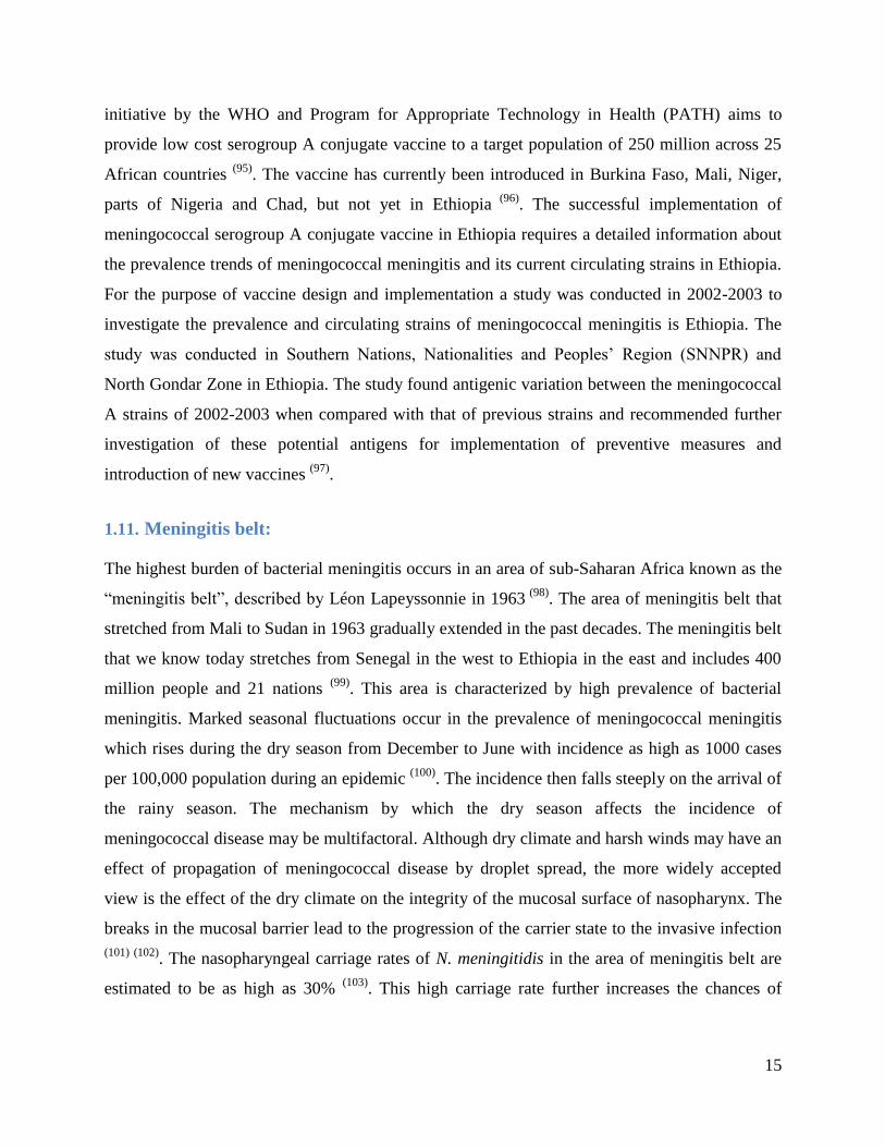
The highest burden of bacterial meningitis occurs in an area of sub-Saharan Africa known as the
“meningitis belt”, described by Léon Lapeyssonnie in 1963 (98)
. The area of meningitis belt that
stretched from Mali to Sudan in 1963 gradually extended in the past decades. The meningitis belt
that we know today stretches from Senegal in the west to Ethiopia in the east and includes 400
million people and 21 nations (99)
. This area is characterized by high prevalence of bacterial
meningitis. Marked seasonal fluctuations occur in the prevalence of meningococcal meningitis
which rises during the dry season from December to June with incidence as high as 1000 cases
per 100,000 population during an epidemic (100)
. The incidence then falls steeply on the arrival of
the rainy season. The mechanism by which the dry season affects the incidence of
meningococcal disease may be multifactoral. Although dry climate and harsh winds may have an
effect of propagation of meningococcal disease by droplet spread, the more widely accepted
view is the effect of the dry climate on the integrity of the mucosal surface of nasopharynx. The
breaks in the mucosal barrier lead to the progression of the carrier state to the invasive infection
(101) (102). The nasopharyngeal carriage rates of N. meningitidis in the area of meningitis belt are
estimated to be as high as 30% (103)
. This high carriage rate further increases the chances of
16
developing or spreading infection during the dry season of the year. Recent studies suggest,
however, that the carriage rate might be much lower in absence of outbreak (104)
.
Figure 1: Map of Meningitis Belt
The part of sub-Saharan Africa that constitutes the meningitis belt is characterized by recurrent
meningococcal outbreaks. The records show that major outbreaks tend to occur every 8–12 years
(97). After the first reported outbreak in 1840 almost 400 epidemics of bacterial meningitis have
been recorded in the meningitis belt. The largest epidemic was reported in 1996 which affected
250,000 people and resulted in almost 25,000 deaths and 50,000 disabilities (105)
. In addition to
the major epidemics, smaller isolated outbreaks involving only a community also occur
frequently. Most of the epidemics have been due to N. meningitidis serogroup A but some
epidemics due to other serogroups such as serogroup C, X and W135 have also been reported

17
(106). The introduction of meningococcal conjugate A vaccine under the MVP may, therefore,
cause a considerable reduction in the incidence of meningococcal disease in the years to come.
18
Chapter two: MENINGITIS IN ETHIOPIA
2.1. Ethiopia: country profile
Located in the Horn of Africa, The Federal Democratic
Republic of Ethiopia lies at the crossroads between Middle
East and Africa. Ethiopia is bounded by Eritrea to the north
and Kenya to the south. The eastern part is bounded by
Somalia and to the west lies Sudan and South Sudan.
Ethiopia covers a vast land area of 1.1 million square
kilometers and is the second most populous country in
Africa with a population of more than 84 million (107)
.
2.1.1. Demographics:
Ethiopia’s population continues to grow at a rate of 2.5%, and has increased from 33.5 million in
1983 to 84 million in 2012 (108)
. The capital city of Ethiopia is Addis Ababa which is also the
largest city with a population of almost 3 million. Other large cities of Ethiopia include Mekele,
Adama, Gondar and Awassa. Ethiopia is one of the least urbanized countries of the world with
only 17% of the population residing in the urban areas. But in the recent decade rapid
urbanization has occurred with the urban population increasing at a rate of 4.1% (108)
.
Ethiopia is famous for her ethnic diversity. People from at least 80 different ethnic backgrounds
reside in Ethiopia. Oromo form the largest ethnic group (34.5%) followed by Amhara (26.9%),
Somali (6.20%) and Tigray (6.07%) (107)
. Due to this reason many native language are spoken in
Ethiopia. These include Oromifa, Tigrinya and Somali. Amharic is the main language spoken
and understood throughout Ethiopia. English is the most commonly spoken foreign language.
The literacy rate still remains low with the adult literacy rate estimated to be about 30%.
19
2.1.2. Climate:
Most of the area of the country is covered by highlands which make the climate much cooler
than the neighboring African countries. Many major cities of Ethiopia, including Addis Ababa
and Gondar, are located at an elevation of more than 2000 meters which provides a considerably
cooler but much uniform temperature throughout the year. The southern part of Ethiopia
including the South Nations and Nationalities Peoples’ Region (SNNPR) is located at a lesser
elevation as compared to the northern part. The capital city of SNNPR, Awassa is located at an
elevation of 1700m (in contrast to 2000m of Addis Ababa and Gondar). Due to this, Awassa has
a climate much hotter than the central and northern part of Ethiopia. The seasons can be defined
by rainfall into rainy season between June to September and a dry season ranging from October
to February.
2.1.3. Economy:
The main domestic product of Ethiopia is agriculture which accounts to about 41% of the total
GDP and makes up to 80% of the total exports. Agriculture is also the major profession in
Ethiopia with almost 80% of the population associated with agriculture. Although Ethiopia
showed highest economical growth within the non-oil dependant African economies in 2007-
2008, the per capita GDP still remains one of the lowest in the world. The poverty rates are very
high with almost 39% of the population living below the poverty line of earning less than US$
1.25 per day.
2.1.4. Health profile:
The main health related problems in Ethiopia are due to communicable diseases. The preventable
causes of death due to communicable diseases account to about 74% of the total deaths. The high
rates of infectious diseases are due to poor sanitation, unavailability of healthcare facilities and
lack of trained staff especially in the rural parts of Ethiopia. Another important dilemma for the
healthcare system is the very high incidence for nutritional deficiencies. The main healthcare
statistics are provided below:
20
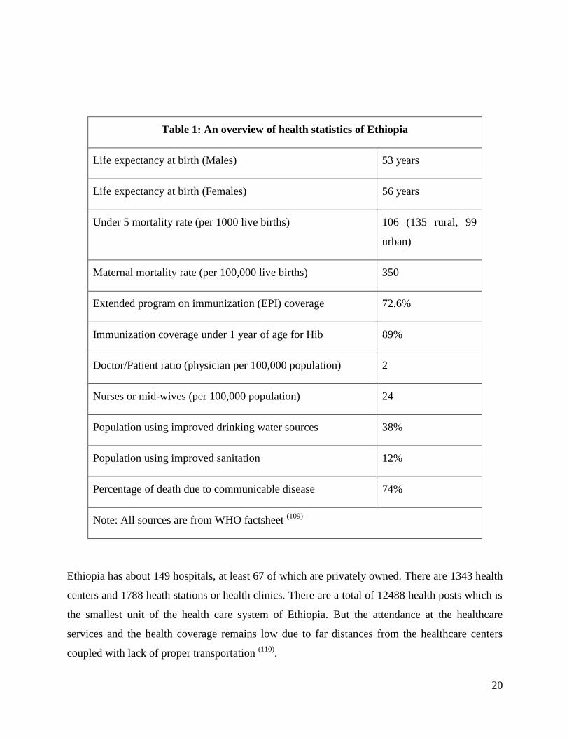
Table 1: An overview of health statistics of Ethiopia
Life expectancy at birth (Males) 53 years
Life expectancy at birth (Females) 56 years
Under 5 mortality rate (per 1000 live births) 106 (135 rural, 99
urban)
Maternal mortality rate (per 100,000 live births) 350
Extended program on immunization (EPI) coverage 72.6%
Immunization coverage under 1 year of age for Hib 89%
Doctor/Patient ratio (physician per 100,000 population) 2
Nurses or mid-wives (per 100,000 population) 24
Population using improved drinking water sources 38%
Population using improved sanitation 12%
Percentage of death due to communicable disease 74%
Note: All sources are from WHO factsheet (109)
Ethiopia has about 149 hospitals, at least 67 of which are privately owned. There are 1343 health
centers and 1788 heath stations or health clinics. There are a total of 12488 health posts which is
the smallest unit of the health care system of Ethiopia. But the attendance at the healthcare
services and the health coverage remains low due to far distances from the healthcare centers
coupled with lack of proper transportation (110)
.
21
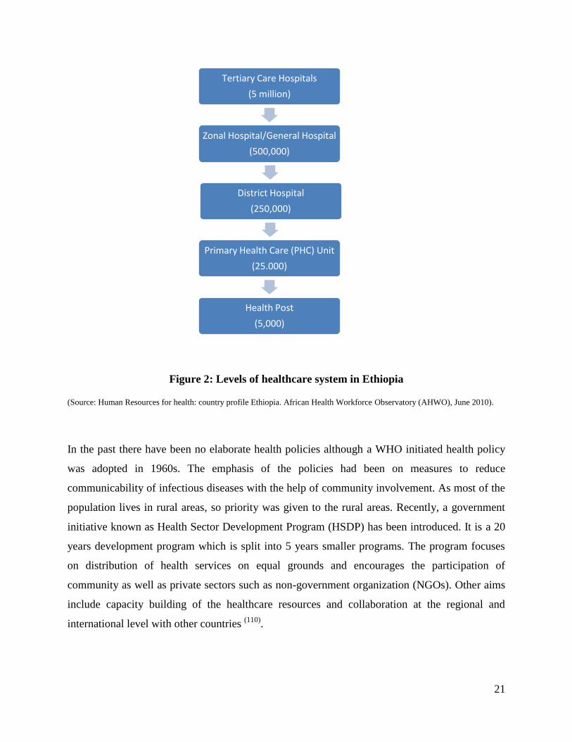
Figure 2: Levels of healthcare system in Ethiopia
(Source: Human Resources for health: country profile Ethiopia. African Health Workforce Observatory (AHWO), June 2010).
In the past there have been no elaborate health policies although a WHO initiated health policy
was adopted in 1960s. The emphasis of the policies had been on measures to reduce
communicability of infectious diseases with the help of community involvement. As most of the
population lives in rural areas, so priority was given to the rural areas. Recently, a government
initiative known as Health Sector Development Program (HSDP) has been introduced. It is a 20
years development program which is split into 5 years smaller programs. The program focuses
on distribution of health services on equal grounds and encourages the participation of
community as well as private sectors such as non-government organization (NGOs). Other aims
include capacity building of the healthcare resources and collaboration at the regional and
international level with other countries (110)
.
Tertiary Care Hospitals
(5 million)
Zonal Hospital/General Hospital
(500,000)
District Hospital
(250,000)
Primary Health Care (PHC) Unit
(25.000)
Health Post
(5,000)
22
2.2. Meningitis in Ethiopia:
Located on the eastern part of meningitis belt, Ethiopia is one of the countries which are most
affected with bacterial meningitis. The first reported outbreak in Ethiopia dates back to 1901,
which was followed by outbreaks in 1935, 1940s, 1950s, 1964 and 1977 (111)
. The largest
epidemics in Ethiopia were reported in 1981 and 1989, each of which affected almost 50,000
people (112)
. Epidemiological studies on the spread on these epidemics in Ethiopia suggest the
introduction of meningococcal disease first in western part of Africa. Earliest recorded outbreaks
of cerebrospinal meningitis occurred in soldiers stationed in Algiers in 1840 and in labourers
working in present day Ghana in 1900 (105)
. The epidemic of 1989 that occurred in the eastern
part of Africa is believed to be spread by pilgrims returning from Mecca (101)
. Since the
introduction of meningococcal disease in Ethiopia, the disease has remained endemic with
frequent outbreaks. The outbreaks prior to 2001 occurred mostly in the north western, western
and south western parts of Ethiopia, the areas that are traditionally included in the meningitis
belt. However, outbreaks in 2001 and afterwards have extended to the eastern parts of the
country as well (113)
. These epidemics were caused mainly by N. meningitidis serogroup A, but
serogroup C has also been isolated from samples during various outbreaks in 2000 and 2003 (114)
.
Apart from N. meningitidis other agents of bacterial meningitis such as Hib and S. pneumoniae
are also very common. During the year 1993-1995, A study conducted in a pediatric hospital in
Addis Ababa in 1993-1995 showed that almost 5.5% of all hospital admissions were bacterial
meningitis. Out of 385 cases diagnosed as bacterial meningitis 74 cases were due to H.
influenzae, 63 cases were recognized as M. tuberculosis and 46 cases were due to S. pneumoniae.
Meningococcal meningitis was very rare and was identified only in 6 cases. However, in 196
cases out of total 385 cases the exact etiology could not be traced (115) (116)
. The study also
reported incidence of antibiotic resistance in S. pneumoniae and H. influenzae. The emerging
resistance among the organisms causing bacterial meningitis is a matter of serious concern.
Studies have reported increased resistance to the commonly used antibiotics such as penicillin G
and chloramphenicol (117) (118)
.
Bacterial meningitis accounts for about 6-8% of all the hospital admissions in Ethiopia and the
case fatality ratio associated with bacterial meningitis is as high as 22-28% (114)
. A study
conducted at Gondar University Hospital over a span of 5 years from 1998-2003 showed the
23
prevalence of various common agents of bacterial meningitis in children up to the age of 14
years. N. meningitidis was the most common cause of meningitis and accounted to about 28% of
the cases. This was followed by S. pneumoniae and H. influenzae which were identified in 7%
and 6.5% of cases, respectively. S. aureus and Salmonella species were responsible for a small
number of cases. Another study conducted in Butajira, a town located in south-central Ethiopia,
reported causes of 10,700 deaths during 1987-2008. The results showed that almost 1% of all
deaths were due to meningitis (119)
. A similar study during the same period reported meningitis as
a cause of almost 1.9% of all deaths (120)
. Both these studies lacked laboratory confirmation and
relied on “verbal autopsy” to acquire information from the relatives of the deceased.
Laboratory-based clinical studies on bacterial meningitis are essential for predicting accurately
the current prevalence of bacterial meningitis and determining the disease causing organisms.
Only a few studies in Ethiopia are supported by laboratory surveillance. A WHO led study was
conducted in various African countries including Ethiopia to study the prevalence of various
agents of bacterial meningitis in infants less than 3 months of age (121)
. Both blood cultures and
CSF cultures were used to identify the causative organism. The study showed S. pneumoniae
(24%) and E. coli (24%) to be the most common organism causing bacterial meningitis in
neonates. Other common agents of bacterial meningitis were Streptococcus pyogenes (22%),
Salmonella (12%) and H. influenzae (7%).
2.3. Gaps in literature:
A critical review of the literature on bacterial meningitis in Ethiopia highlights the efforts of
researchers working on meningitis in this region of the world. The work of the pioneers more
than half a century ago and the subsequent continuation of research with addition of more
advance laboratory techniques has shaped our knowledge and understanding of meningitis in
Ethiopia. This has proven beneficial with respect to the control strategies that are being
implemented now, not only in Ethiopia, but other parts of the world as well. Nevertheless, the
continuously changing epidemiology, emergence of new strains along with the prospects of
newer vaccines and advances in laboratory techniques has led to the demand of new research in
this field. The new research needs to be tailor-made to focus on the changing epidemiology to
achieve the desired results. With this prospective in mind some of the gaps are identified that
24
need to be filled in order to achieve better understanding and control of bacterial meningitis in
Ethiopia.
1) Most of the studies mentioned in the literature were carried out in the epidemic periods, i.e.
from December to June and less emphasis is laid on non-epidemic periods. This implies that
most of those studies have focused on meningococcal meningitis and very few studies focused
on other types of bacterial meningitis, such as H. influenzae and S. pneumoniae. These causes
bacterial meningitis are also endemic in Ethiopia and are associated with high morbidity and
mortality and therefore need to be focused on with priority.
2) A major portion of the studies mentioned have small children as their target group. It is well
known that bacterial meningitis also has high incidence in elderly and immunocompromised
people. Therefore, for implementing nationwide policies and preventive measures, data on other
age groups should also be available.
3) Laboratory surveillance is essential for proper diagnosis of bacterial meningitis and
identifying various causative agents. Not all of the studies were assisted with laboratory
surveillance. The laboratory techniques used in many studies were outdated and did not meet the
criteria of “gold standard” which is considered vital for clinical research.
4) Most of the studies were carried out either in a single hospital or a single city. The results
from one specific area cannot be generalized over the whole population. Generalized results are
needed to take preventive steps.
5) The WHO website for Multi Disease Surveillance Centre (MDSC) show no or limited data on
the etiology of bacterial meningitis in Ethiopia as compared to other countries of meningitis belt
(122). Studies are needed that can provide recent data on bacterial meningitis which is crucial for
its prevention in Ethiopia.
2.4. Rationale of study:
A study is required to investigate the prevalence of different types of bacterial meningitis in
Ethiopia. The current data is limited and does not cover all common types of bacterial
25
meningitis. Most of the studies mentioned in literature are based on the prevalence of meningitis
in the epidemic phases. It is also essential to study the epidemiology of bacterial meningitis in
the non-epidemic phases, i.e. from June to December. Meningococcal meningitis is prevalent
primarily during the epidemic phases, but pneumococcal meningitis and H. influenzae are
endemic during the non-epidemic phases as well. Examples from the literature have shown that
these non-epidemic strains of bacterial meningitis are also responsible for a vast proportion of
cases of bacterial meningitis throughout the year (66)
. Therefore the study is planned to
investigate the prevalence of these strains in the non-epidemic phases, as their incidence may
still be considerably higher than the most other countries of the world.
The incidence of bacterial meningitis varies depending upon the age of the patients, geographical
location, climate and time of the year. Previous studies have focused primarily on children and
data on other age groups is required to carry out preventive measures for bacterial meningitis.
Therefore the study includes all age groups to generate data irrespective of age limitations. Data
is also needed that can be generalized over the whole population of Ethiopia. To meet this
requirement the study is planned to be carried out in two different cities geographically and
climatically different to obtain results that can be generalized. The study has two components, a
retrospective component that covers the past 5 years and a prospective component that provides
laboratory surveillance of bacterial meningitis for one year. This laboratory surveillance is
required to identify the current strains and study their prevalence, with the aim of carrying out
preventive measures or implementing new vaccines in Ethiopia.
Several retrospective studies have been conducted in Ethiopia but a study is needed that can
correlate the clinical symptoms and the severity of disease with the laboratory results to give a
wider view of the factors affecting the severity of bacterial meningitis. Retrospective data from
the previous years can help in predicting the current prevalence of various types of bacterial
meningitis. As Ethiopia is endemic for bacterial meningitis with cases of bacterial meningitis
presenting in the hospitals throughout the year, therefore, the data available in the hospitals’
clinical and laboratory record books can be the most accurate indicator of the current status of
bacterial meningitis in Ethiopia. These data can also serve as a valuable tool and a reliable
indicator of the degree of success of the preventive strategies introduced in the past. Similarly, it

26
can also be used to highlight the short comings and provide a basis for identifying corrective
measures.
27
Chapter three: METHODOLOGY AND STUDY DESIGN
3.1. Brief description of the project:
“Surveillance of bacterial meningitis and factors affecting meningococcal disease severity” is a
project by Norwegian Institute of Public Health (NIPH) in collaboration with Armauer Hansen
Research Institute (AHRI) in Addis Ababa, Ethiopia. The project aims to study the current
patterns of bacterial meningitis in Ethiopia with emphasis on meningococcal disease. The project
has two components: a retrospective study which focuses on the recent trends of bacterial
meningitis in Ethiopia during the last 5 years and a prospective study to provide continuous
surveillance of bacterial meningitis for a period of one year. The prospective study is under
progress with inclusion of new patients and subsequent laboratory analysis on their CSF and
blood specimens. To achieve better understanding of the overall trends of bacterial meningitis in
Ethiopia, a period of one year surveillance is the minimal duration required. A delay had
occurred in the initiation of the prospective study due to the unusually prolonged duration of the
ethical clearance process that had been unaccounted for. Due to this reason the one year
surveillance period has not concluded yet. Therefore, the thesis will focus on the retrospective
component of the study.
3.2. Collaborating institutes:
The collaborating institutes include:
National Institute of Public Health, Norway (NIPH)
Armauer Hansen Research Institute, Addis Ababa, Ethiopia (AHRI)
Gondar University Hospital, Gondar
Awassa Referral Hospital, Awassa
28
Retrospective study
3.3. Objectives of study:
3.3.1. Primary objective:
To study retrospectively the various trends of bacterial meningitis in selected Ethiopian hospitals
in the previous 5 years from 2007-2011.
3.3.2. Secondary objectives:
To explore the trends of bacterial meningitis in terms of number of cases over time.
To find out the prevalence of various etiological factors causing bacterial meningitis.
To find out the distribution of bacterial meningitis in terms of age and sex.
To study the effect of seasonal variability on the incidence of bacterial meningitis.
To describe the most common clinical signs and symptoms associated with bacterial
meningitis.
3.4. Study design:
The study involves a retrospective collection of clinical and laboratory data from selected
hospitals in Ethiopia, to provide a quantitative assessment of the recent epidemiological trends of
bacterial meningitis. The study group constitutes of all consecutive cases of bacterial meningitis
that presented in Gondar University Hospital and Awassa Hospital during a defined study period.
During a visit to the study sites before the start of the study the extent of the laboratory and
clinical data available was assessed with the help of the site co-ordinators of the project. Based
on this initial assessment, a wide range of data comprising clinical records of patients as well as
data on laboratory-based detection of bacterial meningitis was found. Therefore, a
multidimensional approach to study the various clinical, histopathological and biochemical
aspects of bacterial meningitis was adopted.
29
Due to the better laboratory facilities available at Gondar University Hospital, the data on actual
causative organisms of bacterial meningitis was used to interpret the prevalence trends of various
agents of bacterial meningitis. A high input of patients with bacterial meningitis coupled with the
well-maintained clinical records from the in-patients and emergency departments at Awassa
Referral Hospital was observed. The data from Awassa were therefore used to calculate the
prevalence of bacterial meningitis, the frequency of the most common clinical symptoms, rates
of complications and outcome of the disease.
The research design is retrospective. Retrospective studies aim at reasoning the outcome of an
effect back to its antecedent cause. This is in contrast to the prospective studies which aim at
reasoning from a present antecedent to a future outcome or consequence. Retrospective studies
have several advantages. They are less time-consuming, more economical and can provide
valuable results in a very short time period. But they are often subjected to recall bias. This effect
has been minimized in the current research as the data were recorded directly from the clinical
and laboratory records. Another drawback with the retrospective studies is that it is often
difficult to eliminate the effect of confounding variables which affect the outcome of study.
Possible attempts were made to reduce the effect of confounding variable. Patients who were
concomitantly affected by other kind of meningitis, such as tuberculous, viral or aseptic
meningitis and other CNS lesions were not included in the study. However, removing the effects
of all the confounding variables can be difficult to achieve. Retrospective studies cannot prevent
the occurrence of an event which has already occurred, but may be helpful in preventing such
events in the future. Retrospective studies can often be useful in the formation of a hypothesis
which can then be evaluated in further studies. This forms the basis of the relationship of this
retrospective study to the overall project.
The study aimed at providing quantitative assessment of the prevalence of bacterial meningitis in
Ethiopia and to provide results that may be generalized over the whole Ethiopian population. The
only study design that permits these characteristics of the research is a quantitative study.
Quantitative research design can be defined as “the numerical representation and manipulation of
observations for the purpose of describing and explaining the phenomena that those observations
reflect” (123)
. Quantitative research provides details of the direct and indirect variables and allows
the researcher to measure and quantify the effect. The study is basically a prevalence study and
30
demands the inclusion of all the patients of bacterial meningitis during the defined study period.
Consequently, no randomization was used and all patients that fulfilled the inclusion criteria
were included in the study.
3.5. Study sites:
The field work was carried out in two tertiary care hospitals from different cities of Ethiopia i.e.
Gondar University Hospital in Gondar and Awassa Referral Hospital in SNNPR region of
Ethiopia.
3.5.1. University of Gondar Medical Hospital, Gondar:
Gondar is a city in the North-western part of Ethiopia. Administratively, it is located in the
Amhara region. Previously the capital city of Ethiopia, Gondar is now the third largest city in
Ethiopia. Famous for its castles, Gondar is a mountainous area with an elevation of 2100 meters
above the sea level. Due to this reason the climate is much cooler. The average annual
temperature is 19.2oC which ranges from 10
oC (December and January) to 29
oC (March to May).
The total population of Gondar is more than 200,000. Different ethnic groups reside in Gondar,
out of which Amhara form the majority (88.9%) followed by Tigrayan (6.7%) and Qemant
(2.4%). Amharic is the main language spoken in the Gondar region.
The University of Gondar Medical Hospital is the largest hospital in Gondar. It is a 400-bed
teaching hospital associated with a medical college. The medical college was founded in 1954
and is the oldest training institute for health professionals in Ethiopia. The hospital covers a wide
range of area and is a referral hospital for four districts hospitals. It has a range of specialities
including paediatrics, surgical, gynaecological, psychiatric and an HIV centre. With a staff
comprising of 400 healthcare personals, including 50 medical doctors, the hospital covers a
population of four million. The hospital has well established laboratory facilities with a staff of at
least 25 trained laboratory scientists.
3.5.2. Awassa Referral Hospital, Awassa:
Awassa is a city located in the southern part of Ethiopia close to the lake Awassa of the Great
Rift Valley. The city of Awassa lies 270km to the south of the capital city of Addis Ababa.
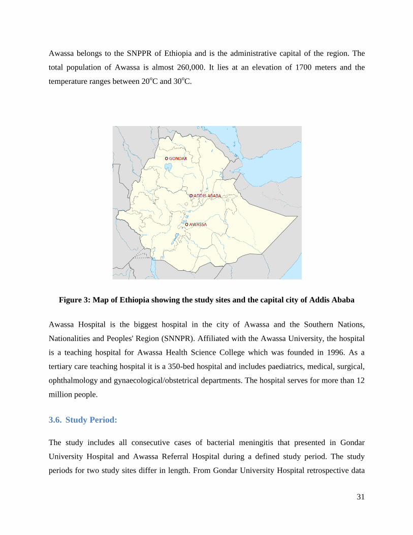
31
Awassa belongs to the SNPPR of Ethiopia and is the administrative capital of the region. The
total population of Awassa is almost 260,000. It lies at an elevation of 1700 meters and the
temperature ranges between 20oC and 30
oC.
Figure 3: Map of Ethiopia showing the study sites and the capital city of Addis Ababa
Awassa Hospital is the biggest hospital in the city of Awassa and the Southern Nations,
Nationalities and Peoples' Region (SNNPR). Affiliated with the Awassa University, the hospital
is a teaching hospital for Awassa Health Science College which was founded in 1996. As a
tertiary care teaching hospital it is a 350-bed hospital and includes paediatrics, medical, surgical,
ophthalmology and gynaecological/obstetrical departments. The hospital serves for more than 12
million people.
3.6. Study Period:
The study includes all consecutive cases of bacterial meningitis that presented in Gondar
University Hospital and Awassa Referral Hospital during a defined study period. The study
periods for two study sites differ in length. From Gondar University Hospital retrospective data
32
were collected for the last 5 years from January 2007 till December 2011 (corresponding to the
Ethiopian calendar from 4th
month of 1999 to 5th
month of 2004). From Awassa Hospital the data
was collected for the last 2.5 years from September 2009 till December 2011 (corresponding to
the Ethiopian calendar from start of 2002 to the 5th
month of 2004). The study periods were
selected to permit the maximum utilization of available data as determined on the preliminary
visits to the study sites.
3.7. Inclusion criteria for cases:
The patients fulfilling the following criteria were selected for the study:
Positive CSF culture or Gram staining results suggestive of bacterial meningitis
Clinically diagnosed cases of bacterial meningitis by a medical specialist in the cases
where a CSF culture or Gram staining were not performed.
All age groups that fulfil the above criteria
3.8. Exclusion criteria:
The patients exhibiting the following characteristics were not included in the study:
Simultaneous infection with another form of meningitis such as tuberculous, viral or
fungal meningitis.
Presence of another CNS infection that can present with symptoms similar to
bacterial meningitis such as cerebral malaria or any other CNS lesion.
3.9. Study population:
All the patients with bacterial meningitis that presented in Gondar University Hospital or Awassa
Referral Hospital during the study period and fulfilling the above mentioned inclusion criteria
were included in the study and defined as the study population.
33
3.10. Target population:
The study sites are two different cities with distinct geographical and climatic features. These
cities are located in the northern and southern part of Ethiopia. Thus the study is carried out with
the intention of providing results that can be generalized over whole of the Ethiopia. Therefore,
the target population for this study is the whole Ethiopian population.
3.11. Sample size:
No predefined sample size was set. The study included all the cases of bacterial meningitis that
presented in Gondar University Hospital and Awassa Referral Hospital during the defined study
period and fulfilled the above mentioned inclusion criteria.
3.12. Data sources:
The clinical and laboratory data were collected from the available hospital clinical and laboratory
records. The patient related clinical data were collected from the hospital clinical records. The
potential cases were identified by the hospital log books present in the emergency wards, the
medical, paediatrics and neonatal in-patient wards and also the out-patient registers. The
laboratory log books for CSF cultures and Gram staining results were also searched to find the
cases of bacterial meningitis. In addition to the routine log books, separate log books that were
used during the off-duty hours such as weekends or public holidays were also searched to avoid
missing any potential study participants. Once the cases were identified from any of these
sources the original hospital clinical records were accessed with the help of the main record
room staff. The records were traced using the patient hospital registration number from the log
books. The original laboratory result slips attached inside these clinical record forms were also
accessed.
3.13. Data collection:
The information obtained from the above mentioned sources were entered into specialized pre-
defined data collection forms (data collection form attached as appendix I). Among the clinical
details, the signs and symptoms at presentation such as stiff neck, headache, fever, altered mental
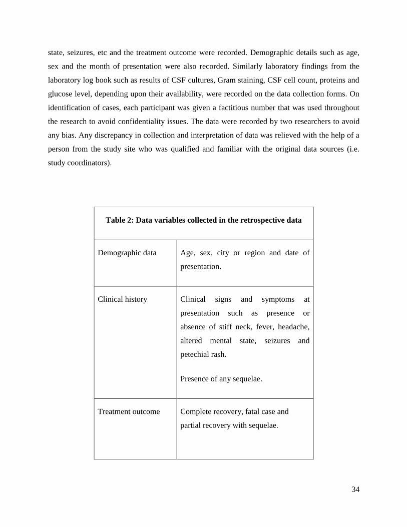
34
state, seizures, etc and the treatment outcome were recorded. Demographic details such as age,
sex and the month of presentation were also recorded. Similarly laboratory findings from the
laboratory log book such as results of CSF cultures, Gram staining, CSF cell count, proteins and
glucose level, depending upon their availability, were recorded on the data collection forms. On
identification of cases, each participant was given a factitious number that was used throughout
the research to avoid confidentiality issues. The data were recorded by two researchers to avoid
any bias. Any discrepancy in collection and interpretation of data was relieved with the help of a
person from the study site who was qualified and familiar with the original data sources (i.e.
study coordinators).
Table 2: Data variables collected in the retrospective data
Demographic data Age, sex, city or region and date of
presentation.
Clinical history Clinical signs and symptoms at
presentation such as presence or
absence of stiff neck, fever, headache,
altered mental state, seizures and
petechial rash.
Presence of any sequelae.
Treatment outcome Complete recovery, fatal case and
partial recovery with sequelae.
35
Laboratory results Gram staining of CSF samples, CSF
culture, CSF cell count, proteins and
glucose level.
3.14. Data handling:
3.14.1. Data entry:
The data from the filled-in data collection forms were then entered into SPSS version 18.0.0 and
later converted to Excel sheets for convenience during discussions with the study co-ordinators.
3.14.2. Statistical analysis:
The results were analyzed using a registered version of SPSS 18.0.0. The statistical analysis was
conducted to find out the exact proportion of various etiological factors of bacterial meningitis in
the patients that presented in Gondar University Hospital and Awassa Referral Hospital during
the defined study period. The various trends of bacterial meningitis over the last 5 years in terms
of age and sex distribution were studied. The hospital-wise prevalence of bacterial meningitis in
relation to seasonal variation and the time of the year were also studied. The study also focused
on the relationship of various clinical signs and symptoms to the overall prognosis and outcome
of disease.
3.15. Ethical considerations:
The approval for the main project “Surveillance of bacterial meningitis and factors affecting
meningococcal disease severity” was granted by REK “Regionale Komiteer for Medisinsk og
Helsefaglig Forskningsetikk” (Regional Committee for medical and healthcare research ethics)
in Norway. Following clearance from REK the protocol was submitted to the National Health
Research Ethics Review Committee (NRERC) in Ethiopia. Abiding by the regulations of the
chief collaborating institute in Ethiopia, the Armauer Hansen Research Institute (AHRI) Addis
Ababa, clearance was also sought from the medical research ethics board of AHRI. Once the
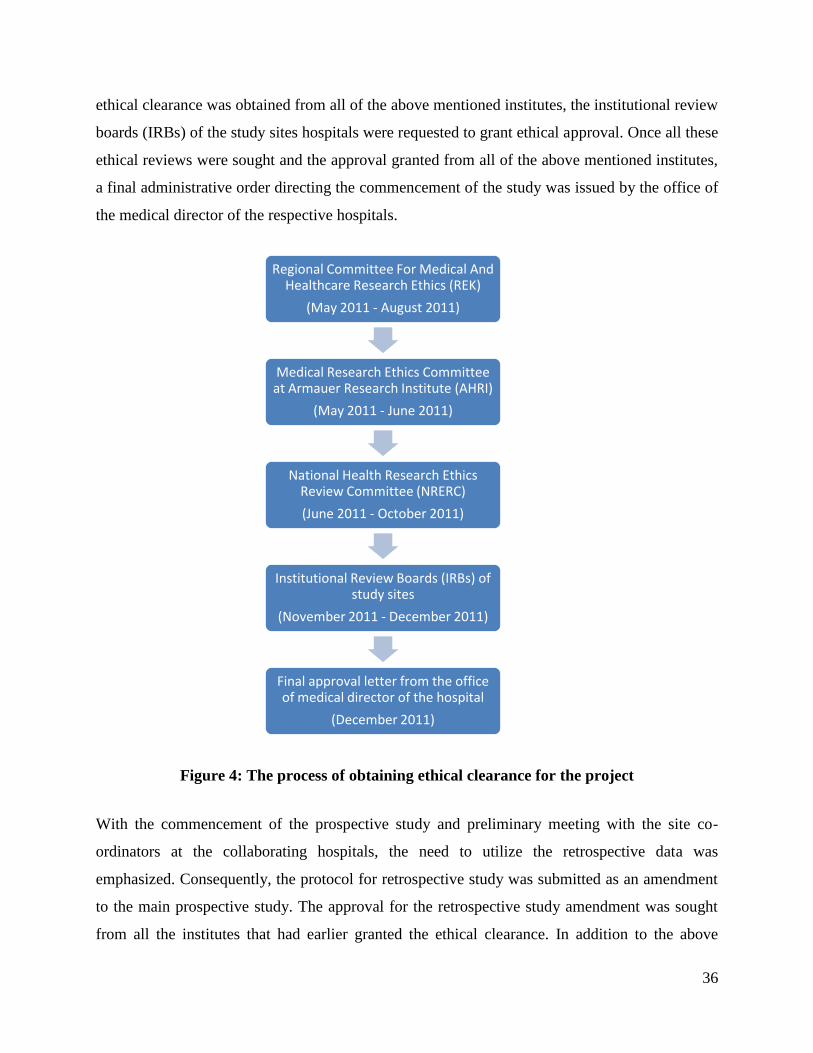
36
ethical clearance was obtained from all of the above mentioned institutes, the institutional review
boards (IRBs) of the study sites hospitals were requested to grant ethical approval. Once all these
ethical reviews were sought and the approval granted from all of the above mentioned institutes,
a final administrative order directing the commencement of the study was issued by the office of
the medical director of the respective hospitals.
Figure 4: The process of obtaining ethical clearance for the project
With the commencement of the prospective study and preliminary meeting with the site co-
ordinators at the collaborating hospitals, the need to utilize the retrospective data was
emphasized. Consequently, the protocol for retrospective study was submitted as an amendment
to the main prospective study. The approval for the retrospective study amendment was sought
from all the institutes that had earlier granted the ethical clearance. In addition to the above
Regional Committee For Medical And Healthcare Research Ethics (REK)
(May 2011 - August 2011)
Medical Research Ethics Committee at Armauer Research Institute (AHRI)
(May 2011 - June 2011)
National Health Research Ethics Review Committee (NRERC)
(June 2011 - October 2011)
Institutional Review Boards (IRBs) of study sites
(November 2011 - December 2011)
Final approval letter from the office of medical director of the hospital
(December 2011)

37
mentioned approvals, an additional approval from the hospital administration was required to
access the hospital main record room and utilize the retrospective data from the clinical records
of the patients. As the research is clinical and involves multiple institutions, therefore, the
process of acquiring ethical clearance was complicated. Taking into account the clinical nature of
the research and the usual application processing times, the whole process of obtaining the
ethical clearance took almost 6 months for the main prospective study. The retrospective
amendment was approved in a course of 3-4 months.
3.16. Participant confidentiality:
Safeguarding the patients’ right to confidentiality should be the foremost responsibility of the
researcher. To ensure this right of confidentiality, all the cases were enrolled with complete
anonymity. Upon inclusion each participant was provided with an identification number that was
used throughout the study and during the data analysis and publication of results. The data record
forms were devised as to not permit the recording of any personal details such as name of the
patient, the hospital registration number and the address of the patients. The original list of the
hospital registration numbers that was used to trace the clinical records of the patients was kept
only with the researcher in a place separate from the collected data. The list will be retained with
the researcher for a period of 6 months after the publication of the results in order to verify any
ambiguity that may arise. The list will be discarded after a period of 6 months from the
publication of results.
3.17. Expected benefits of the study:
The study is intended for the host population as being the sole beneficiaries. The retrospective
data collected from the two study sites will prove helpful in establishing the current prevalence
of bacterial meningitis in those particular settings. The effect of seasonal variability on various
types of bacterial meningitis and the age wise and gender wise distribution of disease burden will
provide useful estimates on the recent trends of bacterial meningitis. The study sites include two
hospitals located in different cities of Ethiopia which imparts generalization to the results.
Therefore, the results obtained from the study will also prove helpful for other hospitals in
Ethiopia working in similar settings. Special emphasis has been laid to ensure the validity of the

38
clinical and laboratory data. This 5-year retrospective study component along with the one year
prospective study that is in progress may provide valuable data that can be used for further
research, formulation of preventive strategies and implementation of new vaccines.
3.18. Funding:
The project is funded by the Norwegian Research Council (PROJECT NO: 192477) under the
GLOBVAC fund. The second visit to Ethiopia was funded by Ivar Helles’ Foundation, Norway.
3.19. Project timeline:
Table 3: Project timeline for the master’s project
Literature search and protocol writing May 2011 – June 2011
First visit to Ethiopia for setting up
prospective study June 2011 – November 2011
Introductory meetings with collaborating
institute regarding retrospective study June 2011 – July 2011
Setting up study start-up, recruiting study staff,
equipment purchase and transfer for the
prospective study.
July 2011 – August 2011
Conducting laboratory optimization and
training for the recruited staff
August 2011 – September
2011
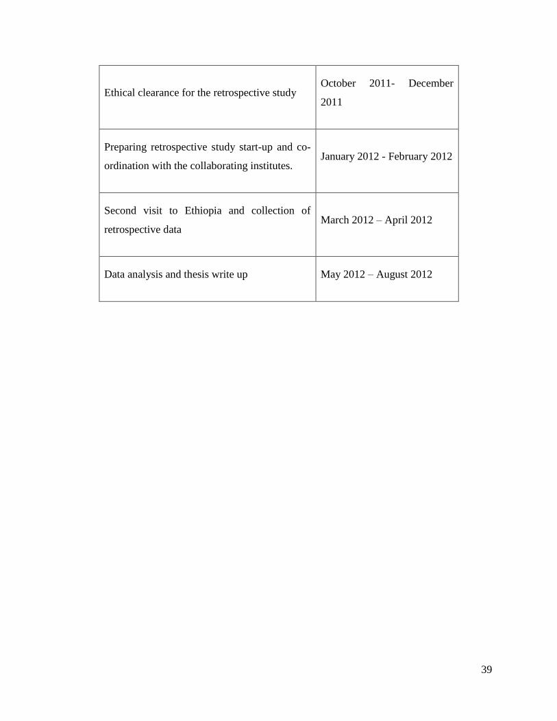
39
Ethical clearance for the retrospective study October 2011- December
2011
Preparing retrospective study start-up and co-
ordination with the collaborating institutes. January 2012 - February 2012
Second visit to Ethiopia and collection of
retrospective data March 2012 – April 2012
Data analysis and thesis write up May 2012 – August 2012
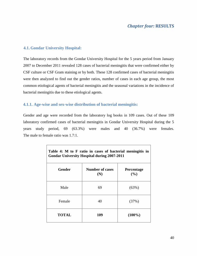
40
Chapter four: RESULTS
4.1. Gondar University Hospital:
The laboratory records from the Gondar University Hospital for the 5 years period from January
2007 to December 2011 revealed 128 cases of bacterial meningitis that were confirmed either by
CSF culture or CSF Gram staining or by both. These 128 confirmed cases of bacterial meningitis
were then analyzed to find out the gender ratios, number of cases in each age group, the most
common etiological agents of bacterial meningitis and the seasonal variations in the incidence of
bacterial meningitis due to these etiological agents.
4.1.1. Age-wise and sex-wise distribution of bacterial meningitis:
Gender and age were recorded from the laboratory log books in 109 cases. Out of these 109
laboratory confirmed cases of bacterial meningitis in Gondar University Hospital during the 5
years study period, 69 (63.3%) were males and 40 (36.7%) were females.
The male to female ratio was 1.7:1.
Table 4: M to F ratio in cases of bacterial meningitis in
Gondar University Hospital during 2007-2011
Gender Number of cases
(N)
Percentage
(%)
Male 69 (63%)
Female 40 (37%)
TOTAL 109 (100%)
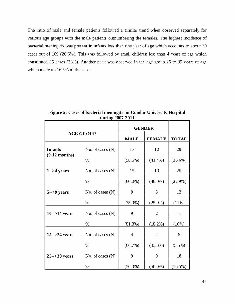
41
The ratio of male and female patients followed a similar trend when observed separately for
various age groups with the male patients outnumbering the females. The highest incidence of
bacterial meningitis was present in infants less than one year of age which accounts to about 29
cases out of 109 (26.6%). This was followed by small children less than 4 years of age which
constituted 25 cases (23%). Another peak was observed in the age group 25 to 39 years of age
which made up 16.5% of the cases.
Figure 5: Cases of bacterial meningitis in Gondar University Hospital
during 2007-2011
AGE GROUP
GENDER
TOTAL MALE FEMALE
Infants
(0-12 months)
No. of cases (N) 17 12 29
% (58.6%) (41.4%) (26.6%)
1-->4 years No. of cases (N) 15 10 25
% (60.0%) (40.0%) (22.9%)
5-->9 years No. of cases (N) 9 3 12
% (75.0%) (25.0%) (11%)
10-->14 years No. of cases (N) 9 2 11
% (81.8%) (18.2%) (10%)
15-->24 years No. of cases (N) 4 2 6
% (66.7%) (33.3%) (5.5%)
25-->39 years No. of cases (N) 9 9 18
% (50.0%) (50.0%) (16.5%)
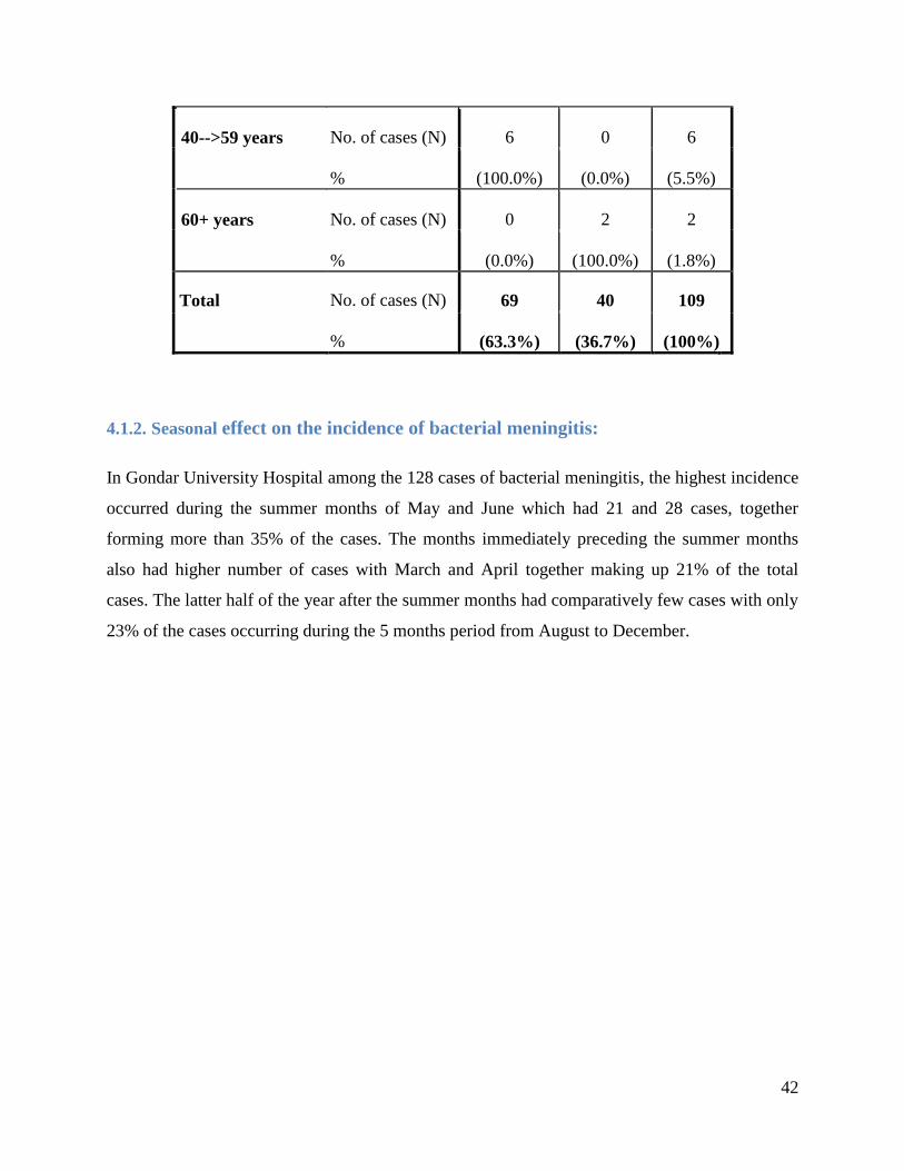
42
40-->59 years No. of cases (N) 6 0 6
% (100.0%) (0.0%) (5.5%)
60+ years No. of cases (N) 0 2 2
% (0.0%) (100.0%) (1.8%)
Total No. of cases (N) 69 40 109
% (63.3%) (36.7%) (100%)
4.1.2. Seasonal effect on the incidence of bacterial meningitis:
In Gondar University Hospital among the 128 cases of bacterial meningitis, the highest incidence
occurred during the summer months of May and June which had 21 and 28 cases, together
forming more than 35% of the cases. The months immediately preceding the summer months
also had higher number of cases with March and April together making up 21% of the total
cases. The latter half of the year after the summer months had comparatively few cases with only
23% of the cases occurring during the 5 months period from August to December.
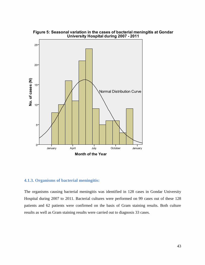
43
4.1.3. Organisms of bacterial meningitis:
The organisms causing bacterial meningitis was identified in 128 cases in Gondar University
Hospital during 2007 to 2011. Bacterial cultures were performed on 99 cases out of these 128
patients and 62 patients were confirmed on the basis of Gram staining results. Both culture
results as well as Gram staining results were carried out to diagnosis 33 cases.
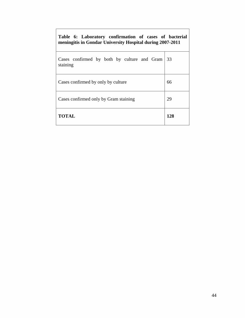
44
Table 6: Laboratory confirmation of cases of bacterial
meningitis in Gondar University Hospital during 2007-2011
Cases confirmed by both by culture and Gram
staining
33
Cases confirmed by only by culture 66
Cases confirmed only by Gram staining 29
TOTAL 128
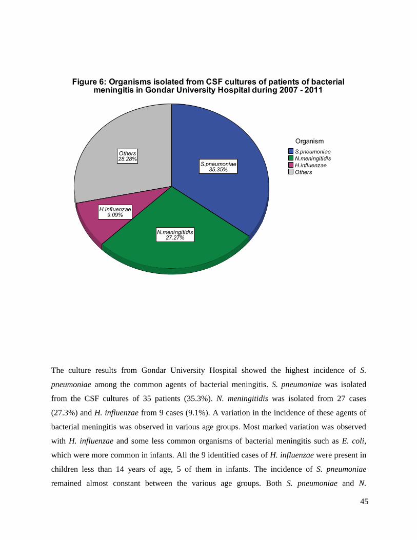
45
The culture results from Gondar University Hospital showed the highest incidence of S.
pneumoniae among the common agents of bacterial meningitis. S. pneumoniae was isolated
from the CSF cultures of 35 patients (35.3%). N. meningitidis was isolated from 27 cases
(27.3%) and H. influenzae from 9 cases (9.1%). A variation in the incidence of these agents of
bacterial meningitis was observed in various age groups. Most marked variation was observed
with H. influenzae and some less common organisms of bacterial meningitis such as E. coli,
which were more common in infants. All the 9 identified cases of H. influenzae were present in
children less than 14 years of age, 5 of them in infants. The incidence of S. pneumoniae
remained almost constant between the various age groups. Both S. pneumoniae and N.
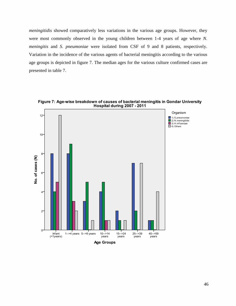
46
meningitidis showed comparatively less variations in the various age groups. However, they
were most commonly observed in the young children between 1-4 years of age where N.
meningitis and S. pneumoniae were isolated from CSF of 9 and 8 patients, respectively.
Variation in the incidence of the various agents of bacterial meningitis according to the various
age groups is depicted in figure 7. The median ages for the various culture confirmed cases are
presented in table 7.

47
Table 7: Median age for the culture positive cases at Gondar University
Hospital during 2007 -2011
Organism Median Age No. of cases (n)
(Range)
N. meningitidis 5 years n = 27
(Range = 0 month – 40 years)
S. pneumoniae 7 years n =35
(Range= 0 month – 48 years)
H. influenzae 1 month n =9
(Range= 0 month – 14 years)
Other organisms 3.5 years n =28
(Range = 0 month – 57 years)
Less common organisms were also isolated from the CSF samples of 28 patients of bacterial
meningitis at Gondar University Hospital. Out of these 28 cases E. coli was the most common
organism and was isolated from the cultures of 8 patients. The next common organisms were S.
aureus and Salmonella sp. that were isolated from 4 and 3 cultures, respectively. The various
uncommon agents of bacterial meningitis that were isolated from cultures of patients of
bacterial meningitis at Gondar University Hospital are presented table 8.
Table 8: Uncommon causes of bacterial
meningitis in patients of Gondar University
Hospital during 2009-2011
Organism Number of
cases (N)
E. coli 8
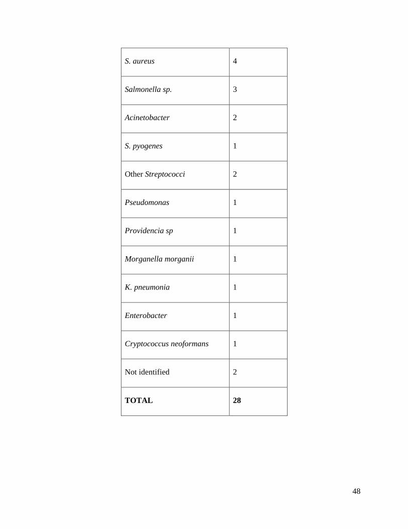
48
S. aureus 4
Salmonella sp. 3
Acinetobacter 2
S. pyogenes 1
Other Streptococci 2
Pseudomonas 1
Providencia sp 1
Morganella morganii 1
K. pneumonia 1
Enterobacter 1
Cryptococcus neoformans 1
Not identified 2
TOTAL 28
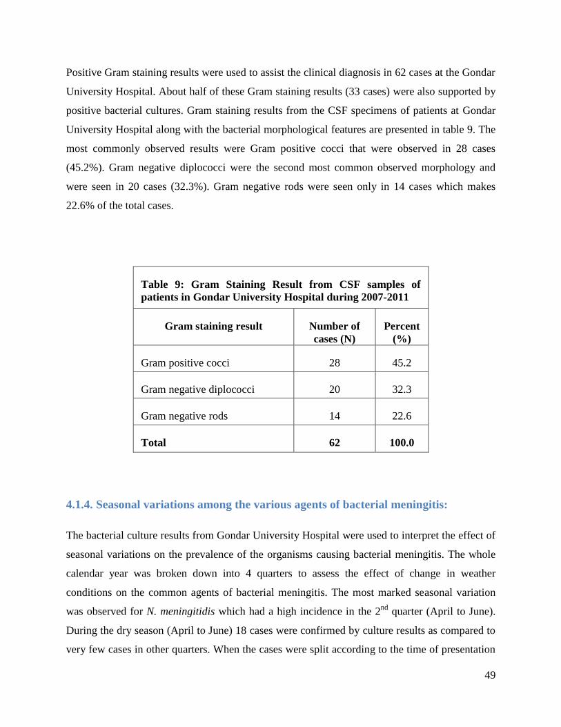
49
Positive Gram staining results were used to assist the clinical diagnosis in 62 cases at the Gondar
University Hospital. About half of these Gram staining results (33 cases) were also supported by
positive bacterial cultures. Gram staining results from the CSF specimens of patients at Gondar
University Hospital along with the bacterial morphological features are presented in table 9. The
most commonly observed results were Gram positive cocci that were observed in 28 cases
(45.2%). Gram negative diplococci were the second most common observed morphology and
were seen in 20 cases (32.3%). Gram negative rods were seen only in 14 cases which makes
22.6% of the total cases.
Table 9: Gram Staining Result from CSF samples of
patients in Gondar University Hospital during 2007-2011
Gram staining result Number of
cases (N)
Percent
(%)
Gram positive cocci 28 45.2
Gram negative diplococci 20 32.3
Gram negative rods 14 22.6
Total 62 100.0
4.1.4. Seasonal variations among the various agents of bacterial meningitis:
The bacterial culture results from Gondar University Hospital were used to interpret the effect of
seasonal variations on the prevalence of the organisms causing bacterial meningitis. The whole
calendar year was broken down into 4 quarters to assess the effect of change in weather
conditions on the common agents of bacterial meningitis. The most marked seasonal variation
was observed for N. meningitidis which had a high incidence in the 2nd
quarter (April to June).
During the dry season (April to June) 18 cases were confirmed by culture results as compared to
very few cases in other quarters. When the cases were split according to the time of presentation
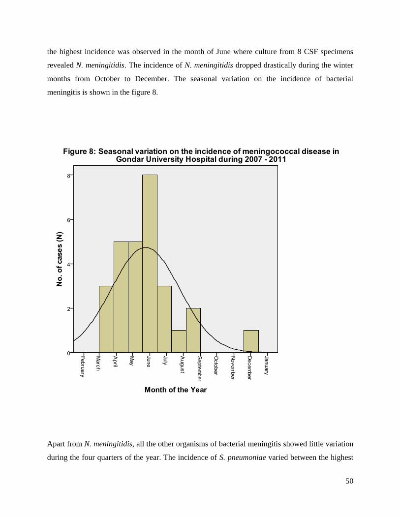
50
the highest incidence was observed in the month of June where culture from 8 CSF specimens
revealed N. meningitidis. The incidence of N. meningitidis dropped drastically during the winter
months from October to December. The seasonal variation on the incidence of bacterial
meningitis is shown in the figure 8.
Apart from N. meningitidis, all the other organisms of bacterial meningitis showed little variation
during the four quarters of the year. The incidence of S. pneumoniae varied between the highest
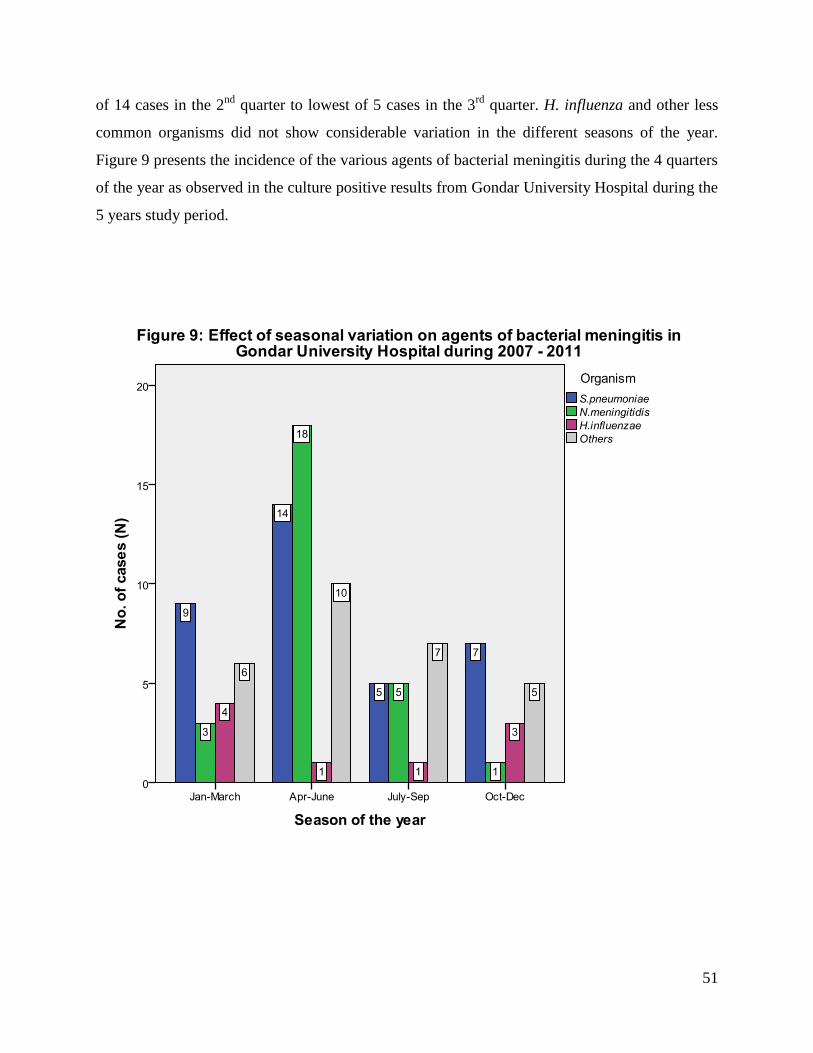
51
of 14 cases in the 2nd
quarter to lowest of 5 cases in the 3rd
quarter. H. influenza and other less
common organisms did not show considerable variation in the different seasons of the year.
Figure 9 presents the incidence of the various agents of bacterial meningitis during the 4 quarters
of the year as observed in the culture positive results from Gondar University Hospital during the
5 years study period.
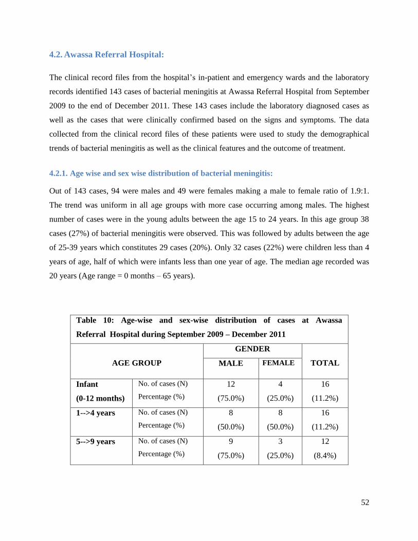
52
4.2. Awassa Referral Hospital:
The clinical record files from the hospital’s in-patient and emergency wards and the laboratory
records identified 143 cases of bacterial meningitis at Awassa Referral Hospital from September
2009 to the end of December 2011. These 143 cases include the laboratory diagnosed cases as
well as the cases that were clinically confirmed based on the signs and symptoms. The data
collected from the clinical record files of these patients were used to study the demographical
trends of bacterial meningitis as well as the clinical features and the outcome of treatment.
4.2.1. Age wise and sex wise distribution of bacterial meningitis:
Out of 143 cases, 94 were males and 49 were females making a male to female ratio of 1.9:1.
The trend was uniform in all age groups with more case occurring among males. The highest
number of cases were in the young adults between the age 15 to 24 years. In this age group 38
cases (27%) of bacterial meningitis were observed. This was followed by adults between the age
of 25-39 years which constitutes 29 cases (20%). Only 32 cases (22%) were children less than 4
years of age, half of which were infants less than one year of age. The median age recorded was
20 years (Age range = 0 months – 65 years).
Table 10: Age-wise and sex-wise distribution of cases at Awassa
Referral Hospital during September 2009 – December 2011
AGE GROUP
GENDER
TOTAL MALE FEMALE
Infant
(0-12 months)
No. of cases (N)
Percentage (%)
12
(75.0%)
4
(25.0%)
16
(11.2%)
1-->4 years No. of cases (N)
Percentage (%)
8
(50.0%)
8
(50.0%)
16
(11.2%)
5-->9 years No. of cases (N)
Percentage (%)
9
(75.0%)
3
(25.0%)
12
(8.4%)

53
10-->14 years No. of cases (N)
Percentage (%)
8
(61.5%)
5
(38.5%)
13
(9.1%)
15-->24 years No. of cases (N)
Percentage (%)
27
(71.1%)
11
(28.9%)
38
(26.6%)
25-->39 years No. of cases (N)
Percentage (%)
16
(55.2%)
13
(44.8%)
29
(20.3%)
40-->59 years No. of cases (N)
Percentage (%)
9
(64.3%)
5
(35.7%)
14
(9.8%)
60+ years No. of cases (N)
Percentage (%)
5
(100.0%)
0
(0.0%)
5
(3.5%)
Total No. of cases (N)
Percentage (%)
94
(65.7%)
49
(34.3%)
143
(100%)
4.2.2. Effect of seasonal variation of the incidence of bacterial meningitis:
In order to assess the effect of seasonal variation on the incidence of bacterial meningitis in
Awassa, two years data from January 2010 to December 2011 were studied (represented in
figure 10). A fairly uniform pattern was observed throughout the year: the incidence remained
constant during this time. However, the highest number of cases were recorded in April (15
cases) with almost 12% of the cases. This was followed by November which has 13 cases (11%).
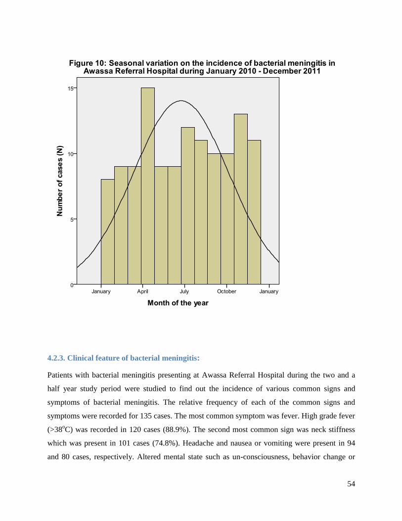
54
4.2.3. Clinical feature of bacterial meningitis:
Patients with bacterial meningitis presenting at Awassa Referral Hospital during the two and a
half year study period were studied to find out the incidence of various common signs and
symptoms of bacterial meningitis. The relative frequency of each of the common signs and
symptoms were recorded for 135 cases. The most common symptom was fever. High grade fever
(>38oC) was recorded in 120 cases (88.9%). The second most common sign was neck stiffness
which was present in 101 cases (74.8%). Headache and nausea or vomiting were present in 94
and 80 cases, respectively. Altered mental state such as un-consciousness, behavior change or
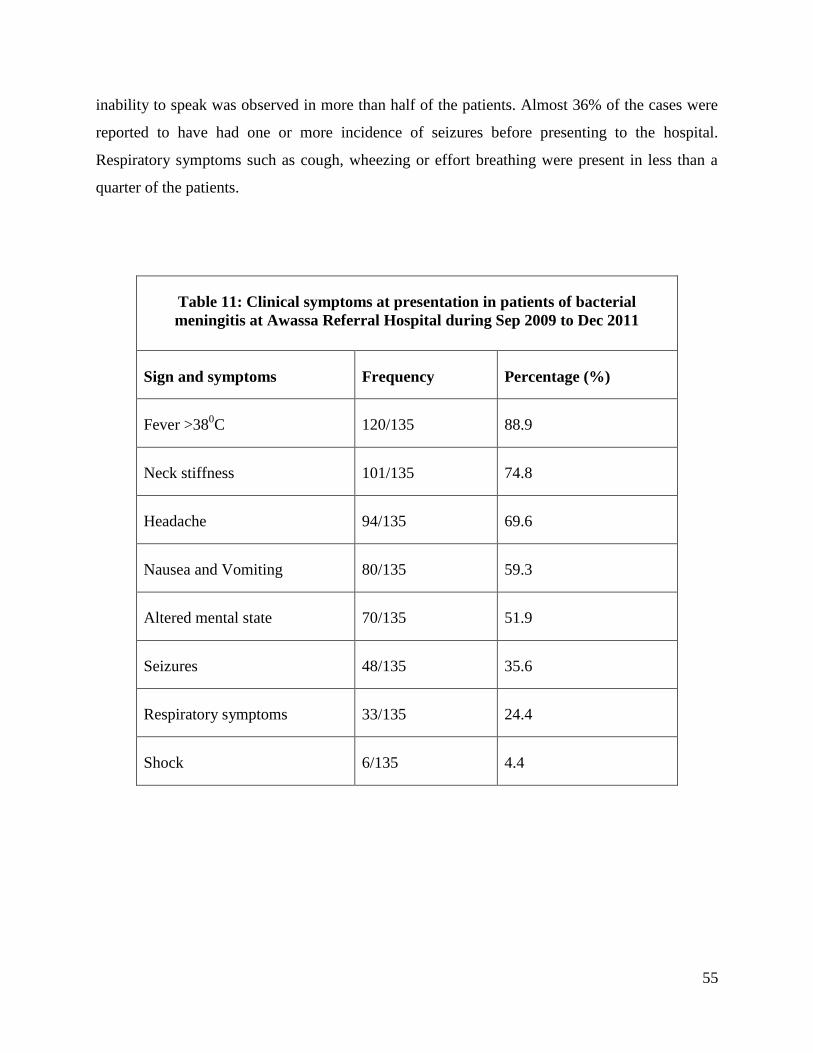
55
inability to speak was observed in more than half of the patients. Almost 36% of the cases were
reported to have had one or more incidence of seizures before presenting to the hospital.
Respiratory symptoms such as cough, wheezing or effort breathing were present in less than a
quarter of the patients.
Table 11: Clinical symptoms at presentation in patients of bacterial
meningitis at Awassa Referral Hospital during Sep 2009 to Dec 2011
Sign and symptoms Frequency Percentage (%)
Fever >380C 120/135 88.9
Neck stiffness 101/135 74.8
Headache 94/135 69.6
Nausea and Vomiting 80/135 59.3
Altered mental state 70/135 51.9
Seizures 48/135 35.6
Respiratory symptoms 33/135 24.4
Shock 6/135 4.4
56
4.2.4. Outcome of bacterial meningitis:
The clinical files of 143 cases of bacterial meningitis at Awassa Referral Hospital were studied
to find out the outcome of the treatment. Final treatment outcomes were recorded in 119 cases.
The most common outcome of the treatment was complete recovery which was observed in 80
(56.6%) out of 119 cases. Death was the second most common outcome with 28 cases recorded
as fatal cases. Neurological or other sequelae were present in 11 cases at the time of discharge
from the hospital.
Outcome could not be recorded in the remaining 24 cases. Out of these 24 cases, 6 cases with
severe life threatening illness were referred to Addis Ababa for further investigation and
management. Treatment outcome could not be recorded in 7 cases which intentionally withdrew
from the treatment and were discharged against the advice of the attending physician. In an
additional 11 cases the medical records from the clinical files were insufficient to reveal the final
outcome of the treatment.
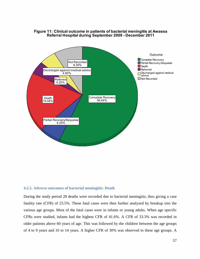
57
4.2.5. Adverse outcomes of bacterial meningitis: Death
During the study period 28 deaths were recorded due to bacterial meningitis, thus giving a case
fatality rate (CFR) of 23.5%. These fatal cases were then further analyzed by breakup into the
various age groups. Most of the fatal cases were in infants or young adults. When age specific
CFRs were studied, infants had the highest CFR of 41.6%. A CFR of 33.3% was recorded in
older patients above 60 years of age. This was followed by the children between the age groups
of 4 to 9 years and 10 to 14 years. A higher CFR of 30% was observed in these age groups. A
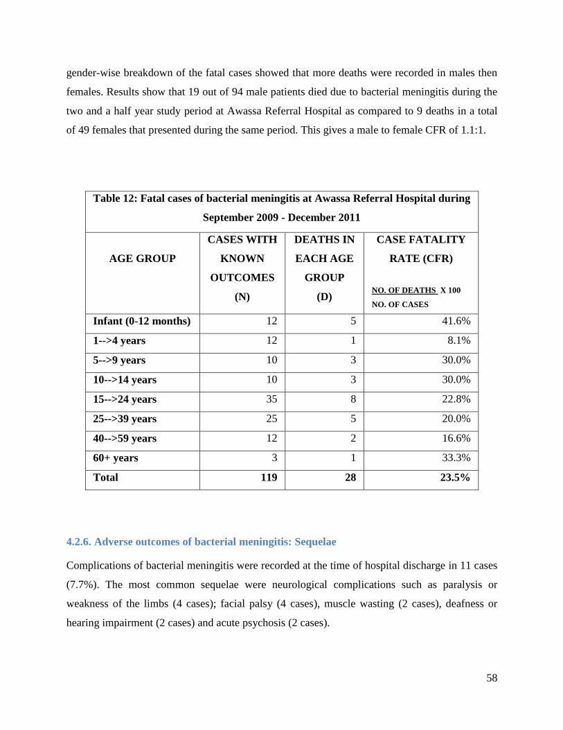
58
gender-wise breakdown of the fatal cases showed that more deaths were recorded in males then
females. Results show that 19 out of 94 male patients died due to bacterial meningitis during the
two and a half year study period at Awassa Referral Hospital as compared to 9 deaths in a total
of 49 females that presented during the same period. This gives a male to female CFR of 1.1:1.
Table 12: Fatal cases of bacterial meningitis at Awassa Referral Hospital during
September 2009 - December 2011
AGE GROUP
CASES WITH
KNOWN
OUTCOMES
(N)
DEATHS IN
EACH AGE
GROUP
(D)
CASE FATALITY
RATE (CFR)
NO. OF DEATHS X 100
NO. OF CASES
Infant (0-12 months) 12 5 41.6%
1-->4 years 12 1 8.1%
5-->9 years 10 3 30.0%
10-->14 years 10 3 30.0%
15-->24 years 35 8 22.8%
25-->39 years 25 5 20.0%
40-->59 years 12 2 16.6%
60+ years 3 1 33.3%
Total 119 28 23.5%
4.2.6. Adverse outcomes of bacterial meningitis: Sequelae
Complications of bacterial meningitis were recorded at the time of hospital discharge in 11 cases
(7.7%). The most common sequelae were neurological complications such as paralysis or
weakness of the limbs (4 cases); facial palsy (4 cases), muscle wasting (2 cases), deafness or
hearing impairment (2 cases) and acute psychosis (2 cases).
59
Chapter five: DISCUSSION AND RECOMMENDATIONS:
5.1. Discussion:
The aim of the study was to explore the various demographical, epidemiological and clinical
aspects of the bacterial meningitis in Ethiopia. The results are based on 128 cases from a 5-year
period (2007 to 2011) from Gondar University Hospital. These 128 cases include only those
cases that had been confirmed by laboratory identification of the causative organism. The data on
clinical signs and symptoms and the treatment outcomes represent a two and a half year
(September 2009 to December 2011) data of the 143 patients at Awassa Referral Hospital.
The data on 128 laboratory confirmed cases of bacterial meningitis from Gondar University
Hospital showed marked male predominance (male to female ratio of 1.7:1). The data from
Awassa reveal an even higher incidence in males (male to female ratio of 1.9:1). Throughout the
various age groups the trend of male predominance tends to persist. Various large scale studies
conducted worldwide show a higher incidence in males. A study on the characteristics of
bacterial meningitis was conducted in four major tertiary care hospitals in Bangladesh during
2003 and 2004 (124)
. The study showed that out of 1841 cases, 1307 were males, giving a male to
female ratio of 2.5:1. Similarly, studies from Ethiopia have also shown a higher incidence in
males. A 5-year retrospective study conducted in Gondar University Hospital during 1998 to
2003 identified 151 children with bacterial meningitis (65)
. This study also reported a higher
incidence in males with a male to female ratio of 2:1. During the meningococcal epidemic in
Gondar in 2001-2002, a total of 1619 probable cases of meningococcal meningitis were reported.
Out of these 1619 cases, 960 were males (male to female ratio of 1.5:1) (125)
. The comparison
between this Gondar study and our study shows a much higher number of patients of bacterial
meningitis in the former study. The reason for this higher number of cases is due to the inclusion
of probable cases which were not laboratory confirmed. The current study focuses on the
etiology and has only included the laboratory confirmed cases from Gondar which were
confirmed either by CSF culture or Gram staining. This explains the difference in the number of
cases in these two studies from Gondar.
60
What has caused this high male to female ratio at both of the study sites may be linked to various
factors. As seen in the literature and from results of the studies mentioned above, the organisms
of bacterial meningitis show susceptibility patterns indicating a higher affinity for males. This
fact may be exaggerated in the settings where males have more access to the health care
facilities, as is presumed to be the cases in this study. The social and cultural factors favor easier
access for males to the healthcare facilities as compared to the females which are more
dependent on the males for travelling to far-located health care centers. Other barriers that may
have affected the female access to the hospitals include fewer financial resources and the
additional responsibilities in the family such as caring for children. The data show a greater
difference in the number of patients of both genders in the infants and small children. At Awassa
75% of the infants were males. This may also highlight the bias or the preference that may be
given by the family to the male children over the female children.
Bacterial meningitis is generally a disease of childhood. In this study the most commonly
affected age group was found to be the infants and small children less than 4 years of age. Data
from Gondar University Hospital show that almost half of the cases (49.5%) were either infants
or children less than 4 years of age. Results from Awassa Referral Hospital show the highest
incidence in young adults 15-24 years of age which make 27% of the total cases. Children less
than 4 years of age, including the infants, make 22% of the cases. Within the various age groups,
a higher incidence in infants has frequently been reported in literature. A 10-year study
conducted in United States in California during 1998 to 2007 showed the highest incidence of
bacterial meningitis in infants less than 2 months of age (126)
. For the year 2006-2007 the
incidence was 80.7 cases per 100,000 population. Children greater than 2 months and less than 2
years of age showed the second higher incidence (6.9 cases per 100,000 population) during the
same year. A large scale retrospective study in Niger showed that in the year 1995-96 (127)
the
incidence of bacterial meningitis was highest in infants less than one year of age (638 cases per
100,000 population), this was followed by children between 1-4 years of age (490 cases per
100,000 population). Older children between 10-14 years and young adults also showed a higher
incidence (each having 476 cases per 100,000 population). In contrast to these studies, a study in
North Gondar during 2001-2002 showed the highest incidence in young adults between 15-30
years of age (52% of cases) (125)
. Generally, these results point out at 2 peaks of age that have
61
high susceptibility for bacterial meningitis i.e. small children and young adults. This also
parallels with the findings in this current study.
Certain agents of bacterial meningitis are known to have higher affinity for some age groups,
particularly H. influenzae, which commonly effects infants. The results of this study from
Gondar show a similar trend in which H. influenzae was more prevalent in the infancy and
younger children (all 9 culture confirmed cases were less than 14 years). Little variation in the
various age groups was observed for N. meningitidis and S. pneumoniae, both of which showed
only a slightly higher incidence in small children between 1-4 years of age. This trend in the
different age groups for these common organisms has frequently been reported in literature. A
study was conducted in Lazio, Italy during the period 2001–2005 to study these variations in age
groups. A slight variation in various age groups was reported for N. meningitidis and S.
pneumoniae. S. pneumoniae had a higher incidence at both extremities of age whereas N.
meningitidis was more prevalent in smaller children less than 4 years. Marked variation was only
observed for H. influenzae which had a considerably higher incidence in smaller children less
than 4 years (25 out of 46 cases) (128)
. Similar results were observed in a study on the laboratory
based surveillance of bacterial meningitis in Sudan during the year 2004-2005. The study
showed that out of a total of 24 culture or PCR confirmed cases of H. influenzae, 21 (87.5%) had
occurred in children less than 5 years of age (11)
.
Seasonal variations are typical of meningococcal meningitis but may occur to a lesser extent with
other agents of bacterial meningitis (100)
. The dry season starting from December and reaching up
till June has invariably been implicated in meningococcal epidemics. When the data from the
study sites were analyzed the highest incidence for meningitis cases was observed in the summer
months. At Gondar University Hospital 35% of the meningococcal meningitis cases presented in
the months of May and June and tapered markedly as the winter season approached. The cases
peaked in second quarter of the year (April to June), followed by a steep decline in the incidence.
This coincides with our knowledge of the seasonal variation in the incidence of meningococcal
meningitis in the countries of meningitis belt. However, such sharp decline in the cases of
bacterial meningitis was not so apparent in Awassa. This may partly be due to the fact that the
data from Awassa represents bacterial meningitis as a whole and the cases of meningococcal
disease could not be studied separately due to the very few cultures performed. As all the agents
62
of bacterial meningitis do not exhibit seasonal variations to similar extent, therefore, when all the
agents of bacterial meningitis were studied collectively, the masking of the seasonal effect on the
incidence of meningococcal disease can be an important factor.
The agents causing bacterial meningitis may vary considerably depending upon the geographical
and climatic conditions, socio-economic status and importantly the immunization schemes in the
host community (16)
. Culture results from Gondar University hospital reveal the prevalence of
various organisms that cause bacterial meningitis. Almost 35% of the cultures were identified as
S. pneumoniae, followed by N. meningitidis and H. influenzae which are isolated from 28% and
9% of the cases, respectively. The predominance of these three agents of bacterial meningitis has
widely been reported in literature. A large scale WHO study conducted in various countries of
meningitis belt including Ethiopia has documented similar patterns with S. pneumoniae being the
most common organism. It was identified in 26% of the cases. S. pyogenes (20%) and E. coli
(18%) were the other common organisms (121)
. A laboratory based study on the surveillance of
bacterial meningitis in Egypt during 1998-2004 depicted a high S. pneumoniae prevalence (42%)
followed by Hib (20%) and N. meningitidis (16%) (129)
. The etiology of bacterial meningitis may
vary with the age of the study group and other characteristics of the population. A 5-year study
on children up to the age of 14 years in Gondar during 1998-2003 reported N. meningitidis as the
leading cause of bacterial meningitis (27.8% of the cases). S. pneumoniae and H. influenzae were
present in 7.1% and 5.6% of the cases (65)
.
The results from these studies show varying etiological trends subjected to multiple factors,
therefore, an estimate of the prevalence of these causative organisms can often be very complex.
A study was conducted by Norwegian Institute of Public Health in North Gondar and SNNPR
Ethiopia in 2002 and 2003 (78)
. A total of 95 cases were included with the aim of characterization
of N. meningitidis from these isolates. Out of 95 cases 71 cases were identified as N.
meningitidis. The remaining 24 cases were identified in February-March 2012 by Real-Time
PCR as a part of this thesis. (Please refer to appendix II to find a detailed account on the RT-PCR
results from those samples).
It is evident from the literature on bacterial meningitis in the meningitis belt countries that in the
lack of availability of laboratory facilities the clinical signs and symptoms play a significant role
63
in the diagnosis of bacterial meningitis. The classic triad of symptoms i.e. fever, headache and
neck stiffness are characteristic of bacterial meningitis (61)
. These were the most common signs
and symptoms that were recorded in the clinical record files of the patients in Awassa Referral
Hospital. Fever was present in 90% of the cases and 75% of the patients had neck stiffness.
Headache was the presenting complaint in almost 70% of the cases. Petechial rash which is a
characteristic of meningococcal sepsis occurred only in a single patient. A very similar study
conducted on a 5-year retrospective clinical data in East England during 2005-2010 (130)
showed
the highest prevalence of fever and headache (82% each), followed by altered mental state
(59%), vomiting (51%), neck or back rigidity (33%). The results follow almost a similar trend as
observed in the current study. However, in the East England study, 10 out of 39 cases (26%) had
purpuric rash, in contrast to only one documented cases of petechial rash in the current study.
The darker skin color may offer some hindrance in spotting petechial rash and this may be a
contributing factor to the lower incidence of the rash at our study sites. As it has already been
mentioned in the literature, that the most common serogroup of N. meningitidis in Ethiopia is
serogroup A; (29)(30)
a serogroup which is not usually associated with petechial rash. As the rash
is a manifestation of severe meningococcal sepsis and a tertiary care hospital may often be
located very far so some of these serious patients may even expire before reaching the hospital.
All these reason may explain the reduced incidence of petechial rash observed in this study.
Data on the clinical outcomes of treatment from Awassa Referral Hospital show a fatality rate
(CFR) of 23.5%. When compared with different studies conducted around the world the CFR
may be highly variable. In the developed world the CFR may be considerably lower than the
CFR observed in this study. In a study conducted in the United States during 2003-2007, a CFR
of 6.9 was observed in pediatric patients whereas a CFR of 16.4 was recorded in adult patients
(126). But earlier studies from Ethiopia have reported a fatality rate of 22-28%
(114). But even the
CFR reported in various studies in Ethiopia varies. In a study on 151 cases of bacterial
meningitis in Gondar, 20 patients died, thus giving a CFR of 13.2% (65)
. The exact estimation of
the fatality rates due to bacterial meningitis may be somewhat difficult as not all the cases
present in the hospitals. These rates represent data from a tertiary care hospital with relatively
better facilities. In isolated smaller healthcare units with minimal facilities the fatality rates may
actually be much higher.
64
As in the case of CFR, the rate of sequelae may also be much lower in tertiary care hospitals.
This may be the reason for a considerably lower rate of sequelae (9.2%) that was observed in this
study as compared to the estimated rates that have been mentioned in the literature (up to 50%)
(69). The most commonly observed sequelae in the current study were neurological complications
such as paralysis or weakness of the limbs (4 cases); facial palsy (4 cases), muscle wasting (2
cases), hearing impairment (2 cases) and acute psychosis (2 cases). The same sequelae, but to a
varying degree, have been reported in various studies. A systematic review of 37 studies from 21
African countries has documented sequelae rate as high as 37% (66)
. The rate was higher with Hib
and S. pneumoniae (44% and 38%, respectively). Therefore, the chances of sequelae may be
highly variable depending upon the treatment facilities, age of the patient (more common in
children) and also the organism causing infection as mentioned earlier. Early detection and
timely treatment is therefore essential for the prevention of these long-lasting sequelae.
5.2. Limitations of the study:
Throughout the study, the emphasis has particularly been on the efforts to ensure the quality,
validity and generalization of the data. Nevertheless, the study is not without its own limitations.
Some main limitations that were encountered during the conduct of this study are listed.
1. Due to the lack of availability of laboratory facilities, not all the cases were confirmed by
laboratory evidence of the bacterial organism. In such instances the diagnosis was based
solely on the clinical diagnosis which may be less accurate then the laboratory assisted
diagnosis.
2. The study period was highly dependent on the availability of the clinical and laboratory
records. The limited data on the laboratory results along with relatively few CSF cultures
and Gram staining confirmed cases were encountered as the major limiting factors,
particularly in Awassa. As the laboratory facilities are still developing, therefore, the very
recent records had relatively more laboratory confirmed cases as compared to earlier
records. Therefore, in order to utilize the data effectively the study period had to be
65
tailored for each study site. This may have an effect on the uniformity of the study
design.
3. Both the study sites that were selected were tertiary care hospitals in larger cities with
relatively better healthcare facilities than many other parts of the country. The situation in
the periphery of these cities and more so in the remotely located villages may be entirely
different. This fact may interfere to some extent with the generalization of the results.
4. Another limitation may be the retrospective nature of the study itself. This implies relying
on the quality and quantity of information that had already been recorded in the hospital
and laboratory records. Some information loss may have occur at multiple steps starting
from the data recorded by attending physician, laboratory staff and finally while
recording the data onto the case record forms.
5. The actual data was not computerized and was rather handwritten manuscripts of clinical
and laboratory data entered by various physicians and laboratory staff. Reading and
interpreting the original data was often difficult. However, to minimize this limitation
help was always sought from a person working in the same settings and who was familiar
with the data.
6. The laboratory records were distributed in various logbooks belonging to different
departments of the laboratory. Often the data were recorded randomly and haphazardly in
these log books without a proper uniform pattern. This may increase the chances of
missing some data or on the contrary the duplication of the data. The hospital registration
numbers and admission dates were used to prevent any possible duplication.
7. The data were entered in predefined data collection forms (attached as appendix I). This
may result in loss of some important information that was not accounted for on the data
collection forms.
66
8. As the study was retrospective and follow up was not a problem, but still some of the
cases were either referred or were discharged against medical advice. In such patients the
final outcome of the treatment could not be recorded.
5.3. Future recommendations:
The research on bacterial meningitis in the meningitis belt countries has evolved persistently in
the past years. The new knowledge thus obtained has reshaped our struggle against meningitis
and undermines the current and future preventive strategies. This study has been conducted with
an aim to pave way for future studies in similar settings. Following are the recommendation from
this study:
1. Similar studies in settings with better laboratory facilities may yield more information on
the recent incidence of the causative organisms or the current serotypes. Combining the
laboratory results with the clinical outcome will help in better understanding of bacterial
meningitis.
2. Studies focusing on other hospitals of Ethiopia may provide results specific to other parts
of Ethiopia. This will be beneficial not only to the respective hospital but also the
Ethiopian population as a whole.
3. Many patients of bacterial meningitis may not present at tertiary care hospitals due to vast
distances from hospitals and lack of transport facilities. Any study that has its focus laid
primarily on the primary healthcare centers in the peripheries will provide valuable
information on a large portion of cases that are easily missed when studies are conducted
only in the tertiary care hospitals.
4. During the course of study many patients with co-existent infectious diseases were found.
The association of bacterial meningitis with other infectious diseases such as Human
Immunodeficiency Virus (HIV) infection or in immunocompromised patients may need
further exploration.
67
5. Standardization of the data entry procedures for hospitals’ clinical and laboratory records
at the study sites along with a more unified collection and storage of records will certainly
prove beneficial for the follow up patients, the hospital staff and even the future
researchers working on disease prevention in these settings.
6. Finally, the laboratory techniques to identify the causative organism form the basis of
treatment of bacterial meningitis. Laboratory surveillance is essential for the control of
meningitis due to continuously changing epidemiology of the disease in the meningitis
belt countries. Limitation of resources and lack of properly trained staff form the main
reason for the underutilization of these laboratory techniques. A steady and step wise
increase in the utilization of these techniques will not only provide prompt and accurate
diagnosis for the patients but will also serve as a more reliable indicator of bacterial
meningitis trends in the host population.
68
Chapter six: REFERENCES
1) World Health Organization (WHO). September, 2005. Wkly. Epidemiol. Rec. 80:313-
320.
2) World Health Organization (WHO). Meningococcal meningitis [online]. Available
from URL: http://www.who.int/mediacentre/factsheets/fs141/en/index.html (accessed
25/07/2012).
3) Gilhus, N. E., Barnes, M. P., and M. Brainin, 2011. Management of community-
acquired bacterial meningitis. European handbook of neurological managament. Vol. 2.
4) Holliday, R. A, 2011. An Invitation to Health: Choosing to Change. J. Nutr. Educ.
Behav. 43:424.
5) Davey, P. 2010. Infections of the central nervous system. Medicine at a Glance 3rd ed.
pg 398-406.
6) Marx, G. E., and E. D. Chan, 2011. Tuberculous Meningitis: Diagnosis and Treatment
Overview. Tuberc. Res. Treat. 2011.
7) Berenguer, J. R., S. Moreno, F. Laguna, T. Vicente, M. Andrados, A. Ortega, J.
González-LaHoz, and E. Bouza, 1992. Tuberculous meningitis in patients infected with
the human immunodeficiency virus. N. Engl. J. Med. 326:668–672.
8) Young, V. B., W. A. Kormos, A. H. Goroll, and D. A. Chick, 2010. Blueprints
medicine: Meningitis 5th ed.
9) Daroff, R. B., G. M. Fenichel, J. Jankovic, and J. C. Mazziotta, 2004. Diagnosis of
neurological disease. Bradley's Neurology in Clinical Practice 6th ed.
10) Philippon, S., H. Broutin, G. Constantin de Magny, K. Toure, C. H. Diakite, N.
Fourquet, M. F. Courel, B. Sultan, and J. F. Guégan, 2009. Meningococcal meningitis
in Mali: a long-term study of persistence and spread. Int. J. Infect. Dis. 13:103–109.
69
11) Affifi, S., M. S. Karsany, M. Wasfy, G. Pimentel, A. Marfin, and R. Hajjeh, 2009.
Laboratory-based surveillance for patients with acute meningitis in Sudan 2004-2005.
Eur. J. Clin. Microbiol. Infect. Dis. 28:429-435.
12) Miranda, J. J., S. Kinra, J. P. Casas, G. D. Smith, and S. Ebrahim, 2008. Non-
communicable diseases in low- and middle-income countries: context, determinants and
health policy. Trop. Med. Int. Health. 13:1225–1234.
13) Segretti, J., and A. A. Harris, 1996. Acute bacterial meningitis. Infect. Dis. Clin. North
Am. 10: 707-809.
14) Patel V., A. Woodward, V. L. Feigin, H. K. Heggenhougen, and S. R. Quah, 2010.
Mental and Neurological Public Health: A Global Perspective 1st ed.
15) Harrison, L. H., C. L. Trotter, and M. E. Ramsay, 2009. Global epidemiology of
meningococcal disease. Vaccine. 24:51-63.
16) Martin, M., J. M. Casellas, S. A. Madhi, T. J. Urquhart, S. D. Delport, F. Ferrero, S.
Chamany, G. H. Dayan, C. E. Rose, O. S. Levine, K. P. Klugman, and D. R. Feikin,
2004. Impact of Haemophilus influenza type b conjugate vaccine in South Africa and
Argentina. Pediatr. Infect. Dis. J. 23: 842-847.
17) Roberts, L., 2008. An Ill Wind, Bringing Meningitis. Science. 320:1710-1715.
18) Agrawal, S., and S. Nadel, 2011. Acute bacterial meningitis in infants and children:
epidemiology and management. Paediatr. Drugs. 13:385–400.
19) Johnson, H. L., M. Deloria-Knol, O. S. Levine, S. K. Stoszek, L. Freimanis Hance, R.
Reithinger, L. R. Muenz, and K. L. O'Brien, 2010. Systematic evaluation of serotypes
causing invasive pneumococcal disease among children under five: the pneumococcal
global serotype project. PLoS Med. 7:10.
20) Lynch, J. P., and G. G. Zhanel, 2010. Streptococcus pneumoniae: epidemiology and
risk factors, evolution of antimicrobial resistance, and impact of vaccines. Curr. Opin.
Pulm. Med. 16: 217-225.
70
21) Centers for Disease Control and Prevention (CDC), 2006. Vaccine preventable deaths
and the global immunization vision and strategy, 2006–2015. MMWR Morb. Mortal.
Wkly. Rep. 55:511–515.
22) O'Brien, K. L., L. J. Wolfson, J. P. Watt, E. Henkle, M. Deloria-Knoll, N. McCall, E.
Lee, K. Mulholland, O. S. Levine, T. Cherian, and Hib and Pneumococcal Global
Burden of Disease Study Team, 2009. Burden of disease caused by Streptococcus
pneumoniae in children younger than 5 years: global estimates. Lancet. 374:893-902.
23) Antonio, M., I. Hakeem, T. Awine, O. Secka, K. Sankareh, D. Nsekpong, G. Lahai, A.
Akisanya, U. Egere, G. Enwere, S. M. Zaman, P. C. Hill, T. Corrah, F. Cutts, B. M.
Greenwood, and R. A. Adegbola, 2008. Seasonality and outbreak of a predominant
Streptococcus pneumoniae serotype 1 clone from The Gambia: expansion of ST217
hypervirulent clonal complex in West Africa. BMC Microbiol. 8:198.
24) Leimkugel, J., A. AdamsForgor, S. Gagneux, V. Pfluger, C. Flierl, E. Awine,
M.Naegeli, J. P. Dangy, T. Smith, A. Hodgson, and G. Pluschke 2005. An outbreak of
serotype 1 Streptococcus pneumoniae meningitis in northern Ghana with features that
are characteristic of Neisseria meningitidis meningitis epidemics. J. Infect. Dis.
192:192-199.
25) Yazdankhah, S. P., and D. A. Caugant, 2004. Neisseria meningitidis: an overview of the
carriage state. J. Med. Microbiol. 53:821-832.
26) Stephens, D. S., B. Greenwood, and P. Brandtzaeg, 2007. Epidemic meningitis,
meningococcaemia and Neisseria meningitidis. Lancet. 369:2196-2210.
27) Rosenstein, N. E., B. A. Perkins, D. S. Stephens, T. Popovic, and J. M. Hughes, 2001.
Meningococcal disease. N. Engl. J. Med. 344:1378-1388.
28) Centers for Diseases Control and Prevention (CDC). Meningococcal disease: technical
and clinical information [online]. Available from website. URL:
http://www.cdc.gov/meningitis/clinical-info.html (Accessed 24/07/2012).
71
29) Centers for Diseases Control and Prevention (CDC), 2006. Active Bacterial Core
surveillance (ABCs) report: emerging infections program network. Neisseria
meningitidis [online]. Available from website URL: http://www.cdc.gov/abcs/ reports-
findings/survreports/mening06.pdf. (Accessed 26/07/2012).
30) Stephens, D. S., 2007. Conquering the meningococcus. FEMS Microbiol. Rev. 31:3-14.
31) The European Union Invasive Bacterial Infections Surveillance Network. Invasive
meningococcal disease [Online]. Available from website URL:
http://www.ecdc.europa.eu/en/publications/Publications/1011_SUR_Annual_Epidemiol
ogical_Report_on_Communicable_Diseases_in_Europe.pdf#page= 149. (Accessed
01/08/2012).
32) Harrison, L. H., 2010. Epidemiological profile of meningococcal disease in the United
States. Clin. Infect. Dis. 50:37-44.
33) LaClaire, L. L., M. L. Tondella, D. S. Beall, C. A. Noble, P. L. Raghunathan, N. E.
Rosenstein, and T. Popovic, 2003. Identification of Haemophilus influenzae serotypes
by standard slide agglutination serotyping and PCR-based capsule typing. J. Clin.
Microbiol. 41:393-396.
34) Watt, J. P., L. J. Wolfson, K. L. O'Brien, E. Henkle, M. Deloria-Knoll, N. McCal, E.
Lee, O. S. Levine, R. Hajjeh, K. Mulholland, T. Cherian, and Hib and Pneumococcal
Global Burden of Disease Study Team, 2009. Burden of disease caused by
Haemophilus influenzae type b in children younger than 5 years: global estimates.
Lancet. 374:903-911.
35) Harvey, D., D. Holt, and H. Bedford, 2009. Bacterial meningitis in the newborn: a
prospective study of mortality and morbidity. Semin. Perin. 23:218-225.
36) Thaver, D., and A. K. Zaidi, 2009. Burden of neonatal infections in developing
countries: a review of evidence from community-based studies. Pediatr. Infect. Dis. J.
28:3-9.
72
37) Puopolo, K. M., L. C. Madoff, and E. C. Eichenwald, 2005. Early onset group B
streptococcal disease in the era of maternal screening. Pediatrics. 115:1240-1246.
38) Bedford, H., J. de Louvois, S. Halket, C. Peckham, R. Hurley, and D. Harvey, 2001.
Meningitis in infancy in England and Wales: follow up at age 5 years. BMJ. 323:533-
536.
39) Zaidi, A. K., D. Thaver, S. A. Ali, and T. A. Khan, 2009. Pathogens associated with
sepsis in newborns and young infants in developing countries. Pediatr. Infect. Dis. J.
28:10-18.
40) Klinger, G., C. N. Chin, J. Beyene, and M. Perlman, 2000. Predicting the outcome of
neonatal bacterial meningitis. Pediatrics. 106:477-482.
41) Centers of Disease Control and Prevention (CDC), 2009. Trends in perinatal group B
streptococcal disease: United States 2000-2006. Morb. Mortal. Wkly. Rep. 58:109-112.
42) Tunkel, A. R., B. J. Hartman, S. L. Kaplan, B. A. Kaufman, K. L. Roos, W. M. Scheld,
and R. J. Whitley, 2004. Practice guidelines for the management of bacterial meningitis.
Clin. Infect. Dis. 39:1267-1284.
43) Johnson, A. J., J. H. Ayvazian, and W. S. Tillett, 1959. Crystalline pancreatic
desoxyribonuclease as an adjunct to the treatment of pneumococcal menignitis. N. Engl.
J. Med. 260:893–900.
44) Kim, K. S., 2003. Pathogenesis of bacterial meningitis: from bacteremia to neuronal
injury. Nat. Rev. Neurosci. 4:376-385.
45) Hoffman, O., and J. R. Weber, 2009. Pathophysiology and treatment of bacterial
meningitis. Ther. Adv. Neurol. Disord. 2:401-412.
46) Daum, R. S., D. W. Scheifele, V. P. Syriopoulou, D. Averill, and A. L. Smith, 1978.
Ventricular involvement in experimental Hemophilus influenzae meningitis. J. Pediatr.
93:927-930.
73
47) Pron, B., M. K. Taha, C. Rambaud, J. C. Fournet, N. Pattey, J. P. Monnet, M. Musilek,
J. L. Beretti, and X. Nassif, 1997. Interaction of Neisseria meningitidis with the
components of the blood-brain barrier correlates with an increased expression of PilC.
J. Infect. Dis. 176:1285-1292.
48) Zwijnenburg, P. J., T. van der Poll, S. Florquin, S. J. van Deventer, J. J. Roord, and A.
M. van Furth, 2001. Experimental pneumococcal meningitis in mice: a model of
intranasal infection. J. Infect. Dis. 183:1143-146.
49) Unkmeir, A., K. Latsch, G. Dietrich, E. Wintermeyer, B. Schinke, S. Schwender, K. S.
Kim, M. Eigenthaler, and M. Frosch, 2002. Fibronectin mediates Opc-dependent
internalization of Neisseria meningitidis in human brain microvascular endothelial cells.
Mol. Microbiol. 46:933-946.
50) Maisey, H. C., M. Hensler, V. Nizet, and K. S. Doran, 2007. Group B streptococcal pili
proteins contribute to adherence and invasion of brain microvascular endothelial cells. J
Bacteriol. 189:1464-1467.
51) Livorsi, D. J., E. Stenehjem, and D. S. Stephens, 2011. Virulence Factors of Gram-
Negative Bacteria in Sepsis with a Focus on Neisseria meningitidis. Contrib. Microbiol.
17:31–47.
52) Kieseier, B. C., R. Paul, U. Koedel, T. Seifert, J. M. Clements, A. J. Gearing, H. W.
Pfister, and H. P. Hartung, 1999. Differential expression of matrix metalloproteinases in
bacterial meningitis. Brain. 122:1579-1587.
53) Polfliet, M. M., P. J. Zwijnenburg, A. M. van Furth, T. van der Poll, E. A. Döpp, C.
Renardel de Lavalette, E. M. van Kesteren-Hendrikx, N. van Rooijen, C. D. Dijkstra,
and T. K. van den Berg, 2001. Meningeal and perivascular macrophages of the central
nervous system play a protective role during bacterial meningitis. J. Immunol.
167:4644-50.
74
54) Hoffmann, O., J. Zweigner, S. H. Smith, D. Freyer, C. Mahrhofer, E. Dagand, E. I.
Tuomanen, and J. R. Weber, 2006. Interplay of pneumococcal hydrogen peroxide and
host-derived nitric oxide. Infect. Immun. 74:5058-5066.
55) Ivey, N. S., E. N. Martin Jr, W. M. Scheld, and B. R. Nathan, 2005. A new method for
measuring BBB permeability demonstrated with Europium bound albumin during
experimental (LPS) induced meningitis in the rat. J. Neurosci. Methods. 142:91-95.
56) van de Beek, D., B. Schmand, J. de Gans, M. Weisfelt, H. Vaessen, J. Dankert, and M.
Vermeulen, 2002. Cognitive impairment in adults with good recovery after bacterial
meningitis. J. Infect. Dis. 186:1047-1052.
57) Roos, K. L., 2010. Handbook of clinical neurology: Bacterial infections of the central
nervous system. Elsevier. vol.18
58) Braun, J. S., J. E. Sublett, D. Freyer, T. J. Mitchell, J. L. Cleveland, E. I. Tuomanen,
and J. R. Weber, 2002. Pneumococcal pneumolysin and H2O2 mediate brain cell
apoptosis during meningitis. J. Clin. Invest. 109:19-27.
59) Braun, J. S., O. Hoffmann, M. Schickhaus, D. Freyer, E. Dagand, D. Bermpohl, T. J.
Mitchell, I. Bechmann, and J. R. Weber, 2007. Pneumolysin causes neuronal cell death
through mitochondrial damage. Infect. Immun. 75:4245-4254.
60) Brandtzaeg, P, P. Kierulf, P. Gaustad, A. Skulberg, J. N. Bruun, S. Halvorsen, and E.
Sorensen, 1989. Plasma endotoxin as a predictor of multiple organ failure and death in
systemic meningococcal disease. J. Infect. Dis. 159:195–204.
61) Heckenberg, S. G., J. de Gans, M. C. Brouwer, M. Weisfelt, J. R. Piet, L. Spanjaard, A.
van der Ende, and D. van de Beek, 2008. Clinical features, outcome, and
meningococcal genotype in 258 adults with meningococcal meningitis: a prospective
cohort study. Medicine 87:185-192.
62) van de Beek, D., J. de Gans, L. Spanjaard, M. Weisfelt, J. B. Reitsma, and M.
Vermeulen, 2004. Clinical features and prognostic factors in adults with bacterial
meningitis. N. Engl. J. Med. 351:1849-1859.
75
63) Cabellos, C., P. F. Viladrich, J. Ariza, J. M. Maiques, R. Verdaguer, and F. Gudiol,
2008. Community acquired bacterial meningitis in cirrhotic patients. Clin. Microbiol.
Infect. 14: 35-40.
64) Royal College of Paedeatrics and Child Health, 2010. NICE Clinical Guideline:
Bacterial meningitis and meningococcal septicemia in children. Website URL:
guidance.nice.org.uk.
65) Amsalu, S., and A. Assefa. 2005. Meningitis in children beyond the neonatal period in
Gondar University Hospital. Ethiop. Med. J. 43:175-180.
66) Ramakrishnan, M., A. J. Ulland, L. C. Steinhardt, J. C. Moisi, F. Were, and O. S.
Levine. 2009. Sequelae due to bacterial meningitis among African children: a
systematic literature review. BMC Medicine. 7:47.
67) Koomen, I., D. E. Grobbee, A. Jennekens-Schinkel, J. J. Roord, and A. M. van Vurth,
2003. Parental perception of educational, behavioural and general health problems in
school-age survivors of bacterial meningitis. Acta Paediatr. 92:177-185.
68) Scheld, W. M., U. Koedel, B. Nathan, and H. W. Pfister, 2002. Pathophysiology of
bacterial meningitis: mechanism of neuronal injury. J. Infect. Dis. 186:225-233.
69) Grimwood K., P. Anderson, V. Anderson, L. Tan, and T. Nolan, 2000. Twelve year
outcomes following bacterial meningitis: further evidence for persisting effects. Arch.
Dis. Child. 83:111-116.
70) Kaplan, S. L., and C. R. Woods, 1992. Neurologic complications of bacterial meningitis
in children. Curr. Clin. Trop. Infect. Dis. 12:37-55.
71) Aronin, S. I., P. Peduzzi, and V. J. Quagliarell, 1998. Community-aquired bacterial
meningitis; risk stratification for adverse clinical outcome and effect of antibiotic
timing. Ann. Inter. Med. 129:862-869.
76
72) Pfister, H. W., W. Feiden, and K. M. Einhaupl, 1993. Spectrum of complications during
bacterial meningitis in adults. Results of a prospective clinical study. Arch. Neurol. 50:
575–581.
73) Namani, S., E. Kuchar, R. Koci, K. Dedushi, M. Mehmeti, and V. Krasniqi, 2011.
Acute neurologic complications and long term sequelae of bacterial meningitis in
children. The Internet Journal of Infectious Diseases. 9:2.
74) Brouwer, M., A. Tunkel, and D. van de Beek, 2010. Epidemiology, diagnosis, and
antimicrobial treatment of acute bacterial meningitis. Clin. Microbiol. Rev. 23:467-492.
75) World Health Organization (WHO), 1999. Laboratory methods for the diagnosis of
meningitis caused by Neisseria meningitidis, Streptococcus pneumoniae, and
Haemophilus influenza 1st ed.
76) Nigrovic, L., R. Malley, G. Macias, J. T. Kanegaye, D. M. Moro-Sutherland, R. D.
Schremmer, S. H. Schwab, D. Agarwal, K. M. Mansoor, and J. E. Bennett, 2008.
Effect of antibiotic pretreatment on cerebrospinal fluid profiles of children with
bacterial meningitis. Pediatrics. 122:726:730.
77) Straus, S. E., K. E. Thorpe, and J. Holroyd-Leduc, 2006. How do I perform a lumbar
puncture and analyze the results to diagnose bacterial meningitis? JAMA. 296:2012-
2022.
78) Norheim, G., E. Rosenqvist, A. Assefa, M. A. Yassin, G. Mengistu, A. Kassu, D.
Fikremariam, W. Tamire, E. A. Høiby, T. Alebel, D. Berhanu, Y. Merid, M. Harboe,
and D. A. Caugant, 2006. Characterization of Neisseria meningitidis isolates from
recent outbreaks in Ethiopia and comparison with those recovered during the epidemic
of 1988 to 1989. J Clin. Microbiol. 44:861-871.
79) Ajello, G., J. C. Feeley, P. S. Hayes, A. L. Reingold, G. Bolan, C. V Broome, and C. J.
Phillips, 1984. Trans-isolate medium: a new medium for primary culturing and
transport of Neisseria meningitidis, Streptococcus pneumoniae, and Haemophilus
influenzae. J. Clin. Microbiol. 20:55-58.
77
80) Hausdorff, W. P., D. R. Feikin, and K. P. Klugman, 2005. Epidemiological differences
among pneumococcal serotypes. Lancet Infect. Dis. 5:83-93.
81) Proulx, N., D. Frechette, B. Toye, J. Chan, and S. Kravcik, 2005. Delays in the
administration of antibiotics are associated with mortality from adult acute bacterial
meningitis. QJM. 98:291-298.
82) Auburtin, M., M. Wolff, J. Charpentier, E. Varon, Y. Le Tulzo, C. Girault, I.
Mohammedi, B. Renard, B. Mourvillier, F. Bruneel, J. D. Ricard, and J. F. Timsit,
2006. Detrimental role of delayed antibiotic administration and penicillin-
nonsusceptible strains in adult intensive care unit patients with pneumococcal
meningitis: the PNEUMOREA prospective multicenter study. Crit. Care Med. 34:2758-
2765.
83) Chaudhuri, A., P. Martinez-Martin, P. G. Kennedy, R. Andrew Seaton, P. Portegies, M.
Bojar, I. Steiner; EFNS Task Force, 2008. EFNS guideline on the management of
community-acquired bacterial meningitis: report of an EFNS Task Force on acute
bacterial meningitis in older children and adults. Eur. J. Neurol. 15:649–659.
84) Woehrl, B., M. Klein, D. Grandgirard, U. Koedel, and S. Leib, 2011. Bacterial
meningitis: current therapy and possible future treatment options. Expert Rev. Anti
Infect. Ther. 9:1053-1065.
85) Adegbola, R. A., O. Secka, G. Lahai, N. Lloyd-Evans, A. Njie, S. Usen, C. Oluwalana,
S. Obaro, M. Weber, T. Corrah, K. Mulholland, K. McAdam, B. Greenwood, and P. J.
Milligan, 2005. Elimination of Haemophilus influenza type b (Hib) disease from The
Gambia after introduction of routine immunization with a Hib conjugate vaccine: a
prospective study. Lancet. 366:144-150.
86) Lexau, C. A., R. Lynfield, R. Danila, T. Pilishvili, R. Facklam, M. M. Farley, L. H.
Harrison, W. Schaffner, A. Reingold, N. M. Bennett, J. Hadler, P. R. Cieslak, C. G.
Whitney; Active Bacterial Core Surveillance Team, 2005. Changing epidemiology of
invasive pneumococcal disease among older adults in the era of pediatric pneumococcal
conjugate vaccine. JAMA. 294:2043-2051.
78
87) Lucero, M. G., V. E. Dulalia, L. T. Nillos, G. Williams, R. A. Parreño, H. Nohynek, I.
D. Riley, and H. Makela, 2009. Pneumococcal conjugate vaccines for preventing
vaccine-type invasive pneumococcal disease and X-ray defined pneumonia in children
less than two years of age. Cochrane Database Syst. Rev. 4:CD004977.
88) Lacapa, R., S. J. Bliss, F. Larzelere-Hinton, K. J. Eagle, D. J. McGinty, A. J. Parkinson,
M. Santosham, M. J. Craig, and K. L. O'Brien, 2008. Changing epidemiology of
invasive pneumococcal disease among White Mountain Apache persons in the era of
the pneumococcal conjugate vaccine. Clin. Infect. Dis. 47:476-84.
89) World Health Organization (WHO), 2008. Progress introducing Haemophilus influenza
type b vaccine in low-income countries, 2004-2008. Wkly. Epidemiol. Rec. 83:61-68.
90) World Health Organization (WHO), 2007. Pneumococcal conjugate vaccine for
childhood immunization: WHO position paper. Wkly. Epidemiol. Rec. 82:93-104.
91) Girard, M. P., M. P. Preziosi, M. T. Aguado, and M. P. Kieny, 2006. A review of
vaccine research and development: meningococcal disease. Vaccine. 24:4692-4700.
92) Ramsay, M. E., N. J. Andrews, C. L. Trotter, E. B. Kaczmarski, and E. Miller, 2003.
Herd immunity from meningococcal serogroup C conjugate vaccination in England.
BMJ. 326:365-366.
93) Kelly, C., R. Arnold, Y. Galloway, and J. O'Hallahan, 2007. A prospective study of the
effectiveness of the New Zealand meningococcal B vaccine. Am. J. Epidemiol.
166:817-823.
94) Norheim, G., G. Tunheim, L. M. Næss, P. A. Kristiansen, D. A. Caugant, and E.
Rosenqvist, 2011. An outer membrane vesicle vaccine for prevention of serogroup A
and W-135 meningococcal disease in the African meningitis belt. Scand. J. Immunol.
76:99-107.
95) World Health Organization (WHO), 2011. Meningitis A conjugate vaccine. Wkly.
Epidemiol. Rec. 86:42-43.
79
96) Meningitis Vaccine Project (MVP). Eliminating epidemic meningitis as a public health
problem in sub-Saharan Africa. [online] Website: http://www.meningvax.org/ (accessed
on 22/07/2012)
97) Norheim, G., A. Assefa, M. Yassin, G. Mengistu, A. Kassu, D. Fikremariam, W.
Tamire, Y. Merid, E. A. Høiby, D. A. Caugant, E. Fritzsønn, T. Tangen, B. Melak, D.
Berhanu, M. Harboe, J. Kolberg, and E. Rosenqvist, 2008. Specificity of subcapsular
antibody responses in Ethiopian patients following disease caused by serogroup a
meningococci. Clin. Vaccine Immunol. 15:863-871.
98) Lapeyssonnie, L., 1963. La me´ ningite ce´ re´ brospinale en Afrique. Bull World
Health Organ. 28:1-114.
99) Molesworth, A. M., M. C. Thomson, S. J. Connor, M. P. Cresswell, A. P. Morse, and L.
E. Cuevas, 2002. Where is the meningitis belt? Defining an area at risk of epidemic
meningitis in Africa. Trans. R. Soc. Trop. Med. Hyg. 96:242-249.
100) Mueller, J. E., and B. D. Gessner, 2009. A hypothetical explanatory model for
meningococcal meningitis in the African meningitis belt. Int. J. Infect. Dis. 14:553-559.
101) Moore, P. S., 1992. Meningococcal meningitis in sub-Saharan Africa: a model for the
epidemic process. Clin. Infect. Dis. 14:515-525.
102) Irving, T. J., K. B. Blyuss, C. Colijn, and C. L. Trotter, 2011. Modelling meningococcal
meningitis in the African meningitis belt. Epidemiol Infect. 140:897-905.
103) Trotter, C. L., and B. M. Greenwood, 2007. Meningococcal carriage in the African
meningitis belt. Lancet Infect. Dis. 7:797-803.
104) Kristiansen, P. A., F. Diomandé, S. C. Wei, R. Ouédraogo, L. Sangaré, I. Sanou, D.
Kandolo, P. Kaboré, T. A. Clark, A. S. Ouédraogo, K. B. Absatou, C. D. Ouédraogo,
M. Hassan-King, J. D. Thomas, C. Hatcher, M. Djingarey, N. Messonnier, M. P.
Préziosi, M. LaForce, and D. A. Caugant, 2011. Baseline meningococcal carriage in
Burkina Faso before the introduction of a meningococcal serogroup A conjugate
vaccine. Clin. Vaccine. Immunol. 18:435-443.
80
105) Greenwood, B., 1999. Society Meeting at Manson House, London, 10 December 1998.
Meningococcal meningitis in Africa. Trans. R. Soc. Trop. Med. Hyg. 3:341-353.
106) Greenwood, B., 2007. The changing face of meningococcal disease in West Africa.
Epidemiol. Infect. 135:703-705.
107) Central Statistical Agency of Ethiopia. [online]. Website: http://www.csa.gov.et.
(Accessed 28/07/2012)
108) United Nations Children's Fund (UNICEF) statistics Ethiopia. [online]. Website:
http://www.unicef.org/infobycountry/ethiopia_statistics.html. (Accessed 3/08/2012)
109) World Health Organization (WHO) statistics Ethiopia. [Online]. Website:
http://www.who.int/countries/eth/en/. (Accessed 3/08/2012)
110) Central Statistical Agency and ORC Macro. Ethiopia Demographic and Health Survey
2005. Addis Ababa and Calverton, MD: Central Statistical Agency and ORC Macro,
2006.
111) Tadele, T., 1993. Meningococcal meningitis. In: Helmut Kloos and Zein Ahmed Zein
(eds.), the ecology of health and disease in Ethiopia. pg 285-293.
112) Habte-Gabr, E., E. Tekle, and M. Mamo, 1984. Meningococcal meningitis in Ethiopia
1974-1983 and strategies of control. Ethiop. J. Health Dev. 1:47-63.
113) United Nations Country Team in Ethiopia (UNCT) April, 2001. Ethiopia Special Alert:
Shortage of supplies and funds as meningitis outbreak spreads. [online] website URL:
http://reliefweb.int/report/ethiopia/ethiopia-special-alert-shortage-supplies-and-funds-
meningitis-outbreak-spreads (accessed 26/07/2012).
114) Hailu, M., and L. Muhe, 2001. Childhood meningitis in a tertiary hospital in Addis
Ababa: clinical and epidemiological features. Ethiop. Med. J. 39:29-38.
81
115) Muhe, L., and K. P. Klugman, 1999. Pneumococcal and Haemophilus influenzae
meningitis in a children's hospital in Ethiopia: serotypes and susceptibility patterns.
Trop. Med. Int. Health. 4:421-427.
116) Mohammed, E., L. Muhe, A. Geyid, A. Dejene, Y. Mekonnen, K. Mammo, A. Afework
and R. Muzein, 2000. Prevalence of bacterial pathogens in children with acute
respiratory infection in Addis Ababa. Ethiop. Med. J. 38:165-174.
117) Klugman, K. P., 1990. Pneumococcal resistance to antibiotics. Clin. Microbiol. Rev.
3:171-196.
118) Pallares, R., F. Gudiol, J. Liñares, J. Ariza, G. Rufi, L. Murgui, J. Dorca, and P. F.
Viladrich, 1987. Risk factors and response to antibiotic therapy in adults with
bacteremic pneumonia caused by penicillin-resistant pneumococci. N. Engl. J. Med.
317:18-22.
119) Byass, P., M. Fantahun, A. Emmelin, M. Molla, and Y. Berhane, 2010. Spatio-temporal
clustering of mortality in Butajira HDSS, Ethiopia, from 1987 to 2008. Glob. Health.
Action. 30:3.
120) Burton, J. L., and J. Underwood, 2007. Clinical, educational, and epidemiological value
of autopsy. The Lancet. 369:1471-1480.
121) The WHO Young Infant Study Group, 1999. Bacterial etiology of serious infections in
young infants in developing countries: Results of a multicenter study. Pediatr. Infect.
Dis. J. 18:17-22.
122) WHO Surveillance Bulletins from Meningitis Vaccine Project (MVP). [Online].
Website: http://www.meningvax.org/epidemic-updates.php. (Accessed: 1/08/2012)
123) Casebeer, A. L., and M. J. Verhoef, 1997. Combining Qualitative and Quantitative
Research Methods: Considering the Possibilities for Enhancing the Study of Chronic
Diseases. Chronic. Dis. Can. 18:130-135.
82
124) Gurley, E, S., M. J. Hossain, S. P. Montgomery, L. R. Petersen, J. J. Sejvar, L. W.
Mayer, A. Whitney, P. Dull, N. Nahar, A. K. Uddin, M. E. Rahman, A. R. Ekram, S. P.
Luby, and R. F. Breiman, 2009. Etiologies of bacterial meningitis in Bangladesh: results
from a hospital-based study. Am. J. Trop. Med. Hyg. 81:475-483.
125) Mengistu, G., K. Mitiku, and W. Teferi, 2003. Analysis and reporting of meningococcal
meningitis epidemic in north Gondar 2001-2002. Ethiop. Med. J. 41:319-331.
126) Thigpen, M. C., C. G. Whitney, N. E. Messonnier, E. R. Zell, R. Lynfield, J. L. Hadler,
L. H. Harrison, M. M. Farley, A. Reingold, N. M. Bennett, A. S. Craig, W. Schaffner,
A. Thomas, M. M. Lewis, E. Scallan, and A. Schuchat; Emerging Infections Programs
Network, 2011. Bacterial meningitis in the United States, 1998-2007. N. Engl. J. Med.
364:2016-2025.
127) Campagne, G., A. Schuchat, S. Djibo, A. Ousséini, L. Cissé, and J. P. Chippaux, 1999.
Epidemiology of bacterial meningitis in Niamey, Niger, 1981-96. Bull. World Health
Organ. 77:499-508.
128) Giorgi Rossi, P., J. Mantovani, E. Ferroni, A. Forcina, E. Stanghellini, F. Curtale, and
P. Borgia, 2009. Incidence of bacterial meningitis (2001-2005) in Lazio, Italy: the
results of an integrated surveillance system. BMC Infect. Dis. 9:13.
129) Afifi, S., M. O. Wasfy, M. A. Azab, F. G. Youssef, G. Pimentel, T. W. Graham, H.
Mansour, N. Elsayed, K. Earhart, R. Hajjeh, and F. Mahoney, 2007. Laboratory-based
surveillance of patients with bacterial meningitis in Egypt (1998-2004). Eur. J. Clin.
Microbiol. Infect. Dis. 26:331-340.
130) Stockdale, A. J., M. P. Weekes, and S. H. Aliyu, 2011. An audit of acute bacterial
meningitis in a large teaching hospital 2005-10. Q.J.M. 104:1055-1063.
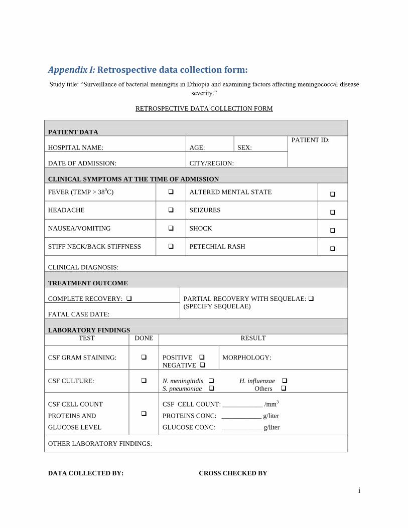
i
Appendix I: Retrospective data collection form:
Study title: “Surveillance of bacterial meningitis in Ethiopia and examining factors affecting meningococcal disease
severity.”
RETROSPECTIVE DATA COLLECTION FORM
PATIENT DATA
HOSPITAL NAME:
AGE:
SEX:
PATIENT ID:
DATE OF ADMISSION:
CITY/REGION:
CLINICAL SYMPTOMS AT THE TIME OF ADMISSION
FEVER (TEMP > 380C) ALTERED MENTAL STATE
HEADACHE SEIZURES
NAUSEA/VOMITING SHOCK
STIFF NECK/BACK STIFFNESS PETECHIAL RASH
CLINICAL DIAGNOSIS:
TREATMENT OUTCOME
COMPLETE RECOVERY:
PARTIAL RECOVERY WITH SEQUELAE:
(SPECIFY SEQUELAE)
FATAL CASE DATE:
LABORATORY FINDINGS
TEST DONE RESULT
CSF GRAM STAINING:
POSITIVE
NEGATIVE
MORPHOLOGY:
CSF CULTURE:
N. meningitidis H. influenzae
S. pneumoniae Others
CSF CELL COUNT
PROTEINS AND
GLUCOSE LEVEL
CSF CELL COUNT: ____________ /mm3
PROTEINS CONC: ____________ g/liter
GLUCOSE CONC: ____________ g/liter
OTHER LABORATORY FINDINGS:
DATA COLLECTED BY: CROSS CHECKED BY
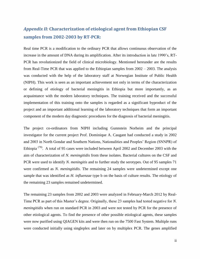
ii
Appendix II: Characterization of etiological agent from Ethiopian CSF
samples from 2002-2003 by RT-PCR:
Real time PCR is a modification to the ordinary PCR that allows continuous observation of the
increase in the amount of DNA during its amplification. After its introduction in late 1990’s, RT-
PCR has revolutionized the field of clinical microbiology. Mentioned hereunder are the results
from Real-Time PCR that was applied to the Ethiopian samples from 2002 – 2003. The analysis
was conducted with the help of the laboratory staff at Norwegian Institute of Public Health
(NIPH). This work is seen as an important achievement not only in terms of the characterization
or defining of etiology of bacterial meningitis in Ethiopia but more importantly, as an
acquaintance with the modern laboratory techniques. The training received and the successful
implementation of this training onto the samples is regarded as a significant byproduct of the
project and an important additional learning of the laboratory techniques that form an important
component of the modern day diagnostic procedures for the diagnosis of bacterial meningitis.
The project co-ordinators from NIPH including Gunnstein Norheim and the principal
investigator for the current project Prof. Dominique A. Caugant had conducted a study in 2002
and 2003 in North Gondar and Southern Nations, Nationalities and Peoples’ Region (SNNPR) of
Ethiopia (78)
. A total of 95 cases were included between April 2002 and December 2003 with the
aim of characterization of N. meningitidis from these isolates. Bacterial cultures on the CSF and
PCR were used to identify N. meningitis and to further study the serotypes. Out of 95 samples 71
were confirmed as N. meningitidis. The remaining 24 samples were undetermined except one
sample that was identified as H. influenzae type b on the basis of culture results. The etiology of
the remaining 23 samples remained undetermined.
The remaining 23 samples from 2002 and 2003 were analyzed in February-March 2012 by Real-
Time PCR as part of this Master’s degree. Originally, these 23 samples had tested negative for N.
meningitidis when run on standard PCR in 2003 and were not tested by PCR for the presence of
other etiological agents. To find the presence of other possible etiological agents, these samples
were now purified using QIAGEN kits and were then run on the 7500 Fast System. Multiple runs
were conducted initially using singleplex and later on by multiplex PCR. The genes amplified
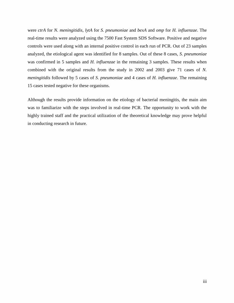
iii
were ctrA for N. meningitidis, lytA for S. pneumoniae and bexA and omp for H. influenzae. The
real-time results were analyzed using the 7500 Fast System SDS Software. Positive and negative
controls were used along with an internal positive control in each run of PCR. Out of 23 samples
analyzed, the etiological agent was identified for 8 samples. Out of these 8 cases, S. pneumoniae
was confirmed in 5 samples and H. influenzae in the remaining 3 samples. These results when
combined with the original results from the study in 2002 and 2003 give 71 cases of N.
meningitidis followed by 5 cases of S. pneumoniae and 4 cases of H. influenzae. The remaining
15 cases tested negative for these organisms.
Although the results provide information on the etiology of bacterial meningitis, the main aim
was to familiarize with the steps involved in real-time PCR. The opportunity to work with the
highly trained staff and the practical utilization of the theoretical knowledge may prove helpful
in conducting research in future.
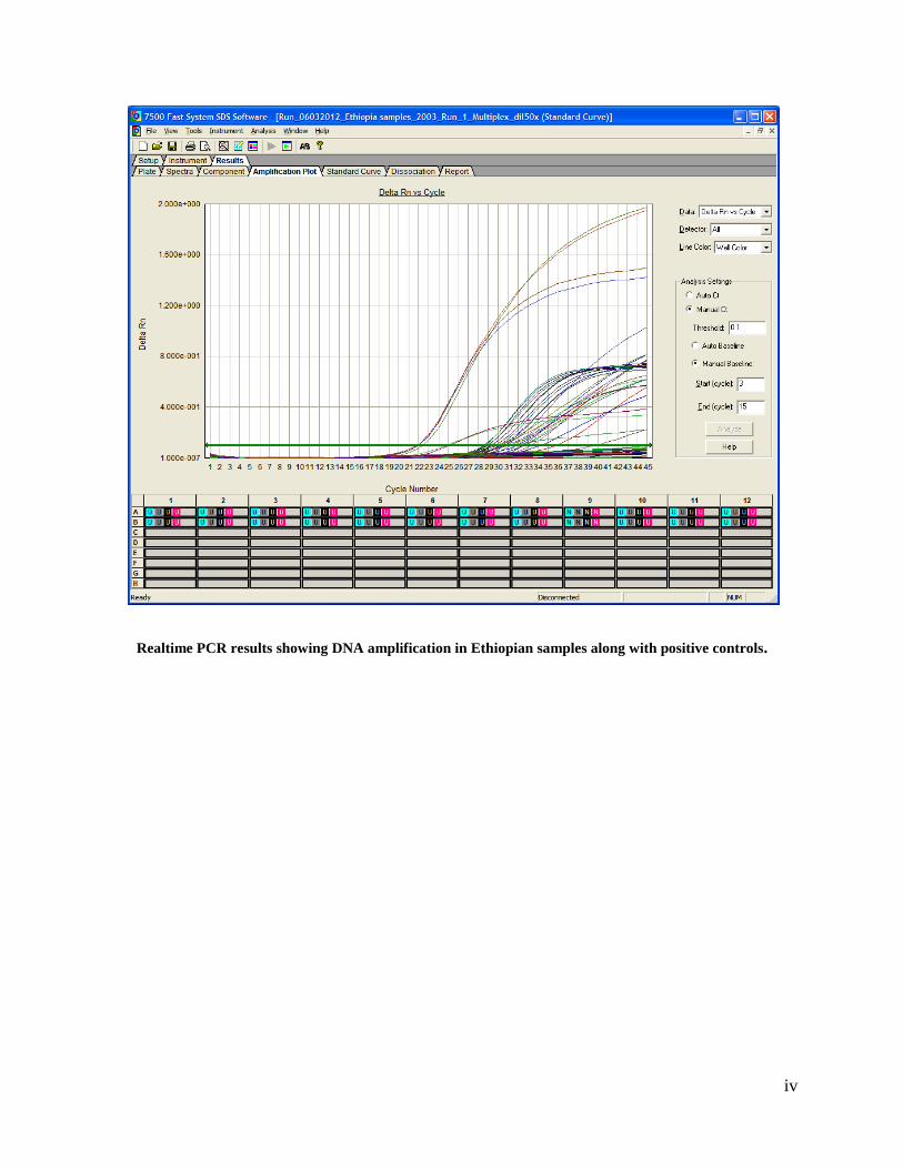
iv
Realtime PCR results showing DNA amplification in Ethiopian samples along with positive controls.
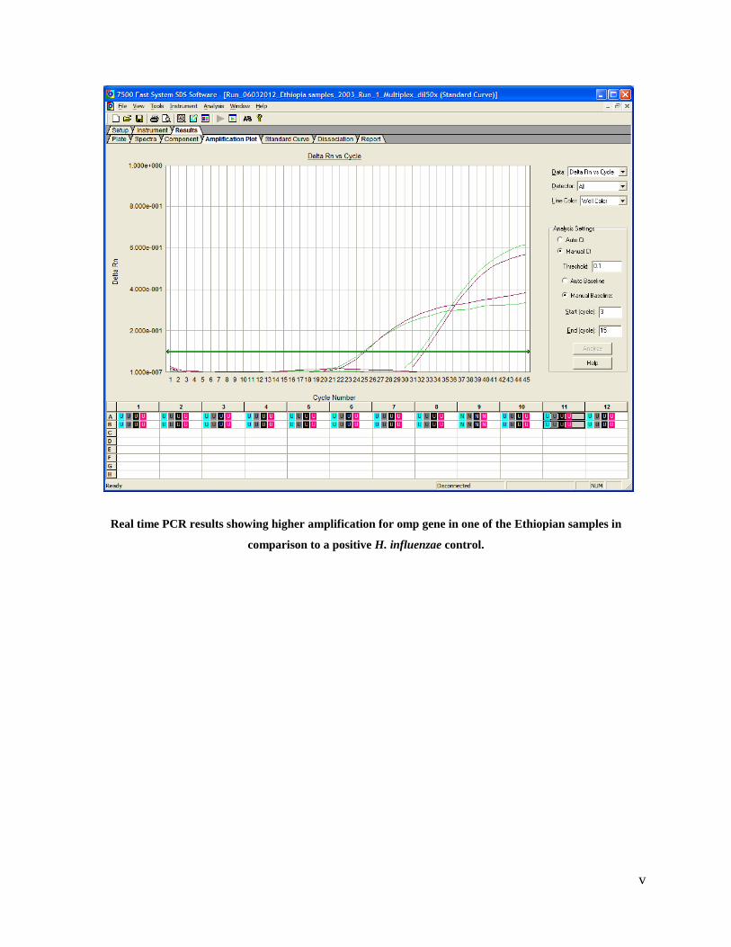
v
Real time PCR results showing higher amplification for omp gene in one of the Ethiopian samples in
comparison to a positive H. influenzae control.