Embed Size (px)
Citation preview

THE EVALUATION OF A LIVE MYCOPLASMA GALLISEPTICUM VACCINE
CANDIDATE AND DNA SEQUENCE ANALYSIS IN THE MOLECULAR
EPIDEMIOLOGY OF MYCOPLASMA GALLISEPTICUM
by
NAOLA MARSHA FERGUSON
(Under the direction of Stanley H. Kleven)
ABSTRACT
A Mycoplasma gallisepticum (MG) isolate from an atypically mild outbreak in turkey breeders was found to be similar to house finch isolates by DNA analyses. A preliminary study in turkeys showed that this isolate (K5054) caused very mild lesions and protected turkeys against subsequent challenge with a virulent MG strain. The safety and efficacy of K5054 was further evaluated in commercial layer-type chickens and turkeys; there was evidence of protection from lesions associated with MG and reduced isolation of R strain post challenge in vaccinated birds. K5054 was further characterized for stability following in vivo passages through chickens; the persistence and the duration of immunity elicited by a single vaccination; and the transmissibility to unvaccinated chickens. K5054 has shown promise as a safe, efficacious, stable vaccine with relatively low transmissibility and long persistence and duration of immunity. In another study, MG isolates from the USA, Israel and Australia were characterized by random amplified polymorphic DNA (RAPD) analysis as well as DNA sequence analysis of portions of the phase-variable putative adhesin protein (pvpA) gene, the cytadhesin gapA gene and an uncharacterized lipoprotein (LP) sequence. The results were compared to reference strains (vaccine and laboratory strains). The RAPD analysis and combined DNA sequence analysis data correlated well, although sequence analysis of any one of the genes did not result in definitive identification of isolates. The Australian isolates appeared to be more similar to the US isolates than were the Israeli isolates.
INDEX WORDS: Mycoplasma gallisepticum, K5054, vaccine, poultry, turkey, house
finch, atypical, random amplified polymorphic DNA, DNA
sequence analysis

THE EVALUATION OF A LIVE MYCOPLASMA GALLISEPTICUM VACCINE
CANDIDATE AND DNA SEQUENCE ANALYSIS IN THE MOLECULAR
EPIDEMIOLOGY OF MYCOPLASMA GALLISEPTICUM
by
NAOLA MARSHA FERGUSON
D.V.M., The University of the West Indies, Trinidad and Tobago, 1996
M.A.M., The University of Georgia, 2001
A Dissertation Submitted to the Graduate Faculty of the University of Georgia in Partial
Fulfillment of the Requirements for the Degree
DOCTOR OF PHILOSOPHY
ATHENS, GEORGIA
2003

� 2003
Naola Marsha Ferguson
All Rights Reserved

THE EVALUATION OF A LIVE MYCOPLASMA GALLISEPTICUM VACCINE
CANDIDATE AND DNA SEQUENCE ANALYSIS IN THE MOLECULAR
EPIDEMIOLOGY OF MYCOPLASMA GALLISEPTICUM
by
NAOLA MARSHA FERGUSON
Major Professor: Stanley H. Kleven
Committee: Maricarmen García
John Glisson
Charles Hofacre
Richard Wooley
Electronic Version Approved:
Maureen Grasso
Dean of the Graduate School
The University of Georgia
August 2003

iv
DEDICATION
This dissertation is dedicated to my family, all of whom helped me to accomplish
my dreams.
To my father, Theodore, for the opportunity and motivation to pursue my passions
fearlessly;
To my mother, Gloria, for being my biggest fan and my biggest inspiration;
To my brothers, Jason and Neron, for helping me to believe that anything is
possible;
And to my wonderful husband, Richard, for never ending support and patience.

v
ACKNOWLEDGEMENTS
I would like to acknowledge the invaluable mentorship of my major professor Dr.
Stanley H. Kleven. I must also thank the members of my committee Dr. Marícarmen
Garcia, Dr. Charles Hofacre, Dr. John Glisson and Dr. Richard Wooley for their support
and guidance. Without the help of the staff and students at the Poultry Diagnostic and
Research Center this would have been a near impossible task. I would like to thank
especially Ms. Victoria Leiting, Mr. Bill Hall, and Mrs. Ruth Wooten.

vi
TABLE OF CONTENTS
Page
ACKNOWLEDGEMENTS………………………………………………….....................v
CHAPTER
1. INTRODUCTION AND LITERATURE REVIEW……………………………...1
2. CHARACTERIZATION OF A NATURALLY OCCURRING INFECTION OF
A MYCOPLASMA GALLISEPTICUM HOUSE FINCH-LIKE STRAIN IN
TURKEY BREEDERS……………………………..……………………………49
3. SAFETY AND EFFICACY OF THE AVIRULENT MYCOPLASMA
GALLISEPTICUM STRAIN K5054 AS A LIVE VACCINE IN
POULTRY……………………………………………………………………….76
4. FURTHER STUDIES OF THE AVIRULENT MYCOPLASMA
GALLISEPTICUM STRAIN K5054 AS A LIVE VACCINE IN
POULTRY……………………………………………………………………...105
5. RANDOM AMPLIFIED POLYMORPHIC DNA (RAPD) ANALYSIS AND
DNA SEQUENCE ANALYSIS OF THREE GENES IN THE MOLECULAR
EPIDEMIOLOGY OF MYCOPLASMA GALLISEPTICUM………………...135
6. DISCUSSION AND CONCLUSIONS………...………………………………161

1
CHAPTER 1
INTRODUCTION AND LITERATURE REVIEW
The Mollicutes (mycoplasmas), the smallest self-replicating organisms, are
characterized by their lack of cell wall, small genome size and low G+C content in their
genomes. The organisms are highly pleomorphic and naturally resistant to antibiotics
affecting cell wall synthesis, for example penicillin (70, 130).
Mycoplasmas are found in multiple hosts, including humans, and many animal species,
plants and insects. Mycoplasmas tend to be host specific and have complex nutritional
requirements (70). They are primarily found as surface parasites on mucous membrane
surfaces of the respiratory tract and urogenital tracts, as well as joints, eyes and mammary
glands (130).
Mollicutes evolved as a branch of gram-positive bacteria by the process of
reductive or degenerative evolution. During this process, the mycoplasmas lost
considerable portions of their ancestors’ chromosomes but retained the genes essential for
life (105, 130).
Unlike other bacteria, all the functions of mycoplasma are expressed from
relatively limited gene sets. Mycoplasmal genomes have so far revealed few of the
complex systems for classic gene regulation and environmental sensing found in other
bacteria. Mycoplasmas do however have systems that provide variation in the expression
and structure of specific gene products. These systems include localized mutable

2
sequences in specific genes or gene sets and the ability to revert to alternative phenotypes
through reversible mutations. The result is that adaptation of variants within the
population is a primary strategy for survival (130).
Tremendous weight has been given to 16S rDNA sequences in the phylogeny,
taxonomy and species identification of Mollicutes (17, 48, 49, 93, 124-127, 142). The
Mollicutes have been divided into 5 phylogenetic units by 16S rRNA sequencing (143),
including the acholeplasmas, the anaeroplasmas, the spiroplasmas, the mycoplasmas and
ureaplasmas.
Numerous avian mycoplasmas have been described but those recognized as
pathogens of domestic poultry include Mycoplasma gallisepticum (MG), Mycoplasma
synoviae, Mycoplasma meleagridis, and Mycoplasma iowae. The most economically
significant of these pathogens is MG, which causes respiratory disease in chickens,
turkeys and other avian species (69).
The disease, in chickens known as chronic respiratory disease (CRD) and turkeys
as infectious sinusitis, can result in respiratory rales, coughing and nasal discharge, as
well as sinusitis in turkeys. Airsacculitis may cause significant economic losses at
processing, there may also be egg production losses, reduced feed efficiency and
medication costs (85).
Mild or subclinical cases of MG, termed ‘atypical’ infections, have been observed
in chickens and turkeys (22, 47, 102, 152). MG infections in turkeys resulting in mild
clinical disease are unusual. Turkeys are more susceptible to MG and often more severely
affected by MG infections than chickens; turkeys may develop severe sinusitis,

3
respiratory distress, depression, decreased feed intake, and weight loss (85). These
atypical infections are often difficult to diagnose (8, 59).
Although MG infection occurs naturally in chickens and turkeys, the organism
has also been isolated from naturally occurring infections in other avian species (85). The
significance of these species in the epidemiology of MG has not been established
although wild passerine species may act as biological carriers (73, 119). This may be a
relatively minor factor in the overall epidemiology of MG.
In early 1994, an epidemic of MG began in wild house finches (Carpodacus
mexicanus) in the mid-Atlantic United States (88). MG had not been previously
associated with clinical disease in wild passerine birds. The disease has become
widespread and has been reported throughout the eastern United States and Canada (30).
Molecular characterization of isolates suggested that the house finch epidemic arose from
a single source and that the MG infection had not been shared between songbirds and
commercial poultry (87).
MG can be transmitted horizontally by direct or indirect contact. In general MG
does not survive outside of the host for extended periods. It has been shown to survive on
straw, cotton, and rubber for up to 2 days and 3-4 days on human hair or feathers (20).
Carrier birds, including backyard flocks, are thought to be the main source of MG
outbreaks (27). MG can also be transmitted vertically in ovo. The highest frequency of
transmission occurs during the acute phase of the disease, but transmission may also
occur at a lower rate during chronic infection (43, 44).

4
Pathogenesis and Virulence Factors
The clinical manifestations of severe MG infection in chickens and turkeys is
generally due to a complicated etiology involving concurrent infections and
environmental factors (68, 85). Colibacillosis, live vaccines, and immunosuppression
may all affect the severity of the disease (46, 113, 116).
Most mycoplasmas adhere tightly to the epithelial linings of the respiratory and
urogenital tract, rarely invading tissues, and are considered surface parasites. Adhesion is
a prerequisite for colonization and infection. Loss of adhesion results in loss of
infectivity, and reversion to a cytadherence phenotype is accompanied by regaining
infectivity and virulence (79, 129).
Early in MG infection in the upper respiratory tract there is release of mucous
granules, destruction and exfoliation of ciliated and nonciliated epithelial cells (23).
Ciliostasis has been observed in vitro (2).
Pathogenic effects that may be attributable to mycoplasma infection include
damage to host cell membranes, clastogenic and oncogenic effects. Adhesion to host
cells, membrane fusion, cell invasion, stimulation or suppression of the host immune
response and antigenic variation may be important factors in mycoplasma disease
pathogenesis.
Mildly toxic by-products of mycoplasma metabolism, such as superoxide and
hydrogen peroxide, may be involved in oxidative damage to host cells membranes in
mycoplasma infections (130).

5
It has also been theorized that mycoplasma-associated preferential loss of
potassium channels occurs resulting in depolarization of the cell membrane leading to the
ciliostasis observed in mycoplasma infected ciliary cells (52).
MG shares similar pathogenic mechanisms with two human mycoplasmas,
Mycoplasma pneumoniae and Mycoplasma genitalium. These mycoplasmas share a flask-
shaped morphology characterized by a unipolar terminal organelle that is involved in
mucosal attachment and gliding motility.
The molecular basis of mycoplasma pathogenicity remains largely elusive. The
clinical picture of mycoplasma infections in humans and animals is suggestive of damage
due to host immune and inflammatory responses rather than direct toxic effects by
mycoplasmal cell components. Various mycoplasmal virulence factors have been
described but there appears to be no clear causal relationship between these factors and
pathogenicity (130).
Antigenic variation and phenotypic switching. Antigenic variation or
phenotypic switching refers to the ability of a microbial species to alter the antigenic
character of its surface components. These surface organelles are the major targets of the
host antibody response; therefore the ability of a microorganism to rapidly change the
surface antigenic repertoire and consequently to vary the immunogenicity of these
structures allows effective avoidance of immune recognition. The molecular switching
events leading to the generation of phenotypic variants are generally reversible.
During their evolution and adaptation to a parasitic mode of life, the mycoplasmas
have developed various genetic systems providing a highly plastic set of variable surface
proteins. The majority of surface proteins involved in generating antigenic variation in

6
mycoplasmas are lipoproteins. The generation of a versatile surface coat through high-
frequency phase and size variation provides the organism with a useful tool for immune
system avoidance, allowing the mycoplasma to escape antibody attack (130).
Many pathogenic mycoplasmas are able to undergo surface variation resulting in
an antigenic shift (11, 41, 53, 118, 133, 134). Epitope switching has been observed for
many MG surface molecules (13, 32, 84).
The high degree of phenotypic variation exhibited by mycoplasmas is considered
a major factor in pathogenicity and chronic infection of the host (128, 132, 135, 155).
Changes in surface topology of MG during host infection and molecular characteristics of
several MG surface proteins have been described (13, 32).
Adhesion and cytadhesins. The cytadhesion process of mycoplasmas appears to
be multifactorial, involving a number of accessory membrane proteins. These act in
concert with cytoskeletal elements to facilitate the lateral movement and concentration of
the adhesion molecules at the attachment tip organelle. Extensive analysis of the
cytadherence process in M. pneumoniae has demonstrated that this involves the
coordinate action of primary adhesin molecules (P1 and P30) in concert with an array of
high-molecular-weight accessory membrane proteins (76).
MG cytadhesins that have been identified include LP64 (31), pMGA (109), PvpA
(14, 154), MGC1 (45, 56), MGC2 (51), and MGC3 (123, 156).
Antigenic variation of cytadhesins allow MG to escape the host immune system
(6).
A surface lipoprotein known as pMGA (106) is expressed in abundance by MG.
The pMGA family of hemagluttinins (adhesins) probably plays an important role in

7
colonization and chronicity of respiratory disease in the avian host. Although each MG
usually expresses only one homogenous, unique pMGA molecule (40), this lipoprotein
appears to exhibit high frequency phase and antigenic variation during culture (especially
when growing in media containing anti-pMGA antibodies) (107) and during the course of
a natural infection (39).
A switch to off expression of pMGA occurs during the acute stages of infection, a
second switch to antigenic variants occurs during the chronic stage of infection. The
variation in expression results from switches in expression of different members of a
repertoire of genes (9, 108, 109).
The number of pMGA gene copies present in the genome varies from 32 to 70 in
different strains (9). Despite the presence of multiple copies of the gene, only one
individual gene is expressed at a time in a given strain. All but one of the genes is
transcriptionally silent. The control of transcription of each member of the gene family
resides in a short GAA trinucleotide repeat region that lies 18 bases 5’ to the -35 box of
the promoter of each gene (38, 98).
PvpA is postulated to be one of the accessory membrane proteins of MG.
Variation within PvpA could affect the specificity or affinity of adherence. It is an
integral membrane surface protein with a free C terminus that is subject to spontaneous
high-frequency phase variation in expression and exhibits size variation among strains
(14, 154). It exists as a single chromosomal copy. PvpA variation of expression is
controlled at the level of translation. A localized nonsense mutation in a poly-GAA tract
of the pvpA coding region was shown to determine PvpA antigenic variation.

8
Another type of variation shown to occur with PvpA results from deletions within
the 3’ end of the pvpA gene and causes size variation of PvpA polypeptide. The size
variation of the PvpA protein was shown to range from 48 to 55kDa. The deletions were
localized at the proline-rich carboxy-terminal region and within two direct repeat
sequences (14),. Tthis domain may be under selective pressure in the host. Several MG
strains differing in their adherence and pathogenicity have varying deletions and sizes of
PvpA. Analysis of pvpA has been used to differentiate between MG strains (99).
The mgc1 gene (56), also referred to as gapA (45), is one of three clustered genes
with adhesin-related functions. It encodes a protein with homology to the P1 cytadhesin
protein of M. pnuemoniae. Immunoblot analysis of various strains has demonstrated
intraspecies variation in the size of GapA (98, 105 and 110 kDa) (45).
The other two genes in the cluster are mgc2 (51) and crmA (123) (also referred to
as mgc3 (156)). MGC2 is a 32 kDa protein with homology to P30 of M. pnuemoniae, the
mgc2 gene is located upstream of the gapA gene. CrmA (or MGC3) is a 120kDa
cytadherence associated membrane protein, the gene is located downstream of the mgc1
gene (156).
CrmA is cotranscribed with GapA, they interact and are essential for cytadherence
(121). CrmA is encoded by the second gene in the gapA operon and shares significant
sequence homology to the ORF6 gene of M.pneumoniae, which has been shown to play
an accessory role in the cytadherence process. GapA and CrmA have been shown to
undergo concomitant phase variation; the underlying genetic mechanism is a reversible
base substitution resulting in a nonsense mutation in the gapA gene that affects the
expression of gapA and the crmA gene located downstream (149).

9
Intracellular location. Human and animal mycoplasmas were shown to be taken
up by polymorphonuclear leukocytes (110). More recently MG was shown to have the
ability to invade human epithelial cells and chicken embryo fibroblasts in vitro. This has
been proposed as a mechanism of resisting host defenses and antibiotic therapy, as well
as the method that MG uses to establish chronic infections and cause systemic infections
(10, 112, 150).
The invasion of mycoplasma into the host cell cytoplasm may affect cell function
and integrity. Lysis of human lung fibroblasts (111) and cell disruption and necrosis
(100) have been demonstrated with M. genitalium and M. penetrans, respectively.
Immune system modulation. Mycoplasmas stimulate both a specific and a non-
specific immune response in the host. The specific anti-mycoplasma reactions have been
shown to play a role in the development of lesions and the exacerbation of disease. The
non-specific responses induced by mycoplasmas include the suppression or polyclonal
stimulation of B and T lymphocytes, induction of cytokines, increasing the cytotoxicity
of macrophages, natural killer cells and T cells, and activating the complement cascade.
The ability of mycoplasmas to modulate the host response may allow them to evade or
suppress host defenses (130).
Immune response
Birds that recover from MG induced disease have some degree of immunity but
remain carriers of the organism (12). The immunogenicity of MG strains varies and is
correlated with virulence (79, 94).

10
Birds lacking a fully functional immune system (neonatal thymectomy or
bursectomy) have significantly higher lesion scores than normal birds following MG
infection (140). It seems that both antibody and cell mediated immunity are important in
the host response to MG (19).
Although the bursa and bursal derived cells have been shown to be essential for a
protective immune response to MG (3, 77), it has also been shown that there is a poor
correlation between systemic antibody levels and protection from challenge (97, 117).
Chickens produce a protective immune response to MG that seems to be localized
to the respiratory tract. MG antibodies in upper and lower respiratory tract washes have
been shown to prevent attachment and establishment of MG in tracheal organ cultures (7)
and in vivo (140, 151).
A significant leukocyte migration into the mucosa of the upper respiratory tract is
a hallmark of MG infection. The lesions usually resolve in 3 to 7 weeks with a
concomitant decrease in MG in the trachea following control of the infection. The
resolution of lesions is correlated with increasing antibodies in tracheal washes (151) and
serum, as well and leukocyte migration into the mucosa (19).
It has been theorized that local immunity mediated by secretory IgA may have a
role in preventing the establishment of infection while CMI may be involved in recovery
(140).
Although a lymphproliferative response in the respiratory tract is a prominent
feature of disease induced by MG, the cell mediated immune response has not been
extensively investigated. It has recently been shown that there is specific stimulation of
CD8+ cells, particularly in the acute phase of the disease (35). It is postulated that the

11
fusion of mycoplasma membranes with the host cells enables presentation to CD8+
lymphocytes.
Diagnosis
Serological screening is routinely used as an indicator of MG infection. Sera
commonly are analyzed for MG antibodies using the serum plate agglutination (SPA)
test, a hemagglutination-inhibition (HI) test and an enzyme-linked immunosorbent assay
(ELISA) test (69). The SPA test is rapid, sensitive and inexpensive but may result in non-
specific reactions (5, 15, 42), so that reactors must generally be confirmed by the HI or
ELISA tests. Serum dilution has also been used to reduce non-specific reactions (136).
The HI test is less sensitive but more specific than the SPA test. It is however, a time
consuming procedure and the reagents are not commercially available. In general the
ELISA test is more sensitive than the HI test and more specific than the SPA test (58,
60).
MG infection is generally confirmed by the isolation and identification of MG or
by DNA based detection methods (83, 92). Isolation and identification of the organism is
generally considered the gold standard for diagnosis. For culture swabs from trachea,
choanal cleft or air sacs are often used. Sinus exudates, as well as swabs of the turbinates,
and lungs and other tissues may also be used (69).
Mycoplasma isolates are commonly identified using direct and indirect
immunofluorescence (69, 138). Mycoplasma species-specific hyperimmune sera is an
essential reagent for these tests and may limit the ability of some laboratories to perform
the test (69).
12
MG species-specific PCR (78, 114), PCR-RFLP (34, 65) and oligonucleotide
probe (29, 33) techniques have been developed.
During the acute stage of the infection the number of organisms in the upper
respiratory tract is high (80, 151); however in chronic infection the number of organisms
is much lower and routine methods may not detect MG (83).
In some situations it may be very difficult to isolate MG consistently from
infected flocks. These instances include chronic MG cases and infections with strains of
low pathogenicity (69, 152). The overgrowth of non-pathogenic mycoplasmas may also
interfere with cultivation of MG from clinical samples in the laboratory (104).
The isolation rates of fastidious MG strains may be enhanced in vivo by bioassays
(103). Susceptible poultry are inoculated with potentially infectious material from suspect
flocks. The organism may have the opportunity to multiply in these birds to levels
detectable by PCR and/or culture. The birds are routinely sampled enhancing MG
detection.
Control of MG
Control MG has generally been based on the eradication of the organism from
breeder flocks and the maintenance of mycoplasma-free status in the breeders and their
progeny by biosecurity. Single-age and all-in all-out production methods allow the
control of MG in this way.
Serology is the primary method for flock screening. Serological monitoring
performed periodically is the basis of voluntary control programs such as the National
Poultry Improvement Plan (NPIP).
13
Large populations of poultry in small geographic areas can make control by
biosecurity alone very difficult. MG vaccines have been used in the control of MG in
areas where eradication is not feasible.
MG vaccines are used to prevent or reduce disease and clinical signs in the
vaccinated birds as well as to prevent egg production losses and egg transmission of MG.
Inactivated MG vaccines. Inactivated MG vaccines have been widely used in
several countries. The results with MG bacterins have been variable (50, 54, 55, 63); they
protect against loss of egg production in layers (25, 43, 44), but do not prevent infection
or provide consistent protection against respiratory disease (1).
Live MG vaccines. One of the options for control is live MG vaccines (67, 83,
145). Eradication of MG is preferable to vaccination wherever possible; however, the use
of live vaccines to displace virulent wild-type MG strains from commercial poultry flocks
may be a useful part of an eradication program (72, 82).
Live vaccines that are currently used worldwide to control MG include F strain
(Schering Plough, Kenilworth, N.J.) (4, 101), 6/85 (Intervet America, Millsboro, Del.)
(24) and ts-11 (Bioproperties, Inc., Australia, marketed in the US by Merial Select
Laboratories, Gainsville, GA.) (146, 147).
The important characteristics of an ideal live MG vaccine include safety in the
target species, efficacy (immunogenicity), the ability to stimulate solid lifelong protection
(preferably from a single dose), and stability following in vivo passages (lack of reversion
of attenuated strains to a virulent form). Vaccines should also be easy and inexpensive to
manufacture. The vaccine should not spread to neighboring flocks (145).

14
It has been established that there is a complex relationship between infectivity,
pathogenicity and immunogenicity of MG strains (79). It has been established that
virulence, invasiveness and immunogenicity of MG strains are directly correlated (94).
Some live vaccines may be so mild as to be incapable of eliciting long lasting protective
immunity. The colonization and persistence of MG in the upper respiratory tract may be
essential to duration of immunity elicited by the vaccine.
Studies have indicated that the level of protection elicited by live vaccines is
directly correlated with the virulence of the vaccine strain (1, 96).
F strain vaccine has been used extensively (92), it is very immunogenic and
mildly virulent in chickens (1, 16, 131), but too virulent for use in turkeys (95, 96). F
strain has been associated with MG outbreaks in commercial turkeys (86). It is effective
in displacing virulent (field) strains from poultry operations (72, 74, 82).
6/85 and ts-11 vaccines have both been shown to elicit protective immunity in
chickens and to possess little or no virulence for chickens and turkeys (24, 146, 147).
F strain persists at higher levels in the upper respiratory tract than either ts-11 or
6/85, and ts-11 appears to colonize more effectively than 6/85 (1, 91). The duration of
immunity elicited by a live vaccine may be associated with the colonization and
persistence of the vaccine in the respiratory tract.
Although 6/85 has been reported not to persist in the trachea and to be poorly
transmissible, “6/85-like” isolates have been recovered from vaccinated and unvaccinated
contact chickens long after vaccination (137) as well as from unvaccinated turkeys
(Kleven, unpublished).

15
In the event that a live vaccine cycles through a flock of poultry it should not
increase in virulence. After years of use there is no evidence that F strain has become
more virulent (145). Experimental passage of 6/85 through chickens and turkeys did not
result in a substantial increase in virulence (24, 26). Attempts to serially passage ts-11 in
birds were unsuccessful but the vaccine appeared to retain its characteristics after three
passages (147).
F strain may be more virulent than 6/85 or ts-11, but it provides better protection
against airsacculitis and persists at higher levels in the upper respiratory tract (1). F strain
also protects against colonization by more virulent challenge strains (21) and is capable
of displacing endemic field strains (72, 74). However, the persistence and transmissibility
of F strain means that it can be isolated from farms long after vaccination has ceased.
There is the potential for spread to susceptible poultry (66), most significantly, turkeys
(86).
F strain is readily transmissible to unvaccinated pen mates and chickens in
adjacent pens (66) and can be isolated from farms long after vaccination has ceased and
has been implicated outbreaks in commercial turkeys (86). Although, in experimental
situations, F strain has been shown to transmit between birds, widespread use of the
vaccine has not resulted in widespread isolations of F strain from field cases in chickens
(37).
The ts-11 and 6/85 vaccines have both been shown to be poorly transmissible to
in contact poultry (90, 147).
The distinct advantage of the milder vaccine strains over F strain is their lack of
virulence in turkeys and their low transmissibility (24, 91, 95, 147). The ts-11 vaccine

16
may be useful in displacing endemic F strain in poultry complexes as part of an
eradication program (141).
Although 6/85 has been reported to transmit poorly (91), not to persist in the
respiratory tract for long periods (72), and to be apathogenic (24), there have been reports
of MG outbreaks in unvaccinated turkeys and chickens from which “6/85-like” MG
strains have been isolated (137).
The vaccination of turkeys against MG has not been shown to be feasible
although there has been limited use of 6/85 (83).
A GapA-negative high passage MG R strain (GT 5) has recently been described
as a potential modified live vaccine (122, 123).
The characteristics of different live MG vaccines have been described and
compared extensively (1, 91, 145). The choice of vaccine should be carefully evaluated in
each situation.
Epidemiology and Strain Differentiation
In general the process of subtyping microbial isolates into strains is important
epidemiologically for recognizing outbreaks of infection, determining the source of the
infection, recognizing particularly virulent strains of organisms, and monitoring
vaccination programs (120).
Methods of strain differentiation must have high differentiation power so that it
can clearly differentiate unrelated strains, as well as demonstrate the relationship of
isolates from individuals infected through the same source. The techniques should also
have a high degree of reproducibility. Reproducibility refers to the ability of a technique

17
to yield the same result when a particular strain is repeatedly tested. It is especially
important for the construction of reliable databases containing known strains within a
species to which unknown organisms can be compared.
Mycoplasma colonies can vary in their surface antigenic phenotype, therefore
mycoplasma strains can differ markedly in their antigen profiles and their potentially
virulence-related surface properties (135).
Intraspecies heterogeneity and antigenic variability can be observed in
mycoplasmas through serological testing (75, 139) and electrophoresis of cell proteins
(62).
The shortcomings of phenotypically based typing methods, such as those based on
a reaction with an antibody (135), have led to the development of typing methods based
on the microbial genotype or DNA sequence, which minimize problems with typeability
and reproducibility and, in some cases, enable the establishment of large databases of
characterized organisms.
Molecular techniques that have been used to identify MG strains restriction
fragment length polymorphisms (RFLP) of DNA (64, 71), DNA and ribosomal RNA
gene probes (61, 153), and PCR with strain-specific primers (115).
Random amplified polymorphic DNA (RAPD). The most widely used method for
differentiating MG strains is random amplified polymorphic DNA (RAPD) or arbitrarily
primed PCR, analysis (18, 28, 36). The RAPD assay was first described by Williams et
al. (148) and Welsh and McClelland (144). RAPD assays are based on the use of short
random sequence primers, which hybridize with sufficient affinity to chromosomal DNA
sequences at low annealing temperatures so that they initiate amplification of regions of

18
the bacterial genome. The number and location of these random primer sites vary for
different strains of a bacterial species. Thus, following separation of the amplification
products by agarose gel electrophoresis, a pattern of bands results. In theory, the patterns
of bands are characteristic of the particular bacterial strain.
RAPD analysis is rapid and sensitive and this method has been used to identify
vaccine strains in MG-vaccinated flocks and for epidemiological studies (57, 81, 87, 89).
Due to the random nature of the primers and the low-stringency conditions of the
RAPD reaction, this assay requires the use of pure cultures of the target mycoplasma.
Isolation of mycoplasma is expensive, time-consuming, and technically complicated in
cases where nonpathogenic mycoplasma species may overgrow the virulent mycoplasma.
The isolation process itself may favor the growth of one strain where more than one MG
subtype may be present. Furthermore, technical factors such as target DNA/primer ratio
may significantly impact the reproducibility of RAPD patterns.
DNA sequence analysis. Ultimately, all molecular genetic methods for
distinguishing organism subtypes are based on differences in the DNA sequence.
Logically, then, DNA sequencing would appear to be the best approach to differentiating
subtypes. DNA sequencing generally begins with PCR amplification of a sample DNA
directed at genetic regions of interest, followed by sequencing reactions with the PCR
products.
DNA sequencing must be directed at a small region of the bacterial genome. It is
impractical to sequence multiple or large regions of the chromosome. Thus, in contrast to
RAPD analysis, which examines the entire chromosome, DNA sequencing examines a
very small portion of the sites that can potentially vary between strains.

19
The variability within the selected sequence must be sufficient to differentiate
different strains of a particular species.
Progress in the molecular biology of mycoplasmas has been achieved in the last
decade, and several surface proteins in virulent mycoplasmas, such as PvpA (14, 154),
MGC1 (45, 56), MGC2 (51), and MGC3 (123, 156), have been described. The DNA
sequences of these genes are under great selective pressure and may be useful in the
molecular epidemiology of MG.
Objectives of the Research
With increasing concentrations of poultry in restricted geographic areas and large
multiple age complexes, the control of MG by purchasing MG-free stock and keeping
flocks MG-free has become much more difficult. There is an increasing need to use live
vaccines to control MG in these situations. Live vaccines prevent production losses by
allowing controlled exposure of flocks to avirulent MG strains resulting in the
development of immunity to subsequent field challenges. Live vaccines can also be part
of an eradication program by displacing the resident virulent MG strain (72, 141);
cessation of vaccination should allow the flocks to return to an MG-free status.
The properties of an ideal MG vaccine include avirulence, immunogenicity, life-
long protection, affordability, easy methods of administration and stability.
Unfortunately, although each of the currently available vaccines has its advantages, none
of them attains the ideal status in every respect (145). F strain is highly efficacious but
moderately virulent in chickens and unsafe for turkeys. 6/85 and ts-11 are both avirulent
and immunogenic, but the level and duration of protection elicited by these milder

20
vaccines may not be as good as F strain, 6/85 more so than ts-11. Also, none of the
currently available vaccines are used in turkeys. Turkeys are very susceptible to MG
infection and more severely affected than chickens. F-strain is too virulent for use in
turkeys. Although there has been restricted use of 6/85, MG isolates similar to this strain
(“6/85-like”) have been isolated from clinically ill commercial turkeys (Kleven,
unpublished). The ts-11 appears to be incapable of infecting turkeys (Kleven, personal
communication).
There is therefore a need for an avirulent, immunogenic, and stable MG vaccine
that is safe for chickens and turkeys.
With the widespread use of live vaccines there is also an increasing need to
differentiate between vaccine strains and field isolates. Methods to easily and clearly
differentiate between pathogens and vaccine strains are necessary to avoid confusion.
Epidemiology and strain differentiation is an essential part of eradication and control of
MG. Precise information about the origin of infectious agents allows cost-effective
control programs to be targeted to the weak points of the current system. This approach
should be more economical than shotgun approaches of control programs. Molecular
epidemiology should aid in pinpointing the source of outbreaks, identify biological
carriers, and increase understanding about the transmission and maintenance of MG in
the environment.
RAPD analysis is a widely used, rapid, sensitive and effective tool in the
molecular epidemiology of MG but this method has its drawbacks and difficulties. Chief
among them is the need for pure cultures of the target organism, as well as difficulty in
the reproducibility and standardization between laboratories.

21
Alternative methods of molecular epidemiology should allow the construction of
a database that allows easy comparison of an unknown isolate to all the known strains in
the database. RAPD analysis restricts the number of strains to which an unknown is
compared. The low reproducibility of RAPD analysis makes the comparison of patterns
from different RAPD reactions or different agarose gels unreliable.
An alternative method should also allow MG strain discrimination at the level of
clinical samples so avoiding the MG isolation step that is necessary for RAPD analysis.
Nucleotide sequence analysis of a specified gene (s) may allow the development of a
PCR that is performed directly on clinical samples to detect MG and identify strains. The
building of a sequence database allows the comparison of many seemingly unrelated
isolates. This method would be reproducible and easily standardized. We must determine
the discriminatory power of this method with respect to selected genes that have shown
intraspecies variability and may be useful in this technique. To be useful DNA
sequencing should be able to differentiate between MG strains.
The aim of the studies described in Chapters 2, 3 and 4 was to evaluate a naturally
occurring MG isolate as a live vaccine in poultry. The aim of the study described in
Chapter 5 is to evaluate DNA sequence analysis of selected genes as a method of
molecular epidemiology of MG.

22
References
1. Abd-El-Motelib, T. Y., and S. H. Kleven. A comparative study of Mycoplasma
gallisepticum vaccines in young chickens. Avian Dis. 37:981-987. 1993.
2. Abdul-Wahab, O. M. S., G. Ross, and J. M. Bradbury. Pathogenicity and cytadherence
of Mycoplasma imitans in chicken and duck embryo tracheal organ cultures. Infect.
Immun. 64:563-568. 1996.
3. Adler, H. E., B. J. Bryant, D. R. Cordy, M. Shifrine, and A. J. DaMassa. Immunity and
mortality in chickens infected with Mycoplasma gallisepticum: Influence of the Bursa of
Fabricius. J. Infect. Dis. 1973.
4. Adler, H. E., D. A. McMartin, and M. Shifrine. Immunization against Mycoplasma
infections of poultry. American Journal of Veterinary Research. 21:482-485. 1960.
5. Adler, H. E., and A. D. Wiggins. Interpretation of serologic tests for Mycoplasma
gallisepticum. World Poult Sci J. 29:345-353. 1973.
6. Athamna, A., R. Rosengarten, S. Levisohn, I. Kahane, and D. Yogev. Adherence of
Mycoplasma gallisepticum involves variable surface membrane proteins. Infect. Immun.
65:2468-2471. 1997.

23
7. Avakian, A., and D. H. Ley. Protective immune response to Mycoplasma gallisepticum
demonstrated in respiratory-tract washings from M. gallisepticum-infected chickens.
Avian Dis. 37:697-705. 1993.
8. Avakian, A. P., D. H. Ley, and M. A. T. McBride. Humoral immune response of
turkeys to strain S6 and a variant Mycoplasma gallisepticum studied by immunoblotting.
Avian Diseases. 36:69-77. 1992.
9. Baseggio, N., M. D. Glew, P. F. Markham, K. G. Whithear, and G. F. Browning. Size
and genomic location of the pMGA multigene family of Mycoplasma gallisepticum.
Microbiol. 142:1429-1435. 1996.
10. Baseman, J. B., and J. G. Tully. Mycoplasmas: Sophisticated,Reemerging, and
Burdened by their Notoriety. Emerging Inf. Dis. 3:21-32. 1997.
11. Behrens, A., M. Heller, H. Kirchhoff, D. Yogev, and R. Rosengarten. A family of
phase-variant and size-variant membrane-surface lipoprotein antigens (VSPS) of
Mycoplasma bovis. Infect. Immun. 62:5075-5084. 1994.
12. Bencina, D., and D. Dorrer. Demonstration of Mycoplasma gallisepticum in tracheas
of healthy carrier chickens by fluorescent-antibody procedure and the significance of
certain serologic tests in estimating antibody response. Avian Dis. 28:574-578. 1984.
24
13. Bencina, D., S. H. Kleven, M. G. Elfaki, A. Snoj, P. Dovc, D. Dorrer, and I. Russ.
Variable expression of epitopes on surface of Mycoplasma gallisepticum demonstrated
with monoclonal antibodies. Avian Pathol. 23:19-36. 1994.
14. Boguslavsky, S., D. Menaker, I. Lysnyansky, T. Liu, S. Levisohn, R. Rosengarten, M.
Garcia, and D. Yogev. Molecular characterization of the Mycoplasma gallisepticum
pvpA gene which encodes a putative variable cytadhesin protein. Infect Immun. 68:3956-
3964. 2000.
15. Boyer, C. I., J. Fabricant, and J. A. Brown. Non-specific plate agglutination reactions
with PPLO antigen. Avian Dis. 4:546-547. 1960.
16. Branton, S. L., B. D. Lott, J. W. Deaton, J. M. Hardin, and W. R. Maslin. F strain
Mycoplasma gallisepticum vaccination of post production peak commercial leghorns and
its effect on egg and eggshell quality. Avian Diseases. 32:304-307. 1988.
17. Brown, D. R., G. S. McLaughlin, and M. B. Brown. Taxonomy of the feline
mycoplasmas Mycoplasma felifaucium, Mycoplasma feliminutum, Mycoplasma felis,
Mycoplasma gateae, Mycoplasma leocaptivus, Mycoplasma leopharyngis, and
Mycoplasma simbae by 16S rRNA gene sequence comparisons. Int J Syst Bacteriol.
45:560-4. 1995.

25
18. Charlton, B. R., A. A. Bickford, R. L. Walker, and R. Yamamoto. Complementary
randomly amplified polymorphic DNA (RAPD) analysis patterns and primer sets to
differentiate Mycoplasma gallisepticum strains. J. Vet. Diag. Invest. 11:158-61. 1999.
19. Chhabra, P. C., and M. C. Goel. Immunological response of chickens to Mycoplasma
gallisepticum infection. Avian Dis. 25:279-293. 1981.
20. Christensen, N. H., C. A. Yavari, A. J. McBain, and J. M. Bradbury. Investigations
into the survival of Mycoplasma gallisepticum, Mycoplasma synoviae and Mycoplasma
iowae on materials found in the poultry house environment. Avian Pathol. 23:127-143.
1994.
21. Cummings, T. S., and S. H. Kleven. Evaluation of protection against Mycoplasma
gallisepticum infection in chickens vaccinated with the F strain of M. gallisepticum.
Avian Diseases. 30:169-171. 1986.
22. Dingfelder, R. S., D. H. Ley, J. M. McLaren, and C. Brownie. Experimental infection
of turkeys with Mycoplasma gallisepticum of low virulence, transmissibility, and
immunogenicity. Avian Dis. 35:910-919. 1991.
23. Dykstra, M. J., S. Levisohn, O. J. Fletcher, and S. H. Kleven. Evaluation of
cytopathologic changes induced in chicken tracheal epithelium by Mycoplasma
gallisepticum in-vivo and in-vitro. Am J Vet Res. 46:116-122. 1985.

26
24. Evans, R. D., and Y. S. Hafez. Evaluation of a Mycoplasma gallisepticum strain
exhibiting reduced virulence for prevention and control of poultry mycoplasmosis. Avian
Dis. 36:197-201. 1992.
25. Evans, R. D., Y. S. Hafez, and F. W. Orthel. Mycoplasma gallisepticum vaccination-
challenge: an egg-production model. Avian Dis. 36:956-963. 1992.
26. Evans, R. D., Y. S. Hafez, and C. S. Schreurs. Demonstration of the genetic stability
of a Mycoplasma gallisepticum strain following in vivo passage. Avian Diseases. 36:554-
560. 1992.
27. Ewing, M. L., S. H. Kleven, and M. B. Brown. Comparison of enzyme-linked
immunosorbent assay and hemagglutination-inhibition for detection of antibody to
Mycoplasma gallisepticum in commercial broiler, fair, and exhibition, and
experimentally infected birds. Avian Dis. 40:13-22. 1996.
28. Fan, H. H., S. H. Kleven, and M. W. Jackwood. Application of polymerase chain
reaction with arbitrary primers to strain identification of Mycoplasma gallisepticum.
Avian Dis. 39:729-735. 1995.
29. Fernandez, C., J. G. Mattsson, G. Bölske, and K.-E. Johansson. Species-specific
oligonucleotide probes complementary to 16S rRNA of Mycoplasma gallisepticum and
Mycoplasma synoviae. Res. Vet. Sci. 55:130-136. 1993.

27
30. Fischer, J. R., D. E. Stallknecht, M. P. Luttrell, A. A. Dohndt, and K. A. Converse.
Mycoplasmal conjunctivitis in wild songbirds: The spread of a new contagious disease in
a mobile host population. Emerging Inf. Dis. 3:69-72. 1997.
31. Forsyth, M. H., M. E. Tourtellotte, and S. J. Geary. Localization of an
immunodominant 64 kDa lipoprotein (LP 64) in the membrane of Mycoplasma
gallisepticum and its role in cytadherence. Molec. Microbiol. 6:2099-2106. 1992.
32. Garcia, M., M. G. Elfaki, and S. H. Kleven. Analysis of the variability in expression
of Mycoplasma gallisepticum surface antigens. Vet. Microbiol. 42:147-158. 1994.
33. García, M., M. W. Jackwood, M. Head, S. Levisohn, and S. H. Kleven. Use of
species-specific oligonucleotide probes to detect Mycoplasma gallisepticum, M.
synoviae, and M. iowae PCR amplification products. J. Vet. Diag. Invest. 8:56-63. 1996.
34. García, M., M. W. Jackwood, S. Levisohn, and S. H. Kleven. Detection of
Mycoplasma gallisepticum, M. synoviae, and M. iowae by multi-species polymerase
chain reaction and restriction fragment length polymorphism. Avian Dis. 39:606-616.
1995.
35. Gaunson, J. E., C. J. Philip, K. G. Whithear, and G. F. Browning. Lymphocytic
infiltration in the chicken trachea in response to Mycoplasma gallisepticum infection.
Microbiol. 146:1223-1229. 2000.

28
36. Geary, S. J., M. H. Forsyth, S. A. Saoud, G. Wang, D. E. Berg, and C. M. Berg.
Mycoplasma gallisepticum strain differentiation by arbitrary primer PCR (RAPD)
fingerprinting. Molec. Cell. Probes. 8:311-316. 1994.
37. Gibbs, P. S., S. H. Kleven, and M. W. Jackwood. Analysis and characterization of
Mycoplasma gallisepticum isolates from Pennsylvania. Avian Dis. 38:475-482. 1994.
38. Glew, M. D., N. Baseggio, P. F. Markham, G. F. Browning, and I. D. Walker.
Expression of the pMGA genes of Mycoplasma gallisepticum is controlled by variation
in the GAA trinucleotide repeat lengths within the 5' noncoding regions. Infect Immun.
66:5833-41. 1998.
39. Glew, M. D., G. F. Browning, P. F. Markham, and I. D. Walker. pMGA phenotypic
variation in mycoplasma gallisepticum occurs In vivo and is mediated by trinucleotide
repeat length variation [In Process Citation]. Infect Immun. 68:6027-33. 2000.
40. Glew, M. D., P. F. Markham, G. F. Browning, and I. D. Walker. Expression studies
on four members of the pMGA multigene family in Mycoplasma gallisepticum S6.
Microbiol. 141:3005-14. 1995.
41. Glew, M. D., L. Papazisi, F. Poumarat, D. Bergonier, R. Rosengarten, and C. Citti.
Characterization of a multigene family undergoing high-frequency DNA rearrangements

29
and coding for abundant variable surface proteins in Mycoplasma agalactiae. Infect
Immun. 68:4539-48. 2000.
42. Glisson, J. R., J. F. Dawe, and S. H. Kleven. The effect of oil emulsion vaccines on
the occurrence on nonspecific plate agglutination reactions for Mycoplasma
gallisepticum and Mycoplasma synoviae. Avian Dis. 28:397-405. 1984.
43. Glisson, J. R., and S. H. Kleven. Mycoplasma gallisepticum vaccination: effects on
egg transmission and egg production. Avian Dis. 28:406-415. 1984.
44. Glisson, J. R., and S. H. Kleven. Mycoplasma gallisepticum vaccination: Further
studies on egg transmission and egg production. Avian Dis. 29:408-415. 1985.
45. Goh, M. S., T. S. Gorton, M. H. Forsyth, K. E. Troy, and S. J. Geary. Molecular and
biochemical analysis of a 105 kDa Mycoplasma gallisepticum cytadhesin (GapA).
Microbiol. 144:2971-2978. 1998.
46. Gross, W. B. Factors affecting the development of respiratory disease complex in
chickens. Avian Dis. 34:607-610. 1990.
47. Hampson, R. J. Case Report – A variant Mycoplasma gallisepticum in breeder
turkeys. Proc. Western Poult. Dis. Conf. 34:16. 1985.
30
48. Harasawa, R. Genetic relationships among mycoplasmas based on the 16S-23S rRNA
spacer sequence. Microbiol Immunol. 43:127-32. 1999.
49. Heldtander, M., B. Pettersson, J. G. Tully, and K. E. Johansson. Sequences of the 16S
rRNA genes and phylogeny of the goat mycoplasmas Mycoplasma adleri, Mycoplasma
auris, Mycoplasma cottewii and Mycoplasma yeatsii. Int. J. Syst. Bacteriol. 48:263-8.
1998.
50. Hildebrand, D. G., D. E. Page, and J. R. Berg. Mycoplasma gallisepticum (MG) —
laboratory and field studies evaluating the safety and efficacy of an inactivated MG
bacterin. Avian Dis. 27:792-802. 1983.
51. Hnatow, L. L., C. L. Keeler, Jr., L. L. Tessmer, K. Czymmek, and J. E. Dohms.
Characterization of MGC2, a Mycoplasma gallisepticum cytadhesin with homology to
the Mycoplasma pneumoniae 30-kilodalton protein P30 and Mycoplasma genitalium P32.
Infect. Immun. 66:3436-3442. 1998.
52. Izutsu, K. T., S. Fatherazi, C. M. Belton, D. Oda, F. D. Cartwright, and G. E. Kenny.
Mycoplasma orale infection affects K+ and Cl- currents in the HSG salivary gland cell
line. In Vitro Cell Dev Biol Anim. 32:361-5. 1996.

31
53. Johansson, K.-E. GENERAL INTRODUCTION TO THE MYCOPLASMAS WITH
SPECIAL REFERENCE TO MOLECULAR GENETICS, PHYLOGENY AND
TAXONOMY. Pharmeuropa. August:7-20. 2000.
54. Karaca, K., and K. M. Lam. Effect of temperature-sensitive Mycoplasma
gallisepticum vaccine preparations and routes of inoculation on resistance of white
leghorns to challenge. Avian Dis. 30:772-5. 1986.
55. Karaca, K., and K. M. Lam. Efficacy of commercial Mycoplasma gallisepticum
bacterin (MG-Bac) in preventing air-sac lesions in chickens. Avian Dis. 31:202-3. 1987.
56. Keeler Jr., C. L., L. L. Hnatow, P. L. Whetzel, and J. E. Dohms. Cloning and
characterization of a putative cytadhesin gene (mgc1) from Mycoplasma gallisepticum.
Infect. Immun. 64:1541-1547. 1996.
57. Kempf, I. DNA amplification methods for diagnosis and epidemiological
investigations of avian Mycoplasmosis. Acta Vet. Hung. 45:373-386. 1997.
58. Kempf, I., and F. Gesbert. Comparison of serological tests for detection of
Mycoplasma gallisepticum antibodies in eggs and chicks hatched from experimentally
infected hens. Vet Microbiol. 60:207-13. 1998.

32
59. Kempf, I., F. Gesbert, and M. Guittet. Experimental infection of chickens with an
atypical Mycoplasma gallisepticum strain: comparison of diagnostic methods. Res. Vet.
Sci. 63:211-3. 1997.
60. Kempf, I., F. Gesbert, M. Guittet, G. Bennejean, and L. Stipkovits. Evaluation of 2
commercial enzyme-linked immunosorbent assay kits for the detection of Mycoplasma
gallisepticum antibodies. Avian Pathol. 23:329-338. 1994.
61. Khan, M. I., B. C. Kirkpatrick, and R. Yamamoto. A Mycoplasma gallisepticum
strain-specific DNA probe. Avian Dis. 31:907-909. 1987.
62. Khan, M. I., K. M. Lam, and R. Yamamoto. Mycoplasma gallisepticum strain
variations detected by sodium dodecyl sulfate-polyacrylamide gel electrophoresis. Avian
Dis. 31:315-20. 1987.
63. Khan, M. I., D. A. McMartin, R. Yamamoto, and H. B. Ortmayer. Observations on
commercial layers vaccinated with Mycoplasma gallisepticum bacterin on a multiple-age
site endemically infected with Mycoplasma gallisepticum. Avian Dis. 30:309-312. 1986.
64. Khan, M. I., and R. Yamamoto. Differentiation of the vaccine F-strain from other
strains of Mycoplasma gallisepticum by restriction endonuclease analysis. Vet Microbiol.
19:167-174. 1989.

33
65. Kiss, I., K. Matiz, E. Kaszanyitzky, Y. Chavez, and K. E. Johansson. Detection and
identification of avian mycoplasmas by polymerase chain reaction and restriction
fragment length polymorphism assay. Vet Microbiol. 58:23-30. 1997.
66. Kleven, S. H. Transmissibility of the F-strain of Mycoplasma gallisepticum in
leghorn chickens. Avian Dis. 25:1005-1018. 1981.
67. Kleven, S. H. Changing expectations in the control of Mycoplasma gallisepticum.
Acta Vet. Hung. 45:299-305. 1997.
68. Kleven, S. H. Mycoplasma in the etiology of multifactorial respiratory diseases.
Poult. Sci. 77:1146-1149. 1998.
69. Kleven, S. H. Mycoplasmosis. In: A Laboratory Manual for the Isolation and
Identification of Avian Pathogens, Fourth ed. D. E. Swayne, J. R. Glisson, M. W.
Jackwood, J. E. Pearson, and W. M. Reed, eds. American Association of Avian
Pathologists, Kennett Square, PA. pp. 74-80. 1998.
70. Kleven, S. H. Mycoplasmosis. Introduction. In: Diseases of Poultry, 11th ed. D. E.
Swayne, eds. Iowa State University Press, Ames, Iowa. pp. 719-721. 2003.
71. Kleven, S. H., G. F. Browning, D. M. Bulach, E. Ghiocas, C. J. Morrow, and K. G.
Whithear. Examination of Mycoplasma gallisepticum strains using restriction

34
endonuclease DNA analysis and DNA-DNA hybridisation. Avian Pathol. 17:559-570.
1988.
72. Kleven, S. H., H.-H. Fan, and K. S. Turner. Pen trial studies on the use of live
vaccines to displace virulent Mycoplasma gallisepticum in chickens. Avian Dis. 42:300-
306. 1998.
73. Kleven, S. H., and W. O. Fletcher. Laboratory infection of house sparrows (Passer
domesticus) with Mycoplasma gallisepticum and Mycoplasma synoviae. Avian Dis.
27:308-311. 1983.
74. Kleven, S. H., M. I. Khan, and R. Yamamoto. Fingerprinting of Mycoplasma
gallisepticum strains isolated from multiple-age layers vaccinated with live F strain.
Avian Diseases. 34:984-990. 1990.
75. Kleven, S. H., C. J. Morrow, and K. G. Whithear. Comparison of Mycoplasma
gallisepticum strains by hemagglutination-inhibition and restriction endonuclease
analysis. Avian Dis. 32:731-741. 1988.
76. Krause, D. C., and M. F. Balish. Structure, function, and assembly of the terminal
organelle of Mycoplasma pneumoniae. FEMS Microbiol Lett. 198:1-7. 2001.
35
77. Lam, K. M., and W. Lin. Resistance of chickens immunized against Mycoplasma
gallisepticum is mediated by bursal dependent lymphoid cells. Vet. Microbiol. 9:509-514.
1984.
78. Lauerman, L. H., A. R. Chilina, J. A. Closser, and D. Johansen. Avian mycoplasma
identification using polymerase chain reaction amplicon and restriction fragment length
polymorphism analysis. Avian Dis. 39:804-811. 1995.
79. Levisohn, S. Early stages in the interaction between Mycoplasma gallisepticum and
the chick trachea as related to pathogenicity and immunogenicity. Isr J Med Sci. 20:982-
984. 1984.
80. Levisohn, S., and M. J. Dykstra. A quantitative study of single and mixed infection of
the chicken trachea by Mycoplasma gallisepticum. Avian Dis. 31:1-12. 1987.
81. Levisohn, S., I. Gerchman, J. E. Berkhoff, and D. H. Ley. Molecular tracking of
Mycoplasma gallisepticum infection by random amplification of polymorphic DNA.
Proceedings of the World Veterinary Poultry Association. 11:68. 1997.
82. Levisohn, S., and S. H. Kleven. Vaccination of chickens with nonpathogenic
Mycoplasma gallisepticum as a means for displacement of pathogenic strains. Isr J Med
Sci. 17:669-669. 1981.

36
83. Levisohn, S., and S. H. Kleven. Avian mycoplasmosis (Mycoplasma gallisepticum).
Rev. sci. tech. Off. int. Epiz. 19:425-442. 2000.
84. Levisohn, S., R. Rosengarten, and D. Yogev. In vivo variation of Mycoplasma
gallisepticum antigen expression in experimentally infected chickens. Vet. Microbiol.
45:219-231. 1995.
85. Ley, D. H. Mycoplasma gallisepticum infection. In: Diseases of Poultry, 11th ed. D.
E. Swayne, eds. Iowa State University Press, Ames, Iowa. pp. 722-744. 2003.
86. Ley, D. H., A. P. Avakian, and J. E. Berkhoff. Clinical Mycoplasma gallisepticum
infection in multiplier breeder and meat turkeys caused by F Strain: Identification by
sodium dodecyl sulfate-polyacrylamide gel electrophoresis, restriction endonuclease
analysis, and the polymerase chain reaction. Avian Dis. 37:854-862. 1993.
87. Ley, D. H., J. E. Berkhoff, and S. Levisohn. Molecular epidemiological investigations
of Mycoplasma gallisepticum conjunctivitis in songbirds by random amplified
polymorphic DNA analysis. Emerging Inf. Dis. 3:375-380. 1997.
88. Ley, D. H., J. E. Berkhoff, and J. M. McLaren. Mycoplasma gallisepticum isolated
from house finches (Carpodacus mexicanus) with conjunctivitis. Avian Dis. 40:480-483.
1996.

37
89. Ley, D. H., J. E. Berkhoff, J. M. McLaren, and S. Levisohn. Random amplified
polymorphic DNA (RAPD) analyses for molecular epidemiological studies of
Mycoplasma gallisepticum anbd Mycoplasma synoviae. Proceedings of the World
Veterinary Poultry Association. 11:65. 1997.
90. Ley, D. H., J. M. McLaren, A. M. Miles, H. J. Barnes, S. Heins Miller, and G. Franz.
Transmissibility of live Mycoplasma gallisepticum vaccine strains ts-11 and 6/85 from
vaccinated layer pullets to sentinel poultry. Proceedings of the World Veterinary Poultry
Association. 11:90. 1997.
91. Ley, D. H., J. M. McLaren, A. M. Miles, H. J. Barnes, S. H. Miller, and G. Franz.
Transmissibility of live Mycoplasma gallisepticum vaccine strains ts-11 and 6/85 from
vaccinated layer pullets to sentinel poultry. Avian Dis. 41:187-194. 1997.
92. Ley, D. H., and H. W. Yoder Jr. Mycoplasma gallisepticum infection. In: Diseases of
Poultry, 9th ed. B. W. Calnek, H. J. Barnes, C. W. Beard, L. R. McDougald, and Y. M.
Saif, eds. Iowa State University Press, Ames, Iowa. pp. 194-207. 1997.
93. Lim, P. O., and B. B. Sears. 16S rRNA sequence indicates that plant-pathogenic
mycoplasmalike organisms are evolutionarily distinct from animal mycoplasmas. J
Bacteriol. 171:5901-5906. 1989.

38
94. Lin, M. Y., and S. H. Kleven. Cross immunity and antigenic relationships among 5
strains of Mycoplasma gallisepticum in young leghorn chickens. Avian Dis. 26:496-507.
1982.
95. Lin, M. Y., and S. H. Kleven. Pathogenicity of two strains of Mycoplasma
gallisepticum in turkeys. Avian Dis. 26:360-364. 1982.
96. Lin, M. Y., and S. H. Kleven. Evaluation of attenuated strains of Mycoplasma
gallisepticum as vaccines in young chickens. Avian Dis. 28:88-99. 1984.
97. Lin, M. Y., and S. H. Kleven. Transferred humoral immunity in chickens to
Mycoplasma gallisepticum. Avian Dis. 28:79-87. 1984.
98. Liu, L., K. Dybvig, V. S. Panangala, V. L. van Santen, and C. T. French. GAA
trinucleotide repeat region regulates M9/pMGA gene expression in Mycoplasma
gallisepticum. Infect Immun. 68:871-6. 2000.
99. Liu, T., M. Garcia, S. Levisohn, D. Yogev, and S. H. Kleven. Molecular Variability
of the Adhesin-Encoding Gene pvpA among Mycoplasma gallisepticum Strains and Its
Application in Diagnosis. J. Clin. Microbiol. 39:1882-1888. 2001.
100. Lo, S. C., M. M. Hayes, H. Kotani, P. F. Pierce, D. J. Wear, P. B. Newton, J. G.
Tully, and J. W. K. Shih. Adhesion onto and invasion into mammalian cells by

39
Mycoplasma penetrans - a newly isolated Mycoplasma from patients with AIDS. Modern
Pathol. 6:276-280. 1993.
101. Luginbuhl, R. E., M. E. Tourtellotte, and M. N. Frazier. Mycoplasma gallisepticum -
Control by immunization. Ann. N. Y. Acad. Sci. 143:234-238. 1967.
102. Mallinson, E. T. Atypical serologic reactions for mycoplasma in breeding flocks.
Avian Dis. 27:330-331. 1983.
103. Mallinson, E. T., R. J. Eckroade, and S. H. Kleven. In vivo bioassay and
supplemental serologic techniques for the detection of Mycoplasma in suspect breeding
chickens. Avian Dis. 25:1077-1082. 1981.
104. Mallinson, E. T., and M. Rosenstein. Clinical, cultural, and serological observations
of avian mycoplasmosis in two chicken breeder flocks. Avian Dis. 20:211-215. 1976.
105. Maniloff, J. Evolution of wall-less prokaryotes. 37:477-499. 1983.
106. Markham, P. F., M. Glew, M. R. Brandon, I. D. Walker, and K. G. Whithear.
Characterization of a major haemagglutinin protein from Mycoplasma gallisepticum.
Infect. Immun. 60:3885-3891. 1992.

40
107. Markham, P. F., M. D. Glew, G. F. Browning, K. G. Whithear, and I. D. Walker.
Expression of two members of the pMGA gene family of Mycoplasma gallisepticum
oscillates and is influenced by pMGA-specific antibodies. Infect. Immun. 66:2845-53.
1998.
108. Markham, P. F., M. D. Glew, J. E. Sykes, T. R. Bowden, T. D. Pollocks, G. F.
Browning, K. G. Whithear, and I. D. Walker. The organisation of the multigene family
which encodes the major cell-surface protein, pMGA, of Mycoplasma gallisepticum.
FEMS Microbiol. Lett. 352:347-352. 1994.
109. Markham, P. F., M. D. Glew, K. G. Whithear, and I. D. Walker. Molecular cloning
of a member of the gene family that encodes pMGA, a hemagglutinin of Mycoplasma
gallisepticum. Infect. Immun. 61:903-909. 1993.
110. Marshall, A. J., R. J. Miles, and L. Richards. The phagocytosis of mycoplasmas. J
Med Microbiol. 43:239-50. 1995.
111. Mernaugh, G. R., S. F. Dallo, S. C. Holt, and J. B. Baseman. Properties of adhering
and nonadhering populations of Mycoplasma genitalium. Clin Infect Dis. 17 Suppl
1:S69-78. 1993.

41
112. Much, P., F. Winner, L. Stipkovits, R. Rosengarten, and C. Citti. Mycoplasma
gallisepticum: Influence of cell invasiveness on the outcome of experimental infection in
chickens. FEMS Immunol Med Microbiol. 34:181-6. 2002.
113. Nakamura, K., H. Ueda, T. Tanimura, and K. Noguchi. Effect of mixed live vaccine
(Newcastle disease and infectious bronchitis) and Mycoplasma gallisepticum on the
chicken respiratory tract and on Escherichia coli infection. J. Comp. Pathol. 111:33-42.
1994.
114. Nascimento, E. R., R. Yamamoto, K. R. Herrick, and R. C. Tait. Polymerase chain
reaction for detection of Mycoplasma gallisepticum. Avian Dis. 35:62-69. 1991.
115. Nascimento, E. R., R. Yamamoto, and M. Khan. Mycoplasma gallisepticum F-
vaccine strain-specific polymerase chain reaction. Avian Dis. 37:203-211. 1993.
116. Naylor, C. J., A. R. Al-Ankari, A. I. Al-Afaleq, J. M. Bradbury, and R. C. Jones.
Exacerbation of Mycoplasma gallisepticum infection in turkeys by rhinotracheitis virus.
Avian Pathol. 21:295-305. 1992.
117. Noormohammadi, A. H., J. E. Jones, G. Underwood, and K. G. Whithear. Poor
systemic antibody response after vaccination of commercial broiler breeders with
Mycoplasma gallisepticum vaccine ts-11 not associated with susceptibility to challenge.
Avian Dis. 46:623-8. 2002.

42
118. Noormohammadi, A. H., P. F. Markham, K. G. Whithear, I. D. Walker, V. A.
Gurevich, D. H. Ley, and G. F. Browning. Mycoplasma synoviae has two distinct phase-
variable major membrane antigens, one of which is a putative hemagglutinin. Infect.
Immun. 65:2542-2547. 1997.
119. O'Connor, R. J., K. S. Turner, J. E. Sander, S. H. Kleven, T. P. Brown, L. Gómez
Jr., and J. L. Cline. Pathogenic effects on domestic poultry of a Mycoplasma
gallisepticum strain isolated from a wild house finch. Avian Dis. 43:640-648. 1999.
120. Olive, D. M., and P. Bean. Principles and applications of methods for DNA-based
typing of microbial organisms. J Clin Microbiol. 37:1661-9. 1999.
121. Papazisi, L., S. Frasca, Jr., M. Gladd, X. Liao, D. Yogev, and S. J. Geary. GapA and
CrmA coexpression is essential for Mycoplasma gallisepticum cytadherence and
virulence. Infect Immun. 70:6839-45. 2002.
122. Papazisi, L., L. K. Silbart, S. Frasca, D. Rood, X. Liao, M. Gladd, M. A. Javed, and
S. J. Geary. A modified live Mycoplasma gallisepticum vaccine to protect chickens from
respiratory disease. Vaccine. 20:3709-19. 2002.
123. Papazisi, L., K. E. Troy, T. S. Gorton, X. Liao, and S. J. Geary. Analysis of
cytadherence-deficient, GapA-negative mycoplasma gallisepticum strain R. Infect.
Immun. 68:6643-6649. 2000.

43
124. Pettersson, B., G. Bolske, F. Thiaucourt, M. Uhlen, and K. E. Johansson. Molecular
evolution of Mycoplasma capricolum subsp. capripneumoniae strains, based on
polymorphisms in the 16S rRNA genes. J. Bacteriol. 180:2350-8. 1998.
125. Pettersson, B., K. E. Johansson, and M. Uhlen. Sequence analysis of 16s rRNA
ribosomal RNA from Mycoplasmas by direct solid-phase DNA sequencing. 60:2456-
2461. 1994.
126. Pettersson, B., J. G. Tully, G. Bolske, and K. E. Johansson. Updated phylogenetic
description of the Mycoplasma hominis cluster (Weisburg et al. 1989) based on 16S
rDNA sequences. Int J Syst Evol Microbiol. 50 Pt 1:291-301. 2000.
127. Pettersson, B., J. G. Tully, G. Bolske, and K. E. Johansson. Re-evaluation of the
classical Mycoplasma lipophilum cluster (Weisburg et al. 1989) and description of two
new clusters in the hominis group based on 16S rDNA sequences. Int J Syst Evol
Microbiol. 51:633-43. 2001.
128. Razin, S. Molecular biology and genetics of mycoplasmas (Mollicutes). Microbiol
Rev. 49:419-455. 1985.
129. Razin, S., and E. Jacobs. Mycoplasma adhesion. J. Gen. Microbiol. 138:407-422.
1992.

44
130. Razin, S., D. Yogev, and Y. Naot. Molecular biology and pathogenicity of
mycoplasmas. Microbiol Mol Biol Rev. 62:1094-156. 1998.
131. Rodriguez, R., and S. H. Kleven. Pathogenicity of two strains of Mycoplasma
gallisepticum in broiler chickens. Avian Diseases. 24:800-807. 1980.
132. Rosengarten, R., A. Behrens, A. Stetefeld, M. Heller, M. Ahrens, K. Sachse, D.
Yogev, and H. Kirchhoff. Antigen heterogeneity among isolates of Mycoplasma bovis is
generated by high-frequency variation of diverse membrane-surface proteins. Infect.
Immun. 62:5066-5074. 1994.
133. Rosengarten, R., and K. S. Wise. Phenotypic switching in mycoplasmas. Phase
variation of diverse surface lipoproteins. Science (Washington D C). 247:315-318. 1990.
134. Rosengarten, R., and K. S. Wise. The Vlp system of Mycoplasma hyorhinis:
Combinatorial expression of distinct size variant lipoproteins generating high-frequency
surface antigenic variation. J. Bacteriol. 173:4782-4793. 1991.
135. Rosengarten, R., and D. Yogev. Variant colony surface antigenic phenotypes within
Mycoplasma strain populations - implications for species identification and strain
standardization. J. Clin. Microbiol. 34:149-158. 1996.
45
136. Ross, T., M. Slavik, G. Bayyari, and J. Skeeles. Elimination of Mycoplasmal plate
agglutination cross-reactions in sera from chickens inoculated with infectious bursal
disease viruses. Avian Dis. 34:663-667. 1990.
137. Steinlage, S. J. T., N. Ferguson, J. E. Sander, M. Garica, S. Subramanian, V. A.
Leiting, and S. H. Kleven. Isolation and Characterization of a 6/85-like Mycoplasma
gallisepticum from Commercial Laying Hens. Avian Dis. In press:2003.
138. Talkington, F. D., and S. H. Kleven. A classification of laboratory strains of avian
Mycoplasma serotypes by direct immunofluorescence. Avian Dis. 27:422-429. 1983.
139. Thomas, C. B., and P. T. Sharp. Detection of antigenic variation among strains of
Mycoplasma gallisepticum by enzyme-linked immunosorbent inhibition assay (ELISIA)
and Western blot analysis. Avian Dis. 32:748-756. 1988.
140. Tiwary, B. K., and M. C. Goel. Experimental Mycoplasma gallisepticum infection in
normal immunoglobulin A and cell-mediated immunity-deficient chickens. Ind J Anim
Sci. 56:719-725. 1986.
141. Turner, K. S., and S. H. Kleven. Eradication of live F strain Mycoplasma
gallisepticum vaccine using live ts-11 on a multiage commercial layer farm. Avian Dis.
42:404-407. 1998.

46
142. van Kuppeveld, F. J. M., J. T. M. van der Logt, A. F. Angulo, M. J. van Zoest, W.
G. V. Quint, H. G. M. Niesters, J. M. D. Galama, and W. J. G. Melchers. Genus- and
species-specific identification of mycoplasmas by 16S rRNA amplification. 58:2606-
2615. 1992.
143. Weisburg, W. G., J. G. Tully, D. L. Rose, J. P. Petzel, H. Oyaizu, D. Yang, L.
Mandelco, J. Sechrest, T. G. Lawrence, J. Van Etten, and et al. A phylogenetic analysis
of the mycoplasmas: basis for their classification. J Bacteriol. 171:6455-67. 1989.
144. Welsh, J., and M. McClelland. Fingerprinting genomes using PCR with arbitrary
primers. Nucleic Acids Res. 18:7213-8. 1990.
145. Whithear, K. G. Control of avian mycoplasmoses by vaccination. Rev. sci. tech. Off.
int. Epiz. 15:1527-1553. 1996.
146. Whithear, K. G., Soeripto, K. E. Harrigan, and E. Ghiocas. Immunogenicity of a
temperature sensitive mutant Mycoplasma gallisepticum vaccine. Aust. Vet. J. 67:168-
174. 1990.
147. Whithear, K. G., Soeripto, K. E. Harrigan, and E. Ghiocas. Safety of temperature
sensitive mutant Mycoplasma gallisepticum vaccine. Aust. Vet. J. 67:159-165. 1990.

47
148. Williams, J. G., A. R. Kubelik, K. J. Livak, J. A. Rafalski, and S. V. Tingey. DNA
polymorphisms amplified by arbitrary primers are useful as genetic markers. Nucleic
Acids Res. 18:6531-5. 1990.
149. Winner, F., I. Markova, P. Much, A. Lugmair, K. Siebert-Gulle, G. Vogl, R.
Rosengarten, and C. Citti. Phenotypic switching in Mycoplasma gallisepticum
hemadsorption is governed by a high-frequency, reversible point mutation. Infect Immun.
71:1265-73. 2003.
150. Winner, F., R. Rosengarten, and C. Citti. In vitro cell invasion of Mycoplasma
gallisepticum. Infect Immun. 68:4238-44. 2000.
151. Yagihashi, T., and M. Tajima. Antibody responses in sera and respiratory secretions
from chickens infected with Mycoplasma gallisepticum. Avian Dis. 30:543-50. 1986.
152. Yoder Jr., H. W. A historical account of the diagnosis and characterization of strains
of Mycoplasma gallisepticum of low virulence. Avian Dis. 30:510-518. 1986.
153. Yogev, D., S. Levisohn, S. H. Kleven, D. Halachmi, and S. Razin. Ribosomal RNA
gene probes to detect intraspecies heterogeneity in Mycoplasma gallisepticum and M.
synoviae. Avian Dis. 32:220-231. 1988.

48
154. Yogev, D., D. Menaker, K. Strutzberg, S. Levisohn, H. Kirchhoff, K.-H. Hinz, and
R. Rosengarten. A surface epitope undergoing high-frequency phase variation is shared
by Mycoplasma gallisepticum and Mycoplasma bovis. Infection and Immunity. 62:4962-
4968. 1994.
155. Yogev, D., R. Rosengarten, M. R. Watson, and K. S. Wise. Molecular basis of
mycoplasma surface antigenic variation a novel set of divergent genes undergo
spontaneous mutation of periodic coding regions and 5' regulatory sequences. EMBO J.
10:4069-4080. 1991.
156. Yoshida, S., A. Fujisawa, Y. Tsuzaki, and S. Saitoh. Identification and expression of
a Mycoplasma gallisepticum surface antigen recognized by a monoclonal antibody
capable of inhibiting both growth and metabolism. Infect Immun. 68:3186-92. 2000.

49
CHAPTER 2
CHARACTERIZATION OF A NATURALLY OCCURRING INFECTION OF
A MYCOPLASMA GALLISEPTICUM HOUSE FINCH-LIKE STRAIN IN
TURKEY BREEDERS1
__________________
1 N.M. Ferguson, D. Hermes, V.A. Leiting, and S.H. Kleven. Accepted by Avian
Diseases.
Reprinted here with permission of publisher 07/28/2003

50
Key words: Mycoplasma gallisepticum, turkey, house finch, atypical
Abbreviations: ELISA = enzyme-linked immunosorbent assay; HI = hemagluttination
inhibition; MG = Mycoplasma gallisepticum; PCR = polymerase chain reaction; RAPD =
random amplified polymorphic DNA; SPA = serum plate agglutination

51
Summary.
An outbreak of Mycoplasma gallisepticum (MG) in commercial turkeys
involving very mild clinical signs was difficult to confirm by routine methods. In the first
part of this study, (Trial A), a bioassay was conducted to increase the likelihood of
detecting MG. Susceptible turkeys were inoculated with sinus exudates from four
different affected commercial turkey flocks. They were evaluated for clinical signs, as
well as by serology and culture of tracheal swabs at 21 and 42 days post challenge. An
MG isolate from one of the sinus exudates used for inoculation, designated K5054, was
very similar to isolates from house finches when characterized by random amplified
polymorphic DNA (RAPD) analysis as well as DNA sequence analysis of portions of the
phase-variable putative adhesin protein (pvpA) gene, a lipoprotein gene (LP), and the
cytadhesin gapA/mgc1 gene. The turkeys inoculated with the K5054 sinus exudate
seroconverted in the absence severe clinical signs. There was a single re-isolation of
K5054 from these turkeys 42 days post challenge. Susceptible contact turkeys were co-
mmingled with the K5054-inoculated turkeys at 49 days post challenge. There was no
evidence of transmission of MG to the contacts by culture or serology at 7, 21 or 35 days
after co-mingling. In the second part of this study, (Trial B), the contacts and K5054
sinus exudate -inoculated turkeys from Trial A were challenged with virulent R strain 88
days after the K5054 sinus exudate inoculation. On necropsy 10 days post challenge, the
evaluation of gross and microscopic lesions, serology and culture showed that the turkeys
previously inoculated with K5054 sinus exudate were protected against disease and re-
infection.

52
Introduction
Mycoplasma gallisepticum (MG) infection may result in chronic respiratory
disease of chickens and infectious sinusitis of turkeys. Although the incidence of MG
infection has decreased significantly in the past years due to extensive control programs
within the poultry industry, MG remains an important concern (26).
Control of MG has been based on the eradication of the organism from breeder
flocks and biosecurity to maintain the mycoplasma-free status in breeders and their
progeny. Serological monitoring is performed periodically and isolation of MG or DNA
based detection methods (e.g. polymerase chain reaction (PCR) (22)) are used to confirm
the presence of MG (23, 26).
In some situations it may be very difficult to isolate MG consistently from
infected flocks. These instances include chronic MG cases or infections with strains of
low pathogenicity (19, 38). The overgrowth of non-pathogenic mycoplasmas may also
interfere with cultivation of MG from clinical samples in the laboratory (32).
The isolation rates of fastidious MG strains may be enhanced in vivo by bioassays
(31). Susceptible poultry are inoculated with potentially infectious material from suspect
flocks. The organism may have the opportunity to multiply in these birds to levels
detectable by PCR and/or culture. The birds are routinely sampled enhancing MG
detection.
MG infections in turkeys resulting in mild clinical disease are unusual. Turkeys
are often more severely affected by MG infections than chickens; turkeys may develop
severe sinusitis, airsacculitis, and tendovaginitis (26). Mild or subclinical cases of MG,
termed ‘atypical’ infections, have been observed naturally and experimentally in chickens

53
and turkeys (7, 13, 30, 38). These atypical infections are often difficult to diagnose (2,
18).
Although MG infection occurs naturally in chickens and turkeys, the organism
has also been isolated from naturally occurring infections in other avian species (26). The
significance of these species in the epidemiology of MG has not been established
although it has been suggested that wild passerine species may act as biological carriers
(20, 34).
In early 1994, an epidemic of MG began in wild house finches (Carpodacus
mexicanus) in the mid-Atlantic United States (25). MG had not been previously
associated with clinical disease in wild passerine birds. The disease has become
widespread and has been reported throughout the eastern United States and Canada (9).
Molecular characterization of isolates suggested that the house finch epidemic arose from
a single source and that the MG infection has not been shared between songbirds and
commercial poultry (24).
In this case report, a bioassay was conducted in susceptible turkeys in an effort to
increase the likelihood of detecting MG from commercial turkeys experiencing an
atypical outbreak with very mild clinical signs (Trial A). MG isolates made during the
bioassay were characterized by molecular methods. In a subsequent trial, (Trial B), some
of the turkeys from Trial A were challenged with a virulent MG strain.
Case History
The MG outbreak occurred in September 2000 on a turkey breeder farm in
northern Indiana. The farm consisted of four houses containing approximately 5000 birds

54
each. The hen flock involved was 39 weeks old. The turkeys were routinely monitored
for MG according to the NPIP serological surveillance program, which consisted of
serum plate agglutination (SPA) screening with hemagglutination inhibition (HI)
confirmation of plate reactors. Prior to and at the onset of clinical signs, no serological
positives were detected on the farm with this protocol.
The MG infection was detected when the breeder farm manager noticed a slight
increase in mortality in one house. The mortality problem was diagnosed as fowl cholera,
but during that visit a very small number of birds were observed to have swollen
infraorbital sinuses. No respiratory noise was heard in the flock. Tracheal swabs taken at
this time were PCR positive for MG but mycoplasma was not isolated from these first
samples. The SPA and HI tests were still negative at this time. All four houses were
depopulated within two weeks of the diagnosis. Although the serum plate test did show
some reactors two weeks after the onset of clinical signs, most of these reactors did not
have geometric mean titers higher than 1:20 on the HI test, although there were a few
titers of 1:40.
Sera from July, August, and September were then tested with an ELISA test
(IDEXX, Westbrook, Maine). From those results it was surmised that the infection had
started in early to mid-August. Commercial flocks with poults from the hen flock
involved that were placed on or after September 11th, 2000 were eventually positive in an
age dependent fashion (the youngest poults seroconverted first and the oldest poults
seroconverted last). The breeder farm may have had more infected hens laying infected
eggs when the last flocks were sourced. Unfortunately, we did not have an isolate from

55
the poults to confirm vertical transmission from the breeders although this was more than
likely.
Another interesting observation in the commercial flocks was that most sisters to
the positive toms never seroconverted. They were more than likely to be infected since
the toms were positive, but they may not have had time to seroconvert. Most hens were
processed at 13 weeks and toms at 19 weeks. One heavy hen flock (19wks) did become
serologically positive.
The MG infection in the commercial turkeys did not adversely affect the
processing of the affected flocks. The turkeys were prophylactically treated with
antibiotics. The producer was able to return to normal operations within a year, despite
having farms with multiple ages of turkeys on each farm.
Materials and Methods
Turkeys. Forty turkeys (Hybrid, Ontario, Canada) were acquired at one day of
age from a commercial source. The turkeys were housed in groups of eight on the floor in
3X3 m2 colony houses with pine shaving litter.
Serology. Sera were analyzed for MG antibodies using the serum plate
agglutination (SPA) test, a hemagglutination-inhibition (HI) test and an enzyme-linked
immunosorbent assay (ELISA) test.
The SPA and HI test were carried out according to procedures described by
Kleven (19). The SPA test was conducted using commercial antigen (Intervet America,
Millsboro, Del). The agglutination was scored from 0 (negative) to 4 (strong, rapid
reaction). An agglutination score > 1 was considered positive. Antigen prepared from the

56
A5969 MG strain and turkey erythrocytes were used in the HI test. A titer of 1:20 was
considered suspect and > 1:40 positive.
The ELISA test was performed using a commercial test kit for turkeys (IDEXX,
Westbrook, Maine).
Isolation and identification of mycoplasma. Cotton swabs from trachea and air
sacs, as well as sinus exudates were used for culture. They were inoculated in Frey’s
modified broth and agar and incubated at 370C. Mycoplasma isolates were identified
using direct immunofluorescence (19).
PCR. MG PCR was performed on the sinus exudates used for challenge as well
as at 42 days post challenge on sinus exudate and pooled samples of tracheal swabs. The
PCR was carried out using primers and a procedure described by Lauerman (22) that was
modified so that the PCR reaction mixture consisted of 32µl of water ultra-pure, 5µl of
10X PCR buffer, 1µl dNTP (10mM), 1µl F Primer (50 µM), 1µl R Primer (50 µM), 1µl
Taq DNA polymerase (5 U/µl), 4µl magnesium chloride (25mM) and 5µl sample (100-
2000ng DNA).
Random amplified polymorphic DNA (RAPD) analysis. MG isolates were
analyzed using random amplified polymorphic DNA (RAPD) analysis. The procedure
and primers used were described by Fan et al. (8). RAPDs were also conducted using
primers described by Geary et al. (10) and Charlton et al. (5).
DNA sequence analysis. DNA base sequences of the pvpA gene (GenBank
accession number AF224059) (3) of the isolates and reference strains were compared as
described by Liu et al. (27) using a polymerase chain reaction with primers 3R and 4F
(given as pvpA 1F and pvpA 2R). DNA sequences from a PCR product from a lipoprotein

57
gene (LP) described by Nascimento et al. (33), GenBank accession number AF075588,
using primers LP-1F (GGA TCC CAT CTC GAC CAC GAG AAA A) and LP-2R (CTT
TCA ATC AGT GAG TAA CTG ATG A) were also compared. Sequences
corresponding to positions 30-541 were analyzed. Finally, sequences from the mgc1 gene
(17), GenBank accession number U34842, using primers Adhs1-3F (TTC TAG CGC
TTT ARC CCT AAA CCC) and Adhs1-4R (CTT GTG GAA CAG CAA CGT ATT
CGC) were compared. Positions 3823-4154 were compared. This gene is analogous to a
gene known as gapA (12). The amplified products of these PCR’s were sequenced at the
Molecular Genetics Instrumentation Facility (MGIF), University of Georgia. Sequence
analysis was performed with MegAlign (DNASTAR, Lasergene, Inc. Madison,
Wisconsin).
Evaluation of lesions. The lesions in turkeys necropsied during the study were
evaluated grossly by air sac lesions scoring on a scale from 0 to 4 (21). The tracheal
lesions were evaluated microscopically by measuring the width of the tracheal mucosa. A
section was collected from the upper third of the trachea (approximately 1 inch distal to
the larynx) and fixed in 10% neutral formalin. The tracheal mucosa thickness was
measured at four equidistant points on histological slides of cross sections of tracheas
(37).
Experimental design. Trial A. Four groups of 8 turkeys were challenged with
sinus exudates collected from affected commercial turkeys on four different farms. A
fifth group of 8 served as negative controls. The sinus exudates used to challenge the
turkeys were cultured for mycoplasma and PCR analysis was performed. The turkeys
were observed for clinical signs, bled for serology, and tracheal swabs were obtained for

58
culture at 21 and 42 days post challenge. Sinus exudate from one bird with mild sinusitis
was also cultured at 42 days post challenge.
At 49 days post challenge, one group of turkeys (previously inoculated with sinus
exudate from which K5054 was isolated) was commingled with 8 susceptible turkeys (the
negative control group). The remaining 24 turkeys in the groups inoculated with sinus
exudates from other farms were sacrificed at this time. The remaining turkeys were
observed for clinical signs, bled for serology, and tracheal swabs were obtained for
mycoplasma culture at 7, 21 and 35 days after commingling (i.e. 56, 70 and 84 days post
inoculation with K5054). Two of the contact turkeys were necropsied 56 days post
challenge (7 days after co-mingling). Swabs of the tracheas and air sacs were obtained for
culture. The remaining turkeys were used in Trial B.
Trial B. Eighty-eight days after the sinus exudate inoculation, 7 of the turkeys (4
principals and 3 contacts) were challenged via fine aerosol (21) with the previously
characterized R strain of MG (35). The 7 remaining turkeys (4 principals and 3 contacts)
were used as controls. All of the turkeys were necropsied 10 days after the R strain
challenge. They were bled for serology, tracheal and air sac swabs were obtained for
culture, and air sac lesions were scored. Tracheal sections were fixed in formalin for
tracheal mucosa thickness measurements.
Turkeys in this study were euthanized with carbon dioxide according to the
animal care and use policies of The University of Georgia.
Statistical analysis. Air sac lesion scores were analyzed using the Kruskal-Wallis
Rank Sums test. The mean tracheal mucosa thickness was analyzed using the Tukey-

59
Kramer HSD test. (JMP® Statistics Made Visual, SAS Institute Inc., SAS Campus Drive,
Cary, NC 27513).
Results
Trial A. MG was isolated from one of the sinus exudates used to challenge one of
the groups of turkeys in Trial A; the isolate was designated K5054.
The turkeys inoculated with sinus exudates from the 3 other farms did not show
any evidence of MG infection throughout the study. They were negative by serology,
culture and PCR throughout the study. No MG was detected by culture or PCR in the
sinus exudates used to inoculate these turkeys.
The serological responses of the turkeys in the group challenged with K5054
sinus exudate and the negative controls are shown in Table 2.1. The turkeys challenged
with the K5054 sinus exudate developed MG antibody titers by 21 days post challenge.
The antibody titers peaked at 56 days post challenge (7 days after co-mingling with
contacts) and declined over time. The negative controls and contact turkeys were
serologically negative by HI and ELISA throughout the study. At 70 and 84 days post
challenge (21 and 35 days after co-mingling), 1 and 2 of the contact turkeys were weakly
positive (agglutination score = 1) by the SPA test.
A single turkey in the group challenged with K5054 sinus exudate had unilaterally
swollen infraorbital sinus at 42 days post challenge. The sinusitis resolved by the end of
the trial. There were no other clinically affected turkeys observed. The 2 contact turkeys
necropsied 56 days post challenge (7 days after co-mingling) showed no gross lesions
associated with MG infection.

60
MG was recovered a single time from the turkeys during this trial at 42 days post
challenge. The isolate (K5074B) was made from sinus exudate collected from the turkey
with unilateral sinusitis. No mycoplasma was isolated from tracheal swabs obtained at 21,
42, 56, 70 and 84 days post inoculation.
The sinus exudates used to challenge the susceptible turkeys were negative for
MG by PCR for all the groups, including the sinus exudate from which K5054 was
eventually cultured. PCR analysis of the K5054 culture was positive. Tracheal swab
samples obtained at 42 days post challenge were negative by PCR.
The RAPD gel patterns of the sinus exudate isolate (K5054), and the isolate made during
the trial (K5074B), were similar to each other and to that of the house finch isolates when
RAPD analyses were performed (Fig. 2.1).
DNA sequencing of the pvpA, LP and gapA/mgc1 genes showed that K5054 and
K5074B had sequence identities of 100% when compared to the house finch isolates of
MG for all three genes.
Trial B. The results of this trial are summarized in Tables 2.2 and 2.3. All of the
challenged turkeys responded serologically. The highest titers were in the contact group
that was challenged with R strain. The principals (K5054 inoculated) that were
challenged with R strain had higher titers than the principals that were not challenged.
The contacts that were not challenged remained serologically negative.
MG was isolated from the tracheas and air sacs of all the turkeys in the group of
contacts that were challenged with R strain. RAPD analysis confirmed that the recovered
isolates were R strain. No MG was recovered from the other groups.

61
The mean air sac lesion score of the challenged contact group was significantly
higher than those of all of the other groups. The mean tracheal mucosa thickness was also
significantly higher for this group (P< 0.05). None of the other groups showed a
significant difference in mean air sac lesion score or mean tracheal mucosa thickness
(P<0.05).
Discussion
MG isolates from poultry that exhibit reduced pathogenicity, transmissibility and
immunogenicity have been described as atypical or variant. Most of these reports have
been in chickens, but there are also reports from turkeys (7, 13, 30, 38). Atypical
serology, very mild or absent clinical signs and lesions, as well as difficulty in making an
isolation by routine culture methods have been reported. In these instances the
confirmation of suspicious cases is very difficult.
In this instance, routine monitoring for MG according to the NPIP serological
surveillance program did not detect the MG infection in the hen flock until late in the
outbreak, although the ELISA test showed MG titers in banked sera.
The K5054 isolate in this study was characterized as similar to the house finch
isolates by multiple analyses. Close examination of RAPD patterns using primer sets
described by Fan et al. (8) and Geary et al. (10), revealed that the patterns of K5054 and
house finch isolates are not completely identical but very similar. Previous studies have
established that the finch MG strain was clearly different from commercial poultry
strains, vaccines and lab strains (24).

62
Wild house finches nest and feed around buildings and farms, so that they may be
in contact with domestic poultry. The house finch strain has been shown not to be host-
specific to the finch although transmission may be slow. There is growing evidence that
songbird species other than house finches are susceptible to MG infection and disease
(14, 15, 29). MG from house finches can also be transmitted from affected finches to
chickens by natural (contact) methods but transmit to domestic poultry quite slowly and
with little pathogenic effect (34, 36).
Inoculation of turkeys with K5054 sinus exudate in this study resulted in
seroconversion with the appearance of very mild clinical signs 42 days after the
challenge. This suggests that K5054 is mildly pathogenic in turkeys, although the
inoculum dose in the bioassay may have been very low. PCR was unable to detect MG in
the sinus exudates used to inoculate the turkeys.
The K5054 strain appeared to be relatively fastidious as are the house finch
isolates (28). This may be a characteristic of the K5054 strain in combination with
minimal replication of the MG in the experimental turkeys.
The infection did not spread among the turkeys in the bioassay. This suggests that
this strain was not as highly transmissible between turkeys as with MG in house finches.
It has been suggested that the MG of finches naturally has a low rate of transmission and
that the rate of transmission is density-dependent (16) resulting in the seasonal fluctuation
of the house finch MG outbreak (6).
The K5054 infection in the turkeys challenged in the study may never have
reached the titers necessary for transmission to the contacts. There were some weakly
positive SPA tests in the contact birds, which may have been due to non-specific

63
reactions (1, 4, 11). The SPA positives disappeared in later samplings and were never
confirmed by the HI or ELISA tests.
It has been shown that minimal biosecurity measures that restrict contact between
domestic poultry and house finches should significantly reduce the potential for MG
transmission between these species (34, 36).
The K5054 strain may be different from the MG isolates from house finches in its
ability to spread to and infect domestic turkeys. There may be future isolations of MG
house finch-like strains from commercial poultry as the characteristics of house finch
strain changes over time. On the other hand, this infection may have occurred under
extreme or unusual circumstances, making it an incidental finding. However, there were
no obvious confounding circumstances (e.g. moribund finches) observed at the farm.
From the trials it appears that infection with the K5054 strain results in positive
serology with very mild clinical disease; confirmation of the infection by isolation of MG
may be difficult. The infection appears to spread very slowly between turkeys. This was
mirrored in the clinical picture of K5054 in the commercial turkeys.
There was a significant difference in resistance to subsequent MG infection and
disease when the K5054 sinus exudate-inoculated turkeys were challenged with virulent
R strain. These results indicate that although the K5054 strain appears to be mildly
virulent in turkeys it is immunogenic enough to provide protection against disease.
This may be the first recorded incident of a naturally occurring infection of a
house finch-like MG strain in commercial poultry.

64
References
1. Adler, H. E., and A. D. Wiggins. Interpretation of serologic tests for Mycoplasma
gallisepticum. World Poult Sci J. 29:345-353. 1973.
2. Avakian, A. P., D. H. Ley, and M. A. T. McBride. Humoral immune response of
turkeys to strain S6 and a variant Mycoplasma gallisepticum studied by immunoblotting.
Avian Diseases. 36:69-77. 1992.
3. Boguslavsky, S., D. Menaker, I. Lysnyansky, T. Liu, S. Levisohn, R. Rosengarten, M.
Garcia, and D. Yogev. Molecular characterization of the Mycoplasma gallisepticum
pvpA gene which encodes a putative variable cytadhesin protein. Infect Immun. 68:3956-
3964. 2000.
4. Boyer, C. I., J. Fabricant, and J. A. Brown. Non-specific plate agglutination reactions
with PPLO antigen. Avian Dis. 4:546-547. 1960.
5. Charlton, B. R., A. A. Bickford, R. L. Walker, and R. Yamamoto. Complementary
randomly amplified polymorphic DNA (RAPD) analysis patterns and primer sets to
differentiate Mycoplasma gallisepticum strains. J. Vet. Diag. Invest. 11:158-61. 1999.
6. Dhondt, A. A., D. L. Tessaglia, and R. L. Slothower. Epidemic mycoplasmal
conjunctivitis in house finches from eastern North America. J. Wildl. Dis. 34:265-280.
1998.

65
7. Dingfelder, R. S., D. H. Ley, J. M. McLaren, and C. Brownie. Experimental infection
of turkeys with Mycoplasma gallisepticum of low virulence, transmissibility, and
immunogenicity. Avian Dis. 35:910-919. 1991.
8. Fan, H. H., S. H. Kleven, and M. W. Jackwood. Application of polymerase chain
reaction with arbitrary primers to strain identification of Mycoplasma gallisepticum.
Avian Dis. 39:729-735. 1995.
9. Fischer, J. R., D. E. Stallknecht, M. P. Luttrell, A. A. Dohndt, and K. A. Converse.
Mycoplasmal conjunctivitis in wild songbirds: The spread of a new contagious disease in
a mobile host population. Emerging Inf. Dis. 3:69-72. 1997.
10. Geary, S. J., M. H. Forsyth, S. A. Saoud, G. Wang, D. E. Berg, and C. M. Berg.
Mycoplasma gallisepticum strain differentiation by arbitrary primer PCR (RAPD)
fingerprinting. Molec. Cell. Probes. 8:311-316. 1994.
11. Glisson, J. R., J. F. Dawe, and S. H. Kleven. The effect of oil emulsion vaccines on
the occurrence on nonspecific plate agglutination reactions for Mycoplasma
gallisepticum and Mycoplasma synoviae. Avian Dis. 28:397-405. 1984.
12. Goh, M. S., T. S. Gorton, M. H. Forsyth, K. E. Troy, and S. J. Geary. Molecular and
biochemical analysis of a 105 kDa Mycoplasma gallisepticum cytadhesin (GapA).
Microbiology. 144:2971-2978. 1998.

66
13. Hampson, R. J. Case Report – A variant Mycoplasma gallisepticum in breeder
turkeys. Proc. Western Poult. Dis. Conf. 34:16. 1985.
14. Hartup, B. K., A. A. Dhondt, K. V. Sydenstricker, W. M. Hochachka, and G. V.
Kollias. Host range and dynamics of mycoplasmal conjunctivitis among birds in North
America. J Wildl Dis. 37:72-81. 2001.
15. Hartup, B. K., G. V. Kollias, and D. H. Ley. Mycoplasmal conjunctivitis in songbirds
from New York. J Wildl Dis. 36:257-64. 2000.
16. Hochachka, W. M., and A. A. Dhondt. Density-dependent decline of host abundance
resulting from a new infectious disease. Proc Natl Acad Sci U S A. 97:5303-6. 2000.
17. Keeler Jr., C. L., L. L. Hnatow, P. L. Whetzel, and J. E. Dohms. Cloning and
characterization of a putative cytadhesin gene (mgc1) from Mycoplasma gallisepticum.
Infect. Immun. 64:1541-1547. 1996.
18. Kempf, I., F. Gesbert, and M. Guittet. Experimental infection of chickens with an
atypical Mycoplasma gallisepticum strain: comparison of diagnostic methods. Res. Vet.
Sci. 63:211-3. 1997.
19. Kleven, S. H. Mycoplasmosis. In: A Laboratory Manual for the Isolation and
Identification of Avian Pathogens, Fourth ed. D. E. Swayne, J. R. Glisson, M. W.

67
Jackwood, J. E. Pearson, and W. M. Reed, eds. American Association of Avian
Pathologists, Kennett Square, PA. pp. 74-80. 1998.
20. Kleven, S. H., and W. O. Fletcher. Laboratory infection of house sparrows (Passer
domesticus) with Mycoplasma gallisepticum and Mycoplasma synoviae. Avian Dis.
27:308-311. 1983.
21. Kleven, S. H., D. D. King, and D. P. Anderson. Airsacculitis in broilers from
Mycoplasma synoviae: effect on air-sac lesions of vaccinating with infectious bronchitis
and Newcastle virus. Avian Dis. 16:915-924. 1972.
22. Lauerman, L. H. Mycoplasma PCR Assays. In: Nucleic Amplification Assays for
Diagnosis of Animal Diseases, ed. L. H. Lauerman, eds. American Association of
Veterinary Laboratory Diagnosticians, Auburn, AL. pp. 41-52. 1998.
23. Levisohn, S., and S. H. Kleven. Avian mycoplasmosis (Mycoplasma gallisepticum).
Rev. sci. tech. Off. int. Epiz. 19:425-442. 2000.
24. Ley, D. H., J. E. Berkhoff, and S. Levisohn. Molecular epidemiological investigations
of Mycoplasma gallisepticum conjunctivitis in songbirds by random amplified
polymorphic DNA analysis. Emerging Inf. Dis. 3:375-380. 1997.

68
25. Ley, D. H., J. E. Berkhoff, and J. M. McLaren. Mycoplasma gallisepticum isolated
from house finches (Carpodacus mexicanus) with conjunctivitis. Avian Dis. 40:480-483.
1996.
26. Ley, D. H., and H. W. Yoder Jr. Mycoplasma gallisepticum infection. In: Diseases of
Poultry, 9th ed. B. W. Calnek, H. J. Barnes, C. W. Beard, L. R. McDougald, and Y. M.
Saif, eds. Iowa State University Press, Ames, Iowa. pp. 194-207. 1997.
27. Liu, T., M. Garcia, S. Levisohn, D. Yogev, and S. H. Kleven. Molecular Variability
of the Adhesin-Encoding Gene pvpA among Mycoplasma gallisepticum Strains and Its
Application in Diagnosis. J. Clin. Microbiol. 39:1882-1888. 2001.
28. Luttrell, M. P., D. E. Stallknecht, J. R. Fischer, C.T. Sewell, and S. H. Kleven.
Natural Mycoplasma gallisepticum infection in a captive flock of house finches. J Wildl
Dis.. 34:289-296. 1998.
29. Luttrell, M. P., D. E. Stallknecht, S. H. Kleven, D. M. Kavanaugh, J. L. Corn, and J.
R. Fischer. Mycoplasma gallisepticum in house finches (Carpodacus mexicanus) and
other wild birds associated with poultry production facilities. Avian Dis. 45:In press.
2001.
30. Mallinson, E. T. Atypical serologic reactions for mycoplasma in breeding flocks.
Avian Dis. 27:330-331. 1983.

69
31. Mallinson, E. T., R. J. Eckroade, and S. H. Kleven. In vivo bioassay and
supplemental serologic techniques for the detection of Mycoplasma in suspect breeding
chickens. Avian Dis. 25:1077-1082. 1981.
32. Mallinson, E. T., and M. Rosenstein. Clinical, cultural, and serological observations
of avian mycoplasmosis in two chicken breeder flocks. Avian Dis. 20:211-215. 1976.
33. Nascimento, E. R., R. Yamamoto, K. R. Herrick, and R. C. Tait. Polymerase chain
reaction for detection of Mycoplasma gallisepticum. Avian Dis. 35:62-69. 1991.
34. O'Connor, R. J., K. S. Turner, J. E. Sander, S. H. Kleven, T. P. Brown, L. Gómez Jr.,
and J. L. Cline. Pathogenic effects on domestic poultry of a Mycoplasma gallisepticum
strain isolated from a wild house finch. Avian Dis. 43:640-648. 1999.
35. Rodriguez, R., and S. H. Kleven. Pathogenicity of two strains of Mycoplasma
gallisepticum in broiler chickens. Avian Dis. 24:800-807. 1980.
36. Stallknecht, D. E., M. P. Luttrell, J. R. Fischer, and S. H. Kleven. Potential for
transmission of the finch strain of Mycoplasma gallisepticum between house finches and
chickens. Avian Dis. 42:352-358. 1998.
37. Whithear, K. G. Control of avian mycoplasmoses by vaccination. Rev. Sci. Tech. Off.
Int. Epiz. 15:1527-1553. 1996.

70
38. Yoder Jr., H. W. A historical account of the diagnosis and characterization of strains
of Mycoplasma gallisepticum of low virulence. Avian Dis. 30:510-518. 1986.
Acknowledgements
We gratefully acknowledge the technical expertise of Mr. Willie Hall, Mr. Hugo
Moscoso, and Ms. Sylva Riblet.

71
Table 2.1.Trial A. Serological responses of turkeys at 21, 42, 56, 70 and 84 days
post challenge with K5054 sinus exudate. Principals (K5054 challenged) and contacts
were co-mingled at 49 days post challenge.
Group Days post
challenge SPA HI ELISA
K5054 21 7/8A (1.6)B 6/8 A (33.6)C ND
Negative Controls 21 0/8 (0.0) 0/8 (0.0) ND
K5054 42 8/8 (2.3) 4/8 (28.2) 7/8 A (1.0+0.6)D
Negative Controls 42 0/8 (0.0) 0/8 (0.0) 0/8 (0.1 + 0.0)
K5054 56 8/8 (2.9) 8/8 (61.7) 8/8 (2.0 + 1.0)
Contacts 56 0/8 (0.0) 0/8 (0.0) 0/8 (0.6 + 0.2)
K5054 70 7/7 (1.3) 0/8 (0.0) 8/8 (1.0 + 0.6)
Contacts 70 2/6 (0.3) 0/6 (0.0) 0/6 (0.0 + 0.1)
K5054 84 7/8 (1.5) 4/8 (9.2) 4/8 (0.7 + 0.9)
Contacts 84 1/6 (0.2) 0/6 (0.0) 0/6 (0.0 + 0.0)
A No. of positive samples/No. of tested samples (SPA: >1, HI: >40, and ELISA: > 0.6)
B Mean agglutination grade (0 to 4).
C Geometric mean titer.
D Geometric mean S/P ratio + standard deviation
ND = not done

72
Table 2.2. Trial B. Serological responses of principal (K5054 challenged) and
contact turkeys 10 days post challenge with R strain.
Group Challenge SPA HI ELISA
Yes 4/4A (2.3)B 4/4 A (56.6)C 4/4 A (2.2 + 1.4)D
Principals No ND 3/4 (33.6) 1/4 (0.6 + 0.3)
Yes 3/3 (2.7) 3/3 (160.0) 3/3 (2.3 + 0.3) Contacts
No ND 0/3 (0.0) 0/3 (0.0 + 0.0)
A No. of positive samples/No. of tested samples (SPA: >1, HI: >40, ELISA: > 0.6)
B Mean agglutination grade (scored from 0 to 4).
C Geometric mean titer.
D Geometric mean S/P ratio + standard deviation

73
Table 2.3. Trial B. Air sac and tracheal lesion evaluation and rate of isolation of
M. gallisepticum from the trachea and air sacs of principal (K5054 challenged) and
contact turkeys 10 days post challenge with R strain.
MG isolation
Group Challenge Air sac lesion
score A
Mean tracheal
mucosa
thicknessB Trachea Air sacs
Yes 0/0 C (0.0+ 0.0)Da 14.44 + 2.68a 0/4 C a 0/4 C a Principals
No 0/0 (0.0 + 0.0)a 13.25 + 1.97a 0/4a 0/4a
Yes 3/3 (4.0 + 0.0)b 31.25 + 1.39b 3/3b 3/3b Contacts
No 0/0 (0.0 + 0.0)a 14.33 + 0.63a 0/3a 0/3a
A Grossly scored from 0 to 4
B Mean thickness for the group in units. 1 unit = 0.0083mm
C No. of positive samples/No. of tested samples (Air sac score > 1)
D Mean score + standard deviation
a,b Values within a column with a different lower case superscript are significantly
different (P< 0.05)

74
Fig. 2.1. RAPD gel patterns of MG isolates using primers described by a) Fan et al.,
b) Geary et al., and c) Charlton et al. Lane 2 = HF-51 (house finch isolate); Lane 3 =
K5054. Lanes 1,4,5,6, 7, 8 = Reference strains R, ts-11, 6/85, F, S6 and A5969
respectively. Lane 9= Negative control; lane 10 = 100bp molecular weight marker.

75
1 2 3 4 5 6 7 8 9 10 1 2 3 4 5 6 7 8 9 10 1 2 3 4 5 6 7 8 9 10

76
CHAPTER 3
SAFETY AND EFFICACY OF THE AVIRULENT MYCOPLASMA
GALLISEPTICUM STRAIN K5054 AS A LIVE VACCINE IN POULTRY2
__________________
2 N.M. Ferguson, V.A. Leiting and S.H. Kleven. Submitted to Avian Diseases,
04/19/2003

77
Key words: Mycoplasma gallisepticum, K5054, vaccine
Abbreviations: ELISA = enzyme-linked immunosorbent assay; MG = Mycoplasma
gallisepticum; HI = hemagglutination inhibition; RAPD = random amplified polymorphic
DNA; SPA = serum plate agglutination

78
Summary
A Mycoplasma gallisepticum (MG) isolate from an atypically mild outbreak in
turkey breeders was found to be similar to house finch isolates by DNA analyses. A
preliminary study in turkeys showed that this isolate (K5054) caused very mild lesions
and protected turkeys against subsequent challenge with a virulent MG strain. In this
study, K5054 was further evaluated as a potential vaccine strain in commercial layer-type
chickens (Trial 1) and turkeys (Trial 2). The safety of K5054 was evaluated by aerosol
challenge followed by evaluation of gross and histopathological lesions, as well as
serological reactions and isolation of MG from the trachea and air sac. Infection of
chickens and turkeys with K5054 resulted in little evidence of MG lesions. There was
weak seroconversion and K5054 was consistently re-isolated from the tracheas of
chickens and turkeys. The efficacy of K5054 as a vaccine was evaluated by aerosol
challenge of vaccinated chickens and turkeys with virulent R strain. There was evidence
of protection from lesions associated with MG and reduced isolation of R strain post
challenge in vaccinated birds.

79
Introduction
Mycoplasma gallisepticum (MG) is a significant pathogen causing respiratory
disease in chickens and turkeys worldwide (28). In most countries control of MG is based
on maintaining breeding stock free of infection, but MG vaccines are used in areas where
this is not feasible (25). Large poultry populations in small geographic areas and
multiple-age farms that never depopulate make eradication and control of MG by
biosecurity alone difficult. In these situations inactivated and live vaccines have also been
used to control MG.
Eradication of MG is preferable to vaccination wherever possible; the use of live
vaccines to displace virulent wild-type MG strains from commercial poultry flocks may
be a useful part of an eradication program (20, 24).
Inactivated MG vaccines have been widely used in several countries. The results
with MG bacterins have been variable (13, 14, 15, 17); they do protect against loss of egg
production in layers (7, 10, 11), but do not prevent infection or provide consistent
protection against respiratory disease (1).
Live vaccines that have been used to control MG include F strain (2, 32), and
more recently, 6/85 (6) and ts-11 (38, 39).
It has been established that there is a complex relationship between infectivity,
pathogenicity and immunogenicity of MG strains (23). Studies have indicated that the
level of protection elicited by live vaccines is directly correlated with the virulence of the
vaccine strain (1, 31).
F strain is very immunogenic but mildly virulent in chickens (1, 4, 34). It has been
shown to be effective in displacing virulent MG strains from poultry operations (20, 24).

80
F strain is too virulent for use in turkeys (30, 31), and has been associated with MG
outbreaks in commercial turkeys (26).
6/85 and ts-11 vaccines have both been shown to elicit protective immunity in
chickens and to possess little or no virulence for chickens or turkeys (6, 38, 39). They are
both poorly transmitted to unvaccinated birds, and do not persist long in the upper
respiratory tract (1, 27).
Vaccination of turkeys against MG has not been shown to be feasible, although
there has been restricted use of 6/85 (25).
In this study chickens and turkeys were vaccinated/challenged with an avirulent
MG isolate from commercial turkeys (K5054) to investigate the safety of this strain. The
vaccinated chickens and turkeys were subsequently challenged with a virulent MG strain
to evaluate the efficacy of K5054 as a live vaccine.
Materials and Methods
Chickens and turkeys. Two hundred and thirty-five layer-type unsexed chickens
(Hy-line, West Des Moines, Iowa) were obtained from a commercial hatchery at one day
of age. They were divided into 4 groups and housed in Horsfal isolator units under
positive pressure.
One hundred and four female turkeys (Hybrid, Ontario, Canada) were acquired at
one day of age from a commercial source. The turkeys were housed together in floor pens
(1.5x3 m2) until 3 weeks of age when they were divided into 3 treatment groups and
moved to different 3x3 m2 colony houses with pine shaving litter.

81
MG strains and isolates. The K5054 isolate was obtained from sinus exudate of
a commercial turkey flock (9). This flock did not exhibit the typical clinical signs of MG
infection. This isolate was found to be similar to house finch strains by random amplified
polymorphic DNA (RAPD) analysis and DNA sequence analysis of the phase-variable
putative adhesin protein gene (pvpA) (3), a lipoprotein gene (LP) (33), and the
gapA/mgc1 gene (12, 16).
The ts-11 vaccine (Merial Select, Gainesville, GA) is commercially available and
was used according to the manufacturer’s directions.
R strain is a virulent MG strain the characteristics of which have already been
described (34).
Serology. Sera were analyzed for MG antibodies using the serum plate
agglutination (SPA) test using commercial antigen (Intervet America, Millsboro, Del).
The hemagglutination-inhibition (HI) test was performed using antigen prepared from
A5969 strain and chicken erythrocytes.
The SPA and HI test were carried out according to procedures described by
Kleven (19). Commercial enzyme-linked immunosorbent assays (ELISA) were also
performed on the sera (IDEXX, Westbrook, Maine).
An SPA score > 1 was considered positive. An HI titer of 1:20 was considered
suspect and > 1:40 was considered positive. A geometric mean sample/positive (S/P)
ratio of > 0.6 on the ELISA test was considered positive.
Isolation and identification of mycoplasma. Cotton swabs from trachea and air
sacs were used for culture. They were inoculated in Frey’s modified broth and agar and
82
incubated at 370C. Mycoplasma isolates were identified using direct immunofluorescence
(19).
Random amplified polymorphic DNA (RAPD) analysis. MG isolates were
analyzed using RAPD analysis. The procedure and primers used were described by Fan et
al. (8). A minimum of 20% of all of the MG isolates recovered after vaccination or
challenge were typed by this method.
Evaluation of lesions. Gross lesions were evaluated by air sac lesion scoring on a
scale from 0 to 4 (22). The tracheal lesions were evaluated microscopically by measuring
the width of the tracheal mucosa. A section of the upper third of the trachea
(approximately 1 inch distal from the larynx) was fixed in 10% neutral formalin. The
tracheal mucosa thickness was measured at four equidistant points on histological slides
of cross sections of tracheas (37).
Experimental design. Two separate trials were conducted in this study. Trial 1
was conducted using chickens and Trial 2 using turkeys. Each trial had a safety phase and
an efficacy phase.
Trial 1. Two hundred and thirty-five commercial layer-type chickens were
divided into 4 groups at 3 weeks of age. One group of 66 chickens was
vaccinated/challenged with ts-11 vaccine by eye drop (1.1 x 10⁴ color changing units
(CCU)/ml). Another group of 66 was vaccinated/challenged with K5054 (1.6 x 10⁸
CCU/ml). A third group of 15 chickens was challenged with the virulent MG R strain
(1.9 x 10⁸ CCU/ml) (positive controls). The K5054 and R challenges were administered
by coarse spray (22) using a commercial paint sprayer (Preval� Sprayer Division,
Precision Valve Corporation, Yonkers, NY). Approximately 1ml of actively growing
83
culture was sprayed per bird. A fourth group of 48 unvaccinated, unchallenged chickens
served as negative controls.
In the safety phase of this trial, 16 chickens from each of the ts-11, K5054 and
negative control groups were removed, necropsied and evaluated along with the 15
positive controls (challenged with R strain) at 10 days post vaccination/challenge, They
were bled for serology, the trachea and air sacs were cultured, the air sac lesions were
scored and a section of trachea removed for histological examination.
In the efficacy phase of Trial 1, at five weeks post challenge/vaccination the
chickens remaining from the first part of the trial (including 40 challenge controls that
were not previously exposed to MG) were challenged with R strain by coarse spray. The
negative controls, 10 chickens from the K5054 group, and 10 chickens from the ts-11
group were not challenged and kept as unchallenged controls. All of the birds were
necropsied and examined 10 days post challenge as described above.
Trial 2. One hundred and four turkeys were divided into 3 groups at 4 weeks of
age. One group of 35 was vaccinated/challenged with K5054 (1.6 x 10⁸ CCU/ml) and 15
turkeys from the challenge control group were challenged with R strain (1.9 x 10⁸
CCU/ml); both were administered by coarse spray. The third group of turkeys served as
negative controls. In the safety phase of this trial, 15 turkeys from each of the
vaccinated/challenged groups and 14 negative controls were necropsied at 10 days post
challenge. They were evaluated by serology, culture of trachea and air sacs, air sac lesion
scoring and measurement of the tracheal mucosa. In the efficacy phase of Trial 2, at 6.5
weeks post vaccination/challenge, the remaining turkeys were challenged with R strain

84
(with the exception of the negative controls). These turkeys were necropsied at 10 days
post challenge and evaluated as before.
The experimental designs of Trials 1 and 2 are summarized in Tables 3.1 and 3.2,
respectively.
Chickens and turkeys that died or were euthanized for humane reasons during the
study were examined for gross lesions of MG.
Chickens and turkeys in this study were euthanized by cervical dislocation or with
carbon dioxide according to the animal care and use policies of The University of
Georgia, Athens, GA.
Statistical analysis. Air sac lesion scores were analyzed using the Kruskal-Wallis
Rank Sums test. The mean tracheal mucosa thickness was analyzed using the Tukey-
Kramer HSD test. JMP® Statistics Made Visual (SAS Institute Inc., SAS Campus Drive,
Cary, NC 27513).
Results
The chickens and turkeys in Trials 1 and 2 did not seroconvert strongly, with the
exception of the birds that were challenged with R strain. The serological data for the
trials are summarized in Tables 3.3, 3.4, 3.5 and 3.6.
Four chickens died during Trial 1, three that were vaccinated with ts-11 and one
that was vaccinated with K5054. There were no gross lesions of mycoplasmosis
associated with these deaths.

85
Two turkeys were euthanized during Trial 2 due to broken wings. Both of these
turkeys had been vaccinated with K5054. There were no gross lesions of MG in either of
the turkeys.
The results of the safety components of Trials 1 and 2 are summarized in Tables
3.7 and 3.8, respectively. The mean lesion scores of chickens in the ts-11 and K5054
groups in Trial 1 were not significantly different from the negative controls (P <0.05).
Similarly, the mean lesion scores of the turkeys in Trial 2 that were
vaccinated/challenged with K5054 were not significantly different from the negative
controls (P <0.05). The isolates that were typed by RAPD analysis were identified as ts-
11, K5054 and R strain corresponding to the respective vaccination/challenge. K5054
was re-isolated from 100% of the tracheal cultures in Trials 1 and 2, but from only 6 of
16 (37.5%) of the air sac cultures of chickens in Trial 1, and 13 of 15 (86.7%) of the air
sac cultures of turkeys in Trial 2.
The results of the efficacy components of Trials 1 and 2 are summarized in Tables
3.9 and 3.10, respectively. The mean lesion scores of the K5054 vaccinated chickens and
turkeys after challenge were not significantly different from the negative controls in both
trials (P <0.05). MG was isolated from significantly fewer K5054-vaccinated chickens
that were challenged as compared to unvaccinated chickens and turkeys that were
challenged (P <0.05). This was also true for tracheal samples from K5054-vaccinated
turkeys as compared to unvaccinated turkeys that were challenged in Trial 2 (P <0.05).
The isolates recovered from groups that were challenged with R strain were identified as
R strain by RAPD analysis. K5054 was re-isolated 6 weeks post vaccination from 10 of

86
10 (100%) of the tracheal swabs from K5054-vaccinated chickens that were not
challenged.
Discussion
The properties of an ideal MG vaccine include avirulence, immunogenicity, life-
long protection, affordability, easy methods of administration and stability.
Unfortunately, although each of the currently available vaccines has advantages, none of
them attains the ideal status in every respect (37).
The characteristics of different live MG vaccines have been described and
compared extensively (1, 27, 37). The choice of vaccine should be carefully evaluated in
each situation.
F strain may be more virulent than 6/85 and ts-11, but it provides better protection
against airsacculitis and persists at higher levels in the upper respiratory tract (1). F strain
also protects against colonization by more virulent challenge strains (5) and is capable of
displacing endemic field strains (20, 21). However, the persistence and transmissibility of
F strain means that it can be isolated from farms long after vaccination has ceased. There
is the potential for spread to susceptible poultry (18), most significantly, turkeys (26).
The distinct advantage of the milder vaccine strains over F strain is their lack of
virulence in turkeys and their low transmissibility (6, 27, 30, 39). The ts-11 vaccine may
be useful in displacing endemic F strain in poultry complexes as part of an eradication
program (36).
Although 6/85 has been reported to transmit poorly (27), not to persist in the
respiratory tract for long periods (20), and to be apathogenic (6), there have been reports

87
of MG outbreaks in unvaccinated turkeys and chickens from which “6/85-like” MG
strains have been isolated (35).
It has been established that virulence, invasiveness and immunogenicity of MG
strains are directly correlated (29). Some live vaccines may be so mild as to be incapable
of eliciting long lasting protective immunity. The colonization and persistence of MG in
the upper respiratory tract may be essential to duration of immunity elicited by the
vaccine.
In this study, infection with K5054 and ts-11 did not result in significant clinical
signs or lesions indicative of MG disease. It can be concluded that these strains are safe
vaccines.
In the group of chickens vaccinated with ts-11, MG was isolated infrequently
from the trachea, and not at all from the air sacs. Strain K5054 was consistently re-
isolated from the tracheas and less consistently from the air sacs of chickens and turkeys
vaccinated with K5054. This may be indicative of the colonization and persistence of
K5054 in the upper respiratory tract of chickens and turkeys.
R strain was re-isolated less frequently from birds that were vaccinated prior to
challenge than those that were unvaccinated, although the difference was only significant
with respect to the turkeys in Trial 2 (P <0.05). Vaccination may prevent subsequent
infection with a virulent MG strain.
The evidence gathered in this preliminary study indicates that the K5054 strain is
relatively avirulent and immunogenic in chickens and turkeys. Further study is warranted
to determine the characteristics of K5054 with respect to those of a good live MG vaccine
candidate, but this study is evidence of significant potential.

88
References
1. Abd-El-Motelib, T. Y., and S. H. Kleven. A comparative study of Mycoplasma
gallisepticum vaccines in young chickens. Avian Dis. 37:981-987. 1993.
2. Adler, H. E., D. A. McMartin, and M. Shifrine. Immunization against Mycoplasma
infections of poultry. American Journal of Veterinary Research. 21:482-485. 1960.
3. Boguslavsky, S., D. Menaker, I. Lysnyansky, T. Liu, S. Levisohn, R. Rosengarten, M.
Garcia, and D. Yogev. Molecular characterization of the Mycoplasma gallisepticum
pvpA gene which encodes a putative variable cytadhesin protein. Infect Immun. 68:3956-
3964. 2000.
4. Branton, S. L., B. D. Lott, J. W. Deaton, J. M. Hardin, and W. R. Maslin. F strain
Mycoplasma gallisepticum vaccination of post production peak commercial leghorns and
its effect on egg and eggshell quality. Avian Diseases. 32:304-307. 1988.
5. Cummings, T. S., and S. H. Kleven. Evaluation of protection against Mycoplasma
gallisepticum infection in chickens vaccinated with the F strain of M. gallisepticum.
Avian Diseases. 30:169-171. 1986.
6. Evans, R. D., and Y. S. Hafez. Evaluation of a Mycoplasma gallisepticum strain
exhibiting reduced virulence for prevention and control of poultry mycoplasmosis. Avian
Dis. 36:197-201. 1992.

89
7. Evans, R. D., Y. S. Hafez, and F. W. Orthel. Mycoplasma gallisepticum vaccination-
challenge: an egg-production model. Avian Dis. 36:956-963. 1992.
8. Fan, H. H., S. H. Kleven, and M. W. Jackwood. Application of polymerase chain
reaction with arbitrary primers to strain identification of Mycoplasma gallisepticum.
Avian Dis. 39:729-735. 1995.
9. Ferguson, N. M., D. Hermes, S. H. Kleven, and V. A. Leiting. The Characterization of
a Naturally Occurring Infection of a Mycoplasma gallisepticum House Finch-Like Strain
In Turkey Breeders. Avian Dis. In press:2003.
10. Glisson, J. R., and S. H. Kleven. Mycoplasma gallisepticum vaccination: effects on
egg transmission and egg production. Avian Dis. 28:406-415. 1984.
11. Glisson, J. R., and S. H. Kleven. Mycoplasma gallisepticum vaccination: Further
studies on egg transmission and egg production. Avian Dis. 29:408-415. 1985.
12. Goh, M. S., T. S. Gorton, M. H. Forsyth, K. E. Troy, and S. J. Geary. Molecular and
biochemical analysis of a 105 kDa Mycoplasma gallisepticum cytadhesin (GapA).
Microbiology. 144:2971-2978. 1998.

90
13. Hildebrand, D. G., D. E. Page, and J. R. Berg. Mycoplasma gallisepticum (MG) —
laboratory and field studies evaluating the safety and efficacy of an inactivated MG
bacterin. Avian Dis. 27:792-802. 1983.
14. Karaca, K., and K. M. Lam. Effect of temperature-sensitive Mycoplasma
gallisepticum vaccine preparations and routes of inoculation on resistance of white
leghorns to challenge. Avian Dis. 30:772-5. 1986.
15. Karaca, K., and K. M. Lam. Efficacy of commercial Mycoplasma gallisepticum
bacterin (MG-Bac) in preventing air-sac lesions in chickens. Avian Dis. 31:202-3. 1987.
16. Keeler Jr., C. L., L. L. Hnatow, P. L. Whetzel, and J. E. Dohms. Cloning and
characterization of a putative cytadhesin gene (mgc1) from Mycoplasma gallisepticum.
Infect. Immun. 64:1541-1547. 1996.
17. Khan, M. I., D. A. McMartin, R. Yamamoto, and H. B. Ortmayer. Observations on
commercial layers vaccinated with Mycoplasma gallisepticum bacterin on a multiple-age
site endemically infected with Mycoplasma gallisepticum. Avian Dis. 30:309-312. 1986.
18. Kleven, S. H. Transmissibility of the F-strain of Mycoplasma gallisepticum in
leghorn chickens. Avian Dis. 25:1005-1018. 1981.

91
19. Kleven, S. H. Mycoplasmosis. In: A Laboratory Manual for the Isolation and
Identification of Avian Pathogens, Fourth ed. D. E. Swayne, J. R. Glisson, M. W.
Jackwood, J. E. Pearson, and W. M. Reed, eds. American Association of Avian
Pathologists, Kennett Square, PA. pp. 74-80. 1998.
20. Kleven, S. H., H.-H. Fan, and K. S. Turner. Pen trial studies on the use of live
vaccines to displace virulent Mycoplasma gallisepticum in chickens. Avian Dis. 42:300-
306. 1998.
21. Kleven, S. H., M. I. Khan, and R. Yamamoto. Fingerprinting of Mycoplasma
gallisepticum strains isolated from multiple-age layers vaccinated with live F strain.
Avian Diseases. 34:984-990. 1990.
22. Kleven, S. H., D. D. King, and D. P. Anderson. Airsacculitis in broilers from
Mycoplasma synoviae: effect on air-sac lesions of vaccinating with infectious bronchitis
and Newcastle virus. Avian Dis. 16:915-924. 1972.
23. Levisohn, S. Early stages in the interaction between Mycoplasma gallisepticum and
the chick trachea as related to pathogenicity and immunogenicity. Isr J Med Sci. 20:982-
984. 1984.

92
24. Levisohn, S., and S. H. Kleven. Vaccination of chickens with nonpathogenic
Mycoplasma gallisepticum as a means for displacement of pathogenic strains. Isr J Med
Sci. 17:669-669. 1981.
25. Levisohn, S., and S. H. Kleven. Avian mycoplasmosis (Mycoplasma gallisepticum).
Rev. sci. tech. Off. int. Epiz. 19:425-442. 2000.
26. Ley, D. H., A. P. Avakian, and J. E. Berkhoff. Clinical Mycoplasma gallisepticum
infection in multiplier breeder and meat turkeys caused by F Strain: Identification by
sodium dodecyl sulfate-polyacrylamide gel electrophoresis, restriction endonuclease
analysis, and the polymerase chain reaction. Avian Dis. 37:854-862. 1993.
27. Ley, D. H., J. M. McLaren, A. M. Miles, H. J. Barnes, S. H. Miller, and G. Franz.
Transmissibility of live Mycoplasma gallisepticum vaccine strains ts-11 and 6/85 from
vaccinated layer pullets to sentinel poultry. Avian Dis. 41:187-194. 1997.
28. Ley, D. H., and H. W. Yoder Jr. Mycoplasma gallisepticum infection. In: Diseases of
Poultry, 9th ed. B. W. Calnek, H. J. Barnes, C. W. Beard, L. R. McDougald, and Y. M.
Saif, eds. Iowa State University Press, Ames, Iowa. pp. 194-207. 1997.
29. Lin, M. Y., and S. H. Kleven. Cross immunity and antigenic relationships among 5
strains of Mycoplasma gallisepticum in young leghorn chickens. Avian Dis. 26:496-507.
1982.

93
30. Lin, M. Y., and S. H. Kleven. Pathogenicity of two strains of Mycoplasma
gallisepticum in turkeys. Avian Dis. 26:360-364. 1982.
31. Lin, M. Y., and S. H. Kleven. Evaluation of attenuated strains of Mycoplasma
gallisepticum as vaccines in young chickens. Avian Dis. 28:88-99. 1984.
32. Luginbuhl, R. E., M. E. Tourtellotte, and M. N. Frazier. Mycoplasma gallisepticum -
Control by immunization. Ann. N. Y. Acad. Sci. 143:234-238. 1967.
33. Nascimento, E. R., R. Yamamoto, K. R. Herrick, and R. C. Tait. Polymerase chain
reaction for detection of Mycoplasma gallisepticum. Avian Dis. 35:62-69. 1991.
34. Rodriguez, R., and S. H. Kleven. Pathogenicity of two strains of Mycoplasma
gallisepticum in broiler chickens. Avian Diseases. 24:800-807. 1980.
35. Steinlage, S. J. T., N. Ferguson, J. E. Sander, M. Garica, S. Subramanian, V. A.
Leiting, and S. H. Kleven. Isolation and Characterization of a 6/85-like Mycoplasma
gallisepticum from Commercial Laying Hens. Avian Dis. In press:2003.
36. Turner, K. S., and S. H. Kleven. Eradication of live F strain Mycoplasma
gallisepticum vaccine using live ts-11 on a multiage commercial layer farm. Avian Dis.
42:404-407. 1998.

94
37. Whithear, K. G. Control of avian mycoplasmoses by vaccination. Rev. sci. tech. Off.
int. Epiz. 15:1527-1553. 1996.
38. Whithear, K. G., Soeripto, K. E. Harrigan, and E. Ghiocas. Immunogenicity of a
temperature sensitive mutant Mycoplasma gallisepticum vaccine. Aust. Vet. J. 67:168-
174. 1990.
39. Whithear, K. G., Soeripto, K. E. Harrigan, and E. Ghiocas. Safety of temperature
sensitive mutant Mycoplasma gallisepticum vaccine. Aust. Vet. J. 67:159-165. 1990.
Acknowledgements
We gratefully acknowledge the technical expertise of Mr. Willie Hall.

95
Table 3.1. Trial 1 - experimental design.
Safety Phase Efficacy Phase
Treatment
Group
No. of
chickens
Vaccine/
Challenge A
No. of
chickens
Challenged (R
strain)B
No. of
chickens
Yes 40 ts-11
66 ts-11
16
No 10
Yes 40 K5054
66 K5054
16
No 10
Challenged
controls
55 R 15 Yes 40
Negative
controls
48 None 16 No 32
A Chickens were vaccinated/challenged at 3 weeks of age by eye drop (ts-11) or coarse
spray (K5054 and R strain); they were examined 10 days post vaccination/challenge.
B Chickens were challenged with R strain by coarse spray at 8 weeks of age (i.e., 5 weeks
post vaccination); they were examined 10 days post challenge.
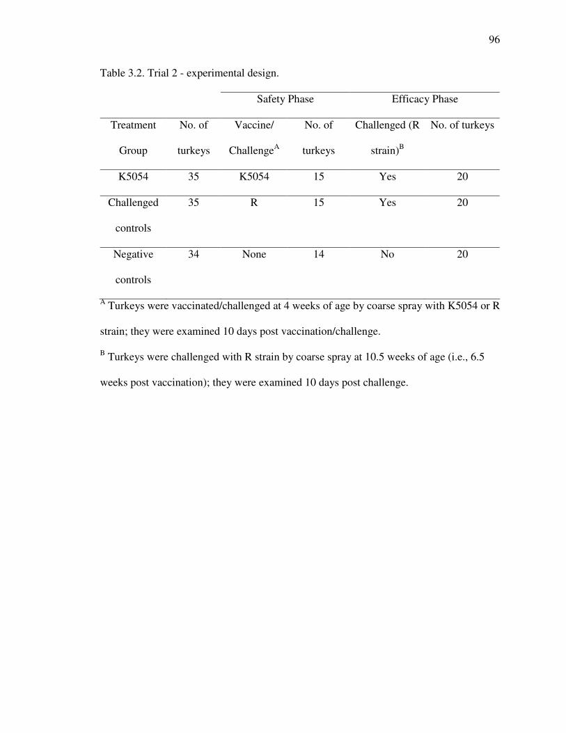
96
Table 3.2. Trial 2 - experimental design.
Safety Phase Efficacy Phase
Treatment
Group
No. of
turkeys
Vaccine/
ChallengeA
No. of
turkeys
Challenged (R
strain)B
No. of turkeys
K5054 35 K5054 15 Yes 20
Challenged
controls
35 R 15 Yes 20
Negative
controls
34 None 14 No 20
A Turkeys were vaccinated/challenged at 4 weeks of age by coarse spray with K5054 or R
strain; they were examined 10 days post vaccination/challenge.
B Turkeys were challenged with R strain by coarse spray at 10.5 weeks of age (i.e., 6.5
weeks post vaccination); they were examined 10 days post challenge.

97
Table 3.3.Trial 1 (Safety). Serological responses of chickens after challenge with ts-11,
K5054 or the R strain at 3 weeks of age A.
Challenge
strain
Days post
challenge SPA HI ELISA
ts-11 10 2/16Ba (0.1)C 0/16a (0.0)D 0/16a (0.8 + 0.0)E
K5054 10 2/16a (0.1) 0/16 a (0.0) 1/16 a (0.3 + 5.1)
R 10 15/15 b (3.0) 5/15 b (20.6) 11/15 b (0.9 + 0.7)
Neg. Controls 10 0/16a (0.0) 0/16 a (0.0) 0/16 a (0.4 + 0.0)
ts-11 35 8/10 a (2.4) 1/10 a (11.8) 7/10 a (0.9 + 0.9)
K5054 35 10/10 a (3.0) 3/10 a (24.6) 3/10 a,b (0.3 + 0.2)
R 35 N/A N/A N/A
Neg. Controls 35 0/5b (0.0) 0/5 a (0.0) 0/5b (0.2 + 0.0)
A Values within a column with a different lower case superscript are significantly
different (P< 0.05)
B No. of positive samples/No. of tested samples (SPA: >1, HI: >40, and ELISA: > 0.6)
C Mean agglutination grade (from 0 to 4).
D Geometric mean titer
E Geometric mean S/P ratio + standard deviation
N/A - not applicable

98
Table 3.4. Trial 2 (Safety). Serological response of turkeys 10 days after challenge with
K5054 or the R strain at 4 weeks of age A.
Challenge strain SPA HI ELISA
K5054 0/15Ba (0.0)C 0/15a (0.0)D 0/15a(0.44 + 0.01)E
R 15/15b (3.1) 0/15a (2.2) 3/15b (0.24 + 0.49)
Neg. Controls 0/14a (0.0) 0/14a (0.0) 0/14a (0.68 + 0.05)
A Values within a column with a different lower case superscript are significantly
different (P< 0.05)
B No. of positive samples/No. of tested samples (SPA: >1, HI: >40, and ELISA: > 0.6)
CMean agglutination grade (0 to 4).
D Geometric mean titer
E Geometric mean S/P ratio + standard deviation

99
Table 3.5. Trial 1 (Efficacy). Serological response of chickens 10 days after challenge
with the R strain of MG at 8 weeks of age A.
Group Challenged SPA HI ELISA
Yes 38/38Ba (4.0)C 37/38a (43.8)D 38/38a(4.6 + 1.1)E
ts-11 No 9/9a (3.8) 0/9b (20.0) 8/9a (1.3 + 0.8)
Yes 39/39a (4.0) 32/39c (37.3) 39/39a (2.9 + 1.1) K5054
No 10/10a (4.0) 1/10b (15.9) 10/10a (1.3 + 0.4)
Yes 40/40a (4.0) 33/38a,c (36.5) 40/40a (3.4 + 1.0) No
Vaccine No 0/32a (0.0) 0/32b (0.0) 0/32b (0.1 + 0.1)
A Values within a column with a different lower case superscript are significantly
different (P< 0.05)
B No. of positive samples/No. of tested samples (SPA: >1, HI: >40, and ELISA: > 0.6)
C Mean agglutination grade (0 to 4)
D Geometric mean titer.
E Geometric mean S/P ratio + standard deviation

100
Table 3.6. Trial 2 (Efficacy). Serological response of turkeys 10 days after challenge with
the R strain at 6 weeks post vaccination A.
Group Challenged SPA HI ELISA
K5054 Yes 18/18Ba(1.3)C 1/18a (2.4)D 17/18a(1.6+0.7)E
No Vaccine Yes 20/20a (2.0) 18/20b (47.0) 20/20a (1.3 + 0.4)
No Vaccine No 0/20b (0.0) 0/20a (0.0) 0/20b (0.1 + 0.1)
A Values within a column with a different lower case superscript are significantly
different (P< 0.05)
B No. of positive samples/No. of tested samples (SPA: >1, HI: >40, and ELISA: > 0.6)
C Mean agglutination grade (0 to 4)
DGeometric mean titer.
E Geometric mean S/P ratio + standard deviation

101
Table 3.7. Trial 1 (Safety). Lesion scores and MG isolation from chickens 10 days after
challenge with ts-11, K5054 or the R strain at 3 weeks of age A.
MG isolationD
Challenge strain Air sac lesions B Tracheal mucosal
thicknessC Trachea Air sacs
ts-11 0/16E (0.0 + 0.0)Fa 6.38 + 1.77a 1/16 Ea 0/16 Ea
K5054 0/16 (0.0 + 0.0)a 5.94 + 1.77a 16/16b 6/16b
R 12/15 (1.8 + 1.15)b 42.75 + 1.83b 15/15b 15/15c
Neg. Controls 0/16 (0.0 + 0.0)a 6.23 + 1.77a 0/16a 0/16a
A Values within a column with a different lower case superscript are significantly
different (P< 0.05)
B Macroscopically scored from 0 to 4
C Mean thickness for the group in units. 1 unit = 0.0083mm
D MG isolates recovered from groups challenged with R strain, ts-11 or K5054 were
identified as R strain, ts-11 or K5054 respectively by RAPD analysis.
E No. of positive samples/No. of tested samples (Air sac score > 1)
FMean score + SD

102
Table 3.8. Trial 2 (Safety). Air sac and tracheal lesion evaluation and MG isolation from
turkeys 10 days post challenge with K5054 or the R strain at 4 weeks of age A.
MG isolationD Challenge
strain Air sac lesions B
Tracheal mucosal
thicknessC Trachea Air sacs
K5054 2/15 E (0.2 + 0.7)Fa 12.77 + 2.52a 15/15 Ea 13/15 Ea
R 13/15 (2.2 + 1.3)b 20.87 + 4.36b 15/15a 15/15a
Neg. Controls 0/14 (0.0 + 0.0)a 8.29 + 1.02a 0/14b 0/14b
A Values within a column with a different lower case superscript are significantly
different (P< 0.05)
B Macroscopically scored from 0 to 4
C Mean thickness for the group in units. 1 unit = 0.0083mm
D MG isolates recovered from groups challenged with R strain or K5054 were identified
as R strain or K5054 respectively by RAPD analysis.
E No. of positive samples/No. of tested samples (Air sac score > 1)
F Mean score + SD
103
Table 3.9. Trial 1 (Efficacy). Mean lesion scores and MG isolation from chickens 10 days
after challenge with the R strain of MG at 6 weeks of age A.
MG isolationD
Group Challenged Air sac lesionsB
Tracheal
mucosal
thicknessC Trachea Air sacs
Yes 17/38 E (0.7 + 1.0)Fa 16.32 + 19.27a 38/38 a 33/38 a ts-11
No 0/9 (0.0 + 0.0)b 7.28 + 1.77a 5/9b 0/9 b
Yes 13/39 (0.4 + 0.5)a 8.88 + 2.91a 39/39a 36/39a K5054
No 0/10 (0.0 + 0.0) b 7.50 + 1.72a 10/10a 0/10 b
Yes 40/40 (2.9+ 0.4)c 57.23 + 22.17b 40/40a 40/40c No
Vaccine No 0/32 (0.0 + 0.0)b 6.94 + 1.42a 0/32c 0/31b
A Values within a column with a different lower case superscript are significantly
different (P< 0.05)
B Macroscopically scored from 0 to 4
B Mean thickness for the group in units + standard deviation. 1 unit = 0.0083mm
C MG isolates from groups challenged with R strain were all identified as R strain by
RAPD analysis; MG isolates from groups vaccinated with ts-11 or K5054 and not
challenged with R strain were identified as ts-11 or K5054 respectively by RAPD
analysis.
D No. of positive samples/No. of tested samples (Air sac score > 1)
EMean score + SD
a,b.c Values within a column with a different lower case superscript are significantly
different (P< 0.05)

104
Table 3.10. Trial 2 (Efficacy). Mean lesion scores and MG isolation from turkeys 10 days
after challenge with the R strain at 10.5 weeks of age A.
MG isolationD
Group Challenged Air sac lesionsB
Tracheal
mucosal
thicknessC Trachea Air sacs
K5054 Yes 12/18E (1.4 + 1.3)Fa 13.90 + 2.10a 14/18Ea 11/18Ea
Challenge
d Controls Yes 20/20 (3.8 + 0.4)b 22.06 + 3.46b 20/20b 20/20b
Negative
Controls No 0/20 (0.0 + 0.0)a 13.55 + 2.00a 0/20c 0/20c
A Values within a column with a different lower case superscript are significantly
different (P< 0.05
B Macroscopically scored from 0 to 4
C Mean thickness for the group in units. 1 unit = 0.0083mm
D MG isolates from groups challenged with R strain were all identified as R strain by
RAPD analysis.
E No. of positive samples/No. of tested samples (Air sac score > 1)
FMean score + SD

105
CHAPTER 4
FURTHER STUDIES OF AN AVIRULENT MYCOPLASMA
GALLISEPTICUM STRAIN AS A LIVE VACCINE IN POULTRY3
__________________
3 N.M. Ferguson, V.A. Leiting and S.H. Kleven. To be submitted to Avian Diseases.

106
Key words: Mycoplasma gallisepticum, K5054, vaccine
Abbreviations: ELISA = enzyme-linked immunosorbent assay; HI = hemagglutination
inhibition; MG = Mycoplasma gallisepticum; RAPD = random amplified polymorphic
DNA; SPA = serum plate agglutination

107
Summary
A Mycoplasma gallisepticum (MG) isolate (K5054) showed promise as an
efficacious and safe live vaccine in poultry in previous studies. In this study, K5054 was
further characterized in three trials. In Trial 1, the stability of K5054 was evaluated by in
vivo passages through chickens. An isolate from the tenth chicken passage (K5383-2)
was used to challenge chickens. The virulence of the chicken-passaged isolate was not
significantly increased when compared to the parental K5054 isolate. In Trial 2, the
persistence and the duration of immunity elicited by a single vaccination with K5054
were evaluated. Chickens vaccinated with K5054 at 3 weeks of age were evaluated over a
seven-month period with 5 periodic samplings for serology and tracheal culture for
mycoplasma. Ten of the vaccinated chickens were removed at each sampling period and
challenged with virulent R strain. K5054 persisted in the trachea and the vaccinated
chickens were protected from disease for the duration of the experiment. In Trial 3, the
transmissibility of K5054 to unvaccinated chickens was evaluated. Seeders were
inoculated with K5054 via eye-drop and placed so that there were groups of chickens in
direct contact (within the same pen) and indirect contact (adjacent or with an empty pen
separating). R strain was used as a control in a similar experimental design. Tracheal
culture and serological monitoring of chickens from each group of contacts evaluated the
transmissibility of K5054 and R strain. K5054 did not transmit to in contact chickens.

108
Introduction
Mycoplasma gallisepticum (MG) is a significant pathogen that affects poultry
worldwide. The control of MG is generally by isolation and maintenance of breeding
stock free of MG. Live MG vaccines have been used to control MG in areas where
isolation of poultry flocks and eradication is not feasible (15, 21, 31). Eradication of MG
is preferable to vaccination wherever possible, and the use of live vaccines to displace
virulent wild-type MG strains from commercial poultry flocks may be a useful part of an
eradication program (17, 20, 30).
Live vaccines that are currently used worldwide to control MG include F strain
(Schering Plough, Kenilwoth, N.J.) (26), 6/85 (Intervet America, Millsboro, Del.) (5) and
ts-11 (Bioproperties, Inc., Australia, marketed in the US by Merial Select Laboratories,
Gainsville, GA.) (32).
An MG isolate, designated K5054, from an atypically mild outbreak in
commercial turkey breeders has shown promise as an efficacious and safe live vaccine in
poultry in previous studies (8, 9).
The important characteristics of an ideal live MG vaccine, aside from safety in the
target animals and efficacy, include the ability to stimulate solid lifelong protection,
preferably from a single dose, and stability following in vivo passages (lack of reversion
of attenuated strains to a virulent form). The vaccine should not spread to neighboring
flocks (31).
The duration of immunity elicited by a live vaccine may be associated with the
colonization and persistence of the vaccine in the respiratory tract. F strain persists at

109
higher levels in the upper respiratory tract than either ts-11 or 6/85, and ts-11 appears to
colonize more effectively than 6/85 (1, 24).
The transmissibility of live MG vaccines is an important characteristic in
determining the likelihood of the strain spreading to unvaccinated neighboring chickens
and turkeys. F strain is readily transmissible to unvaccinated pen mates and chickens in
adjacent pens (14). It can be isolated from farms long after vaccination has ceased and
has been implicated in MG outbreaks in commercial turkeys (22). Although
experimentally F strain has been shown to transmit from bird to bird, the widespread use
of the vaccine has not resulted in widespread isolations of F strain from field cases in
chickens (10).
The ts-11 and 6/85 vaccines have both been shown to be poorly transmissible to
in contact poultry (23, 33).
Although 6/85 has been reported not to persist in the trachea and to be poorly
transmissible, a “6/85-like” isolate was isolated from vaccinated and unvaccinated
contact chickens long after vaccination (29).
In the event that a live vaccine cycles through a flock of poultry it should not
increase in virulence. After years of use there is no evidence that F strain has become
more virulent (31). Experimental passage of 6/85 through chickens and turkeys did not
result in a substantial increase in virulence (5, 6). Attempts to serially passage ts-11 in
birds were unsuccessful but the vaccine appeared to retain its characteristics after three
passages (33).
In this study we conducted three trials to further characterize K5054 with respect
to stability by in vivo passages through chickens (Trial 1); persistence and the duration of

110
immunity elicited by a single vaccination (Trial 2); and transmissibility to unvaccinated
chickens (Trial 3).
Materials and Methods
MG strains and isolates. K5054 was isolated from sinus exudate of a
commercial turkey flock. This flock did not exhibit typical clinical signs of MG infection
(8). R strain is a well-characterized virulent MG strain (28).
Serology. Sera were analyzed for MG antibodies using the serum plate
agglutination (SPA) test using commercial antigen (Intervet America, Millsboro, Del),
and the hemagglutination-inhibition (HI) test with antigen prepared from A5969 strain
and chicken erythrocytes.
The SPA and HI test were carried out according to procedures described by
Kleven (16). An SPA score > 1 was considered positive. An HI titer of 1:20 was
considered suspect and > 1:40 was considered positive.
Commercial enzyme-linked immunosorbent assays (ELISA) were also performed
on the sera (IDEXX, Westbrook, Maine).
Isolation and identification of mycoplasma. Cotton swabs from trachea and air
sacs were used for culture. They were inoculated in Frey’s modified broth and agar and
incubated at 37 C. Mycoplasma isolates were identified using direct immunofluorescence
(16).
Polymerase chain reaction (PCR). MG PCR was performed on pooled samples
of tracheal swabs in Trial 3 at 12 weeks post vaccination/challenge. The PCR was carried
out with modified primers and procedure described by Lauerman et al. (8, 19)

111
Random amplified polymorphic DNA (RAPD) analysis. MG isolates were
characterized by RAPD analysis. The procedure and primers used were described by Fan
et al.(7). A minimum of 20% of isolates recovered from vaccinated and challenged
chickens were typed by this method.
DNA sequence analysis. DNA sequences of chicken-passaged isolates in Trial 1
and reference strains were compared as previously described (8). The sequences of the
pvpA gene (GenBank accession number AF224059) (3) as described by Liu et al. (25)
using a polymerase chain reaction with primers 3R and 4F (given as pvpA 1F and pvpA
2R). DNA sequences from a PCR product from a lipoprotein sequence (LP) described by
Nascimento et al. (27), GenBank accession number AF075588, using primers described
(8) were also compared. Sequences from the mgc1 gene (13), GenBank accession number
U34842, using described primers (8) were compared. This gene is analogous to a gene
known as gapA (12). Sequence analysis was performed with MegAlign (DNASTAR,
Lasergene, Inc. Madison, Wisconsin).
Evaluation of lesions. The lesions in chickens necropsied during the study were
evaluated grossly by air sac lesions scoring on a scale from 0 to 4 (18). The tracheal
lesions were evaluated microscopically by measuring the width of the tracheal mucosa. A
section was collected from the upper third of the trachea (approximately 1 inch distal
from the larynx) and fixed in 10% neutral formalin. The tracheal mucosa thickness was
measured at four equidistant points on histological slides of cross sections of tracheas
(31).
Chickens and Experimental Design. Trial 1. In the first part of Trial 1, fifteen
layer-type male chickens (Hy-line, West Des Moines, Iowa) were obtained from a

112
commercial hatchery at one day of age. One week later and every week for 9 weeks,
fifteen more chickens were obtained from the same hatchery. They were housed in
Horsfal isolator units under positive pressure. The fifteen oldest chickens were inoculated
with K5054 (8.4 x 10⁸ color changing units (CCU)/ml) via eye drop at three weeks of
age. One week later tracheal swabs from these chickens were used to inoculate 15 naive
chickens (3 weeks of age) via the intratracheal route. This process was repeated until the
isolate was passaged 10 times at 7-day intervals through chickens.
In the second part of this trial, one hundred and twenty layer-type male chickens
were obtained from the hatchery at one day of age. These chickens were divided into 4
treatment groups and housed in four 3x3-m2 colony houses with pine shaving litter. An
isolate from the 10th in vivo passage (K5383-2; 4.0 x10⁷ CCU/ml) was used to challenge
a group of 30 naïve chickens. Two other groups of 30 chickens were challenged with
K5054 (2.3 x10⁸ CCU/ml) or R strain (6.2 x10⁸ CCU/ml). These challenges were
administered by coarse spray (18) using a commercial paint sprayer (Preval� Sprayer
Division, Precision Valve Corporation, Yonkers, NY); approximately 1ml of culture was
administered per bird. The response to challenge was compared to that of 30
unchallenged negative controls. Ten days post challenge the chickens were bled for
serology; swabs were taken of the trachea and air sacs for culture, the air sac lesions were
scored and a section of trachea taken for histological examination.
Trial 2. One hundred and eighty layer-type male chickens (Hy-line, West Des
Moines, Iowa) were acquired from a commercial hatchery at one day of age. Ninety of
these chickens were housed in two floor pens (1.5x3 m2) with pine shaving litter. The
remaining ninety chickens were divided in 3 groups of thirty chickens and housed in

113
three 3x3-m2 colony houses with pine shaving litter. The ninety chickens in the colony
houses were vaccinated with K5054 (8.42 x 10⁸ CCU/ml) via coarse spray at 3 weeks of
age. At 4, 8 12, 16 and 28 weeks post vaccination 10 vaccinated birds from the colony
houses and 10 unvaccinated birds from the floor pens (positive controls) were moved into
two different colony houses (3x3-m2) for challenge with virulent R strain. Five
unvaccinated and unchallenged chickens served as negative controls. The titers of the R
strain used in each of the challenges were 6.2 x 10⁸ CCU/ml, 1.1 x 10⁹ CCU/ml, 1.2 x 10⁹
CCU/ml, 1.0 x 10⁸ CCU/ml and 4.7 x 10⁸ CCU/ml, respectively. Ten days post challenge
the chickens were evaluated as in Trial 1. At 4, 8, 12, 16 and 28 weeks post vaccination
10 vaccinated (unchallenged) chickens were also bled for serology and cultured for
mycoplasma. At the end of the experiment remaining chickens were euthanized.
Trial 3. One hundred and eighty layer-type female chickens (Hy-line, West Des
Moines, Iowa) were acquired from a commercial hatchery at one day of age. These birds
were housed in two floor pens (1.5x3 m2) with pine shaving litter until at approximately 5
weeks of age they were divided into 6 treatment groups of 30 and moved to 6 different
floor pens (1.5x3 m2) with pine shaving litter.
Five chickens in one group were inoculated with K5054 (4.7 x 10⁸ CCU/ml) via
eye drop. These chickens (seeders) were placed in a pen with 25 naïve chickens (direct
contacts), 30 chickens were placed in a pen immediately adjacent to the seeders and
direct contacts (across-wire contacts). Another group of 30 chickens were placed in a pen
separated from the direct contacts and seeders by an empty pen (across-empty-pen
contacts). Five chickens were also inoculated with R strain (2.4 x 10⁸ CCU/ml) and
placed with direct contacts, across-wire contacts and across-empty-pen contacts in a

114
similar configuration. Five chickens from each of the 6 groups were bled for serology and
tracheal swabs were obtained for mycoplasma culture at 1, 2, 4 and 8 weeks post
inoculation of the seeders. At 12 weeks post inoculation all of the birds were bled,
cultured and subsequently euthanized.
Chickens that died during the course of the trials were examined for gross lesions
associated with MG.
The chickens in these trials were euthanized by cervical dislocation or with
carbon dioxide according to the animal care and use policies of The University of
Georgia.
Statistical analysis. Air sac lesion scores were analyzed using the Kruskal-Wallis
Rank Sums test. The mean tracheal mucosa thickness was analyzed using the Tukey-
Kramer HSD test. JMP® Statistics Made Visual (SAS Institute Inc., SAS Campus Drive,
Cary, NC 27513). A P value < 0.05 was considered significant.
Results
Trial 1. The results of this experiment are summarized in Tables 4.1 and 4.2. The
air sac lesion scores and tracheal mucosa measurements of the K5383-2 (bird-passaged)
group were not significantly different from the K5054 or negative control groups (P<
0.05). The mean agglutination grade on the SPA of the K5383-2 group was higher than
that of the K5054 group. There was also a higher proportion of MG isolations from the
air sacs of the group challenged with K5383-2. The chicken-passaged isolates form the
first, fifth and tenth passages had RAPD patterns that were identical to the K5054 strain.

115
DNA sequence analysis also showed that the DNA sequence of pvpa, mgc1 and LP were
100% similar to K5054.
Trial 2. The results of this trial are summarized in Tables 4.3, 4.4 and 4.5. The
K5054 strain persisted in the tracheas of vaccinated birds; it was isolated from tracheal
swabs up to 28 weeks post-vaccination (Table 4.3). The proportion of tracheal samples
from which MG was isolated declined to 6/9 (67%) at 16 weeks post vaccination and
5/10 (50%) at 28 weeks post vaccination. MG antibodies could be detected in the
vaccinated chickens by SPA and ELISA up to 28 weeks post vaccination.
The serological responses of chickens vaccinated with K5054 and challenged
with R strain at 4, 8 12, 16 or 28 weeks postvaccination are summarized in Table 4.4. The
chickens vaccinated with K5054 and challenged with R strain did not have lesion scores
that were significantly different from the negative controls in any of the five challenges
during the trial (P< 0.05) (Table 4.5).
Trial 3. The results of this trial are summarized in Tables 4.6 and 4.7. The R strain
spread to the direct contacts and could be detected by culture 2 weeks post challenge. At
12 weeks postchallenge R strain was isolated from all of the seeders and 23 of 25 (92%)
penmates. This strain did not spread to chickens separated from the seeders by a wire
fence or an empty pen.
K5054 failed to transmit to any of the in contact groups for the duration of the
experiment but was reisolated from all of the seeders at 12 weeks post vaccination.
116
Discussion
Previous studies have shown that K5054 is a safe and efficacious vaccine in layer-
type chickens and turkeys. To be an effective live MG vaccine K5054 must also have a
low rate of transmission to unvaccinated poultry, be stable and consistently avirulent, and
provide long lasting immunity.
K5054 did not transmit to in contact chickens in Trial 3 although it was re-
isolated from all of the vaccinated birds (seeders). The SPA tests that were weakly
positive at 8 weeks postvaccination were likely due to non-specific reactions (2, 4, 11).
These positives were unconfirmed by the HI or ELISA tests and disappeared in the
subsequent samplings.
R strain was transmitted to many of the direct contacts (92%) although it did not
spread to the indirect contacts. In our experience with R strain, it sometimes spreads very
well to contacts and sometimes it does not. The conditions of this study may not have
been ideal for the transmission of MG. The transmission of MG strains may be variable
and unpredictable from study to study. Whithear et. al., was unable to show transmission
of ts-11 to contacts (33) although in a different study Ley et. al., showed transmission of
ts-11 to 0-40% of commingled pullets (23).
It must be noted that K5054 did spread, albeit very slowly, among the commercial
turkeys from which it was originally isolated (8), although it did not spread
experimentally between the chickens in this study or the turkeys in a previous study (8).
A greater challenge to the seeders or a higher proportion of seeders may eventually allow
the transmission of K5054 to contacts. It can be concluded however, that K5054 has a
relatively low rate of transmission.

117
K5054 did not increase in virulence after ten passages in chickens and was also
genetically stable; there was no change in the RAPD pattern or DNA sequence of
selected genes.
We believe that a naturally occurring MG strain of low virulence, such as K5054,
may have the advantage of increased stability over many in vivo passages as compared to
laboratory attenuated strains. F strain is likely to have originated from a naturally
occurring strain of moderate virulence and there is no published evidence of an increase
in virulence although it has been used for many years. However, the possibility of
“escape” and some increase in the level of virulence cannot be ignored with any live
mycoplasma vaccine in poultry production situations. Diligence should always be applied
when using a live vaccine; which can spread, multiply and change its characteristics
when given the opportunity, however small the possibility.
A vaccine strain should persist and multiply in the trachea long enough to elicit a
protective immune response (31). The duration of immunity elicited may be associated
with the colonization and persistence of the vaccine in the respiratory tract. In this study
K5054 persisted in the upper respiratory tract and elicited protective immunity for the
duration of the study (7 months). With respect to persistence K5054 seems to be
comparable to F strain and ts-11.
To conclude K5054 appears to be not only a safe and efficacious vaccine but also
to be stable following ten in vivo passages, it has a low rate of transmission, the vaccine
persists in the upper respiratory tract for long periods, and a single vaccination results in
long lasting immunity. Further study is needed to evaluate the amenability of this strain
to commercial production and application.
118
References
1. Abd-El-Motelib, T. Y., and S. H. Kleven. A comparative study of Mycoplasma
gallisepticum vaccines in young chickens. Avian Dis. 37:981-987. 1993.
2. Adler, H. E., and A. D. Wiggins. Interpretation of serologic tests for Mycoplasma
gallisepticum. World Poult Sci J. 29:345-353. 1973.
3. Boguslavsky, S., D. Menaker, I. Lysnyansky, T. Liu, S. Levisohn, R. Rosengarten, M.
Garcia, and D. Yogev. Molecular characterization of the Mycoplasma gallisepticum
pvpA gene which encodes a putative variable cytadhesin protein. Infect Immun. 68:3956-
3964. 2000.
4. Boyer, C. I., J. Fabricant, and J. A. Brown. Non-specific plate agglutination reactions
with PPLO antigen. Avian Dis. 4:546-547. 1960.
5. Evans, R. D., and Y. S. Hafez. Evaluation of a Mycoplasma gallisepticum strain
exhibiting reduced virulence for prevention and control of poultry mycoplasmosis. Avian
Dis. 36:197-201. 1992.

119
6. Evans, R. D., Y. S. Hafez, and C. S. Schreurs. Demonstration of the genetic stability of
a Mycoplasma gallisepticum strain following in vivo passage. Avian Diseases. 36:554-
560. 1992.
7. Fan, H. H., S. H. Kleven, and M. W. Jackwood. Application of polymerase chain
reaction with arbitrary primers to strain identification of Mycoplasma gallisepticum.
Avian Dis. 39:729-735. 1995.
8. Ferguson, N. M., D. Hermes, V. A. Leiting, and S. H. Kleven. Characterization of a
Naturally Occurring Infection of a Mycoplasma gallisepticum House Finch-Like Strain In
Turkey Breeders. Avian Dis. In press:2003.
9. Ferguson, N. M., V. A. Leiting, and S. H. Kleven. Saftey and Efficacy of an Avirulent
Mycoplasma gallisepticum Strain as a Live vaccine in Poultry. Avian Dis.
Submitted:2003.
10. Gibbs, P. S., S. H. Kleven, and M. W. Jackwood. Analysis and characterization of
Mycoplasma gallisepticum isolates from Pennsylvania. Avian Dis. 38:475-482. 1994.

120
11. Glisson, J. R., J. F. Dawe, and S. H. Kleven. The effect of oil emulsion vaccines on
the occurrence on nonspecific plate agglutination reactions for Mycoplasma
gallisepticum and Mycoplasma synoviae. Avian Dis. 28:397-405. 1984.
12. Goh, M. S., T. S. Gorton, M. H. Forsyth, K. E. Troy, and S. J. Geary. Molecular and
biochemical analysis of a 105 kDa Mycoplasma gallisepticum cytadhesin (GapA).
Microbiol. 144:2971-2978. 1998.
13. Keeler Jr., C. L., L. L. Hnatow, P. L. Whetzel, and J. E. Dohms. Cloning and
characterization of a putative cytadhesin gene (mgc1) from Mycoplasma gallisepticum.
Infect. Immun. 64:1541-1547. 1996.
14. Kleven, S. H. Transmissibility of the F-strain of Mycoplasma gallisepticum in
leghorn chickens. Avian Dis. 25:1005-1018. 1981.
15. Kleven, S. H. Changing expectations in the control of Mycoplasma gallisepticum.
Acta Vet. Hung. 45:299-305. 1997.
16. Kleven, S. H. Mycoplasmosis. In: A Laboratory Manual for the Isolation and
Identification of Avian Pathogens, Fourth ed. D. E. Swayne, J. R. Glisson, M. W.

121
Jackwood, J. E. Pearson, and W. M. Reed, eds. American Association of Avian
Pathologists, Kennett Square, PA. pp. 74-80. 1998.
17. Kleven, S. H., H.-H. Fan, and K. S. Turner. Pen trial studies on the use of live
vaccines to displace virulent Mycoplasma gallisepticum in chickens. Avian Dis. 42:300-
306. 1998.
18. Kleven, S. H., D. D. King, and D. P. Anderson. Airsacculitis in broilers from
Mycoplasma synoviae: effect on air-sac lesions of vaccinating with infectious bronchitis
and Newcastle virus. Avian Dis. 16:915-924. 1972.
19. Lauerman, L. H. Mycoplasma PCR Assays. In: Nucleic Amplification Assays for
Diagnosis of Animal Diseases, ed. L. H. Lauerman, eds. American Association of
Veterinary Laboratory Diagnosticians, Auburn, AL. pp. 41-52. 1998.
20. Levisohn, S., and S. H. Kleven. Vaccination of chickens with nonpathogenic
Mycoplasma gallisepticum as a means for displacement of pathogenic strains. Isr J Med
Sci. 17:669-669. 1981.
21. Levisohn, S., and S. H. Kleven. Avian mycoplasmosis (Mycoplasma gallisepticum).
Rev. sci. tech. Off. int. Epiz. 19:425-442. 2000.

122
22. Ley, D. H., A. P. Avakian, and J. E. Berkhoff. Clinical Mycoplasma gallisepticum
infection in multiplier breeder and meat turkeys caused by F Strain: Identification by
sodium dodecyl sulfate-polyacrylamide gel electrophoresis, restriction endonuclease
analysis, and the polymerase chain reaction. Avian Dis. 37:854-862. 1993.
23. Ley, D. H., J. M. McLaren, A. M. Miles, H. J. Barnes, S. Heins Miller, and G. Franz.
Transmissibility of live Mycoplasma gallisepticum vaccine strains ts-11 and 6/85 from
vaccinated layer pullets to sentinel poultry. Proceedings of the World Veterinary Poultry
Association. 11:90. 1997.
24. Ley, D. H., J. M. McLaren, A. M. Miles, H. J. Barnes, S. H. Miller, and G. Franz.
Transmissibility of live Mycoplasma gallisepticum vaccine strains ts-11 and 6/85 from
vaccinated layer pullets to sentinel poultry. Avian Dis. 41:187-194. 1997.
25. Liu, T., M. Garcia, S. Levisohn, D. Yogev, and S. H. Kleven. Molecular Variability
of the Adhesin-Encoding Gene pvpA among Mycoplasma gallisepticum Strains and Its
Application in Diagnosis. J. Clin. Microbiol. 39:1882-1888. 2001.
26. Luginbuhl, R. E., M. E. Tourtellotte, and M. N. Frazier. Mycoplasma gallisepticum -
Control by immunization. Ann. N. Y. Acad. Sci. 143:234-238. 1967.

123
27. Nascimento, E. R., R. Yamamoto, K. R. Herrick, and R. C. Tait. Polymerase chain
reaction for detection of Mycoplasma gallisepticum. Avian Dis. 35:62-69. 1991.
28. Rodriguez, R., and S. H. Kleven. Pathogenicity of two strains of Mycoplasma
gallisepticum in broiler chickens. Avian Dis. 24:800-807. 1980.
29. Steinlage, S. J. T., N. Ferguson, J. E. Sander, M. Garica, S. Subramanian, V. A.
Leiting, and S. H. Kleven. Isolation and Characterization of a 6/85-like Mycoplasma
gallisepticum from Commercial Laying Hens. Avian Dis. In press:2003.
30. Turner, K. S., and S. H. Kleven. Eradication of live F strain Mycoplasma
gallisepticum vaccine using live ts-11 on a multiage commercial layer farm. Avian Dis.
42:404-407. 1998.
31. Whithear, K. G. Control of avian mycoplasmoses by vaccination. Rev. sci. tech. Off.
int. Epiz. 15:1527-1553. 1996.
32. Whithear, K. G., Soeripto, K. E. Harrigan, and E. Ghiocas. Immunogenicity of a
temperature sensitive mutant Mycoplasma gallisepticum vaccine. Aust. Vet. J. 67:168-
174. 1990.

124
33. Whithear, K. G., Soeripto, K. E. Harrigan, and E. Ghiocas. Safety of temperature
sensitive mutant Mycoplasma gallisepticum vaccine. Aust. Vet. J. 67:159-165. 1990.

125
Table 4.1. Trial 1 (Stability). Serological response of chickens 10 days postchallenge
with R strain, K5054 or K5383-2 (isolate from 10th in vivo passage of K5054).
Challenge SPA HI ELISA
R strain 30/30 A (4.0)B 2/30 (4.2) C 26/30 (0.9 + 0.5) D
K5054 0/30 (0.0) 0/30 (0.0) 0/30 (0.1 + 0.0)
K5383-2 27/30 (1.6) 0/30 (0.0) 3/30 (0.1 + 0.3)
None 0/30 (0.0) 0/30 (0.0) 0/30 (0.1 + 0.0)
A No. of positive samples/No. of tested samples (SPA: >1, HI: >40, and ELISA: > 0.6)
B Mean agglutination grade (from 0 to 4).
C Geometric mean titer
D Geometric mean sample/positive ratio + standard deviation

126
Table 4.2. Trial 1 (Stability). Lesion scores and MG isolation from chickens 10 days post
challenge with R strain, K5054 or K5383-2 (isolate from 10th in vivo passage of K5054)
A.
MG isolationD
Challenge Air sac lesion scoreB
Tracheal
mucosal
thicknessC Trachea Air sacs
R strain 27/30E (2.0 + 1.2)Fa 32.27 + 14.30a 30/30a 30/30a
K5054 0/30 (0.0 + 0.0) b 5.88 + 1.04b 29/29a 4/30b
K5383-2 6/30 (0.2 + 0.5) b 6.32 + 2.05 b 30/30a 23/30c
None 0/30 (0.0 + 0.0) b 6.63 + 1.49 b 0/30b 0/30d
A Values within a column with a different lower case superscript are significantly
different (P< 0.05)
B Macroscopically scored from 0 to 4
C Mean thickness for the group in units. 1 unit = 0.0083mm
D MG isolates from group challenged with R strain were identified as R strain by RAPD
analysis; MG isolates from groups challenged with K5054 or K5383-2 were identified as
similar to K5054 by RAPD analysis.
ENo. of positive samples/No. of tested samples (Air sac score > 1)
FMean score + SD

127
Table 4.3. Trial 2 (Persistence). Serological response and MG isolation from the tracheas
of chickens post vaccination with K5054.
Wk post
vaccination SPA HI ELISA MG isolationA
4 9/10B (1.6)C 0/10 (0.0)D 1/10 (0.1 + 0.2)E 10/10
8 10/10 (3.5) 0/10 (3.3) 10/10 (1.6 + 0.4) 9/9
12 10/10 (4.0) 0/10 (0.0) 10/10 (2.3 + 0.8) 8/8
16 10/10 (4.0) 0/10 (0.0) 10/10 (1.3 + 0.6) 6/9
28 10/10 (2.7) 4/10 (26.4) 6/10 (0.8 + 1.0) 5/10
A Recovered MG isolates were all identified as K5054 by RAPD analysis.
BNo. of positive samples/No. of tested samples (SPA: >1, HI: >40, and ELISA: > 0.6)
C Mean agglutination grade (from 0 to 4).
D Geometric mean titer
E Geometric mean sample/positive ratio + standard deviation

128
Table 4.4. Trial 2 (Duration of Immunity). Serological response of chickens vaccinated
with K5054 and 10 days post challenge with R strain.
Wk
PV Group Challenged SPA HI ELISA
K5054 Yes 10/10A (4.0)B 0/10 (1.4)C 5/10 (0.6 + 0.6)D
No Vaccine Yes 10/10 (4.0) 2/10 (8.6) 4/10 (0.4 + 0.3) 4
No Vaccine No 0/5 (0.0) 0/5 (0.0) 0/5 (0.0 + 0.0)
K5054 Yes 10/10 (4.0) 2/10 (6.9) 10/10 (2.7 + 1.1)
No vaccine Yes 10/10 (4.0) 1/10 (2.0) 10/10 (1.0 + 0.5) 8
No vaccine No 0/5 (0.0) 0/5 (0.0) 0/5 (0.1 + 0.0)
K5054 Yes 10/10 (4.0) 1/10 (4.8) 8/10 (1.2 + 0.9)
No vaccine Yes 10/10(3.8) 0/10 (2.5) 3/10 (0.4 + 0.5) 12
No vaccine No 0/5 (0.0) 0/5 (0.0) 0/5 (0.1 + 0.0)
K5054 Yes 10/10 (4.0) 4/10 (6.3) 10/10 (3.9 + 1.6)
No vaccine Yes 10/10 (3.0) 0/10 (0.0) 10/10 (1.0 + 0.3) 16
No vaccine No 0/5 (0.0) 0/5 (0.0) 0/5 (0.2 + 0.0)
K5054 Yes 10/10 (4.0) 10/10 (60.6) 10/10 (2.4 + 1.8)
No vaccine Yes 10/10 (2.4) 8/10 (29.7) 5/10 (0.4 + 0.8) 28
No vaccine No 0/5 (0.0) 0/5 (0.0) 0/5 (0.2 + 0.2)
A No. of positive samples/No. of tested samples (SPA: >1, HI: >40, and ELISA: > 0.6)
B Mean agglutination grade (from 0 to 4).
C Geometric mean titer
D Geometric mean sample/positive ratio + standard deviation

129
Table 4.5. Trial 2 (Duration of Immunity). Lesion scores and MG isolation from K5054-
vaccinated chickens 10 days post challenge with R strain A.
MG isolationD
Wk
PV Group Challenged
Air sac lesion
scoreB
Tracheal
mucosal
thicknessC Trachea
Air
sac
K5054 Yes 4/10E (0.4 + 0.5)Fa 9.45 + 3.21a 10/10a 8/10a
No Vaccine Yes 10/10 (3.2 + 0.8)b 25.44 + 10.67b 10/10a 10/10a 4
No Vaccine No 0/5 (0.0 + 0.0)a 10.35 + 2.23a 0/5b 0/5b
K5054 Yes 1/10 (0.1 + 0.3) a 11.85 + 4.31a 10/10a 5/10a
No vaccine Yes 10/10 (2.3 + 0.8)b 28.53 + 15.49b 10/10a 10/10b 8
No vaccine No 0/5 (0.0 + 0.0)a 10.05 + 1.19a 0/5b 0/5c
K5054 Yes 0/10 (0.0 + 0.0)a 10.70 + 1.38a 10/10 a 10/10 a
No vaccine Yes 10/10(2.5 + 0.7)b 20.22 + 5.45b 8/8 a 10/10 a 12
No vaccine No 0/5 (0.0 + 0.0)a 10.73 + 1.20a 0/5 b 0/5 b
K5054 Yes 6/10 (0.9 + 0.9)a 12.97+ 2.36a 6/6a 7/9a
No vaccine Yes 10/10 (3.0 + 0.0)b 34.82 + 17.04b 10/10a 9/10a 16
No vaccine No 0/5 (0.0 + 0.0)c 12.95 + 1.41a 0/5b 0/5b
K5054 Yes 7/10 (0.8 + 0.6)a 13.83 + 3.14a 9/10a 9/10a
No vaccine Yes 10/10 (3.3 + 0.7)b 36.18 + 16.44b 10/10a 10/10a 28
No vaccine No 0/5 (0.0 + 0.0)c 13.56 + 3.65a 0/10b 0/10b
A Values within a column with a different lower case superscript are significantly
different (P< 0.05)
B Macroscopically scored from 0 to 4

130
C Mean thickness for the group in units. 1 unit = 0.0083mm
D MG isolates recovered from groups challenged with R strain were all identified as R
strain by RAPD analysis.
E No. of positive samples/No. of tested samples (Air sac score > 1)
F Mean score + SD

131
Table 4.6. Trial 3 (Transmissibility). Serological response and MG isolation from the
tracheas of chickens postvaccination with K5054 by eye drop (seeders), direct contact,
indirect contact between neighboring pens (adjacent) or with an empty pen separating.
Weeks
PC
K5054
Contact SPA HI ELISA
MG
isolation
Direct 2/5 A
(0.4)B 0/5 (0.0) C 0/5 (0.1 + 0.0) D 0/5
Adjacent 2/5 (0.4) 0/5 (0.0) 0/5 (0.0 + 0.0) 0/5 1
Empty pen 3/5 (0.6) 0/5 (0.0) 0/5 (0.1 + 0.1) 0/5
Direct 0/5 (0.0) 0/5 (0.0) 0/5 (0.1 + 0.0) 0/5
Adjacent 0/5 (0.0) 0/5 (0.0) 0/5 (0.1 + 0.0) 0/5 2
Empty pen 0/5 (0.0) 0/5 (0.0) 0/5 (0.2 + 0.0) 0/5
Direct 0/5 (0.0) 0/5 (0.0) 0/5 (0.1 + 0.0) 0/5
Adjacent 0/5 (0.0) 0/5 (0.0) 0/5 (0.2 + 0.1) 0/5 4
Empty pen 0/5 (0.0) 0/5 (0.0) 0/5 (0.1 + 0.1) 0/5
Direct 5/5 (1.6) 0/5 (0.0) 0/5 (0.1 + 0.0) 0/5
Adjacent 5/5 (2.2) 0/5 (0.0) 0/5 (0.0 + 0.0) 0/5 8
Empty pen 5/5 (1.2) 0/5 (0.0) 0/5 (0.1 + 0.1) 0/5
Seeders 5/5 (3.4) 0/5 (0.0) 0/5 (0.1 + 0.1) 5/5
Direct 0/25 (0.0) 0/25 (0.0) 0/25 (0.1 + 0.1) 0/25
12
Adjacent 0/30 (0.0) 0/30 (0.0) 2/30 (0.1 + 0.3) 0/30

132
Empty pen 0/26 (0.0) 0/26 (0.0) 0/26 (0.1 + 0.1) 0/26
A No. of positive samples/No. of tested samples (SPA: >1, HI: >40, and ELISA: > 0.6)
B Mean agglutination grade (from 0 to 4).
C Geometric mean titer
D Geometric mean sample/positive ratio + standard deviation

133
Table 4.7. Trial 3 (Transmissibility). Serological response and MG isolation from the
tracheas of chickens post challenge with R strain by eye drop (seeders), direct contact,
indirect contact between neighboring pens (adjacent) or with an empty pen separating.
Wk
PC
R strain
Contact SPA HI ELISA
MG
isolationA
Direct 0/5B (0.0)C 0/5 (0.0)D 0/5 (0.0 + 0.0)E 0/5
Adjacent 1/5 (0.2) 0/5 (0.0) 0/5 (0.0 + 0.1) 0/5 1
Empty pen 0/5 (0.0) 0/5 (0.0) 0/5 (0.1 + 0.0) 0/5
Direct 0/5 (0.0) 0/5 (0.0) 0/5 (0.0 + 0.0) 3/5
Adjacent 0/5 (0.0) 0/5 (0.0) 0/5 (0.1 + 0.0) 0/5 2
Empty pen 0/5 (0.0) 0/5 (0.0) 0/5 (0.1 + 0.0) 0/5
Direct 3/5 (2.2) 0/5 (0.0) 0/5 (0.2 + 0.1) 5/5
Adjacent 0/5 (0.0) 0/5 (0.0) 0/5 (0.2 + 0.0) 0/5 4
Empty pen 0/5 (0.0) 0/5 (0.0) 1/5 (0.1 + 0.2) 0/5
Direct 5/5 (3.6) 0/5 (0.0) 0/5 (0.2 + 0.1) 5/5
Adjacent 4/5 (2.4) 0/5 (0.0) 0/5 (0.1 + 0.0) 0/5 8
Empty pen 4/5 (1.4) 0/5 (0.0) 0/5 (0.1 + 0.1) 0/5
Seeders 5/5 (4.0) 5/5 (69.6) 5/5 (2.1 + 0.6) 5/5
Direct 25/25 (4.0) 17/25 (35.1) 25/25 (1.3 + 0.6) 23/25
Adjacent 0/30 (0.0) 4/30 (2.4) 0/30 (0.0 + 0.1) 0/30 12
Empty pen 0/30 (0.0) 0/30 (0.0) 0/30 (0.1 + 0.1) 0/30
A MG isolates recovered from the seeders or groups in contact with R strain or K5054
seeders were identified as R strain or K5054 respectively by RAPD analysis.

134
B No. of positive samples/No. of tested samples (SPA: >1, HI: > 40, and ELISA: > 0.6)
C Mean agglutination grade (from 0 to 4).
D Geometric mean titer
E Geometric mean sample/positive ratio + standard deviation

135
CHAPTER 5
RANDOM AMPLIFIED POLYMORPHIC DNA (RAPD) ANALYSIS AND DNA
SEQUENCE ANALYSIS OF THREE GENES IN THE MOLECULAR
EPIDEMIOLOGY OF MYCOPLASMA GALLISEPTICUM4
__________________
4 N.M. Ferguson, M. García, S. Levisohn, S.H. Kleven, and N.Ikuta. To be submitted to
Infection and Immunity

136
Key words: Mycoplasma gallisepticum, random amplified polymorphic DNA
Abbreviations: RAPD = random amplified polymorphic DNA; PCR= polymerase chain
reaction

137
Summary
Mycoplasma gallisepticum isolates from the USA, Israel and Australia were
characterized by random amplified polymorphic DNA (RAPD) as well as DNA sequence
analysis of portions of the phase-variable putative adhesin protein (pvpA) gene, the
cytadhesin gapA gene and an uncharacterized lipoprotein (LP) sequence. The results were
compared to reference strains (vaccine and laboratory strains). Sequence analysis of any
one of the genes did not result in definitive identification of isolates. However, by
combining the data from DNA sequencing of all three genes, patterns could be identified
in recognizing related isolates and unique strains. The RAPD analysis and combined
DNA sequence analysis data correlated well. The Australian isolates appeared to be more
similar to the US isolates than were the Israeli isolates. It appeared that the LP and gapA
genes are more highly conserved than pvpA gene.

138
Introduction
Control of Mycoplasma gallisepticum has generally been based on the eradication
of the organism from breeder flocks and the maintenance of mycoplasma-free status in
the breeders and their progeny by biosecurity. Serological monitoring performed
periodically and isolation of M. gallisepticum or DNA based detection methods used to
confirm the presence of M. gallisepticum (16, 19).
One of the options for control of M. gallisepticum in situations where complete
eradication is difficult to attain is live vaccines (13, 30). Three live M. gallisepticum
vaccines are currently used worldwide, they are: F-strain (Schering Plough, Kenilwoth,
N.J.), ts-11 (Bioproperties, Inc., Australia, marketed in the US by Merial select
Laboratories, Gainsville, GA.) and 6/85 (Intervet America, Millsboro, Del.).
In recent years, a reemergence of mycoplasma infection has necessitated a
reevaluation of control strategies for M. gallisepticum and increased the need to
differentiate between vaccine strains and field isolates (13).
In general the process of subtyping microbial isolates into strains is important
epidemiologically for recognizing outbreaks of infection, determining the source of the
infection, recognizing particularly virulent strains of organisms, and monitoring
vaccination programs (25).
Mycoplasma colonies can vary in their surface antigenic phenotype; therefore
mycoplasma strains can differ markedly in their antigen profiles and their potentially
virulence-related surface properties (28).

139
Molecular techniques that have been used to identify M. gallisepticum strains
include restriction fragment length polymorphisms (RFLP) of DNA (12, 14), DNA and
ribosomal RNA gene probes (11, 34), and PCR with strain-specific primers (24).
The most widely used method for differentiating M. gallisepticum strains is
random amplified polymorphic DNA (RAPD) or arbitrarily primed PCR, analysis (3, 5,
6). The RAPD assay was first described by Williams et al. (33) and Welsh and
McClelland (29). RAPD analysis is quick and sensitive and this method has been used to
identify vaccine strains in M. gallisepticum -vaccinated flocks and for epidemiological
studies (10, 15, 17, 18).
However, due to the random nature of the primers and the low-stringency
conditions of the RAPD reaction, this assay requires the use of pure cultures of the target
mycoplasma. Isolation of mycoplasma is expensive, time-consuming, and technically
complicated in cases where nonpathogenic mycoplasma species may overgrow the
virulent mycoplasma. The isolation process itself may favor the growth of one strain
where more than one M. gallisepticum subtype may be present. Furthermore, technical
factors such as target DNA/primer ratio may significantly impact the reproducibility of
RAPD patterns. The amplification process of the RAPD reaction is extremely sensitive to
slight changes in the annealing temperature; this can also lead to variability in the
banding patterns. The use of empirically designed primers, each with its own optimal
reaction conditions and reagents, also makes standardization of the technique difficult.
Ultimately, all molecular genetic methods for distinguishing organism subtypes
are based on differences in the DNA sequence. DNA sequencing would therefore appear
to be an appropriate approach to differentiating subtypes.

140
Progress in the molecular biology of mycoplasmas has resulted in the description
of several surface proteins in virulent mycoplasmas, many of them cytadhesins including
pMGA (22), PvpA (35), MGC1 (7, 9) and MGC2 (8). Adhesion is prerequisite for
colonization and for infection. Loss of adhesion by mutation results in loss of infectivity,
reversion to cytadherence phenotype accompanied by regaining infectivity and virulence
(26).
PvpA is postulated to be one of the accessory membrane proteins involved in
cytadherence of M. gallisepticum. Variation within PvpA could affect the specificity or
affinity of adherence. It is an integral membrane surface protein with a free C terminus
that is subject to spontaneous high-frequency phase variation in expression and exhibits
size variation among strains (2, 35). Deletions within the 3’ end of the pvpA gene results
in size variation of the PvpA polypeptide from 48 to 55 kDa. The deletions were
localized at the proline-rich carboxy-terminal region and within two direct repeat
sequences (2), this domain may be under selective pressure in the host. Several M.
gallisepticum strains differing in their adherence and pathogenicity have varying
deletions and sizes of PvpA. Analysis of pvpA has been used to differentiate between M.
gallisepticum strains (20).
The gapA gene (7), also know as mgc1 (9), is one of three clustered genes. The
two associated genes are mgc2 and mgc3 (also known as crmA). The gapA gene encodes
a 105-kDa protein with homology to the P1 cytadhesin protein of M. pneumoniae.
Immunoblot analysis of various strains has demonstrated intraspecies variation in the size
of the GapA protein (98, 105 and 110 kDa) (7).
141
The LP nucleotide sequence analyzed in this paper was first described by
Nasciemto et al. in a diagnostic PCR for M. gallisepticum (23). The open reading frame is
thought to code for an uncharacterized lipoprotein.
In this study, reference strains and isolates form the USA, Israel and Australia
were analyzed by RAPD analysis as well as by comparison of nucleotide sequence data
of the pvpA and gapA genes as well as LP sequences. The results of RAPD analysis were
compared to that of DNA sequencing.
Materials and Methods
Mycoplasma isolates. Sixty-four M. gallisepticum isolates were analyzed; there
were 52 from the USA, 4 from Australia and 8 from Israel. The isolates from the USA
were obtained from the depository at the Poultry Diagnostic and Research Center
(PDRC) in Athens, GA. These isolates were from 16 different states and ranged from
1975 to 2001. The isolates were from broiler breeders, commercial layers, turkey
breeders, meat-type turkeys, house finches and an American goldfinch. The Israeli
isolates were obtained through the Division of Avian and Aquatic Diseases, Kimron
Veterinary Institute, Bet Dagan 50250, Israel. Most of these isolates are from outbreaks
that had no specific connection. The isolates were from broiler breeders, turkey breeders
and meat-type turkeys. The Australian isolates were acquired courtesy of K.G. Whithear,
the University of Melbourne, Australia. The origins of the isolates are listed in Table 5.1.
The reference strains used included vaccine strains such as F strain (1, 21), 6/85
(4) and ts-11 (31, 32). Laboratory strains such as HF-51 (a house finch isolate) R strain
(27), A5969, and S6 (36) were also included.

142
Random amplified polymorphic DNA (RAPD) analysis. The procedure and
primers used for RAPD analysis were described by Fan et al. (5). RAPD analysis was
also conducted using primers described by Geary et al. (6) and Charlton et al. (3).
Isolates that appeared identical with Fan’s primers were then tested with the primers
described by Geary and Charlton. Isolates were considered identical when major band
differences could not be visualized with any of the three primer sets.
PCR and sequence analysis. DNA base sequences of the pvpA gene (GenBank
accession number AF224059) (2) of the isolates and reference strains were compared as
described by Liu et al. (20) using a polymerase chain reaction with primers 3R and 4F
(given as pvpA 1F and pvpA 2R). DNA sequences from a PCR product from a lipoprotein
gene (LP) described by Nascimento et al. (23), GenBank accession number AF075588,
using primers LP-1F (GGA TCC CAT CTC GAC CAC GAG AAA A) and LP-2R (CTT
TCA ATC AGT GAG TAA CTG ATG A) were also compared. Sequences
corresponding to positions 30-541 were analyzed. Finally, sequences from the mgc1 gene
(9), GenBank accession number U34842, using primers Adhs1-3F (TTC TAG CGC TTT
ARC CCT AAA CCC) and Adhs1-4R (CTT GTG GAA CAG CAA CGT ATT CGC)
were compared. Positions 3823-4154 were compared. This gene is analogous to a gene
known as gapA (7). The amplified products of these PCR’s were sequenced at the
Molecular Genetics Instrumentation Facility (MGIF), University of Georgia. Sequence
analysis was performed with MegAlign (DNASTAR, Lasergene, Inc. Madison,
Wisconsin).

143
RESULTS
RAPD analysis. RAPD analysis using 3 different primer sets resulted in a
relatively high level discrimination between M. gallisepticum strains (Fig 5.1). One
primer set did not always elucidate differences between M. gallisepticum strains; two
isolates may have appeared very similar with one set of primers and definitely different
with another set of primers. We therefore first conducted RAPD’s using Fan’s primers
(5); if there was significant similarity between the patterns generated we then conducted
RAPD’s with primers described by Geary et al. (6) and Charlton et al. (3) to confirm or
dispute the similarity between strains.
RAPD analysis identified 32 different patterns (strains) when the 64 isolates were
analyzed. The patterns were designated A to AH. (Table 5.1).
DNA sequence analysis. These results are summarized and compared to the
RAPD data in Table 5.2. The pvpA gene sequence analysis exhibited the most variability
in nucleotide sequence differences among the strains (66.1 – 100% range of identities).
The LP and gapA sequence analysis resulted in the less differentiation between the strains
(95.1– 100%) and (94.3– 100%) range of identities respectively.
The reference strains S6 and HF-51 are 100% identical to each other with respect
to the LP sequence. The reference strains 6/85, ts-11 and HF-51 are 100% identical to
each other with respect to the gapA sequence.
Analysis of the combined sequence of all three genes allowed further
discrimination between M. gallisepticum strains and correlated better with RAPD
analysis than comparison of single DNA sequence data.

144
Eleven of 52 (21%) of the US field isolates were characterized as very similar to
vaccine strains by RAPD and DNA sequence analysis. The sequence identities ranged
from 98.8 – 100 % for each of the three DNA sequences.
The isolates that had RAPD patterns different from the reference strains (RAPD
types E to AG) ranged in sequence identity to the reference strains from 93.4% to 100%
for each of the 3 sequences analyzed.
Forty-one of 52 (79%) of the US field isolates were characterized as different
from the reference strains. These isolates could be grouped within outbreaks by RAPD
analysis or the combined sequence analysis data. 23 of these 41 isolates (56%) were
similar to HF-51 with respect to the pvpA sequence (98.8 – 99.5% identity). Thirteen of
these isolates also had LP (98.8-100% identity) and gapA (100% identity) sequences that
were similar to HF-51.
There were some isolates that were very similar to one reference strain sequence
and very similar to a different reference strain when a different region was compared. For
example, the isolates that generated RAPD type I were 100% similar to R strain with
respect to pvpA and 100% identical to 6/85, ts-11 and HF-51 with respect to the gapA
sequence.
The isolates from house finches appeared to be closely related to each other with
the exception of K4409. The pvpA gene of this isolate has a relatively large deletion of
170 nucleotides when compared to the other house finch isolates. K4409 however
generates a RAPD pattern (D) indistinguishable from that of the other isolates.
Some isolates generated similar RAPD patterns although the DNA sequence data
showed differences in the nucleotide sequences. These were isolates with RAPD patterns
145
designated A, E and H. Two isolates with RAPD type A have 4 nucleotide differences
from the other isolates in the gapA sequence (T7→C7; A16→C16; C320→G320; and C328
→A328). The A pattern is generated by the F strain vaccine.
Two isolates with the RAPD type E had a single nucleotide substitution in the
gapA sequence (C328 →A328) that was different from the other isolates in this group.
One isolate with the RAPD type H had a single nucleotide substitution in the
pvpA sequence (T375 →A375).
One isolate (K5120) generated a unique RAPD pattern (M) although on DNA
sequencing of all three regions it was 100% identical to the isolates that generated a
different RAPD type (L).
The RAPD types K and L were similar although not indistinguishable from the
6/85 vaccine type (B), and designated “6/85-like”. On sequence analysis these isolates
were more similar to HF-51, ts-11 or F strain (98.0 – 99.8% identity) than 6/85 when the
pvpA and LP sequences were analyzed.
The Israeli isolates were characterized as relatively similar to each other but
different from the reference strains, the US and Australian isolates by both RAPD and
DNA sequence analysis. The isolates could be differentiated into 6 types (AB to AH),
although not easily, by RAPD analysis with multiple primer sets. The sequence analysis
consistently resulted in clustering of these isolates away from the US and Australian
isolates. The sequence data correlated with the RAPD analysis.
The Australian isolates could be differentiated from each other the other isolates
by both RAPD and sequence analysis. The isolates were more similar to the reference

146
strains and US isolates than the Israeli isolates. The sequence data for these isolates did
not result in a cluster of Australian isolates as did the Israeli.
Discussion
The ultimate goal of researchers involved in M. gallisepticum strain
differentiation and epidemiology is a method that will differentiate strains easily, rapidly
and reproducibly from clinical samples.
Methods of strain differentiation must have high differentiation power so that it
can clearly differentiate unrelated strains, as well as demonstrate the relationship of
isolates from individuals infected through the same source. The techniques should also
have a high degree of reproducibility. It is especially important for the construction of
reliable databases containing known strains within a species to which unknown
organisms can be compared.
In this study, it appeared that the LP and gapA sequences are more highly
conserved than pvpA, these results are similar to those of a previous study (20).
The RAPD analysis and combined DNA sequence analysis data correlated well,
although there were some isolates that indistinguishable on RAPD analysis but DNA
sequencing revealed small differences (1 to 4 nucleotide substitutions). There was also an
isolate that could not be differentiated by the DNA sequence data but RAPD analysis.
generated a unique pattern.
Some of the USA field isolates were very similar to vaccine strains (6/85, ts-11
and F strain) with respect to RAPD patterns and nucleotide sequences. The number of
isolates that were similar to vaccine strains may not be a true representation of the M.

147
gallisepticum epidemiology in the US because vaccine-like isolates are more likely to be
selected for further study thereby skewing the sample away from “wild-type” isolates.
Similar to the results from previous studies (17), the isolates from house finches
were closely related to each other and different from the reference strains and other field
isolates with respect to RAPD patterns and DNA sequences. There was one US isolate
(K5054) from turkeys that was also very similar to the house finch strains.
Most of the US isolates were not very similar to any of the reference strains,
however similarities among isolates within and between M. gallisepticum outbreaks
could be identified.
The Australian isolates appeared to be more closely related to the US isolates than
were the Israeli isolates.
RAPD analysis is rapid and sensitive method and although RAPD analysis can
successfully differentiate between M. gallisepticum strains there are disadvantages to the
method that make amplification and sequencing of specific genes an attractive alternative
for differentiation. A sequence database allows the comparison of many seemingly
unrelated isolates. This method would have greater reproducibility and be more easily
standardized.
However, DNA sequencing must be directed at a small region of the bacterial
genome. It is impractical to sequence multiple or large regions of the chromosome. Thus,
in contrast to RAPD analysis, which examines the entire chromosome, DNA sequencing
examines a very small portion of the sites that can potentially vary between strains and
the variability within the selected sequence must be sufficient to differentiate different
strains of a particular species.

148
In this study, analysis of any one sequence did not result in definitive
identification of the isolates. However, when all three sequences were analyzed the
results were more useful and closely approached the discriminatory power of RAPD
analysis.

149
References
1. Adler, H. E., D. A. McMartin, and M. Shifrine. Immunization against Mycoplasma
infections of poultry. American Journal of Veterinary Research. 21:482-485. 1960.
2. Boguslavsky, S., D. Menaker, I. Lysnyansky, T. Liu, S. Levisohn, R. Rosengarten, M.
Garcia, and D. Yogev. Molecular characterization of the Mycoplasma gallisepticum
pvpA gene which encodes a putative variable cytadhesin protein. Infect Immun. 68:3956-
3964. 2000.
3. Charlton, B. R., A. A. Bickford, R. L. Walker, and R. Yamamoto. Complementary
randomly amplified polymorphic DNA (RAPD) analysis patterns and primer sets to
differentiate Mycoplasma gallisepticum strains. J. Vet. Diag. Invest. 11:158-61. 1999.
4. Evans, R. D., and Y. S. Hafez. Evaluation of a Mycoplasma gallisepticum strain
exhibiting reduced virulence for prevention and control of poultry mycoplasmosis. Avian
Dis. 36:197-201. 1992.
5. Fan, H. H., S. H. Kleven, and M. W. Jackwood. Application of polymerase chain
reaction with arbitrary primers to strain identification of Mycoplasma gallisepticum.
Avian Dis. 39:729-735. 1995.

150
6. Geary, S. J., M. H. Forsyth, S. A. Saoud, G. Wang, D. E. Berg, and C. M. Berg.
Mycoplasma gallisepticum strain differentiation by arbitrary primer PCR (RAPD)
fingerprinting. Molec. Cell. Probes. 8:311-316. 1994.
7. Goh, M. S., T. S. Gorton, M. H. Forsyth, K. E. Troy, and S. J. Geary. Molecular and
biochemical analysis of a 105 kDa Mycoplasma gallisepticum cytadhesin (GapA).
Microbiol. 144:2971-2978. 1998.
8. Hnatow, L. L., C. L. Keeler, Jr., L. L. Tessmer, K. Czymmek, and J. E. Dohms.
Characterization of MGC2, a Mycoplasma gallisepticum cytadhesin with homology to
the Mycoplasma pneumoniae 30-kilodalton protein P30 and Mycoplasma genitalium P32.
Infect. Immun. 66:3436-3442. 1998.
9. Keeler Jr., C. L., L. L. Hnatow, P. L. Whetzel, and J. E. Dohms. Cloning and
characterization of a putative cytadhesin gene (mgc1) from Mycoplasma gallisepticum.
Infect. Immun. 64:1541-1547. 1996.
10. Kempf, I. DNA amplification methods for diagnosis and epidemiological
investigations of avian Mycoplasmosis. Acta Vet. Hung. 45:373-386. 1997.
11. Khan, M. I., B. C. Kirkpatrick, and R. Yamamoto. A Mycoplasma gallisepticum
strain-specific DNA probe. Avian Dis. 31:907-909. 1987.

151
12. Khan, M. I., and R. Yamamoto. Differentiation of the vaccine F-strain from other
strains of Mycoplasma gallisepticum by restriction endonuclease analysis. Vet Microbiol.
19:167-174. 1989.
13. Kleven, S. H. Changing expectations in the control of Mycoplasma gallisepticum.
Acta Vet. Hung. 45:299-305. 1997.
14. Kleven, S. H., G. F. Browning, D. M. Bulach, E. Ghiocas, C. J. Morrow, and K. G.
Whithear. Examination of Mycoplasma gallisepticum strains using restriction
endonuclease DNA analysis and DNA-DNA hybridisation. Avian Pathol. 17:559-570.
1988.
15. Levisohn, S., I. Gerchman, J. E. Berkhoff, and D. H. Ley. Molecular tracking of
Mycoplasma gallisepticum infection by random amplification of polymorphic DNA.
Proceedings of the World Veterinary Poultry Association. 11:68. 1997.
16. Levisohn, S., and S. H. Kleven. Avian mycoplasmosis (Mycoplasma gallisepticum).
Rev. sci. tech. Off. int. Epiz. 19:425-442. 2000.
17. Ley, D. H., J. E. Berkhoff, and S. Levisohn. Molecular epidemiological investigations
of Mycoplasma gallisepticum conjunctivitis in songbirds by random amplified
polymorphic DNA analysis. Emerging Inf. Dis. 3:375-380. 1997.

152
18. Ley, D. H., J. E. Berkhoff, J. M. McLaren, and S. Levisohn. Random amplified
polymorphic DNA (RAPD) analyses for molecular epidemiological studies of
Mycoplasma gallisepticum anbd Mycoplasma synoviae. Proceedings of the World
Veterinary Poultry Association. 11:65. 1997.
19. Ley, D. H., and H. W. Yoder Jr. Mycoplasma gallisepticum infection. In: Diseases of
Poultry, 9th ed. B. W. Calnek, H. J. Barnes, C. W. Beard, L. R. McDougald, and Y. M.
Saif, eds. Iowa State University Press, Ames, Iowa. pp. 194-207. 1997.
20. Liu, T., M. Garcia, S. Levisohn, D. Yogev, and S. H. Kleven. Molecular Variability
of the Adhesin-Encoding Gene pvpA among Mycoplasma gallisepticum Strains and Its
Application in Diagnosis. J. Clin. Microbiol. 39:1882-1888. 2001.
21. Luginbuhl, R. E., M. E. Tourtellotte, and M. N. Frazier. Mycoplasma gallisepticum -
Control by immunization. Ann. N. Y. Acad. Sci. 143:234-238. 1967.
22. Markham, P. F., M. D. Glew, K. G. Whithear, and I. D. Walker. Molecular cloning of
a member of the gene family that encodes pMGA, a hemagglutinin of Mycoplasma
gallisepticum. Infect. Immun. 61:903-909. 1993.
23. Nascimento, E. R., R. Yamamoto, K. R. Herrick, and R. C. Tait. Polymerase chain
reaction for detection of Mycoplasma gallisepticum. Avian Dis. 35:62-69. 1991.

153
24. Nascimento, E. R., R. Yamamoto, and M. Khan. Mycoplasma gallisepticum F-
vaccine strain-specific polymerase chain reaction. Avian Dis. 37:203-211. 1993.
25. Olive, D. M., and P. Bean. Principles and applications of methods for DNA-based
typing of microbial organisms. J Clin Microbiol. 37:1661-9. 1999.
26. Razin, S., and E. Jacobs. Mycoplasma adhesion. J. Gen. Microbiol. 138:407-422.
1992.
27. Rodriguez, R., and S. H. Kleven. Pathogenicity of two strains of Mycoplasma
gallisepticum in broiler chickens. Avian Dis. 24:800-807. 1980.
28. Rosengarten, R., and D. Yogev. Variant colony surface antigenic phenotypes within
Mycoplasma strain populations - implications for species identification and strain
standardization. J. Clin. Microbiol. 34:149-158. 1996.
29. Welsh, J., and M. McClelland. Fingerprinting genomes using PCR with arbitrary
primers. Nucleic Acids Res. 18:7213-8. 1990.
30. Whithear, K. G. Control of avian mycoplasmoses by vaccination. Rev. sci. tech. Off.
int. Epiz. 15:1527-1553. 1996.

154
31. Whithear, K. G., Soeripto, K. E. Harrigan, and E. Ghiocas. Immunogenicity of a
temperature sensitive mutant Mycoplasma gallisepticum vaccine. Aust. Vet. J. 67:168-
174. 1990.
32. Whithear, K. G., Soeripto, K. E. Harrigan, and E. Ghiocas. Safety of temperature
sensitive mutant Mycoplasma gallisepticum vaccine. Aust. Vet. J. 67:159-165. 1990.
33. Williams, J. G., A. R. Kubelik, K. J. Livak, J. A. Rafalski, and S. V. Tingey. DNA
polymorphisms amplified by arbitrary primers are useful as genetic markers. Nucleic
Acids Res. 18:6531-5. 1990.
34. Yogev, D., S. Levisohn, S. H. Kleven, D. Halachmi, and S. Razin. Ribosomal RNA
gene probes to detect intraspecies heterogeneity in Mycoplasma gallisepticum and M.
synoviae. Avian Dis. 32:220-231. 1988.
35. Yogev, D., D. Menaker, K. Strutzberg, S. Levisohn, H. Kirchhoff, K.-H. Hinz, and R.
Rosengarten. A surface epitope undergoing high-frequency phase variation is shared by
Mycoplasma gallisepticum and Mycoplasma bovis. Infection and Immunity. 62:4962-
4968. 1994.
36. Zander, D. V. Origin of S6 strain Mycoplasma. Avian Dis. 5:154-156. 1961.

155
Table 5.1. Origin and RAPD type of the isolates analyzed in this study.
Isolate Species State/Country
of Isolation
Year of
Isolation
RAPD type
K5104 Turkey VA, USA 2001 A (F)
K4781A Turkey VA, USA 1999 A
K5058E Turkey VA, USA 2001 A
K3944 Turkey NC, USA 1995 B (6/85)
K4029 Turkey NE, USA 1995 B
K4043 Turkey NE, USA 1995 B
K4421A Turkey MI, USA 1997 B
K4423 Turkey MI, USA 1997 B
K4465 Turkey OH, USA 1997 B
K4688 Chicken NC, USA 1999 C (ts-11)
K5109 Turkey PA, USA 2001 C
K3839 Housefinch MD, USA 1994 D (HF-51)
K4013 Housefinch PA, USA 1995 D
K4094 Housefinch TN, USA 1996 D
K4366 American
Goldfinch
SC, USA 1997 D
K4409 Housefinch TX, USA 1997 D
K4593 Housefinch MD, USA 1998 D
K5054 Turkey IN, USA 2001 D
K4110A Turkey NC, USA 1996 E

156
K4110B Turkey NC, USA 1996 E
K4110F Turkey NC, USA 1996 E
K4158C Turkey MO, USA 1996 E
K4181B Chicken AR, USA 1996 E
K4649A Turkey CO, USA 1998 G
K4649B Turkey CO, USA 1998 G
K4669A Turkey CO, USA 1998 G
K5011 Turkey MD, USA 2000 H
K5027B Turkey IN, USA 2000 H
K5033A Turkey IN, USA 2000 H
K5033F Turkey IN, USA 2000 H
K2101 Chicken CO, USA 1994 I
K4385 Turkey CO, USA 1997 I
K4414A Turkey CO, USA 1997 I
K4902 Turkey CO, USA 2000 I
K4181C Chicken AR, USA 1996 J
K4246 Turkey AR, USA 1996 J
K4280 Chicken MO, USA 1996 J
K4311 Turkey AR, USA 1996 J
K5029B Turkey PA, USA 2000 K
K5037A Turkey PA, USA 2000 K
K5039H Turkey PA, USA 2000 K
K4236 Turkey VA, USA 1996 L

157
K5120 Turkey GA, USA 2001 M
K4657 Chicken GA, USA 1998 N
K4705 Chicken AR, USA 1999 O
K4931 Turkey VA, USA 2000 P
K435 Turkey GA, USA 1973 Q
K503 Chicken GA, USA 1974 S
K703 Chicken MD, USA 1975 T
K730 Chicken GA, USA 1975 U
K3020 Turkey CA, USA 1990 V
K4355 Chicken CA, USA 1996 W
97019 E3 Chicken Australia 1997 X
94043 30-5a Chicken Australia 1994 Y
95002 16a Chicken Australia 1995 Z
96002 6-3a Chicken Australia 1996 AA
99169 2NSb Chicken Australia 1999 AB
BRT-14 Chicken Israel 2000 AC
DSD-6 Turkey Israel 2000 AC
KS-2 Chicken Israel 2001 AD
KSC-3 Chicken Israel 1999 AE
UHP-1 Chicken Israel 1999 AF
YBS-2 Turkey Israel 2000 AF
MK-8 Chicken Israel 2001 AG
OR-2 Chicken Israel 2001 AH

158
Table 5.2. Summary of the RAPD and DNA sequence analysis results.
RAPD
TypeA
No. of
Isolates
pvpAB
(467)C
LP
(490)
gapA
(332)
A (F) 3 F (100%) F (100%) F (98.8% -100%)
B (6/85) 6 6/85 (100%) 6/85 (99.8 - 100%) 6/85 (100%)
C (ts-11) 2 ts-11 (100%) ts-11(100%) ts-11(100%)
D (HF-51) 7 HF-51 (97.9 - 100%) HF-51/S6 (100%) HF-51 (100%)
E 5 HF-51 (99.5%) HF-51/S6 (100%) R (99.4%)
G 3 HF-51 (98.8%) HF-51/S6 (100%) A5969 (99.4%)
H 4 HF-51 (99.3 - 99.5%) HF-51/S6 (99.0%) R (99.7%)
I 4 R (100%) F (98.0%) 6/85/ts-11/HF-51 (100%)
J 4 HF-51 (99.5%) ts-11(100%) 6/85/ts-11/HF-51 (100%)
K 3 HF-51 (98.8%) F (98.0%) 6/85/ts-11/HF-51 (100%)
L 1 HF-51 (99.5%) ts-11(99.8%) 6/85/ts-11/HF-51 (100%)
M 1 HF-51 (99.5%) ts-11(100%) 6/85/ts-11/HF-51 (100%)
N 1 F (98.7%) 6/85 (99.6%) R (99.7%)
O 1 HF-51 (99.0%) HF-51/S6 (99.8%) A5969 (99.4%)
P 1 HF-51 (99.5%) R (99.0%) 6/85/ts-11/HF-51 (100%)
Q 1 R (99.8%) R (100%) A5969 (99.4%)
S 1 F (89.0%) HF-51/S6 (94.3%) F (99.7%)
T 1 F (88.6%) HF-51/S6 (94.1%) F (98.8%)
U 1 F (89.0%) 6/85 (94.7%) F (98.5%)

159
V 1 6/85 (97.5%) R (100%) R (99.7%)
W 1 S6 (100%) HF-51/S6 (100%) S6 (97.6%)
XD 1 F (99.2%) ts-11 (98.6%) 6/85/ts-11/HF-51 (100%)
YD 1 6/85 (99.3%) HF-51/S6 (100%) 6/85/ts-11/HF-51 (100%)
ZD 1 ts-11 & R (98.1%) ts-11 (99.8%) 6/85/ts-11/HF-51 (100%)
AAD 1 ts-11(98.5%) HF-51/S6 (100%) A5969 (99.1%)
ABD 1 ts-11(99.1%) HF-51/S6 (100%) 6/85/ts-11/HF-51 (100%)
ACE 2 F (91.5%) F (98.2%) 6/85/ts-11/HF-51 (96.4%)
ADE 1 F (91.1%) F (98.2%) 6/85/ts-11/HF-51 (96.4%)
AEE 2 F (91.5%) F (98.0%) 6/85/ts-11/HF-51 (96.4%)
AFE 1 F (91.5%) F (98.0%) 6/85/ts-11/HF-51 & F (97.0%)
AGE 1 F (91.5%) F (98.2%) 6/85/ts-11/HF-51 (96.4%)
AHE 1 F (91.5%) F (98.2%) 6/85/ts-11/HF-51 (96.4%)
A Similar to reference strains (F, 6/85, ts-11, HF-51, R strain, A5969 or S6) or unique
patterns (A to AH)
B Reference strain with the highest identity (range of % identities)
C Number of nucleotides compared
D Isolates from Australia
EIsolates from Israel

160
Figure 5.1. Example of RAPD analysis with primers desciribed by Fan et al. Lane 1 =
HF-51; Lane 2 = K4158C; Lane 3 = K4110A; Lane 4 = K4110B; Lane 5 = K4110F; Lane
6 = K4181B; Lane 7 = K5029B; Lane 8 = K5037A; Lane 9 = K5039H; Lane 10 = 6/85;
Lane 11 = ts-11; Lane 12 = R; Lane 13 = F; Lane 14 = negative control; and Lane 15 =
molecular weight marker.
1 2 3 4 5 6 7 8 9 10 11 12 13 14 15

161
CHAPTER 6
DISCUSSION AND CONCLUSIONS
K5054 was isolated from an atypical MG outbreak in commercial turkeys. MG
isolates from poultry that exhibit reduced pathogenicity, transmissibility and
immunogenicity have been described as atypical or variant. Most of these reports have
been in chickens, but there are also reports from turkeys (2, 5, 13, 18). Atypical serology,
very mild or absent clinical signs and lesions, as well as difficulty in making an isolation
by routine culture methods have been reported. In these instances the confirmation of
suspicious cases is very difficult.
In the case of K5054, routine monitoring for MG according to the NPIP
serological surveillance program did not detect the MG infection in the hen flock until
late in the outbreak, although the ELISA test showed MG titers in banked sera.
The K5054 isolate was characterized as similar to the house finch strains by
multiple analyses. Close examination of RAPD patterns using primer sets described by
Fan et al. (3) and Geary et al. (4), revealed that the patterns of K5054 and house finch
isolates are not completely identical but very similar. Previous studies have established
that the finch MG strain was clearly different from commercial poultry strains, vaccines
and lab strains (9).

162
Wild house finches nest and feed around buildings and farms, so that they may be
in contact with domestic poultry. The house finch strain has been shown not to be host-
specific to the finch although transmission may be slow. There is growing evidence that
songbird species other than house finches are susceptible to MG infection and disease (6,
7, 12). MG from house finches can also be transmitted from affected finches to chickens
by natural (contact) methods but transmit to domestic poultry quite slowly and with little
pathogenic effect (14, 15).
From the bioassay in Chapter 2, it appeared that infection with the K5054 strain
results in seroconversion with very mild clinical disease; confirmation of the infection by
isolation of MG may be difficult. The infection appears to spread very slowly between
turkeys. This was mirrored in the clinical picture of K5054 in the commercial turkeys.
This may be the first recorded incident of a naturally occurring infection of a
house finch-like MG strain in commercial poultry.
Inoculation of turkeys with K5054 sinus exudate in Chapter 2 resulted in
seroconversion with the appearance of very mild clinical signs 42 days after the
challenge. This suggests that K5054 is mildly pathogenic in turkeys, although the
inoculum dose in the bioassay may have been very low. PCR was unable to detect MG in
the sinus exudates used to inoculate the turkeys. In Chapters 3 and 4, infection of
chickens and turkeys with K5054 also did not result in significant clinical signs or lesions
indicative of MG disease. It can be concluded that K5054 is a relatively avirulent MG
strain for chickens and turkeys.

163
It has been established that virulence, invasiveness and immunogenicity of MG
strains are directly correlated (11). Some live vaccines may be so mild as to be incapable
of eliciting long lasting protective immunity.
K5054 was shown to elicit protective immunity in turkeys and chickens. Turkeys
and chickens inoculated with K5054 were consistently protected against subsequent
challenge with a virulent MG strain.
Also, despite the heavy challenge, R strain was re-isolated less frequently from
birds that were vaccinated prior to challenge than those that were unvaccinated, although
the difference was not always significant. Vaccination with K5054 may prevent
subsequent infection with a virulent MG strain.
A vaccine strain should persist and multiply in the trachea long enough to elicit a
protective immune response (16). The duration of immunity elicited may be associated
with the colonization and persistence of the vaccine in the respiratory tract. K5054 was
consistently re-isolated from the tracheas and less consistently from the air sacs of
chickens and turkeys vaccinated with K5054 (Chapters 3 and 4). K5054 persisted in the
upper respiratory tract and elicited protective immunity for the duration of the study (7
months) in Chapter 4. With respect to persistence K5054 seems to be comparable to F
strain and ts-11.
K5054 did not spread among the turkeys in the bioassay (Chapter 2). K5054 also
did not transmit to in contact chickens in Chapter 4 although it was re-isolated from all of
the vaccinated birds (seeders).
This suggests that this strain was not as highly transmissible between turkeys or
chickens as with MG in house finches. It has been suggested that the MG of finches

164
naturally has a low rate of transmission and that the rate of transmission is density-
dependent (8) resulting in the seasonal fluctuation of the house finch MG outbreak (1).
The K5054 strain may be different from the MG isolates from house finches in its
ability to spread to and infect domestic poultry. There may be future isolations of MG
house finch-like strains from commercial poultry as the characteristics of house finch
strain changes over time. On the other hand, this infection may have occurred under
extreme or unusual circumstances, making it an incidental finding. However, there were
no obvious confounding circumstances (e.g. moribund finches) observed at the farm.
It must be noted that K5054 did spread, albeit very slowly, among the commercial
turkeys from which it was originally isolated. The K5054 infection in the turkeys
challenged in Chapter 2 may never have reached the titers necessary for transmission to
the contacts. A greater challenge to the seeders or a higher proportion of seeders in
Chapter 4 may have allowed the transmission of K5054 to contacts. It can be concluded
however, that K5054 has a relatively low rate of transmission.
In this study (Chapter 4), R strain was transmitted to many of the direct contacts
(92%) although it did not spread to the indirect contacts. In our experience with R strain,
in some instances it spreads very well to contacts and sometimes it does not. The
conditions of this study may not have been ideal for the transmission of MG. The
transmission of MG strains may be variable and unpredictable from study to study.
Whithear et. al., was unable to show transmission of ts-11 to contacts (17) although in a
different study Ley et. al., showed transmission of ts-11 to 0-40% of commingled pullets
(10).

165
As described in Chapter 4, K5054 did not increase in virulence after ten passages
in chickens and was also genetically stable; there was no change in the RAPD pattern or
DNA sequence of selected genes.
We believe that a naturally occurring MG strain of low virulence, such as K5054,
may have the advantage of increased stability over many in vivo passages as compared to
laboratory attenuated strains. F strain is likely to have originated from a naturally
occurring strain of moderate virulence and there is no published evidence of an increase
in virulence although it has been used for many years. However, the possibility of
“escape” and some increase in the level of virulence cannot be ignored with any live
mycoplasma vaccine in poultry production situations. Diligence should always be applied
when using a live vaccine; which can spread, multiply and change its characteristics
when given the opportunity, however small the possibility.
To conclude this part of the studies, K5054 appears to be not only a safe and
efficacious vaccine but also to be stable following ten in vivo passages, it has a low rate
of transmission, the vaccine persists in the upper respiratory tract for long periods, and a
single vaccination results in long lasting immunity. Further study is needed to evaluate
the amenability of this strain to commercial production and application.
In the study described in chapter 5, DNA sequence analysis of selected genes was
compared to RAPD analysis. The ultimate goal of researchers involved in MG strain
differentiation and epidemiology is a method that will differentiate strains easily, rapidly
and reproducibly from clinical samples.

166
It was found that sequence analysis any one sequence did not result in definitive
identification of the isolates. However, when all three genes were analyzed the results
were more useful and closely approached the discriminatory power of RAPD analysis.
The RAPD analysis and combined DNA sequence analysis data correlated well
and there were no isolates that were classified as similar by the DNA sequence data that
were identified as different by RAPD analysis.
There were some isolates that resulted in very similar RAPD patterns and differed
very slightly in the nucleotide sequence (1 nucleotide change).
It appeared that the LP and gapA genes are more highly conserved than pvpA
gene, resulting in fewer nucleotide changes in LP and gapA between isolates as compared
to pvpA.
Some of the USA field isolates were very similar to vaccine strains (6/85, ts-11
and F strain) with respect to RAPD patterns and nucleotide sequences. The number of
isolates that were similar to vaccine strains may not truly reflect the MG epidemiology
picture in the US because vaccine-like isolates may be selected for further study thereby
skewing the sample away from “wild-type” isolates.
The US isolates from house finches were closely related to each other and
different from the reference strains and other field isolates with respect to RAPD patterns
and DNA sequences. There was one US isolate from turkeys (K5054) that was also very
similar to the house finch strains.
Other US isolates were not very similar to any of the reference strains; however
similarities among isolates within and between MG outbreaks could be identified.

167
Many of the US field isolates that were not similar to a reference strain overall
were very similar to a reference strain with respect to a single DNA sequence.
To conclude, RAPD analysis and combined DNA sequence analysis data
correlated well, although comparison of any one DNA sequence did not result in
definitive identification of the isolates.

168
References
1. Dhondt, A. A., D. L. Tessaglia, and R. L. Slothower. Epidemic mycoplasmal
conjunctivitis in house finches from eastern North America. J. Wildl. Dis. 34:265-280.
1998.
2. Dingfelder, R. S., D. H. Ley, J. M. McLaren, and C. Brownie. Experimental infection
of turkeys with Mycoplasma gallisepticum of low virulence, transmissibility, and
immunogenicity. Avian Dis. 35:910-919. 1991.
3. Fan, H. H., S. H. Kleven, and M. W. Jackwood. Application of polymerase chain
reaction with arbitrary primers to strain identification of Mycoplasma gallisepticum.
Avian Dis. 39:729-735. 1995.
4. Geary, S. J., M. H. Forsyth, S. A. Saoud, G. Wang, D. E. Berg, and C. M. Berg.
Mycoplasma gallisepticum strain differentiation by arbitrary primer PCR (RAPD)
fingerprinting. Molec. Cell. Probes. 8:311-316. 1994.
5. Hampson, R. J. Case Report – A variant Mycoplasma gallisepticum in breeder turkeys.
Proc. Western Poult. Dis. Conf. 34:16. 1985.
6. Hartup, B. K., A. A. Dhondt, K. V. Sydenstricker, W. M. Hochachka, and G. V.
Kollias. Host range and dynamics of mycoplasmal conjunctivitis among birds in North
America. J Wildl Dis. 37:72-81. 2001.

169
7. Hartup, B. K., G. V. Kollias, and D. H. Ley. Mycoplasmal conjunctivitis in songbirds
from New York. J Wildl Dis. 36:257-64. 2000.
8. Hochachka, W. M., and A. A. Dhondt. Density-dependent decline of host abundance
resulting from a new infectious disease. Proc Natl Acad Sci U S A. 97:5303-6. 2000.
9. Ley, D. H., J. E. Berkhoff, and S. Levisohn. Molecular epidemiological investigations
of Mycoplasma gallisepticum conjunctivitis in songbirds by random amplified
polymorphic DNA analysis. Emerging Inf. Dis. 3:375-380. 1997.
10. Ley, D. H., J. M. McLaren, A. M. Miles, H. J. Barnes, S. Heins Miller, and G. Franz.
Transmissibility of live Mycoplasma gallisepticum vaccine strains ts-11 and 6/85 from
vaccinated layer pullets to sentinel poultry. Proceedings of the World Veterinary Poultry
Association. 11:90. 1997.
11. Lin, M. Y., and S. H. Kleven. Cross immunity and antigenic relationships among 5
strains of Mycoplasma gallisepticum in young leghorn chickens. Avian Dis. 26:496-507.
1982.
12. Luttrell, M. P., D. E. Stallknecht, S. H. Kleven, D. M. Kavanaugh, J. L. Corn, and J.
R. Fischer. Mycoplasma gallisepticum in house finches (Carpodacus mexicanus) and
other wild birds associated with poultry production facilities. Avian Diseases. 45:321-
329. 2001.

170
13. Mallinson, E. T. Atypical serologic reactions for mycoplasma in breeding flocks.
Avian Dis. 27:330-331. 1983.
14. O'Connor, R. J., K. S. Turner, J. E. Sander, S. H. Kleven, T. P. Brown, L. Gómez Jr.,
and J. L. Cline. Pathogenic effects on domestic poultry of a Mycoplasma gallisepticum
strain isolated from a wild house finch. Avian Dis. 43:640-648. 1999.
15. Stallknecht, D. E., M. P. Luttrell, J. R. Fischer, and S. H. Kleven. Potential for
transmission of the finch strain of Mycoplasma gallisepticum between house finches and
chickens. Avian Dis. 42:352-358. 1998.
16. Whithear, K. G. Control of avian mycoplasmoses by vaccination. Rev. sci. tech. Off.
int. Epiz. 15:1527-1553. 1996.
17. Whithear, K. G., Soeripto, K. E. Harrigan, and E. Ghiocas. Safety of temperature
sensitive mutant Mycoplasma gallisepticum vaccine. Aust. Vet. J. 67:159-165. 1990.
18. Yoder Jr., H. W. A historical account of the diagnosis and characterization of strains
of Mycoplasma gallisepticum of low virulence. Avian Dis. 30:510-518. 1986.