Embed Size (px)
Citation preview
The evolutionary diversification of cyanobacteria:Molecular–phylogenetic andpaleontological perspectivesAkiko Tomitani†‡, Andrew H. Knoll§¶, Colleen M. Cavanaugh§, and Terufumi Ohno†
†The Kyoto University Museum, Kyoto University, Yoshida-honmachi, Sakyo, Kyoto 606-8501, Japan; and §Department of Organismic and EvolutionaryBiology, Harvard University, 16 Divinity Avenue, Cambridge, MA 02138
Contributed by Andrew H. Knoll, February 6, 2006
Cyanobacteria have played a significant role in Earth history asprimary producers and the ultimate source of atmospheric oxygen.To date, however, how and when the group diversified hasremained unclear. Here, we combine molecular phylogenetic andpaleontological studies to elucidate the pattern and timing of earlycyanobacterial diversification. 16S rRNA, rbcL, and hetR genes weresequenced from 20 cyanobacterial strains distributed among 16genera, with particular care taken to represent the known diversityof filamentous taxa. Unlike most other bacteria, some filamentouscyanobacteria evolved a degree of cell differentiation, producingboth specialized cells for nitrogen fixation (heterocysts) and rest-ing cells able to endure environmental stress (akinetes). Phyloge-netic analyses support the hypothesis that cyanobacteria capableof cell differentiation are monophyletic, and the geological recordprovides both upper and lower bounds on the origin of this clade.Fossil akinetes have been identified in 1,650- to 1,400-mega-annum(Ma) cherts from Siberia, China, and Australia, and what may be theearliest known akinetes are preserved in �2,100-Ma chert fromWest Africa. Geochemical evidence suggests that oxygen firstreached levels that would compromise nitrogen fixation (andhence select for heterocyst differentiation) 2,450–2,320 Ma. Inte-grating phylogenetic analyses and geological data, we suggestthat the clade of cyanobacteria marked by cell differentiationdiverged once between 2,450 and 2,100 Ma, providing an internalbacterial calibration point for studies of molecular evolution inearly organisms.
molecular evolution � phylogeny � Proterozoic
Cyanobacteria are oxygenic photosynthetic bacteria that arewidely distributed in aquatic and terrestrial environments,
including such extreme habitats as hot springs, deserts, and polarregions (1). They are globally important primary producerstoday and have been through much of our planet’s history (2, 3).Some diazotrophic cyanobacteria are reported to be importantagents in the global nitrogen budget (3, 4); therefore, the groupplays a significant role in the nitrogen cycle as well as in the cyclesof oxygen and carbon. Moreover, it is generally accepted that thechloroplasts of plants and algae are derived from a cyanobac-terial ancestor (5), implicating the blue–green bacteria in eu-karyotic evolution. Without doubt, then, cyanobacteria are keyto any understanding of Earth’s early biological and environ-mental history.
Cyanobacteria are monophyletic but morphologically diverse.In traditional classifications, morphological distinctions havebeen used to divide the group into five subsections (6, 7).Cyanobacteria of subsection I (formerly Chroococcales) and II(Pleurocapsales) are unicellular coccoids. Cells of subsection Idivide by binary fission, whereas those of subsection II can alsoundergo multiple fission to produce small, easily dispersed cellscalled baeocytes. Cyanobacteria of subsections III–V form fila-ments of varying morphological complexity. Filamentous cya-nobacteria of subsection III (Oscillatoriales) have only vegeta-tive cells, but in subsections IV (Nostocales) and V
(Stigonematales), vegetative cells can differentiate into morpho-logically and ultrastructurally distinct heterocysts, cells thatspecialize in nitrogen fixation under aerobic conditions (8), orakinetes, resting cells that survive environmental stresses such ascold and desiccation (9), depending on growth conditions. Inaddition, filaments of subsection V have complicated branchingpatterns. Their developmental variety and complexity are thusamong the most highly developed in prokaryotes.
The geologic record offers some clues to the reconstruction ofearly cyanobacterial history. 2-methylhopanoids, molecular fos-sils known to be sourced by cyanobacteria, have been identifiedin 2,700-mega-annum (Ma) shales from Australia (10), althoughincomplete phylogenetic sampling leaves open the possibilitythat other bacteria might also produce this biomarker (11).Consistent with the cyanobacterial interpretation of early2-methylhopanoids, 2,700-Ma lacustrine stromatolites, also fromAustralia, display features interpreted as products of carbonateaccretion by cyanobacterial mats in a setting where electrondonors other than water were limited (12). Microfossils withdetailed and diagnostic morphological counterparts among mod-ern cyanobacteria in subsections I–III occur in the �1,900-MaBelcher Supergroup of Canada (13). However, given the gen-erally poor preservation of microfossils in older rocks, thesefossils must be regarded as a minimum constraint on cyanobac-terial diversification.
Molecular phylogeny has become a powerful tool in elucidat-ing evolutionary pattern, and analyses based on 16S rRNAsequences (14–16) indicate that cyanobacteria producing baeo-cytes (subsection II), heterocysts (subsections IV and V), andtrue-branching filaments (subsection V) are each phylogeneti-cally coherent. In contrast, phylogenies reconstructed by usingnifH (17) and nifD (18), structural genes for nitrogenase, theenzyme that catalyses biological nitrogen fixation, do not sup-port monophyly of subsection V; the nifH phylogeny also indi-cates paraphyly of subsection II. Not all cyanobacteria fixnitrogen and, therefore, genes such as nifH or nifD cannot beused to analyze non-nitrogen-fixing species. Because even wellresolved molecular phylogenies show only the relative timing ofdiversification events, direct evidence from the geologic recordis required to constrain actual divergence times.
Conflict of interest statement: No conflicts declared.
Abbreviations: Ma, mega-annum (millions of years ago); ML, maximum likelihood; NJ,neighbor joining; MP, maximum parsimony; PO2, partial pressure of oxygen; atm,atmosphere.
Data deposition: The sequences reported in this paper have been deposited in the GenBankdatabase (accession nos. AB75806–AB75818, AB75905–AB75924, AB0075979–AB0075992,and AB0075994–AB0075999).
‡To whom correspondence may be sent at the present address: Institute for Researchon Earth Evolution, Japan Agency for Marine-Earth Science and Technology, 2-15Natsushima-cho, Yokosuka 237-0061, Japan. E-mail: [email protected].
¶To whom correspondence may be addressed. E-mail: [email protected].
© 2006 by The National Academy of Sciences of the USA
5442–5447 � PNAS � April 4, 2006 � vol. 103 � no. 14 www.pnas.org�cgi�doi�10.1073�pnas.0600999103
To reveal how and when cyanobacteria evolved, we carried outmolecular phylogenetic and paleontological studies in tandem.First, to elucidate the phylogenetic pattern of cyanobacterialdiversification, we conducted a molecular phylogenetic study,choosing 20 cyanobacterial strains from 15 genera to representthe known diversity of filamentous taxa. We focused on fila-mentous cyanobacteria because of their developmental andpaleoenvironmental interest. Because differentiated cell typeshave played an important role in the classification of both livingand fossil cyanobacteria, we asked whether these characters fallwithin a single clade whose members can be recognized fromwell preserved fossils. In addition, cyanobacterial developmentis controlled by growth conditions, so the evolution of celldifferentiation may reflect key events in Earth’s environmentalhistory. Three genes were analyzed here: 16S rRNA; rbcL, whichcodes for the large subunit of ribulose 1,5-bisphosphate carbox-ylase�oxygenase, the key enzyme for CO2 fixation by means ofthe Calvin cycle; and hetR, a gene essential for heterocystdifferentiation. Trees obtained in these analyses were calibratedby using both original paleontological observations and pub-lished geochemical data that place upper and lower constraintson the timing of cyanobacterial diversification.
ResultsMolecular Phylogenetic Analyses of Cyanobacterial 16S rRNA, rbcL,and hetR Sequences. Phylogenetic analyses of the 20 16S rRNAsequences show that filamentous cyanobacteria without cell
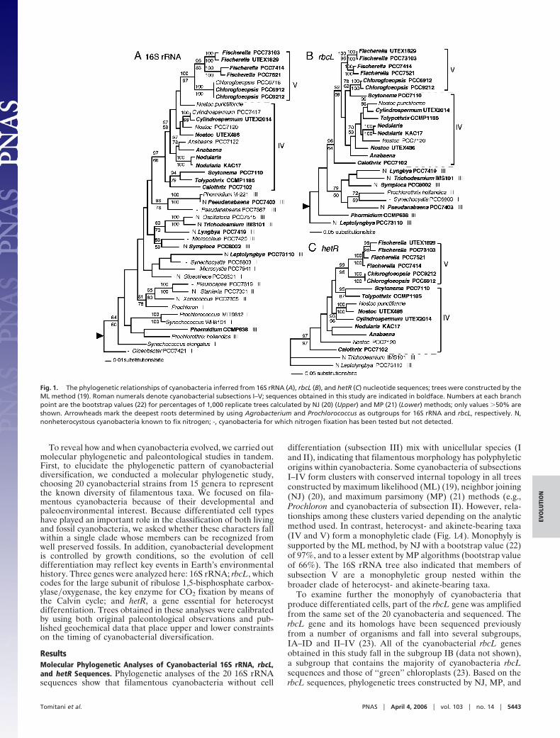
differentiation (subsection III) mix with unicellular species (Iand II), indicating that filamentous morphology has polyphyleticorigins within cyanobacteria. Some cyanobacteria of subsectionsI–IV form clusters with conserved internal topology in all treesconstructed by maximum likelihood (ML) (19), neighbor joining(NJ) (20), and maximum parsimony (MP) (21) methods (e.g.,Prochloron and cyanobacteria of subsection II). However, rela-tionships among these clusters varied depending on the analyticmethod used. In contrast, heterocyst- and akinete-bearing taxa(IV and V) form a monophyletic clade (Fig. 1A). Monophyly issupported by the ML method, by NJ with a bootstrap value (22)of 97%, and to a lesser extent by MP algorithms (bootstrap valueof 66%). The 16S rRNA tree also indicated that members ofsubsection V are a monophyletic group nested within thebroader clade of heterocyst- and akinete-bearing taxa.
To examine further the monophyly of cyanobacteria thatproduce differentiated cells, part of the rbcL gene was amplifiedfrom the same set of the 20 cyanobacteria and sequenced. TherbcL gene and its homologs have been sequenced previouslyfrom a number of organisms and fall into several subgroups,IA–ID and II–IV (23). All of the cyanobacterial rbcL genesobtained in this study fall in the subgroup IB (data not shown),a subgroup that contains the majority of cyanobacteria rbcLsequences and those of ‘‘green’’ chloroplasts (23). Based on therbcL sequences, phylogenetic trees constructed by NJ, MP, and
Fig. 1. The phylogenetic relationships of cyanobacteria inferred from 16S rRNA (A), rbcL (B), and hetR (C) nucleotide sequences; trees were constructed by theML method (19). Roman numerals denote cyanobacterial subsections I–V; sequences obtained in this study are indicated in boldface. Numbers at each branchpoint are the bootstrap values (22) for percentages of 1,000 replicate trees calculated by NJ (20) (Upper) and MP (21) (Lower) methods; only values �50% areshown. Arrowheads mark the deepest roots determined by using Agrobacterium and Prochlorococcus as outgroups for 16S rRNA and rbcL, respectively. N,nonheterocystous cyanobacteria known to fix nitrogen; -, cyanobacteria for which nitrogen fixation has been tested but not detected.
Tomitani et al. PNAS � April 4, 2006 � vol. 103 � no. 14 � 5443
EVO
LUTI
ON
ML methods all support the monophyly of heterocystous cya-nobacteria (Fig. 1B). Monophyly also was supported when thethird nucleotide in each codon was excluded from the analyses(data not shown). Several topological differences occur amongthe trees constructed by the three methods: (i) the positions ofTolypothrix and Nostoc PCC 7120 (formerly described asAnabaena) in the heterocystous clade in the NJ tree differ fromMP and ML, (ii) Phormidium and Leptolyngbya form a cluster inthe NJ and MP trees but are distributed separately in ML, and(iii) positions of Symploca and Pseudanabaena vary in a clustercomposed of Lyngbya, Trichodesmium, Symploca, Prochlorothrix,Synechocystis PCC 6803, and Pseudanabaena, depending on theanalytical method.
Partial hetR genes were amplified and sequenced from the 13heterocystous cyanobacteria among our 20 experimental strains.Interestingly, genes homologous to hetR have been detectedfrom some nonheterocystous nitrogen-fixing filamentous cya-nobacteria (subsection III), although the function of those genesis not yet certain (24). The obtained sequences were aligned withhetR sequences of two nostocalean cyanobacteria whose ge-nomes are completely sequenced and hetR-like genes of theoscillatorialeans Leptolyngbya (formerly described as Plec-tonema) PCC 73110 and Trichodesmium IMS 101. No geneknown so far has significant similarity to hetR, and so anoutgroup was not included in the calculation. Overall topologyis similar among trees constructed by the NJ, MP, and MLmethods. The members of subsection V form a monophyleticclade in the hetR tree constructed by ML (Fig. 1C). Monophylyof subsection V is also supported by the NJ and MP methods,with bootstrap values of 99% and 96%, respectively. Twocyanobacteria of subsection III fall outside of a cluster ofheterocyst-producing species. The differences in topologyamong the trees constructed by the three methods are (i)relationships within Fischerella strains and (ii) the position of acluster composed of Nodularia KAC17 and Anabaena. The outerlocation of subsection III and the monophyly of subsection Vwere also supported when the third nucleotide in each codon wasexcluded from hetR analyses, although relationships within sub-section IV varied depending on the analytical method used (datanot shown).
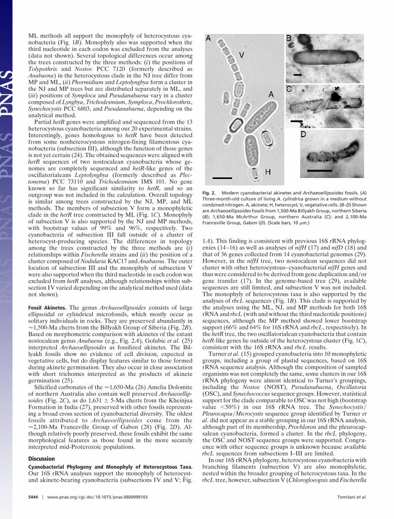
Fossil Akinetes. The genus Archaeoellipsoides consists of largeellipsoidal or cylindrical microfossils, which mostly occur assolitary individuals in rocks. They are preserved abundantly in�1,500-Ma cherts from the Billyakh Group of Siberia (Fig. 2B).Based on morphometric comparison with akinetes of the extantnostocalean genus Anabaena (e.g., Fig. 2 A), Golubic et al. (25)interpreted Archaeoellipsoides as fossilized akinetes. The Bil-lyakh fossils show no evidence of cell division, expected invegetative cells, but do display features similar to those formedduring akinete germination. They also occur in close associationwith short trichomes interpreted as the products of akinetegermination (25).
Silicified carbonates of the �1,650-Ma (26) Amelia Dolomiteof northern Australia also contain well preserved Archaeoellip-soides (Fig. 2C), as do 1,631 � 5-Ma cherts from the KheinjuaFormation in India (27), preserved with other fossils represent-ing a broad cross section of cyanobacterial diversity. The oldestfossils attributed to Archaeoellipsoides come from the�2,100-Ma Franceville Group of Gabon (28) (Fig. 2D). Al-though relatively poorly preserved, these fossils exhibit the samemorphological features as those found in the more securelyinterpreted mid-Proterozoic populations.
DiscussionCyanobacterial Phylogeny and Monophyly of Heterocystous Taxa.Our 16S rRNA analyses support the monophyly of heterocyst-and akinete-bearing cyanobacteria (subsections IV and V; Fig.
1A). This finding is consistent with previous 16S rRNA phylog-enies (14–16) as well as analyses of nifH (17) and nifD (18) andthat of 36 genes collected from 14 cyanobacterial genomes (29).However, in the nifH tree, two nostocalean sequences did notcluster with other heterocystous–cyanobacterial nifH genes andthus were considered to be derived from gene duplication and�orgene transfer (17). In the genome-based tree (29), availablesequences are still limited, and subsection V was not included.The monophyly of heterocystous taxa is also supported by theanalyses of rbcL sequences (Fig. 1B). This clade is supported bythe analyses using the ML, NJ, and MP methods for both 16SrRNA and rbcL (with and without the third nucleotide positions)sequences, although the MP method showed lower bootstrapsupport (66% and 64% for 16S rRNA and rbcL, respectively). Inthe hetR tree, the two oscillatorialean cyanobacteria that containhetR-like genes lie outside of the heterocystous cluster (Fig. 1C),consistent with the 16S rRNA and rbcL results.
Turner et al. (15) grouped cyanobacteria into 10 monophyleticgroups, including a group of plastid sequences, based on 16SrRNA sequence analysis. Although the composition of sampledorganisms was not completely the same, some clusters in our 16SrRNA phylogeny were almost identical to Turner’s groupings,including the Nostoc (NOST), Pseudanabaena, Oscillatoria(OSC), and Synechococcus sequence groups. However, statisticalsupport for the clade comparable to OSC was not high (bootstrapvalue �50%) in our 16S rRNA tree. The Synechocystis�Pleurocapsa�Microcystis sequence group identified by Turner etal. did not appear as a stable grouping in our 16S rRNA analysis,although part of its membership, Prochloron and the pleurocap-salean cyanobacteria, formed a cluster. In the rbcL phylogeny,the OSC and NOST sequence groups were supported. Congru-ence with other sequence groups is unknown because availablerbcL sequences from subsections I–III are limited.
In our 16S rRNA phylogeny, heterocystous cyanobacteria withbranching filaments (subsection V) are also monophyletic,nested within the broader grouping of heterocystous taxa. In therbcL tree, however, subsection V (Chlorogloeopsis and Fischerella
Fig. 2. Modern cyanobacterial akinetes and Archaeoellipsoides fossils. (A)Three-month-old culture of living A. cylindrica grown in a medium withoutcombined nitrogen. A, akinete; H, heterocyst; V, vegetative cells. (B–D) Shownare Archaeoellipsoides fossils from 1,500-Ma Billyakh Group, northern Siberia(B); 1,650-Ma McArthur Group, northern Australia (C); and 2,100-MaFranceville Group, Gabon (D). (Scale bars, 10 �m.)
5444 � www.pnas.org�cgi�doi�10.1073�pnas.0600999103 Tomitani et al.
in this study) did not form a cluster; instead, the two Chlorog-loeopsis strains clustered with the nostocalean Scytonema. hetR,which plays a key role in the early stage of heterocyst differen-tiation (8) and is unique to filamentous cyanobacteria, shouldprovide better resolution of the relationship between subsectionsIV and V. Sampled members of subsection V formed a mono-phyletic clade in our hetR trees, consistent with the 16S rRNAphylogeny (Fig. 1 A), but not with analyses of rbcL (Fig. 1B), nifH(17), and nifD (18) phylogenies. 16S rRNA analysis of taxonom-ically more diverse stigonematalean cyanobacteria indicatedpolyphyletic origins among filamentous taxa (30); further inves-tigation is necessary to settle their phylogenetic placement.
Species able to fix nitrogen (all taxa in subsections IV and Vand some among I–III, marked with an ‘‘N’’ in Fig. 1) do notform a monophyletic group, indicating either that the ability tofix nitrogen existed in the common ancestor of cyanobacteriaand was subsequently lost independently in multiple descendantsor that this capability spread through the group by lateral genetransfer. Phylogenetic analyses of nifH and nifD from variousnitrogen-fixing bacteria and archaea suggest that cyanobacterialgenes form a cluster (17, 18). Although all cyanobacterialnitrogen fixation may have a common origin, it remains unclearwhether the present distribution of this trait reflects verticaldescent and secondary losses or horizontal gene transfer withinthe cyanobacteria.
The Upper Time Limit of Divergence of Cyanobacteria with Differen-tiated Cells. With varying degrees of confidence, many Precam-brian (�543 Ma) microfossils have been linked to moderncyanobacteria of subsections I–III (31). Putative subsection Vmicrofossils with complex branching filaments and possibleheterocysts are preserved in the Lower Devonian (�400 Ma)Rhynie Chert in Scotland (32), but fossil heterocysts have notbeen reliably identified in Precambrian rocks (25).
In contrast to heterocysts, akinetes survive postmortem decaybetter than vegetative cells (9, 25), and, as noted above, fossil
cells interpreted as akinetes can be traced back well into thePaleoproterozoic Era. Akinetes of extant cyanobacteria occuronly in the heterocystous species (7). Although phylogeny pro-vides no way of determining unambiguously the relative timingof akinete and heterocyst origins, geochemical data discussedbelow suggest that the paleoenvironmental drivers of heterocystevolution were in place before the earliest known records ofakinetes.
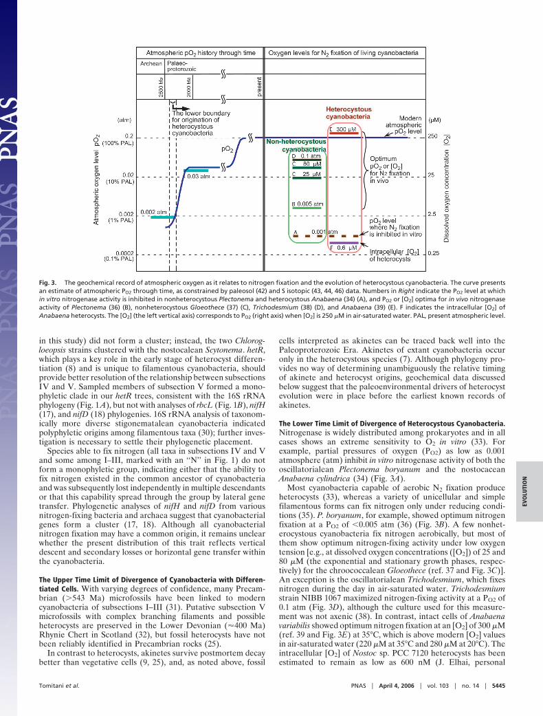
The Lower Time Limit of Divergence of Heterocystous Cyanobacteria.Nitrogenase is widely distributed among prokaryotes and in allcases shows an extreme sensitivity to O2 in vitro (33). Forexample, partial pressures of oxygen (PO2) as low as 0.001atmosphere (atm) inhibit in vitro nitrogenase activity of both theoscillatorialean Plectonema boryanum and the nostocaceanAnabaena cylindrica (34) (Fig. 3A).
Most cyanobacteria capable of aerobic N2 fixation produceheterocysts (33), whereas a variety of unicellular and simplefilamentous forms can fix nitrogen only under reducing condi-tions (35). P. boryanum, for example, showed optimum nitrogenfixation at a PO2 of �0.005 atm (36) (Fig. 3B). A few nonhet-erocystous cyanobacteria fix nitrogen aerobically, but most ofthem show optimum nitrogen-fixing activity under low oxygentension [e.g., at dissolved oxygen concentrations ([O2]) of 25 and80 �M (the exponential and stationary growth phases, respec-tively) for the chroococcalean Gloeothece (ref. 37 and Fig. 3C)].An exception is the oscillatorialean Trichodesmium, which fixesnitrogen during the day in air-saturated water. Trichodesmiumstrain NIBB 1067 maximized nitrogen-fixing activity at a PO2 of0.1 atm (Fig. 3D), although the culture used for this measure-ment was not axenic (38). In contrast, intact cells of Anabaenavariabilis showed optimum nitrogen fixation at an [O2] of 300 �M(ref. 39 and Fig. 3E) at 35°C, which is above modern [O2] valuesin air-saturated water (220 �M at 35°C and 280 �M at 20°C). Theintracellular [O2] of Nostoc sp. PCC 7120 heterocysts has beenestimated to remain as low as 600 nM (J. Elhai, personal
Fig. 3. The geochemical record of atmospheric oxygen as it relates to nitrogen fixation and the evolution of heterocystous cyanobacteria. The curve presentsan estimate of atmospheric PO2 through time, as constrained by paleosol (42) and S isotopic (43, 44, 46) data. Numbers in Right indicate the PO2 level at whichin vitro nitrogenase activity is inhibited in nonheterocystous Plectonema and heterocystous Anabaena (34) (A), and PO2 or [O2] optima for in vivo nitrogenaseactivity of Plectonema (36) (B), nonheterocystous Gloeothece (37) (C), Trichodesmium (38) (D), and Anabaena (39) (E). F indicates the intracellular [O2] ofAnabaena heterocysts. The [O2] (the left vertical axis) corresponds to PO2 (right axis) when [O2] is 250 �M in air-saturated water. PAL, present atmospheric level.
Tomitani et al. PNAS � April 4, 2006 � vol. 103 � no. 14 � 5445
EVO
LUTI
ON
communication; Fig. 3F), suggesting that heterocyst formershave a more efficient system to protect nitrogenase from oxygenthan do nonheterocystous taxa. Considering that heterocystdevelopment is arrested under anaerobic conditions (40) andthat the rate of heterocyst differentiation is enhanced by in-creases in PO2 (41), the evolution of heterocysts may haveaccompanied the initial appearance of oxygen-rich environ-ments in Earth’s history.
Chemical analyses of iron retention in ancient weatheringhorizons suggest that PO2 rose from �0.002 atm to as much as0.03 atm between �2,400 and 2,200 Ma (42). Sulfur-isotopeanalyses independently indicate a rapid increase of atmosphericoxygen between �2,450 and 2,320 Ma (43, 44). One might arguethat heterocysts could have evolved earlier, in oxic habitatislands within thick mats, but the ecology of extant heterocyst-formers militates against this occurrence. Most heterocyst-forming cyanobacteria live in soil or freshwater environmentswhere they are in direct contact with air and oxic waters; they areuncommon in marine microbial mats where oxic microhabitatsmight develop (45). Accordingly, cyanobacterial heterocystslikely appeared after 2,450–2,320 Ma, when PO2 first reachedlevels that inhibit nitrogenase activity (Fig. 3).
Time Calibration for the Tree of Life. Integrating molecular phylo-genetic, physiological, paleontological, and geochemical data, wepropose that the clade of cyanobacteria marked by heterocystand akinete differentiation evolved once between 2,450 and2,100 Ma. This constraint on the timing of a relatively late branchwithin the cyanobacteria provides a calibration point that mayilluminate molecular evolutionary studies of deep nodes in theTree of Life. As complete genome sequences of several cya-nobacterial species have been determined, including the hetero-cystous Nostoc sp. PCC 7120 (47) and Nostoc punctiforme (48),the calibration can also be applied to expanding research incomparative genomics. Continuing study of Precambrian micro-fossils and taxon-specific biomarkers promises to place increas-ingly precise constraints on the timing of early cyanobacterialevolution. Conversely, continuing molecular biological researchon living populations will add depth to the portrait of cyanobac-terial evolution gleaned from an incomplete geological record.
Materials and MethodsGene Isolation and Sequencing. Twenty filamentous cyanobacteriawere selected for sequencing: six strains from subsection III,eight from IV, and six from V (shown in boldface in Fig. 1).Axenic cultures of Fischerella strain UTEX 1829, Nostoc UTEX486, and Cylindrospermum UTEX 2014 were obtained from theCulture Collection of Algae at the University of Texas, Austin.Chlorogloeopsis PCC 6912 and 9212; Fischerella PCC 73103,7414, and 7521; and Pseudanabaena PCC 7403 came from thePasteur Culture Collection of Cyanobacteria (Paris). Isolates ofAnabaena and Nodularia, together with Calothrix PCC 7102,Lyngbya PCC 7419, and Scytonema PCC 7110, were provided byJ. B. Waterbury (Woods Hole Oceanographic Institution,Woods Hole, MA). DNA from cyanobacterial cells was extractedby standard methods designed for bacteria (49). Genomic DNAfrom Tolypothrix CCMP 1185 and Phormidium CCMP 638 wasobtained from the Provasoli–Guillard National Center for Cul-ture of Marine Phytoplankton (Boothbay Harbor, ME).Genomic DNA from Nodularia strain KAC17, Symploca PCC8002, Leptolyngbya PCC 73110, and Trichodesmium IMS 101 wasprovided by S. Janson (University of Kalmar, Kalmar, Sweden).16S rRNA was amplified by using a pair of primers designedspecially for cyanobacteria (PLG1-1 and PLG2-1) (50).
Two pairs of primers for rbcL (GF-AB:GARTCTTCIA-CYGGTACYTGGAC and GR-D:TGCCAIACGTGGATAC-CACC; GF-C:CTTYACYCAAGACTGGGCTTC and GR-E:AACTCRAACTTGATTTCYTTCC) were designed based
on the conserved regions in rbcL sequences of Nostoc sp. PCC7120 (GenBank accession no. L02522), Synechocystis PCC6803 (GenBank accession no. D64000), and Prochlorothrix(GenBank accession no. X57359). A pair of primers for hetR(HETR-F1:TATCTGGCTTTTAGYGCCATG and HETR-R1:CTTGGTGATATTTATCWGCCC) was designed basedon conserved regions between hetR sequences of Nostoc PCC7120 (GenBank accession no. M37779) and N. punctiforme(GenBank accession no. AF318069). PCRs were performed ina T-gradient thermal cycler (Biometra, Tampa, FL) by usingthe AmpliTaq Gold kit (Applied Biosystems). Amplified PCRproducts were cloned by using either the TOPO TA cloning kit(Invitrogen) or the pSTBlue-1 AccepTor kit (Novagen). Bothstrands of a minimum of three clones from each PCR productwere sequenced with the Big Dye Terminator Cycle Sequenc-ing kit version 2 (Applied Biosystems) by using a DNAsequencer (3100 Genetic Analyser, Applied Biosystems).
Phylogenetic Analyses. Twenty-one 16S rRNA sequences of cya-nobacteria (GenBank accession nos. AB003165, AB039009,AB39006, AF001480, AF027655, AF053398, AF091108,AF091150, AF132777, AF132783, AF132789, AF132790,AF132792, AF132933, D64000, D83715, U40340, X59559,X63141, X70770, and X78680) and that of Agrobacterium (Gen-Bank accession no. D14500) were obtained from GenBank, aswere partial rbcL sequences of Nostoc PCC 7120 (GenBankaccession no. L02522), Synechocystis PCC 6803 (GenBank ac-cession no. D64000), Prochlorothrix (GenBank accession no.X57359), and Prochlorococcus MED 4 (accession no.BX572091), two hetR genes of Nostoc PCC 7120 (GenBankaccession no. M37779) and N. punctiforme (GenBank accessionno. AF318069), and two hetR-like genes of Leptolyngbya PCC73110 (GenBank accession no. AF410433) and TrichodesmiumIMS 101 (accession no. AF410432). The rbcL sequence of N.punctiforme was obtained from the web site of the Departmentof Energy Joint Genome Institute (Walnut Creek, CA).
Nucleotide sequences were aligned by using CLUSTAL X (51)with manual refinement. Sites where gaps existed or sequenceswere ambiguous were excluded. Secondary structures (52) weretaken into consideration when the 16S rRNA sequences werealigned with Agrobacterium as an outgroup. The 20 rbcL se-quences were aligned with the form IB rbcL sequences fromthree cyanobacteria (Synechocystis PCC 6803, Nostoc sp. PCC7120, and N. punctiforme) whose genomes are completely se-quenced and that of Prochlorothrix, with Prochlorococcus MED4 (a form IA rbcL) as an outgroup (data not shown). The hetRsequences were aligned with sequences of two nostocaleancyanobacteria whose genomes are completely sequenced andhetR-like genes of the oscillatorialeans Leptolyngbya PCC 73110and Trichodesmium IMS 101 (data not shown). Sequence lengthsobtained here were as follows (base pairs of full sequence�basepairs used for analyses): 16S rRNA (1,145–1,160�1,117), rbcL(1,183�1,181), and hetR (742�737).
Phylogenetic trees were generated by using PAUP* 4.0b10 (53).The Hasegawa–Kishino–Yano 1985 model was used to modelnucleotide substitution in the ML (19) and NJ (20) analyses. Basefrequencies and transition transversion ratios were estimatedfrom each data set. Each MP (21) calculation was performed byheuristic search of 100 replicates with the TBR (tree bisectionand reconnection) option. For 16S rRNA, ML analyses were firstdone by heuristic search with the NNI (nearest-neighbor inter-change) option, using the NJ and MP results as starting trees. Tofurther investigate the 16S rRNA tree topology, heuristic searchby ML with the TBR option was performed, using a topologyconstraint file that fixed groupings that appeared in all 16SrRNA trees obtained by the NJ, MP, and ML (NNI option)methods. The result obtained by ML with the TBR option wasthe same as that by ML with NNI. For rbcL and hetR, ML
5446 � www.pnas.org�cgi�doi�10.1073�pnas.0600999103 Tomitani et al.
analyses were carried out by heuristic search with the TBRoption, using results obtained from the NJ and MP analyses asstarting trees.
Nitrogen fixation ability of PCC strains in subsections I–III(‘‘N’’ in Fig. 1) is based on information provided by the PasteurCulture Collection.
Paleontological Analyses. Fossils were identified by optical mi-croscopy on thin sections of silicified carbonates deposited incoastal marine environments in a series of Paleoproterozoic andMesoproterozoic basins. Materials from the �1,500-Ma BillyakhGroup of northern Siberia and the 1,600- to 1,500-Ma McArthurGroup of northern Australia collected by A.H.K. are repositedin the Paleobotanical Collections of the Harvard University
Herbaria, as are fossiliferous cherts of the 2,100-Ma FrancevilleGroup of West Africa. Additional fossiliferous samples of theAmelia Dolomite (McArthur Group) are reposited in the Com-monwealth Palaeontological Collection in Canberra, Australia(collection CPC 15609 for the specimen shown in Fig. 2). A livingcyanobacterial culture of A. cylindrica was provided by D. G.Adams (University of Leeds, Leeds, U.K.).
We thank J. B. Waterbury and D. G. Adams for cyanobacterial cultures;S. Janson for genomic DNAs; and J. C. Meeks, D. E. Canfield,J. B. Waterbury, and D. J. Des Marais for constructive criticism. Thiswork was supported by Research Fellowships of the Japan Society for thePromotion of Science for Young Scientists (to A.T.), the NationalAeronautics and Space Administration (NASA) Astrobiology Institute(A.H.K.), and the NASA Exobiology program (C.M.C.).
1. Whitton, B. A. & Potts, M. (2000) in The Ecology of Cyanobacteria, eds.Whitton, B. A. & Potts, M. (Kluwer, Dordrecht, The Netherlands), pp. 1–11.
2. Liu, H., Nolla, H. A. & Campbell, L. (1997) Aquat. Microb. Ecol. 12, 39–47.3. Capone, D. G., Zehr, J. P., Paerl, H. W., Bergman, B. & Carpenter, E. J. (1997)
Science 276, 1221–1229.4. Karl, D., Michaels, A., Bergman, B., Capone, D., Carpenter, E., Letelier, R.,
Lipschultz, F., Paerl, H., Sigman, D. & Stal, L. (2002) Biogeochemistry 57�58,47–98.
5. Delwiche, C. F. & Palmer, J. D. (1997) in Origins of the Algae and Their Plastids,ed. Bhattachartya, D. (Springer, Berlin), pp. 53–96.
6. Rippka, R., Deruelles, J., Waterbury, J. B., Herdman, M. & Stanier, R. Y.(1979) J. Gen. Microbiol. 111, 1–61.
7. Castenholz, R. W. (2001) in Bergey’s Manual of Systematic Bacteriology, eds.Boone, D. R. & Castenholz, R. W. (Springer, New York), 2nd Ed., Vol. 1, pp.474–487.
8. Wolk, C. P., Ernst, A. & Elhai, J. (1994) in The Molecular Biology ofCyanobacteria, ed. Bryant, D. A. (Kluwer, Dordrecht, The Netherlands), pp.769–823.
9. Herdman, M. (1987) in The Cyanobacteria, eds. Fay, P. & Van Baalen, C.(Elsevier, Amsterdam), pp. 227–250.
10. Brocks, J. J., Logan, G. A., Buick, R. & Summons, R. E. (1999) Science 285,1033–1036.
11. Knoll, A. H. (2003) Geobiology 1, 3–14.12. Buick, R. (1992) Science 255, 74–77.13. Hofmann, H. J. (1976) J. Paleontol. 50, 1040–1073.14. Giovannoni, S. J., Turner, S., Olsen, G. J., Barns, S., Lane, D. J. & Pace, N. R.
(1988) J. Bacteriol. 170, 3584–3592.15. Turner, S., Prayer, K. M., Miao, V. P. & Palmer, J. D. (1999) J. Eukaryot.
Microbiol. 46, 327–338.16. Wilmotte, A. & Herdman, M. (2001) in Bergey’s Manual of Systematic Bacte-
riology, eds. Boone, D. R. & Castenholz, R. W. (Springer, New York), 2nd Ed.,Vol. 1, pp. 487–493.
17. Zehr, J. P., Mellon, M. T. & Hiorns, W. D. (1997) Microbiology 143, 1443–1450.18. Henson, B. J., Watson, L. E. & Barnum, S. R. (2004) J. Mol. Evol. 58, 390–399.19. Felsenstein, J. (1981) J. Mol. Evol. 17, 368–376.20. Saitou, N. & Nei, M. (1987) Mol. Biol. Evol. 4, 406–425.21. Fitch, W. M. (1977) Am. Nat. 111, 223–257.22. Felsenstein, J. (1985) Evolution (Lawrence, Kans.) 39, 783–791.23. Tabita, F. R. (1999) Photosynth. Res. 60, 1–28.24. Janson, S., Matveyev, A. & Bergman, B. (1998) FEMS Microbiol. Lett. 168,
173–179.25. Golubic, S., Sergeev, V. N. & Knoll, A H. (1995) Lethaia 28, 285–298.26. Page, R. W., Jackson, M. J. & Krassay, A. A. (2000) Aust. J. Earth Sci. 47,
431–459.
27. Srivastava, P. (2005) Orig. Life Evol. Biosph. 35, 175–185.28. Amard, B. & Bertrand-Sarfati, J. (1997) Precambrian Res. 81, 197–221.29. Sanchez-Baracaldo, P., Hayes, P. K. & Blank, C. E. (2005) Geobiology 3,
145–165.30. Gugger, M. F. & Hoffmann, L. (2004) Int. J. Syst. Evol. Microbiol. 54, 349–357.31. Knoll, A. H. & Golubic, S. (1992) in Early Organic Evolution: Implications for
Mineral and Energy Resources, eds. Schidlowski, M., Golubic, S. & Kimberley,M. M. (Springer, Heidelberg), pp. 450–462.
32. Croft, W. N. & George, E. A. (1959) Bull. Brit. Mus. Nat. Hist. Geol. 3, 339–353.33. Gallon, J. R. (1992) New Phytol. 122, 571–609.34. Haystead, A., Robinson, R. & Stewart, W. D. P. (1970) Arch. Mikrobiol. 74,
235–243.35. Rippka, R. & Waterbury, J. B. (1977) FEMS Microbiol. Lett. 2, 83–86.36. Weare, N. M. & Benemann, J. R. (1974) J. Bacteriol. 119, 258–265.37. Maryan, P. S., Eady, R. R., Charplin, A. E. & Gallon, J. R. (1986) J. Gen.
Microbiol. 132, 789–796.38. Ohki, K. & Fujita, Y. (1988) Marine Biol. 98, 111–114.39. Jensen, B. B. & Cox, R. P. (1983) Arch. Microbiol. 135, 287–292.40. Rippka, R. & Stanier, R. Y. (1978) J. Gen. Microbiol. 105, 83–94.41. Fay, P. (1992) Microbiol. Rev. 56, 340–373.42. Rye, R. & Holland, H. D. (1998) Am. J. Sci. 298, 621–672.43. Farquhar, J., Bao, H. & Thiemens, M. (2000) Science 289, 756–759.44. Bekker, A., Holland, H. D., Wang, P. L., Rumble, D., III, Stein, H. J., Hannah,
J. L., Coetzee, L. L. & Beukes, N. J. (2004) Nature 427, 117–120.45. Stal, L. J. (2000) in The Ecology of Cyanobacteria: Their Diversity in Space and
Time, ed. Whitton, B. A. (Luver, Dordrecht, The Netherlands), pp. 61–121.46. Canfield, D. E. & Teske, A. (1996) Nature 382, 127–132.47. Kaneko, T., Nakamura, Y., Wolk, C. P., Kuritz, T., Sasamoto, S., Watanabe,
A., Iriguchi, M., Ishikawa, A., Kawashima, K., Kimura, T., et al. (2001) DNARes. 8, 205–213, 227–253.
48. Meeks, J. C., Elhai, J., Thiel, T., Potts, M., Larimer, F., Lamerdin, J., Predki,P. & Atlas, R. (2001) Photosynth. Res. 70, 85–106.
49. Ausubel, F. M., Brent, R., Kingston, R. E., Moore, D., Seidmann, J. G., Smith,J. A. & Sturuhl, K., eds. (1987) in Current Protocols in Molecular Biology (Wiley,New York), Sect. 2.4.
50. Urbach, E., Robertson, D. L. & Chisholm, S. W. (1992) Nature 355,267–270.
51. Jeanmougin, F., Thompson, J. D., Gouy, M., Higgins, D. G. & Gibson, T. J.(1998) Trends Biochem. Sci. 23, 403–405.
52. Wilmotte, A., Van der Auwera, G. & De Wacheter, R. (1993) FEBS Lett. 317,96–100.
53. Swofford, D. L. (2002) PAUP*: Phylogenetic Analysis Using Parsimony (*andOther Methods) (Sinauer, Sunderland, MA), Version 4.
Tomitani et al. PNAS � April 4, 2006 � vol. 103 � no. 14 � 5447
EVO
LUTI
ON