Embed Size (px)
Citation preview
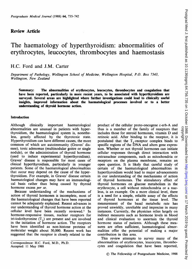
Postgraduate Medical Journal (1988) 64, 735-742
Review Article
The haematology of hyperthyroidism: abnormalities oferythrocytes, leucocytes, thrombocytes and haemostasis
H.C. Ford and J.M. CarterDepartment of Pathology, Wellington School of Medicine, Wellington Hospital, P.O. Box 7343,Wellington, New Zealand
Summary: The abnormalities of erythrocytes, leucocytes, thrombocytes and coagulation thathave been reported, particularly in more recent years, to be associated with hyperthyroidism aresurveyed. Several areas are highlighted where further investigations could lead to clinically usefulinsights, improved information about the haematological processes involved or to a betterunderstanding of thyroid hormone action.
Introduction
Although clinically, important haematologicalabnormalities are unusual in patients with hyper-thyroidism, the haematological system is, nonethe-less, greatly affected by the thyrotoxic state.Hyperthyroidism can have different causes, the morecommon of which are autoimmunity (Graves' dis-ease), toxic adenomas (multinodular goitre or singlenodule), or the administration of thyroid hormone(used to induce experimental hyperthyroidism).Graves' disease is responsible for most cases ofclinical hyperthyroidism, particularly in youngerpatients. Some of the haematological abnormalitiesthat occur may depend on the cause of the hyper-thyroidism. For example, in Graves' disease certainhaematological changes may have an immunologi-cal basis rather than being caused by thyroidhormone excess per se.
Because understanding of the mechanisms ofaction of thyroid hormones is incomplete, most ofthe haematological changes that have been reportedcannot be adequately explained. Recent advances inour understanding of thyroid hormone action at thecellular level have been recently reviewed.1 Inhormone-responsive tissues, nuclear receptors fortri-iodothyronine (T3) are present and are involvedin the initiation of hormone action. The receptorshave been identified as non-histone proteins ofmolecular weight about 50,000. Recent work hassuggested that the receptor is closely related to the
product of the cellular proto-oncogene c-erb-A andthus is a member of the family of receptors thatincludes those for steroid hormones, vitamin D andretinoic acid. After binding to the receptor, it ispostulated that the T3-receptor complex binds tospecific regions of the DNA and alters gene expres-sion. Whether or not thyroid hormones can initiatecellular responses through direct interaction withextranuclear components, such as mitochondria orreceptors on the plasma membrane, remains anopen question. It is likely that careful study ofcertain of the haematological abnormalities inhyperthyroidism would lead to major advancementsin our understanding of the mechanisms of actionof thyroid hormones. The stimulatory effect ofthyroid hormones on glucose metabolism in theerythrocyte, a cell without mitochondria or a nuc-leus, is an example. On a more clinical level, thereis a need for a simple, reliable assay of the effectsof thyroid hormones at the tissue level. Themeasurement of the basal metabolic rate hasproved unwieldy, unreliable and imprecise in manyinstances. Currently, the physician relies largely onindirect measures such as hormone levels in bloodand clinical evaluation to ascertain the thyroidhormone status of patients. Although these mea-sures are often sufficient, haematological abnor-malities offer the potential of making a majorcontribution in this area.
This review attempts to survey many of theabnormalities of erythrocytes, leucocytes, thrombo-cytes and coagulation that have been reported,
©) The Fellowship of Postgraduate Medicine, 1988
Correspondence: H.C. Ford, M.D., Ph.D.Accepted: 11 May 1988
copyright. on 17 M
arch 2019 by guest. Protected by
http://pmj.bm
j.com/
Postgrad M
ed J: first published as 10.1136/pgmj.64.756.735 on 1 O
ctober 1988. Dow
nloaded from
736 H.C. FORD AND J.M. CARTER
particularly in more recent years, to be associatedwith hyperthyroidism and to highlight the areaswhere further investigations are needed to increaseour understanding of the mechanisms involved. Inmost instances, we have not compared the haema-tological change seen in hyperthyroidism with thechange, or lack thereof, that has been reported inhypothyroidism. To do so regularly would haveextended the review unduly. The well-known asso-ciation of pernicious anaemia with Graves' diseasehas been extensively reviewed2'3 and will not befurther discussed in this review.
Erythrocytes
Kinetics and indices (Table I)
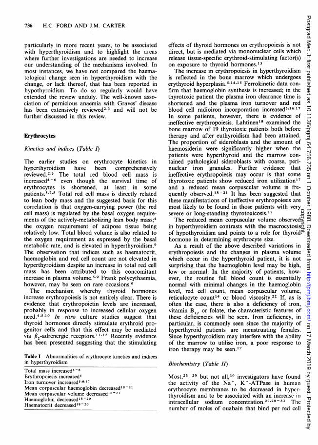
The earlier studies on erythrocyte kinetics inhyperthyroidism have been comprehensivelyreviewed.2'3 The total red blood cell mass isincreased4-6 even though the survival time oferythrocytes is shortened, at least in somepatients.5 7'8 Total red cell mass is directly relatedto lean body mass and the suggested basis for thiscorrelation is that oxygen-carrying power (the redcell mass) is regulated by the basal oxygen require-ments of the actively-metabolizing lean body mass;4the oxygen requirement of adipose tissue beingrelatively low. Total blood volume is also related tothe oxygen requirement as expressed by the basalmetabolic rate, and is elevated in hyperthyroidism.9The observation that indices such as haematocrit,haemoglobin and red cell count are not elevated inhyperthyroidism despite an increase in total red cellmass has been attributed to this concomitantincrease in plasma volume.5'9 Frank polycythaemia,however, may be seen on rare occasions.6The mechanism whereby thyroid hormones
increase erythropoiesis is not entirely clear. There isevidence that erythropoietin levels are increased,probably in response to increased cellular oxygenneed.4'5'10 In vitro culture studies suggest thatthyroid hormones directly stimulate erythroid pro-genitor cells and that this effect may be mediatedvia fl2-adrenergic receptors.I11 12 Recently evidencehas been presented suggesting that the stimulating
Table I Abnormalities of erythrocyte kinetics and indicesin hyperthyroidismTotal mass increased4-6Erythropoiesis increased5Iron turnover increased5'6"17Mean corpuscular haemoglobin decreased'8- 21
Mean corpuscular volume decreased' 8 -21Haemoglobin decreased18 -20Haematocrit decreased' 8 - 20
effects of thyroid hormones on erythropoiesis is notdirect, but is mediated via mononuclear cells whichrelease tissue-specific erythroid-stimulating factor(s)on exposure to thyroid hormones.13The increase in erythropoiesis in hyperthyroidism
is reflected in the bone marrow which undergoeserythyroid hyperplasia.5",4'15 Ferrokinetic data con-firm that haemoglobin synthesis is increased; in thethyrotoxic patient the plasma iron clearance time isshortened and the plasma iron turnover and redblood cell radioiron incorporation increased5l1617In some patients, however, there is evidence ofineffective erythropoiesis. Lahtinen18 examined thebone marrow of 19 thyrotoxic patients both beforetherapy and after euthyroidism had been attained.The proportion of sideroblasts and the amount ofhaemosiderin were significantly higher when thepatients were hyperthyroid and the marrow con-tained pathological sideroblasts with coarse, peri-nuclear iron granules. Further evidence thatineffective erythropoiesis may occur is that somethyrotoxic patients show reduced iron utilization'7and a reduced mean corpuscular volume is fre-quently observed.18 -21 It has been suggested thatthese manifestations of ineffective erythropoiesis aremost likely to be found in those patients with verysevere or long-standing thyrotoxicosis.17The reduced mean corpuscular volume observed
in hyperthyroidism contrasts with the macrocytosisof hypothyroidism and points to a role for thyroidhormone in determining erythrocyte size.As a result of the above described variations in
erythropoiesis and the changes in plasma volumewhich occur in the hyperthyroid patient, it is notsurprising that the haemoglobin level may be high,low or normal. In the majority of patients, how-ever, the routine full blood count is essentiallynormal with minimal changes in the haemoglobinlevel, red cell count, mean corpuscular volume,reticulocyte count14 or blood viscosity.22 If, as isoften the case, there is also a deficiency of iron,vitamin B12 or folate, the characteristic features ofthese deficiencies will be seen. Iron deficiency, inparticular, is commonly seen since the majority ofhyperthyroid patients are menstruating females.Since hyperthyroidism may interfere with the abilityof the marrow to utilise iron, a poor response toiron therapy may be seen.'7
Biochemistry (Table II)
Most,23-29 but not all,30 investigators have foundthe activity of the Na+, K +-ATPase in humanerythrocyte membranes to be decreased in hyper-thyroidism and to be associated with an increasc illintracellular sodium concentration.27'29 - 33 Thcnumber of moles of ouabain that bind per red cell
copyright. on 17 M
arch 2019 by guest. Protected by
http://pmj.bm
j.com/
Postgrad M
ed J: first published as 10.1136/pgmj.64.756.735 on 1 O
ctober 1988. Dow
nloaded from

HAEMATOLOGY OF HYPERTHYROIDISM 737
Table II Biochemical abnormalities of erythrocytes inhyperthyroidismNa +, K + -ATPase activity decreased 23 - 29Sodium concentration increased27'29 - 33Ca2+-ATPase activity increased39Zinc concentration decreased27'32'40'42B isoenzyme of carbonic anhydrase decreased43-46Oxygen consumption and heat production increased47'48Increased activity of glucose-6-phosphate dehydrogenaseand other enzymes that metabolise glucose32'49
2,3-Diphosphoglycerate concentration increased56'58'59Catalase activity increased62Monoester lipase activity increased63Percent haemoglobin A2 increased64'65Percent fetal haemoglobin increased65'66
is decreased26027'29'34 which suggests that a
decrease in the number of Na , K + -ATPasesodium pumps per cell is responsible for thedecrease in sodium efflux rather than a defect inperformance of the pumps. A significant inverserelationship has been observed between the numberof sodium pumps per erythrocyte and the plasmathyroxine and tri-iodothyronine levels.27'34 It hasbeen suggested25-28 that the effects of thyroidhormone on the activity of the Na+, K+-ATPaseare not specific but are a manifestation of a
widespread effect on the metabolism or membraneenvironment of most surface proteins of the erythro-cyte. However, extensive studies of the function andenvironment of erythrocyte surface proteins inhyperthyroidism have not been carried out andsome of the existing data are controversial. Forexample, one group28 has reported a significantreduction in the number of insulin receptors per redblood cell in hyperthyroidism together with a signi-ficant increase in their average affinity, whereasanother group35 has reported a marked decline ininsulin binding affinity without change in insulinreceptor number. The proposal36'37 that a general-ized increase in Na +, K + -ATPase activity isresponsible for a major proportion of the increasedenergy demand in hyperthyroidism appears not toinclude the erythrocyte.33 It may be26 that theerythrocyte, being unable to carry out protein syn-thesis, cannot adequately replace certain proteins,such as components of the Na+, K+-ATPasesodium pump, whose rate of catabolism is increasedin hyperthyroidism.38 On the other hand, Ca2 +-ATPase activity of erythrocyte membranes was
found to be increased in hyperthyroidism39 whichsuggests that no single explanation for the effect ofthyroid hormone on components of the plasmamembrane of the erythrocyte is likely.
There is general agreement that erythrocyte zinc-concentration is reduced in hyperthyroid-iSM27 32,40,42 and that urinary zinc excretion is
increased.41'42 The concentration of the B isoen-zyme of carbonic anhydrase, which is by far themost abundant zinc metalloenzyme in the erythro-cyte, is decreased in hyperthyroidism, whereas theconcentration of the genetically distinct C isoen-zyme is near normal.43-46 For example, Funakoshiand Deutsch46 found the mean levels of the B andC isoenzymes to be 37% and 80% of normal,respectively. The overall activity of carbonic anhyd-rase is decreased, but the activities of a number ofother zinc metalloenzymes of the erythrocyte areunchanged in hyperthyroidism.40 It is likely thatthyroid hormones specifically inhibit the synthesisof the B isoenzyme. This effect has not beenassociated with a change in erythrocyte functionnor is it of sufficient magnitude to explain thenegative zinc balance that occurs in thyrotoxico-sis,41'42 a phenomenon which has been correlatedwith the overall catabolic state.41
Erythrocytes, which lack mitochondria, metabo-lise glucose via the Embden-Meyerhof glycolyticpathway and the hexose monophosphate shunt(also called the pentose phosphate shunt). ATP, themajor energy source, is produced by the Embden-Meyerhof pathway and the hexose monophosphateshunt yields NADPH which is used as a cofactor ina number of reactions. Oxygen consumption andheat production by erythrocytes from hyperthyroidpatients are both increased4748 and increasedactivities of a number of enzymes of intermediarymetabolism in erythrocytes from hyperthyroidpatients have been reported.32'49 Normally, most ofthe available glucose is utilized by the Embden-Meyerhof pathway50 and recently it was reported51that the metabolic activities of both pathways areproportionately increased in hyperthyroidismso that the Embden-Meyerhoff pathway continuesto predominate. The mechanism by which thyroidhormones increase the metabolic activity of theerythrocyte is unknown but is of special interestsince the erythrocyte lacks the two organelles whichare usually postulated to be involved in the mecha-nism of action of thyroid hormones in other tissues,namely mitochondria and a nucleus.
In erythrocytes from hyperthyroid patients andfrom normal subjects treated with thyroid hormonethe haemoglobin dissociation curve is shifted to theright indicating a decreased affinity for oxygen.52'53A result of this shift is that oxygen is more readilydelivered to peripheral tissues, perhaps to meet theincreased metabolic demands of thyrotoxicosis. It isknown54'55 that an increase in the concentration oforganic phosphates within erythrocytes displacesthe oxyhaemoglobin dissociation curve to the right.It has been reported that the concentration of 2,3-diphosphoglycerate, the major source of organicphosphate in the erythrocyte, is increased after
copyright. on 17 M
arch 2019 by guest. Protected by
http://pmj.bm
j.com/
Postgrad M
ed J: first published as 10.1136/pgmj.64.756.735 on 1 O
ctober 1988. Dow
nloaded from

738 H.C. FORD AND J.M. CARTER
incubation in vitro of red cell preparations fromnormal subjects in medium containing thyroid hor-mones56'57 and in the erythrocytes of some,56'58'59but not all,60'61 patients with hyperthyroidism.The activities of erythrocyte catalase62 and
monoester lipase63 are increased in hyperthyroidpatients but the mechanism and possible signifi-cance of these changes are unclear. The meanpercent haemoglobin A2 level (3.21) in a group ofuntreated hyperthyroid subjects was significantly(P<0.001) higher than in a group made euthyroidwith antithyroid drugs (2.42).64 These results, whichconfirmed an earlier report,65 suggest that thyroidhormones can modulate the synthesis of globinchains. A slight elevation of the foetal haemoglobinpercentage has also been reported.65'66
Leucocytes
Absolute and relative numbers
An association between neutropenia and Graves'disease has been recognized for at least 80years.67'68 In relatively recent studies, the incidenceof absolute neutropenia in hyperthyroid patientsvaried from less than 5%16.69 to 18%.70 A signifi-cant relative neutropenia in thyrotoxic patients,originally described in 1908,68 has also been con-firmed in more recent years.69 Treatment of theneutropenic thyrotoxic patient with radioiodine orpropylthiouracil usually results in a return of theneutrophil count to normal and neutropeniadetected before treatment does not contraindicatethe use of thioamide drugs.70 The aetiology of theneutropenia is unknown, but does not appear toinvolve depressed or ineffective myelopoiesis. 11.12.70Although there is general agreement that a rela-
tive lymphocytosis is often seen in hyperthyroidpatients,16.68.69 there is less consensus regardingwhether an absolute lymphocytosis is 16.68.71.72 oris not69 73 associated. There has also been disagree-ment regarding the relative and absolute numbersof T lymphocytes and subsets. Table III gives thefindings of several recent reports. The conflicting
Table III T and B lymphocytes in Graves' disease
T lymphocytes (total number)
Suppressor T lymphocytes (%)
Helper T lymphocytes (%)
Activated T lymphocytes (%)B lymphocytes (%)
- normal73'74- decreased72'75- normal72'74'76- decreased73'7'- normal72'76- increased73- increased73.76- normal73'76- increased71
results are further complicated by the fact that themean changes reported in a study often do notoccur in all patients in that study. For example, onegroup" found a decrease in the proportion ofsuppressor T lymphocytes in only about 50% ofpatients. The lack of consistent findings in peri-pheral blood may be due to the fact that thepathogenetic immunological events in Graves' dis-ease take place within the thyroid gland.77 Atypicallymphocytes may be seen in some patients withthyrotoxicosis.1'16 Monocyte and eosinophil countsare usually normal, 16 although eithermay occasionallybe modestly raised.' .72
Biochemistry
There have been surprisingly few investigations ofpossible changes in the metabolism of leucocytes inhyperthyroidism. In one study26 it was found thatleucocyte Na t, Kt-ATPase activity, [3H]ouabainbinding capacity (an indicator of the number ofsodium pumps) and total active rubidium influx(used to measure potassium flux) were all signifi-cantly increased in untreated hyperthyroidism.Therewas no change in leucocyte sodium concen-tration. These results contrasted with those foundin erythrocytes which showed decreased Na+, K+-ATPase activity, decreased [3H]ouabain bindingcapacity and decreased active potassium flux rates.A statistically significant decrease in phagocytic
ability and an increase in nitroblue tetrazoliumreduction by neutrophils from hyperthyroid patientscompared to normal control subjects have beenreported.78 Cytochemical observations suggestedincreased activity of leucocyte acid phosphataseand fl-glucuronidase. Further studies of granulocytefunction and metabolism in hyperthyroidism areneeded.
Insulin binding to monocytes was found to besignificantly decreased in a group of hyperthyroidpatients as compared to a control group.79 Thedecrease was attributed to a decrease in receptornumber rather than receptor affinity. It was sug-gested that peripheral insulin resistance might con-tribute to the glucose intolerance of thyrotoxicosis.
Several groups have studied the effects of thyro-toxicosis on the adrenergic receptor system ofmononuclear leucocytes. It is generally agreed thatthere is an increase in the number of #-adrenergic receptors per cell.80 - 82 If, as somebelieve, hyperthyroidism is associated with anincreased sensitivity to circulating catecholamines, itis possible that the mechanism involves a thyroidhormone-induced increase in adrenergic receptordensity in a variety of responsive tissues.
copyright. on 17 M
arch 2019 by guest. Protected by
http://pmj.bm
j.com/
Postgrad M
ed J: first published as 10.1136/pgmj.64.756.735 on 1 O
ctober 1988. Dow
nloaded from
HAEMATOLOGY OF HYPERTHYROIDISM 739
Platelets and other coagulation factors
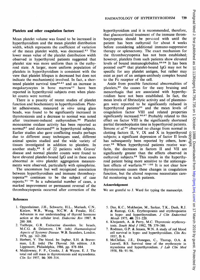
Mean platelet volume was found to be increased inhyperthyroidism and the mean.platelet distributionwidth, which represents the coefficient of variationof the mean platelet width, was decreased.83 Thelower mean value of the platelet distribution widthobserved in hyperthyroid patients suggested thatplatelet size was more uniform than in the euthy-roid state. A larger, more uniform population ofplatelets in hyperthyroidism is consistent with theview that platelet lifespan is decreased but does notindicate the mechanism(s) involved. In fact, a shor-tened platelet survival time84'85 and an increase inmegakaryocytes in bone marrow 2 have beenreported in hyperthyroid subjects even when plate-let counts were normal.
There is a paucity of recent studies of plateletfunction and biochemistry in hyperthyroidism. Plate-let adhesiveness, measured in vitro using glassbeads, was found to be significantly increased inthyrotoxicosis and a decrease to normal was notedafter treatment-induced euthyroidism.86 Plateletmonoamine oxidase activity has been reported asnormal87 and decreased88 in hyperthyroid subjects.Earlier studies also gave conflicting results perhapsdue to different assay techniques used, differentanimal species studied and the wide variety oftissues investigated in addition to platelets. Inanother study,89 8 of 22 patients with Graves'disease and normal platelet counts were found tohave elevated platelet-bound IgG and in these casesabnormal in vitro platelet aggregation measure-ments were observed, particularly with epinephrine.The uncommon but well recognized association
between hyperthyroidism and immune thrombocy-topenia90 continues to be the subject of casereports.91 -94 In a substantial number of cases, amarked improvement or permanent reversal of thethrombocytopenia occurred after correction of the
hyperthyroidism and it is recommended, therefore,that glucocorticoid treatment of the immune throm-bocytopenia should be persisted with until thepatient has been euthyroid for about 4 weeksbefore considering additional immuno-suppressivetherapy or splenectomy. The exact mechanism forthe thrombocytopenia has not been established;however, platelets from such patients show elevatedlevels of bound immunoglobulins.89'94 It has beenpointed out89 that platelet-bound IgG may not bespecific for any platelet antigen, but rather mayexist as part of an antigen-antibody complex boundto the Fc receptor of the cell.
Aside from possible functional abnormalities ofplatelets,89 the causes for the easy bruising andmenorrhagia that are associated with hyperthy-roidism have not been established. In fact, themean levels of fibrinolytic activity and of plasmino-gen were reported to be significantly reduced inhyperthyroid patients95 and the mean levels ofcoagulation factor VIII and related propertiessignificantly increased.96'97 Probably related to thiseffect on factor VIII is the significantly shortenedpartial thromboplastin time in hyperthyroid patients.Simone et al.96 observed no change from normal inclotting factors II, V, IX and X in hyperthyroidsubjects; a significant depression of factor II levelshas subsequently been reported by others, how-ever.98 When hyperthyroid patients receive war-farin, the decreases in factors II and VII aresignificantly greater than the effects observed ineuthyroid subjects.98 This results in the hyperthy-roid patient being more sensitive to the anticoagu-lant effects of warfarin.98-100 It is not clear howthyrotoxicosis causes these changes in coagulationfunction, but the altered response necessitates care-ful monitoring in such patients.
AcknowledgementWe are grateful to J. Ward for typing the manuscript.
References
1. Oppenheimer, J.H., Schwartz, H.L., Mariash, C.N.,Kinlaw, W.B., Wong, N.C.W. & Freake, H.C.Advances in our understanding of thyroid hormoneaction at the cellular level. Endocrine Rev 1987, 8:288-308.
2. Tudhope, G.R. Endocrine disorders. In: Israels,M.C.G. & Delamore, I.W. (eds) HaematologicalAspects of Systemic Disease. W.B. Saunders, London,1976, pp. 162-200.
3. Herbert, V. The blood. In: Ingbar, S.H. & Braver-man, L.E. (eds) The Thyroid, 5th edition. J.B.Lippincott, Philadelphia, 1986, pp. 878-884.
4. Muldowney, F. P., Crooks, J. & Wayne, E. J. Thetotal red cell mass in thyrotoxicosis and myxoedema.Clin Sci 1957, 16: 309-314.
5. Das, K.C., Mukherjee, M., Sarkar, T.K., Dash, R.J.& Rastogi, G.K. Erythropoiesis and erythropoietinin hypo- and hyperthyroidism. J Clin EndocrinolMetab 1975, 40: 211-220.
6. Khojasteh, A. & Perry, M.C. Thyrotoxic erythrocy-tosis. South Med J 1982, 75: 379-380.
7. Rodman, G.P. & Jensen, W.N. A study of red bloodcell survival in hypo- and hyperthyroidism. Clin Res1957, 5: 8.
8. McClellan, J.E., Donegan, C., Thorup, O.A. &Leavell, B.S. Survival time of the erythrocyte inmyxedema and hyperthyroidism. J Lab Clin Med1958, 51: 91-96.
copyright. on 17 M
arch 2019 by guest. Protected by
http://pmj.bm
j.com/
Postgrad M
ed J: first published as 10.1136/pgmj.64.756.735 on 1 O
ctober 1988. Dow
nloaded from
740 H.C. FORD AND J.M. CARTER
9. Gibson, J.G. II & Harris, A.W. Clinical studies ofthe blood volume. V. Hyperthyroidism and myx-edema. J Clin Invest 1939, 18: 59-65.
10. Peschle, C., Zanjani, E.D., Gidarl, A.S., McLaurin,W.D. & Gordon, A.S. Mechanism of thyroxineaction on erythropoiesis. Endocrinology 1971, 89:609-612.
11. Golde, D.W., Bersh, N., Chopra, I.J. & Cline, M.J.Thyroid hormones stimulate erythropoiesis in vitro.Br J Haematol 1977, 37: 173-177.
12. Popovic, W.J., Brown, J.E. & Adamson, J.W. Theinfluence of thyroid hormones on in vitro erythro-poiesis. J Clin Invest 1977, 60: 907-913.
13. Dainiak, N., Sutter, D. & Kreczko, S. L-Triiodothyronine augments erythropoietic growthfactor release from peripheral blood and bonemarrow leukocytes. Blood 1986, 68: 1289-1297.
14. Bistrom, O. On the morphology of blood and bone-marrow in thyrotoxicosis. Acta Chir Scand 1946,(Suppl.) 114.
15. Axelrod, A.R. & Berman, L. The bone marrow inhyperthyroidism and hypothyroidism. Blood 1951, 6:436-453.
16. Donati, R.M., Warnecke, M.A. & Gallagher, N.I.Ferrokinetics in hyperthyroidism. Ann Intern Med1965, 63: 945-950.
17. Rivlin, R.S. & Wagner, H.N. Anemia in hyperthyr-oidism. Ann Intern Med 1969, 70: 507-516.
18. Lahtinen, R. Sideroblasts and haemosiderin in thyro-toxicosis. Scand J Haematol 1980, 25: 237-243.
19. Nightingale, S., Vitek, P.J. & Himsworth, R.L. Thehaematology of hyperthyroidism. Q J Med 1978, 47:35-47.
20. How, J., Davidson, R.J.L. & Bewsher, P.D. Red cellchanges in hyperthyroidism. Scand J Haematol 1979,23: 323-328.
21. Reddy, J., Brownlie, B.E.W., Heaton, D.C., Hamer,J.W. & Turner, J.G. The peripheral blood picture inthyrotoxicosis. N Z Med J 1981, 93: 143-145.
22. Dintenfass, L., Forbes, C.D. & McDougall, I.R.Blood viscosity in hyperthyroid and hypothyroidpatients. Haemostasis 1974, 3: 348-352.
23. Cole, C.H. & Waddell, R.W. Alteration in intracellu-lar sodium concentration and ouabain-sensitiveATPase in erythrocytes from hyperthyroid patients. JClin Endocrinol Metab 1976, 42: 1056-1063.
24. Smith, E.K.M. & Samuel, P.D. Abnormalities in thesodium pump of erythrocytes from patients withhyperthyroidism. Clin Sci 1970, 38: 49-61.
25. Michels, R.C., Ober, K.P. & Hennessy, J.F. Cationtransport in intact erythrocytes of hyperthyroidpatients: role of the NaK-ATPase pump. HormMetab Res 1981, 13: 635-638.
26. DeLuise, M. & Flier, J.S. Status of the red cell Na,K-pump in hyper- and hypothyroidism. Metabolism1983, 32: 25-30.
27. Rubython, E.J., Cumberbatch, M. & Morgan, D.B..Changes in the number and activity of sodiumpumps in erythrocytes'from patients with hyperthyr-oidism. Clin Sci 1983, 64: 441-447.
28. Suzuki, K. & Fujino, R. Alteration in activities ofNa, K, ATPase, sugar transport and insulin recep-tors in erythrocytes from hyperthyroid patients.Metabolism 1986, 35: 371-377.
29. Khan, F.A. & Baron, D.N. Ion influx and Na', K+-ATPase activity of erythrocytes and leucocytes inthyroid disease. Clin Sci 1987, 72: 171-179.
30. Sutterlin, U., Gless, K-H., Schaz, K., Hufner, M.,Schutz, V. & Hunstein, W. Peripheral effects ofthyroid hormones: alteration of intracellular Na-concentration, ouabain-sensitive Na-transport, andNa-Li countertransport in human red blood cells.Klin Wochenschr 1984, 62: 598-601.
31. Boekelman, W.A. Sodium content of erythrocytes inhyperthyroidism. Nature 1958, 181: 1136.
32. Swaminathan, R., Chapman, C., Segall, N.H. &Morgan, D.B. Red-blood-cell composition in thyroiddisease. Lancet 1976, ii: 1382-1385.
33. Monti, M., Hedner, P., Ikomi-Kumm, J. & Valde-marsson, S. Erythrocyte thermogenesis in hyperthyr-oid patients: microcalorimetric investigation ofsodium/potassium pump and cell metabolism. Meta-bolism 1987, 36: 155-159.
34. Suzuki, K., Kadowaki, T., Sekimizu, M. & Shishiba,Y. Erythrocyte ouabain binding capacity as a poss-ible cellular index of hyperthyroid status. EndocrinolJpn 1983; 30: 609-614.
35. Laville, M., Riou, J.P., Bougneres, P.F. et al. Glu-cose metabolism in experimental hyperthyroidism:intact in vivo sensitivity to insulin with abnormalbinding and increased glucose turnover. J ClinEndocrinol Metab 1984, 58: 960-965.
36. Edelman, I.S. Thyroid thermogenesis. N Engl J Med1974, 290: 1303-1308.
37. Smith, T.J. & Edelman, I.S. The role of sodiumtransport in thyroid thermogenesis. Fed Proc 1979,38: 2150-2153.
38. Flaim, K.E., Li, J.B. & Jefferson, L.S. Effects ofthyroxine on protein turnover in rat skeletal muscle.Am J Physiol 1978, 235: E231-E237.
39. Dube, M.P., Davis, F.B., Davis, P.J. Schoenl, M. &Blas, S.D. Effects of hyperthyroidism and hypothyr-oidism on human red blood cell Ca2 +-ATPaseactivity. J Clin Endocrinol Metab 1986, 62: 253-257.
40. Pangaro, J.A., Weinstein, M., Devetak, M.C. &Soto, R.J. Red cell zinc and red cell zinc metalloen-zymes in hyperthyroidism. Acta Endocrinol 1974, 76:645-650.
41. Bremner, W.F. & Fell, G.S. Zinc metabolism andthyroid status. Postgrad Med J 1977, 53: 143-145.
42. Nishi, Y., Kawate, R. & Usui, T. Zinc metabolism inthyroid disease. Postgrad Med J 1980, 56: 833-837.
43. Weatherall, D.J. & McIntyre, P.A. Developmentaland acquired variations in erythrocyte carbonicanhydrase isozymes. Br J Haem 1967, 13: 106-114.
44. Magid, E. Determination of erythrocyte carbonicanhydrase B & C: an aid in the diagnosis of thyroiddisorders? Scand J Clin Lab Invest 1970, 26: 257-262.
45. Funakoshi, S. & Deutsch, H.F. Human carbonicanhydrase. V. Levels in erythrocytes in variousstates. J Lab Clin Med 1971, 77: 39-45.
46. Anker, N. & Mondrup, M. Carbonic anhydraseisoenzyme B in erythrocytes of subjects with thyroiddisorders. Clin Chim Acta 1974, 54: 277-282.
47. Angelone, L., Watkins, D.H. & Angerer, C.A. Oxy-gen consumption of erythrocytes from patients withvarious thyroid conditions related to their respective
copyright. on 17 M
arch 2019 by guest. Protected by
http://pmj.bm
j.com/
Postgrad M
ed J: first published as 10.1136/pgmj.64.756.735 on 1 O
ctober 1988. Dow
nloaded from
HAEMATOLOGY OF HYPERTHYROIDISM 741
serum protein-bound iodine concentrations. Blood1954, 9: 953-958.
48. Monti, M. & Wadso, I. Microcalorimetric measure-ments of heat production in human erythrocytes. II.Hyperthyroid patients before, during and after treat-ment. Acta Med Scand 1976, 200: 301-308.
49. Goebel, K.M., Goebel, F.D., Neitzert, A., Haus-mann, L. & Schneider, J. Adaptation of red cellenzymes and intermediates in metabolic disorders.Enzyme 1975, 19: 201-211.
50. Murphy, J.R. Erythrocyte metabolism. II. Glucosemetabolism and pathways. J Lab Clin Med 1960, 55:286-302.
51. Monti, M., Hedner, P., Ikomi-Kumm, J. & Valde-marsson, S. Erythrocyte metabolism in hyperthyr-oidism: a microcalorimetric study on changes in theEmbden-Meyerhof and the hexose monophosphatepathways. Acta Endocrinol 1987, 115: 87-90.
52. Bansi, H.W. & Groscurth, G. Veranderungen derSauerstoffbindungskurven des Blutes beiStoffwechsel- und Blutkrankheiten (Anamie undPolycythamie). Z Klin Med 1930, 113: 560-575.
53. Gahlenbeck, H. & Bartels, H. Veranderung derSauerstoffbindungskurven des Blutes bei Hyperthyr-eosen und nach Gabe von Trijodthyronin bei Gesun-den und bei Ratten. Klin Wochenschr 1968, 46:547-548.
54. Benesch, R. & Benesch, R.E. The effect of organicphosphates from the human erythrocyte on the allos-teric properties of hemoglobin. Biochem Biophys ResCommun 1967, 26: 162-167.
55. Chanutin, A. & Curnish, R.R. Effect of organic andinorganic phosphates on the oxygen equilibrium ofhuman erythrocytes. Arch Biochem Biophys 1967,121: 96-102.
56. Miller, L.D., Sugarman, H.J., Miller, W.W. et al.Increased peripheral oxygen delivery in thyrotoxico-sis: role of red cell 2,3-diphosphoglycerate. Ann Surg1970, 172: 1051-1058.
57. Snyder, L.M. & Reddy, W.J. Thyroid hormonecontrol of erythrocyte 2,3-diphosphoglyceric acidconcentrations. Science 1970, 169: 879-880.
58. Monti, M. Red cell 2,3-diphosphoglycerate inpatients with hyperthyroidism before and after treat-ment. Acta Med Scand 1974, 196: 263-266.
59. Alvarez-Sala, J.L., Urban, M.A., Sicilia, J.J., DiazFdez, A.J., Fdez Mendieta, F. & Espinos, D. Red-cell 2,3-diphosphoglycerate in patients with hyper-thyroidism. Acta Endocrinol 1980, 93: 424-429.
60. Viherkoski, M. & Lamberg, B-A. The glucose-6-phosphate dehydrogenase activity (G-6-PD) of thered blood cells in hyperthyroidism and hypothyr-oidism. Scand J Clin Lab Invest 1970, 25: 137-143.
61. Zaroulis, C.G., Kourides, I.A. & Valeri, C.R. Redcell 2,3-diphosphoglycerate and oxygen affinity ofhemoglobin in patients with thyroid disorders. Blood1978, 52: 181-185.
62. Kurasaki, M., Saito, T., Kaji, H., Kojima, Y. &Saito, K. Increased erythrocyte catalase activity inpatients with hyperthyroidism. Horm Metab Res1986, 18: 56-59.
63. Cohen, J., Somma-Delpero, C., Verine, A., Codac-cioni, J.L. & Boyer, J. Increased monoester lipase
activity in red blood cells during hyperthyroidism. JEndocrinol 1986, 108: 357-359.
64. Kuhn, J-M., Rieu, M., Rochette, J. et al. Influenceof thyroid status on hemoglobin A2 expression. JClin Endocrinol Metab 1983, 57: 344-348.
65. Kendall, A.G. & Bastomsky, C.H. Hemoglobin A2in hyperthyroidism. Hemoglobin 1981, 5: 571-577.
66. Fuhr, J.E. & Dunn, C.D.R. Control of hemoglobinsynthesis in fetal erythroid cells by L-thyroxine. AmJ Hematol 1978, 5: 163-168.
67. Caro-Posen, L. Ein Fall von malignem MorbusBasedowii kombiniert mit den Symptomen der Pseu-doleukamie. Berl Klin Wochenschr 1907, 54: 519-520.
68. Kocher, T. Blutintersuchungen bei Morbus Base-dowii mit Beitrage zur Fruhdiagnose und Theorieder Krankheit. Arch Klin Chirurg 1908, 87: 131-157.
69. Irvine, W.J., Wu, F.C.W., Urbaniak, S.J. & Toolis,F. Peripheral blood leucocytes in thyrotoxicosis(Graves' disease) as studied by conventional lightmicroscopy. Clin Exp Immunol 1977, 27: 216-221.
70. Eakin, D.L., Peake, R.L. & Weiss, G.B. Effect oftherapy on the neutropenia of hyperthyroidism.South Med J 1983, 76: 335-340.
71. Mori, H., Amino, N. Iwatani, Y. et al. Increase ofperipheral B lymphocytes in Graves' disease. ClinExp Immunol 1980, 42: 33-40.
72. Iwatani, Y., Amino, N., Mori, H. et al. T lympho-cyte subsets in autoimmune thyroid diseases andsubacute thyroiditis detected with monoclonal anti-bodies. J Clin Endocrinol Metab 1983, 56: 251-254.
73. Chan, J.Y.C. & Walfish, P.G. Activated (Ia')T-lymphocytes and their subsets in autoimmunethyroid diseases: analysis by dual laser flow micro-fluorocytometry. J Clin Endocrinol Metab 1986, 62:403-409.
74. Van Ouwerkerk, B.M., Krenning, E.P., Doctor, R.,et al. Cellular and humoral immunity in patientswith hyperthyroid Graves' disease before, during andafter antithyroid drug treatment. Clin Endocrinol1987, 26: 385-394.
75. Madec, A.M., Allannic, H., Genetet, N. et al. Tlymphocyte subsets at various stages of hyperthyroidGraves' disease: effect of carbimazole treatment andrelationship with thyroid-stimulating antibody levelsor HLA status. J Clin Endocrinol Metab 1986, 62:117-121.
76. Buse, J.B. & Eisenbarth, G.S. Autoimmune endoc-rine disease. Vitam Horm 1985, 42: 253-314.
77. Volpe, R. Immunoregulation in autoimmune thyroiddisease. N Engl J Med 1987, 316: 44-46.
78. Hrycek, A. & Kalina, Z. Changes in the metabolismand functions of peripheral blood neutrophils inpatients with thyroid diseases. Endokrynol Pol 1984,35: 45-50.
79. Schernthaner, G., Prager, R., Weissel, M. & Hofer,R. Decreased insulin receptor binding in hyperthyr-oidism. Klin Wochenschr 1984, 62: 1074-1080.
80. Ginsberg, A.M., Clutter, W.E., Shah, S.D. & Cryer,P.E. Triiodothyronine-induced thyrotoxicosisincreases mononuclear leukocyte /,-adrenergic recep-tor density in man. J Clin Invest 1981, 67:1785-1791.
81. Andersson, R.G.G., Nilsson, O.R. & Kuo, J.F.
copyright. on 17 M
arch 2019 by guest. Protected by
http://pmj.bm
j.com/
Postgrad M
ed J: first published as 10.1136/pgmj.64.756.735 on 1 O
ctober 1988. Dow
nloaded from
742 H.C. FORD AND J.M. CARTER
f3-Adrenoreceptor-adenoside, 3',5'-monophosphatesystem in human leucocytes before and after treat-ment for hyperthyroidism. J Clin Endocrinol Metab1983, 56: 42-45.
82. Ratge, D., Hansel-Bessey, S. & Wisser, H. Alteredplasma catecholamines and number of a- and /l-adrenergic receptors in platelets and leucocytes inhyperthyroid patients normalized under antithyroidtreatment. Acta Endocrinol 1985, 110: 75-82.
83. Ford, H.C., Toomath, R.J., Carter, J.M. et al. Meanplatelet volume is increased in hyperthyroidism. AmJ Hematol 1988, 27: 190-193.
84. Lamberg, B-A., Kivikangas, V., Pelkonen, R. &Vuopio, P. Thrombocytopenia and decreased life-span of thrombocytes in hyperthyroidism. Ann ClinRes 1971, 3: 98-102.
85. Kurata, Y., Nishioeda, Y., Tsubakio, T. & Kitani, T.Thrombocytopenia in Graves' disease: effect of T3on platelet kinetics. Acta Haematol 1980, 63:185-190.
86. Hellem, A.J., Segaard, E. & Solem, J.H. The adhesi-veness of human blood platelets and thyroid func-tion. Acta Med Scand 1975, 197: 15-17.
87. Feldman, J.M. & Roche, J. Effect of hyper- andhypothyroidism on platelet monoamine oxidaseactivity and serotonin metabolism. Metabolism 1977,26: 657-664.
88. Singh, H.C., Sarkar, F.H., Singh, R.H. & Udupa,K.N. Blood platelet monoamine oxidase and acetyl-cholinesterase in thyrotoxicosis. Indian J Med Res1980, 71: 384-386.
89. Hymes, K., Blum, M., Lackner, H. & Karpatkin, S.Easy bruising, thrombocytopenia, and elevated plate-let immunoglobulin G in Graves' disease and Hashi-moto's thyroiditis. Ann Intern Med 1981, 94: 27-30.
90. Jackson, A.S. Acute hemorrhagic purpura associatedwith exophthalmic goiter. JAMA 1931, 96: 38-39.
91. Herman, J., Resnitzky, P. & Fink, A. Associationbetween thyrotoxicosis and thrombocytopenia. IsraelJ Med Sci 1978, 14: 469-475.
92. Adrouny, A., Sandler, R.M. & Carmel, R. Variablepresentation of thrombocytopenia in Graves' disease.Arch Intern Med 1982, 142: 1460-1464.
93. Liechty, R.D. The thyrotoxicosis/thrombocytopeniaconnection. Surgery 1983, 94: 966-968.
94. Jacobs, P., Majoos, F. & Perrotta, A. Hyperthyr-oidism and immune thrombocytopenia. PostgradMed J 1984, 60: 657-661.
95. Rennie, J.A.N., Bewsher, P.D., Murchison, L.E. &Ogston, D. Coagulation and fibrinolysis in thyroiddisease. Acta Haematol 1978, 59: 171-177.
96. Simone, J.V., Abildgaard, C.F. & Schulman, I.Blood coagulation in thyroid dysfunction. N Engl JMed 1965, 273: 1057-1061.
97. Rogers, J.S., Shane, S.R. & Jencks, F.S. Factor VIIIactivity and thyroid function. Ann Intern Med 1982,97: 713-716.
98. Kellett, H.A., Sawers, J.S.A., Boulton, F.E. et al.Problems of anticoagulation with warfarin inhyperthyroidism. Q J Med 1986, 58: 43-51.
99. Self, T., Weisburst, M., Wooten, E., Straughn, A. &Oliver, J. Warfarin-induced hypoprothrombinemia-potentiation by hyperthyroidism. JAMA 1975, 231:1165-1166.
100. Shenfield, G.M. Influence of thyroid dysfunction ondrug pharmacokinetics. Clin Pharmacokinet 1981, 6:275-297.
copyright. on 17 M
arch 2019 by guest. Protected by
http://pmj.bm
j.com/
Postgrad M
ed J: first published as 10.1136/pgmj.64.756.735 on 1 O
ctober 1988. Dow
nloaded from