Embed Size (px)
Citation preview
1
3 Major Regions of the Brain
• The Forebrain• The Midbrain• The Hindbrain• Not reflective of their position in the adult
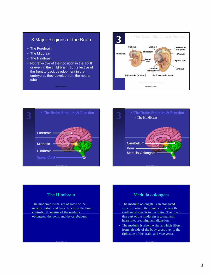
or even in the child brain. But reflective of the front to back development in the embryo as they develop from the neural tube.
Biological Basis 3
•• The Brain: Structure & FunctionThe Brain: Structure & Function333
Neural DevelopmentBiological Basis 3
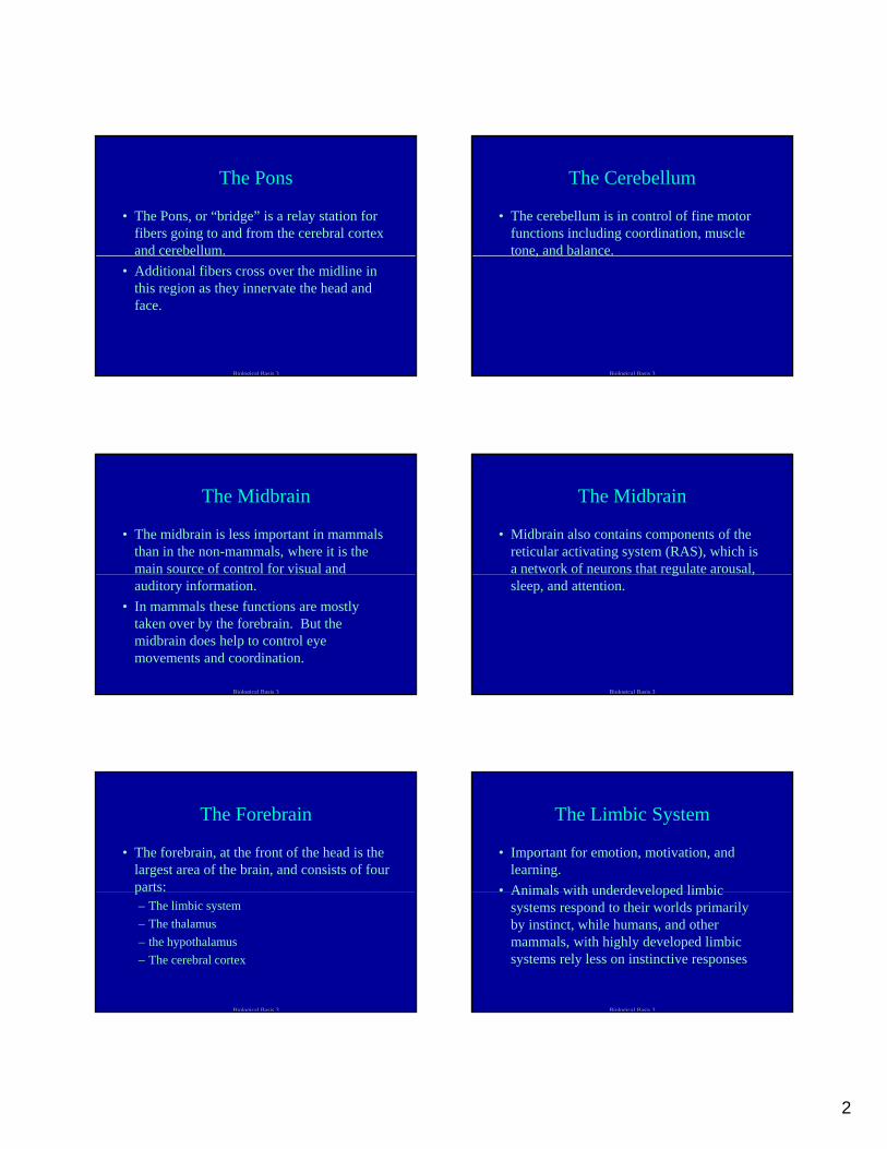
•• The Brain: Structure & FunctionThe Brain: Structure & Function
ForebrainForebrain
333
MidbrainMidbrain
HindbrainHindbrain
Spinal CordSpinal Cord
Biological Basis 3
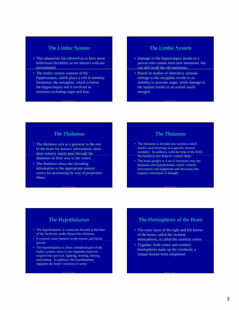
•• The Brain: Structure & FunctionThe Brain: Structure & Function–– The HindbrainThe Hindbrain333
Medulla OblongataMedulla OblongataPonsPonsCerebellumCerebellum
Biological Basis 3
The Hindbrain
• The hindbrain is the site of some of the most primitive and basic functions the brain controls. It consists of the medulla oblongata, the pons, and the cerebellum.
Biological Basis 3
Medulla oblongata
• The medulla oblongata is an elongated structure where the spinal cord enters the skull and connects to the brain. The role of this part of the hindbrain is to maintain heart rate, breathing and digestion.
• The medulla is also the site at which fibers from left side of the body cross over to the right side of the brain, and vice versa.
Biological Basis 3
2
The Pons
• The Pons, or “bridge” is a relay station for fibers going to and from the cerebral cortex and cerebellum.
• Additional fibers cross over the midline in this region as they innervate the head and face.
Biological Basis 3
The Cerebellum
• The cerebellum is in control of fine motor functions including coordination, muscle tone, and balance.,
Biological Basis 3
The Midbrain
• The midbrain is less important in mammals than in the non-mammals, where it is the main source of control for visual and auditory information.
• In mammals these functions are mostly taken over by the forebrain. But the midbrain does help to control eye movements and coordination.
Biological Basis 3
The Midbrain
• Midbrain also contains components of the reticular activating system (RAS), which is a network of neurons that regulate arousal, g ,sleep, and attention.
Biological Basis 3
The Forebrain
• The forebrain, at the front of the head is the largest area of the brain, and consists of four parts:p– The limbic system– The thalamus– the hypothalamus– The cerebral cortex
Biological Basis 3
The Limbic System
• Important for emotion, motivation, and learning.
• Animals with underdeveloped limbicAnimals with underdeveloped limbic systems respond to their worlds primarily by instinct, while humans, and other mammals, with highly developed limbic systems rely less on instinctive responses
Biological Basis 3
3
The Limbic System
• This adaptation has allowed us to have more behavioral flexibility as we interact with our environment
• The limbic system consists of the hippocampus, which plays a role in memory formation; the amygdala, which is below the hippocampus and is involved in emotions including anger and fear.
Biological Basis 3
The Limbic System
• Damage to the hippocampus results in a person who cannot form new memories, but can still recall the old memories.
• Based on studies of laboratory animals, damage to the amygdala results in an inability to provoke anger, while damage to the septum results in an animal easily enraged.
Biological Basis 3
The Thalamus
• The thalamus acts as a gateway to the rest of the brain for sensory information, since most sensory inputs pass through the y p p gthalamus on their way to the cortex.
• The thalamus relays the incoming information to the appropriate sensory cortex for processing by way of projection fibers.
Biological Basis 3
The Thalamus
• The thalamus is divided into sections called nuclei, each focusing on a specific sensory modality. In addition, with the help of the RAS, h h l l h l l lthe thalamus also helps to control sleep
• The basal ganglia is a set of structures near the thalamus and hypothalamus which controls movements and judgments and decisions that require a minimum of thought.
Biological Basis 3
The Hypothalamus
• The hypothalamus is a structure located at the base of the forebrain, under (hypo) the thalamus.
• It controls water balance in the tissues and blood stream.
• The hypothalamus is often considered part of the limbic system, since it also regulates behavior required for survival: fighting, feeding, fleeing, and mating. In addition, the hypothalamus regulates the body’s reaction to stress
Biological Basis 3
The Hemispheres of the Brain
• The outer layer of the right and left halves of the brain, called the cerebral hemispheres, is called the cerebral cortexp ,
• Together, both cortex and cerebral hemispheres make up the cerebrum, a unique human brain adaptation
Biological Basis 3
4
The Cerebral Cortex
• The cerebral cortex layer is 2 mm thick and enfolds the surface of the brain. In humans, this layer is highly convoluted and comprises about 80% f h h b i80% of the human brain.
• This layer is responsible for most of our higher functions, such as our ability to plan, coordinate thoughts and actions, perceived sensory information, use language, and, in general, think.
Biological Basis 3
The Cerebral Cortex
• The cerebral cortex is comprised of gray and white tissue, with a gray tissue being the cell bodies of neurons and the white tissue being the axons of those neurons covered in myelin. Both white matter and gray matter are important for human intelligence.
Biological Basis 3
The Cerebral Cortex
• The cerebral cortex is the outer covering of both the left and right cerebral hemispheres, which are similar in appearance, and are pp ,connected by a dense fiber pathway, called the corpus callosum.
• Although similar looking, the two hemispheres actually process different types of information.
Biological Basis 3
The Cerebral Cortex
• Fibers coming from, or going to, the left side of the body are processed by the right hemisphere, while the left hemisphere p , pprocesses information pertaining to the right side of the body.
Biological Basis 3
The Cerebral Cortex
• Not all information transmission is contralateral in this fashion, some information is ipsilateral, or stays on the p , ysame side of the brain from which the pathway originated.
• The corpus callosum allows for the easy transfer of information between the two hemispheres
Biological Basis 3
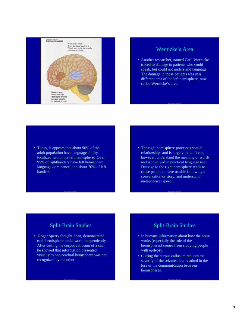
Broca’s Area
• Paul Broca demonstrated in 1861 that damage on the left hemisphere could result in aphasia, or loss of speech. This finding p , p gstood the test of time and the area where the lesions were found is called a Broca’s area.
Biological Basis 3
5
Biological Basis 3
Wernicke’s Area
• Another researcher, named Carl Wernicke traced to damage in patients who could speak, but could not understand language. p , g gThe damage in these patients was in a different area of the left hemisphere, now called Wernicke’s area.
Biological Basis 3
• Today, it appears that about 90% of the adult population have language ability localized within the left hemisphere. Over p95% of righthanders have left hemisphere language dominance, and about 70% of left-handers.
Biological Basis 3
• The right hemisphere processes spatial relationships and is largely mute. It can, however, understand the meaning of words , gand is involved in practical language use. Damage to the right hemisphere tends to cause people to have trouble following a conversation or story, and understand metaphorical speech.
Biological Basis 3
Split-Brain Studies
• Roger Sperry thought, then, demonstrated each hemisphere could work independently. After cutting the corpus callosum of a cat, g p ,he showed that information presented visually to one cerebral hemisphere was not recognized by the other.
Biological Basis 3
Split-Brain Studies
• In humans information about how the brain works (especially the role of the hemispheres) comes from studying people p ) y g p pwith epilepsy.
• Cutting the corpus callosum reduces the severity of the seizures, but resulted in the loss of the communication between hemispheres.
Biological Basis 3
6
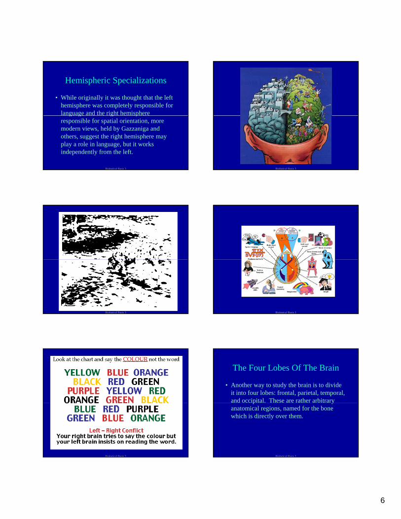
Hemispheric Specializations
• While originally it was thought that the left hemisphere was completely responsible for language and the right hemisphere g g g presponsible for spatial orientation, more modern views, held by Gazzaniga and others, suggest the right hemisphere may play a role in language, but it works independently from the left.
Biological Basis 3 Biological Basis 3
Biological Basis 3 Biological Basis 3
Biological Basis 3
The Four Lobes Of The Brain
• Another way to study the brain is to divide it into four lobes: frontal, parietal, temporal, and occipital. These are rather arbitrary p yanatomical regions, named for the bone which is directly over them.
Biological Basis 3

7
Biological Basis 3
The Four Lobes Of The Brain
• Generally abstract thought and motor processing occurs in the frontal lobe, somatosensory processing of sensations y p goccurs in the parietal, and auditory and visual processing occurring in the temporal and occipital lobes, respectively.
Biological Basis 3
The Frontal Lobe
• The frontal lobe contains the primary motor cortex, which specializes in the planning, control, and execution of movement to ,requiring a delayed response.
• Control a body movements is contralateral and originates in the primary motor cortex.
Biological Basis 3 Biological Basis 3
Biological Basis 3
The Frontal Lobe
• An inverse mapping occurs from top to bottom, with the lower portions of the body represented on the upper side of the motor cortex and the upper
f h b d d h l id fpart of the body represented on the lower side of the motor cortex.
• This map is called a hommunculus, which means the little person. The map motor functioning is mirrored by a map of sensory functioning, controlled by the parietal lobe.
Biological Basis 3

8
Biological Basis 3
The Parietal Lobe
• The parietal lobe contains the primary sensory cortex, which receives input from the senses regarding pressure, texture, g g p , ,temperature, and pain.
• It is located behind the primary motor cortex.
Biological Basis 3
The Parietal Lobe
• Stimulation of the primary sensory cortex would most likely lead to a report of being touched.
• As with the motor cortex, the more sensitivity and fine control we need in a particular part of the body, the larger the neural representation of that body part in the primary cortex
Biological Basis 3
The Temporal Lobe
• The region of the cerebral cortex pertaining to hearing is in the temporal lobe, just above the ear.
• The temporal lobe is specialized in such a way that the different aspects of pitch are processed in different regions.
Biological Basis 3
The Temporal Lobe
• The sense of hearing is primarily contralateral, but each auditory cortex has some representation of sound for both the ears; therefore damage to the
l l b di h bili d dtemporal lobe can disrupt the ability to understand speech, or reduce the ability to hear, in general. Hearing is coordinated with vision, controlled by the occipital lobe, in order for us to understand movies and television through the combination of auditory and visual cues
Biological Basis 3
The Occipital Lobe
• The occipital lobe is the visual region of the cerebral cortex, located at the back of the head. Some neurons carrying visual i f i f h i il linformation from the eyes stay ipsilateral, from the left eye to the left occipital cortex, but others cross at the optic chiasm to form contralateral projections. Stimulation of the occipital lobe results in the perception of disorganized light patterns.
Biological Basis 3
9
Biological Basis 3
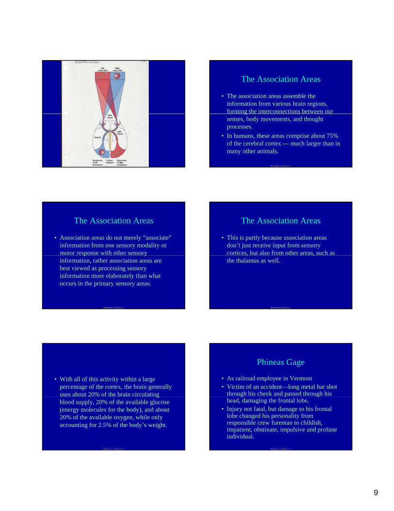
The Association Areas
• The association areas assemble the information from various brain regions, forming the interconnections between our gsenses, body movements, and thought processes.
• In humans, these areas comprise about 75% of the cerebral cortex --- much larger than in many other animals.
Biological Basis 3
The Association Areas
• Association areas do not merely “associate” information from one sensory modality or motor response with other sensory p yinformation, rather association areas are best viewed as processing sensory information more elaborately than what occurs in the primary sensory areas.
Biological Basis 3
The Association Areas
• This is partly because association areas don’t just receive input from sensory cortices, but also from other areas, such as , ,the thalamus as well.
Biological Basis 3
• With all of this activity within a large percentage of the cortex, the brain generally uses about 20% of the brain circulating gblood supply, 20% of the available glucose (energy molecules for the body), and about 20% of the available oxygen, while only accounting for 2.5% of the body’s weight.
Biological Basis 3
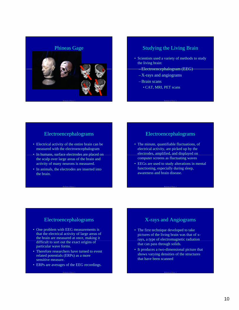
Phineas Gage
• As railroad employee in Vermont• Victim of an accident—long metal bar shot
through his cheek and passed through his g p ghead, damaging the frontal lobe.
• Injury not fatal, but damage to his frontal lobe changed his personality from responsible crew foreman to childish, impatient, obstinate, impulsive and profane individual.
Biological Basis 3
10
Phineas Gage
Biological Basis 3
Studying the Living Brain
• Scientists used a variety of methods to study the living brain:
Electroencephalogram (EEG)– Electroencephalogram (EEG)– X-rays and angiograms– Brain scans
• CAT, MRI, PET scans
Biological Basis 3
Electroencephalograms
• Electrical activity of the entire brain can be measured with the electroencephalogram
• In humans surface electrodes are placed onIn humans, surface electrodes are placed on the scalp over large areas of the brain and activity of many neurons is measured.
• In animals, the electrodes are inserted into the brain.
Biological Basis 3
Electroencephalograms
• The minute, quantifiable fluctuations, of electrical activity, are picked up by the electrodes, amplified, and displayed on , p , p ycomputer screens as fluctuating waves
• EEGs are used to study alterations in mental functioning, especially during sleep, awareness and brain disease.
Biological Basis 3
Electroencephalograms
• One problem with EEG measurements is that the electrical activity of large areas of the brain are measured at once, making it diffi l h i i fdifficult to sort out the exact origins of particular wave forms.
• Therefore researchers have turned to event related potentials (ERPs) as a more sensitive measure.
• ERPs are averages of the EEG recordings.
Biological Basis 3
X-rays and Angiograms
• The first technique developed to take pictures of the living brain was that of x-rays, a type of electromagnetic radiation y , yp gthat can pass through solids.
• It produces a two-dimensional picture that shows varying densities of the structures that have been scanned
Biological Basis 3
11
X-rays and Angiograms
• Most areas of the brain have roughly the same density so x-rays of the head are useful for showing small fractures of the gskull but little else of concern to psychologists.
Biological Basis 3
X-rays and Angiograms
• Angiograms are x-rays that provide more contrast due to special dyes injected into the blood vessels.
• Angiograms are usually used to study the heart, but are also used to study vascular diseases, and to locate particular kinds of brain tumors, and indicate what parts of the brain are active during various tasks.
Biological Basis 3
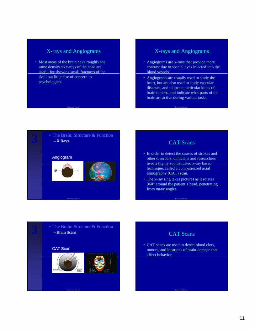
•• The Brain: Structure & FunctionThe Brain: Structure & Function–– X RaysX Rays
AngiogramAngiogram
333
Biological Basis 3
CAT Scans
• In order to detect the causes of strokes and other disorders, clinicians and researchers used a highly sophisticated x-ray based g y p ytechnique, called a computerized axial tomography (CAT) scan.
• The x-ray ring takes pictures as it rotates 360° around the patient’s head, penetrating from many angles.
Biological Basis 3
•• The Brain: Structure & FunctionThe Brain: Structure & Function–– Brain ScansBrain Scans
CAT ScanCAT Scan
333
Biological Basis 3
CAT Scans
• CAT scans are used to detect blood clots, tumors, and locations of brain-damage that affect behavior.
Biological Basis 3
12
MRIs
• Another sophisticated technique is magnetic resonance imaging (MRI).
• The MRI scanner resembles the CATThe MRI scanner resembles the CAT scanner, but it uses no radiation. Instead, it uses magnetic fields to take clearer, more detailed pictures of the brain.
Biological Basis 3
MRIs
• These fields change the orbits of nuclear particles in the molecules of the body, causing bursts of energy which can be g gydetected and analyzed by computer.
• The computer then generates a highly precise, three-dimensional picture.
Biological Basis 3
MRIs
• There are different types of MRI. – Structural MRIs take static anatomical pictures
and functional MRI (f MRI) measures changes ( ) gin the magnetic state of the blood as a function of its degree of oxygenation. Psychologists use fMRIs in an attempt to uncover how the brain works during mental tasks.
Biological Basis 3
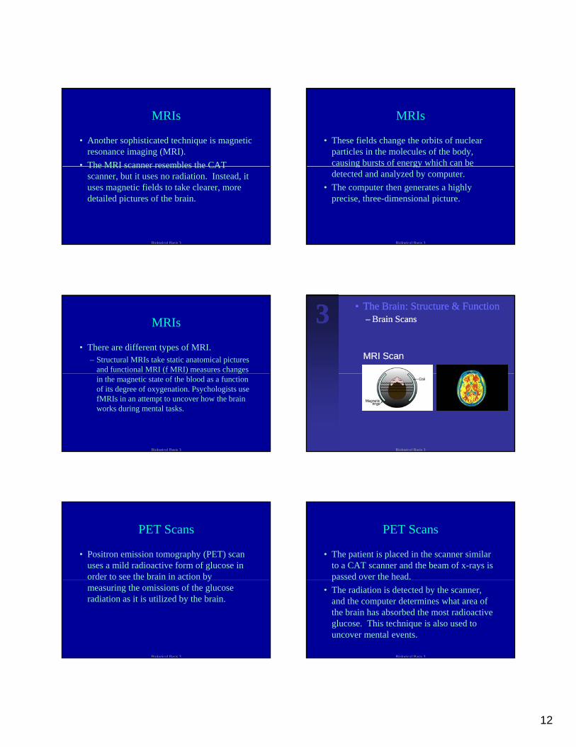
•• The Brain: Structure & FunctionThe Brain: Structure & Function–– Brain ScansBrain Scans
MRI ScanMRI Scan
333
Biological Basis 3
PET Scans
• Positron emission tomography (PET) scan uses a mild radioactive form of glucose in order to see the brain in action by ymeasuring the omissions of the glucose radiation as it is utilized by the brain.
Biological Basis 3
PET Scans
• The patient is placed in the scanner similar to a CAT scanner and the beam of x-rays is passed over the head.p
• The radiation is detected by the scanner, and the computer determines what area of the brain has absorbed the most radioactive glucose. This technique is also used to uncover mental events.
Biological Basis 3
13
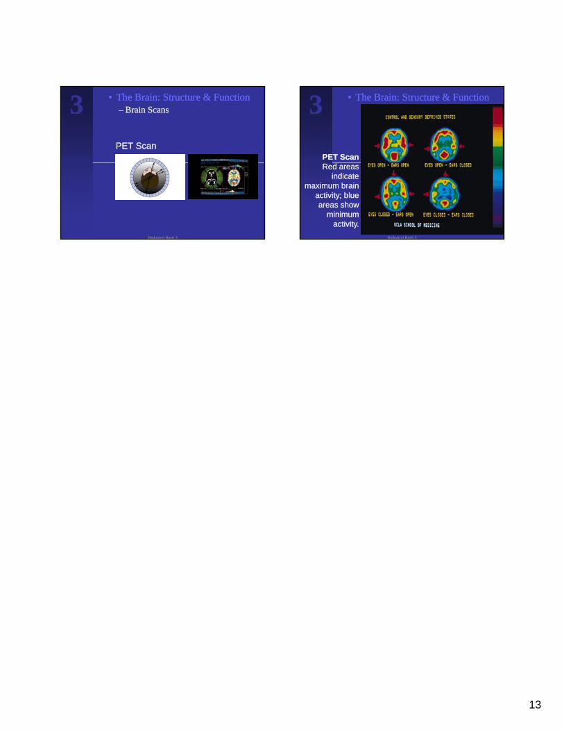
•• The Brain: Structure & FunctionThe Brain: Structure & Function–– Brain ScansBrain Scans
PET ScanPET Scan
333
Biological Basis 3
•• The Brain: Structure & FunctionThe Brain: Structure & Function333
PET ScanPET ScanRed areas Red areas
indicate indicate maximum brain maximum brain
activity; blue activity; blue areas show areas show
minimum minimum activity.activity.
Biological Basis 3