Embed Size (px)
Citation preview
Immunity
Article
The Histone Methyltransferase Wbp7Controls Macrophage Functionthrough GPI Glycolipid Anchor SynthesisLiv Austenaa,1 Iros Barozzi,1 Agnieszka Chronowska,1 Alberto Termanini,1 Renato Ostuni,1 Elena Prosperini,1
A. Francis Stewart,2 Giuseppe Testa,1 and Gioacchino Natoli1,*1Department of Experimental Oncology, European Institute of Oncology (IEO), Via Adamello 16, 20139 Milan, Italy2Genomics, BioInnovationsZentrum, Technische Universitaet Dresden (TUD), Tatzberg 47, 01307 Dresden, Gerrmany*Correspondence: [email protected]
DOI 10.1016/j.immuni.2012.02.016
SUMMARY
Histone methyltransferases catalyze site-specificdeposition of methyl groups, enabling recruitmentof transcriptional regulators. In mammals, trimethy-lation of lysine 4 in histone H3, a modification local-ized at the transcription start sites of active genes,is catalyzed by six enzymes (SET1a and SET1b,MLL1–MLL4) whose specific functions are largelyunknown. By using a genomic approach, we foundthat in macrophages, MLL4 (also known as Wbp7)was required for the expression of Pigp, an essentialcomponent of the GPI-GlcNAc transferase, theenzyme catalyzing the first step of glycosylphospha-tidylinositol (GPI) anchor synthesis. Impaired Pigpexpression in Wbp7�/� macrophages abolished GPIanchor-dependent loading of proteins on the cellmembrane. Consistently, loss of GPI-anchoredCD14, the coreceptor for lipopolysaccharide (LPS)and other bacterial molecules, markedly attenuatedLPS-triggered intracellular signals and gene expres-sion changes. These data link a histone-modifyingenzyme to a biosynthetic pathway and indicatea specialized biological role for Wbp7 in macrophagefunction and antimicrobial response.
INTRODUCTION
The inherent structure of chromatin imposes the need for dedi-
cated mechanisms to control accessibility and usage of the
underlying genetic material (Kornberg and Lorch, 1999). At the
same time it provides regulatory opportunities that are exploited
by eukaryotes to control both differential usage of the genomic
information during differentiation and the deployment of specific
gene expression programs in response to changing environ-
ments. At the heart of this control system are the N-terminal tails
of core histones, which undergo a wide array of posttranslational
modifications catalyzed by enzymes involved in multiple steps of
transcriptional control (Jenuwein and Allis, 2001; Kouzarides,
2007).
Histone tail methylation is dynamically controlled by site-
specific methylases and demethylases whose effect on trans-
572 Immunity 36, 572–585, April 20, 2012 ª2012 Elsevier Inc.
cription is dependent on which lysine (or arginine) is modified
and on the degree of methylation (mono-, di-, or trimethylation)
eventually achieved (Cloos et al., 2008; Klose and Zhang,
2007). For instance, trimethylation of K9 and K27 in histone H3
(H3K9me3 and H3K27me3) control transcriptional repression
and silencing (Boyer et al., 2006; Bracken et al., 2006; Lee
et al., 2006b; Peters et al., 2001), and the interaction between
H3K9me3 and HP1 proteins has become the paradigm for the
recognition of a histone modification by a specific ‘‘reader’’ in
charge of translating the modification into a functional outcome
(in this case repression and heterochromatin formation) (Lachner
et al., 2001).
In higher eukaryotes, an additional element of complexity is
represented by the appearance of multiple paralogs and/or
evolutionarily distinct enzymes effecting the same modification.
In some cases these paralogs act redundantly, as exemplified by
the two heterochromatic H3K9 trimethylases Suv39h1 and
Suv39h2 (Peters et al., 2001). However, clear functional differ-
ences can usually be seen among evolutionary unrelated
enzymes acting on the same site: for instance G9a (also known
as Ehmt2) and its dimerization partner (and paralog) Glp (also
known as Ehmt1) selectively catalyze H3K9 methylation in
euchromatin (Tachibana et al., 2002, 2005), thus demonstrating
a different biological function from the heterochromatin-specific
Suv39h1 and Suv39h2 (Peters et al., 2003).
H3K4me3 is associated with the transcription start sites (TSS)
of active or poised genes (Bernstein et al., 2005; Santos-Rosa
et al., 2002) and is detected by several recognition domains
present in transcriptional regulators, including the PHD finger,
the chromodomain, and the tudor domain (Huang et al., 2006;
Kim et al., 2006; Pena et al., 2006; Ruthenburg et al., 2007).
In spite of its correlation with gene activity, the functional role
of H3K4me3 is unclear. In vitro data indicate that this modifica-
tion has no direct effect on transcription (Pavri et al., 2006),
and deletion of the only H3K4 methyltransferase in the yeast
genome, Set1, hasmarginal consequences on the transcriptome
(Miller et al., 2001). A possible role in tuning splicing rates is sug-
gested by the association of an H3K4me3 binding protein, Chd1,
with spliceosome components (Sims et al., 2007). Rather than
promoting gene activation, in higher eukaryotes H3K4me3 may
prevent inappropriate silencing: in Drosophila, deletion of tri-
thorax groupH3K4methyltransferases causes loss of Hox genes
expression, which can be rescued by simultaneous deletion of
Polycomb group proteins (which control H3K27me3-dependent
repression) (Klymenko and Muller, 2004).

Immunity
MLL4 (Wbp7) Controls Macrophage Function
In mammals, H3K4me3 deposition is controlled by the Set1-
MLL family of enzymes, which includes six members: Set1a
and Set1b andMLL1–MLL4. Set-MLL proteins are found in three
multimolecular complexes, each one containing either of two
highly related paralogs as the catalytically active subunit: the
Set1a-Set1b complex, the MLL1-MLL4 complex, and the
MLL2-MLL3 complex (Supplemental Discussion available on-
line; Cho et al., 2007; Hughes et al., 2004; Smith et al., 2011;
Wu et al., 2008). Different subunit composition is responsible
for specific properties of different complexes. Set1a-Set1b
complexes contain a subunit, Wdr82 (Lee and Skalnik, 2008;
Wu et al., 2008), that mediates crosstalk between transcrip-
tion-coupled histone H2B monoubiquitination and H3K4
trimethylation (Kim et al., 2009). Because of this mechanism,
Set1a-Set1b complexes are recruited after transcriptional acti-
vation has initiated and therefore in general they have little
impact on transcription. However, Set1a-Set1b complexes can
also be recruited by a transcription-independent mechanism
that relies on direct recognition of unmethylated CpG dinucleo-
tides in CpG islands by the CXXC domain of the Cfp1 subunit
(Thomson et al., 2010). MLL complexes may not be recruited
by a transcription-dependent mechanism at all. In the case of
MLL1 and MLL4, a CXXC domain in the MLL subunits them-
selves may be essential for binding unmethylated CpG dinucle-
otides and for recruitment to CpG islands. The MLL2 and MLL3
complexes, instead, contain subunits that mediate association
with nuclear receptors (Cho et al., 2007).
Although MLL1 and MLL4 are produced from paralogous
genes related to Drosophila Trithorax (Trx) and are part of iden-
tical complexes, they have both redundant and nonredundant
functions. At Hox loci, Mll1 is required for H3K4 methylation of
a small subset of genes. However, absence of menin, a compo-
nent of MLL1 andMLL4 complexes (Hughes et al., 2004), results
in near global loss of H3K4me3 at Hox loci, indicating redun-
dancy between the two enzymes (Wang et al., 2009). Mouse
phenotypes resulting from loss of Mll1 are invariably severe but
differ depending on the mutant allele engineered in the mouse
(Ayton et al., 2001; Yagi et al., 1998; Yu et al., 1995). Mll1 is
required for definitive hematopoiesis, in part through mainte-
nance of the expression of a subset of Hox genes (Ernst et al.,
2004a, 2004b). Conversely, Mll4 has unique and nonredundant
roles in controlling bulk H3K4me3 in oocytes (Andreu-Vieyra
et al., 2010), and its loss in adults results in both male and female
sterility (Glaser et al., 2009). Moreover, deletion of Mll4 in utero
before E10.5 is lethal and associated with widespread develop-
mental and growth defects, suggesting a specific requirement
for Mll4 in early development (Glaser et al., 2006, 2009). Consis-
tent with the notion that MLL1 and MLL4 exert exquisitely timed
roles during development, deletion of Mll4 after E11.5 did not
have any obvious phenotypic consequence (Glaser et al., 2009).
By using a combination of epigenomic and transcriptomic
analyses in Mll4 mutant mice, we found that Mll4 loss in mouse
macrophages causes hyporesponsiveness to lipopolysaccha-
ride (LPS). Amechanistic dissection of the phenotype uncovered
a specific underlying defect, namely the absence of GPI-
anchored proteins on the cell surface. The glycosylphosphatidy-
linositol (GPI) anchor is a glycolipidic structure composed of
a linear glycan containing phosphoethanolamine (PE), three
mannose residues (MR), and a nonacetylated glucosamine
(GlcN) linked to phosphatidylinositol (PI) (Kinoshita et al., 2008).
The PI moiety contains two saturated fatty acid chains critical
for association with membrane microdomains. GPI anchor
precursors are synthesized in the endoplasmic reticulum
through nine sequential steps and attached to the C termini of
proteins bearing a GPI-attachment signal (Kinoshita et al.,
2008). The first step of GPI biosynthesis is controlled by the
GPI-GlcNAc transferase complex (GPI-GnT), which consists of
eight distinct proteins. We found that the expression of one
essential component of this enzyme, Pigp (Watanabe et al.,
2000), requires Mll4-mediated antisilencing. Defective GPI
anchor synthesis in Mll4 mutant macrophages caused loss of
membrane association of CD14, a glycoprotein retained on the
surface of myelomonocytic cells via a GPI anchor. CD14 is
involved in recognition of microbial molecules, most notably
LPS (Wright et al., 1990), which are then transferred to the corre-
sponding Toll-like receptor (TLR) to trigger downstream
signaling events. The role of CD14 is therefore to enhance sensi-
tivity of innate immune cells to LPS and other TLR agonists
(Haziot et al., 1996; Lee et al., 1992). In fact, lack of
membrane-anchored CD14 in Mll4�/� macrophages globally
impaired the LPS response. Thus, through the control of GPI-
anchor synthesis, Mll4 enables optimal responsiveness of
macrophages to microbial stimuli.
RESULTS
Gene-Selective H3K4me3 Loss inWbp7–/– MacrophagesTo delete Wbp7 in adult mice, we used a floxed Wbp7 allele
based on the ‘‘knock-out first strategy’’ (Testa et al., 2004) in
which exon 2 is flanked by loxP sites (Glaser et al., 2006).
Wbp7fl/fl mice were crossed to Rosa26-CreERT2 heterozygous
mice to induce Cre-mediated recombination upon in vivo admin-
istration (by gavage feeding) of tamoxifen. Recombination
results in a frame-shifted transcript carrying a premature stop
codon (Glaser et al., 2006). Detection of the genomic recombina-
tion product and analysis of exon 2 containing transcripts by
qRT-PCR indicated that recombination is both very efficient
and virtually complete (Figure S1A). Hereinafter, mice and cells
generated from Cre-mediated recombination of the floxed
Wbp7 allele will be indicated asWbp7�/�. Throughout the study,
Wbp7+/� macrophages harboring the Rosa26-CreERT2 allele
were used as control.
Macrophages generated in vitro fromWbp7�/� bone marrows
were indistinguishable from Wbp7+/� macrophages (Figures
S1B and S1C), indicating that Wbp7 is not required for their
differentiation.
H3K4me3 profiles were analyzed inWbp7�/� versusWbp7+/�
macrophages with chromatin immunoprecipitation (ChIP)
coupled to high-throughput sequencing (ChIP-Seq) (Barski
et al., 2007; Mikkelsen et al., 2007). Data were obtained both in
untreated (UT) and LPS-stimulated (4 hr) macrophages. LPS is
the prototypical microbial molecule triggering the deployment
of an inflammatory gene expression program through a specific
surface receptor, Toll-like receptor 4 (TLR4), which signals
through multiple signaling pathways controlling the activation
of several transcription factors (NF-kB, Irf3, AP-1, etc.) (Lee
and Kim, 2007). Overall, deletion ofWbp7 abrogated or reduced
a substantial fraction of H3K4me3 peaks in both untreated
Immunity 36, 572–585, April 20, 2012 ª2012 Elsevier Inc. 573
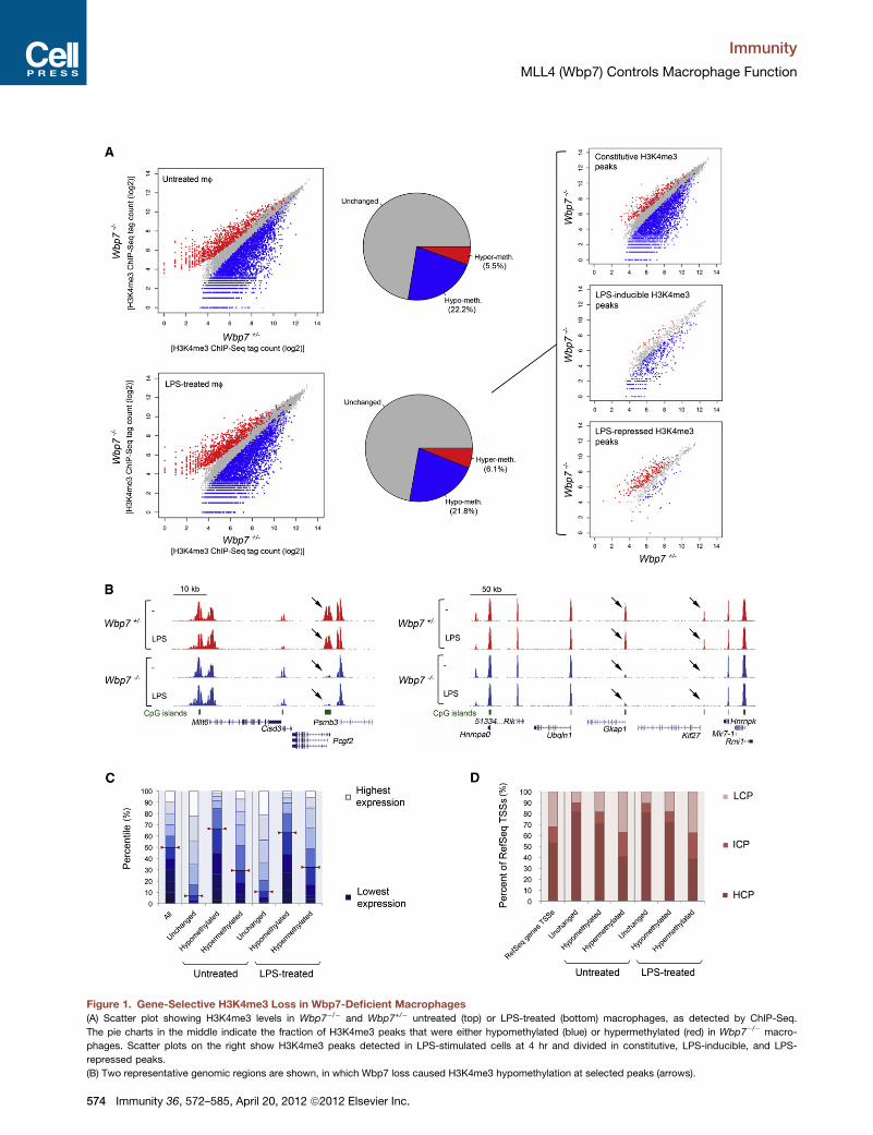
Figure 1. Gene-Selective H3K4me3 Loss in Wbp7-Deficient Macrophages
(A) Scatter plot showing H3K4me3 levels in Wbp7�/� and Wbp7+/� untreated (top) or LPS-treated (bottom) macrophages, as detected by ChIP-Seq.
The pie charts in the middle indicate the fraction of H3K4me3 peaks that were either hypomethylated (blue) or hypermethylated (red) in Wbp7�/� macro-
phages. Scatter plots on the right show H3K4me3 peaks detected in LPS-stimulated cells at 4 hr and divided in constitutive, LPS-inducible, and LPS-
repressed peaks.
(B) Two representative genomic regions are shown, in which Wbp7 loss caused H3K4me3 hypomethylation at selected peaks (arrows).
Immunity
MLL4 (Wbp7) Controls Macrophage Function
574 Immunity 36, 572–585, April 20, 2012 ª2012 Elsevier Inc.
Immunity
MLL4 (Wbp7) Controls Macrophage Function
(22.2%, corresponding to 3,948 out of 17,824 total peaks at
p < 10�5) and LPS-treated (21.8%; 3,896/17,899) macrophages
(Figure 1A, left; Table S1). By using a completely unrelated bio-
logical replicate (data not shown), overlap of hypomethylation
was 88% at p < 10�20 and 77.9% at p < 10�5. We also detected
low-magnitude increases in H3K4me3 at 5.5% of peaks in
untreatedmacrophages and 6.1% in LPS-treatedmacrophages,
probably reflecting secondary effects of Wbp7 loss. The domi-
nance of H3K4me3 losses over H3K4me3 gains is indicated by
the asymmetry of the scatter plots in Figure 1A. H3K4me3 is
induced at a few hundred LPS-activated genes in macrophages
(De Santa et al., 2009; Foster et al., 2007). By distinguishing
constitutive from LPS-inducible H3K4me3 peaks, it became
clear that Wbp7 affected both groups to a similar extent (Fig-
ure 1A, top and middle scatter plots on the right), even though
most high-magnitude hypomethylations (those closer to the
bottom-right corner of the plot) were detected in unstimulated
cells. Intriguingly, H3K4me3 peaks that were diminished upon
LPS stimulation in Wbp7+/� cells were conversely increased in
Wbp7�/� macrophages (Figure 1A, bottom scatter plot on the
right). As documented below, this apparently paradoxical result
is readily explainable as an indirect effect. Representative
genomic snapshots are shown in Figure 1B.
Overall, Wbp7 was required to maintain H3K4me3 levels at
about one-fifth of genes in mouse macrophages, with a prefer-
ence for moderately expressed genes containing CpG islands.
First, gene expression (obtained from cDNA microarray data)
was correlated to the status of hypo-, hyper-, or unchanged
methylation according to H3K4me3 levels at the TSS (±2.5 kbp)
in Wbp7�/� versus Wbp7+/� cells. The hypomethylated gene
group showed a moderate enrichment of genes with interme-
diate expression and a strong underrepresentation of highly
expressed genes (p < 13 10�15 in a chi-square test) (Figure 1C).
Second high-CpG-content promoters were moderately overrep-
resented among the RefSeq genes showing hypomethylation in
Wbp7�/�, suggesting that CpG islands may represent preferen-
tial Wbp7 targets (p < 13 10�15 in a chi-square test) (Figure 1D).
However, it is clear that differential CpG content does not suffice
as a satisfactory explanation for the selectivity of the effects of
Wbp7 deletion. We therefore searched for transcription factor
(TF) binding sites overrepresented in the different groups.
Although we could not identify any TF binding sites strongly
enriched in hypomethylated genes, the group of genes showing
no H3K4me3 changes in Wbp7�/� macrophages was instead
characterized by a strong overrepresentation of multiple Ets
family TF binding sites (Table S2 and Figure S1D). These data
suggest that distinct compositions in TF binding sites may in
fact underlie selective coupling of individual promoters to
specific MLL proteins and that Ets TFs are uncoupled from
Wbp7 in macrophages (or compensate for its loss). When the
analyses reported in Figures 1C and 1D were repeated with
a more restrictive threshold for the identification of hypomethy-
lated peaks (p < 10�10, see Supplemental Experimental Proce-
(C) Genes were divided in ten groups based on their expression level in unstimul
the distribution of all genes (left) or the genes where loss of Wbp7 caused no
unstimulated or stimulated conditions.
(D) Bar graph showing the distribution of high-, intermediate-, or low-density C
unchanged, hypomethylated, or hypermethylated in Wbp7�/� macrophages.
dures), the conclusions were fully consistent with those reported
above.
Finally, when the four Hox genes clusters were analyzed, small
H3K4me3 changes were observed only at Hoxa4 (Figure S1E).
This result is consistent with the already described redundancy
between Mll1 and Wbp7 at these loci (Wang et al., 2009).
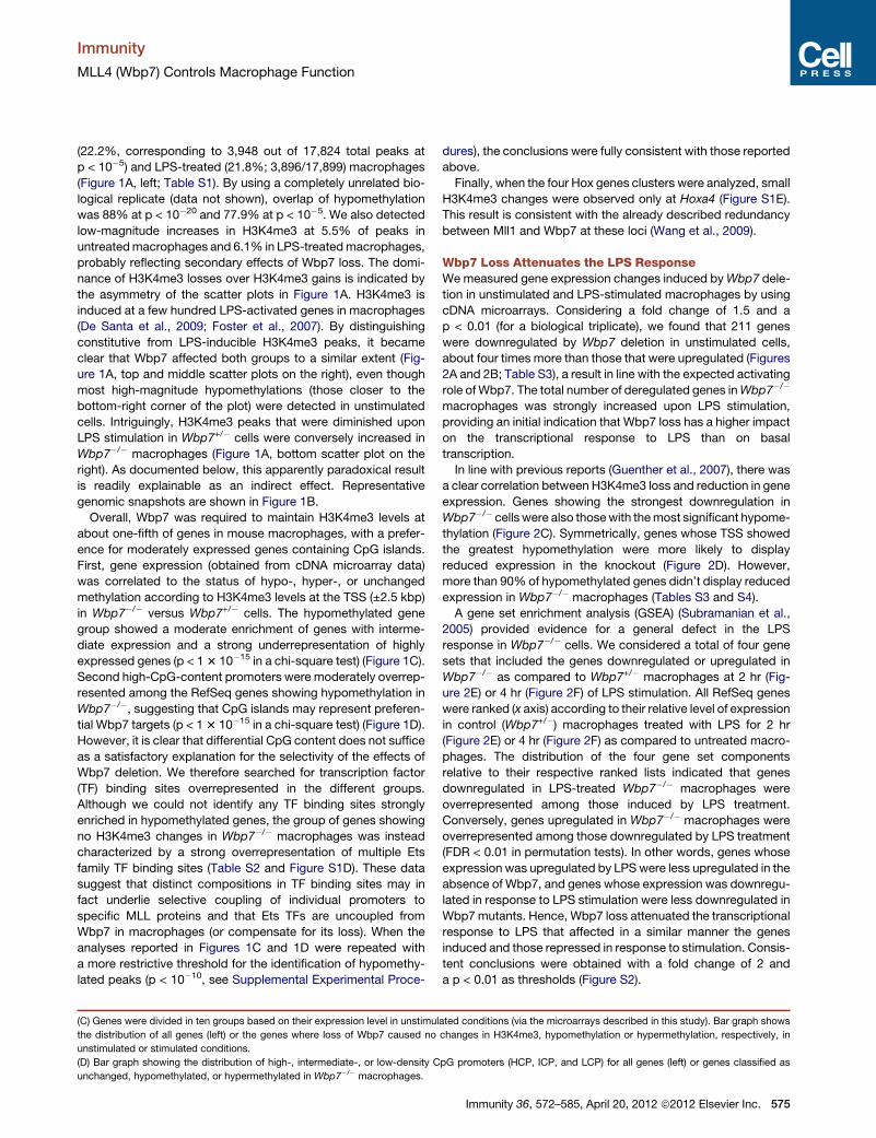
Wbp7 Loss Attenuates the LPS ResponseWemeasured gene expression changes induced byWbp7 dele-
tion in unstimulated and LPS-stimulated macrophages by using
cDNA microarrays. Considering a fold change of 1.5 and a
p < 0.01 (for a biological triplicate), we found that 211 genes
were downregulated by Wbp7 deletion in unstimulated cells,
about four times more than those that were upregulated (Figures
2A and 2B; Table S3), a result in line with the expected activating
role ofWbp7. The total number of deregulated genes inWbp7�/�
macrophages was strongly increased upon LPS stimulation,
providing an initial indication that Wbp7 loss has a higher impact
on the transcriptional response to LPS than on basal
transcription.
In line with previous reports (Guenther et al., 2007), there was
a clear correlation between H3K4me3 loss and reduction in gene
expression. Genes showing the strongest downregulation in
Wbp7�/� cells were also thosewith themost significant hypome-
thylation (Figure 2C). Symmetrically, genes whose TSS showed
the greatest hypomethylation were more likely to display
reduced expression in the knockout (Figure 2D). However,
more than 90% of hypomethylated genes didn’t display reduced
expression in Wbp7�/� macrophages (Tables S3 and S4).
A gene set enrichment analysis (GSEA) (Subramanian et al.,
2005) provided evidence for a general defect in the LPS
response in Wbp7�/� cells. We considered a total of four gene
sets that included the genes downregulated or upregulated in
Wbp7�/� as compared to Wbp7+/� macrophages at 2 hr (Fig-
ure 2E) or 4 hr (Figure 2F) of LPS stimulation. All RefSeq genes
were ranked (x axis) according to their relative level of expression
in control (Wbp7+/�) macrophages treated with LPS for 2 hr
(Figure 2E) or 4 hr (Figure 2F) as compared to untreated macro-
phages. The distribution of the four gene set components
relative to their respective ranked lists indicated that genes
downregulated in LPS-treated Wbp7�/� macrophages were
overrepresented among those induced by LPS treatment.
Conversely, genes upregulated in Wbp7�/� macrophages were
overrepresented among those downregulated by LPS treatment
(FDR < 0.01 in permutation tests). In other words, genes whose
expression was upregulated by LPSwere less upregulated in the
absence of Wbp7, and genes whose expression was downregu-
lated in response to LPS stimulation were less downregulated in
Wbp7mutants. Hence, Wbp7 loss attenuated the transcriptional
response to LPS that affected in a similar manner the genes
induced and those repressed in response to stimulation. Consis-
tent conclusions were obtained with a fold change of 2 and
a p < 0.01 as thresholds (Figure S2).
ated conditions (via the microarrays described in this study). Bar graph shows
changes in H3K4me3, hypomethylation or hypermethylation, respectively, in
pG promoters (HCP, ICP, and LCP) for all genes (left) or genes classified as
Immunity 36, 572–585, April 20, 2012 ª2012 Elsevier Inc. 575
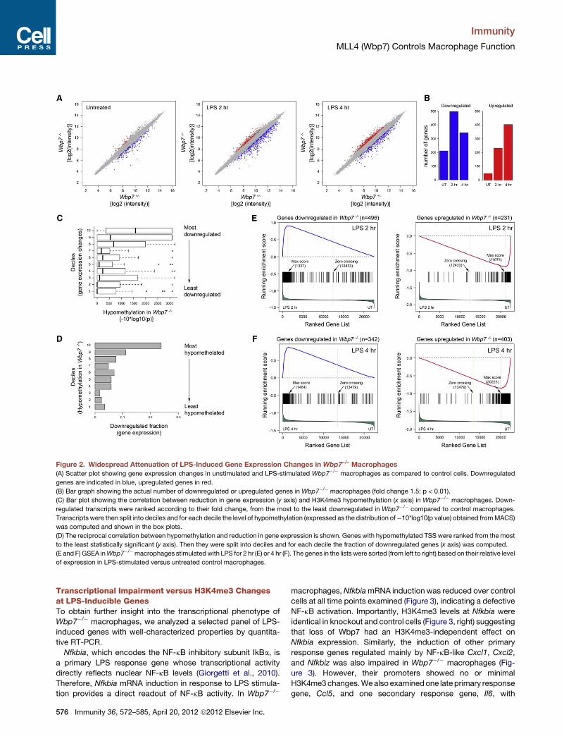
Figure 2. Widespread Attenuation of LPS-Induced Gene Expression Changes in Wbp7–/– Macrophages
(A) Scatter plot showing gene expression changes in unstimulated and LPS-stimulated Wbp7�/� macrophages as compared to control cells. Downregulated
genes are indicated in blue, upregulated genes in red.
(B) Bar graph showing the actual number of downregulated or upregulated genes in Wbp7�/� macrophages (fold change 1.5; p < 0.01).
(C) Bar plot showing the correlation between reduction in gene expression (y axis) and H3K4me3 hypomethylation (x axis) in Wbp7�/� macrophages. Down-
regulated transcripts were ranked according to their fold change, from the most to the least downregulated in Wbp7�/� compared to control macrophages.
Transcripts were then split into deciles and for each decile the level of hypomethylation (expressed as the distribution of�10*log10(p value) obtained fromMACS)
was computed and shown in the box plots.
(D) The reciprocal correlation between hypomethylation and reduction in gene expression is shown. Genes with hypomethylated TSS were ranked from the most
to the least statistically significant (y axis). Then they were split into deciles and for each decile the fraction of downregulated genes (x axis) was computed.
(E and F) GSEA inWbp7�/�macrophages stimulated with LPS for 2 hr (E) or 4 hr (F). The genes in the lists were sorted (from left to right) based on their relative level
of expression in LPS-stimulated versus untreated control macrophages.
Immunity
MLL4 (Wbp7) Controls Macrophage Function
Transcriptional Impairment versus H3K4me3 Changesat LPS-Inducible GenesTo obtain further insight into the transcriptional phenotype of
Wbp7�/� macrophages, we analyzed a selected panel of LPS-
induced genes with well-characterized properties by quantita-
tive RT-PCR.
Nfkbia, which encodes the NF-kB inhibitory subunit IkBa, is
a primary LPS response gene whose transcriptional activity
directly reflects nuclear NF-kB levels (Giorgetti et al., 2010).
Therefore, Nfkbia mRNA induction in response to LPS stimula-
tion provides a direct readout of NF-kB activity. In Wbp7�/�
576 Immunity 36, 572–585, April 20, 2012 ª2012 Elsevier Inc.
macrophages,NfkbiamRNA induction was reduced over control
cells at all time points examined (Figure 3), indicating a defective
NF-kB activation. Importantly, H3K4me3 levels at Nfkbia were
identical in knockout and control cells (Figure 3, right) suggesting
that loss of Wbp7 had an H3K4me3-independent effect on
Nfkbia expression. Similarly, the induction of other primary
response genes regulated mainly by NF-kB-like Cxcl1, Cxcl2,
and Nfkbiz was also impaired in Wbp7�/� macrophages (Fig-
ure 3). However, their promoters showed no or minimal
H3K4me3changes.Wealso examinedone late primary response
gene, Ccl5, and one secondary response gene, Il6, with
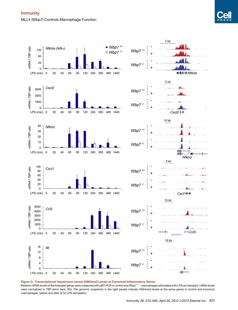
Figure 3. Transcriptional Impairment versus H3K4me3 Levels at Canonical Inflammatory Genes
Relative mRNA levels of the indicated genes were measured with qRT-PCR in control andWbp7�/�macrophages stimulated with LPS as indicated. mRNA levels
were normalized to TBP (error bars: SD). The genomic snapshots in the right panels indicate H3K4me3 levels at the same genes in control and knockout
macrophages, before and after (4 hr) LPS stimulation.
Immunity
MLL4 (Wbp7) Controls Macrophage Function
Immunity 36, 572–585, April 20, 2012 ª2012 Elsevier Inc. 577

Figure 4. LPS-Triggered Signaling Events in
Macrophages Lacking Wbp7
(A) Protein immunoblots were carried out with the
indicated antibodies on cytosolic lysates of control
and Wbp7�/� macrophages stimulated with LPS
as indicated. Images were acquired with a Li-Cor
Odyssey imaging system. Arrowheads, phospho-
IKK; n.s., nonspecific band. For JNK, ERK, and
IRF3, protein immunoblots are shown with both
a phospho-specific antibody (p-JNK, p-ERK, and
p-IRF3) and an antibody recognizing the proteins
irrespective of phosphorylation. Tubulin: loading
control.
(B) Digital quantification (normalized for the
respective control) for some of the images.
Immunity
MLL4 (Wbp7) Controls Macrophage Function
a complex (and therefore more fragile) regulation reflecting the
convergence of multiple signaling pathways. Also at these genes
Wbp7 loss caused a strong, albeit incomplete, transcriptional
impairment without any strong reduction in H3K4me3 levels (Fig-
ure 3). Altogether, we conclude that loss of Wbp7 impairs the
induction of inflammatory genes in a manner that is not directly
related to the deposition of H3K4me3 at these genes.
Consistent with these data, we found that de novoH3K4 trime-
thylation induced by LPS at genes like Il6 and Ccl5 (and at nearly
all other genes where this mark was induced by stimulation)
was entirely dependent on Set1a-Set1b and that it could be
completely ablated without significantly affecting mRNA levels
(data not shown). Finally, when using agonists of TLRs other
than TLR4, transcriptional responses were similarly impaired
(Figure S3).
A Signaling Defect in Wbp7–/– MacrophagesTwo major clues pointed to a signaling defect in Wbp7�/�
macrophages. First, GSEA showed a global attenuation of the
transcriptional response to LPS that similarly impacted induced
and repressed genes. Second, impaired NfkbiamRNA induction
suggested a reduced NF-kB activation and/or nuclear import in
response to LPS stimulation. Therefore, we analyzed the activa-
tion of LPS-inducible signaling pathways in Wbp7�/� versus
Wbp7+/� macrophages. Phosphorylation of the IkB kinases
(IKK) was reduced several fold in Wbp7�/� cells (Figures 4A
and 4B). Consistent with this result, although in control cells
degradation of IkBawas complete at 40 min and initial resynthe-
sis (which is NF-kB dependent) was already observed at 60 min,
inWbp7�/�macrophages the whole process of degradation and
resynthesis was delayed and impaired (Figure 4A). Impaired
signaling in Wbp7�/� cells was not restricted to the IKK/NF-kB
578 Immunity 36, 572–585, April 20, 2012 ª2012 Elsevier Inc.
pathway. Indeed, activation of extracel-
lular signal-regulated kinases (Erk1 and
Erk2) and c-Jun N-terminal kinases
(JNK1 and JNK2) were also reduced in
mutant cells (Figures 4A and 4B). Most
importantly, TLR4-induced phosphoryla-
tion of the transcription factor IRF3, which
requires the adaptor TRIF and is essential
for the activation of the interferon beta
(Ifnb1) gene, was severely reduced in
Wbp7-deficient cells. Consistent with
this result, LPS induction of the Ifnb1 mRNA was almost
completely abolished in Wbp7�/� macrophages (Figure S4).
Overall, Wbp7�/� macrophages displayed a general signaling
defect in response to LPS stimulation.
Transcription and H3K4me3 Levels of PigpRequire Wbp7To determine the mechanistic basis for impaired signaling in
Wbp7 knockoutmacrophages, we inspected the list of the genes
most affected by Wbp7 deletion. Notably, this list includes
Magohb, the only gene whose expression is completely depen-
dent on Wbp7 in embryonic stem cells (Glaser et al., 2009),
thereby suggesting that some Wbp7 targets may be shared
across cell types.
Two genes were strongly downregulated in unstimulated
Wbp7�/� macrophages: Pigp and Taf9b (Figure 5A). The latter
is a paralog of Taf9, a general transcription factor that binds to
an RNA polymerase II core promoter element, the DPE (down-
stream core promoter element) (Frontini et al., 2005). Although
the specific role of Taf9b in relation to Taf9 is still incompletely
defined, it may be involved in bringing about some of the tran-
scriptional consequences of Wbp7 deletion.
Pigp was the main candidate to account for the signaling defi-
ciency we observed in Wbp7�/� cells. Pigp is an essential
component of the GPI-GlcNAc transferase (Watanabe et al.,
2000), the enzyme that controls the first and rate-limiting step
in GPI anchor synthesis (Kinoshita et al., 2008). Because
CD14, which enhances responsiveness of TLR4 to LPS (Lee
et al., 1992), is a GPI-anchored protein, we hypothesized that
the loss of GPI anchor synthesis and the consequent loss of
membrane-bound CD14 may underlie the signaling impairment
of Wbp7�/� macrophages.
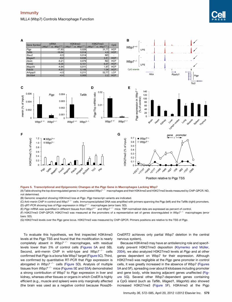
Figure 5. Transcriptional and Epigenomic Changes at the Pigp Gene in Macrophages Lacking Wbp7
(A) Table showing the top downregulated genes in unstimulatedWbp7�/�macrophages and their H3K4me3 andH3K27me3 levelsmeasured byChIP-QPCR.ND,
not determined.
(B) Genomic snapshot showing H3K4me3 loss at Pigp. Pigp transcript variants are indicated.
(C) Anti-menin ChIP in control andWbp7�/� cells. Immunoprecipitated DNA was amplified with primers spanning the Pigp (left) and the Taf9b (right) promoters.
(D) qRT-PCR showing loss of Pigp expression in Wbp7�/� macrophages (error bars: SD).
(E) Pigp mRNA was quantified in different tissues from Wbp7+/� and Wbp7�/� mice. TBP-normalized data are expressed as percent of control.
(F) H3K27me3 ChIP-QPCR. H3K27me3 was measured at the promoters of a representative set of genes downregulated in Wbp7�/� macrophages (error
bars: SD).
(G) H3K27me3 levels over the Pigp gene locus. H3K27me3 was measured by ChIP-QPCR. Primers positions are relative to the TSS of Pigp.
Immunity
MLL4 (Wbp7) Controls Macrophage Function
To evaluate this hypothesis, we first inspected H3K4me3
levels at the Pigp TSS and found that the modification is nearly
completely absent in Wbp7�/� macrophages, with residual
levels lower than 3% of control cells (Figures 5A and 5B).
Second, anti-menin ChIP in wild-type and Wbp7�/� cells
confirmed thatPigp is a bona fideWbp7 target (Figure 5C). Third,
we confirmed by quantitative RT-PCR that Pigp expression is
abrogated in Wbp7�/� cells (Figure 5D). Analysis of multiple
tissues from Wbp7�/� mice (Figures 5E and S5A) demonstrated
a strong contribution of Wbp7 to Pigp expression in liver and
kidney, whereas other tissues or organs in which CreER is highly
efficient (e.g., muscle and spleen) were only marginally affected
(the brain was used as a negative control because Rosa26-
CreERT2 achieves only partial Wbp7 deletion in the central
nervous system).
Because H3K4me3 may have an antisilencing role and specif-
ically prevent H3K27me3 deposition (Klymenko and Muller,
2004), we also analyzed H3K27me3 levels at Pigp and at other
genes dependent on Wbp7 for their expression. Although
H3K27me3 was negligible at the Pigp gene promoter in control
cells, it was greatly increased in the absence of Wbp7 (Figures
5A and 5F), spreading over about 8 kilobases including promoter
and gene body, while leaving adjacent genes unaffected (Fig-
ure 5G). Several other Wbp7-dependent genes containing
a CpG island (such as Taf9b, Maged1, Magohb) also showed
increased H3K27me3 (Figure 5F). H3K4me3 at the Pigp
Immunity 36, 572–585, April 20, 2012 ª2012 Elsevier Inc. 579
Immunity
MLL4 (Wbp7) Controls Macrophage Function
promoter was unaffected by LPS activation of wild-type macro-
phages (Figure S5B); however, promoter acetylation and mRNA
levels were moderately reduced upon sustained stimulation
(Figures S5C and S5D).
Global Loss of GPI-Anchored Proteins andMembrane-Bound CD14 in Wbp7–/– MacrophagesBecause Pigp is essential for the activity of the 8-subunit GPI-
GlcNAc transferase complex, the enzyme catalyzing the first
step of GPI anchor synthesis (Watanabe et al., 2000), we
analyzed surface expression of GPI-anchored proteins in
Wbp7�/� macrophages. To this aim, we exploited the ability of
a bacterial toxin, aerolysin, to bind the GPI anchor with high
affinity (Diep et al., 1998). A fluorescently labeled inactive aeroly-
sin precursor (FLAER) is routinely used for diagnosis of parox-
ysmal nocturnal hemoglobinuria, in which lack of Piga (another
component of the GPI-GlcNAc transferase) results in loss of
GPI-anchored proteins on erythrocytes (Brodsky et al., 2000).
FLAER staining was stable during a prolonged LPS stimulation
of WT macrophages (Figure S5E), suggesting that the moderate
reduction in PigpmRNA levels observed in these conditions (Fig-
ure S5D) is not sufficient to affect the GPI anchor synthesis
capacity of macrophages. Conversely, flow cytometry analysis
demonstrated a nearly complete absence of FLAER staining in
Wbp7�/� macrophages (Figure 6A), thus revealing a global
reduction of GPI-anchored proteins on the cell surface. In
keeping with this result, staining of CD14, a GPI-anchored
protein, was almost completely lost in Wbp7�/� macrophages
(Figure 6A). Another GPI-anchored protein tested, uPAR, simi-
larly showed strongly reduced surface staining in Wbp7�/�
macrophages (Figure 6A).
CD14mRNAwas unaffected byWbp7 deletion, indicating that
loss of surface staining is not due to a transcriptional effect of
Wbp7 loss (Figure 6B). Similarly, TLR4 mRNA was not altered
by Wbp7 loss (Figure 6B) and its surface expression was unaf-
fected (Figure S6).
CD14 captures LPS and transfers it to TLR4, thus facilitating
TLR4 triggering and downstream signaling at low agonist
concentrations (Haziot et al., 1996; Lee et al., 1992). If the impair-
ment in LPS-regulated gene expression in Wbp7�/� macro-
phages is mainly due to a CD14-linked defect in signaling,
a simple prediction is that the defect should be partially rescued
by increasing LPS concentration. In keeping with this prediction,
a 10-fold increase in LPS concentration rescued or attenuated
the gene expression defect at several genes tested (Figure 6C).
In some cases gene induction at the maximal LPS concentration
was even higher than in control cells, reflecting a role of CD14 in
postinduction TLR4 downregulation (Zanoni et al., 2011).
RestoringMembrane CD14 Rescues the TranscriptionalDefectsTo determine whether Pigp re-expression in Wbp7�/� macro-
phages is sufficient to rescue the transcriptional phenotype,
we infectedWbp7�/� macrophages with a retrovirus expressing
the Pigp cDNA. Flow cytometry analysis showed that Pigp
expression in Wbp7�/� cells restored surface levels of CD14
similar to those of wild-type cells (Figure 7A). Strikingly, induction
of genes that are strongly dependent on Wbp7 for maximal
expression was rescued in Pigp reconstituted cells (Figure 7B).
580 Immunity 36, 572–585, April 20, 2012 ª2012 Elsevier Inc.
We also reconstituted surface CD14 by retroviral transduction
of CD14 fused to the transmembrane domain of mouse TLR4
(Figure 7C). Also in this case, defective LPS induction of induc-
ible genes could be rescued to wild-type levels (Figure 7D).
Overall, these data demonstrated a central role for loss of GPI-
anchored CD14 in the impaired response of Wbp7-deficient
macrophages to LPS.
DISCUSSION
In this study, we identified a specific role for Wbp7 in the
synthesis of GPI anchors in macrophages. This role of Wbp7 is
exercised through the tight transcriptional control of a single
gene, Pigp, encoding an essential component of the enzymatic
complex that catalyzes the first step of GPI-anchor synthesis.
GPI anchors are indirectly implicated in microbial recognition,
as shown by the fact that CD14, which enhances detection of
LPS (Wright et al., 1990) and other microbial molecules
(Baumann et al., 2010; Lee et al., 2006a) through transfer to
the competent TLRs, requires a GPI anchor for membrane
loading. Moreover, CD14 itself may deliver activation signals
(despite the lack of a cytoplasmic tail), as demonstrated by its
requirement for TLR4-independent, LPS-induced NFAT activa-
tion in dendritic cells (Zanoni et al., 2009). Whereas membrane
anchoring of CD14 appears to represent a particularly relevant
aspect of GPI anchor biosynthesis in macrophages, it is likely
that loss of GPI anchors will affect localization and function of
many other biologically relevant proteins in this system, such
as the urokinase plasminogen activator receptor (uPAR), which
regulates the proteolytic activity of the uPA serine protease (Blasi
and Sidenius, 2010).
Lack of membrane-associated CD14 and the ensuing defect
in LPS recognition resulted in a straightforward cascade of
effects: (1) a reduced activation of intracellular signaling
pathways, including the IKK-NF-kB pathway, the MAPK path-
ways, and the TRIF/IRF3 pathway, followed by (2) a broad atten-
uation of both positive and negative gene expression changes
triggered by LPS. The transcriptional defects were partially
rescued simply by increasing LPS concentration, a result consis-
tent with the central role of CD14 in this phenotype. More
directly, re-expression of Pigp or membrane-anchored CD14 in
Wbp7�/� cells reverted the transcriptional defects.
The central role played in this phenotype by the impact of
Wbp7 on signaling is underscored by the observation that
Wbp7 loss attenuated not only LPS-induced increases in
H3K4me3 (which may reflect a direct role of Wbp7 in de novo
deposition of H3K4me3) but also LPS-induced reductions of
this histone modification, which probably represent an indirect
consequence of the signaling defect. Because of its requirement
for GPI anchor synthesis and CD14 attachment to the macro-
phage membrane, Wbp7 can thus be considered as a compo-
nent of the innate immune system in its own right.
The impact of Wbp7 loss on the macrophage epigenome was
broad, with about 20% of active genes showing reduced
H3K4me3 levels in mutant cells. Attempts to identify specific
sequence features responsible for Wbp7 dependence of
H3K4me3 failed to generate clear-cut results, even though hypo-
methylation tended to occur at high CpG-containing promoters
and at moderately but not highly expressed genes. Moreover,
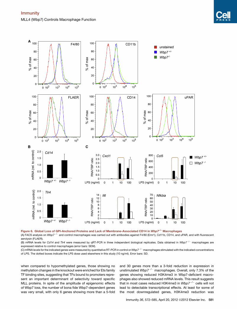
Figure 6. Global Loss of GPI-Anchored Proteins and Lack of Membrane-Associated CD14 in Wbp7–/– Macrophages
(A) FACS analysis on Wbp7�/� and control macrophages was carried out with antibodies against F4/80 (Emr1), Cd11b, CD14, and uPAR, and with fluorescent
aerolysin (FLAER).
(B) mRNA levels for Cd14 and Tlr4 were measured by qRT-PCR in three independent biological replicates. Data obtained in Wbp7�/� macrophages are
expressed relative to control macrophages (error bars: SEM).
(C)mRNA levels for the indicated genesweremeasured by quantitative RT-PCR in control orWbp7�/�macrophages stimulatedwith the indicated concentrations
of LPS. The dotted boxes indicate the LPS dose used elsewhere in this study (10 ng/ml). Error bars: SD.
Immunity
MLL4 (Wbp7) Controls Macrophage Function
when compared to hypomethylated genes, those showing no
methylation changes in the knockout were enriched for Ets family
TF binding sites, suggesting that TFs bound to promoters repre-
sent an important determinant of selectivity toward specific
MLL proteins. In spite of the amplitude of epigenomic effects
of Wbp7 loss, the number of bona fide Wbp7-dependent genes
was very small, with only 6 genes showing more than a 5-fold
and 30 genes more than a 3-fold reduction in expression in
unstimulated Wbp7�/� macrophages. Overall, only 7.3% of the
genes showing reduced H3K4me3 in Wbp7-deficient macro-
phages also showed reduced mRNA levels. This result suggests
that in most cases reduced H3K4me3 in Wbp7�/� cells will not
lead to detectable transcriptional effects. At least for some of
the most downregulated genes, H3K4me3 reduction was
Immunity 36, 572–585, April 20, 2012 ª2012 Elsevier Inc. 581
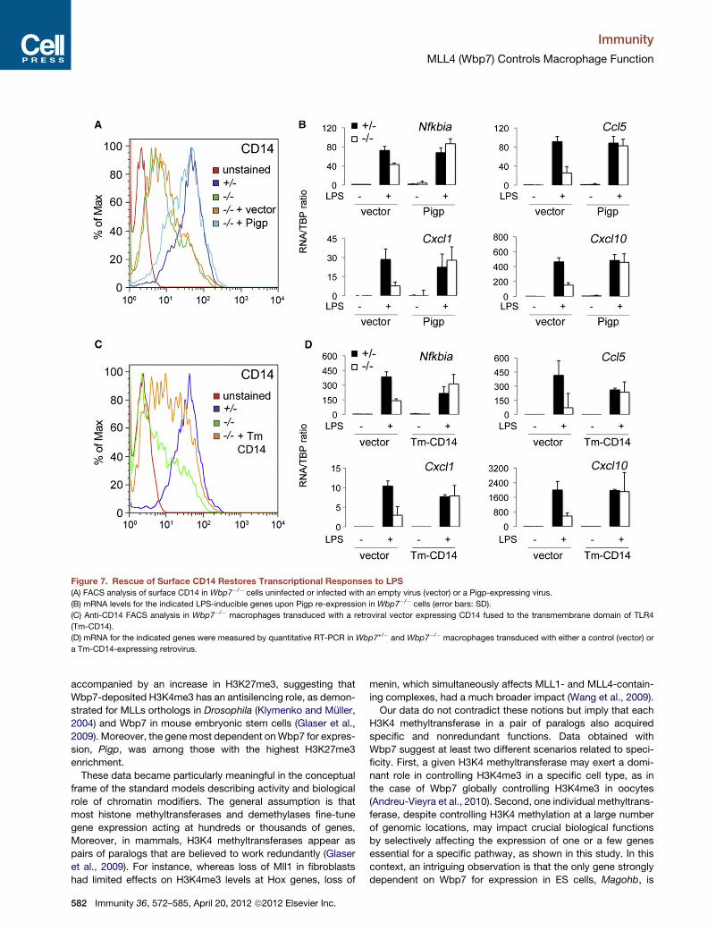
Figure 7. Rescue of Surface CD14 Restores Transcriptional Responses to LPS
(A) FACS analysis of surface CD14 in Wbp7�/� cells uninfected or infected with an empty virus (vector) or a Pigp-expressing virus.
(B) mRNA levels for the indicated LPS-inducible genes upon Pigp re-expression in Wbp7�/� cells (error bars: SD).
(C) Anti-CD14 FACS analysis in Wbp7�/� macrophages transduced with a retroviral vector expressing CD14 fused to the transmembrane domain of TLR4
(Tm-CD14).
(D) mRNA for the indicated genes were measured by quantitative RT-PCR in Wbp7+/� and Wbp7�/� macrophages transduced with either a control (vector) or
a Tm-CD14-expressing retrovirus.
Immunity
MLL4 (Wbp7) Controls Macrophage Function
accompanied by an increase in H3K27me3, suggesting that
Wbp7-deposited H3K4me3 has an antisilencing role, as demon-
strated for MLLs orthologs in Drosophila (Klymenko and Muller,
2004) and Wbp7 in mouse embryonic stem cells (Glaser et al.,
2009). Moreover, the gene most dependent onWbp7 for expres-
sion, Pigp, was among those with the highest H3K27me3
enrichment.
These data became particularly meaningful in the conceptual
frame of the standard models describing activity and biological
role of chromatin modifiers. The general assumption is that
most histone methyltransferases and demethylases fine-tune
gene expression acting at hundreds or thousands of genes.
Moreover, in mammals, H3K4 methyltransferases appear as
pairs of paralogs that are believed to work redundantly (Glaser
et al., 2009). For instance, whereas loss of Mll1 in fibroblasts
had limited effects on H3K4me3 levels at Hox genes, loss of
582 Immunity 36, 572–585, April 20, 2012 ª2012 Elsevier Inc.
menin, which simultaneously affects MLL1- and MLL4-contain-
ing complexes, had a much broader impact (Wang et al., 2009).
Our data do not contradict these notions but imply that each
H3K4 methyltransferase in a pair of paralogs also acquired
specific and nonredundant functions. Data obtained with
Wbp7 suggest at least two different scenarios related to speci-
ficity. First, a given H3K4 methyltransferase may exert a domi-
nant role in controlling H3K4me3 in a specific cell type, as in
the case of Wbp7 globally controlling H3K4me3 in oocytes
(Andreu-Vieyra et al., 2010). Second, one individual methyltrans-
ferase, despite controlling H3K4 methylation at a large number
of genomic locations, may impact crucial biological functions
by selectively affecting the expression of one or a few genes
essential for a specific pathway, as shown in this study. In this
context, an intriguing observation is that the only gene strongly
dependent on Wbp7 for expression in ES cells, Magohb, is
Immunity
MLL4 (Wbp7) Controls Macrophage Function
dependent onWbp7 inmacrophages aswell. Therefore,Wbp7 is
connected to the expression of this specific gene independently
of the differentiation state of the cell, a result consistent with the
idea of an extreme functional specialization of a subset of histone
methyltransferases.
In conclusion, our mechanistic analysis of the transcriptional
and epigenomic phenotype of Wbp7-deficient macrophages
revealed that a housekeeping biosynthetic pathway essential
for membrane localization of hundreds of molecules, including
a coreceptor important for microbial recognition, is under tight
and nonredundant control of a single H3K4 methyltransferase,
which is otherwise dispensable for viability of adult mice.
EXPERIMENTAL PROCEDURES
Wbp7-Deficient Mice
Animal experiments were performed in accordance with the Italian Laws
(D.L.vo 116/92 and following additions), which enforce EU 86/609 Directive.
The Wbp7 floxed allele has been described (Glaser et al., 2006, 2009). The
two loxP sites flank the 73 bp of Wbp7 exon 2 and recombination leads to
a frame shift and the generation of a premature stop codon. Wbp7fl/fl mice
were crossed to Rosa26-CreERT2+/� mice and Cre was activated by gavage
feeding of 5mg tamoxifen (Sigma T5648) dissolved in peanut oil (SigmaP2144)
for 5 consecutive days. Mice were sacrificed and bone marrow cells isolated
7–10 days after tamoxifen treatment. Efficiency of recombination was evalu-
ated by Taqman assay (Figure S1A).
Cell Culture and Retroviral Infections
Macrophage cultures and retroviral infections were carried out as described
(De Santa et al., 2007). LPS from E. coli serotype EH100 (Alexis) was used at
10 ng/ml unless otherwise indicated. The cDNA encoding Flag-tagged Pigp
(NM_001159616) was obtained by standard cloningwhereas the cDNA encod-
ing CD14 fused to the transmembrane domain of mouse TLR4 was obtained
by in vitro gene synthesis (MWG). Both cDNAs were subcloned into pMSCV-
puro (Clontech).
Quantitative RT-PCR
RNA was extracted from macrophages with Trizol (Invitrogen) and reverse
transcribed with random hexamers. Primers sequences are in Table S5.
FACS Analysis
Cells were stained with the following antibodies: F4/80 (Abcam ab6640; 1:50
dilution); biotin-labeled CD11b (BD #557395; 1:50 dilution) detected with
streptavidin-PE-Cy5 (BD #554062); FITC-labeled CD14 (Abcam ab65087,
1:50 dilution); uPAR (a gift from N. Sidenius); and fluorescently labeled
aerolysin (FLAER-Alexa488, Pinewood Scientific) at a concentration of
50 nM. The TLR4 antibody is a kind gift of J. Kagan (Harvard Medical School).
The cells were analyzed by FACScalibur 4 or 10 channel flow cytometer (BD
Biosciences).
Chromatin Immunoprecipitation and Sequencing
ChIP experiments were carried out as described starting from 2 3 107 cells
(Ghisletti et al., 2010). Lysates were immunoprecipitated with 5 mg of
H3K4me3 antibody (Active Motif #39159), 10 mg of H3K27me3 antibody
(Active motif #39155), or menin antibody (Bethyl Labs., A300-105A).
Antibodies were prebound to G protein-coupled paramagnetic beads (Dyna-
beads) in PBS/BSA 0.5% and incubated with lysates overnight. Beads were
washed six times in a modified RIPA buffer (50 mM HEPES [pH 7.6],
500 mM LiCl, 1 mM EDTA, 1% NP-40, 0.7% Na-deoxycholate) and once in
TE containing 50 mM NaCl. DNA was eluted in TE-2% SDS and crosslinks
reversed by incubation overnight at 65�C. DNA was then purified by Qiaquick
columns (QIAGEN) and quantified with PicoGreen (Invitrogen). Yield was
�10 ng/107 cells for H3K4me3. For validation by ChIP-QPCR, 0.4 ml of purified
DNA was used for amplification on an ABI 7500 machine. Primers used for
ChIP QPCR are in Table S5. ChIP DNA was prepared for Solexa 2G
sequencing as described (Ghisletti et al., 2010). DNA quantified both with an
Agilent Bioanalyzer and Picogreen was diluted to 10 nM. Cluster generation
was performed and loaded into individual lanes of a flow cell (4 pmoles/
sample).
A detailed description of the computational analyses is provided in the
Supplemental Information. Statistics referring to the sequencing runs are in
Table S6.
cDNA Microarrays
A biological triplicate was used for cDNA microarray analyses. RNA was puri-
fied with an RNeasy-QIAGEN kit. Quality analysis of total RNA, cRNA
synthesis, hybridization, and data extraction were performed at the Cogentech
Microarray Core Facility. The mouse gene ST1.0 Affymetrix array (Affymetrix)
was used for gene expression screening. Data analysis is described in the
Supplemental Information.
Immunoblot
The following antibodies were used: IkBa (Santa Cruz, sc-371), phospho-IKKa
(S176, S180) (Upstate, #07-837), phospho-JNK (T183, Y185) (CST, #9251),
JNK (CST, #9252), ERK1 (Santa Cruz, sc-94), phospho-ERK1/2 (E10) (CST,
#9106), phospho-IRF3 (CST, #4947), IRF3 (CST #4302), and Tubulin (Sigma,
T9026). For quantified images, secondary IRDye antibodies from Li-Cor
were utilized (cat. no. 926-68021 and 926-32210).
ACCESSION NUMBERS
Raw data sets are available for download at the Gene Expression Omnibus
(GEO) database (http://www.ncbi.nlm.nih.gov/gds/) under the accession
number GSE30973, which comprises expression data (GSE30971) and
ChIP-seq data (GSE30972).
SUPPLEMENTAL INFORMATION
Supplemental Information includes Supplemental Discussion, Supplemental
Experimental Procedures, six figures, and six tables and can be found with
this article online at doi:10.1016/j.immuni.2012.02.016.
ACKNOWLEDGMENTS
We thank B. Amati for critically reading this manuscript; N. Sidenius (IFOM,
Milan) and J. Kagan (Harvard Medical School, Boston, MA) for antibodies;
and G. Bucci (Cogentech, Milan) for initial microarray data analysis. This
work was supported by the FP6 program of the European Community (Marie
Curie Excellence Grant Trans-Tar to G.N.), the Italian Association for Research
on Cancer, AIRC (G.N. and G.T.), the European Research Council (ERC,
project NORM; G.N.), and the Italian Health Ministry (G.T.).
Received: October 19, 2011
Revised: January 20, 2012
Accepted: February 4, 2012
Published online: April 5, 2012
REFERENCES
Andreu-Vieyra, C.V., Chen, R., Agno, J.E., Glaser, S., Anastassiadis, K.,
Stewart, A.F., and Matzuk, M.M. (2010). MLL2 is required in oocytes for bulk
histone 3 lysine 4 trimethylation and transcriptional silencing. PLoS Biol. 8, 8.
Ayton, P., Sneddon, S.F., Palmer, D.B., Rosewell, I.R., Owen, M.J., Young, B.,
Presley, R., and Subramanian, V. (2001). Truncation of the Mll gene in exon 5
by gene targeting leads to early preimplantation lethality of homozygous
embryos. Genesis 30, 201–212.
Barski, A., Cuddapah, S., Cui, K., Roh, T.Y., Schones, D.E., Wang, Z., Wei, G.,
Chepelev, I., and Zhao, K. (2007). High-resolution profiling of histone methyl-
ations in the human genome. Cell 129, 823–837.
Baumann, C.L., Aspalter, I.M., Sharif, O., Pichlmair, A., Bluml, S., Grebien, F.,
Bruckner, M., Pasierbek, P., Aumayr, K., Planyavsky, M., et al. (2010). CD14 is
a coreceptor of Toll-like receptors 7 and 9. J. Exp. Med. 207, 2689–2701.
Immunity 36, 572–585, April 20, 2012 ª2012 Elsevier Inc. 583
Immunity
MLL4 (Wbp7) Controls Macrophage Function
Bernstein, B.E., Kamal, M., Lindblad-Toh, K., Bekiranov, S., Bailey, D.K.,
Huebert, D.J., McMahon, S., Karlsson, E.K., Kulbokas, E.J., 3rd, Gingeras,
T.R., et al. (2005). Genomic maps and comparative analysis of histone modifi-
cations in human and mouse. Cell 120, 169–181.
Blasi, F., and Sidenius, N. (2010). The urokinase receptor: focused cell surface
proteolysis, cell adhesion and signaling. FEBS Lett. 584, 1923–1930.
Boyer, L.A., Plath, K., Zeitlinger, J., Brambrink, T., Medeiros, L.A., Lee, T.I.,
Levine, S.S., Wernig, M., Tajonar, A., Ray, M.K., et al. (2006). Polycomb
complexes repress developmental regulators in murine embryonic stem cells.
Nature 441, 349–353.
Bracken, A.P., Dietrich, N., Pasini, D., Hansen, K.H., and Helin, K. (2006).
Genome-wide mapping of Polycomb target genes unravels their roles in cell
fate transitions. Genes Dev. 20, 1123–1136.
Brodsky, R.A., Mukhina, G.L., Li, S., Nelson, K.L., Chiurazzi, P.L., Buckley,
J.T., and Borowitz, M.J. (2000). Improved detection and characterization of
paroxysmal nocturnal hemoglobinuria using fluorescent aerolysin. Am. J.
Clin. Pathol. 114, 459–466.
Cho, Y.W., Hong, T., Hong, S., Guo, H., Yu, H., Kim, D., Guszczynski, T.,
Dressler, G.R., Copeland, T.D., Kalkum,M., andGe, K. (2007). PTIP associates
with MLL3- and MLL4-containing histone H3 lysine 4 methyltransferase
complex. J. Biol. Chem. 282, 20395–20406.
Cloos, P.A., Christensen, J., Agger, K., and Helin, K. (2008). Erasing the methyl
mark: histone demethylases at the center of cellular differentiation and
disease. Genes Dev. 22, 1115–1140.
De Santa, F., Totaro, M.G., Prosperini, E., Notarbartolo, S., Testa, G., and
Natoli, G. (2007). The histone H3 lysine-27 demethylase Jmjd3 links inflamma-
tion to inhibition of polycomb-mediated gene silencing. Cell 130, 1083–1094.
De Santa, F., Narang, V., Yap, Z.H., Tusi, B.K., Burgold, T., Austenaa, L., Bucci,
G., Caganova, M., Notarbartolo, S., Casola, S., et al. (2009). Jmjd3 contributes
to the control of gene expression in LPS-activated macrophages. EMBO J. 28,
3341–3352.
Diep, D.B., Nelson, K.L., Raja, S.M., Pleshak, E.N., and Buckley, J.T. (1998).
Glycosylphosphatidylinositol anchors of membrane glycoproteins are binding
determinants for the channel-forming toxin aerolysin. J. Biol. Chem. 273,
2355–2360.
Ernst, P., Fisher, J.K., Avery, W., Wade, S., Foy, D., and Korsmeyer, S.J.
(2004a). Definitive hematopoiesis requires the mixed-lineage leukemia gene.
Dev. Cell 6, 437–443.
Ernst, P., Mabon, M., Davidson, A.J., Zon, L.I., and Korsmeyer, S.J. (2004b).
An Mll-dependent Hox program drives hematopoietic progenitor expansion.
Curr. Biol. 14, 2063–2069.
Foster, S.L., Hargreaves, D.C., and Medzhitov, R. (2007). Gene-specific
control of inflammation by TLR-induced chromatin modifications. Nature
447, 972–978.
Frontini, M., Soutoglou, E., Argentini, M., Bole-Feysot, C., Jost, B., Scheer, E.,
and Tora, L. (2005). TAF9b (formerly TAF9L) is a bona fide TAF that has unique
and overlapping roles with TAF9. Mol. Cell. Biol. 25, 4638–4649.
Ghisletti, S., Barozzi, I., Mietton, F., Polletti, S., De Santa, F., Venturini, E.,
Gregory, L., Lonie, L., Chew, A., Wei, C.L., et al. (2010). Identification and char-
acterization of enhancers controlling the inflammatory gene expression
program in macrophages. Immunity 32, 317–328.
Giorgetti, L., Siggers, T., Tiana, G., Caprara, G., Notarbartolo, S., Corona, T.,
Pasparakis, M., Milani, P., Bulyk, M.L., and Natoli, G. (2010).
Noncooperative interactions between transcription factors and clustered
DNA binding sites enable graded transcriptional responses to environmental
inputs. Mol. Cell 37, 418–428.
Glaser, S., Schaft, J., Lubitz, S., Vintersten, K., van der Hoeven, F., Tufteland,
K.R., Aasland, R., Anastassiadis, K., Ang, S.L., and Stewart, A.F. (2006).
Multiple epigenetic maintenance factors implicated by the loss of Mll2 in
mouse development. Development 133, 1423–1432.
Glaser, S., Lubitz, S., Loveland, K.L., Ohbo, K., Robb, L., Schwenk, F., Seibler,
J., Roellig, D., Kranz, A., Anastassiadis, K., and Stewart, A.F. (2009). The
histone 3 lysine 4 methyltransferase, Mll2, is only required briefly in develop-
ment and spermatogenesis. Epigenetics Chromatin 2, 5.
584 Immunity 36, 572–585, April 20, 2012 ª2012 Elsevier Inc.
Guenther, M.G., Levine, S.S., Boyer, L.A., Jaenisch, R., and Young, R.A.
(2007). A chromatin landmark and transcription initiation at most promoters
in human cells. Cell 130, 77–88.
Haziot, A., Ferrero, E., Kontgen, F., Hijiya, N., Yamamoto, S., Silver, J.,
Stewart, C.L., and Goyert, S.M. (1996). Resistance to endotoxin shock and
reduced dissemination of gram-negative bacteria in CD14-deficient mice.
Immunity 4, 407–414.
Huang, Y., Fang, J., Bedford, M.T., Zhang, Y., and Xu, R.M. (2006).
Recognition of histone H3 lysine-4 methylation by the double tudor domain
of JMJD2A. Science 312, 748–751.
Hughes, C.M., Rozenblatt-Rosen, O., Milne, T.A., Copeland, T.D., Levine,
S.S., Lee, J.C., Hayes, D.N., Shanmugam, K.S., Bhattacharjee, A., Biondi,
C.A., et al. (2004). Menin associates with a trithorax family histonemethyltrans-
ferase complex and with the hoxc8 locus. Mol. Cell 13, 587–597.
Jenuwein, T., and Allis, C.D. (2001). Translating the histone code. Science 293,
1074–1080.
Kim, J., Daniel, J., Espejo, A., Lake, A., Krishna, M., Xia, L., Zhang, Y., and
Bedford, M.T. (2006). Tudor, MBT and chromo domains gauge the degree of
lysine methylation. EMBO Rep. 7, 397–403.
Kim, J., Guermah, M., McGinty, R.K., Lee, J.S., Tang, Z., Milne, T.A.,
Shilatifard, A., Muir, T.W., and Roeder, R.G. (2009). RAD6-mediated transcrip-
tion-coupled H2B ubiquitylation directly stimulates H3K4 methylation in
human cells. Cell 137, 459–471.
Kinoshita, T., Fujita, M., and Maeda, Y. (2008). Biosynthesis, remodelling and
functions of mammalian GPI-anchored proteins: recent progress. J. Biochem.
144, 287–294.
Klose, R.J., and Zhang, Y. (2007). Regulation of histone methylation by deme-
thylimination and demethylation. Nat. Rev. Mol. Cell Biol. 8, 307–318.
Klymenko, T., and Muller, J. (2004). The histone methyltransferases Trithorax
and Ash1 prevent transcriptional silencing by Polycomb group proteins.
EMBO Rep. 5, 373–377.
Kornberg, R.D., and Lorch, Y. (1999). Twenty-five years of the nucleosome,
fundamental particle of the eukaryote chromosome. Cell 98, 285–294.
Kouzarides, T. (2007). Chromatin modifications and their function. Cell 128,
693–705.
Lachner, M., O’Carroll, D., Rea, S., Mechtler, K., and Jenuwein, T. (2001).
Methylation of histone H3 lysine 9 creates a binding site for HP1 proteins.
Nature 410, 116–120.
Lee, M.S., and Kim, Y.J. (2007). Signaling pathways downstream of pattern-
recognition receptors and their cross talk. Annu. Rev. Biochem. 76, 447–480.
Lee, J.H., and Skalnik, D.G. (2008). Wdr82 is a C-terminal domain-binding
protein that recruits the Setd1A Histone H3-Lys4 methyltransferase complex
to transcription start sites of transcribed human genes. Mol. Cell. Biol. 28,
609–618.
Lee, J.D., Kato, K., Tobias, P.S., Kirkland, T.N., and Ulevitch, R.J. (1992).
Transfection of CD14 into 70Z/3 cells dramatically enhances the sensitivity
to complexes of lipopolysaccharide (LPS) and LPS binding protein. J. Exp.
Med. 175, 1697–1705.
Lee, H.K., Dunzendorfer, S., Soldau, K., and Tobias, P.S. (2006a). Double-
stranded RNA-mediated TLR3 activation is enhanced by CD14. Immunity
24, 153–163.
Lee, T.I., Jenner, R.G., Boyer, L.A., Guenther, M.G., Levine, S.S., Kumar, R.M.,
Chevalier, B., Johnstone, S.E., Cole, M.F., Isono, K., et al. (2006b). Control of
developmental regulators by Polycomb in human embryonic stem cells. Cell
125, 301–313.
Mikkelsen, T.S., Ku, M., Jaffe, D.B., Issac, B., Lieberman, E., Giannoukos, G.,
Alvarez, P., Brockman, W., Kim, T.K., Koche, R.P., et al. (2007). Genome-wide
maps of chromatin state in pluripotent and lineage-committed cells. Nature
448, 553–560.
Miller, T., Krogan, N.J., Dover, J., Erdjument-Bromage, H., Tempst, P.,
Johnston, M., Greenblatt, J.F., and Shilatifard, A. (2001). COMPASS:
a complex of proteins associated with a trithorax-related SET domain protein.
Proc. Natl. Acad. Sci. USA 98, 12902–12907.
Immunity
MLL4 (Wbp7) Controls Macrophage Function
Pavri, R., Zhu, B., Li, G., Trojer, P., Mandal, S., Shilatifard, A., and Reinberg, D.
(2006). Histone H2B monoubiquitination functions cooperatively with FACT to
regulate elongation by RNA polymerase II. Cell 125, 703–717.
Pena, P.V., Davrazou, F., Shi, X., Walter, K.L., Verkhusha, V.V., Gozani, O.,
Zhao, R., and Kutateladze, T.G. (2006). Molecular mechanism of histone
H3K4me3 recognition by plant homeodomain of ING2. Nature 442, 100–103.
Peters, A.H., O’Carroll, D., Scherthan, H., Mechtler, K., Sauer, S., Schofer, C.,
Weipoltshammer, K., Pagani, M., Lachner, M., Kohlmaier, A., et al. (2001). Loss
of the Suv39h histone methyltransferases impairs mammalian heterochro-
matin and genome stability. Cell 107, 323–337.
Peters, A.H., Kubicek, S., Mechtler, K., O’Sullivan, R.J., Derijck, A.A., Perez-
Burgos, L., Kohlmaier, A., Opravil, S., Tachibana, M., Shinkai, Y., et al.
(2003). Partitioning and plasticity of repressive histone methylation states in
mammalian chromatin. Mol. Cell 12, 1577–1589.
Ruthenburg, A.J., Allis, C.D., andWysocka, J. (2007). Methylation of lysine 4 on
histone H3: intricacy of writing and reading a single epigenetic mark. Mol. Cell
25, 15–30.
Santos-Rosa, H., Schneider, R., Bannister, A.J., Sherriff, J., Bernstein, B.E.,
Emre, N.C., Schreiber, S.L., Mellor, J., and Kouzarides, T. (2002). Active genes
are tri-methylated at K4 of histone H3. Nature 419, 407–411.
Sims, R.J., 3rd, Millhouse, S., Chen, C.F., Lewis, B.A., Erdjument-Bromage,
H., Tempst, P., Manley, J.L., and Reinberg, D. (2007). Recognition of trimethy-
lated histone H3 lysine 4 facilitates the recruitment of transcription postinitia-
tion factors and pre-mRNA splicing. Mol. Cell 28, 665–676.
Smith, E., Lin, C., and Shilatifard, A. (2011). The super elongation complex
(SEC) and MLL in development and disease. Genes Dev. 25, 661–672.
Subramanian, A., Tamayo, P., Mootha, V.K., Mukherjee, S., Ebert, B.L.,
Gillette, M.A., Paulovich, A., Pomeroy, S.L., Golub, T.R., Lander, E.S., and
Mesirov, J.P. (2005). Gene set enrichment analysis: a knowledge-based
approach for interpreting genome-wide expression profiles. Proc. Natl.
Acad. Sci. USA 102, 15545–15550.
Tachibana, M., Sugimoto, K., Nozaki, M., Ueda, J., Ohta, T., Ohki, M., Fukuda,
M., Takeda, N., Niida, H., Kato, H., and Shinkai, Y. (2002). G9a histone meth-
yltransferase plays a dominant role in euchromatic histone H3 lysine 9 methyl-
ation and is essential for early embryogenesis. Genes Dev. 16, 1779–1791.
Tachibana, M., Ueda, J., Fukuda, M., Takeda, N., Ohta, T., Iwanari, H.,
Sakihama, T., Kodama, T., Hamakubo, T., and Shinkai, Y. (2005). Histone
methyltransferases G9a and GLP form heteromeric complexes and are both
crucial for methylation of euchromatin at H3-K9. Genes Dev. 19, 815–826.
Testa, G., Schaft, J., van der Hoeven, F., Glaser, S., Anastassiadis, K., Zhang,
Y., Hermann, T., Stremmel,W., andStewart, A.F. (2004). A reliable lacZ expres-
sion reporter cassette for multipurpose, knockout-first alleles. Genesis 38,
151–158.
Thomson, J.P., Skene, P.J., Selfridge, J., Clouaire, T., Guy, J., Webb, S., Kerr,
A.R., Deaton, A., Andrews, R., James, K.D., et al. (2010). CpG islands influence
chromatin structure via the CpG-binding protein Cfp1. Nature 464, 1082–1086.
Wang, P., Lin, C., Smith, E.R., Guo, H., Sanderson, B.W., Wu, M., Gogol, M.,
Alexander, T., Seidel, C., Wiedemann, L.M., et al. (2009). Global analysis of
H3K4 methylation defines MLL family member targets and points to a role
for MLL1-mediated H3K4 methylation in the regulation of transcriptional initi-
ation by RNA polymerase II. Mol. Cell. Biol. 29, 6074–6085.
Watanabe, R., Murakami, Y., Marmor, M.D., Inoue, N., Maeda, Y., Hino, J.,
Kangawa, K., Julius, M., and Kinoshita, T. (2000). Initial enzyme for glycosyl-
phosphatidylinositol biosynthesis requires PIG-P and is regulated by DPM2.
EMBO J. 19, 4402–4411.
Wright, S.D., Ramos, R.A., Tobias, P.S., Ulevitch, R.J., and Mathison, J.C.
(1990). CD14, a receptor for complexes of lipopolysaccharide (LPS) and LPS
binding protein. Science 249, 1431–1433.
Wu, M., Wang, P.F., Lee, J.S., Martin-Brown, S., Florens, L., Washburn, M.,
and Shilatifard, A. (2008). Molecular regulation of H3K4 trimethylation by
Wdr82, a component of human Set1/COMPASS. Mol. Cell. Biol. 28, 7337–
7344.
Yagi, H., Deguchi, K., Aono, A., Tani, Y., Kishimoto, T., and Komori, T. (1998).
Growth disturbance in fetal liver hematopoiesis of Mll-mutant mice. Blood 92,
108–117.
Yu, B.D., Hess, J.L., Horning, S.E., Brown, G.A., and Korsmeyer, S.J. (1995).
Altered Hox expression and segmental identity in Mll-mutant mice. Nature
378, 505–508.
Zanoni, I., Ostuni, R., Capuano, G., Collini, M., Caccia, M., Ronchi, A.E.,
Rocchetti, M., Mingozzi, F., Foti, M., Chirico, G., et al. (2009). CD14 regulates
the dendritic cell life cycle after LPS exposure through NFAT activation. Nature
460, 264–268.
Zanoni, I., Ostuni, R., Marek, L.R., Barresi, S., Barbalat, R., Barton, G.M.,
Granucci, F., and Kagan, J.C. (2011). CD14 controls the LPS-induced endocy-
tosis of Toll-like receptor 4. Cell 147, 868–880.
Immunity 36, 572–585, April 20, 2012 ª2012 Elsevier Inc. 585