Embed Size (px)
Citation preview
RESEARCH ARTICLES
SDG714, a Histone H3K9 Methyltransferase, Is Involved inTos17 DNA Methylation and Transposition in Rice W
Yong Ding,a,b Xia Wang,a Lei Su,c,d JiXian Zhai,a,b ShouYun Cao,a DongFen Zhang,a,b ChunYan Liu,a YuPing Bi,c,d
Qian Qian,e ZhuKuan Cheng,a ChengCai Chu,a,1 and XiaoFeng Caoa,1
a State Key Laboratory of Plant Genomics and National Center for Plant Gene Research, Institute of Genetics and
Developmental Biology, Chinese Academy of Sciences, Beijing 100101, Chinab Graduate School of the Chinese Academy of Sciences, Beijing 100039, Chinac High-Tech Research Center, Shandong Academy of Agricultural Sciences, Key Laboratory for Genetic Improvement
of Crop, Animal, and Poultry of Shandong Province, Ji’nan 250100, Chinad College of Life Sciences, Shandong Normal University, Ji’nan 250014, Chinae State Key Laboratory of Rice Biology, China National Rice Research Institute, Hangzhou 310006, China
Although the role of H3K9 methylation in rice (Oryza sativa) is unclear, in Arabidopsis thaliana the loss of histone H3K9
methylation by mutation of Kryptonite [also known as SU(VAR)3-9 homolog] reduces genome-wide DNA methylation and
increases the transcription of transposable elements. Here, we report that rice SDG714 (for SET Domain Group Protein714)
encodes a histone H3K9-specific methyltransferase. The C terminus of SDG714 confers enzymatic activity and substrate
specificity, whereas the N terminus localizes it in the nucleus. Loss-of-function mutants of SDG714 (SDG714IR transform-
ants) generated by RNA interference display a mostly glabrous phenotype as a result of the lack of macro trichomes in
glumes, leaves, and culms compared with control plants. These mutants also show decreased levels of CpG and CNG
cytosine methylation as well as H3K9 methylation at the Tos17 locus, a copia-like retrotransposon widely used for the
generation of rice mutants. Most interestingly, loss of function of SDG714 can enhance transcription and cause the
transposition of Tos17. Together, these results suggest that histone H3K9 methylation mediated by SDG714 is involved in
DNA methylation, the transposition of transposable elements, and genome stability in rice.
INTRODUCTION
The organization of eukaryotic genomes can be classified into
two distinct structural and functional domains, known as eu-
chromatin and heterochromatin, referring, respectively, to the
permissive and repressive potential for gene transcription within
these regions. This transcriptional regulation is often correlated
with distinct posttranslational modification at the N-terminal tails
of histones (Strahl and Allis, 2000), including acetylation, meth-
ylation, phosphorylation, ubiquitination, ADP-ribosylation, and
sumoylation (Jenuwein and Allis, 2001; Fischle et al., 2003). In
recent years, significant progress has been achieved in under-
standing the functional importance of histone methylation in
regulating genome organization (Peters et al., 2001; Zhang and
Reinberg, 2001). In general, methylation of H3K4, H3K36, and
H3K79 primarily correlates with transcriptionally active chroma-
tin, whereas methylation of H3K9 and H3K27, as well as H4K20,
is associated with transcriptionally silenced regions (Fischle
et al., 2003). In addition to the different Lys residues, each Lys
residue can have an added monomethyl, dimethyl, and trimethyl
group (Bannister et al., 2001; Czermin et al., 2002; Peters et al.,
2003; Tamaru et al., 2003; Xiao et al., 2003). As a result, the net
effect of the combination of methylated Lys residues, level of
methylation, and potential interaction with other types of mod-
ifications creates a complex epigenetic code for transcriptional
repression or activation (Fischle et al., 2005).
All identified histone Lys methyltransferases contain a SET
domain except for Dot1, which is specific for H3K79 (Fang et al.,
2004). The SETdomain iscomposedof;130aminoacid residues,
named after three Drosophila genes, Su(var), E(z), and TRX (for
Trithorax), which are involved in regulating position-effect variega-
tion. SU(VAR)3-9 from Drosophila was the first identified histone
methyltransferase specific for K9 of histone H3 (Rea et al., 2000).
Thereafter, multiple H3K9 histone methyltransferases, such as
ESET, G9a, Suv39 h1, Suv39 h2, and Eu-HMTase, have been
identified inmammals and were shown tobe of great importance in
the control of gene expression and development (Sims et al., 2003;
Dodge et al., 2004). For example, suv39 h–defective mice display
severely impaired viability and increased risk of tumorigenesis
resulting from chromosomal instability (Peters et al., 2001).
1 To whom correspondence should be addressed. E-mail [email protected] or [email protected]; fax 86-10-64873428.The authors responsible for distribution of materials integral to thefindings presented in this article in accordance with the policy describedin the Instructions for Authors (www.plantcell.org) are: ChengCai Chu([email protected]) and XiaoFeng Cao ([email protected]).W Online version contains Web-only data.www.plantcell.org/cgi/doi/10.1105/tpc.106.048124
The Plant Cell, Vol. 19: 9–22, January 2007, www.plantcell.org ª 2007 American Society of Plant Biologists
In Arabidopsis thaliana, 39 SET domain proteins have been
identified (Baumbusch et al., 2001) (http://www.chromdb.org/),
and some of them play important roles in developmental pro-
cesses, such as MEA in seed development (Grossniklaus et al.,
1998), ATX1 in flower organ formation (Alvarez-Venegas et al.,
2003), and SDG8/EFS in preventing the transition from vegeta-
tive to reproductive growth (Kim et al., 2005; Zhao et al., 2005).
Overexpression of SUVH2 leads to a dwarf phenotype in
Arabidopsis (Naumann et al., 2005). In tobacco (Nicotiana
tabacum), overexpression of SET1 delays root and leaf growth
(Shen and Meyer, 2004). In rice (Oryza sativa), SET1 represses
plant growth, as overexpression of rice SET1 in Arabidopsis
leads to retarded plant growth (Liang et al., 2003).
Besides their important roles in the regulation of normal growth
and development, SET proteins encoding H3K9 methyltransfer-
ases also play pivotal roles in heterochromatin formation and
genome integrity. In Neurospora crassa, DIM-5, encoding an
H3K9 methyltransferase, is essential for all DNA methylation
(Tamaru and Selker, 2001). In Arabidopsis, the H3K9 methyl-
transferase Kryptonite [also known as SU(VAR)3-9 homolog]
(KYP/SUVH4) is required for efficient CNG methylation (Jackson
et al., 2002; Malagnac et al., 2002). In addition, kyp/suvh4 mu-
tants also reactivate the transcription of some retrotransposons,
such as TSI, At COPIA4, and At SN1 (Jackson et al., 2002;
Lippman et al., 2003; Mathieu et al., 2005). Two additional
SU(VAR)3-9 homologs, SUVH5 and SUVH6, also encode H3K9
methyltransferases that are required for the maintenance of
heterochromatin formation through H3K9 methylation and
CMT3-mediated non-CpG DNA methylation in a locus-specific
manner (Jackson et al., 2004; Ebbs et al., 2005; Ebbs and
Bender, 2006).
Rice, as one of the most important crop species in the world, has
become the model monocot species for functional genomic analysis.
The rice genome is 430 Mb in size, and >40% of the genome is
composed of repetitive sequences or transposable elements
(Hirochika et al., 1992; Wang et al., 1999; Goff et al., 2002). Based
on the mechanism of transposition, the transposable elements are
classified into two groups, DNA transposable elements (class II) and
retrotransposons (class I). The movement of a DNA transposon is
primarily mediated through a cut-and-paste mechanism, whereas
retrotransposons undergo mobilization through an RNA intermediate,
resulting in an increase of copy number (Hirochika, 1997; Wang et al.,
1999; Jiang et al., 2003). Tos17 is a copia-like retrotransposon, one of
only a few retrotransposons that can mobilize under prolonged cell
culture conditions (Hirochika et al., 1996). It has been shown that
Tos17 has heavy DNA methylation under normal growth conditions,
and transposition only occurs during prolonged tissue culture. The
activated Tos17 is resilenced during plant regeneration from explants
(Hirochika et al., 1996; Hirochika, 1997; Han et al., 2004; Liu et al.,
2004; Cheng et al., 2006). A recent study showed that an increase in
Tos17 transcripts isaccompaniedbya lossofTos17DNAmethylation
(Cheng et al., 2006). Although Tos17 mobilization has been widely
used as a tool to generate rice mutants for functional genomic anal-
ysis, the mechanism of its transposition remains unclear (Hirochika,
1997; Izawa et al., 1997; Kumar and Hirochika, 2001).
Here, we report that SDG714, a histone H3 methyltransferase
specific for H3K9, is involved in histone H3K9 methylation, DNA
methylation, and transposition of Tos17 in rice.
RESULTS
SDG714 Encodes a Conserved SET Domain Protein in Rice
There are 38 SET domain proteins, SDG701 to SDG741, in the
plant chromatin database (http://www.chrombd.org). Phyloge-
netic analysis suggests that SDG714 is the most similar to KYP/
SUVH4, the major Su(var)3-9 class of histone H3K9 methyltrans-
ferase in Arabidopsis (Jackson et al., 2002; Malagnac et al., 2002)
(see Supplemental Figure 1 online). Full-length cDNA for SDG714
(accession number AK106700) was obtained by RT-PCR.
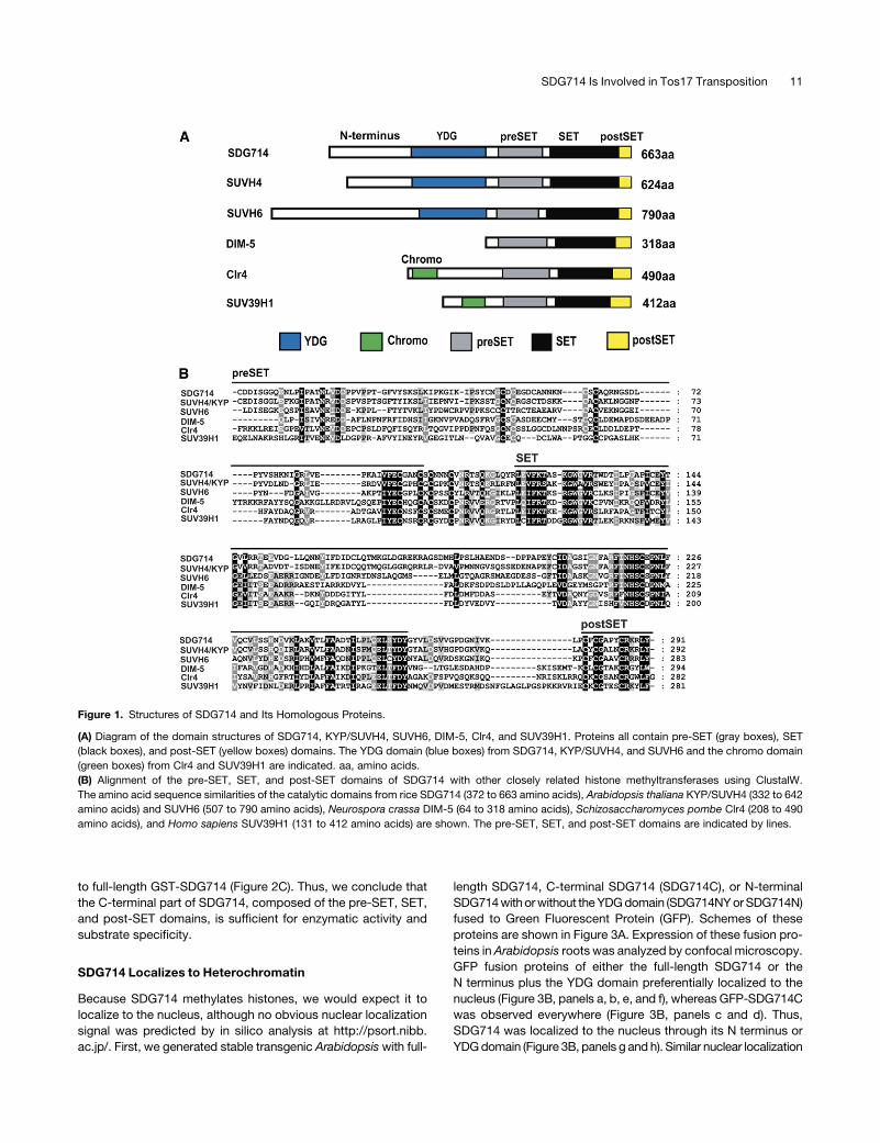
SDG714 encodes a protein of 663 amino acids and contains an
YDG domain and a conserved catalytic domain including the SET
domain and two Cys-rich motifs, pre-SET and post-SET, critical
for histone methyltransferase activity in the C terminus (Rea
et al., 2000) (Figure 1A).
The YDG domain is conserved in the SUVH class of histone
methyltransferases in Arabidopsis but is not found in other his-
tone H3K9 methyltransferases, such as DIM-5 from the fungus
N. crassa, Clr4 from yeast, and SUV39H1 from human. DIM-5
lacks an N-terminal conserved domain, whereas Clr4 and
SUV39H1 contain a chromo domain at their N termini (Rea
et al., 2000) (Figure 1A). Alignment of SDG714 with KYP/SUVH4,
SUVH6, DIM-5, Clr4, and SUV39H1 shows very high similarity in
the pre-SET, SET, and post-SET motifs, indicating that SDG714
could be an active histone methyltransferase (Figure 1B).
SDG714 Specifically Methylates Histone H3K9
To test whether SDG714 is an active histone methyltransferase,
full-length SDG714 fused to glutathione-S-transferase (GST-
SDG714) was expressed and purified from Escherichia coli. It
has been shown that different histone methyltransferases have
different substrate specificities (Nishioka and Reinberg, 2003).
For example, SET8 preferentially methylates nucleosomal sub-
strates (oligonucleosomes) at H4K20 (Fang et al., 2002). There-
fore, we performed in vitro methylation assays of SDG714 with
different substrates, including oligonucleosomes and core his-
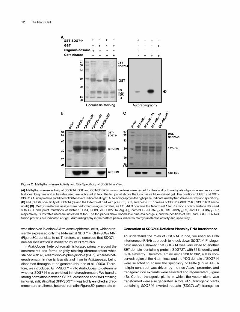
tones (Figure 2A). GST-SDG714 strongly methylated histone H3
from core histones but had only weak activity on histone H3 from
oligonucleosomes (Figure 2A). As a negative control, GST alone
did not methylate any of the substrates. Therefore, we conclude
that core histones are the optimal substrate for SDG714 in vitro.
Because SDG714 is similar to KYP/SUVH4, we wondered
whether it also has similar enzymatic specificity. The N terminus
of histone H3 was GST-tagged, and GST-H3N1-57 and GST-
H3N1-57 with a K-to-R replacement at K4 (GST-H3N1-57R4), K9
(GST-H3N1-57R9), or K27 (GST-H3N1-57R27) were used to test
the site specificity of SDG714. As expected, SDG714 methylated
GST-H3N1-57, GST-H3N1-57R4, and GST-H3N1-57R27 but not
GST-H3N1-57R9, suggesting that SDG714 is a methyltransferase
specific for histone H3 at K9 (Figure 2B).
The YDG domain is highly conserved in a group of histone
methyltransferases, and we asked whether it is required for en-
zymatic activity or substrate recognition. The GST fusion protein
containing just the pre-SET, SET, and post-SET domains of
SDG714 (GST-SDG714C without the N terminus or YDG domain)
showed histone methyltransferase activity and specificity similar
10 The Plant Cell
to full-length GST-SDG714 (Figure 2C). Thus, we conclude that
the C-terminal part of SDG714, composed of the pre-SET, SET,
and post-SET domains, is sufficient for enzymatic activity and
substrate specificity.
SDG714 Localizes to Heterochromatin
Because SDG714 methylates histones, we would expect it to
localize to the nucleus, although no obvious nuclear localization
signal was predicted by in silico analysis at http://psort.nibb.
ac.jp/. First, we generated stable transgenic Arabidopsis with full-
length SDG714, C-terminal SDG714 (SDG714C), or N-terminal
SDG714 with or without the YDG domain (SDG714NY or SDG714N)
fused to Green Fluorescent Protein (GFP). Schemes of these
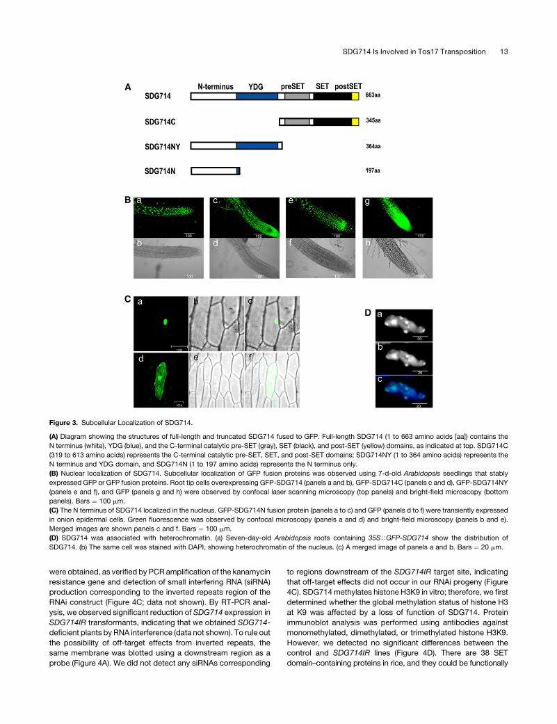
proteins are shown in Figure 3A. Expression of these fusion pro-
teins in Arabidopsis roots was analyzed by confocal microscopy.
GFP fusion proteins of either the full-length SDG714 or the
N terminus plus the YDG domain preferentially localized to the
nucleus (Figure 3B, panels a, b, e, and f), whereas GFP-SDG714C
was observed everywhere (Figure 3B, panels c and d). Thus,
SDG714 was localized to the nucleus through its N terminus or
YDG domain (Figure 3B, panels g and h). Similar nuclear localization
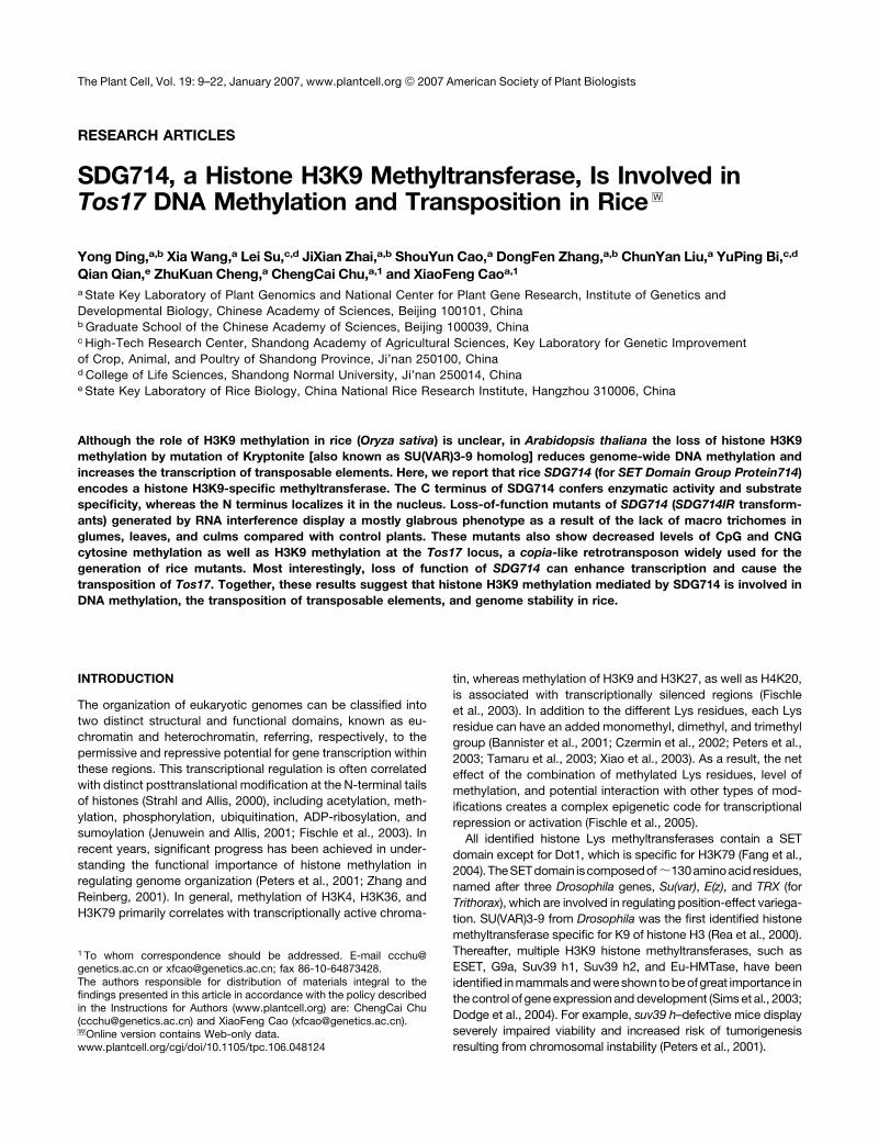
Figure 1. Structures of SDG714 and Its Homologous Proteins.
(A) Diagram of the domain structures of SDG714, KYP/SUVH4, SUVH6, DIM-5, Clr4, and SUV39H1. Proteins all contain pre-SET (gray boxes), SET
(black boxes), and post-SET (yellow boxes) domains. The YDG domain (blue boxes) from SDG714, KYP/SUVH4, and SUVH6 and the chromo domain
(green boxes) from Clr4 and SUV39H1 are indicated. aa, amino acids.
(B) Alignment of the pre-SET, SET, and post-SET domains of SDG714 with other closely related histone methyltransferases using ClustalW.
The amino acid sequence similarities of the catalytic domains from rice SDG714 (372 to 663 amino acids), Arabidopsis thaliana KYP/SUVH4 (332 to 642
amino acids) and SUVH6 (507 to 790 amino acids), Neurospora crassa DIM-5 (64 to 318 amino acids), Schizosaccharomyces pombe Clr4 (208 to 490
amino acids), and Homo sapiens SUV39H1 (131 to 412 amino acids) are shown. The pre-SET, SET, and post-SET domains are indicated by lines.
SDG714 Is Involved in Tos17 Transposition 11
was observed in onion (Allium cepa) epidermal cells, which tran-
siently expressed only the N-terminal SDG714 (GFP-SDG714N)
(Figure 3C, panels a to c). Therefore, we conclude that SDG714
nuclear localization is mediated by its N terminus.
In Arabidopsis, heterochromatin is located primarily around the
centromeres and forms brightly staining chromocenters when
stained with 49,6-diamidino-2-phenylindole (DAPI), whereas het-
erochromatin in rice is less distinct than in Arabidopsis, being
dispersed throughout the genome (Houben et al., 2003). There-
fore, we introduced GFP-SDG714 into Arabidopsis to determine
whether SDG714 was enriched in heterochromatin. We found a
strong correlation between GFP fluorescence and DAPI staining
in nuclei, indicating that GFP-SDG714 was highly enriched in chro-
mocenters and hence heterochromatin (Figure 3D, panels a to c).
Generation of SDG714-Deficient Plants by RNA Interference
To understand the roles of SDG714 in rice, we used an RNA
interference (RNAi) approach to knock down SDG714. Phyloge-
netic analysis showed that SDG714 was very close to another
SET domain–containing protein, SDG727, with 36% identity and
52% similarity. Therefore, amino acids 238 to 362, a less con-
served region at the N terminus, and the YDG domain of SDG714
were selected to ensure the specificity of RNAi (Figure 4A). A
hairpin construct was driven by the rice Actin1 promoter, and
transgenic rice explants were selected and regenerated (Figure
4B). Control transgenic plants in which the vector alone was
transformed were also generated. A total of 13 transgenic plants
containing SDG714 inverted repeats (SDG714IR) transgenes
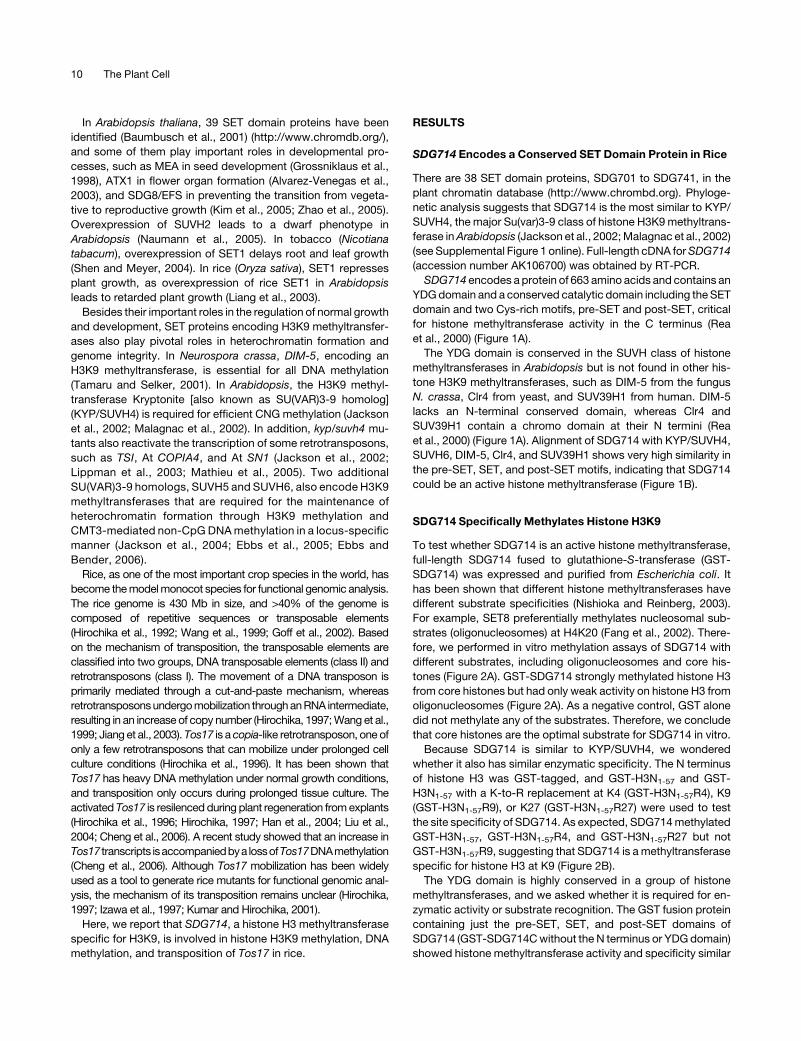
Figure 2. Methyltransferase Activity and Site Specificity of SDG714 in Vitro.
(A) Methyltransferase activity of SDG714. GST and GST-SDG714 fusion proteins were tested for their ability to methylate oligonucleosomes or core
histones. Enzymes and substrates used are indicated at top. The left panel shows the Coomassie blue–stained gel. The positions of GST and GST-
SDG714 fusion proteins and different histones are indicated at right. Autoradiography in the right panel indicates methyltransferase activity and specificity.
(B) and (C) Site specificity of SDG714 (B) and the C-terminal part with pre-SET, SET, and post-SET domains of SDG714 (SDG714C; 319 to 663 amino
acids) (C). Methyltransferase assays were performed using substrates, as GST-NH3 contains the N-terminal 1 to 57 amino acids of histone H3 fused
with GST and point mutations at histone H3K4, H3K9, or H3K27 to Arg (R), named GST-H3N1-57R4, GST-H3N1-57R9, and GST-H3N1-57R27
respectively. Substrates used are indicated at top. The top panels show Coomassie blue–stained gels, and the positions of GST and GST-SDG714C
fusion proteins are indicated at right. Autoradiography in the bottom panels indicates methyltransferase activity and specificity.
12 The Plant Cell
were obtained, as verified by PCR amplification of the kanamycin
resistance gene and detection of small interfering RNA (siRNA)
production corresponding to the inverted repeats region of the
RNAi construct (Figure 4C; data not shown). By RT-PCR anal-
ysis, we observed significant reduction of SDG714 expression in
SDG714IR transformants, indicating that we obtained SDG714-
deficient plants by RNA interference (data not shown). To rule out
the possibility of off-target effects from inverted repeats, the
same membrane was blotted using a downstream region as a
probe (Figure 4A). We did not detect any siRNAs corresponding
to regions downstream of the SDG714IR target site, indicating
that off-target effects did not occur in our RNAi progeny (Figure
4C). SDG714 methylates histone H3K9 in vitro; therefore, we first
determined whether the global methylation status of histone H3
at K9 was affected by a loss of function of SDG714. Protein
immunoblot analysis was performed using antibodies against
monomethylated, dimethylated, or trimethylated histone H3K9.
However, we detected no significant differences between the
control and SDG714IR lines (Figure 4D). There are 38 SET
domain–containing proteins in rice, and they could be functionally
Figure 3. Subcellular Localization of SDG714.
(A) Diagram showing the structures of full-length and truncated SDG714 fused to GFP. Full-length SDG714 (1 to 663 amino acids [aa]) contains the
N terminus (white), YDG (blue), and the C-terminal catalytic pre-SET (gray), SET (black), and post-SET (yellow) domains, as indicated at top. SDG714C
(319 to 613 amino acids) represents the C-terminal catalytic pre-SET, SET, and post-SET domains; SDG714NY (1 to 364 amino acids) represents the
N terminus and YDG domain, and SDG714N (1 to 197 amino acids) represents the N terminus only.
(B) Nuclear localization of SDG714. Subcellular localization of GFP fusion proteins was observed using 7-d-old Arabidopsis seedlings that stably
expressed GFP or GFP fusion proteins. Root tip cells overexpressing GFP-SDG714 (panels a and b), GFP-SDG714C (panels c and d), GFP-SDG714NY
(panels e and f), and GFP (panels g and h) were observed by confocal laser scanning microscopy (top panels) and bright-field microscopy (bottom
panels). Bars ¼ 100 mm.
(C) The N terminus of SDG714 localized in the nucleus. GFP-SDG714N fusion protein (panels a to c) and GFP (panels d to f) were transiently expressed
in onion epidermal cells. Green fluorescence was observed by confocal microscopy (panels a and d) and bright-field microscopy (panels b and e).
Merged images are shown panels c and f. Bars ¼ 100 mm.
(D) SDG714 was associated with heterochromatin. (a) Seven-day-old Arabidopsis roots containing 35STGFP-SDG714 show the distribution of
SDG714. (b) The same cell was stained with DAPI, showing heterochromatin of the nucleus. (c) A merged image of panels a and b. Bars ¼ 20 mm.
SDG714 Is Involved in Tos17 Transposition 13
redundant. Alternatively, SDG714 preferentially acts at specific
sequences but not globally in rice.
SDG714IR Transformants Are Defective in Macro
Trichome Development
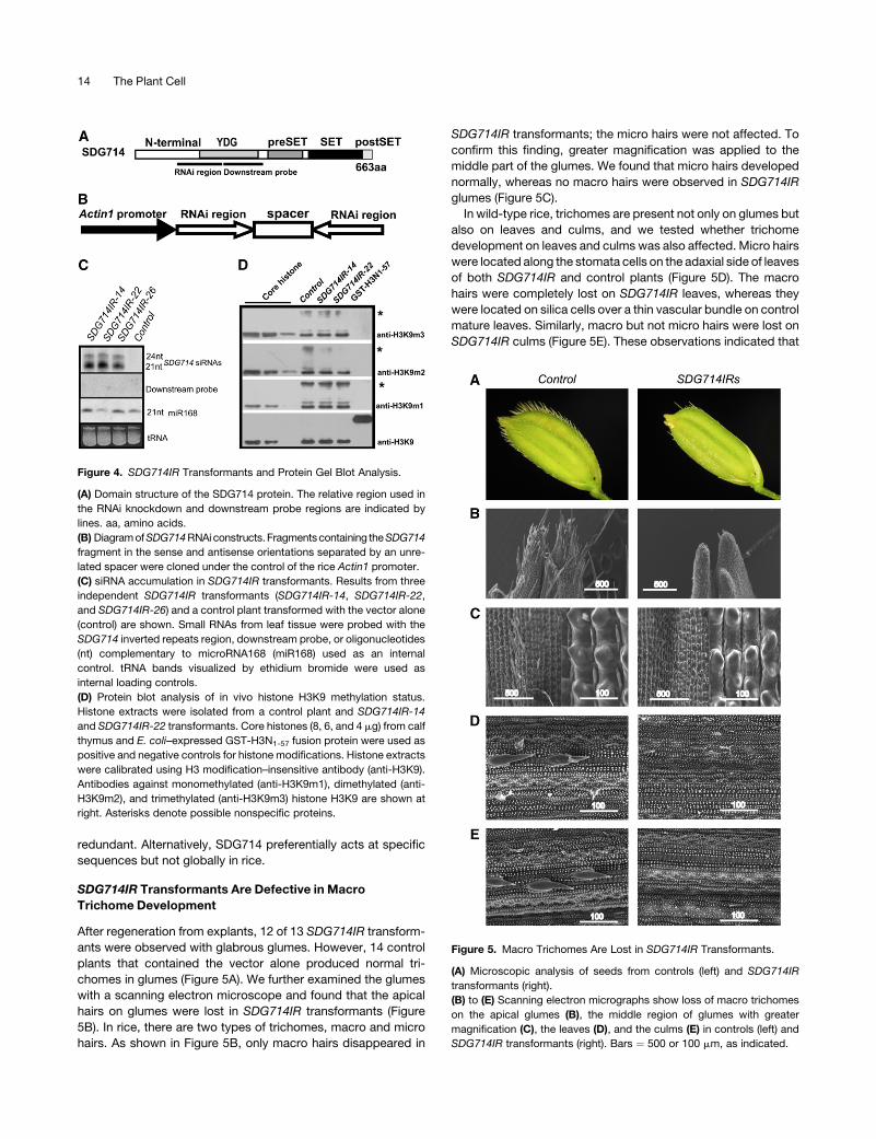
After regeneration from explants, 12 of 13 SDG714IR transform-
ants were observed with glabrous glumes. However, 14 control
plants that contained the vector alone produced normal tri-
chomes in glumes (Figure 5A). We further examined the glumes
with a scanning electron microscope and found that the apical
hairs on glumes were lost in SDG714IR transformants (Figure
5B). In rice, there are two types of trichomes, macro and micro
hairs. As shown in Figure 5B, only macro hairs disappeared in
SDG714IR transformants; the micro hairs were not affected. To
confirm this finding, greater magnification was applied to the
middle part of the glumes. We found that micro hairs developed
normally, whereas no macro hairs were observed in SDG714IR
glumes (Figure 5C).
In wild-type rice, trichomes are present not only on glumes but
also on leaves and culms, and we tested whether trichome
development on leaves and culms was also affected. Micro hairs
were located along the stomata cells on the adaxial side of leaves
of both SDG714IR and control plants (Figure 5D). The macro
hairs were completely lost on SDG714IR leaves, whereas they
were located on silica cells over a thin vascular bundle on control
mature leaves. Similarly, macro but not micro hairs were lost on
SDG714IR culms (Figure 5E). These observations indicated that
Figure 4. SDG714IR Transformants and Protein Gel Blot Analysis.
(A) Domain structure of the SDG714 protein. The relative region used in
the RNAi knockdown and downstream probe regions are indicated by
lines. aa, amino acids.
(B) Diagram of SDG714 RNAi constructs.Fragments containing the SDG714
fragment in the sense and antisense orientations separated by an unre-
lated spacer were cloned under the control of the rice Actin1 promoter.
(C) siRNA accumulation in SDG714IR transformants. Results from three
independent SDG714IR transformants (SDG714IR-14, SDG714IR-22,
and SDG714IR-26) and a control plant transformed with the vector alone
(control) are shown. Small RNAs from leaf tissue were probed with the
SDG714 inverted repeats region, downstream probe, or oligonucleotides
(nt) complementary to microRNA168 (miR168) used as an internal
control. tRNA bands visualized by ethidium bromide were used as
internal loading controls.
(D) Protein blot analysis of in vivo histone H3K9 methylation status.
Histone extracts were isolated from a control plant and SDG714IR-14
and SDG714IR-22 transformants. Core histones (8, 6, and 4 mg) from calf
thymus and E. coli–expressed GST-H3N1-57 fusion protein were used as
positive and negative controls for histone modifications. Histone extracts
were calibrated using H3 modification–insensitive antibody (anti-H3K9).
Antibodies against monomethylated (anti-H3K9m1), dimethylated (anti-
H3K9m2), and trimethylated (anti-H3K9m3) histone H3K9 are shown at
right. Asterisks denote possible nonspecific proteins.
Figure 5. Macro Trichomes Are Lost in SDG714IR Transformants.
(A) Microscopic analysis of seeds from controls (left) and SDG714IR
transformants (right).
(B) to (E) Scanning electron micrographs show loss of macro trichomes
on the apical glumes (B), the middle region of glumes with greater
magnification (C), the leaves (D), and the culms (E) in controls (left) and
SDG714IR transformants (right). Bars ¼ 500 or 100 mm, as indicated.
14 The Plant Cell
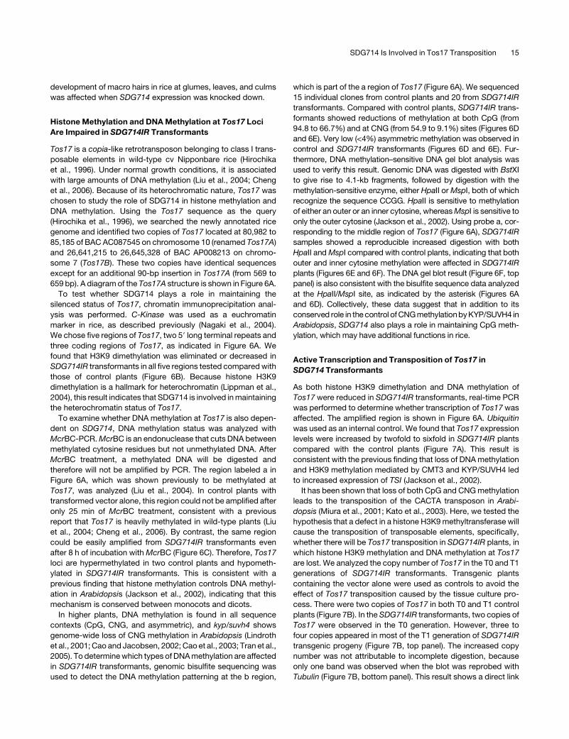
development of macro hairs in rice at glumes, leaves, and culms
was affected when SDG714 expression was knocked down.
Histone Methylation and DNA Methylation at Tos17 Loci
Are Impaired in SDG714IR Transformants
Tos17 is a copia-like retrotransposon belonging to class I trans-
posable elements in wild-type cv Nipponbare rice (Hirochika
et al., 1996). Under normal growth conditions, it is associated
with large amounts of DNA methylation (Liu et al., 2004; Cheng
et al., 2006). Because of its heterochromatic nature, Tos17 was
chosen to study the role of SDG714 in histone methylation and
DNA methylation. Using the Tos17 sequence as the query
(Hirochika et al., 1996), we searched the newly annotated rice
genome and identified two copies of Tos17 located at 80,982 to
85,185 of BAC AC087545 on chromosome 10 (renamed Tos17A)
and 26,641,215 to 26,645,328 of BAC AP008213 on chromo-
some 7 (Tos17B). These two copies have identical sequences
except for an additional 90-bp insertion in Tos17A (from 569 to
659 bp). A diagram of the Tos17A structure is shown in Figure 6A.
To test whether SDG714 plays a role in maintaining the
silenced status of Tos17, chromatin immunoprecipitation anal-
ysis was performed. C-Kinase was used as a euchromatin
marker in rice, as described previously (Nagaki et al., 2004).
We chose five regions of Tos17, two 59 long terminal repeats and
three coding regions of Tos17, as indicated in Figure 6A. We
found that H3K9 dimethylation was eliminated or decreased in
SDG714IR transformants in all five regions tested compared with
those of control plants (Figure 6B). Because histone H3K9
dimethylation is a hallmark for heterochromatin (Lippman et al.,
2004), this result indicates that SDG714 is involved in maintaining
the heterochromatin status of Tos17.
To examine whether DNA methylation at Tos17 is also depen-
dent on SDG714, DNA methylation status was analyzed with
McrBC-PCR. McrBC is an endonuclease that cuts DNA between
methylated cytosine residues but not unmethylated DNA. After
McrBC treatment, a methylated DNA will be digested and
therefore will not be amplified by PCR. The region labeled a in
Figure 6A, which was shown previously to be methylated at
Tos17, was analyzed (Liu et al., 2004). In control plants with
transformed vector alone, this region could not be amplified after
only 25 min of McrBC treatment, consistent with a previous
report that Tos17 is heavily methylated in wild-type plants (Liu
et al., 2004; Cheng et al., 2006). By contrast, the same region
could be easily amplified from SDG714IR transformants even
after 8 h of incubation with McrBC (Figure 6C). Therefore, Tos17
loci are hypermethylated in two control plants and hypometh-
ylated in SDG714IR transformants. This is consistent with a
previous finding that histone methylation controls DNA methyl-
ation in Arabidopsis (Jackson et al., 2002), indicating that this
mechanism is conserved between monocots and dicots.
In higher plants, DNA methylation is found in all sequence
contexts (CpG, CNG, and asymmetric), and kyp/suvh4 shows
genome-wide loss of CNG methylation in Arabidopsis (Lindroth
et al., 2001; Cao and Jacobsen, 2002; Cao et al., 2003; Tran et al.,
2005). To determine which types of DNA methylation are affected
in SDG714IR transformants, genomic bisulfite sequencing was
used to detect the DNA methylation patterning at the b region,
which is part of the a region of Tos17 (Figure 6A). We sequenced
15 individual clones from control plants and 20 from SDG714IR
transformants. Compared with control plants, SDG714IR trans-
formants showed reductions of methylation at both CpG (from
94.8 to 66.7%) and at CNG (from 54.9 to 9.1%) sites (Figures 6D
and 6E). Very low (<4%) asymmetric methylation was observed in
control and SDG714IR transformants (Figures 6D and 6E). Fur-
thermore, DNA methylation–sensitive DNA gel blot analysis was
used to verify this result. Genomic DNA was digested with BstXI
to give rise to 4.1-kb fragments, followed by digestion with the
methylation-sensitive enzyme, either HpaII or MspI, both of which
recognize the sequence CCGG. HpaII is sensitive to methylation
of either an outer or an inner cytosine, whereas MspI is sensitive to
only the outer cytosine (Jackson et al., 2002). Using probe a, cor-
responding to the middle region of Tos17 (Figure 6A), SDG714IR
samples showed a reproducible increased digestion with both
HpaII and MspI compared with control plants, indicating that both
outer and inner cytosine methylation were affected in SDG714IR
plants (Figures 6E and 6F). The DNA gel blot result (Figure 6F, top
panel) is also consistent with the bisulfite sequence data analyzed
at the HpaII/MspI site, as indicated by the asterisk (Figures 6A
and 6D). Collectively, these data suggest that in addition to its
conserved role in the control of CNG methylation by KYP/SUVH4 in
Arabidopsis, SDG714 also plays a role in maintaining CpG meth-
ylation, which may have additional functions in rice.
Active Transcription and Transposition of Tos17 in
SDG714 Transformants
As both histone H3K9 dimethylation and DNA methylation of
Tos17 were reduced in SDG714IR transformants, real-time PCR
was performed to determine whether transcription of Tos17 was
affected. The amplified region is shown in Figure 6A. Ubiquitin
was used as an internal control. We found that Tos17 expression
levels were increased by twofold to sixfold in SDG714IR plants
compared with the control plants (Figure 7A). This result is
consistent with the previous finding that loss of DNA methylation
and H3K9 methylation mediated by CMT3 and KYP/SUVH4 led
to increased expression of TSI (Jackson et al., 2002).
It has been shown that loss of both CpG and CNG methylation
leads to the transposition of the CACTA transposon in Arabi-
dopsis (Miura et al., 2001; Kato et al., 2003). Here, we tested the
hypothesis that a defect in a histone H3K9 methyltransferase will
cause the transposition of transposable elements, specifically,
whether there will be Tos17 transposition in SDG714IR plants, in
which histone H3K9 methylation and DNA methylation at Tos17
are lost. We analyzed the copy number of Tos17 in the T0 and T1
generations of SDG714IR transformants. Transgenic plants
containing the vector alone were used as controls to avoid the
effect of Tos17 transposition caused by the tissue culture pro-
cess. There were two copies of Tos17 in both T0 and T1 control
plants (Figure 7B). In the SDG714IR transformants, two copies of
Tos17 were observed in the T0 generation. However, three to
four copies appeared in most of the T1 generation of SDG714IR
transgenic progeny (Figure 7B, top panel). The increased copy
number was not attributable to incomplete digestion, because
only one band was observed when the blot was reprobed with
Tubulin (Figure 7B, bottom panel). This result shows a direct link
SDG714 Is Involved in Tos17 Transposition 15
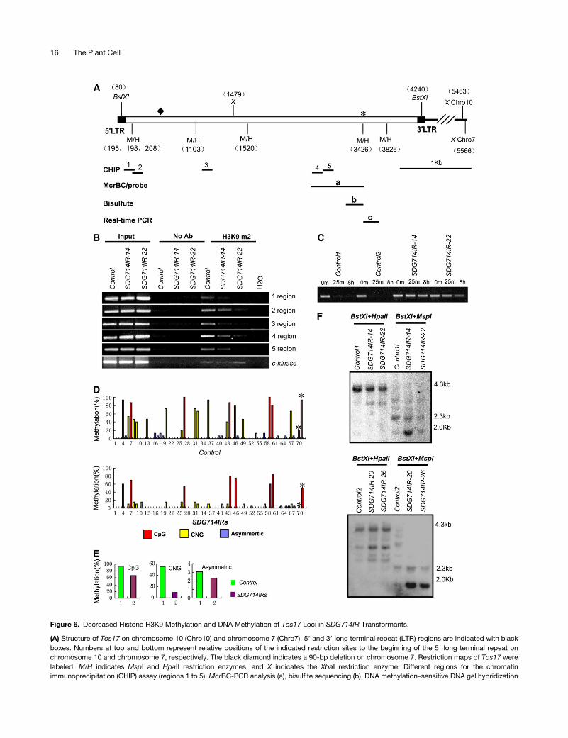
Figure 6. Decreased Histone H3K9 Methylation and DNA Methylation at Tos17 Loci in SDG714IR Transformants.
(A) Structure of Tos17 on chromosome 10 (Chro10) and chromosome 7 (Chro7). 59 and 39 long terminal repeat (LTR) regions are indicated with black
boxes. Numbers at top and bottom represent relative positions of the indicated restriction sites to the beginning of the 59 long terminal repeat on
chromosome 10 and chromosome 7, respectively. The black diamond indicates a 90-bp deletion on chromosome 7. Restriction maps of Tos17 were
labeled. M/H indicates MspI and HpaII restriction enzymes, and X indicates the XbaI restriction enzyme. Different regions for the chromatin
immunoprecipitation (CHIP) assay (regions 1 to 5), McrBC-PCR analysis (a), bisulfite sequencing (b), DNA methylation–sensitive DNA gel hybridization
16 The Plant Cell
between histone methylation, DNA methylation, transcription,
and transposition of a retrotransposon.
DISCUSSION
Differences between SDG714 and KYP/SUVH4
It has been shown that tagged KYP/SUVH4, SUVH1, and SUVH2
in Arabidopsis and tobacco SET1 are localized to heterochromatin
(Yu et al., 2004; Naumann et al., 2005). Here, we demonstrate
that SDG714 is also a nuclear protein that associates with
heterochromatic chromocenters in Arabidopsis. Still, the exact
mechanism of how SDG714 is targeted to heterochromatin
remains to be addressed. The similar enzymatic properties and
cellular localization indicate that SDG714 and KYP/SUVH4 gene
diversification occurred before the divergence of monocots and
dicots. After the monocot and dicot split, rice and Arabidopsis
evolved many species-specific sequences in each genome.
These unique sequences may require different cofactors along
with either SDG714 or KYP/SUVH4 to function. The differences
between SDG714 and KYP/SUVH4 were also supported by the
observation that DNA methylation patterns maintained by SDG714
and KYP/SUVH4 may not be identical, as SDG714 is required for
both CpG and CNG methylation at Tos17 and KYP/SUVH4 acts
mainly on CNG methylation. As a result, SDG714 and KYP/
SUVH4 may not be orthologous in terms of developmental
regulation, because SDG714 could not complement Arabidopsis
KYP/SUVH4 in kyp clk-st double mutants (for details, see Sup-
plemental Results online). In addition, we observed that reduc-
tion of SDG714 mRNA leads to defects in macro trichome
development, whereas there is no obvious developmental defect
in plants without KYP, its closest homolog in Arabidopsis. It
would be interesting to identify genes causing altered macrohair
defects in the loss of function of SDG714 in rice.
Significance of SDG714 in Controlling Tos17 Silencing
In eukaryotes, the presence of 59 methylcytosine is an important
component of transcriptionally silent chromatin and is widely
considered to be a mechanism that the genome uses to defend
against transposable elements and retroviruses (Constancia
et al., 1998; Covey and Al-Kaff, 2000; Lorincz et al., 2001). For
example, loss of function of CMT3, a plant-specific DNA meth-
yltransferase that mainly controls non-CpG methylation, causes
transcriptional activation of retroelements at Ta3 and TSI in
Arabidopsis (Lindroth et al., 2001). Loss of CpG and CNG
methylation leads to mobilization of the CACTA transposon in
cmt3 and met1 double mutants (Kato et al., 2003). Besides DNA
methylation, transcriptionally silenced retroelements are also
maintained through histone methylation and siRNA-mediated
gene silencing within a gene regulation network (Tran et al.,
2005). In the kyp/suvh4 mutant, the retrotransposons TSI, At
COPIA4, and At SN1 were actively transcribed, indicating a role
for histone and DNA methylation in transcriptional silencing of
retrotransposons (Lindroth et al., 2001; Jackson et al., 2002;
Lippman et al., 2003; Mathieu et al., 2005). In addition, large-
scale DNA methylation analysis also showed that KYP/SUVH4
preferentially regulates transposable elements (Tran et al., 2005).
And SUVH5 and SUVH6 were identified to be partially redundant
with KYP/SUVH6 in controlling CMT3-mediated non-CpG meth-
ylation and repressing the transcription of transposable elements
in heterochromatin (Jackson et al., 2004; Ebbs et al., 2005; Ebbs
and Bender, 2006).
In rice, Tos17 is a copia-like retrotransposon that is activated
and transposed during prolonged cell culture and subsequently
remethylated and silenced in regenerated plants (Hirochika et al.,
1996; Cheng et al., 2006). Because of its ability to transpose,
Tos17 has been widely used as a tool for generating mutants in
rice (Hirochika, 1997; Kumar and Hirochika, 2001). However, the
exact mechanism of how this transposition is controlled is un-
known. Here, we show that decreased expression of SDG714
results in a decrease in histone methylation and DNA methylation
and an increase in Tos17 expression. Our work highlights the
important connection between histone methylation, DNA meth-
ylation, transcription, and the transposition of a retrotransposon.
Surprisingly, our real-time PCR results showed that Tos17
transcript was enhanced by only twofold to sixfold between RNAi
and control plants but led to significant differences, between no
Figure 6. (continued).
probe (a), and real-time PCR analysis (c) are shown. The asterisk represents the HpaII/MspI site, which was also determined by bisulfite sequencing
analysis in (D).
(B) Chromatin immunoprecipitation analysis of H3K9 dimethylation in SDG714IR transformants (SDG714IR-14 and SDG714IR-22). Chromatin
immunoprecipitation assays were performed with antibodies against dimethyl Lys-9 of histone H3 (H3K9m2). Primers specific for regions 1 to 5 of Tos17
are shown in (A). The euchromatin marker C-Kinase was used as an internal control. No Ab, no antibody.
(C) McrBC-PCR analysis of DNA methylation at Tos17 loci. Equal amounts of genomic DNAs from two controls (control 1 and control 2) and two
SDG714IR transformants (SDG714IR-14 and SDG714IR-22) were digested with McrBC for 0 min, 25 min, and 8 h, followed by PCR amplification of
region a shown in (A).
(D) Profile of DNA methylation at a 400-bp region (shown in [A]) of Tos17. The numbers on the x axis represent cytosine positions in the analyzed region,
and the y axis represents methylation levels in controls (top panel) and SDG714IR transformants (bottom panel). Red, blue, and yellow bars indicate
CpG, CNG, and asymmetric methylation, respectively. Asterisks indicate methylation at the CCGG position corresponding to the HpaII/MspI site (3426 bp
from the 59 long terminal repeat on chromosome 10) in control plants and SDG714IR transformants.
(E) Histograms represent the percentage of CpG, CNG, and asymmetric methylation in control plants (green) and SDG714IR transformants (purple).
(F) DNA gel blot analysis of DNA methylation in SDG714IR transformants. Genomic DNAs of two controls or SDG714IR transformants (SDG714IR-14
and SDG714IR-22 in the top panel, SDG714IR-20 and SDG714IR-26 in the bottom panel) were digested with BstXI, followed by methylation-sensitive
enzyme HpaII or MspI, and the blot was hybridized with the probes shown in (A). DNA markers are shown at right.
SDG714 Is Involved in Tos17 Transposition 17
transposition in the controls and active transposition in the RNAi
lines (Figures 7A and 7B). One possibility is that Tos17 transpo-
sition requires a certain transcript threshold and that loss of
function of SDG714 increases the Tos17 transcriptover the thresh-
old, resulting in a high frequency of transposition in SDG714IR
plants. A similar phenomenon was observed in CACATA trans-
position in the met1 and cmt3 backgrounds (Kato et al., 2003).
Alternatively, transcript accumulation is not the major barrier to
the transposition of retrotransposons. Instead, active transposi-
tion might be affected mainly through the epigenetic regula-
tion of chromatin, such as DNA methylation, histone covalent
modifications, or ATP-dependent chromatin remodeling. Here, we
show that SDG714 targeted to heterochromatin and loss of
function of SDG714 decreased histone methylation and DNA
methylation at Tos17 loci, which might in turn modulate heter-
ochromatin, resulting in higher levels of transposition. It is
reported that Tos17 can be activated and transposed during
prolonged cell culture. Although the tissue culture process is
required for the generation of SDG714IR plants, no transposition
was observed in the control plants transformed with vector only
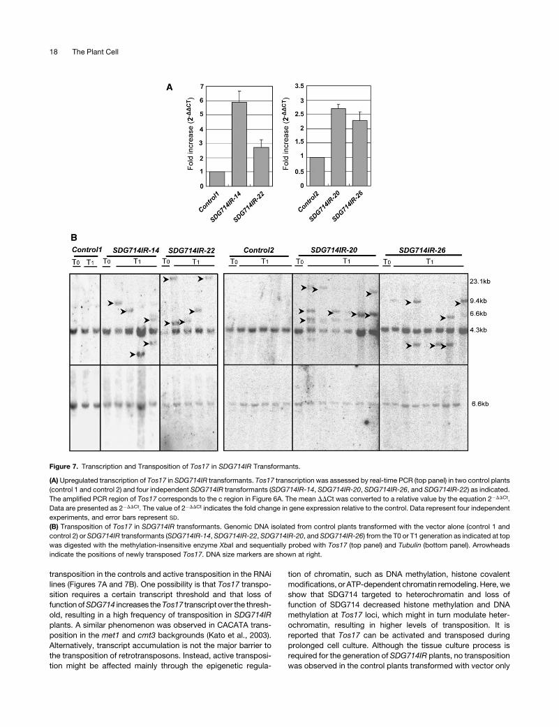
Figure 7. Transcription and Transposition of Tos17 in SDG714IR Transformants.
(A) Upregulated transcription of Tos17 in SDG714IR transformants. Tos17 transcription was assessed by real-time PCR (top panel) in two control plants
(control 1 and control 2) and four independent SDG714IR transformants (SDG714IR-14, SDG714IR-20, SDG714IR-26, and SDG714IR-22) as indicated.
The amplified PCR region of Tos17 corresponds to the c region in Figure 6A. The mean DDCt was converted to a relative value by the equation 2�DDCt.
Data are presented as 2�DDCt. The value of 2�DDCt indicates the fold change in gene expression relative to the control. Data represent four independent
experiments, and error bars represent SD.
(B) Transposition of Tos17 in SDG714IR transformants. Genomic DNA isolated from control plants transformed with the vector alone (control 1 and
control 2) or SDG714IR transformants (SDG714IR-14, SDG714IR-22, SDG714IR-20, and SDG714IR-26) from the T0 or T1 generation as indicated at top
was digested with the methylation-insensitive enzyme XbaI and sequentially probed with Tos17 (top panel) and Tubulin (bottom panel). Arrowheads
indicate the positions of newly transposed Tos17. DNA size markers are shown at right.
18 The Plant Cell
(Figure 7B). Therefore, we proposed that during the transforma-
tion of rice, condensed chromatin might be loosened, decreasing
the threshold for the activation of Tos17 expression and trans-
position. However, transformation alone would not reach the
threshold for transposition of Tos17, because no transposition of
Tos17 was observed in control plants in which the identical
transformation process was applied. By contrast, when SDG714
was knocked down, in addition to partially released silencing by
transformation, Tos17 was further modulated as a result of the
loss of histone H3K9 and DNA methylation, resulting in the
upregulation of gene expression and activated transposition.
Therefore, a synergic effect between the loss of function of
SDG714 and transformation processing led to stronger activa-
tion of Tos17 expression and activated transposition.
It has been shown that Tos17 preferentially jumps into eu-
chromatic regions, which would be harmful to the rice genome if
it were not properly silenced (Miyao et al., 2003). Here, we find a
significant role for SDG714 in rice genome stability, similar to
what has been observed in mice. suv39 h–defective mice display
severely impaired viability and chromosomal instability, with an
increased risk of tumorigenesis (Peters et al., 2001). Practically,
loss of function of SDG714 can potentially be used as a tool for
the generation of mutants in rice. The advantage of using the
SDG714IR system to generate mutants is that prolonged tissue
culture is no longer needed, as several extra copies of Tos17 can
be observed in the T1 generation. Once the SDG714IR transgene
is outcrossed in the T2 generation, Tos17 should be resilenced,
and the homozygous progeny of Tos17 insertions will give rise to
insertional mutations.
METHODS
Plant Materials and Growth Conditions
Rice plants used in this study were Oryza sativa spp japonica cv
Nipponbare. The plants were all regenerated from the callus. T1 gener-
ation plants were generated from self-pollinated T0 generation seeds.
Plant materials were harvested from leaf tissue grown in the field.
Molecular Cloning of SDG714
cDNAs encoding amino acids 1 to 663 of SDG714 were PCR-amplified
from reverse-transcribed rice cDNA by forward primer CX187 (59-TAGa-
gatctATGGAGGTGATGGATTCGGTGGCCGT-39; lowercase and under-
lined letters represent the restriction sites added for subsequent cloning)
and reverse primer CX188 (59-GTCtctagaCTAATAAAGTCGCTTCCGG-
CAGTAC-39), and PCR products were cloned into Topo-PCR4.0
(XF00204). SDG714 was then digested with BglII and EcoRI and sub-
cloned into pGEX-4T-1 via BamHI and EcoRI sites, giving GST-SDG714
(XF00206). To generate the C-terminal fragment of SDG714, the cDNA
fragments encoding amino acids 319 to 663 of SDG714 were amplified by
forward primer CX48 (59-GAGgaattcTACAAGGTTGTAGATGACTGGG-
TGCAG-39) and reverse primer CX47 (59-GTCgtcgacATCTAATAAAGT-
CGCTTCCGGCAGTAC-39). PCR products digested with EcoRI and SalI
were further cloned into pGEX-4T-1 at EcoRI and SalI sites, giving GST-
SDG714C (XF00163).
Protein Expression and Purification
The plasmids GST-SDG714 and GST-SDG714C, containing either the full
length or the C-terminal end of SDG714, respectively, were transformed
into BL21(RIL), and single clones were cultured in 23 YT medium plus
100 mg/mL ampicillin at 378C to OD 0.6, then induced by 0.1 mM
isopropylthio-b-galactoside at 188C for 12 h. GST fusion proteins were
purified as reported previously (Jackson et al., 2002).
In Vitro Histone Methyltransferase Assay
The detailed histone methyltransferase assay was performed according
to the methods described by Rea et al. (2000) with slight modification.
Briefly, 10 mg of oligonucleosomes (containing 2.5 mg of H2A, H2B, H3,
and H4 each, wrapped with DNA; kindly provided by Y. Zhang) and core
histones (containing 2.5 mg of H2A, H2B, H3, and H4 each) as substrates,
1 mCi of [3H]S-adenosyl-L-[methyl-3H]L-Met (79 Ci/mmol; TRK865; Amer-
sham Biosciences) as methyl donor, and 2 to 5 mg of recombinant protein
were used in the histone methyltransferase assay. Histone methyltrans-
ferase reactions on GST-NH3 fusion proteins (gifts of Y. Shinkai) were
performed using similar reaction conditions. After incubation at 308C for
1 h, reactions were stopped with SDS loading buffer, and protein was
separated by 15 or 12% SDS-PAGE, stained with Coomassie Brilliant
Blue R 250, treated with Amplifier (Amersham Biosciences), dried, and
fluorographed on x-ray film.
Construction of Binary Vectors
To generate pCAMBIA1300-35S-GFP, 35S-GFP from pAVA321 vector
was cloned into pCAMBIA1300 vector as a GFP control and named
pCAMBIA1300-35S-GFP (XF00183). To generate GFP fusion proteins,
primers used for the amplification of each fragment were as follow: CX187
and CX188 for full-length SDG714 (SDG714-GFP; 1 to 663 amino acids);
CX48 and CX47 for the C terminus of SDG714 (SDG714C-GFP; 319 to
663 amino acids); CX187 and reverse primer CX317 (59-GACtctagaAT-
CGTGGTGGGAGCTTCAGCACGAG-39) for the NY terminus of SDG714
(SDG714NY-GFP; 1 to 364 amino acids); and CX187 and reverse primer
CX1366 (59-AGCtctagaTGATCCCCAACATCAACACCAGGTA-39) for the
N terminus of SDG714 (SDG714N-GFP; 1 to 197 amino acids). Each
fragment was subcloned into binary vector pCAMBIA1300-35S-GFP
(XF00183) with appropriate restriction enzymes. The detailed process will
be provided upon request. Binary vectors of pCAMBIA1300-35S-GFP,
SDG714-GFP, SDG714NY-GFP, and SDG714C-GFP were transferred
into Agrobacterium tumefaciens strain AGL0 and used to transform
Arabidopsis thaliana ecotype Columbia plants by vacuum infiltration. T1
seeds were selected on half-strength Murashige and Skoog medium
supplemented with hygromycin (25 mg/mL). To generate the SDG714
RNAi vector, the fragment was amplified with primers CX52 (59-ATTctc-
gagGCAACTTGTATTGTCATGTCGGG-39) and CX53 (59-AGCagatctGG-
GAGCTTCAGCACGAGTAA-39), then sequentially cloned into pUCRNAi
vector with XhoI/BglII and BamHI/SalI. Finally, the full stem-loop fragment
was cloned into pCAMBIA2300-Actin vector (Liu et al., 2005).
Transient Expression of SDG714N-GFP
The SDG714N-GFP construct was coated onto gold particles (0.1 mm)
and delivered into onion (Allium cepa) epidermal cells with a Helios Gene
Gun System (Bio-Rad Laboratories). The bombardment parameters were
as follows: discharge pressure of 150 p.s.i.; and distance to target tissue
of 3.5 cm. Onion epidermal cells were placed onto half-strength Mura-
shige and Skoog agar plates before bombardment, incubated at 238C for
24 h, and analyzed with a Zeiss LSM 510 confocal microscope.
Protein Immunoblot Analysis
Leaves were homogenized in histone extraction buffer (0.25 M sucrose,
60 mM KCl, 15 mM NaCl, 5 mM MgCl2, 1 mM CaCl2, 15 mM PIPES,
pH 6.8, 0.8% Triton X-100, 0.1 mM Pefablock, and protease inhibitor
cocktail), as modified from Tariq et al. (2003). After centrifugation at
SDG714 Is Involved in Tos17 Transposition 19
10,000g for 10 min, the pellet was resuspended with 0.4 M H2SO4 and
centrifuged again; the soluble proteins were precipitated with 12 volumes of
acetone, left at �208C overnight, and then collected by centrifugation at
8000g for 15 min. The pellet was resuspended with 4 M urea (100 mL),
separatedona 15% PAGE gel,detectedbymonomethylated, dimethylated,
or trimethylated specific H3K9 and methylation-insensitive H3 antibodies
(catalog Nos. 07-450, 07-441, 07-442, and 06-755; Upstate). Peroxidase-
conjugated goat anti-rabbit antibodies (Pierce) were used as secondary
antibody, and the SuperSignal Western Pico detection system (Pierce) was
used for signal detection. The same blot was stripped before probing the
next antibody. Escherichia coli–expressed GST-H3 (1 to 57 amino acids)
fusion protein and diluted core histones were used as loading controls.
Microscopy
Seven-day-old seedlings were used for GFP analysis. The GFP fluores-
cence of protein was visualized by confocal laser scanning microscopy
(Olympus). Heterochromatin sublocation was analyzed with Arabidopsis
roots. The roots were stained with 2 mg/mL DAPI, and GFP images were
captured with the Olympus BX61 fluorescence microscope in conjunc-
tion with a micro CCD camera. Gray-scale images were captured for each
color channel and then merged using Image-Pro Plus software.
Scanning Electron Microscopy
Scanning electron microscopy was performed as described previously
with slight modifications (Li et al., 2003). Rice tissue was placed into 5%
formaldehyde dissolved in 13 PBS for 18 h, dried with 30, 50, 70, 90, and
100% ethanol, each for 10 min, and then placed into glutaraldehyde for 12 h.
The material was then point-dried with a CO2 critical point dryer (Hitachi
HCP-2), spotted with gold powder with ion sputter (Hitachi E-1010), and
observed with a scanning electron microscope (Hitachi S-3000N).
Reverse Transcription and Real-Time PCR
Total RNA isolation and RT-PCR were performed as described previously
(Liu et al., 2005). Real-time PCR analysis was performed using the
Chromo4 real-time PCR instrument (PTC-200; Bio-Rad) and SYBR Green
I (Invitrogen S-7567). The relative expressions of specific genes were
quantitated using 2�DDCt calculation, where DDCt is the difference in the
threshold cycles of the test and housekeeping gene Ubiquitin. DCt is the
threshold cycle of the target gene subtracted from the threshold cycle of
the housekeeping gene. The mean threshold cycle values for the genes of
interest were calculated from three experiments. Primers used were as
follows: for Ubiquitin, CX2137 (59-GCCCAAGAAGAAGATCAAGAAC-39)
and CX2138 (59-AGATAACAACGGAAGCATAAAAGTC-39); for Tos17,
CX2141 (59-TGTCAGTGCTCCGATTTCAGTTCA-39) and CX2142 (59-AAA-
TACAATAGCCAGTGACAGAGCG-39).
Small RNA Gel Blot Analysis
Small RNA gel blot analysis was performed as described by Liu et al.
(2005). Probes corresponding to the inverted repeated and downstream
region of SDG714 were PCR-amplified with CX52 and CX53 or with
CX2095 (59-CACGATTTCGGAATTACCTG-39) and CX2096 (59-AAGCGA-
TACTGCAGACCTTT-39), respectively. The PCR products were then cloned
into pGEM-T easy vector (Promega), transcribed with T7 polymerase in
vitro, and labeled with [a-32P]ATP. Probe corresponding to microRNA168
(CX161, 59-GTCCCGATCTGCACCAAGCGA-39) was end-labeled with
[g-32P]ATP.
Chromatin Immunoprecipitation Assay
Chromatin immunoprecipitation was performed as described previously
(Johnson et al., 2002). Briefly, 2 g of leaves was fixed with formaldehyde,
and the DNA/protein complex was immunoprecipitated with a-dimethyl
H3K9. After reverse cross-linking and proteinase K treatment, the
immunoprecipitated DNA was extracted with phenol/chloroform. All
PCR procedures were performed in 30-mL volumes, started with 2 min
at 948C, and followed with 30 to 40 cycles. The PCR product was elec-
trophoresed on a 2 to 3% agarose gel. The primers were as follows:
for region 1, CX1675 (59-TGTACTGTATAGTTGGCCCATGTCC-39) and
CX1676 (59-GATGGGGAATTGGCAGCTAG-39); for region 2, CX1250
(59-GTTAGGTTGCAAGTTAGTTAAGA-39) and CX1251 (59-GTCAACGA-
CAAATCGCGCTG-39); for region 3, CX1668 (59-ACTCAGGAGCCTCC-
TTTCAT-39) and CX1669 (59-GTTTTGGAACAAGTGACACA-39); for region
4, CX430 (59-GCTACCCGTTCTTGGACTAT-39) and CX1665 (59-ATA-
CATGTCCTGGTGGAGCT-39); for region 5, CX1692 (59-GATCTTCAT-
GAGGAGGTATA-39) and CX1693 (59-TATGAATGAATAGTGCTGGG-39);
and for C-Kinase, CX1262 (59-CGACTAAACCACTCCAATCATC-39) and
CX1263 (59-CCAATCAAAACTTCTCCTGTAA-39).
McrBC-PCR
Genomic DNA was isolated from leaf tissue. For McrBC-PCR analysis of
Tos17, 500 ng of genomic DNA was digested with 20 units of McrBC
restriction enzyme (New England Biolabs) for 0 min, 25 min, and 8 h. Equal
amounts of McrBC-digested DNAs were used for PCR amplification by
primers CX430 and CX431 (59-CTGAAATCGGAGCACTGACA-39).
Bisulfite Sequencing
For Tos17 bisulfite sequencing, 3 mg of genomic DNA was digested with
EcoRI (New England Biolabs). Samples were extracted once with phenol/
chloroform and then precipitated with 3 volumes of ethanol and 30 mg of
tRNA. After centrifugation, pellets were dissolved in 40 mL of water,
heated at 978C for 5 min, and quenched on ice. Two microliters of NaOH
(6.3 M; freshly prepared) was added and incubated at 398C for 30 min,
followed by the addition of 416 mL of bisulfite solution to the denatured
DNA. Bisulfite solution was prepared as follows: 40.5 g of sodium bisulfite
(Fisher S654-500) was dissolved in 80 mL of water, adjusted to pH 5.1,
3.3 mL of 20 mM hydroquinone was added (Sigma-Aldrich H-9003), and
the volume was adjusted to 100 mL with water. Samples were incubated
in a PCR machine for five cycles of 558C for 3 h and 958C for 5 min. After
bisulfite conversion, the Wizard DNA clean-up system was used to
remove extra salt (Promega). Then, NaOH was added to a final concen-
tration of 0.3 M and incubated at 378C for 15 min. The bisulfite-treated
DNA was precipitated with 3 volumes of ethanol plus 20 mg of tRNA. DNA
was dissolved in 100 mL of TE (10 mM Tris-HCl and 1 mM EDTA, pH 8.0).
PCR was performed, and the PCR products were cloned into pGEM-T
easy vector (Promega) and sequenced. The primers used for Tos17
were CX1787 (59-GTTGATTAtAGGGGATGATTTGGAGTAtATTGtTT-39)
and CX1103 (59-CaTAAaACACaAAaCAAaTaACCATAaTaAACTa-39), with
lowercase letters representing C-to-T or G-to-A substitution in Tos17,
respectively.
DNA Gel Blot Analysis
Genomic DNA (5 to 10 mg) was digested with 30 units of the appropriate
restriction enzymes (New England Biolabs) for 3 h. The digested DNAs
were separated on 0.8% agarose gels and transferred onto Hybond Nþ
(Amersham). The preparation of blots, hybridization, and probes were as
described previously (Cao and Jacobsen, 2002). The PCR product used
for Tos17 probe in DNA methylation and the copy number assay was
amplified with CX430 and CX431 from genomic DNA as a template. The
probes for tubulin were amplified with primers CX1201 (59-GAGAGA-
GATCCTGCACATCCA-39) and CX1202 (59-ACTCCTCCCTGATCTTTGAT-
ATC-39).
20 The Plant Cell
Accession Numbers
Sequence data from this article can be found in the GenBank/EMBL data
libraries under the following accession numbers: Tos17, D85876;
SDG714, AK106700; Arabidopsis thaliana KYP/SUVH4, AAK28969;
SUVH6, AAK28971; Neurospora crassa DIM-5, Q8X225; Schizosacchar-
omyces pombe Clr4, CAA07709; Homo sapiens SUV39H1, CAG46546.
Supplemental Data
The following materials are available in the online version of this article.
Supplemental Results. SDG714 Fails to Complement KYP/SUVH4 in
Arabidopsis.
Supplemental Figure 1. Phylogenetic Analysis of SET Domain
Proteins between Arabidopsis and Oryza sativa.
Supplemental Figure 2. Complementary Analysis of kyp-2 clk-st with
SDG714.
ACKNOWLEDGMENTS
We thank Y. Shinkai for the GST-NH3 expression vector, T. Jenuwein for
histone H3K9 dimethylated antibody, Y. Zhang for oligonucleosome,
Ying Lan for scanning electron microscopy analysis, and YuXiang Wen
for GFP analysis. The confocal images from this research were acquired
with the Zeiss LSM 510 META confocal microscope at the Institute of
Genetics and Developmental Biology, Chinese Academy of Sciences.
We acknowledge Dong-Qiao Shi and Wei-Cai Yang for assistance with
confocal microscopy. We thank Steve Jacobsen and Lianna Johnson at
the University of California Los Angeles and Falong Lu at the Institute of
Genetics and Developmental Biology, Chinese Academy of Sciences,
for critically reading the manuscript. This research was supported by
National Basic Research Program of China Grants 2005CB522400 and
2005CB120806 and National Natural Science Foundation of China
Grants 30430410, 30325015, and 30621001 to X.C. and by Chinese
Academy of Sciences Grant CXTD-S2005-2.
Received October 17, 2006; revised December 11, 2006; accepted
January 10, 2007; published January 26, 2007.
REFERENCES
Alvarez-Venegas, R., Pien, S., Sadder, M., Witmer, X., Grossniklaus,
U., and Avramova, Z. (2003). ATX-1, an Arabidopsis homolog of
trithorax, activates flower homeotic genes. Curr. Biol. 13: 627–637.
Bannister, A.J., Zegerman, P., Partridge, J.F., Miska, E.A., Thomas,
J.O., Allshire, R.C., and Kouzarides, T. (2001). Selective recognition
of methylated lysine 9 on histone H3 by the HP1 chromo domain.
Nature 410: 120–124.
Baumbusch, L.O., Thorstensen, T., Krauss, V., Fischer, A., Naumann,
K., Assalkhou, R., Schulz, I., Reuter, G., and Aalen, R.B. (2001). The
Arabidopsis thaliana genome contains at least 29 active genes en-
coding SET domain proteins that can be assigned to four evolution-
arily conserved classes. Nucleic Acids Res. 29: 4319–4333.
Cao, X., Aufsatz, W., Zilberman, D., Mette, M.F., Huang, M.S., Matzke,
M., and Jacobsen, S.E. (2003). Role of the DRM and CMT3 methyltrans-
ferases in RNA-directed DNA methylation. Curr. Biol. 13: 2212–2217.
Cao, X., and Jacobsen, S.E. (2002). Locus-specific control of asym-
metric and CpNpG methylation by the DRM and CMT3 methyltrans-
ferase genes. Proc. Natl. Acad. Sci. USA 99 (suppl. 4): 16491–16498.
Cheng, C., Daigen, M., and Hirochika, H. (2006). Epigenetic regulation
of the rice retrotransposon Tos17. Mol. Genet. Genomics 276: 378–390.
Constancia, M., Pickard, B., Kelsey, G., and Reik, W. (1998). Imprint-
ing mechanisms. Genome Res. 8: 881–900.
Covey, S.N., and Al-Kaff, N.S. (2000). Plant DNA viruses and gene
silencing. Plant Mol. Biol. 43: 307–322.
Czermin, B., Melfi, R., McCabe, D., Seitz, V., Imhof, A., and Pirrotta,
V. (2002). Drosophila enhancer of Zeste/ESC complexes have a
histone H3 methyltransferase activity that marks chromosomal Poly-
comb sites. Cell 111: 185–196.
Dodge, J.E., Kang, Y.K., Beppu, H., Lei, H., and Li, E. (2004). Histone
H3-K9 methyltransferase ESET is essential for early development.
Mol. Cell. Biol. 24: 2478–2486.
Ebbs, M.L., Bartee, L., and Bender, J. (2005). H3 lysine 9 methylation is
maintained on a transcribed inverted repeat by combined action of
SUVH6 and SUVH4 methyltransferases.Mol. Cell. Biol.25: 10507–10515.
Ebbs, M.L., and Bender, J. (2006). Locus-specific control of DNA
methylation by the Arabidopsis SUVH5 histone methyltransferase.
Plant Cell 18: 1166–1176.
Fang, J., Feng, Q., Ketel, C.S., Wang, H., Cao, R., Xia, L., Erdjument-
Bromage, H., Tempst, P., Simon, J.A., and Zhang, Y. (2002). Purifi-
cation and functional characterization of SET8, a nucleosomal histone
H4-lysine 20-specific methyltransferase. Curr. Biol. 12: 1086–1099.
Fang, J., Wang, H., and Zhang, Y. (2004). Purification of histone
methyltransferases from HeLa cells. Methods Enzymol. 377: 213–226.
Fischle, W., Tseng, B.S., Dormann, H.L., Ueberheide, B.M., Garcia,
B.A., Shabanowitz, J., Hunt, D.F., Funabiki, H., and Allis, C.D.
(2005). Regulation of HP1-chromatin binding by histone H3 methyl-
ation and phosphorylation. Nature 438: 1116–1122.
Fischle, W., Wang, Y., and Allis, C.D. (2003). Binary switches and modi-
fication cassettes in histone biology and beyond. Nature 425: 475–479.
Goff, S.A., et al. (2002). A draft sequence of the rice genome (Oryza
sativa L. ssp. japonica). Science 296: 92–100.
Grossniklaus, U., Vielle-Calzada, J.P., Hoeppner, M.A., and Gagliano,
W.B. (1998). Maternal control of embryogenesis by MEDEA, a poly-
comb group gene in Arabidopsis. Science 280: 446–450.
Han, F.P., Liu, Z.L., Tan, M., Hao, S., Fedak, G., and Liu, B. (2004).
Mobilized retrotransposon Tos17 of rice by alien DNA introgression
transposes into genes and causes structural and methylation altera-
tions of a flanking genomic region. Hereditas 141: 243–251.
Hirochika, H. (1997). Retrotransposons of rice: Their regulation and use
for genome analysis. Plant Mol. Biol. 35: 231–240.
Hirochika, H., Fukuchi, A., and Kikuchi, F. (1992). Retrotransposon
families in rice. Mol. Gen. Genet. 233: 209–216.
Hirochika, H., Sugimoto, K., Otsuki, Y., Tsugawa, H., and Kanda, M.
(1996). Retrotransposons of rice involved in mutations induced by
tissue culture. Proc. Natl. Acad. Sci. USA 93: 7783–7788.
Houben, A., Demidov, D., Gernand, D., Meister, A., Leach, C.R., and
Schubert, I. (2003). Methylation of histone H3 in euchromatin of plant
chromosomes depends on basic nuclear DNA content. Plant J. 33:
967–973.
Izawa, T., et al. (1997). Transposon tagging in rice. Plant Mol. Biol. 35:
219–229.
Jackson, J.P., Johnson, L., Jasencakova, Z., Zhang, X., PerezBurgos,
L., Singh, P.B., Cheng, X., Schubert, I., Jenuwein, T., and Jacobsen,
S.E. (2004). Dimethylation of histone H3 lysine 9 is a critical mark for
DNA methylation and gene silencing in Arabidopsis thaliana. Chro-
mosoma 112: 308–315.
Jackson, J.P., Lindroth, A.M., Cao, X., and Jacobsen, S.E. (2002).
Control of CpNpG DNA methylation by the KRYPTONITE histone H3
methyltransferase. Nature 416: 556–560.
Jenuwein, T., and Allis, C.D. (2001). Translating the histone code.
Science 293: 1074–1080.
SDG714 Is Involved in Tos17 Transposition 21
Jiang, N., Bao, Z., Zhang, X., Hirochika, H., Eddy, S.R., McCouch,
S.R., and Wessler, S.R. (2003). An active DNA transposon family in
rice. Nature 421: 163–167.
Johnson, L., Cao, X., and Jacobsen, S. (2002). Interplay between two
epigenetic marks. DNA methylation and histone H3 lysine 9 methyl-
ation. Curr. Biol. 12: 1360–1367.
Kato, M., Miura, A., Bender, J., Jacobsen, S.E., and Kakutani, T.
(2003). Role of CG and non-CG methylation in immobilization of
transposons in Arabidopsis. Curr. Biol. 13: 421–426.
Kim, S.Y., He, Y., Jacob, Y., Noh, Y.S., Michaels, S., and Amasino, R.
(2005). Establishment of the vernalization-responsive, winter-annual
habit in Arabidopsis requires a putative histone H3 methyl transferase.
Plant Cell 17: 3301–3310.
Kumar, A., and Hirochika, H. (2001). Applications of retrotransposons
as genetic tools in plant biology. Trends Plant Sci. 6: 127–134.
Li, Y., Qian, Q., Zhou, Y., Yan, M., Sun, L., Zhang, M., Fu, Z., Wang,
Y., Han, B., Pang, X., Chen, M., and Li, J. (2003). BRITTLE CULM1,
which encodes a COBRA-like protein, affects the mechanical prop-
erties of rice plants. Plant Cell 15: 2020–2031.
Liang, Y.K., Wang, Y., Zhang, Y., Li, S.G., Lu, X.C., Li, H., Zou, C., Xu,
Z.H., and Bai, S.N. (2003). OsSET1, a novel SET-domain-containing
gene from rice. J. Exp. Bot. 54: 1995–1996.
Lindroth, A.M., Cao, X., Jackson, J.P., Zilberman, D., McCallum,
C.M., Henikoff, S., and Jacobsen, S.E. (2001). Requirement of
CHROMOMETHYLASE3 for maintenance of CpXpG methylation.
Science 292: 2077–2080.
Lippman, Z., et al. (2004). Role of transposable elements in hetero-
chromatin and epigenetic control. Nature 430: 471–476.
Lippman, Z., May, B., Yordan, C., Singer, T., and Martienssen, R.
(2003). Distinct mechanisms determine transposon inheritance and
methylation via small interfering RNA and histone modification. PLoS
Biol. 1: E67.
Liu, B., Li, P., Li, X., Liu, C., Cao, S., Chu, C., and Cao, X. (2005). Loss
of function of OsDCL1 affects microRNA accumulation and causes
developmental defects in rice. Plant Physiol. 139: 296–305.
Liu, Z.L., Han, F.P., Tan, M., Shan, X.H., Dong, Y.Z., Wang, X.Z.,
Fedak, G., Hao, S., and Liu, B. (2004). Activation of a rice endog-
enous retrotransposon Tos17 in tissue culture is accompanied by
cytosine demethylation and causes heritable alteration in methylation
pattern of flanking genomic regions. Theor. Appl. Genet. 109: 200–209.
Lorincz, M.C., Schubeler, D., and Groudine, M. (2001). Methylation-
mediated proviral silencing is associated with MeCP2 recruitment and
localized histone H3 deacetylation. Mol. Cell. Biol. 21: 7913–7922.
Malagnac, F., Bartee, L., and Bender, J. (2002). An Arabidopsis SET
domain protein required for maintenance but not establishment of
DNA methylation. EMBO J. 21: 6842–6852.
Mathieu, O., Probst, A.V., and Paszkowski, J. (2005). Distinct regu-
lation of histone H3 methylation at lysines 27 and 9 by CpG meth-
ylation in Arabidopsis. EMBO J. 24: 2783–2791.
Miura, A., Yonebayashi, S., Watanabe, K., Toyama, T., Shimada, H.,
and Kakutani, T. (2001). Mobilization of transposons by a mutation
abolishing full DNA methylation in Arabidopsis. Nature 411: 212–214.
Miyao, A., Tanaka, K., Murata, K., Sawaki, H., Takeda, S., Abe, K.,
Shinozuka, Y., Onosato, K., and Hirochika, H. (2003). Target site
specificity of the Tos17 retrotransposon shows a preference for
insertion within genes and against insertion in retrotransposon-rich
regions of the genome. Plant Cell 15: 1771–1780.
Nagaki, K., Cheng, Z., Ouyang, S., Talbert, P.B., Kim, M., Jones,
K.M., Henikoff, S., Buell, C.R., and Jiang, J. (2004). Sequencing of a
rice centromere uncovers active genes. Nat. Genet. 36: 138–145.
Naumann, K., Fischer, A., Hofmann, I., Krauss, V., Phalke, S., Irmler,
K., Hause, G., Aurich, A.C., Dorn, R., Jenuwein, T., and Reuter, G.
(2005). Pivotal role of AtSUVH2 in heterochromatic histone methyla-
tion and gene silencing in Arabidopsis. EMBO J. 24: 1418–1429.
Nishioka, K., and Reinberg, D. (2003). Methods and tips for the
purification of human histone methyltransferases. Methods 31: 49–58.
Peters, A.H., Kubicek, S., Mechtler, K., O’Sullivan, R.J., Derijck,
A.A., Perez-Burgos, L., Kohlmaier, A., Opravil, S., Tachibana, M.,
Shinkai, Y., Martens, J.H., and Jenuwein, T. (2003). Partitioning and
plasticity of repressive histone methylation states in mammalian
chromatin. Mol. Cell 12: 1577–1589.
Peters, A.H., et al. (2001). Loss of the Suv39h histone methyltransfer-
ases impairs mammalian heterochromatin and genome stability. Cell
107: 323–337.
Rea, S., Eisenhaber, F., O’Carroll, D., Strahl, B.D., Sun, Z.W.,
Schmid, M., Opravil, S., Mechtler, K., Ponting, C.P., Allis, C.D.,
and Jenuwein, T. (2000). Regulation of chromatin structure by site-
specific histone H3 methyltransferases. Nature 406: 593–599.
Shen, W.H., and Meyer, D. (2004). Ectopic expression of the NtSET1
histone methyltransferase inhibits cell expansion, and affects cell
division and differentiation in tobacco plants. Plant Cell Physiol. 45:
1715–1719.
Sims, R.J., III, Nishioka, K., and Reinberg, D. (2003). Histone lysine
methylation: A signature for chromatin function. Trends Genet. 19:
629–639.
Strahl, B.D., and Allis, C.D. (2000). The language of covalent histone
modifications. Nature 403: 41–45.
Tamaru, H., and Selker, E.U. (2001). A histone H3 methyltransferase
controls DNA methylation in Neurospora crassa. Nature 414: 277–283.
Tamaru, H., Zhang, X., McMillen, D., Singh, P.B., Nakayama, J.,
Grewal, S.I., Allis, C.D., Cheng, X., and Selker, E.U. (2003).
Trimethylated lysine 9 of histone H3 is a mark for DNA methylation
in Neurospora crassa. Nat. Genet. 34: 75–79.
Tariq, M., Saze, H., Probst, A.V., Lichota, J., Habu, Y., and
Paszkowski, J. (2003). Erasure of CpG methylation in Arabidopsis
alters patterns of histone H3 methylation in heterochromatin. Proc.
Natl. Acad. Sci. USA 100: 8823–8827.
Tran, R.K., Zilberman, D., de Bustos, C., Ditt, R.F., Henikoff, J.G.,
Lindroth, A.M., Delrow, J., Boyle, T., Kwong, S., Bryson, T.D.,
Jacobsen, S.E., and Henikoff, S. (2005). Chromatin and siRNA
pathways cooperate to maintain DNA methylation of small transpos-
able elements in Arabidopsis. Genome Biol. 6: R90.
Wang, S., Liu, N., Peng, K., and Zhang, Q. (1999). The distribution and
copy number of copia-like retrotransposons in rice (Oryza sativa L.)
and their implications in the organization and evolution of the rice
genome. Proc. Natl. Acad. Sci. USA 96: 6824–6828.
Xiao, B., Jing, C., Wilson, J.R., Walker, P.A., Vasisht, N., Kelly, G.,
Howell, S., Taylor, I.A., Blackburn, G.M., and Gamblin, S.J. (2003).
Structure and catalytic mechanism of the human histone methyltrans-
ferase SET7/9. Nature 421: 652–656.
Yu, Y., Dong, A., and Shen, W.H. (2004). Molecular characteriza-
tion of the tobacco SET domain protein NtSET1 unravels its role in
histone methylation, chromatin binding, and segregation. Plant J. 40:
699–711.
Zhang, Y., and Reinberg, D. (2001). Transcription regulation by histone
methylation: Interplay between different covalent modifications of the
core histone tails. Genes Dev. 15: 2343–2360.
Zhao, Z., Yu, Y., Meyer, D., Wu, C., and Shen, W.H. (2005). Prevention
of early flowering by expression of FLOWERING LOCUS C requires
methylation of histone H3 K36. Nat. Cell Biol. 7: 1156–1160.
22 The Plant Cell
DOI 10.1105/tpc.106.048124; originally published online January 26, 2007; 2007;19;9-22Plant Cell
Qian Qian, ZhuKuan Cheng, ChengCai Chu and XiaoFeng CaoYong Ding, Xia Wang, Lei Su, JiXian Zhai, ShouYun Cao, DongFen Zhang, ChunYan Liu, YuPing Bi,
Transposition in Rice DNA Methylation andTos17SDG714, a Histone H3K9 Methyltransferase, Is Involved in
This information is current as of June 19, 2018
Supplemental Data /content/suppl/2007/01/22/tpc.106.048124.DC1.html
References /content/19/1/9.full.html#ref-list-1
This article cites 62 articles, 21 of which can be accessed free at:
Permissions https://www.copyright.com/ccc/openurl.do?sid=pd_hw1532298X&issn=1532298X&WT.mc_id=pd_hw1532298X
eTOCs http://www.plantcell.org/cgi/alerts/ctmain
Sign up for eTOCs at:
CiteTrack Alerts http://www.plantcell.org/cgi/alerts/ctmain
Sign up for CiteTrack Alerts at:
Subscription Information http://www.aspb.org/publications/subscriptions.cfm
is available at:Plant Physiology and The Plant CellSubscription Information for
ADVANCING THE SCIENCE OF PLANT BIOLOGY © American Society of Plant Biologists