Embed Size (px)
Citation preview

The
Journ
al o
f Exp
erim
enta
l M
edic
ine
JEM © The Rockefeller University Press $8.00Vol. 202, No. 9, November 7, 2005 1249–1260 www.jem.org/cgi/doi/10.1084/jem.20050864
ARTICLE
1249
The immunogenicity of a viral cytotoxic T cell epitope is controlled by its MHC-bound conformation
Fleur E. Tynan,
1
Diah Elhassen,
2
Anthony W. Purcell,
3
Jacqueline M. Burrows,
4
Natalie A. Borg,
1
John J. Miles,
4,5
Nicholas A. Williamson,
3
Kate J. Green,
4
Judy Tellam,
4
Lars Kjer-Nielsen,
2
James McCluskey,
2
Jamie Rossjohn,
1
and Scott R. Burrows
4
1
The Protein Crystallography Unit, Department of Biochemistry and Molecular Biology, School of Biomedical Sciences, Monash University, Clayton, Victoria 3800, Australia
2
Department of Microbiology and Immunology, University of Melbourne, Parkville, Victoria 3010, Australia
3
Department of Biochemistry and Molecular Biology, The Bio21 Molecular Science and Biotechnology Institute, University of Melbourne, Parkville, Victoria 3010, Australia
4
Cellular Immunology Laboratory, Queensland Institute of Medical Research, Brisbane, Queensland 4029, Australia
5
School of Population Health, University of Queensland, Brisbane, Queensland 4006, Australia
Thousands of potentially antigenic peptides are encoded by an infecting pathogen; however, only a small proportion induce measurable CD8
�
T cell responses. To investigate the factors that control peptide immunogenicity, we have examined the cytotoxic T lymphocyte (CTL) response to a previously undefined epitope (
77
APQPAPENAY
86
) from the BZLF1 protein of Epstein-Barr virus (EBV). This peptide binds well to two human histocompatibility leukocyte antigen (HLA) allotypes, HLA-B*3501 and HLA-B*3508, which differ by a single amino acid at position 156 (
156
Leucine vs.
156
Arginine, respectively). Surprisingly, only individuals expressing HLA-B*3508 show evidence of a CTL response to the
77
APQPAPENAY
86
epitope even though EBV-infected cells expressing HLA-B*3501 process and present similar amounts of peptide for CTL recognition, suggesting that factors other than peptide presentation levels are influencing immunogenicity. Functional and structural analysis revealed marked conformational differences in the peptide, when bound to each HLA-B35 allotype, that are dictated by the polymorphic HLA residue 156 and that directly affected T cell receptor recognition. These data indicate that the immunogenicity of an antigenic peptide is influenced not only by how well the peptide binds to major histocompatibility complex (MHC) molecules but also by its bound conformation. It also illustrates a novel mechanism through which MHC polymorphism can further diversify the immune response to infecting pathogens.
The CD8
�
T cell response to an infectingpathogen is generally focused toward a limitedsubset of antigenic peptides presented on thesurface of infected cells. Furthermore, a hierarchyof immunodominance that is maintained in un-related individuals is often observed betweenthose peptides that are the targets of CTL rec-ognition (1). There appear to be three majorfactors that control the immunogenicity of aforeign peptide: the specificity of the antigenprocessing machinery, the peptide binding pref-erences of MHC class I molecules, and limita-tions in the diversity of the TCR repertoire (1).
How these parameters focus the CTL responsetoward a limited number of determinants withinan antigen is not completely understood.
The dominant factor controlling the magni-tude of the CTL response to a foreign peptideis the quantity of peptide presented on the sur-face of the APC. MHC class I molecules showstrict binding specificity because of the highlevel of polymorphism concentrated in theantigen-binding cleft (2). The pockets (A–F)of the peptide-binding groove vary in theirdepth, electrostatic potential, and hydrophobic-ity, thereby determining the individual specificityof the peptide–MHC interaction (3). For mostMHC alleles, two of these pockets display amarked preference for one or two amino acids,
F. Tynan and D. Elhassen contributed equally to this work.The online version of this article contains supplemental material.
CORRESPONDENCEScott R. Burrows:[email protected] Rossjohn:[email protected]
Abbreviations used: LCL, lym-phoblastoid cell line; MFI, mean fluorescence intensity; v.d.w., van der Waals.
Dow
nloaded from http://rupress.org/jem
/article-pdf/202/9/1249/1154963/jem20291249.pdf by guest on 16 D
ecember 2021

THE INFLUENCE OF PEPTIDE CONFORMATION ON A CYTOTOXIC T CELL RESPONSE | Tynan et al.
1250
termed anchor residues, at certain positions within the pep-tide. For example, the common class I molecule HLA-B
*
3501 prefers peptide ligands, with proline as a dominantanchor residue at position 2 (P2) and tyrosine (or less com-monly phenylalanine, methionine, leucine, or isoleucine) atP
�
(the COOH terminus) (4, 5). Peptide amino acids atother secondary anchor positions can also influence allele-specific binding (6).
Although immunodominance of antigenic CTL epitopesusually correlates with the abundance of peptide presentedon the surface of the APC (7), there have been several re-ports where this is clearly not the case (8, 9). In these in-stances, the major factors controlling immunodominancehave been proposed to be limitations and bias in the TCRrepertoire imposed by thymic or peripheral selection (1, 10–12). There has also been a suggestion that some immuno-dominant determinants may be intrinsically more immu-nogenic because of an innate propensity to interact withTCRs, perhaps through the orientation or nature of sidechains available for interaction (1); however, no evidence forthis theory has been presented to date.
We have defined a CTL epitope (
77
APQPAPENAY
86
,referred to as APQP) from the BZLF1 or Z EBV replicationactivator protein of EBV that binds well to both HLA-B
*
3501 and HLA-B
*
3508, two closely related moleculesthat differ by a single amino acid at position 156 (
156
Leucinevs.
156
Arginine, respectively). This epitope was found to beimmunogenic in individuals expressing HLA-B
*
3508 but,unexpectedly, no response could be detected in HLA-B
*
3501
�
EBV-exposed individuals, indicating that factorsother than the level of peptide presentation are influencingimmunogenicity. Structural analysis revealed major differ-ences in the peptide conformation when bound to eachHLA-B35 allotype but, surprisingly, there were no importantdifferences in the MHC class I heavy chain conformation.These data indicate that T cell responsiveness to a foreignpeptide can be influenced by its MHC-bound conformation.
RESULTSThe influence of a single MHC amino acid difference on the immunogenicity of a CTL epitope from BZLF1
A highly immunogenic CTL epitope (BZLF1 54–64, EPL-PQGQLTAY) that binds to HLA-B
*
3501 has recently beenidentified from the BZLF1 antigen of EBV (13, 14). A closeexamination of the BZLF1 protein sequence revealed a 10-mer sequence (BZLF1 77–86, APQPAPENAY) down-stream from this previously defined 11-mer epitope that alsoconforms to the HLA-B35 peptide-binding motif (4, 5). Apeptide corresponding to this 10-mer sequence was testedfor recognition by a CTL line raised from an HLA-B35
�
EBV-sero
�
donor (SB) by in vitro stimulation of PBMCswith the peptide for 10 d. Our earlier studies have shownthat this stimulation protocol with EBV peptides only in-duces a measurable in vitro CTL response in donors whohave been previously exposed to the antigenic determinantthrough prior infection with EBV, and any peptide-specific
CTL lysis observed in these assays reflects a substantial invivo CTL response in these individuals (unpublished data).This CTL line recognized autologous PHA blasts (SB) onlyafter pretreatment with the APQP peptide, thereby demon-strating that this sequence includes an EBV CTL epitope(Fig. 1 A). To determine if the HLA-B35 allele expressed bydonor SB (HLA-B
*
3508) is responsible for presenting this
Figure 1. Identification of a CTL epitope from the BZLF1 antigen of EBV that is immunogenic in HLA-B*3508� individuals. (A) Cytotoxicity by a CTL line raised against the APQP peptide of autologous PHA blast target cells (SB) and PHA blasts sharing one HLA allele with the CTLs (underlined). The target cells were tested with or without the addition of 0.1 �M of the APQP peptide. E/T ratio � 10:1. (B) Peptide dose-response cytotoxicity assay using a CTL line from donor SB (HLA-B*3508�) raised against the APQP pep-tide, and PHA blast target cells also from donor SB. E/T ratio � 10:1. (C) CTL recognition at a variety of E/T ratios by polyclonal T cell lines raised against the APQP peptide from PBMCs from eight HLA-B*3501� donors, three HLA-B*3501� donors, and one HLA-B*3501/-B*3508 coexpressing donor. The target cells were autologous PHA blasts that were pretreated with 0.1 �M of the APQP peptide or left untreated.
Dow
nloaded from http://rupress.org/jem
/article-pdf/202/9/1249/1154963/jem20291249.pdf by guest on 16 D
ecember 2021

JEM VOL. 202, November 7, 2005
1251
ARTICLE
EBV epitope, cells expressing a range of other HLA alleleswere also included as targets for this CTL line. PHA blastssharing HLA-B
*
3508 with donor SB were recognized effi-ciently by the CTLs only after the addition of the APQPpeptide, whereas target cells sharing other class I alleles withdonor SB were not lysed, confirming that this peptide is pre-sented by HLA-B
*
3508 (Fig. 1 A).Because the APQP 10-mer peptide includes an 8-mer
sequence that also conforms to the HLA-B35 binding mo-tif, experiments were conducted to determine the minimallength of this CTL epitope. All possible 8- and 9-mer pep-tides from within the APQP 10-mer sequence were testedfor recognition by a CTL line raised from donor SB. Thisdose-response experiment clearly shows that the 10-merpeptide is recognized most efficiently, with high levels of ly-sis induced with peptide concentrations down to 0.001
�
M(Fig. 1 B), suggesting that APQPAPENAY is the naturallypresented epitope.
To determine if other EBV-sero
�
HLA-B35
�
donors re-spond to this BZLF1 epitope, CTL lines were raised againstthe APQP peptide using PBMCs from a panel of such do-nors to test for reactivity with the peptide. The donors ex-pressed either HLA-B
*
3501 or HLA-B
*
3508, two allotypesof the HLA-B35 serotype that diverge by a single amino aciddifference at position 156. Surprisingly, this micropolymor-phism appeared to have a major impact on responsiveness tothe APQP epitope, because no evidence of a response to thepeptide could be seen with these cytotoxicity assays in any ofeight HLA-B
*
3501
�
donors. In contrast, three donors ex-pressing the much less common HLA-B
*
3508 respondedwell to the peptide, as did a donor positive for both HLA-B
*
3501 and HLA-B
*
3508 (Fig. 1 C).
Peptide conformation, not peptide binding efficiency, controls the immunogenicity of a viral CTL epitope
To determine if these dramatic differences in responsiveness tothis 10-mer epitope in individuals expressing HLA-B
*
3501 orHLA-B
*
3508 are related to how well the peptide associateswith each HLA-B35 subtype, MHC–peptide binding assayswere conducted. These assays used the T2 cell line transfectedwith either HLA-B
*
3501 or HLA-B
*
3508. These antigen-presenting mutant cells express stable class I HLA moleculeson their surface upon addition of exogenous HLA bindingpeptide (15). As positive controls for these experiments, pep-tides known to be highly immunogenic EBV epitopes inHLA-B
*
3501
�
or HLA-B
*
3508
�
individuals were used forcomparison. These included the BZLF1 11-mer EPLPQGQ-LTAY (13, 14), which is immunogenic in HLA-B
*
3501
�
in-dividuals, and the unusually long 13-mer peptide LPEP-LPQGQLTAY was included for HLA-B
*
3508 (13, 16). Anearlier study has shown that this 13-mer epitope binds toHLA-B
*
3508 more efficiently than the completely overlap-ping EPLPQGQLTAY 11-mer peptide. Consistent with thisobservation, EBV-exposed individuals expressing HLA-B
*
3508show a strong CTL response to the 13-mer epitope in prefer-ence to the overlapping 11-mer peptide (13, 16).
As shown in Fig. 2 A, the APQP peptide stabilized theexpression of both HLA-B
*
3501 and HLA-B
*
3508 on thesurface of T2 cells, indicating efficient binding to each class Imolecule. In comparison to the immunogenic 11- or 13-mer BZLF1 epitopes, the APQP 10-mer bound with similarefficiency to both HLA-B
*
3501 and HLA-B
*
3508.To investigate the possibility that the APQP peptide may
bind unstably to HLA-B
*
3501 in comparison to an EBVepitope that is immunogenic in the context of this class Imolecule, peptide–MHC dissociation rates were examinedon living cells. T2.B
*
3501 cells were loaded with eitherAPQP or the highly immunogenic EPLPQGQLTAY pep-tide and were then washed and assessed for B
*
3501 surfaceexpression levels at various time points. The data show thatthe two peptides dissociate from HLA-B
*
3501 at essentiallysimilar rates (Fig. 2 B), indicating that the contrasting re-sponses to the APQP epitope in individuals expressingHLA-B
*
3501 or HLA-B
*
3508 is unrelated to peptide–MHC binding stability.
To determine if the T cells that respond to the APQPpeptide in HLA-B
*
3508
�
donors have the capacity to recog-nize the peptide bound to HLA-B
*
3501, peptide-specific
Figure 2. The APQP peptide binds efficiently to both HLA-B*3501 and HLA-B*3508. (A) Peptide–MHC binding assay comparing APQP with other peptides for their ability to stabilize HLA-B*3501 or HLA-B*3508 expression on T2 cells that had been transfected with these HLA genes. These other peptides included EPLPQGQLTAY, an EBV epitope immunogenic in HLA-B*3501� individuals (tested only with T2.B*3501), and LPEPLPQGQLTAY, an EBV epitope immunogenic in HLA-B*3508� individuals (tested only with T2.B*3508). As negative controls, the HLA-A24/-A23–binding EBV epitope PYLFWLAAI and a truncated version of the APQP peptide (APQPAPENA_) were also included. The T2.B*3501 data were calculated relative to the EPLPQGQLTAY peptide used at 100 �M, and the T2.B*3508 data were calculated relative to the LPEPLPQGQLTAY peptide used at 100 �M. (B) Peptide–MHC dissociation rates were examined for the APQP and the EPLPQGQLTAY peptides using T2.B*3501. Data were calculated relative to EPLPQGQLTAY at time 0.
Dow
nloaded from http://rupress.org/jem
/article-pdf/202/9/1249/1154963/jem20291249.pdf by guest on 16 D
ecember 2021

THE INFLUENCE OF PEPTIDE CONFORMATION ON A CYTOTOXIC T CELL RESPONSE | Tynan et al.
1252
CTL clones were raised from an HLA-B
*
3508
�
donor andscreened for recognition of the epitope presented on targetcells expressing either HLA-B
*
3501 or HLA-B
*
3508 (Fig. 3A). These peptide dose-response cytotoxicity assays show thatfive out of five CTL clones from donor SB recognized thepeptide several orders of magnitude more efficiently in thecontext of self–HLA-B
*
3508 compared with HLA-B
*
3501,suggesting that the peptide binds with distinct conformationson these closely related HLA molecules. To examine thissame issue with a broader range of APQP-specific T cells,multiple CTL microcultures were generated from donors SB(HLA-B
*
3508
�
) and MB (HLA-B
*
3501
�
and HLA-B
*
3508
�
) by stimulating PBMCs at a limiting dilution withthe APQP peptide. On day 13, the microcultures werescreened for cross-recognition of the peptide presented by ei-ther HLA-B
*
3501
� or HLA-B*3508� PHA blast target cells(peptide concentration � 0.02 �g/ml). The data shown inFig. 3 B are from CTL microcultures raised from responderPBMC densities from which �50% of the wells producedCTL specific for the EBV epitope; thus, most were likely tohave been generated from a single peptide-specific CTL.Consistent with data presented in Fig. 3 A, the vast majorityof these CTLs recognized the APQP peptide much more ef-ficiently when bound to HLA-B*3508� target cells com-pared with HLA-B*3501� target cells. These data indicatethat the HLA-B*3501–APQP and HLA-B*3508–APQPstructures are quite distinct targets for T cell recognition.
To determine if the APQP peptide is processed and pre-sented from endogenously expressed viral antigen in HLA-B*3501� cells, the single T cell microculture from donor SB(HLA-B*3508�) that could efficiently cross-recognize theexogenously added peptide presented by HLA-B*3501 (re-ferred to as CTL 2D6; Fig. 3 B) was used as a probe forHLA-B*3501–APQP complexes. Because only a small pro-portion of cells in lymphoblastoid cell lines (LCLs) expressthe BZLF1 lytic antigen of EBV, it was impossible to useconventional cytotoxicity assays to test for T cell recognitionof endogenously expressed antigen. Instead we used a re-cently described method (17) that measures IFN-� release inan ELISPOT assay where the effector T cells were challengedwith LCLs expressing HLA-B*3501 and infected with eitherWT EBV (BZLF1� LCLs) or a recombinant EBV in whichthe BZLF1 gene was deleted (BZLF1� LCLs). As shown inFig. 3 C, the 2D6 CTLs responded strongly to the BZLF1�
LCLs but not the BZLF1� LCLs, demonstrating that theAPQP peptide is processed from endogenously expressed an-tigen and presented by HLA-B*3501 at sufficient levels for Tcell activation. This data supports the notion that factorsother than peptide presentation levels control the immunoge-nicity of this viral CTL epitope.
To examine levels of the APQP peptide processed andpresented from endogenously expressed viral antigen in
Figure 3. CTL recognition of the APQP peptide in the context of HLA-B*3508 or HLA-B*3501. (A) Peptide dose-response cytotoxicity assay using five APQP-specific CTL clones from donor SB (HLA-B*3508�), and PHA blast target cells expressing either HLA-B*3501 or HLA-B*3508. E/T ratio � 2:1. (B) Multiple CTL microcultures were established from the HLA-B*3508� donor SB and the HLA-B*3501/-B*3508 coexpressing donor MB by stimulating PBMCs at limiting dilution with the APQP peptide. On day 13, the microcultures were screened for cross-recognition of the peptide (used at 0.02 �g/ml) presented on either HLA-B*3501� or HLA-B*3508� PHA blasts. The data are from CTL microcultures raised from responder PBMC concentrations from which �50% of the wells produced CTLs specific for the stimulator EBV epitope; thus, most were likely to have been generated from a single peptide-specific CTL. Data from any CTL microcultures that killed the PHA blast target cells without peptide addition was discarded, and data are only shown for microcultures that displayed considerable lysis of one or both of the peptide-coated target cells. (C) The CTL microculture 2D6 that could efficiently recognize exogenously added APQP peptide in the context of HLA B*3501 was used in an IFN-� ELISPOT assay to determine if this epitope is naturally presented on EBV-infected cells expressing this HLA allele. The target cells were HLA B*3501� LCLs carrying either the WT
EBV genome (BZLF1� LCL) or an EBV genome that had been rendered inca-pable of lytic cycle entry by disruption of the BZLF1 gene (BZLF1� LCL). The number of spots per well is shown.
Dow
nloaded from http://rupress.org/jem
/article-pdf/202/9/1249/1154963/jem20291249.pdf by guest on 16 D
ecember 2021

JEM VOL. 202, November 7, 2005 1253
ARTICLE
HLA-B*3501� versus HLA-B*3508� cells, we acid elutedclass I–bound peptides from EBV-infected cells expressingeach of these HLA alleles and subjected extracts to HPLCfractionation. As mentioned in the previous paragraph, onlya small proportion of cells in LCLs express the BZLF1 anti-gen, which posed a potential problem for these experiments.Because levels vary slightly between different cell lines, weused flow cytometry and an mAb for BZLF1 to test severalHLA-B*3501� and HLA-B*3508� LCLs from our storedpanel, and those expressing the highest levels were selectedfor further analysis (HLA-B*3501� LCL, 7.3% BZLF1�;B*3508� LCL, 3.8% BZLF1�). Ionizing radiation has beenshown to increase the amount of viral reactivation in LCLs(18); thus, to ensure that levels of BZLF1 expression weresufficient for the peptide elution experiments, the LCLswere �-irradiated (200 rads) and incubated at 37�C over-night before peptide recovery. This procedure nearly dou-bled the percentage of cells in our cell lines that were posi-tive for BZLF1 expression (HLA-B*3501� LCL, 12.3%BZLF1�; B*3508� LCL, 6.5% BZLF1�). Additional controlexperiments using ELISPOT assays similar to those shown inFig. 3 C showed that processing and presentation of theAPQP peptide were not inhibited by the irradiation step(unpublished data).
The HPLC fractions from extracts from the two cell linesthat were predicted to include the APQP peptide (based on aparallel HPLC run with synthetic APQP peptide) were testedat varying dilutions for recognition by an APQP-specific CTLclone (Fig. 4, A and B). To allow an estimate of the concentra-tion of APQP in each fraction, synthetic APQP was tested inparallel for CTL recognition at varying concentrations (Fig. 4C). Half-maximum lysis was observed with 48 pg/ml of syn-thetic peptide (Fig. 4 C), and similar levels of lysis were re-corded when HLA-B*3501 fraction 32 was used at a dilutionof 1:32 (Fig. 4 A), suggesting that the concentration of APQPin this fraction was �1,540 pg/ml. Similar extrapolation withdata from the other fractions allowed an estimate of the APQPconcentration in each (Fig. 4 D). The total amount of APQPpeptide eluted off each cell line (from �8 108 cells) was esti-mated at 90 pg for HLA-B*3501 and 40 pg for B*3508. Be-cause there were approximately twice as many BZLF1-express-ing cells in the original HLA-B*3501� population, these datasuggest that similar levels of the APQP peptide are presentedby virus-infected cells expressing these highly homologousHLA alleles, indicating that other factors must control the im-munogenicity of this EBV T cell epitope. Further analysis byliquid chromatography–mass spectrometry of the peptide rep-ertoire in fraction 32 from the B*3501� LCLs confirmed thatthe APQP 10-mer peptide was present (not depicted).
Structural analysesTo determine the effect that the polymorphic residue 156has on peptide conformation, we determined the structuresof HLA-B*3501 and HLA-B*3508 bound to APQP to 2.0 Å(Rfactor, 21.3%; Rfree, 23.3%) and 1.8 Å (Rfactor, 20.9%; Rfree,23.4%), respectively (Table I). These two binary complexes
crystallize in the same space group under identical conditionswith isomorphous unit cells and, importantly, the boundepitopes do not participate in crystal contacts. Accordingly,conformational differences that are observed between thesestructures can be attributed to the polymorphic amino acidat position 156. The electron density for the bound peptidesand contacting residues was very clear in the HLA-B*3501-APQP and the HLA-B*3508-APQP structures (Fig. 5). Theanalysis below links changes in peptide conformation to thelocal effect of the polymorphism at position 156.
Like the previously determined HLA-B35 structure (19),the APQP epitope is bound in a slightly bulged mode, withthe N and COOH termini anchored in the A and F pockets,respectively. 10 out of the 12 direct H bonds between thepeptide and HLA-B*3508 are located within these pockets(Table II), the majority of which are conserved between allsolved HLA B35 structures (16, 19–21). The P1-C/P�-C distance in the HLA-B*3508–APQP complex compares
Figure 4. EBV-infected cells expressing HLA-B*3501 or HLA-B*3508 present similar levels of the APQP peptide after endogenous pro-cessing. Approximately 8 108 LCLs from the HLA-B3501� donor MW or the HLA-B3508� donor CA were irradiated and incubated overnight to enhance expression of BZLF1 (donor MW, 12.3% of cells BZLF1�; donor CA, 6.5% of cells BZLF1�). Cells were then lysed in 0.5% TFA, homogenized, and subjected to ultrafiltration and HPLC fractionation. Fractions 30–33 from (A) the HLA-B*3501� LCL and (B) the HLA-B*3508� LCL that were predicted to include the APQP peptide (based on a parallel HPLC run with synthetic APQP peptide) were tested at varying dilutions for their ability to sensitize HLA-B*3508� target cells to lysis by an APQP-specific CTL clone (see panel D for the graph legend; E/T ratio � 2:1). The broken lines at the points of inflection on the dose-response curves mark the fraction dilution that led to 37% lysis. Toxicity controls consisting of target cells incubated with fractions in the absence of CTLs were negative (not depicted). (C) To allow an estimate of the concentration of the APQP peptide in each fraction, synthetic APQP was tested in parallel for CTL recognition at varying con-centrations. Half-maximum lysis (37%) was measured at a synthetic peptide concentration of 48 pg/ml. (D) The concentration of synthetic peptide that led to 37% lysis (48 pg/ml) was divided by the fraction dilution that also led to 37% lysis to give an estimate of the amount of APQP in each fraction and a total amount of the peptide eluted from each cell populations.
Dow
nloaded from http://rupress.org/jem
/article-pdf/202/9/1249/1154963/jem20291249.pdf by guest on 16 D
ecember 2021

THE INFLUENCE OF PEPTIDE CONFORMATION ON A CYTOTOXIC T CELL RESPONSE | Tynan et al.1254
closely with that observed in the previously determinedHLA-B35 complexes (19, 20). The binding pockets for P2-Pro and P�-Tyr, primary anchor residues for HLA-B35, areconserved between this structure and that previously de-scribed (20, 21). Positions P4-Pro, P5-Ala, P6-Pro, P8-Asn,and P9-Ala of the peptide are solvent exposed and thusrepresent possible TCR contact points. Interestingly, only21.4% of the 10-mer is solvent exposed when bound toHLA-B*3508, compared with 25% for the previously de-scribed nonameric HLA-B35 complex. This may be attrib-uted to the apparent secondary anchor residue in the HLA-B*3508–APQP complex, where the P7-Glu side chainpoints down toward the antigen-binding cleft, forming an Hbond with Tyr 74 and salt bridging to Arg 97 and the poly-morphic Arg 156. This tethering of the central-bulged re-gion is distinct from several previously determined bulgedpeptides where the central bulge participates minimally incontacts with the antigen-binding cleft (16, 21).
In HLA-B*3508, Arg 156 forms an integral part of anunusual charged cluster of residues, with its guanadiniumgroup stacking antiparallel to the guanadinium group of Arg97. Interestingly, the high-resolution structure has enabledus to visualize discrete mobility of the Arg 97 and Arg 156residues, creating two conformations of these residues (Fig. 6A and Fig. S1, available at http://www.jem.org/cgi/content/full/jem.20050864/DC1). The Arg 97–Arg 156 interaction
is flanked by two salt-bridging residues: Asp 114, a residuelocated within the E-pocket, and P7-Glu from the peptide.The extended conformation of the aliphatic moiety of Arg156 is further stabilized by van der Waals (v.d.w.) interac-tions with Leu 126, Trp 133, and Val 152. In one conforma-tion, Arg 156 also forms a direct H bond with the P3-Gln(Fig. 6 A). The two conformations of Arg 156 and Arg 97 inHLA-B*3508 do not affect the conformation of the solvent-exposed residues of the APQP peptide where no such dis-crete mobility was observed.
In HLA-B*3501, the positively charged Arg at position156 is replaced by the hydrophobic Leu residue that formsv.d.w. contacts with Trp 133, Val 152, and the aliphaticmoiety of Asp 114 and unfavorable v.d.w. interactions withthe charged Arg 97; Leu 156 does not contact the peptide(Fig. 6 B). To avoid unfavorable interactions with the hy-drophobic Leu 156, the carboxylate moiety of Asp 114 ro-tates away to form a more favorable interaction with Arg 97.Accommodating this polymorphic residue at position 156requires minimal changes in the HLA-B*3501 heavy chain(the heavy chains in the two complexes are virtually identi-cal; C root mean square deviation � 0.25 Å2). However,the polymorphism at position 156 greatly affects the confor-mation of the peptide. Somewhat surprisingly, in the HLA-B*3501–APQP complex, the P7-Glu/Arg 97 salt bridge ismaintained despite the loss of the Arg 156 salt bridge. How-
Figure 5. High resolution structures of HLA-B*3501 and HLA-B*3508 presenting APQPAPENAY show that the peptides are presented in different conformations. Structures of APQP complexed to (A) HLA-B*3508 and (B) HLA-B*3501. For clarity, the 2 helix has been removed. 2Fo–Fc electron density, displayed in mesh format, clearly shows the accurate
modeling of peptide residues. (C and D) Superposition of the APQP peptides presented by HLA-B*3501 (yellow) and HLA-B*3508 (green) show a dramatic difference in peptide presentation, including (C) a switch from the P5-Ala C� group pointing toward the 1 helix (HLA-B*3508) to the 2 helix (HLA-B*3501) and (D) a change from cis–P6-Pro to trans–P6-Pro.
Dow
nloaded from http://rupress.org/jem
/article-pdf/202/9/1249/1154963/jem20291249.pdf by guest on 16 D
ecember 2021

JEM VOL. 202, November 7, 2005 1255
ARTICLE
ever, it is the loss of the heavy chain interaction with the P3-Gln that considerably affects the peptide conformation. Thispolymorphism-induced change in peptide interactions cul-minates in a dramatic shift of residues P4-Pro, P5-Ala, andP6-Pro of the peptide (Fig. 5, C and D). The side chain ofP3-Gln within the HLA-B*3501–APQP complex swingsupwards, forming H bonds to P5-AlaN and P5-AlaO. To ac-commodate the new conformation of the P3-Gln, the P4-Pro is pushed away toward the 1 helix of HLA-B*3501.The movement of the P4-Pro also includes a dramatic flip inthe main chain carbonyl group P4-Pro peptide main chain,which then results in a marked shift in the main chain con-formation of P5-Ala, such that the P5-Ala C� groups are 6.5 Åapart in the HLA-B*3501 and HLA-B*3508 structures. This
change in peptide conformation also moves the P5-Ala andP6-Pro closer toward the 2 helix of HLA-B*3501 (Fig. 5D) and results in a switch from cis–P6-Pro to trans–P6-Pro.These conformational changes are important enough todrastically reduce recognition of the HLA-B*3501–APQPcomplex by the majority of T cells raised against HLA-B*3508–APQP.
CTLs specific for the APQP epitope interact with peptide residues that are oriented differently when bound to HLA-B*3501 and HLA-B*3508To investigate why these peptide structural differences hadsuch a major impact on CTL recognition, three APQP-spe-cific CTL clones raised from the HLA-B*3508� donor SB
Table I. Data collection and refinement statistics
Statistics HLA-B*3501 HLA-B*3508
Data collectionTemperature (K) 100 100Space group P212121 P212121
Cell dimensions(a,b,c) (Å) 50.81, 81.56, 110.38 50.83, 81.53, 110.73
Resolution (Å) 50–2 50–1.8Total number of
observations 115,720 158,895Number of unique
observations 28,293 43,173Multiplicity 4.1 3.7Data completeness (%) 88 (82.9) 99.1 (97.7)Number of data �2 I 72.4 81.7I/ I 21.4 (3.9) 31.7 (2.82)Rmerge
a (%) 9.7 (42) 4.3 (49.5)Refinement
Nonhydrogen atomsProtein 3160 3185Water 342 516
Resolution (Å) 2 1.8Rfactor
b (%) 21.3 20.85Rfree
b (%) 23.33 23.42Rms deviations from
idealityBond lengths (Å) 0.007 0.005Bond angles (�) 1.43 1.24Impropers (�) 25.05 24.96Dihedrals (�) 0.98 0.88
Ramachandran plot (%)Most-favored region 90.7 91.8Allowed region 9.3 8.2
B-factors (Å2)Average main chain 29.52 27.02Average side chain 31.86 29.73Average water molecule 43.04 42.55Rmsd of bonded Bs 1.76 1.76
aRmerge � � | Ihkl � � Ihkl � | / �Ihkl.bRfactor � �hkl | | Fo | � | Fc | | / �hkl | Fo | for all data except �4% that were used for Rfree calculation.
Figure 6. Local impact of the 156 polymorphism on peptide pre-sentation. (A) An extensive network of H bonding is seen involving Arg 156 in HLA-B*3508. Particularly important is the interaction with P3-Gln. (B) The polymorphic residue, Leu 156, makes no direct contacts with the peptide in HLA-B*3501. The side chain of P3-Gln interacts instead by pushing P4-Pro toward the 1 helix and pulling P5-Ala and P6-Pro in the direction if the 2 helix. Particularly evident is the switch from cis–P6-Pro to trans–P6-Pro. Residues are in ball-and-stick format. Polar interactions are depicted as dotted lines. The polymorphic residue is green, the peptide is yellow, and other MHC heavy chain residues are shown in gray.
Dow
nloaded from http://rupress.org/jem
/article-pdf/202/9/1249/1154963/jem20291249.pdf by guest on 16 D
ecember 2021
THE INFLUENCE OF PEPTIDE CONFORMATION ON A CYTOTOXIC T CELL RESPONSE | Tynan et al.1256
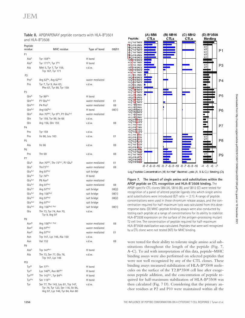
were tested for their ability to tolerate single amino acid sub-stitutions throughout the length of the peptide (Fig. 7,A–C). To aid with interpretation of this data, peptide–MHCbinding assays were also performed on selected peptides thatwere not well recognized by any of the CTL clones. Thesebinding assays measured stabilization of HLA-B*3508 mole-cules on the surface of the T2.B*3508 cell line after exoge-nous peptide addition, and the concentration of peptide re-quired for half-maximum stabilization of HLA-B*3508 wasthen calculated (Fig. 7 D). Considering that the primary an-chor residues at P2 and P10 were maintained within all the
Table II. APQPAPENAY peptide contacts with HLA-B*3501 and HLA-B*3508Peptideresidue MHC residue Type of bond 08/01
P1
AlaO Tyr 159O� H bond
AlaN Tyr 171O�, Tyr 7O� H bond
Ala Met 5, Tyr 7, Tyr 159,Trp 167, Tyr 171
v.d.w.
P2
ProO Arg 62N�, Arg 62N�2 water mediated
Pro Tyr 7, Tyr 9, Asn 63,Phe 67, Tyr 99, Tyr 159
v.d.w.
P3
GlnN Tyr 99O� H bond
GlnO�1 P7 GluO�2 water mediated 01
GlnN�2 P4 ProO water mediated 08
GlnO�1 Arg156N�1 H bond 08(1)
GlnO Asn 70N�2, Tyr 9O�, P7 GluO�2 water mediated
Gln Tyr 159, Tyr 99, Ile 66 v.d.w.
Gln Arg 156, Gln 155 v.d.w. 08
P4
Pro Tyr 159 v.d.w.
Pro Ile 66, Leu 163 v.d.w. 01
P5
Ala Ile 66 v.d.w. 08
P6
Pro Thr 69 v.d.w. 08
P7
GluN Asn 70O�1, Thr 73O�1, P7 GluO water mediated 01
GluO Thr73O�1 water mediated 08
GluO�1 Arg 97N�1 salt bridge
GluO�1 Tyr 74O� H bond
GluO�1 P8 AsnO water mediated
GluO�1 Arg 97N�1 water mediated 08
GluO�1 Arg 97N�2 salt bridge 08(2)
GluO�1 Arg 156N�2 salt bridge 08(1)
GluO�2 Arg 97N�2 salt bridge 08(2)
GluO�2 Arg 97N�1 salt bridge
GluO�2 Arg 156N�2, N�1 salt bridge 08(1)
Glu Thr 73, Tyr 74, Asn 70,Tyr 9, Arg 97
v.d.w.
P8
AsnN Arg 156N�1, N�2 water mediated 08
AsnO Arg 97N�1 water mediated
AsnO Arg 97N�2 water mediated 01
Asn Trp 147, Lys 146, Ala 150 v.d.w.
Asn Val 152 v.d.w. 08
P9
AlaO Trp 147N�1 H bond
Ala Thr 73, Ser 77, Glu 76,Trp 147, Lys 146
v.d.w.
P�
TyrN Ser 77O� H bond
TyrO Lys 146N�, Asn 80O�1 H bond
TyrOXT Thr 143O�1, Tyr 84O� H bond
TyrO� Ser 116O� H bond
Tyr Ser 77, Thr 143, Leu 81, Trp 147,Tyr 74, Tyr 123, Ser 116, Ile 95,Arg 97, Lys 146, Tyr 84, Asn 80
v.d.w.
Figure 7. The impact of single amino acid substitutions within the APQP peptide on CTL recognition and HLA-B*3508 binding. The APQP-specific CTL clones SB8 (A), SB16 (B), and SB12 (C) were tested for recognition of a panel of altered peptide ligands into which single amino acid substitutions were introduced (E/T ratio � 2:1). A range of peptide concentrations were used in these chromium release assays, and the con-centration required for half-maximum lysis was calculated from this dose-response data. (D) MHC–peptide binding assays were also conducted by testing each peptide at a range of concentrations for its ability to stabilize HLA-B*3508 expression on the surface of the antigen-processing mutant T2 cell line. The concentration of peptide required for half-maximum HLA-B*3508 stabilization was calculated. Peptides that were well recognized by a CTL clone were not tested (NT) for MHC binding.
Dow
nloaded from http://rupress.org/jem
/article-pdf/202/9/1249/1154963/jem20291249.pdf by guest on 16 D
ecember 2021

JEM VOL. 202, November 7, 2005 1257
ARTICLE
analogues, a surprising number of amino acid substitutions atother positions within the peptide reduced binding to HLA-B*3508. Consistent with the structural analysis, the glutamateat P7 appears to be a particularly important secondary anchorresidue for this peptide because most substitutions at this po-sition resulted in reduced MHC binding.
A total of 48 analogues of the 10-mer peptide weretested for CTL recognition over a range of concentrationsusing chromium release assays, and the concentration ofpeptide required for half-maximum lysis was calculated (Fig.7, A–C). Each CTL clone displayed a unique pattern of finespecificity for the 48 analogues, indicating that each ex-presses a different TCR. CTL recognition by the SB8 CTLclone was particularly affected by amino acid replacementstoward the COOH terminus of the peptide (Fig. 7 A),whereas the SB16 clone was focused toward central residues,with recognition less affected by substitutions at the extrem-ities of the peptide (Fig. 7 B). The SB12 CTL clone was par-ticularly tolerant of substitutions toward the COOH termi-nus, suggesting that the highly exposed peptide residues atP8 and P9 do not contact the TCR expressed by this clone(Fig. 7 C). It is notable that all three CTL clones have diffi-culty recognizing peptides with substitutions at P5 and P6,suggesting that the alanine and proline residues at these posi-tions within the EBV peptide might interact closely with theantigen receptors of these clones. Therefore, these data helpexplain why these clones fail to recognize the APQP peptidebound to B*3501 because the side chains of the residues atP5 and P6 are oriented quite differently when bound toB*3501 and B*3508.
DISCUSSIONPolymorphism at the MHC locus enhances immune defenseacross the population by ensuring wide variation in the Tcell response to infecting pathogens through presentation ofa broad array of target epitopes (22, 23). This report hasdemonstrated another mechanism through which MHCpolymorphism can diversify the immune response to an in-fecting pathogen. Thus, polymorphic MHC residues canmarkedly affect peptide binding conformation as well asMHC–peptide binding affinity, and this can have a majorimpact on the T cell response. Although previous studieshave also demonstrated peptide structural alterations inducedby MHC polymorphism (24, 25), none have shown thatsuch changes can influence a peptide-specific immune re-sponse to this extent.
Our data demonstrate that a single residue polymorphismbetween HLA-B*3501 and HLA-B*3508 controls respon-siveness to the APQP epitope through a mechanism unrelatedto peptide–MHC binding efficiency/stability (Fig. 2). Fur-thermore, HLA-B*3501�, EBV-infected lymphoblasts clearlypresent the APQP epitope from endogenously expressed anti-gen with similar efficiency to HLA-B*3508� LCLs (Fig. 3 Cand Fig. 4), ruling out differences in epitope processing withinEBV-infected cells. It should be noted, however, that the re-
sponse to this epitope could be initiated by cross-primingthrough dendritic cells that could process and present a differ-ent repertoire of EBV peptides compared with infected Bcells. Nonetheless, direct stimulation by EBV-infected B cellsis likely to have the major role in restimulating and maintain-ing these T cell populations during persistent infection. Wealso consider it highly unlikely that polymorphic antigen pro-cessing genes are influencing this response in our study sub-jects because our HLA-B*3501� donors were all unrelatedand the HLA-B*3508� donors were from two unrelated fam-ilies from different ethnic groups.
Another factor with the potential to influence the im-munogenicity of a T cell epitope is immunodominationwhereby the T cell response to an immunodominant deter-minant suppresses the response to another epitope (26, 27).It was therefore feasible that HLA-B*3501, but not HLA-B*3508, presents another unknown EBV epitope that ishighly immunogenic, thereby suppressing the response toAPQP in HLA-B*3501� individuals. Arguing against thispossibility are data presented in Fig. 1 C and Fig. 3 B show-ing that the HLA-B*3501/-B*3508–coexpressing donor MBresponds to the APQP epitope.
The potential role of peptide conformation in controllingthe differential responsiveness to this epitope became apparentafter functional assays demonstrated that APQP-specific CTLclones from an HLA-B*3508� donor were unable to recog-nize this peptide efficiently in the context of HLA-B*3501(Fig. 3 A). Thus, although these two closely related HLAmolecules present this identical peptide sequence very effi-ciently, they appear to be structurally distinct from the per-spective of many T cells. This conclusion was strengthened byour analysis of multiple CTL microcultures, most of whichrecognized the peptide much more efficiently when presentedon HLA-B*3508� target cells compared with HLA-B*3501�
targets. It is notable that one of the donors used for this analy-sis coexpressed HLA-B*3501 and HLA-B*3508, yet therewas no indication that T cells had been selected for the abilityto cross-recognize the peptide on each of these HLA mole-cules (Fig. 3 B). Subsequent structural analysis confirmed ma-jor conformational differences between the peptide bound toHLA-B*3501 and HLA-B*3508 resulting from direct peptideinteractions with the polymorphic MHC residue (Fig. 5 andFig. 6). Furthermore, the conformational differences wereparticularly striking at highly exposed peptide residues thatwere shown to be important TCR contact residues for threeout of three APQP-specific CTL clones (Fig. 7).
There are several possible mechanisms through whichsuch peptide conformational differences could influence im-munogenicity. It is possible that thymic and postthymic se-lection influence the relative number of naive T cells withthe capacity to recognize each complex. For example, theremay be an abundantly presented self-peptide that binds toboth HLA-B*3501 and HLA-B*3508 that negatively selectsCTLs with the potential to recognize the HLA-B*3501–bound conformation of the viral peptide or that positively
Dow
nloaded from http://rupress.org/jem
/article-pdf/202/9/1249/1154963/jem20291249.pdf by guest on 16 D
ecember 2021

THE INFLUENCE OF PEPTIDE CONFORMATION ON A CYTOTOXIC T CELL RESPONSE | Tynan et al.1258
selects for T cells that recognize the viral peptide presentedby HLA-B*3508 but not HLA-B*3501. Alternatively, cer-tain self-peptides may be presented preferentially by HLA-B*3508 that positively select T cells specific for HLA-B*3508–APQP (11, 12). In any case, the unique conformation of theAPQP peptide when bound to HLA-B*3508 appears to becritical for recognition by most of the selected T cells and is,therefore, a major controlling influence over the immunoge-nicity of this EBV epitope.
Another possible explanation for our data, unrelated toTCR repertoire differences between HLA-B*3501� andHLA-B*3508� individuals, is that the conformation of thisEBV epitope on HLA-B*3508 is intrinsically more immu-nogenic through structural features that enable it to interactfavorably with a higher frequency of TCRs (1). It is nowclear that certain antigenic peptides present challenging tar-get structures for TCR recognition and are recognized by avery limited TCR repertoire (16, 28–30). It is therefore notunreasonable to expect that some peptide conformations willpresent docking surfaces complementary to a higher propor-tion of the TCR repertoire compared with others. Althoughit is impossible to decipher the exact mechanisms underlyingthe phenomenon, this study clearly demonstrates that pep-tide conformation can have a dramatic impact on the immu-nogenicity of an MHC–peptide complex and could there-fore play a major role in controlling determinant selectionand immunodominance in other T cell responses.
MATERIALS AND METHODSCell lines. LCLs were established by exogenous transformation of periph-eral B cells with EBV and were maintained in growth medium (RPMI1640 with 10% FCS). The mutant LCL T lymphoblastoid hybrid cellline, 174 CEM.T2 (referred to as T2 cells) (31), expressing either HLA-B*3501 (T2.B*3501) or HLA-B*3508 (T2.B*3508) was also used in thisstudy and has been described elsewhere (13, 32). PHA blasts were generatedas previously described (10). Blood donors used in this study were healthylaboratory staff selected for particular HLA alleles and prior exposure toEBV as assessed by standard virus-specific antibody tests.
CTL cultures. CTL clones were generated by agar cloning as previouslydescribed (10) after initial stimulation of PBMCs for 1 h with 0.1 �M of theAPQP peptide. Clones were maintained with biweekly restimulation withrIL-2 and the �-irradiated (8,000 rads) autologous LCLs that had beenprelabeled with the APQP peptide at 0.1 �M for 1 h and washed threetimes. Short-term CTL bulk cultures were also used as effectors in cytotox-icity assays. These were generated by culturing 2 106 PBMCs/2-ml wellin growth medium with autologous PBMCs that had been precoated for1 h with 0.1 �M of the APQP peptide (responder/stimulator � 2:1). Cul-tures were supplemented with 20 U/ml rIL-2 on day 3, split on day 7, andanalyzed on day 10. CTL cultures were tested in duplicate or triplicate forcytotoxicity in the standard 5-h chromium release assay. In brief, CTLswere assayed against 51Cr-labeled LCL or PHA blast targets that were pre-treated with synthetic peptide (Mimotopes Ltd.) or left untreated. Themean spontaneous lysis for target cells in culture medium was always �20%,and the variation from the mean specific lysis was �10%.
Short-term CTL microcultures were generated by limiting dilution asfollows: PBMCs were distributed in roundbottom microtiter plates ingrowth medium at cell numbers ranging from 103 to 4 104 cells/well.Approximately 5 104 �-irradiated (2,000 rads) autologous PBMCs thathad been preincubated for 1 h with 1 �M of the APQP peptide were added
to each well to give a total volume of 100 �l. Cultures were fed on days 4,7, and 10 with 50 �l of medium supplemented with 20 U of rIL-2 and 30%supernatant from MLA-144 cultures. On day 13, each CTL microculturewas split into multiple replicates and used as effectors in a standard 5-h 51Cr-release assay against target PHA blasts that had been treated with the APQPpeptide or left untreated.
HLA class I peptide binding assays. To assess peptide binding to thedifferent HLA-B35 subtypes, T2.B*3501 and T2.B*3508 cells were incu-bated in AIM V serum-free medium (Invitrogen) with various concentra-tions (0.01, 0.1, 1, 10, and 100 �M) of peptides at 26�C for 14–16 h, fol-lowed by incubation at 37�C for 2–3 h and staining for HLA-B35 surfaceexpression. To determine peptide dissociation rates, T2.B*3501 cells werepulsed with 100 �M of each peptide at 26�C for 14–16 h, followed by incu-bation at 37�C for 2–3 h. Cells were then either stained immediately forHLA-B35 surface expression (0 h) or were washed three times and incubatedat 37�C for 1–6 h before staining. HLA-B35 surface expression was mea-sured by a flow cytometer (FACSCalibur; Becton Dickinson) using an mAbto HLA-Bw6 (SFR8 Bw6). Data were expressed relative to the mean fluo-rescence intensity (MFI) measured using 100 �M of a reference peptide thatwas known to bind to HLA-B*3501 or HLA-B*3508, using the followingformula: ([MFI with test peptide � MFI without peptide addition] 100) /(MFI with reference peptide [at 100 �M] � MFI without peptide addition).
ELISPOT assays. IFN-� ELISPOT assays were performed using cytokinecapture and detection reagents according to the manufacturer’s instructions(Mabtech). In brief, anti–IFN-� antibodies were coated on the wells of a 96-well nitrocellulose plate, and duplicate wells were seeded with 1,000 CD8� Tcells and 50,000 target cells. Two LCLs raised from an HLA B*3501� indi-vidual were used as target cells in these experiments: one carried a WT B95.8virus genome (BZLF1� LCL), and the other carried a B95.8 genome that hadbeen rendered incapable of lytic cycle entry by disruption of the BZLF1 gene(BZLF1� LCL; a gift from A. Hislop and A. Rickinson, University of Bir-mingham, UK) (17). After incubation for 16 h, captured IFN-� was detectedwith a biotinylated anti–IFN-� antibody followed by development withstreptavidin–horseradish peroxidase complex and chromogenic substrate, andspots were counted using an automated plate counter (AID).
Expression, purification, and crystallization of HLA-B35 alleleswith APQPAPENAY. Soluble HLA-B*3501 and HLA-B*3508 mole-cules (residues 1–276) and full-length �2-microglobulin (residues 1–99)were expressed, refolded with the APQP peptide, purified, and concen-trated to 10 mg/ml as previously described (33). Crystals were obtained bythe hanging drop vapor diffusion technique. Block-shaped crystals grewwithin 4 d in a condition containing 0.2 M ammonium acetate and 16%wt/vol PEG 3350 (100 mM cacodylate, pH 7.6) at 4�C.
X-ray data collection and structure determination. Crystals were soakedin reservoir solution containing increasing increments of glycerol as a cryo-protectant (5, 10, and 15%) and then flash frozen before data collection.Data were collected on an in-house radiation source and was processed andscaled using the HKL suite (34).
The HLA-B35 complex structures were refined from an HLA-B*3501structure that was previously determined in our laboratory (unpublished data).The model was manually built using the program “O” (35) and improvedthrough multiple rounds of refinement using the CNS suite (36). The progressof refinement was monitored by Rfactor and Rfree values. Rigid-body refinementand simulated annealing were used in the first instance, but in later rounds en-ergy minimization and B-individual refinement were used to improve thequality of the model. See Table I for the final refinement and model statistics.The structures have been deposited in the Protein Data Bank under accessionno. 2AXF (HLA-B*3508–APQP) and 2AXG (HLA-B*3501–APQP).
Purification of HLA complexes and peptide analysis. Purification ofHLA molecules was performed from �8 108 HLA-B*3501� LCLs (do-
Dow
nloaded from http://rupress.org/jem
/article-pdf/202/9/1249/1154963/jem20291249.pdf by guest on 16 D
ecember 2021

JEM VOL. 202, November 7, 2005 1259
ARTICLE
nor MW: HLA A1, A3, B8, and B*3501) and HLA-B*3508� LCLs (donorCA: HLA A30, A32, B42, and B*3508) that had been irradiated (200 rads)and incubated at 37�C overnight to enhance BZLF1 expression (18). TheseLCLs were selected for these experiments on the basis of relatively high lev-els of BZLF1 expression as measured using flow cytometry and a mousemAb to BZLF1 (BZ.1). Peptides were recovered from cell lysates aftertreatment with 0.5% TFA and acid extraction from the cell pellet as de-scribed elsewhere (37). After ultrafiltration (3-kD cutoff) and preconcentra-tion, peptides were collected in 50-�l fractions as described previously (38).Peptides were separated by RP-HPLC using a SMART system HPLC (GEHealthcare) with a vydac C18 column (1 mm [inside diameter] 25 cm)and eluted using an optimized linear gradient of acetonitrile in aqueous0.09% TFA. Peptide fractions were used in chromium release assays to sen-sitize HLA-B*3508� LCL target cells for lysis by a CTL clone from theB*3508� donor SB. In brief, 5 �l of each undiluted or diluted fraction wasadded to 15 �l of target cells for 90 min before CTL addition. To test fortoxicity, each fraction was also added to chromium-labeled target cellswithout CTL addition, but no toxicity was detected, even at the lowest di-lution (1:4; unpublished data). HLA-B*3501 fraction 32 was also character-ized by liquid chromatography–mass spectrometry using an Ultra ion trapmass spectrometer coupled to an 1100 nanoLC (both Agilent Technologies)as described previously (38, 39) and found to contain the APQP 10-merpeptide after MS/MS fragmentation of the [M�2H]2� species of target massm/z � 529.2 D (unpublished data).
Online supplemental material. Fig. S1 shows an alternate conformationof the polymorphic residue 156 in the HLA-B*3508–APQP complex. On-line supplemental material is available at http://www.jem.org/cgi/content/full/jem.20050864/DC1.
We would like to thank the BioCars staff for assistance in data collection at the Advanced Photon Source and Wendy van Zuylen and Geoff Connolly for technical assistance.
This work was supported by grants from the Australian National Health and Medical Research Council (NHMRC), the Roche Organ Transplantation Research Fund, the Juvenile Diabetes Research Foundation, and the Australian Research Council. S.R. Burrows is a recipient of an NHMRC Career Development award, and J. Rossjohn is a Wellcome Trust Senior Research Fellow.
The authors have no conflicting financial interests.
Submitted: 2 May 2005Accepted: 2 September 2005
REFERENCES1. Yewdell, J.W., and J.R. Bennink. 1999. Immunodominance in major
histocompatibility complex class I-restricted T lymphocyte responses.Annu. Rev. Immunol. 17:51–88.
2. Parham, P., and T. Ohta. 1996. Population biology of antigen presen-tation by MHC class I molecules. Science. 272:67–74.
3. Saper, M.A., P. Bjorkman, and D.C. Wiley. 1991. Refined structure ofthe human histocompatibility antigen HLA-A2 at 2.6A resolution. J.Mol. Biol. 219:277–319.
4. Hill, A.V., J. Elvin, A.C. Willis, M. Aidoo, C.E. Allsopp, F.M. Gotch,X.M. Gao, M. Takiguchi, B.M. Greenwood, A.R. Townsend, et al.1992. Molecular analysis of the association of HLA-B53 and resistanceto severe malaria. Nature. 360:434–439.
5. Falk, K., O. Rotzschke, B. Grahovac, D. Schendel, S. Stevanovic, G.Jung, and H.G. Rammensee. 1993. Peptide motifs of HLA-B35 and-B37 molecules. Immunogenetics. 38:161–162.
6. Ruppert, J., J. Sidney, E. Celis, R.T. Kubo, H.M. Grey, and A. Sette.1993. Prominent role of secondary anchor residues in peptide bindingto HLA-A2.1 molecules. Cell. 74:929–937.
7. Chen, W., S. Khilko, J. Fecondo, D.H. Margulies, and J. McCluskey.1994. Determinant selection of major histocompatibility complex classI–restricted antigenic peptides is explained by class I–peptide affinityand is strongly influenced by nondominant anchor residues. J. Exp.Med. 180:1471–1483.
8. Busch, D.H., I.M. Pilip, S. Vijh, and E.G.P. Am. 1998. Coordinateregulation of complex T cell populations responding to bacterial infec-tion. Immunity. 8:353–362.
9. Crotzer, V.L., R.E. Christian, J.M. Brooks, J. Shabanowitz, R.E. Sett-lage, J.A. Marto, F.M. White, A.B. Rickinson, D.F. Hunt, and V.H.Engelhard. 2000. Immunodominance among EBV-derived epitopes re-stricted by HLA-B27 does not correlate with epitope abundance in EBV-transformed B-lymphoblastoid cell lines. J. Immunol. 164:6120–6129.
10. Burrows, S.R., S.L. Silins, D.J. Moss, R. Khanna, I.S. Misko, and V.P.Argaet. 1995. T cell receptor repertoire for a viral epitope in humans isdiversified by tolerance to a background major histocompatibility com-plex antigen. J. Exp. Med. 182:1703–1715.
11. Messaoudi, I., J.A. Guevara Patino, R. Dyall, J. LeMaoult, and J. Ni-kolich-Zugich. 2002. Direct link between mhc polymorphism, T cellavidity, and diversity in immune defense. Science. 298:1797–1800.
12. Nikolic-Zugic, J., and M.J. Bevan. 1990. Role of self-peptides in posi-tively selecting the T-cell repertoire. Nature. 344:65–67.
13. Green, K.J., J.J. Miles, J. Tellam, W.J.M. van Zuylen, G. Connolly,and S.R. Burrows. 2004. Potent T cell response to a class-I-binding13-mer viral epitope and the influence of HLA micropolymorphism incontrolling epitope length. Eur. J. Immunol. 34:2510–2519.
14. Saulquin, X., C. Ibisch, M.A. Peyrat, E. Scotet, M. Hourmant, H. Vie,M. Bonneville, and E. Houssaint. 2000. A global appraisal of immuno-dominant CD8 T cell responses to Epstein-Barr virus and cytomega-lovirus by bulk screening. Eur. J. Immunol. 30:2531–2539.
15. Stuber, G., G.H. Leder, W.T. Storkus, M.T. Lotze, S. Modrow, L.Szekely, H. Wolf, E. Klein, K. Karre, and G. Klein. 1994. Identificationof wild-type and mutant p53 peptides binding to HLA-A2 assessed by apeptide loading-deficient cell line assay and a novel major histocompati-bility complex class I peptide binding assay. Eur. J. Immunol. 24:765–768.
16. Tynan, F.E., N.A. Borg, J.J. Miles, T. Beddoe, D. El-Hassen, S.L. Si-lins, W.J.M. van Zuylen, A.W. Purcell, L. Kjer-Nielsen, J. McClus-key, et al. 2005. High resolution structures of highly bulged viralepitopes bound to the major histocompatability class I: implications forT-cell receptor engagement and T-cell immunodominance. J. Biol.Chem. 280:23900–23909.
17. Pudney, V.A., A.M. Leese, A.B. Rickinson, and A.D. Hislop. 2005.CD8� immunodominance among Epstein-Barr virus lytic cycle anti-gens directly reflects the efficiency of antigen presentation in lyticallyinfected cells. J. Exp. Med. 201:349–360.
18. Ferrieu, C., B. Ballester, J. Mathieu, and E. Drouet. 2003. Flow cy-tometry analysis of gamma-radiation-induced Epstein-Barr virus reacti-vation in lymphocytes. Radiat. Res. 159:268–273.
19. Menssen, R., P. Orth, A. Ziegler, and W. Saenger. 1999. Decamer-like conformation of a nona-peptide bound to HLA-B*3501 due tonon-standard positioning of the C terminus. J. Mol. Biol. 285:645–653.
20. Smith, K.J., S.W. Reid, D.I. Stuart, A.J. McMichael, E.Y. Jones, andJ.I. Bell. 1996. An altered position of the 2 helix of MHC class I is re-vealed by the crystal structure of HLA-B*3501. Immunity. 4:203–213.
21. Miles, J.J., D. Elhassen, N.A. Borg, S.L. Silins, F.E. Tynan, J.M. Bur-rows, A.W. Purcell, L. Kjer-Nielsen, J. Rossjohn, S.R. Burrows, and J.McCluskey. 2005. CTL recognition of a bulged viral peptide involvesbiased TCR selection. J. Immunol. 175:3826–3834.
22. Germain, R.N., and D.H. Margulies. 1993. The biochemistry and cellbiology of antigen processing and presentation. Annu. Rev. Immunol.11:403–450.
23. Lawlor, D.A., J. Zemmour, P.D. Ennis, and P. Parham. 1990. Evolu-tion of class-I MHC genes and proteins: from natural selection to thy-mic selection. Annu. Rev. Immunol. 8:23–63.
24. Hulsmeyer, M., M.T. Fiorillo, F. Bettosini, R. Sorrentino, W. Saen-ger, A. Ziegler, and B. Uchanska-Ziegler. 2004. Dual, HLA-B27 sub-type–dependent conformation of a self-peptide. J. Exp. Med. 199:271–281.
25. Luz, J.G., M. Huang, K.C. Garcia, M.G. Rudolph, V. Apostolopoulos,L. Teyton, and I.A. Wilson. 2002. Structural comparison of allogeneicand syngeneic T cell receptor–peptide–major histocompatibility com-plex complexes: a buried alloreactive mutation subtly alters peptidepresentation substantially increasing V� interactions. J. Exp. Med. 195:
Dow
nloaded from http://rupress.org/jem
/article-pdf/202/9/1249/1154963/jem20291249.pdf by guest on 16 D
ecember 2021
THE INFLUENCE OF PEPTIDE CONFORMATION ON A CYTOTOXIC T CELL RESPONSE | Tynan et al.1260
1175–1186.26. Lewicki, H.A., G. von Herrath, C.F. Evans, J.L. Whitton, and M.B.
Oldstone. 1995. CTL escape viral variants. II. Biologic activity in vivo.Virology. 211:443–450.
27. van der Most, R.G., R.J. Concepcion, C. Oseroff, J. Alexander, S.Southwood, J. Sidney, R.W. Chesnut, R. Ahmed, and A. Sette. 1997.Uncovering subdominant cytotoxic T-lymphocyte responses in lym-phocytic choriomeningitis virus-infected BALB/c mice. J. Virol. 71:5110–5114.
28. Stewart-Jones, G.B., A.J. McMichael, J.I. Bell, D.I. Stuart, and E.Y.Jones. 2003. A structural basis for immunodominant human T cell re-ceptor recognition. Nat. Immunol. 4:657–663.
29. Kjer-Nielsen, L., C.S. Clements, A.W. Purcell, A.G. Brooks, J.C.Whisstock, S.R. Burrows, J. McCluskey, and J. Rossjohn. 2003. Astructural basis for the selection of dominant alphabeta T cell receptorsin antiviral immunity. Immunity. 18:53–64.
30. Turner, S.J., K. Kedzierska, H. Komodromou, N.L. La Gruta, M.A.Dunstone, A.I. Webb, R. Webby, H. Walden, W. Xie, J. McCluskey,et al. 2005. Lack of prominent peptide-major histocompatibility com-plex features limits repertoire diversity in virus-specific CD8� T cellpopulations. Nat. Immunol. 6:382–389.
31. Salter, R.D., and P. Cresswell. 1986. Impaired assembly and transportof HLA-A and -B antigens in a mutant TxB cell hybrid. EMBO J.5:943–949.
32. Khanna, R., S.L. Silins, Z. Weng, D. Gatchell, S.R. Burrows, and L.Cooper. 1999. Cytotoxic T cell recognition of allelic variants of HLAB35 bound to an Epstein-Barr virus epitope: influence of peptide confor-mation and TCR-peptide interaction. Eur. J. Immunol. 29:1587–1597.
33. Macdonald, W., D.S. Williams, C.S. Clements, J.J. Gorman, L. Kjer-Nielsen, A.G. Brooks, J. McCluskey, J. Rossjohn, and A.W. Purcell.2002. Identification of a dominant self-ligand bound to three HLA B44alleles and the preliminary crystallographic analysis of recombinantforms of each complex. FEBS Lett. 527:27–32.
34. Rossmann, M.G., and C.G. van Beek. 1999. Data processing. ActaCrystallogr. D Biol. Crystallogr. 55:1631–1640.
35. Jones, T.A., J.Y. Zou, S.W. Cowan, and M. Kjeldgaard. 1991. Improvedmethods for building protein models in electron density maps and the lo-cation of errors in these models. Acta. Crystallogr. A. 47:110–119.
36. Brunger, A.T., P.D. Adams, G.M. Clore, W.L. DeLano, P. Gros,R.W. Grosse-Kunstleve, J.S. Jiang, J. Kuszewski, M. Nilges, N.S.Pannu, et al. 1998. Crystallography & NMR system: a new softwaresuite for macromolecular structure determination. Acta Crystallogr. DBiol. Crystallogr. 54:905–921.
37. Purcell, A.W. 2004. Isolation and characterization of naturally pro-cessed MHC-bound peptides from the surface of antigen-presentingcells. Methods Mol. Biol. 251:291–306.
38. Purcell, A.W., J.J. Gorman, M. Garcia-Peydro, A. Paradela, S.R. Bur-rows, G.H. Talbo, N. Laham, C.A. Peh, E.C. Reynolds, J.A. Lopez deCastro, and J. McCluskey. 2001. Quantitative and qualitative influencesof tapasin on the class I peptide repertoire. J. Immunol. 166:1016–1027.
39. Macdonald, W.A., A.W. Purcell, N. Mifsud, L.K. Ely, D.S. Williams,L. Chang, J.J. Gorman, C.S. Clements, L. Kjer-Nielsen, D.M. Koelle,et al. 2003. A naturally selected dimorphism within the HLA-B44 su-pertype alters class I structure, peptide repertoire, and T cell recogni-tion. J. Exp. Med. 198:679–691.
Dow
nloaded from http://rupress.org/jem
/article-pdf/202/9/1249/1154963/jem20291249.pdf by guest on 16 D
ecember 2021