Embed Size (px)
Citation preview
lable at ScienceDirect
Neurobiology of Stress 1 (2015) 184e194
Contents lists avai
Neurobiology of Stress
journal homepage: http : / /www.journals .e lsevier .com/neurobiology-of-stress/
The impact of developmental timing for stress and recovery
Dylan G. Gee*, B.J. CaseySackler Institute for Developmental Psychobiology, Weill Cornell Medical College, 1300 York Ave, New York, NY 10065, USA
a r t i c l e i n f o
Article history:Received 15 September 2014Received in revised form10 February 2015Accepted 18 February 2015Available online 19 February 2015
Keywords:Early-life stressDevelopmentResilienceSensitive periodNeuroplasticityAmygdalaPrefrontal cortex
* Corresponding author.E-mail address: [email protected] (D.G. Gee).
http://dx.doi.org/10.1016/j.ynstr.2015.02.0012352-2895/© 2015 The Authors. Published by Elsevier
a b s t r a c t
Stress can have lasting effects on the brain and behavior. Delineating the impact of stress on thedeveloping brain is fundamental for understanding mechanisms through which stress induces persistenteffects on behavior that can lead to psychopathology. The growing field of translational developmentalneuroscience has revealed a significant role of the timing of stress on risk, resilience, and neuroplasticity.Studies of stress across species have provided essential insight into the mechanisms by which the brainchanges and the timing of those changes on outcome. In this article, we review the neurobiologicaleffects of stress and propose a model by which sensitive periods of neural development interact withstressful life events to affect plasticity and the effects of stress on functional outcomes. We then highlighthow early-life stress can alter the course of brain development. Finally, we examine mechanisms ofbuffering against early-life stress that may promote resilience and positive outcomes. The findings arediscussed in the context of implications for early identification of risk and resilience factors and devel-opment of novel interventions that target the biological state of the developing brain to ultimatelyameliorate the adverse consequences of stress during childhood and adolescence.© 2015 The Authors. Published by Elsevier Inc. This is an open access article under the CC BY-NC-ND
license (http://creativecommons.org/licenses/by-nc-nd/4.0/).
1. Introduction
Stress is a potent environmental risk factor for both mental andphysical illness. The effects of stress on the brain depend criticallyon the timing (age of onset and duration). When stress occurs earlyin life it can have profound and lasting effects on brain organizationand function. Approximately 10% of youth have anxiety and stress-related disorders (Newman et al., 1996; Kim-Cohen et al., 2003;Kessler et al., 2005), and early childhood adversity accounts for over30% of all mental illnesses (Green et al., 2010). Yet not all childrenwho experience stressful life events develop mental illness. Un-derstanding the mechanisms by which stress alters the developingbrain is fundamental for: 1) delineating adaptive and maladaptivechanges; 2) identifying resilience and risk factors; and 3) devel-oping interventions for ameliorating risk. This article highlightsrecent studies that examine the neurobiological effects of thetiming and buffering of stressful life events.
Inc. This is an open access article u
2. Brain development and sensitive periods
The brain undergoes dynamic changes throughout the course ofdevelopment, with important implications for how stress in-fluences the brain and the efficacy of treatments targeting stress-related mental illness at different developmental time points.Nonhuman primate studies show that typical brain development ismarked by an initial period of overproduction of synapses, followedby selective stabilization and elimination of a substantial propor-tion of synapses (Huttenlocher, 1979; Huttenlocher et al., 1982;Bourgeois and Rakic, 1993; LaMantia and Rakic, 1994). Humanneuroimaging studies show corresponding patterns, in which graymatter volumes typically peak around 10e12 years of age (Gieddet al., 1999), with significant gray matter loss throughout adoles-cence and adulthood (Sowell et al., 2001, 2003). Simultaneously,increases in white matter occur through myelination of axons(Brody et al., 1987; Benes et al., 1994). Substantial regional variationexists, with maturation of low-level sensory and motor corticesoccurring prior to prefrontal and temporal cortices involved inhigher-level cognition and regulation of behavior (Yakovlev andLecours, 1967; Benes et al., 1994; Sowell et al., 1999, 2001; Gogtayet al., 2004). Such regional changes in brain structure and func-tion across development, as well as changes in the availability ofneurochemicals and patterns of cortical cell firing, are posited tolead to transient imbalances that underlie behavioral changes
nder the CC BY-NC-ND license (http://creativecommons.org/licenses/by-nc-nd/4.0/).
D.G. Gee, B.J. Casey / Neurobiology of Stress 1 (2015) 184e194 185
during adolescence (Galvan et al., 2006; Casey, Galvan, Getz, 2008).These dynamic changes in brain and behavioral development likelyinfluence how stress at unique developmental time periods altersthe brain and how children and adolescents cope with stressors.Exacerbations of transient imbalances in brain circuitry, such as theeffects of acute or chronic stress, may lead to altered stress reac-tivity and ultimately increase the risk for mental illness.
Understanding neurodevelopmental changes that influencestress reactivity and recovery are critical for enhancing mentalhealth. Sensitive periods refer to times in development whenheightened neuroplasticity renders the brain especially amenableto environmental influences (Moriceau and Sullivan, 2006;Callaghan and Richardson, 2011; Yang et al., 2012). The timing ofsensitive periods differs by neural circuit and behavioral system,but it may be that sensitive periods occur when brain developmentis most dynamic, such as infancy and adolescence (Fig. 1). Duringthese periods, environmental input can lead to a series of devel-opmental cascades (Masten and Cicchetti, 2010) that ultimatelyhave significant influences on behavior, of a positive or negativenature. A sensitive period may render the brain more capable ofresponding to stress in adaptive ways. It could also magnify con-sequences of stressful life events in maladaptive ways. By contrast,stress that occurs during windows of reduced plasticity (e.g., afterthe closing of a sensitive period) may yield a brain that is lesscapable of remodeling itself. Thus, sensitive periods in neuro-development may render the developing brain more vulnerable tothe effects of later stress, but they could also serve as windows ofopportunity, during which there is increased potential for positiveadaptation or effective intervention.
Delineating sensitive periods could reveal how the effects ofstress differ depending on when in development and what type ofstress occurs, as well as when in development certain types ofintervention may be most effective for buffering against maladap-tive consequences of stress. In this way we may begin to direct thetiming and type of interventions at the level of the individual andthe nature of the stressor. The extent to which neuroplasticity andbrain function change throughout childhood and adolescencesuggests that interventions based on the adult brain cannot be
Fig. 1. Model of sensitive periods of brain development. Periods of rapid and substantial chain gray), may provide the most opportunity for adaptive behavioral changes. These sensitive pthe effects of stress. Figure adapted with permission from Lee et al., 2014 (Copyright 2014
simply applied to youth who experience stress-related mentalhealth disorders (Lee et al., 2014). Understanding how sensitiveperiods shift, constrict, or expand in individuals at different pointsin development will allow treatments to precisely target the bio-logical state of the developing brain to optimize stress-relatedinterventions.
3. Neurobiology of stress
Studies of mature animals have provided the majority of extantknowledge on the effects of stress at the cellular level and showthat stress can significantly remodel brain structure and function(reviewed in McEwen, 2012). Stress results in changes in fronto-limbic circuitry that are regional in nature. Chronic stress can leadto hypermetabolism and morphological changes within theamygdala, which is critical for learning about the emotional sig-nificance of environmental cues and helping the organism react tothe challenge or threat of these cues. In contrast, chronic stressdownregulates the hippocampus and prefrontal cortex (PFC),which regulate the stress response. Specifically, studies of rodentsshow that stress increases dendritic arborization and spine densityof the amygdala, with concomitant increases in anxiety-like be-haviors (Vyas et al., 2002; Vyas et al., 2003; Mitra et al., 2005). Bycontrast, stress results in atrophy of the hippocampus and medialPFC (mPFC) (Magari~nos et al., 1997; Vyas et al., 2002; Radley et al.,2006). Parallel findings of increased amygdala volume and func-tional reactivity, smaller hippocampal volume, and altered pre-frontal function and connectivity have been observed in humansfollowing stress (Ganzel et al., 2007, 2008; Liston et al., 2006; Listonet al., 2009; Sheridan et al., 2012a,b).
The reversibility of the effects of stress is regional as well. Thereis a growing body of evidence to suggest that the hippocampus andPFC may have greater capacity for change or plasticity followingstress with many of the effects being reversible following thetermination of stress (McEwen, 1999; Vyas et al., 2004; Liston et al.,2009). In contrast, stress-induced amygdala morphology and vol-ume changes seem to persist (Vyas et al., 2002; Adamec et al., 2005;Tottenham et al., 2010). Due to its cellular properties, the amygdala
nges in brain development, such as the first three years of life and adolescence (shadederiods of neural development may also render the developing brain most vulnerable toAAAS).
D.G. Gee, B.J. Casey / Neurobiology of Stress 1 (2015) 184e194186
might be particularly sensitive to stress (Plotsky et al., 2005;Sabatini et al., 2007; reviewed in Tottenham and Sheridan, 2009)and therefore more resistant to recovery following chronic stress(Ganzel et al., 2007; Lupien et al., 2011; Malter Cohen et al.,2013a,b).
These inverse effects of stress on frontolimbic regions are due inpart to complex interactions within the neuroendocrine system ofthe Limbic-Hypothalamic-Pituitary-Adrenal Axis (LHPA). Animportant function of the LHPA stress response is to release glu-cocorticoids that facilitate mobilizationwith threat and by doing soinhibit “non-essential” systems for immediate survival such asgrowth, reproduction, and immunity. Under non-stressful or basalconditions, the LHPA functions to support growth and development(De Kloet et al., 1998). Under conditions of threat or challenge, LHPAactivity increases resulting in the release of hormones and peptidesthat suppress growth and repair in order to support functionsnecessary for immediate survival. Failure to activate the stressresponse places the organism in a vulnerable state, and failure toinhibit the stress response results in adverse effects on growth anddevelopment and can lead to diseased states. The amygdala iscritical in activating the LHPA axis in response to threat and stress(Dunn and Whitener, 1986; Feldman et al., 1995; Redgate andFahringer, 1973), and levels of glucocorticoids are regulated vianegative feedback loops at several levels of the axis including thehippocampus and PFC (Diorio et al., 1993; Jacobson and Sapolsky,1991). Opposing regulatory actions occur in amygdala and fronto-hippocampal regions with upregulation of the former and down-regulation of the latter providing a partial explanation for inverseeffects of stress within frontolimbic circuitry. This review focuseson the impact of psychological stressors on neuroplasticity,although glucocorticoids, and their directmanipulation, canmodifythe brain in anatomically selective ways (Sapolsky, 1986; Listonet al., 2013) and alter the expression of neurotrophic factorsessential for neuroplasticity (Smith et al., 1995).
4. Developmental changes in the effects of acute stressors
Adolescence is a unique period in development with manyimplications for the effects of stress. As adolescents transition fromdependence on their caregivers to a more independent state, theyface many new challenges to which they must adapt (Romeo, 2010;Spear, 2010; Malter Cohen et al., 2013b). Several studies demon-strate changes in emotional reactivity and frontoamygdala circuitryin adolescence with important implications for how stress affectsadolescents. For example, we have provided evidence of height-ened emotional reactivity during adolescence that leads to anxietywhen that reactivity persists long after a potential threat isremoved (Hare et al., 2008). These findings parallel findings ofincreased hormonal stress reactivity during puberty and adoles-cence (Romeo et al., 2006; Folib et al., 2011).
Potential threats can be stressors depending on how they areperceived. Fear conditioning and extinction paradigms provide apowerful way to examine stress reactivity to and regulation of acutethreat. During fear extinction, cues previously associated withthreat are presented without the threatening stimulus until thecues are learned to be safe and fear responses decrease. This pro-cess is critical to the etiology and treatment of anxiety disorderssuch as phobias and posttraumatic stress disorder (PTSD), whichare characterized by an inappropriate fear response to a cue that isno longer dangerous (Rothbaum and Davis, 2003).
Recently we examined fear learning in mice and humans acrossdevelopment. Consistent with work in rats (McCallum et al., 2010;Kim et al., 2011) we showed differential effects of fear extinction inadolescent mice and humans, relative to younger and older ages.Although all groups showed similar acquisition of cued fear, the
adolescents showed attenuated fear extinction learning relative tochildren and adults (Pattwell et al., 2012). Parallel findings wereobserved in mice, such that adolescent (postnatal day (P) 29) miceshowed diminished fear extinction compared with pre- (P23) andpost-adolescent (P70) mice. Examination of frontolimbic circuitryin the mice suggested reduced infralimbic prefrontal activity inadolescence during extinction learning. Taken together, this worksuggests that adolescence is marked by prominent changes inneurodevelopment that are likely to interact with the effects ofstress to influence behavioral phenotypes later in life.
5. Developmental changes in the effects of chronic stress
The timing of stress and its interactions with dynamic devel-opmental processes are critical to subsequent outcomes (e.g.,Lupien et al., 2009; Monk, 2008; Monk et al., 2002; Pechtel andPizzagalli, 2011; Eiland and Romeo, 2013). Manipulating thetiming of stress is challenging in humans. However, a series ofstudies in developing nonhuman primates has shed new light onthe effects of stress as a function of timing. The stress manipulationwas amaternal separation paradigm that occurred at either 1 week,1 month, or 3 months after birth (Cameron, 2001; McCormick et al.,2005). The results showed qualitatively distinct behavioral out-comes depending on the timing of the separation. Monkeys whoexperienced maternal separation at 1 week exhibited less social-contact behaviors than maternally reared animals. By contrast,monkeys who experiencedmaternal separation at 1month showedsignificantly more social behavior. Examination of gene expressionchanges in the amygdala at 3 months of age in each group indicateddownregulation of mRNA expression throughout the amygdala inthe monkeys who were separated from their mothers the earliest(Sabatini et al., 2007). These results suggest that the timing (andduration) of stressors may interact with dynamically changingbrain systems to alter behavior in complex and unique ways.Moreover, evidence from rodent models suggests that early-lifestress may affect different phenotypes in childhood than adoles-cence (Raineki et al., 2012; Rinc�on-Cort�es and Sullivan, 2014).
Investigations of naturally occurring stressors in humans pro-vide evidence that the onset and duration of stress matters. Instudies of children reared in orphanages abroad and later adoptedinto stable families, the findings consistently suggest that earlieradoption is better (Rutter, 1998; Gunnar et al., 2000; Tottenhamet al., 2010). It remains unclear whether earlier adoption is asso-ciated with increased resilience due to a shorter duration of stressor because the stress may interact with sensitive periods foremotional development, or both. It may be that the malleability ofthe brain decreases over time, such that stress and remediationoccurring later in development have differential consequences dueto changes in the brain's ability to adapt or recover.
A study of the impact of the 9/11 terrorist attacks on healthyadults provides further evidence of the importance of timing ofstress on its neural and behavioral effects (Ganzel et al., 2007).More than three years after 9/11, individuals who were within 1.5miles of the disaster had higher amygdala reactivity than thosewhowere over 200 miles away. Notably, the association betweenproximity and amygdala activation was accounted for by the timesince the last worst trauma. These findings show that recovery,even in healthy adults, occurs across many years, while also high-lighting the importance of the recency of trauma.
6. Lasting effects of early-life chronic stress
Stress can arise through any number of environments thatchallenge an individual cognitively, emotionally, or physically, suchas uncontrollable or unpredictable settings (e.g., Lupien et al., 2000;

D.G. Gee, B.J. Casey / Neurobiology of Stress 1 (2015) 184e194 187
McEwen, 2012; Pollak, 2008; Sheridan et al., 2013; Teicher et al.,2006). However, environments that result in a mismatch betweenthe expected and actual environment may prove particularlystressful (Finlay, 2007; Casey et al., 2010). Environmental stabilityacross a long evolutionary history has led to species-expected ex-periences, such as caregiving for humans early in life. Consistentwith this idea, poor caregiving is one of the most potent stressorsfor an infant and has long-lasting effects on the brain and behavior(e.g., Sheridan et al., 2012a,b; reviewed in Tottenham, 2012).Maternal separation in rodent pups is associated with greater LHPAaxis reactivity (Moriceau et al., 2010), accelerated amygdaladevelopment (Moriceau and Sullivan, 2006; Ono et al., 2008),increased anxiety-like behaviors (Romeo et al., 2003), and moresocial instability in adulthood (Kikusui and Mori, 2009). Humanstudies of maternal deprivation early in life have shown atypicalfrontoamygdala development and function with greater amygdalavolume, amygdala hyperactivity, and less prefrontal activity toemotional stimuli, as well as long-term impairments in anxiety andsocial behavior (Mehta et al., 2009; Zeanah et al., 2009; Tottenhamet al., 2010, 2011). The enhanced amygdala activity and decreasedprefrontal activity in the children with a history of maternaldeprivation may suggest that they are less able to suppress irrele-vant emotional information leading to dysregulation of emotions.By preschool these children have a rate of mental health disordersthat is more than twice that in children who did not experienceinstitutional rearing (Zeanah et al., 2009).
Until recently, less has been known about changes in the long-term course of brain development following early-life stress.Recent studies in our respective laboratories indicate that early-lifestress has lasting effects on the organization of frontolimbic cir-cuitry. We specifically examined the effects of orphanage rearing ondevelopment of frontoamygdala activity and connectivity. Withtypical development, task-based frontoamygdala functional con-nectivity switches from positive coupling in childhood to inversecoupling during the transition to adolescence (Gee et al., 2013a,b)(Fig. 2). This mature pattern of inverse amygdala-mPFC functionalconnectivity is consistent with the inverse connectivity observed inthe literature of emotion regulation in healthy adults (Banks et al.,2007; Hare et al., 2008; Hariri et al., 2003; Kim et al., 2003).
Based on evidence that early-life stress accelerates amygdaladevelopment in rodents, we hypothesized that children who
Fig. 2. Mature frontoamygdala functional connectivity following maternal deprivation. Leftthat group differences emerged when participants viewed fearful faces. Right) Unlike compawith a history of early-life stress (previous institutionalized care) exhibited the mature paadolescents. The results suggest an early closure of a sensitive period in frontoamygdala dreproduced with permission from Gee et al., 2013a (Copyright 2013 Proceedings of the Nati
experienced maternal deprivation early in life would displayaltered development of frontoamygdala circuitry. Children whowere reared in international orphanages as infants and were sub-sequently adopted into stable families in the U.S. provided a meansof examining an isolated period of early-life stress (i.e., institu-tionalized care) on later brain development and behavior. Incontrast to the immature positive functional connectivity displayedin comparison children, the children who experienced early-lifestress showed the adult-like pattern of inverse amygdala-mPFCfunctional connectivity (Gee et al., 2013a,b) (Fig. 2). This markedshift in connectivity may reflect early closure of a neural sensitiveperiod that could have long-term consequences for later affectivebehaviors.
To better understand the functional significance of acceleratedneural circuit development we tested whether amygdala-mPFCfunctional connectivity was related to anxiety. Children with ahistory of early-life stress had higher levels of anxiety than com-parison children, consistent with prior findings. However, youth inthe early-life stress group with the mature phenotype of inversefunctional connectivity had lower anxiety than those with theimmature phenotype of positive functional connectivity. It may bethat the earlier emergence of mature connectivity is adaptive in thecontext of early-life stress. Cortisol levels mediated the relationshipbetween early-life experience and frontoamygdala connectivity,suggesting that stress-related modifications of the LHPA axis mayshape the early development of amygdala-mPFC connections.Accelerated frontoamygdala development may serve as an onto-genetic adaptation that reprioritizes development to cope with anearly adverse environment. However, the long-term consequencesof this accelerated development remain unclear.
7. Translational studies of early-life stress
Naturalistic studies of stress effects in humans have providedcritical insight into the neurobiological mechanisms through whichstress has lasting effects on emotional behavior. However, theinterpretation of these studies is limited by confounds of uncon-trolled genetic and environmental factors. To address these con-cerns we recently conducted a translational study in mice in whichwewere able tomanipulate the type and timing of stress in rodentsto mimic the orphanage-rearing environment in humans and
) A group by emotion interaction was observed in the mPFC (p < 0.01, corrected), suchrison children who showed immature (positive) amygdala-mPFC connectivity, childrenttern of inverse amygdala-mPFC coupling, such that the stressed children resembledevelopment following early-life stress. Error bars ¼ þ/� 1 SEM; *p < 0.05. Data areonal Academy of Sciences of the United States of America).
D.G. Gee, B.J. Casey / Neurobiology of Stress 1 (2015) 184e194188
examine the long-term effects (Malter Cohen et al., 2013a,b). Theearly-life stress manipulation involved limiting the nesting mate-rial provided to the dams, which disrupted maternal care of thepups (Gilles et al., 1996; Ivy et al., 2008; Rice et al., 2008). Thisstressor was limited to the pre-weaning period (P2eP21) thatparalleled the adoption of most children from orphanages duringearly childhood.
To capture the heightened emotional reactivity and slowing ofresponse latencies in anticipation of negative emotional informa-tion in children reared in the orphanage (Tottenham et al., 2011)
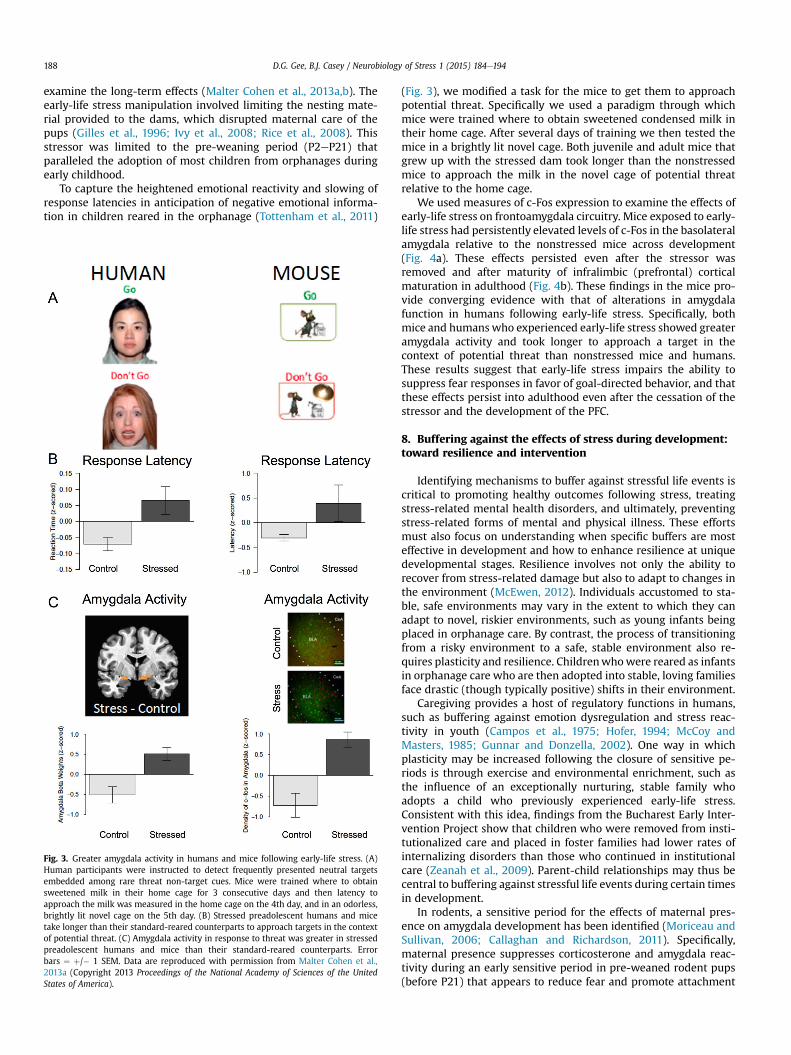
Fig. 3. Greater amygdala activity in humans and mice following early-life stress. (A)Human participants were instructed to detect frequently presented neutral targetsembedded among rare threat non-target cues. Mice were trained where to obtainsweetened milk in their home cage for 3 consecutive days and then latency toapproach the milk was measured in the home cage on the 4th day, and in an odorless,brightly lit novel cage on the 5th day. (B) Stressed preadolescent humans and micetake longer than their standard-reared counterparts to approach targets in the contextof potential threat. (C) Amygdala activity in response to threat was greater in stressedpreadolescent humans and mice than their standard-reared counterparts. Errorbars ¼ þ/� 1 SEM. Data are reproduced with permission from Malter Cohen et al.,2013a (Copyright 2013 Proceedings of the National Academy of Sciences of the UnitedStates of America).
(Fig. 3), we modified a task for the mice to get them to approachpotential threat. Specifically we used a paradigm through whichmice were trained where to obtain sweetened condensed milk intheir home cage. After several days of training we then tested themice in a brightly lit novel cage. Both juvenile and adult mice thatgrew up with the stressed dam took longer than the nonstressedmice to approach the milk in the novel cage of potential threatrelative to the home cage.
We used measures of c-Fos expression to examine the effects ofearly-life stress on frontoamygdala circuitry. Mice exposed to early-life stress had persistently elevated levels of c-Fos in the basolateralamygdala relative to the nonstressed mice across development(Fig. 4a). These effects persisted even after the stressor wasremoved and after maturity of infralimbic (prefrontal) corticalmaturation in adulthood (Fig. 4b). These findings in the mice pro-vide converging evidence with that of alterations in amygdalafunction in humans following early-life stress. Specifically, bothmice and humanswho experienced early-life stress showed greateramygdala activity and took longer to approach a target in thecontext of potential threat than nonstressed mice and humans.These results suggest that early-life stress impairs the ability tosuppress fear responses in favor of goal-directed behavior, and thatthese effects persist into adulthood even after the cessation of thestressor and the development of the PFC.
8. Buffering against the effects of stress during development:toward resilience and intervention
Identifying mechanisms to buffer against stressful life events iscritical to promoting healthy outcomes following stress, treatingstress-related mental health disorders, and ultimately, preventingstress-related forms of mental and physical illness. These effortsmust also focus on understanding when specific buffers are mosteffective in development and how to enhance resilience at uniquedevelopmental stages. Resilience involves not only the ability torecover from stress-related damage but also to adapt to changes inthe environment (McEwen, 2012). Individuals accustomed to sta-ble, safe environments may vary in the extent to which they canadapt to novel, riskier environments, such as young infants beingplaced in orphanage care. By contrast, the process of transitioningfrom a risky environment to a safe, stable environment also re-quires plasticity and resilience. Childrenwhowere reared as infantsin orphanage care who are then adopted into stable, loving familiesface drastic (though typically positive) shifts in their environment.
Caregiving provides a host of regulatory functions in humans,such as buffering against emotion dysregulation and stress reac-tivity in youth (Campos et al., 1975; Hofer, 1994; McCoy andMasters, 1985; Gunnar and Donzella, 2002). One way in whichplasticity may be increased following the closure of sensitive pe-riods is through exercise and environmental enrichment, such asthe influence of an exceptionally nurturing, stable family whoadopts a child who previously experienced early-life stress.Consistent with this idea, findings from the Bucharest Early Inter-vention Project show that children who were removed from insti-tutionalized care and placed in foster families had lower rates ofinternalizing disorders than those who continued in institutionalcare (Zeanah et al., 2009). Parent-child relationships may thus becentral to buffering against stressful life events during certain timesin development.
In rodents, a sensitive period for the effects of maternal pres-ence on amygdala development has been identified (Moriceau andSullivan, 2006; Callaghan and Richardson, 2011). Specifically,maternal presence suppresses corticosterone and amygdala reac-tivity during an early sensitive period in pre-weaned rodent pups(before P21) that appears to reduce fear and promote attachment
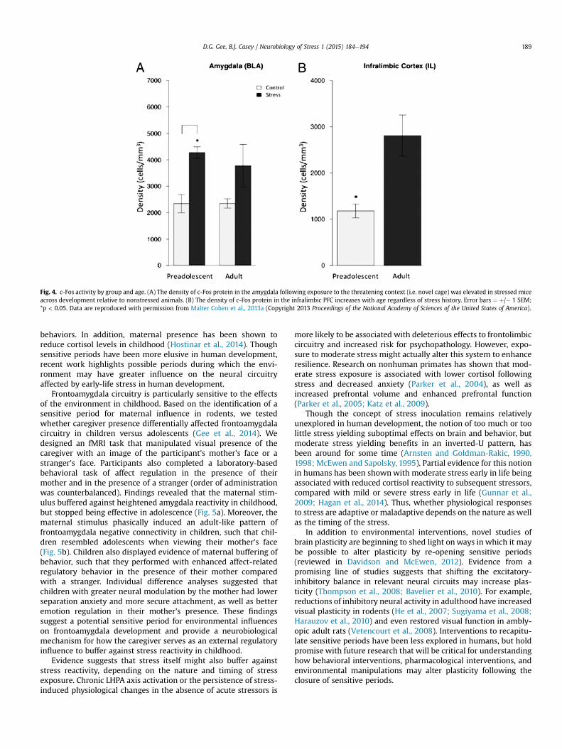
Fig. 4. c-Fos activity by group and age. (A) The density of c-Fos protein in the amygdala following exposure to the threatening context (i.e. novel cage) was elevated in stressed miceacross development relative to nonstressed animals. (B) The density of c-Fos protein in the infralimbic PFC increases with age regardless of stress history. Error bars ¼ þ/� 1 SEM;*p < 0.05. Data are reproduced with permission from Malter Cohen et al., 2013a (Copyright 2013 Proceedings of the National Academy of Sciences of the United States of America).
D.G. Gee, B.J. Casey / Neurobiology of Stress 1 (2015) 184e194 189
behaviors. In addition, maternal presence has been shown toreduce cortisol levels in childhood (Hostinar et al., 2014). Thoughsensitive periods have been more elusive in human development,recent work highlights possible periods during which the envi-ronment may have greater influence on the neural circuitryaffected by early-life stress in human development.
Frontoamygdala circuitry is particularly sensitive to the effectsof the environment in childhood. Based on the identification of asensitive period for maternal influence in rodents, we testedwhether caregiver presence differentially affected frontoamygdalacircuitry in children versus adolescents (Gee et al., 2014). Wedesigned an fMRI task that manipulated visual presence of thecaregiver with an image of the participant's mother's face or astranger's face. Participants also completed a laboratory-basedbehavioral task of affect regulation in the presence of theirmother and in the presence of a stranger (order of administrationwas counterbalanced). Findings revealed that the maternal stim-ulus buffered against heightened amygdala reactivity in childhood,but stopped being effective in adolescence (Fig. 5a). Moreover, thematernal stimulus phasically induced an adult-like pattern offrontoamygdala negative connectivity in children, such that chil-dren resembled adolescents when viewing their mother's face(Fig. 5b). Children also displayed evidence of maternal buffering ofbehavior, such that they performed with enhanced affect-relatedregulatory behavior in the presence of their mother comparedwith a stranger. Individual difference analyses suggested thatchildren with greater neural modulation by the mother had lowerseparation anxiety and more secure attachment, as well as betteremotion regulation in their mother's presence. These findingssuggest a potential sensitive period for environmental influenceson frontoamygdala development and provide a neurobiologicalmechanism for how the caregiver serves as an external regulatoryinfluence to buffer against stress reactivity in childhood.
Evidence suggests that stress itself might also buffer againststress reactivity, depending on the nature and timing of stressexposure. Chronic LHPA axis activation or the persistence of stress-induced physiological changes in the absence of acute stressors is
more likely to be associated with deleterious effects to frontolimbiccircuitry and increased risk for psychopathology. However, expo-sure to moderate stress might actually alter this system to enhanceresilience. Research on nonhuman primates has shown that mod-erate stress exposure is associated with lower cortisol followingstress and decreased anxiety (Parker et al., 2004), as well asincreased prefrontal volume and enhanced prefrontal function(Parker et al., 2005; Katz et al., 2009).
Though the concept of stress inoculation remains relativelyunexplored in human development, the notion of too much or toolittle stress yielding suboptimal effects on brain and behavior, butmoderate stress yielding benefits in an inverted-U pattern, hasbeen around for some time (Arnsten and Goldman-Rakic, 1990,1998; McEwen and Sapolsky, 1995). Partial evidence for this notionin humans has been shownwith moderate stress early in life beingassociated with reduced cortisol reactivity to subsequent stressors,compared with mild or severe stress early in life (Gunnar et al.,2009; Hagan et al., 2014). Thus, whether physiological responsesto stress are adaptive or maladaptive depends on the nature as wellas the timing of the stress.
In addition to environmental interventions, novel studies ofbrain plasticity are beginning to shed light on ways in which it maybe possible to alter plasticity by re-opening sensitive periods(reviewed in Davidson and McEwen, 2012). Evidence from apromising line of studies suggests that shifting the excitatory-inhibitory balance in relevant neural circuits may increase plas-ticity (Thompson et al., 2008; Bavelier et al., 2010). For example,reductions of inhibitory neural activity in adulthood have increasedvisual plasticity in rodents (He et al., 2007; Sugiyama et al., 2008;Harauzov et al., 2010) and even restored visual function in ambly-opic adult rats (Vetencourt et al., 2008). Interventions to recapitu-late sensitive periods have been less explored in humans, but holdpromise with future research that will be critical for understandinghow behavioral interventions, pharmacological interventions, andenvironmental manipulations may alter plasticity following theclosure of sensitive periods.
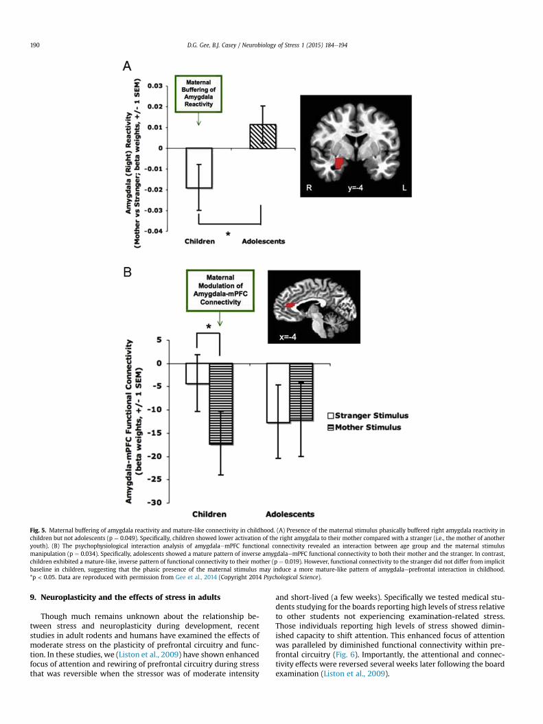
Fig. 5. Maternal buffering of amygdala reactivity and mature-like connectivity in childhood. (A) Presence of the maternal stimulus phasically buffered right amygdala reactivity inchildren but not adolescents (p ¼ 0.049). Specifically, children showed lower activation of the right amygdala to their mother compared with a stranger (i.e., the mother of anotheryouth). (B) The psychophysiological interaction analysis of amygdalaemPFC functional connectivity revealed an interaction between age group and the maternal stimulusmanipulation (p ¼ 0.034). Specifically, adolescents showed a mature pattern of inverse amygdalaemPFC functional connectivity to both their mother and the stranger. In contrast,children exhibited a mature-like, inverse pattern of functional connectivity to their mother (p ¼ 0.019). However, functional connectivity to the stranger did not differ from implicitbaseline in children, suggesting that the phasic presence of the maternal stimulus may induce a more mature-like pattern of amygdalaeprefrontal interaction in childhood.*p < 0.05. Data are reproduced with permission from Gee et al., 2014 (Copyright 2014 Psychological Science).
D.G. Gee, B.J. Casey / Neurobiology of Stress 1 (2015) 184e194190
9. Neuroplasticity and the effects of stress in adults
Though much remains unknown about the relationship be-tween stress and neuroplasticity during development, recentstudies in adult rodents and humans have examined the effects ofmoderate stress on the plasticity of prefrontal circuitry and func-tion. In these studies, we (Liston et al., 2009) have shown enhancedfocus of attention and rewiring of prefrontal circuitry during stressthat was reversible when the stressor was of moderate intensity
and short-lived (a few weeks). Specifically we tested medical stu-dents studying for the boards reporting high levels of stress relativeto other students not experiencing examination-related stress.Those individuals reporting high levels of stress showed dimin-ished capacity to shift attention. This enhanced focus of attentionwas paralleled by diminished functional connectivity within pre-frontal circuitry (Fig. 6). Importantly, the attentional and connec-tivity effects were reversed several weeks later following the boardexamination (Liston et al., 2009).
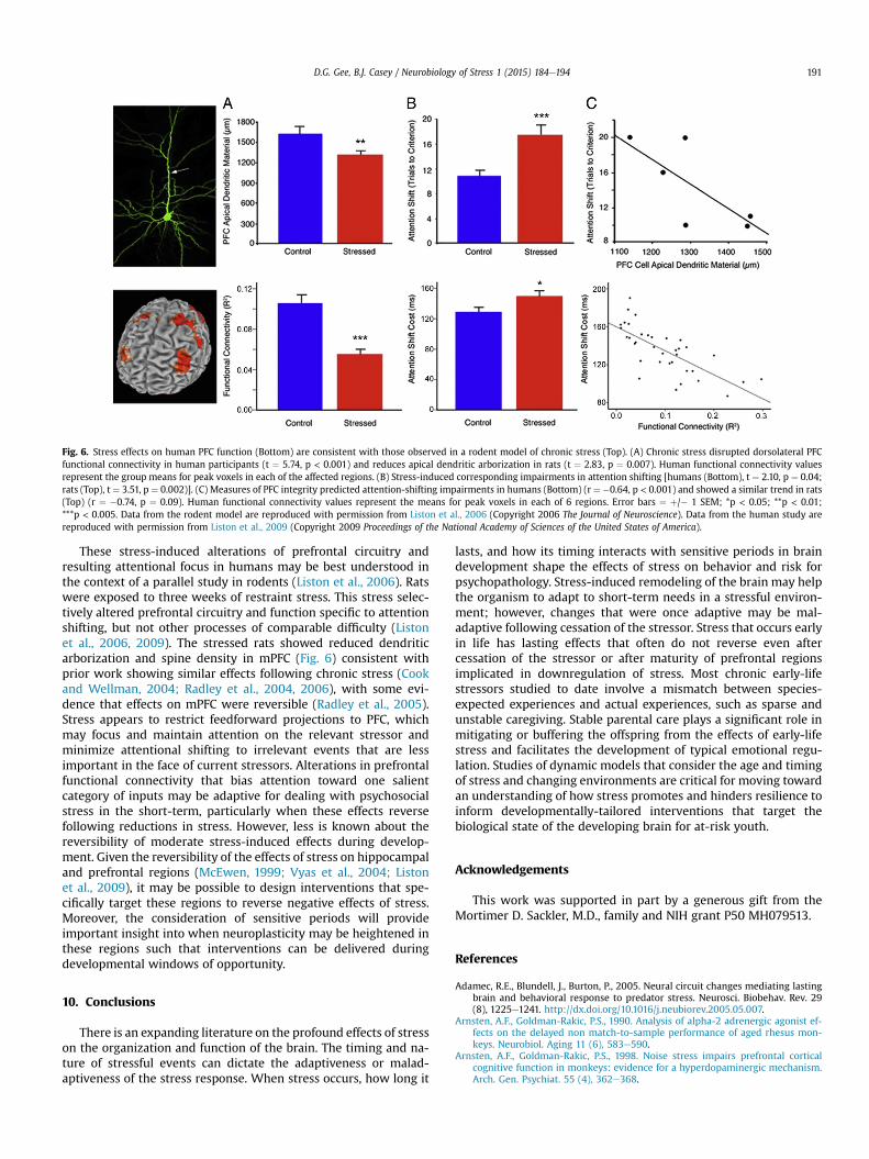
Fig. 6. Stress effects on human PFC function (Bottom) are consistent with those observed in a rodent model of chronic stress (Top). (A) Chronic stress disrupted dorsolateral PFCfunctional connectivity in human participants (t ¼ 5.74, p < 0.001) and reduces apical dendritic arborization in rats (t ¼ 2.83, p ¼ 0.007). Human functional connectivity valuesrepresent the group means for peak voxels in each of the affected regions. (B) Stress-induced corresponding impairments in attention shifting [humans (Bottom), t ¼ 2.10, p ¼ 0.04;rats (Top), t ¼ 3.51, p ¼ 0.002)]. (C) Measures of PFC integrity predicted attention-shifting impairments in humans (Bottom) (r ¼ �0.64, p < 0.001) and showed a similar trend in rats(Top) (r ¼ �0.74, p ¼ 0.09). Human functional connectivity values represent the means for peak voxels in each of 6 regions. Error bars ¼ þ/� 1 SEM; *p < 0.05; **p < 0.01;***p < 0.005. Data from the rodent model are reproduced with permission from Liston et al., 2006 (Copyright 2006 The Journal of Neuroscience). Data from the human study arereproduced with permission from Liston et al., 2009 (Copyright 2009 Proceedings of the National Academy of Sciences of the United States of America).
D.G. Gee, B.J. Casey / Neurobiology of Stress 1 (2015) 184e194 191
These stress-induced alterations of prefrontal circuitry andresulting attentional focus in humans may be best understood inthe context of a parallel study in rodents (Liston et al., 2006). Ratswere exposed to three weeks of restraint stress. This stress selec-tively altered prefrontal circuitry and function specific to attentionshifting, but not other processes of comparable difficulty (Listonet al., 2006, 2009). The stressed rats showed reduced dendriticarborization and spine density in mPFC (Fig. 6) consistent withprior work showing similar effects following chronic stress (Cookand Wellman, 2004; Radley et al., 2004, 2006), with some evi-dence that effects on mPFC were reversible (Radley et al., 2005).Stress appears to restrict feedforward projections to PFC, whichmay focus and maintain attention on the relevant stressor andminimize attentional shifting to irrelevant events that are lessimportant in the face of current stressors. Alterations in prefrontalfunctional connectivity that bias attention toward one salientcategory of inputs may be adaptive for dealing with psychosocialstress in the short-term, particularly when these effects reversefollowing reductions in stress. However, less is known about thereversibility of moderate stress-induced effects during develop-ment. Given the reversibility of the effects of stress on hippocampaland prefrontal regions (McEwen, 1999; Vyas et al., 2004; Listonet al., 2009), it may be possible to design interventions that spe-cifically target these regions to reverse negative effects of stress.Moreover, the consideration of sensitive periods will provideimportant insight into when neuroplasticity may be heightened inthese regions such that interventions can be delivered duringdevelopmental windows of opportunity.
10. Conclusions
There is an expanding literature on the profound effects of stresson the organization and function of the brain. The timing and na-ture of stressful events can dictate the adaptiveness or malad-aptiveness of the stress response. When stress occurs, how long it
lasts, and how its timing interacts with sensitive periods in braindevelopment shape the effects of stress on behavior and risk forpsychopathology. Stress-induced remodeling of the brain may helpthe organism to adapt to short-term needs in a stressful environ-ment; however, changes that were once adaptive may be mal-adaptive following cessation of the stressor. Stress that occurs earlyin life has lasting effects that often do not reverse even aftercessation of the stressor or after maturity of prefrontal regionsimplicated in downregulation of stress. Most chronic early-lifestressors studied to date involve a mismatch between species-expected experiences and actual experiences, such as sparse andunstable caregiving. Stable parental care plays a significant role inmitigating or buffering the offspring from the effects of early-lifestress and facilitates the development of typical emotional regu-lation. Studies of dynamic models that consider the age and timingof stress and changing environments are critical for moving towardan understanding of how stress promotes and hinders resilience toinform developmentally-tailored interventions that target thebiological state of the developing brain for at-risk youth.
Acknowledgements
This work was supported in part by a generous gift from theMortimer D. Sackler, M.D., family and NIH grant P50 MH079513.
References
Adamec, R.E., Blundell, J., Burton, P., 2005. Neural circuit changes mediating lastingbrain and behavioral response to predator stress. Neurosci. Biobehav. Rev. 29(8), 1225e1241. http://dx.doi.org/10.1016/j.neubiorev.2005.05.007.
Arnsten, A.F., Goldman-Rakic, P.S., 1990. Analysis of alpha-2 adrenergic agonist ef-fects on the delayed non match-to-sample performance of aged rhesus mon-keys. Neurobiol. Aging 11 (6), 583e590.
Arnsten, A.F., Goldman-Rakic, P.S., 1998. Noise stress impairs prefrontal corticalcognitive function in monkeys: evidence for a hyperdopaminergic mechanism.Arch. Gen. Psychiat. 55 (4), 362e368.
D.G. Gee, B.J. Casey / Neurobiology of Stress 1 (2015) 184e194192
Banks, S.J., Eddy, K.T., Angstadt, M., Nathan, P.J., Phan, K.L., 2007. Amygdalaefrontalconnectivity during emotion regulation. Soc. Cognitive Affect. Neurosci. 2 (4),303e312. http://dx.doi.org/10.1093/scan/nsm029.
Bavelier, D., Levi, D.M., Li, R.W., Dan, Y., Hensch, T.K., 2010. Removing brakes onadult brain plasticity: from molecular to behavioral interventions. J. Neurosci.Off. J. Soc. Neurosci. 30 (45), 14964e14971. http://dx.doi.org/10.1523/JNEUR-OSCI.4812-10.2010.
Benes, F.M., Turtle, M., Khan, Y., Farol, P., 1994. Myelination of a key relay zone in thehippocampal formation occurs in the human brain during childhood, adoles-cence, and adulthood. Arch. Gen. Psychiatry 51, 477e484.
Bourgeois, J.P., Rakic, P., 1993. Changes of synaptic density in the primary visualcortex of the macaque monkey from fetal to adult stage. J. Neurosci. 13,2801e2820.
Brody, B.A., Kinney, H.C., Kloman, A.S., Gilles, F.H., 1987. Sequence of central nervoussystem myelination in human infancy. I. An autopsy study of myelination. J.Neuropathol. Exp. Neurol. 46, 283e301.
Callaghan, B.L., Richardson, R., 2011. Maternal separation results in early emergenceof adult-like fear and extinction learning in infant rats. Behav. Neurosci. 125 (1),20e28. http://dx.doi.org/10.1037/a0022008.
Cameron, J.L., 2001. Critical periods for social attachment: deprivation and neuralsystems in rhesus monkeys. Soc. Res. Child. Dev. Abstr 2e054.
Campos, J.J., Emde, R.N., Gaensbauer, T., Henderson, C., 1975. Cardiac and behavioralinterrelationships in the reactions of infants to strangers. Dev. Psychol. 11 (5),589e601.
Casey, B.J., Duhoux, S., Malter Cohen, M., 2010. Adolescence: what do transmission,transition, and translation have to do with it? Neuron 67 (5), 749e760. http://dx.doi.org/10.1016/j.neuron.2010.08.033.
Cook, S.C., Wellman, C.L., 2004. Chronic stress alters dendritic morphology in ratmedial prefrontal cortex. J. Neurobiol. 60 (2), 236e248. http://dx.doi.org/10.1002/neu.20025.
Davidson, R.J., McEwen, B.S., 2012. Social influences on neuroplasticity: stress andinterventions to promote well-being. Nat. Neurosci. 15 (5), 689e695. http://dx.doi.org/10.1038/nn.3093.
De Kloet, E.R., Vreugdenhil, E., Oitzl, M.S., Jo€els, M., 1998. Brain corticosteroid re-ceptor balance in health and disease. Endocr. Rev. 19 (3), 269e301. http://dx.doi.org/10.1210/edrv.19.3.0331.
Diorio, D., Viau, V., Meaney, M.J., 1993. The role of the medial prefrontal cortex(cingulate gyrus) in the regulation of hypothalamic-pituitary-adrenal responsesto stress. J. Neurosci. Off. J. Soc. Neurosci. 13 (9), 3839e3847.
Dunn, J.D., Whitener, J., 1986. Plasma corticosterone responses to electrical stimu-lation of the amygdaloid complex: cytoarchitectural specificity. Neuroendocri-nology 42 (3), 211e217.
Eiland, L., Romeo, R.D., 2013. Stress and the developing adolescent brain. Neuro-science 249, 162e171.
Feldman, S., Conforti, N., Weidenfeld, J., 1995. Limbic pathways and hypothalamicneurotransmitters mediating adrenocortical responses to neural stimuli. Neu-rosci. Biobehav. Rev. 19 (2), 235e240.
Finlay, B.L., 2007. Endless minds most beautiful. Dev. Sci. 10 (1), 30e34. http://dx.doi.org/10.1111/j.1467-7687.2007.00560.x.
Folib, A.R., Lui, P., Romeo, R.D., 2011. The transformation of hormonal stress re-sponses throughout puberty and adolescence. J. Endocrinol. 210 (3), 391e398.
Galvan, A., Hare, T.A., Parra, C.E., Penn, J., Voss, H., Glover, G., Casey, B.J., 2006. Earlierdevelopment of the accumbens relative to orbitofrontal cortex might underlierisk-taking behavior in adolescents. J. Neurosci. 26, 6885e6892.
Ganzel, B., Casey, B.J., Glover, G., Voss, H.U., Temple, E., 2007. The aftermath of 9/11:effect of intensity and recency of trauma on outcome. Emotion Washington.D.C. 7 (2), 227e238. http://dx.doi.org/10.1037/1528-3542.7.2.227.
Ganzel, B.L., Kim, P., Glover, G.H., Temple, E., 2008. Resilience after 9/11: multimodalneuroimaging evidence for stress-related change in the healthy adult brain.NeuroImage 40 (2), 788e795. http://dx.doi.org/10.1016/j.neuroimage.2007.12.010.
Gee, D.G., Gabard-Durnam, L.J., Flannery, J., Goff, B., Humphreys, K.L., Telzer, E.H.,Tottenham, N., 2013a. Early developmental emergence of human amygdala-prefrontal connectivity after maternal deprivation. Proc. Natl. Acad. Sci. U. S.A. 110 (39), 15638e15643. http://dx.doi.org/10.1073/pnas.1307893110.
Gee, D.G., Humphreys, K.L., Flannery, J., Goff, B., Telzer, E.H., Shapiro, M., ,et al.Tottenham, N., 2013b. A developmental shift from positive to negativeconnectivity in human amygdala-prefrontal circuitry. J. Neurosci. 33 (10),4584e4593. http://dx.doi.org/10.1523/JNEUROSCI.3446-12.2013.
Gee, D.G., Gabard-Durnam, L., Telzer, E.H., Humphreys, K.L., Goff, B., Flannery, J.,Shapiro, M., Lumian, D.S., Fareri, D.S., Caldera, C., Tottenham, N., 2014. Maternalbuffering of amygdala-prefrontal circuitry during childhood but not adoles-cence. Psychol. Sci. 25 (11), 2067e2078.
Giedd, J.N., Blumenthal, J., Jeffries, N.O., Castellanos, F.X., Liu, H., Zijdenbos, A., et al.,1999. Brain development during childhood and adolescence: a longitudinal MRIstudy. Nat. Neurosci. 2, 861e863.
Gilles, E.E., Schultz, L., Baram, T.Z., 1996. Abnormal corticosterone regulation in animmature rat model of continuous chronic stress. Pediatr. Neurol. 15 (2),114e119.
Gogtay, N., Giedd, J.N., Lusk, L., Hayashi, K.M., Greenstein, D., Vaituzis, A.C., et al.,2004. Dynamic mapping of human cortical development during childhoodthrough early adulthood. Proc. Natl Acad. Sci. 101, 8174e8179.
Green, J.G., McLaughlin, K.A., Berglund, P.A., Gruber, M.J., Sampson, N.A.,Zaslavsky, A.M., Kessler, R.C., 2010. Childhood adversities and adult psychiatricdisorders in the national comorbidity survey replication I: associations with
first onset of DSM-IV disorders. Arch. Gen. Psychiatry 67 (2), 113e123. http://dx.doi.org/10.1001/archgenpsychiatry.2009.186.
Gunnar, M.R., Donzella, B., 2002. Social regulation of the cortisol levels in earlyhuman development. Psychoneuroendocrinology 27 (1e2), 199e220.
Gunnar, M.R., Bruce, J., Grotevant, H.D., 2000. International adoption of institu-tionally reared children: research and policy. Dev. Psychopathol. 12 (4),677e693.
Gunnar, M.R., Frenn, K., Wewerka, S.S., Van Ryzin, M.J., 2009. Moderate versus se-vere early life stress: associations with stress reactivity and regulation in10e12-year-old children. Psychoneuroendocrinology 34 (1), 62e75. http://dx.doi.org/10.1016/j.psyneuen.2008.08.013.
Hagan, M.J., Roubinov, D.S., Purdom Marreiro, C.L., Luecken, L.J., 2014. Childhoodinterparental conflict and HPA axis activity in young adulthood: examiningnonlinear relations. Dev. Psychobiol. 56 (4), 871e880. http://dx.doi.org/10.1002/dev.21157.
Harauzov, A., Spolidoro, M., DiCristo, G., De Pasquale, R., Cancedda, L., Pizzorusso, T.,Maffei, L., 2010. Reducing intracortical inhibition in the adult visual cortexpromotes ocular dominance plasticity. J. Neurosci. Off. J. Soc. Neurosci. 30 (1),361e371. http://dx.doi.org/10.1523/JNEUROSCI.2233-09.2010.
Hare, T.A., Tottenham, N., Galvan, A., Voss, H.U., Glover, G.H., Casey, B.J., 2008.Biological substrates of emotional reactivity and regulation in adolescenceduring an emotional Go-Nogo task. Biol. Psychiatry 63 (10), 927e934. http://dx.doi.org/10.1016/j.biopsych.2008.03.015.
Hariri, A.R., Mattay, V.S., Tessitore, A., Fera, F., Weinberger, D.R., 2003. Neocorticalmodulation of the amygdala response to fearful stimuli. Biol. Psychiatry 53 (6),494e501. http://dx.doi.org/10.1016/S0006-3223(02)01786-9.
He, H.-Y., Ray, B., Dennis, K., Quinlan, E.M., 2007. Experience-dependent recovery ofvision following chronic deprivation amblyopia. Nat. Neurosci. 10 (9),1134e1136. http://dx.doi.org/10.1038/nn1965.
Hofer, M.A., 1994. Early relationships as regulators of infant physiology andbehavior. Acta Paediatr. 83, 9e18.
Hostinar, C.E., Sullivan, R.M., Gunnar, M.R., 2014. Psychobiological mechanismsunderlying the social buffering of the hypothalamic-pituitary-adrenocorticalaxis: a review of animal models and human studies across development. Psy-chol. Bull. 140 (1), 256e282. http://dx.doi.org/10.1037/a0032671.
Huttenlocher, P.R., 1979. Synaptic density in human frontal cortex e developmentalchanges and effects of aging. Brain Res. 163, 195e205.
Huttenlocher, P.R., De Courten, C., Garey, L.J., van der Loos, H., 1982. Synapticdevelopment in human cerebral cortex. Int. J. Neurol. 16e17, 144e154.
Ivy, A.S., Brunson, K.L., Sandman, C., Baram, T.Z., 2008. Dysfunctional nurturingbehavior in rat dams with limited access to nesting material: a clinically rele-vant model for early-life stress. Neuroscience 154 (3), 1132e1142. http://dx.doi.org/10.1016/j.neuroscience.2008.04.019.
Jacobson, L., Sapolsky, R., 1991. The role of the hippocampus in feedback regulationof the hypothalamic-pituitary-adrenocortical axis. Endocr. Rev. 12 (2), 118e134.http://dx.doi.org/10.1210/edrv-12-2-118.
Katz, M., Liu, C., Schaer, M., Parker, K.J., Ottet, M.-C., Epps, A., Lyons, D.M., 2009.Prefrontal plasticity and stress inoculation-induced resilience. Dev. Neurosci. 31(4), 293e299. http://dx.doi.org/10.1159/000216540.
Kessler, R.C., Demler, O., Frank, R.G., Olfson, M., Pincus, H.A., Walters, E.E.,Zaslavsky, A.M., 2005. Prevalence and treatment of mental disorders, 1990 to2003. N. Engl. J. Med. 352 (24), 2515e2523. http://dx.doi.org/10.1056/NEJMsa043266.
Kikusui, T., Mori, Y., 2009. Behavioural and neurochemical consequences of earlyweaning in rodents. J. Neuroendocrinol 21 (4), 427e431. http://dx.doi.org/10.1111/j.1365-2826.2009.01837.x.
Kim, H., Somerville, L.H., Johnstone, T., Alexander, A.L., Whalen, P.J., 2003. Inverseamygdala and medial prefrontal cortex responses to surprised faces. Neuro-report 14 (18), 2317e2322. http://dx.doi.org/10.1097/01.wnr.0000101520.44335.20.
Kim, J.H., Li, S., Richardson, R., 2011. Immunohistochemical analyses of long-termextinction of conditioned fear in adolescent rats. Cereb. Cortex 21 (3),530e538. http://dx.doi.org/10.1093/cercor/bhq116.
Kim-Cohen, J., Caspi, A., Moffitt, T.E., Harrington, H., Milne, B.J., Poulton, R., 2003.Prior juvenile diagnoses in adults with mental disorder: developmental follow-back of a prospective-longitudinal cohort. Arch. Gen. Psychiatry 60 (7),709e717. http://dx.doi.org/10.1001/archpsyc.60.7.709.
Lee, F.S., Heimer, H., Giedd, J.N., Lein, E.S., Sestan, N., Weinberger, D., Casey, B., 2014.Adolescent mental health: an opportunity and an obligation. Science 346,547e549.
Liston, C., Miller, M.M., Goldwater, D.S., Radley, J.J., Rocher, A.B., Hof, P.R.,McEwen, B.S., 2006. Stress-induced alterations in prefrontal cortical dendriticmorphology predict selective impairments in perceptual attentional set-shift-ing. J. Neurosci. Off. J. Soc. Neurosci. 26 (30), 7870e7874. http://dx.doi.org/10.1523/JNEUROSCI.1184-06.2006.
Liston, C., McEwen, B.S., Casey, B.J., 2009. Psychosocial stress reversibly disruptsprefrontal processing and attentional control. Proc. Natl. Acad. Sci. U. S. A. 106(3), 912e917. http://dx.doi.org/10.1073/pnas.0807041106.
Liston, C.L., Cichon, J.M., Jeanneteau, F., Jia, Z., Chao, M.V., Gan, W., 2013. Circadianglucocorticoid oscillations promote learning-dependent synapse formation andmaintenance. Nat. Neurosci. 16, 698e705.
Lupien, S.J., King, S., Meaney, M.J., McEwen, B.S., 2000. Child's stress hormone levelscorrelate with mother's socioeconomic status and depressive state. Biol. Psy-chiatry 48 (10), 976e980.
D.G. Gee, B.J. Casey / Neurobiology of Stress 1 (2015) 184e194 193
Lupien, S.J., McEwen, B.S., Gunnar, M.R., Heim, C., 2009. Effects of stress throughoutthe lifespan on the brain, behaviour and cognition. Nat. Rev. Neurosci. 10 (6),434e445. http://dx.doi.org/10.1038/nrn2639.
Lupien, S.J., Parent, S., Evans, A.C., Tremblay, R.E., Zelazo, P.D., Corbo, V., S�eguin, J.R.,2011. Larger amygdala but no change in hippocampal volume in 10-year-oldchildren exposed to maternal depressive symptomatology since birth. Proc.Natl. Acad. Sci. U. S. A. 108 (34), 14324e14329. http://dx.doi.org/10.1073/pnas.1105371108.
Magari~nos, A.M., Verdugo, J.M., McEwen, B.S., 1997. Chronic stress alters synapticterminal structure in hippocampus. Proc. Natl. Acad. Sci. U. S. A. 94 (25),14002e14008.
Malter Cohen, M., Jing, D., Yang, R.R., Tottenham, N., Lee, F.S., Casey, B.J., 2013a.Early-life stress has persistent effects on amygdala function and development inmice and humans. Proc. Natl. Acad. Sci. U. S. A. 110 (45), 18274e18278. http://dx.doi.org/10.1073/pnas.1310163110.
Malter Cohen, M., Tottenham, N., Casey, B.J., 2013b. Translational developmentalstudies of stress on brain and behavior: implications for adolescent mentalhealth and illness? Neuroscience 249, 53e62. http://dx.doi.org/10.1016/j.neuroscience.2013.01.023.
Masten, A.S., Cicchetti, D., 2010. Developmental cascades. Dev. Psychopathol. 22,491e495.
McCallum, J., Kim, J.H., Richardson, R., 2010. Impaired extinction retention inadolescent rats: effects of D-cycloserine. Neuropsychopharmacol. Off. Publ. Am.Coll. Neuropsychopharmacol. 35 (10), 2134e2142. http://dx.doi.org/10.1038/npp.2010.92.
McCormick, K., Gualano, M.F., Kerr, D., Rockcastle, N., Cameron, J.L., 2005. Socialbond disruption in early life has behavioral consequences which remainevident through puberty. Soc. Neurosci. Abstr. 31:873.9.
McCoy, C.L., Masters, J.C., 1985. The development of children's strategies for thesocial control of emotion. Child Dev. 56 (5), 1214e1222.
McEwen, B.S., 1999. Stress and hippocampal plasticity. Annu. Rev. Neurosci. 22,105e122. http://dx.doi.org/10.1146/annurev.neuro.22.1.105.
McEwen, B.S., 2012. Brain on stress: how the social environment gets under theskin. Proc. Natl. Acad. Sci. U. S. A. 109 (Suppl. 2), 17180e17185. http://dx.doi.org/10.1073/pnas.1121254109.
McEwen, B.S., Sapolsky, R.M., 1995. Stress and cognitive function. Curr. Opin. Neu-robiol. 5 (2), 205e216.
Mehta, M.A., Golembo, N.I., Nosarti, C., Colvert, E., Mota, A., Williams, S.C.R., Sonuga-Barke, E.J.S., 2009. Amygdala, hippocampal and corpus callosum size followingsevere early institutional deprivation: the English and Romanian Adopteesstudy pilot. J. Child Psychol. Psychiatry Allied Discip. 50 (8), 943e951. http://dx.doi.org/10.1111/j.1469-7610.2009.02084.x.
Mitra, R., Jadhav, S., McEwen, B.S., Vyas, A., Chattarji, S., 2005. Stress durationmodulates the spatiotemporal patterns of spine formation in the basolateralamygdala. Proc. Natl. Acad. Sci. U. S. A. 102 (26), 9371e9376. http://dx.doi.org/10.1073/pnas.0504011102.
Monk, C.S., 2008. The development of emotion-related neural circuitry in healthand psychopathology. Dev. Psychopathol. 20 (Special Issue 04), 1231e1250.http://dx.doi.org/10.1017/S095457940800059X.
Monk, C.S., Pine, D.S., Charney, D.S., 2002. A developmental and neurobiologicalapproach to early trauma research. Seminars Clin. Neuropsychiatry 7 (2),137e146.
Moriceau, S., Sullivan, R.M., 2006. Maternal presence serves as a switch betweenlearning fear and attraction in infancy. Nat. Neurosci. 9 (8), 1004e1006. http://dx.doi.org/10.1038/nn1733.
Moriceau, S., Roth, T.L., Sullivan, R.M., 2010. Rodent model of infant attachmentlearning and stress. Dev. Psychobiol. 52 (7), 651e660. http://dx.doi.org/10.1002/dev.20482.
Newman, D.L., Moffitt, T.E., Caspi, A., Magdol, L., Silva, P.A., Stanton, W.R., 1996.Psychiatric disorder in a birth cohort of young adults: prevalence, comorbidity,clinical significance, and new case incidence from ages 11 to 21. J. Consult. Clin.Psychol. 64 (3), 552e562.
Ono, M., Kikusui, T., Sasaki, N., Ichikawa, M., Mori, Y., Murakami-Murofushi, K.,2008. Early weaning induces anxiety and precocious myelination in the anteriorpart of the basolateral amygdala of male Balb/c mice. Neuroscience 156 (4),1103e1110. http://dx.doi.org/10.1016/j.neuroscience.2008.07.078.
Parker, K.J., Buckmaster, C.L., Schatzberg, A.F., Lyons, D.M., 2004. Prospectiveinvestigation of stress inoculation in young monkeys. Arch. Gen. Psychiatry 61(9), 933e941. http://dx.doi.org/10.1001/archpsyc.61.9.933.
Parker, K.J., Buckmaster, C.L., Justus, K.R., Schatzberg, A.F., Lyons, D.M., 2005. Mildearly life stress enhances prefrontal-dependent response inhibition in monkeys.Biol. Psychiatry 57 (8), 848e855. http://dx.doi.org/10.1016/j.biopsych.2004.12.024.
Pattwell, S.S., Duhoux, S., Hartley, C.A., Johnson, D.C., Jing, D., Elliott, M.D., Lee, F.S.,2012. Altered fear learning across development in both mouse and human.Proc. Natl. Acad. Sci. 109 (40), 16318e16323. http://dx.doi.org/10.1073/pnas.1206834109.
Pechtel, P., Pizzagalli, D.A., 2011. Effects of early life stress on cognitive and affectivefunction: an integrated review of human literature. Psychopharmacology 214(1), 55e70. http://dx.doi.org/10.1007/s00213-010-2009-2.
Plotsky, P.M., Thrivikraman, K.V., Nemeroff, C.B., Caldji, C., Sharma, S., Meaney, M.J.,2005. Long-term consequences of neonatal rearing on central corticotropin-releasing factor systems in adult male rat offspring. Neuropsychopharmacol.Off. Publ. Am. Coll. Neuropsychopharmacol. 30 (12), 2192e2204. http://dx.doi.org/10.1038/sj.npp.1300769.
Pollak, S.D., 2008. Mechanisms linking early experience and the emergence ofemotions: illustrations from the study of maltreated children. Curr. Dir. Psychol.Sci. 17 (6), 370e375. http://dx.doi.org/10.1111/j.1467-8721.2008.00608.x.
Radley, J.J., Sisti, H.M., Hao, J., Rocher, A.B., McCall, T., Hof, P.R., Morrison, J.H., 2004.Chronic behavioral stress induces apical dendritic reorganization in pyramidalneurons of the medial prefrontal cortex. Neuroscience 125 (1), 1e6. http://dx.doi.org/10.1016/j.neuroscience.2004.01.006.
Radley, J.J., Rocher, A.B., Janssen, W.G.M., Hof, P.R., McEwen, B.S., Morrison, J.H.,2005. Reversibility of apical dendritic retraction in the rat medial prefrontalcortex following repeated stress. Exp. Neurol. 196 (1), 199e203. http://dx.doi.org/10.1016/j.expneurol.2005.07.008.
Radley, J.J., Arias, C.M., Sawchenko, P.E., 2006. Regional differentiation of the medialprefrontal cortex in regulating adaptive responses to acute emotional stress.J. Neurosci. Off. J. Soc. Neurosci. 26 (50), 12967e12976. http://dx.doi.org/10.1523/JNEUROSCI.4297-06.2006.
Raineki, C., Cort�es, M.R., Belnoue, L., Sullivan, R.M., 2012. Effects of early-life abusediffer across development: infant social behavior deficits are followed byadolescent depressive-like behaviors mediated by the amygdala. J. Neurosci.Off. J. Soc. Neurosci. 32 (22), 7758e7765. http://dx.doi.org/10.1523/JNEUR-OSCI.5843-11.2012.
Redgate, E.S., Fahringer, E.E., 1973. A comparison of the pituitary adrenal activityelicited by electrical stimulation of preoptic, amygdaloid and hypothalamic sitesin the rat brain. Neuroendocrinology 12 (6), 334e343.
Rice, C.J., Sandman, C.A., Lenjavi, M.R., Baram, T.Z., 2008. A novel mouse model foracute and long-lasting consequences of early life stress. Endocrinology 149 (10),4892e4900. http://dx.doi.org/10.1210/en.2008-0633.
Rinc�on-Cort�es, M., Sullivan, R.M., 2014. Early life trauma and attachment: imme-diate and enduring effects on neurobehavioral and stress axis development.Front. Endocrinol. 5, 33. http://dx.doi.org/10.3389/fendo.2014.00033.
Romeo, R.D., 2010. Adolescence: a central event in shaping stress reactivity. Dev.Psychobiol. 52 (3), 244e253. http://dx.doi.org/10.1002/dev.20437.
Romeo, R.D., Mueller, A., Sisti, H.M., Ogawa, S., McEwen, B.S., Brake, W.G., 2003.Anxiety and fear behaviors in adult male and female C57BL/6 mice aremodulated by maternal separation. Hormones Behav. 43 (5), 561e567.
Romeo, R.D., Bellani, R., Karatsoreos, I.N., Chhua, N., Vernov, M., Conrad, C.D.,McEwen, B.S., 2006. Stress history and pubertal development interact to shapehypothalamic-pituitary-adrenal axis plasticity. Endocrinology 147 (4),1664e1674.
Rothbaum, B.O., Davis, M., 2003. Applying learning principles to the treatment ofpost-trauma reactions. Ann. N. Y. Acad. Sci. 1008, 112e121.
Rutter, M., 1998. Developmental catch-up, and deficit, following adoption aftersevere global early privation. J. Child Psychol. Psychiatry Allied Discip. 39 (04),465e476.
Sabatini, M.J., Ebert, P., Lewis, D.A., Levitt, P., Cameron, J.L., Mirnics, K., 2007.Amygdala gene expression correlates of social behavior in monkeys experi-encing maternal separation. J. Neurosci. Off. J. Soc. Neurosci. 27 (12),3295e3304. http://dx.doi.org/10.1523/JNEUROSCI.4765-06.2007.
Sapolsky, R.M., 1986. Glucocorticoid toxicity in the hippocampus: reversal by sup-plementation with brain fuels. J. Neurosci. Official J. Soc. Neurosci. 6 (8),2240e2244.
Sheridan, M.A., Fox, N.A., Zeanah, C.H., McLaughlin, K.A., Nelson, C.A., 2012a. Vari-ation in neural development as a result of exposure to institutionalization earlyin childhood. Proc. Natl. Acad. Sci. U. S. A. 109 (32), 12927e12932. http://dx.doi.org/10.1073/pnas.1200041109.
Sheridan, M.A., Sarsour, K., Jutte, D., D'Esposito, M., Boyce, W.T., 2012b. The impactof social disparity on prefrontal function in childhood. PloS One 7 (4). http://dx.doi.org/10.1371/journal.pone.0035744 e35744.
Sheridan, M.A., How, J., Araujo, M., Schamberg, M.A., Nelson, C.A., 2013. What arethe links between maternal social status, hippocampal function, and HPA axisfunction in children? Dev. Sci. 16 (5), 665e675. http://dx.doi.org/10.1111/desc.12087.
Smith, M.A., Makino, S., Kvetnansky, R., Post, R.M., 1995. Stress and glucocorticoidsaffect the expression of brain-derived neurotrophic factor and neurotrophin-3mRNAs in the hippocampus. J. Neurosci. Off. J. Soc. Neurosci. 15 (3), 1768e1777.
Sowell, E.R., Thompson, P.M., Holmes, C.J., Jernigan, T.L., Toga, A.W., 1999. In vivoevidence for post-adolescent brain maturation in frontal and striatal regions.Nat. Neurosci. 2, 859e861.
Sowell, E.R., Thompson, P.M., Tessner, K.D., Toga, A.W., 2001. Mapping continuedbrain growth and gray matter density reduction in dorsal frontal cortex: inverserelationships during postadolescent brain maturation. J. Neurosci. 21,8819e8829.
Spear, L., 2010. The Behavioral Neuroscience of Adolescence. W. W. Norton &Company.
Sugiyama, S., Di Nardo, A.A., Aizawa, S., Matsuo, I., Volovitch, M., Prochiantz, A.,Hensch, T.K., 2008. Experience-dependent transfer of Otx2 homeoprotein intothe visual cortex activates postnatal plasticity. Cell 134 (3), 508e520. http://dx.doi.org/10.1016/j.cell.2008.05.054.
Teicher, M.H., Samson, J.A., Polcari, A., McGreenery, C.E., 2006. Sticks, stones, andhurtful words: relative effects of various forms of childhood maltreatment. Am.J. Psychiatry 163 (6), 993e1000. http://dx.doi.org/10.1176/appi.ajp.163.6.993.
Thompson, J.V., Sullivan, R.M., Wilson, D.A., 2008. Developmental emergence of fearlearning corresponds with changes in amygdala synaptic plasticity. Brain Res.1200, 58e65. http://dx.doi.org/10.1016/j.brainres.2008.01.057.
D.G. Gee, B.J. Casey / Neurobiology of Stress 1 (2015) 184e194194
Tottenham, N., 2012. Human amygdala development in the absence of species-expected caregiving. Dev. Psychobiol. 54 (6), 598e611. http://dx.doi.org/10.1002/dev.20531.
Tottenham, N., Sheridan, M., 2009. A review of adversity, the amygdala and thehippocampus: a consideration of developmental timing. Front. Hum. Neurosci.3, 68. http://dx.doi.org/10.3389/neuro.09.068.2009.
Tottenham, N., Hare, T.A., Quinn, B.T., McCarry, T.W., Nurse, M., Gilhooly, T.,Casey, B.J., 2010. Prolonged institutional rearing is associated with atypicallylarge amygdala volume and difficulties in emotion regulation. Dev. Sci. 13 (1),46e61. http://dx.doi.org/10.1111/j.1467-7687.2009.00852.x.
Tottenham, N., Hare, T.A., Millner, A., Gilhooly, T., Zevin, J.D., Casey, B.J., 2011.Elevated amygdala response to faces following early deprivation. Dev. Sci. 14(2), 190e204. http://dx.doi.org/10.1111/j.1467-7687.2010.00971.x.
Vetencourt, Maya, Sale, J.F., Viegi, A., Baroncelli, A., De Pasquale, L., O'Leary OF, R.,Maffei, L., 2008. The antidepressant fluoxetine restores plasticity in the adultvisual cortex. Sci. (New York, N.Y.) 320 (5874), 385e388. http://dx.doi.org/10.1126/science.1150516.
Vyas, A., Mitra, R., Shankaranarayana Rao, B.S., Chattarji, S., 2002. Chronic stressinduces contrasting patterns of dendritic remodeling in hippocampal and
amygdaloid neurons. J. Neurosci. Off. J. Soc. Neurosci. 22 (15), 6810e6818 doi:20026655.
Vyas, A., Bernal, S., Chattarji, S., 2003. Effects of chronic stress on dendritic arbor-ization in the central and extended amygdala. Brain Res. 965 (1e2), 290e294.
Vyas, A., Pillai, A.G., Chattarji, S., 2004. Recovery after chronic stress fails to reverseamygdaloid neuronal hypertrophy and enhanced anxiety-like behavior.Neuroscience 128 (4), 667e673. http://dx.doi.org/10.1016/j.neuroscience.2004.07.013.
Yang, E.-J., Lin, E.W., Hensch, T.K., 2012. Critical period for acoustic preference inmice. Proc. Natl. Acad. Sci. U. S. A. 109 (Suppl. 2), 17213e17220. http://dx.doi.org/10.1073/pnas.1200705109.
Yakovlev, P.I., Lecours, A.R., 1967. The myelogenetic cycles of regional maturation ofthe brain. In: Minkowski, A. (Ed.), Regional Development of the Brain in EarlyLife. Blackwell Scientific, Oxford, pp. 3e70.
Zeanah, M.D., Egger, M.D., Smyke, P.D., Nelson, P.D., Fox, P.D., Marshall, P.D.,Guthrie, P.D., 2009. Institutional rearing and psychiatric disorders in Romanianpreschool children. Am. J. Psychiatry 166 (7), 777e785. http://dx.doi.org/10.1176/appi.ajp.2009.08091438.


![An m6A-YTH Module Controls Developmental …An m6A-YTH Module Controls Developmental Timing and Morphogenesis in Arabidopsis[OPEN] Laura Arribas-Hernández,a,b Simon Bressendorff,a,b](https://img.pdfslide.net/doc/110x75/5f03a0e97e708231d409fdc3/an-m6a-yth-module-controls-developmental-an-m6a-yth-module-controls-developmental.jpg)

![arXiv:1306.5297v1 [q-bio.PE] 22 Jun 2013automated and manual annotation to determine the timing of 34 milestones during em-bryogenesis. Developmental timing is highly temperature-dependent](https://img.pdfslide.net/doc/110x75/602e51e68cf2be557c1778fc/arxiv13065297v1-q-biope-22-jun-2013-automated-and-manual-annotation-to-determine.jpg)