Embed Size (px)
Citation preview
The Journal of
Functional and
Fixed Orthodontics Volume 1 Number 2
December 2014
This Edi�on features ar�cles by
Dr Derek Mahony,
Dr Bill Clark
and Graham Manley
Merry Christmas and a Happy and
Prosperous New Year
Ortholab will be closing for Christmas Holidays from 3.30pm on
Friday 19th December un$l 9am on Monday 12th January. Please note that the last
day for accep$ng cases to be finished before the Christmas break will be
Friday 12th December.

The Journal of Func�onal and
Fixed Orthodon�cs is published
twice yearly and is available
free by subscrip�on. To sub-
scribe send an email to
Editor Graham Manley
Publisher Ortholab
The journal of Func�onal and
Fixed Orthodon�cs is copyright
2013.
Published quarterly by
ORTHOLAB,
297 Canterbury Rd,
Canterbury,
Victoria 3126, Australia.
Ar�cles published are the ex-
press opinion of the writer un-
der whose name they appear
and are not necessarily the
opinion of the publisher or edi-
tor of this journal.
Cover picture Felicity Manley
The Journal of
Functional and
Fixed Orthodontics
Volume 1 Number 2 December 2014
Contents
Editorial. . . . . . . . . . . . . . . . . . . . . . . . . . . . . . . Page 4
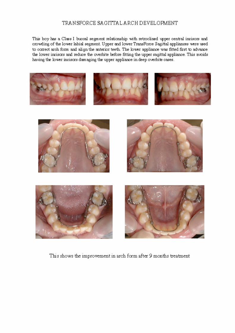
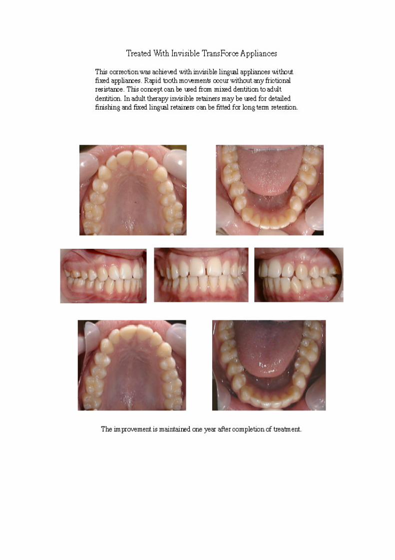
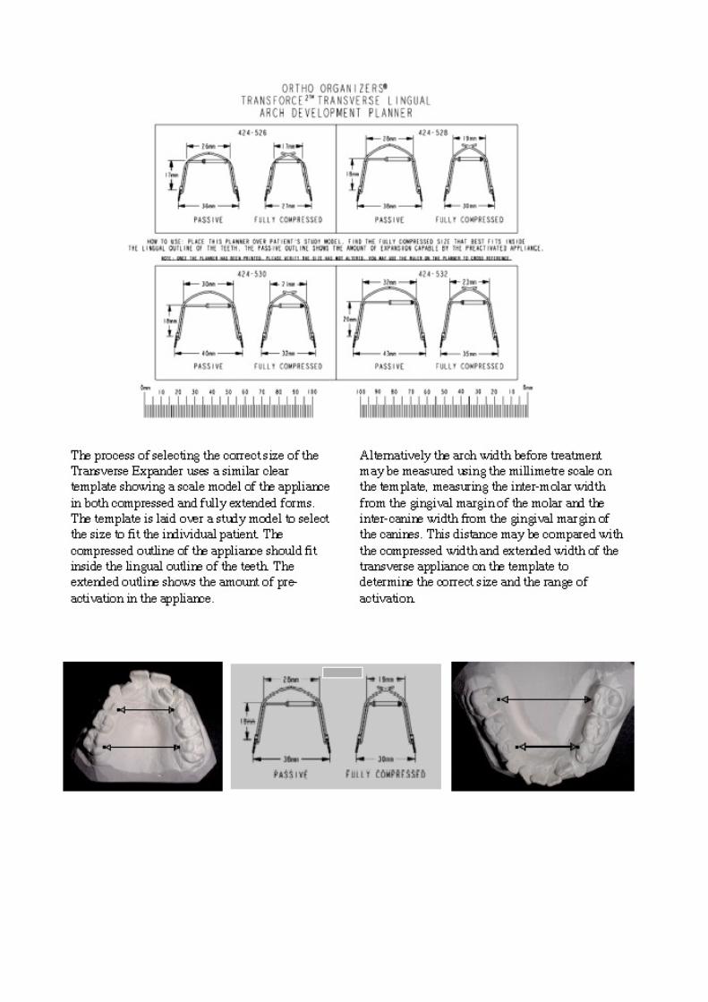
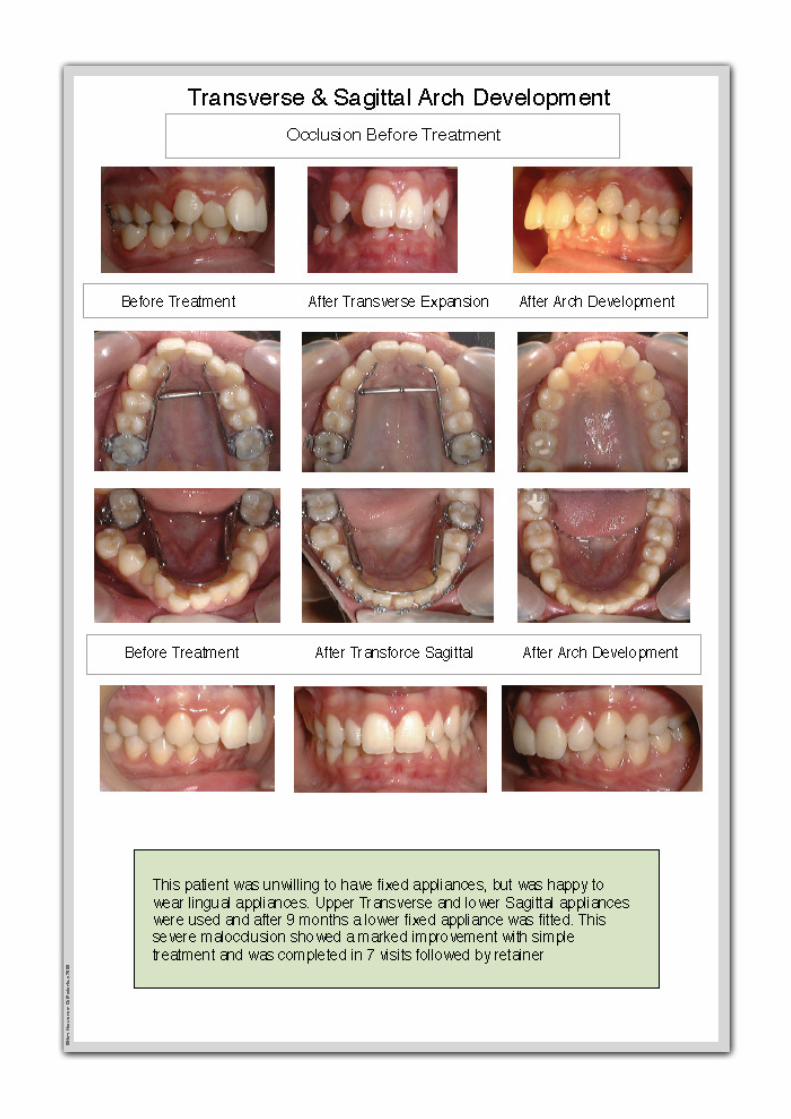
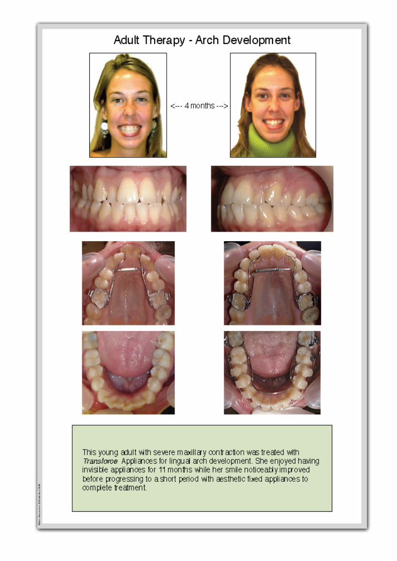
Transforce Appliances. . . . . . . . . . . . . . . . . . . Page 5
Re-establishing a physiologic ver�cal dimension for
an overclosed pa�ent . . . . . . . . . . . . . . . . . .Page 16
It’s a Wrap. . . . . . . . . . . . . . . . . . . . . . . . . . . Page 31
E d i t o r i a l
Page 4
Graham Manley Editor
Hello and welcome to our journal!
Christmas is almost upon us!! As I’ve got
older, it seems they come around so much
faster. As December comes, ever increasing
pa�ents want to get their braces off so as to
show off their new smile for Christmas. In
the lab, the number of new cases winds
down but the number of retainers more
than make up for it. It’s a hec�c �me, Haw-
ley’s, Essix, Clearbow and many other types
are made with increasing frequency as the
last day draws nearer.
Stress levels increase in the lab as I’m sure
they do in the clinic as impressions flood in
and retainers flood out. And at the back of
everyone mind is finish �me for the last day
this year. At last, a �me for extended relaxa-
�on, Christmas celebra�on with family and
friends and, if you’re lucky, a holiday. Time
to chill out, take stock of the year just gone
and think about your grand plans for the
year ahead.
What will you do different next year? Will
you start more cases, increase your mar-
ke�ng to aBract new pa�ents, buy that new
piece of equipment you’ve been thinking
about, what will it be?
Den�stry is the fastest changing medi
cal science and orthodon�cs is the fast
est changing dental science. Keeping up
to date is becoming more and more
difficult. Courses are run, some cos�ng
way too much, and some for free. Why
not, in this upcoming year, make a
promise to yourself to aBend a few
more courses. Educa�on is a life long
process, it’s also a very powerful asset,
one you can use in your prac�ce to
make life easier for you, for beBer
treatment for your pa�ent and to
increase your boBom line.
Think about making educa�on one of
your priori�es for 2015. Have a fantas
�c Christmas, New Year and Holiday
Season.
Graham Manley
Welcome to our Journal
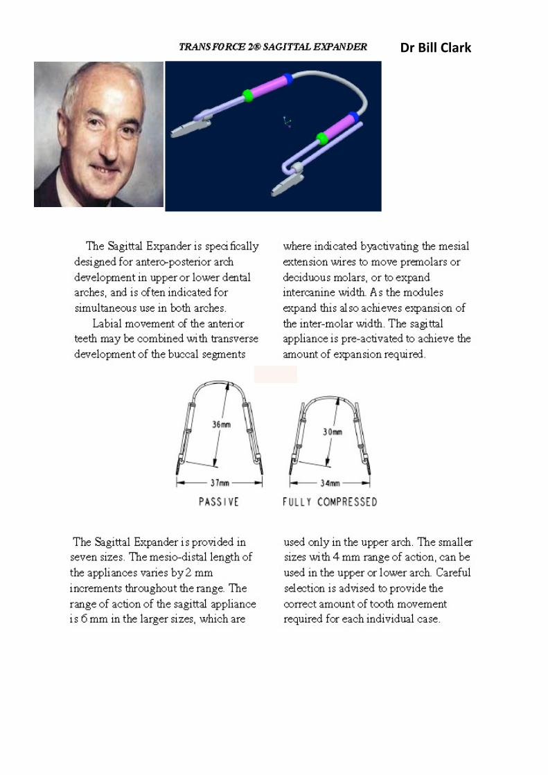
Dr Bill Clark

ORTHOLAB
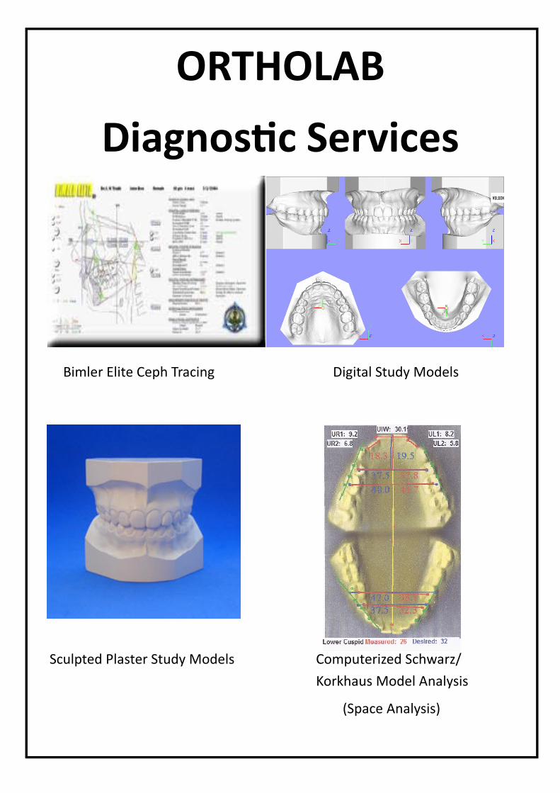
Diagnos$c Services
Bimler Elite Ceph Tracing Digital Study Models
Sculpted Plaster Study Models Computerized Schwarz/
Korkhaus Model Analysis
(Space Analysis)
Re-establishing a Physiologic Ver$cal
Dimension for an Overclosed Pa$ent
Dr. Derek Mahony
Specialist Orthodon$st
BDS(Syd) MScOrth(Lon) DOrth RCS(Edin) MDOrth RCSP(Glas) MOrth RCS(Eng)
MOrth RCS(Edin)/FCDS(HK) FRCD(Can) IBO FICD FICCDE
49 Botany Street
Randwick NSW 2031
Sydney, Australia
www.derekmahony.com
Introduc$on
The term neuromuscular occlusion has become associated with certain limited methodologies that are
used to obtain a muscle-compa�ble occlusal rela�onship. In reality, there are several different approach-
es that can be used to determine a "neuromuscular" maxillo-mandibular rela�onship, even with a fully
edentulous case. Within each method, however, the common basis for all muscle-oriented approaches
involves first determining the res�ng length of the mas�catory muscles.
Historically, opening the bite has been considered hazardous and/or foolhardy by many den�sts and with
good reason. Arbitrary opening of the bite, especially when accomplished strictly on an ar�culator, can
result in a difficult, uncomfortable and unapprecia�ve pa�ent. Some den�sts have recommended
against ever opening a bite, perhaps aLer an especially troublesome experience with a pa�ent.
In spite of the risks, there are some advantages associated with opening an over-closed bite. The iden�fi-
ca�on can be traced back at least 70 years to an ENT physician, Dr. J. B. Costen.1-3
Dr. Costen discovered,
perhaps quite by accident aLer referring many of his symptoma�c, edentulous pa�ents to a local den�st
for new dentures, that many returned with their head and ear pain symptoms greatly relieved. His publi-
ca�ons were posi�vely received at the �me and, in fact, what we refer to today as temporomandibular
disorders (TMDs) were originally referred to as "Costen's Syndrome." While we know today that many
TMD pa�ents are not over-closed, over-closed pa�ents do oLen exhibit some of the signs and symptoms
commonly associated with TMD. Thus, although over-closure in and of itself is not pathognomonic of
TMD, it should be considered as a risk factor.
The use of the pa�ent's own muscles to determine the ver�cal dimension of occlusion was already being
explored in the 1940s by people like orthodon�st John R. Thompson.4
Sears5 introduced the concept of
the "Pivot Appliance" in the 1950s, which was designed to open the bite enough to allow the pa�ent’s
muscles to
reposi�on the mandible. Following their lead, others6-28
have subsequently evolved the current array of neu-
romuscular registra�on methods presently in use. At the same �me several studies29-32
have demonstrated
that a muscle-determined posi�on, although similar, is not iden�cal to centric rela�on.
Common Signs and Symptoms of Over-closure
When asked, over-closed pa�ents oLen report symptoms such as frequent headaches, dull pain of the eleva-
tor muscles and pain or s�ffness in their neck muscles. Ear stuffiness, �nnitus and/or ver�go are also com-
monly reported. A more subtle symptom, less oLen reported, is frequent gastrointes�nal distress in various
forms that has no clear, iden�fiable cause. This may also be accompanied by a report of difficulty in chewing
and/or swallowing. An overclosed pa�ent will usually report several, but not all, of the following symptoms.
1. Frequent headaches with no iden�fiable cause
2. Ear stuffiness with no indica�on of ear pathology
3. Difficulty in chewing tough foods
4. Difficulty or discomfort in swallowing
5. Frequent gastrointes�nal distress
6. Ver�go
7. Tinnitus
8. Persistent dull pain in mas�catory elevator muscles
9. Neck pain or s�ffness
10. Possible increased wear of incisor teeth
Under examina�on, a number of signs indica�ng over-closure may appear. These include; 1) a measured free-
way space greater than 3 mm, 2) EMG or visual iden�fica�on of a tongue-thrust swallow, 3) the appearance
of less than fully erupted molars, 4) a deep curve of Spee, 5) one or more posterior edentulous spaces, 6) lin-
gually �pped mandibular molars, 7) EMG iden�fica�on of elevator muscle hyperac�vity at rest
of more than 2.0 microvolts average (or 2.2 microvolts RMS), 8) worn and shortened teeth (there is no scien�fic
evidence that human teeth "grow out" in response to wear in the way that elephant's teeth do), 9) horizontal skin
creasing and saliva weeping at the corners of the mouth, 10) a so-called "Shimbashi" measurement (in centric oc-
clusion) of less than 16 mm from the cemento-enamel junc�on of the maxillary central incisor to the cemento-
enamel junc�on of its opposing mandibular tooth and 11) long-term chronic internal derangement of the TM Joint
(s). However, pa�ents rarely seek dental treatment for any of these objec�ve signs. Instead, they are more likely
to seek rehabilita�ve treatment for headache, jaw-ache, ear-ache, difficulty in chewing/swallowing or for purely
esthe�c reasons. In other cases they are unaware of their condi�on, apparently due to their excellent adaptability.
In the over-closed pa�ent the “reason” for treatment, either cosme�c or func�onal, is oLen dependent more on
his/her individual adaptability than on the dental condi�ons present. While some signs simply indicate the
“progress of the destruc�on” that a pathological maxillo-mandibular rela�onship fosters, other signs may indicate
a successful adapta�on.
1. Freeway space > 3 mm [if pain level is low, it is an adapta�on, otherwise it is not]
2. Tongue thrust swallow [if full arch tongue thrust, usually a successful compensa�on]
3. The appearance of less than fully erupted molars [tongue inhibi�on of natural erup�on]
4. A deep curve of Spee [oLen associated with one or more missing molars or a deep anterior overbite
with retroclined upper incisors]
5. One or more posterior edentulous spaces [leads to deep curve of Spee]
6. Lingually �pped posterior teeth [tongue thrust during swallow, restricted maxillary arch]
7. Hyperac�vity of elevator muscles at "rest." [an adapta�on, successful if no elevator muscle pain]
8. Worn/short teeth, abfrac�ons (ground off) [not a successful adapta�on]
9. Skin creasing at corners of mouth [may appear as aesthe�c problem only, not an adapta�on]
10. Saliva weeping at corners of mouth [an esthe�c and func�onal problem, not an adapta�on]
11. CEJ (cemento-enamel junc�on) to CEJ in C.O. < 16 mm. [less than the normal adap�ve range]
12. Internal derangement(s) of the TMJ [if no degenera�on, may be a successful adapta�on]

Maxillo-mandibular Bite Rela$onships
Centric Occlusion (CO = habitual)
The maxillo-mandibular posi�on of maximum intercuspa�on is most oLen the dental treatment posi�on, primarily
by default. This is of necessity whenever single tooth prepara�ons or small restora�ons are involved, since they
must fit within the pa�ents exis�ng occlusal scheme. It is only at �mes of major reconstruc�ve, orthodon�c and/or
surgical treatments that the op�on of opening a bite or establishing a new maxillo-mandibular rela�on may present
itself. However, many clinicians s�ll prefer to "play it safe" and retain the exis�ng habitual (CO) maxillo-mandibular
rela�onship, even during major rehabilita�ve procedures. By defini�on, the use of centric occlusion as a treatment
posi�on excludes re-establishing a proper ver�cal dimension in an over-closed pa�ent. However, if the pa�ents
condi�on is ac�vely deteriora�ng this may not be a safe op�on at all, as the con�nued physiologic breakdown may
lead to failed den�stry and/or a flair up of craniofacial pain.
Centric Rela$on (CR)
The concept of centric rela�on has a very long history and was originally devised, at least in part, to accommodate
the use of ar�culators during prosthodon�c treatment. Although we now know that the jaw doesn't func�on like a
hinge, originally it was convenient to make that assump�on when using ar�culators to make prostheses. Today,
one clear difference between centric rela�on procedures and strictly muscle-oriented methodologies is the priority
given by CR methods to evalua�ng the func�on of the temporomandibular joints. Typically, centric rela�on opera-
tors give first priority to establishing stable joint func�on, while muscle-oriented (neuromuscular) approaches tend
to focus almost exclusively on muscle comfort.
Muscle-related Centric (MC)
In general, muscle-oriented approaches consider joint posi�on and/or stability secondary to muscle func�on. In
the extreme, it is simply assumed that crea�ng "happy muscles" will automa�cally provide good or at
least adequate joint func�on. In a more prac�cal view, both joint func�on and
muscle func�on are seriously evaluated and, when indicated, a compromise is
sought to provide both joint and muscle compa�bility. This represents an ap-
proach that bridges the gap between strict CR and rigid MC approaches. Con-
sequently, a variety of methods have evolved to capture and establish a mus-
cle-related centric posi�on, while maintaining favorable joint func�on.
The requirements of proper Neuromuscular Occlusion (NMO)
The first step in all approaches to NMO requires inducing relaxa�on in the
mas�catory musculature, however, there is no ra�onal excuse for not evalu-
a�ng TM joint func�on prior to beginning the process. This can be accom-
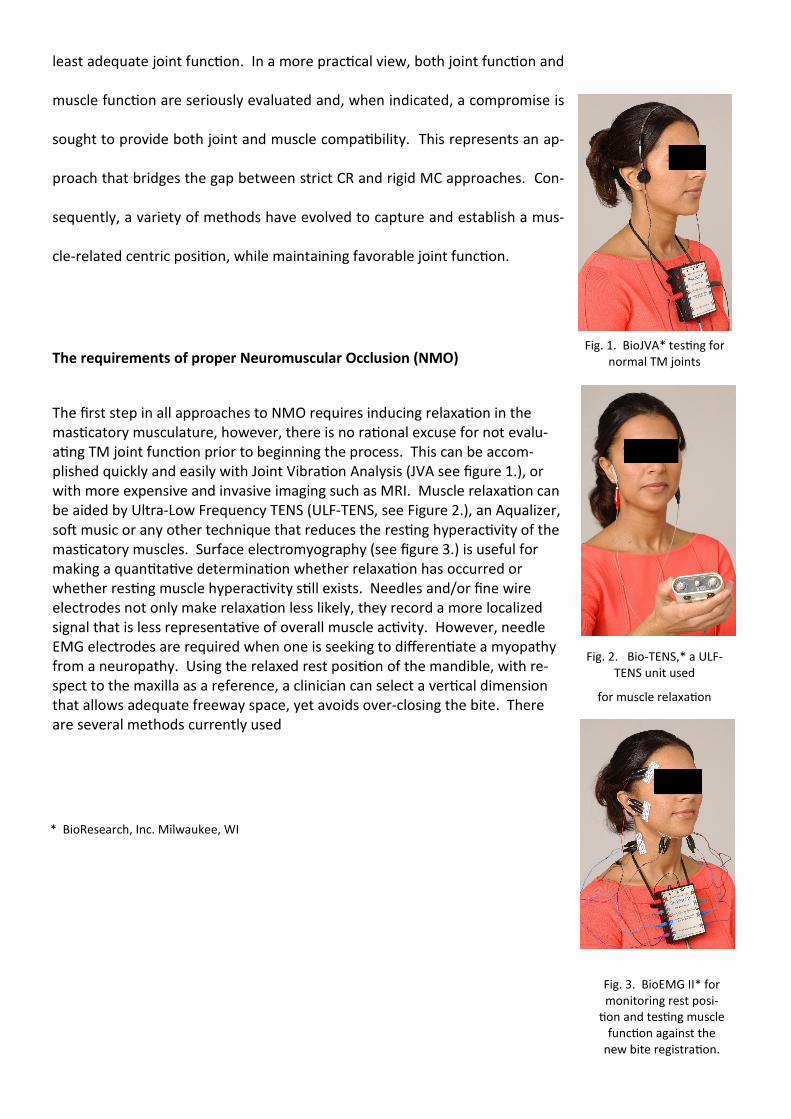
plished quickly and easily with Joint Vibra�on Analysis (JVA see figure 1.), or
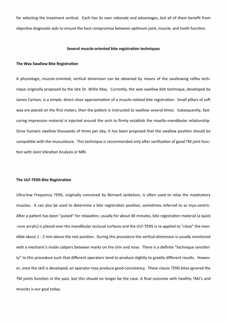
with more expensive and invasive imaging such as MRI. Muscle relaxa�on can
be aided by Ultra-Low Frequency TENS (ULF-TENS, see Figure 2.), an Aqualizer,
soL music or any other technique that reduces the res�ng hyperac�vity of the
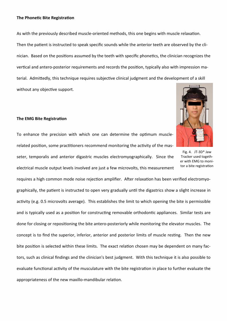
mas�catory muscles. Surface electromyography (see figure 3.) is useful for
making a quan�ta�ve determina�on whether relaxa�on has occurred or
whether res�ng muscle hyperac�vity s�ll exists. Needles and/or fine wire
electrodes not only make relaxa�on less likely, they record a more localized
signal that is less representa�ve of overall muscle ac�vity. However, needle
EMG electrodes are required when one is seeking to differen�ate a myopathy
from a neuropathy. Using the relaxed rest posi�on of the mandible, with re-
spect to the maxilla as a reference, a clinician can select a ver�cal dimension
that allows adequate freeway space, yet avoids over-closing the bite. There
are several methods currently used
Fig. 2. Bio-TENS,* a ULF-
TENS unit used
for muscle relaxa�on
Fig. 1. BioJVA* tes�ng for
normal TM joints
Fig. 3. BioEMG II* for
monitoring rest posi-
�on and tes�ng muscle
func�on against the
new bite registra�on.
* BioResearch, Inc. Milwaukee, WI
for selec�ng the treatment ver�cal. Each has its own ra�onale and advantages, but all of them benefit from
objec�ve diagnos�c aids to ensure the best compromise between op�mum joint, muscle, and tooth func�on.
Several muscle-oriented bite registra$on techniques
The Wax Swallow Bite Registra$on
A physiologic, muscle-oriented, ver�cal dimension can be obtained by means of the swallowing reflex tech-
nique originally proposed by the late Dr. Willie May. Currently, the wax swallow bite technique, developed by
James Carlson, is a simple, direct close approxima�on of a muscle-related bite registra�on. Small pillars of soL
wax are placed on the first molars, then the pa�ent is instructed to swallow several �mes. Subsequently, fast-
curing impression material is injected around the arch to firmly establish the maxillo-mandibular rela�onship.
Since humans swallow thousands of �mes per day, it has been proposed that the swallow posi�on should be
compa�ble with the musculature. This technique is recommended only aLer verifica�on of good TM joint func-
�on with Joint Vibra�on Analysis or MRI.
The ULF-TENS Bite Registra$on
Ultra-low Frequency TENS, originally conceived by Bernard Jankelson, is oLen used to relax the mas�catory
muscles. It can also be used to determine a bite registra�on posi�on, some�mes referred to as myo-centric.
ALer a pa�ent has been "pulsed" for relaxa�on, usually for about 40 minutes, bite registra�on material (a quick
-cure acrylic) is placed over the mandibular occlusal surfaces and the ULF-TENS is re-applied to "close" the man-
dible about 1 - 2 mm above the rest posi�on. During this procedure the ver�cal dimension is usually monitored
with a mechanic's inside calipers between marks on the chin and nose. There is a definite “technique sensi�vi-
ty” to this procedure such that different operators tend to produce slightly to greatly different results. Howev-
er, once the skill is developed, an operator may produce good consistency. These classic TENS bites ignored the
TM joints func�on in the past, but this should no longer be the case. A final outcome with healthy TMJ’s and
muscles is our goal today.
The Phone$c Bite Registra$on
As with the previously described muscle-oriented methods, this one begins with muscle relaxa�on.
Then the pa�ent is instructed to speak specific sounds while the anterior teeth are observed by the cli-
nician. Based on the posi�ons assumed by the teeth with specific phone�cs, the clinician recognizes the
ver�cal and antero-posterior requirements and records the posi�on, typically also with impression ma-
terial. AdmiBedly, this technique requires subjec�ve clinical judgment and the development of a skill
without any objec�ve support.
The EMG Bite Registra$on
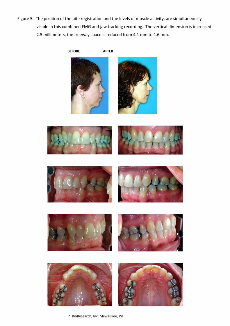
To enhance the precision with which one can determine the op�mum muscle-
related posi�on, some prac��oners recommend monitoring the ac�vity of the mas-
seter, temporalis and anterior digastric muscles electromyographically. Since the
electrical muscle output levels involved are just a few microvolts, this measurement
requires a high common mode noise rejec�on amplifier. ALer relaxa�on has been verified electromyo-
graphically, the pa�ent is instructed to open very gradually un�l the digastrics show a slight increase in
ac�vity (e.g. 0.5 microvolts average). This establishes the limit to which opening the bite is permissible
and is typically used as a posi�on for construc�ng removable orthodon�c appliances. Similar tests are
done for closing or reposi�oning the bite antero-posteriorly while monitoring the elevator muscles. The
concept is to find the superior, inferior, anterior and posterior limits of muscle res�ng. Then the new
bite posi�on is selected within these limits. The exact rela�on chosen may be dependent on many fac-
tors, such as clinical findings and the clinician's best judgment. With this technique it is also possible to
evaluate func�onal ac�vity of the musculature with the bite registra�on in place to further evaluate the
appropriateness of the new maxillo-mandibular rela�on.
Fig. 4. JT-3D* Jaw
Tracker used togeth-
er with EMG to moni-
tor a bite registra�on

The Instrument Monitored Bite Registra$on
To maximize the precision with which one can determine the bite registra�on posi�on, clinicians can
ac�vely monitor the posi�on of the mandible using a magne�c jaw tracker while simultaneously record-
ing EMG ac�vity. ALer the muscles are relaxed, a recording is made of the movement from rest to cen-
tric occlusion, light tapping in CO and protrusive guidance. Next, the registra�on posi�on is selected
and targeted on the computer screen. The treatment posi�on chosen can reflect all of the informa�on
available regarding the pa�ent's current condi�on. Finally, the registra�on material is placed in the
mouth and the pa�ent is instructed to close into it while the posi�on of the mandible and the muscle
ac�vi�es are monitored on the computer screen. (Figure 5). This allows the clinician to immediately
see the three dimensional rela�onship between the old centric occlusal posi�on and the new bite posi-
�on. The saved recording can be recalled later and u�lized to evaluate an appliance, provisional resto-
ra�ons or the prosthesis at try-in.
* BioResearch, Inc. Milwaukee, WI
Figure 5. The posi�on of the bite registra�on and the levels of muscle ac�vity, are simultaneously
visible in this combined EMG and jaw tracking recording. The ver�cal dimension is increased
2.5 millimeters, the freeway space is reduced from 4.1 mm to 1.6 mm.
BEFORE AFTER
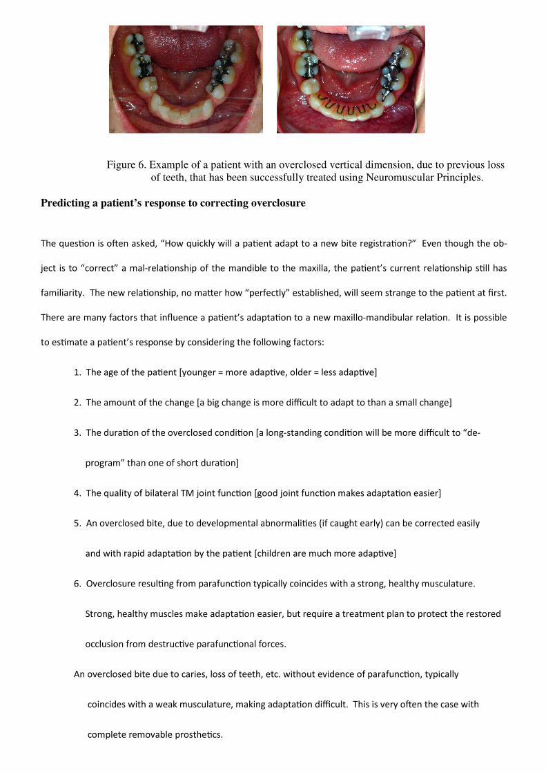
Figure 6. Example of a patient with an overclosed vertical dimension, due to previous loss
of teeth, that has been successfully treated using Neuromuscular Principles.
Predicting a patient’s response to correcting overclosure
The ques�on is oLen asked, “How quickly will a pa�ent adapt to a new bite registra�on?” Even though the ob-
ject is to “correct” a mal-rela�onship of the mandible to the maxilla, the pa�ent’s current rela�onship s�ll has
familiarity. The new rela�onship, no maBer how “perfectly” established, will seem strange to the pa�ent at first.
There are many factors that influence a pa�ent’s adapta�on to a new maxillo-mandibular rela�on. It is possible
to es�mate a pa�ent’s response by considering the following factors:
1. The age of the pa�ent [younger = more adap�ve, older = less adap�ve]
2. The amount of the change [a big change is more difficult to adapt to than a small change]
3. The dura�on of the overclosed condi�on [a long-standing condi�on will be more difficult to “de-
program” than one of short dura�on]
4. The quality of bilateral TM joint func�on [good joint func�on makes adapta�on easier]
5. An overclosed bite, due to developmental abnormali�es (if caught early) can be corrected easily
and with rapid adapta�on by the pa�ent [children are much more adap�ve]
6. Overclosure resul�ng from parafunc�on typically coincides with a strong, healthy musculature.
Strong, healthy muscles make adapta�on easier, but require a treatment plan to protect the restored
occlusion from destruc�ve parafunc�onal forces.
An overclosed bite due to caries, loss of teeth, etc. without evidence of parafunc�on, typically
coincides with a weak musculature, making adapta�on difficult. This is very oLen the case with
complete removable prosthe�cs.
Summary
Overclosure is a common condi�on among pa�ents seeking restora�ve and/or orthodon�c rehabilita-
�on. By evalua�ng the pa�ent for common signs and symptoms associated with overclosure, one can
determine the need for re-establishing a physiologic ver�cal dimension. Opening of the bite can be ac-
complished in a number of ways by following specific guidelines. The use of objec�ve diagnos�c aids
are extremely helpful by allowing the clinician to op�mize TMJ and craniofacial muscle func�on at the
new VDO. The correc�on of the ver�cal dimension during a rehabilita�ve procedure should result in
enhanced comfort and improved func�on in the finished case.

Bibliography:
Costen JB: A syndrome of ear and sinus symptoms dependent upon disturbed func�on of the temporomandibular joint.
Ann Otol Rhin and Laryngol 1934 Mar; 43:1-15
Costen JB: Glossodynia: Reflex irrita�on from the mandibular joint as the principal e�ologic factor. Arch Otolaryg 1935
Nov;22:554-564
Costen JB: Neuralgias and ear symptoms. J Am Med Assn 1936 Jul;107:252-255
Thompson JR: Concepts regarding the func�on of the stomatognathic system. JADA 1954 Jun; 48:626-637
Sears VH: Occlusal Pivots. J Prosthet Dent 1956 6:332-338
Gourion GR. [A new occlusal concept: myocentric rela�on and the Myo-monitor] Rev Fr Odontostomatol. 1971 Oct;18
(8):995-1004. French.
Fujii H, Mitani H. Reflex responses of the masseter and temporal muscles in man. J Dent Res. 1973 Sep-Oct;52(5):1046-
50
Vesanen E, Vesanen R. The Jankelson Myo-Monitor and its clinical use. Proc Finn Dent Soc. 1973 Dec;69(6):244-7.
Wessberg GA, Dinham R. The Myo-Monitor and the Myofacial Pain Dysfunc�on Syndrome. J Hawaii Dent Assoc. 1977
Aug;10(2):10-3.
Jankelson B, Radke JC. The myo-monitor: its use and abuse (I). Quintessence Int. 1978 Feb;9(2):47-52.
Jankelson B, Radke JC. The Myo-monitor: its use and abuse (II). Quintessence Int. 1978 Mar;9(3):35-9.
Kobayashi Y, Nakano Y, Komatsu Y, Ando N. [Clinical study of Myo-monitor. Part 1. An evalua�on in the treatment of
dysfunc�on of the mas�catory system] Shigaku. 1978 Dec;66(4):539-47. Japanese.
Rogers JL. Pa�ent's facial pain treated by Myo-monitor and dentures. Dent Surv. 1979 May;55(5):54.
Gernet W, Reither W, Gilde H. [Use of the Myo-Monitor in the func�onally disturbed stomatognathic system] Dtsch
Zahnarztl Z. 1980 Jun;35(6):595-8. German.
Shen WW. [A study of the myo-monitor and its clinical applica�on] Zhonghua Kou Qiang Ke Za Zhi. 1982 Dec;17(4):193-
6. Chinese.
Yoshida M, Higashi H, Yamauchi M, Takigawa H, Murakami M, Kawano J. [Effect of Myo-monitor pulsing on jaw opening
in pa�ents with trismus] Gifu Shika Gakkai Zasshi. 1983 Aug;11(1):157-69. Japanese.

Dinham GA. Myocentric. A clinical appraisal. Angle Orthod. 1984 Jul;54(3):211-7.
Boschiero R, Fraccari F, Pagnacco O. [Analysis of the results of using the Myo-Monitor on pa�ents with a reduced mouth
opening] Minerva Stomatol. 1986 Sep;35(9):857-64 Italian.
Allgood JP. Transcutaneous electrical neural s�mula�on (TENS) in dental prac�ce. Compend Con�n Educ Dent 1986 Oct;7
(9):640, 642-4
Bremerich A, Wiegel W, Thein T, Dietze T. Transcutaneous electric nerve s�mula�on (TENS) in the therapy of chronic facial
pain. Preliminary report. J Craniomaxillofac Surg 1988 Nov;16(8):379-81
Donegan SJ, Carr AB, Christensen LV, Ziebert GJ. An electromyographic study of aspects of 'deprogramming' of human jaw
muscles. J Oral Rehabil 1990 Nov;17(6):509-18
Gomez CE, Christensen LV. S�mulus-response latencies of two instruments delivering transcutaneous electrical neuromus-
cular s�mula�on (TENS). J Oral Rehabil 1991 Jan;18(1):87-94
Carr AB, Donegan SJ, Christensen LV, Ziebert GJ. An electrognathographic study of aspects of 'deprogramming' of human jaw
muscles. J Oral Rehabil 1991 Mar;18(2):143-8
Michelo\ A, Farella M, Vollaro S, Mar�na R. Mandibular rest posi�on and electrical ac�vity of the mas�catory muscles. J
Prosthet Dent. 1997 Jul;78(1):48-53
Rilo B, Santana U, Mora MJ, Cadarso CM. Myoelectrical ac�vity of clinical rest posi�on and jaw muscle ac�vity in young
adults. J Oral Rehabil. 1997 Oct;24(10):735-40
Sgobbi de Faria CR, Berzin F. Electromyographic study of the temporal, masseter and suprahyoid muscles in the mandibular
rest posi�on. J Oral Rehabil 1998 Oct;25(10):776-80
Eble OS, Jonas IE, Kappert HF. [Transcutaneous electrical nerve s�mula�on (TENS): its short-term and long-term effects on
the mas�catory muscles.] J Orofac Orthop 2000;61(2):100-11 [Ar�cle in English, German]
Kamyszek G, Ketcham R, Garcia R Jr, Radke J. Electromyographic evidence of reduced muscle ac�vity when ULF-TENS is ap-
plied to the Vth and VIIth cranial nerves. Cranio 2001 Jul;19(3):162-8
BesseBe RW, Quinlivan JT. Electromyographic evalua�on of the Myo-Monitor. J Prosthet Dent. 1973 Jul;30(1):19-24.
Remien JC 2nd, Ash M Jr. "Myo-Monitor centric": an evalua�on. J Prosthet Dent. 1974 Feb;31(2):137-45.
Noble WH. Anteroposterior posi�on of "Myo-Monitor centric". J Prosthet Dent. 1975 Apr;33(4):398-402.
Azarbal M. Comparison of Myo-Monitor centric posi�on to centric rela�on and centric occlusion. J Prosthet Dent. 1977
Sep;38(3):331-7.
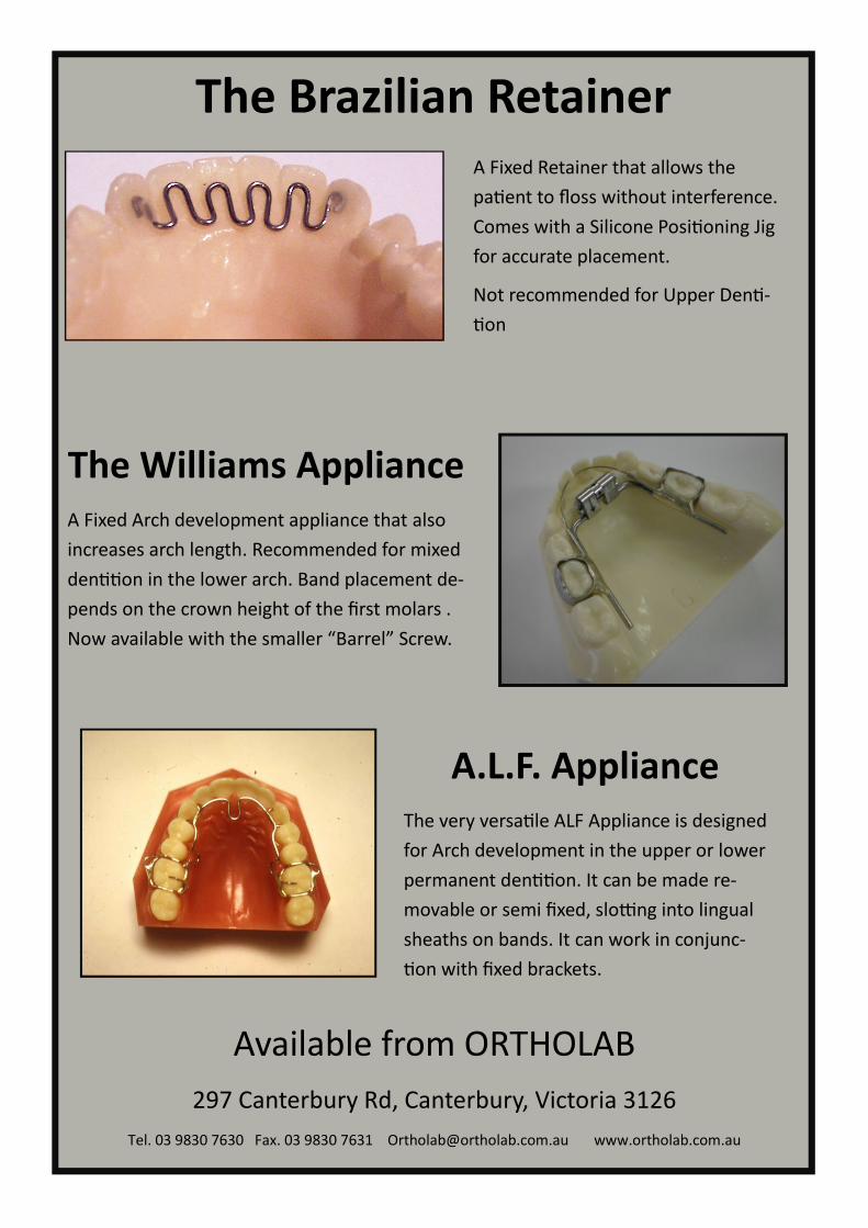
The Brazilian Retainer
A Fixed Retainer that allows the
pa�ent to floss without interference.
Comes with a Silicone Posi�oning Jig
for accurate placement.
Not recommended for Upper Den�-
�on
The Williams Appliance
A Fixed Arch development appliance that also
increases arch length. Recommended for mixed
den��on in the lower arch. Band placement de-
pends on the crown height of the first molars .
Now available with the smaller “Barrel” Screw.
A.L.F. Appliance
The very versa�le ALF Appliance is designed
for Arch development in the upper or lower
permanent den��on. It can be made re-
movable or semi fixed, slo\ng into lingual
sheaths on bands. It can work in conjunc-
�on with fixed brackets.
Available from ORTHOLAB
297 Canterbury Rd, Canterbury, Victoria 3126
Tel. 03 9830 7630 Fax. 03 9830 7631 [email protected] www.ortholab.com.au
From the Lab
If there’s one thing that’s guaranteed to get
under the skin of every lab owner, it’s badly
wrapped models that have been sent to the lab and
have arrived in an inappropriate container and are
now a mass of dust and pieces of a jigsaw, ready for
the applica�on of a liberal amount of Superglue.
OLen it’s an absence of training for the person re-
sponsible for dispatch, as simple as it seems,
properly wrapping models or impressions is not
very common. Common sense is, aLer all, not very
common.
Take away containers from the local Chinese restau-
rant are great for transpor�ng the Friday night take-
away to your house, but as a container for dental
models, they suck! We get 10-20 every week and
they always arrive smashed to bits. They are very
briBle. The contents within are in various stages of
demoli�on. Using a take away container is only
slightly beBer than no container at all (about 30 per
week). I some�mes wonder if the person wrapping
the models thinks the courier will carefully collect
the satchel and sit it on his lap during the flight to
the lab, and then hand deliver it himself. Nothing
could be further from the truth.
Major courier companies collect thousands of satch-
els, sort them into des�na�ons by throwing them
into large bins and oLen have heavier satchels
thrown on top of them. Unprotected models will be
crushed. They are flown to the des�na�on airport
and re sorted by throwing them into other large
bins. They are collected by van drivers who throw
them into the van and deliver them to the des�na-
�on. Write “Fragile” if you want, but it makes no
difference.
I know you don’t want a call from the lab asking for
another model. And I know your pa�ent doesn’t
want a call from your clinic to come in for another
impression. It’s a waste of �me and money for eve-
ryone involved. It doesn’t give your pa�ent a posi-
�ve impression of the prac�ce.
Wrapping models and impressions correctly is very
easy if you just follow a few rules. As I’ve said al-
ready, it’s just common sense, but it just isn’t hap-
pening too oLen.
Don’t laugh, but let me state the obvious, don’t put
the lab sheet in the bag with impressions and wet
�ssue. They tend to become “One”, by the �me we
get them. If you are sending impressions, put them
in a sealed (zip lock) plas�c bag with some wet �s-
sue if they are alginate, and double check the zip is
fully zipped. Then staple the lab sheet to the bag.
You don’t need to half fill the bag with water (yes it
does happen, oLen).
If you are sending models, firstly check the model to
make sure it’s good enough. By that I mean, no big
air blows or voids, no teeth trimmed off by an en-
thusias�c assistant on the model trimmer, no bro-
ken or missing teeth and not a warped or dragged
impression. Check par�cularly the molars as they
are most oLen the teeth that are malformed due to
It’s a Wrap
Graham Manley
A dragged or voided impression. You will need
to strike a balance between having enough im-
pression material in the posterior region and
too much material that would cause discomfort
to the pa�ent.
Once you are happy the model is good enough,
write the pa�ents name in pencil on the base.
Then wrap each model individually with bubble
wrap, put it in a plas�c bag with the bite and
then put all the cases you are sending in a box.
Tape the box shut and put it in the courier
satchel.
This is a VERY IMPORTANT POINT. Never ever
send two models together in occlusion, even
with a bite in between them. They are guaran-
teed to break several teeth.
When you receive cases back from the lab (I as-
sume other labs do it similar to Ortholab) each
model is wrapped in bubble wrap and put in a
plas�c bag in a box. You are expected to save
the box (they are expensive to buy), the bag
and the bubblewrap to use to send your next
cases back to us. Recycle the wrapping material.
Buy yourself a roll of bubblewrap, it’s dirt
cheap. Buy some sandwich bags, they’re cheap,
and reuse the box. You are receiving the best
wrapping material from the lab, just reuse it.
When all is said and done, I assume that you as-
sume your staff know this stuff, aLer all it’s in-
credibly basic. But believe me, they don’t. Let
them read this ar�cle or show them exactly how
you want it done. The very best appliances can
only be made on great models. I know you are
sending great models, but by the �me I get
them in the lab, they are not so great.
We have put together a youtube video showing
you how to wrap models, and another showing
you how to pour models from impressions.
They are available to view on our website,
www.ortholab.com.au click on “Video Library”.
Graham Manley is the owner of Ortholab
Enhance your Orthodon$c Experience by Joining a Study Club
Mee$ngs are held once a month and we have clubs star$ng in
Sydney, Melbourne, Brisbane, Adelaide and Canberra
Free membership, join us for Supper and a short lecture followed by round table
discussion of doctors cases, product demonstra$ons, hands on opportuni$es
and much more.
Call 03 9830 7630 for more informa$on
New eBook now Available
for Download or on CD
This Great new eBook shows you 20 ways to Distalize Molars.
There are plenty of options using Removable or Fixed Appliances,
this book is packed with colour pictures as well as explanations of how
to use and adjust each appliance. Indications and contraindications are
included. This ebook serves as a fantastic reference as well as a very
informative guide to Molar Distalization. Available for immediate
download from www.ortholab.com.au
Download $25.00
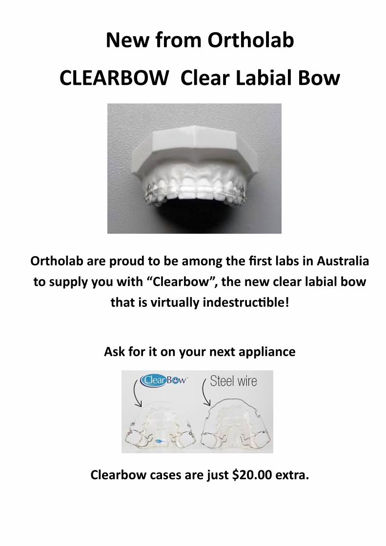
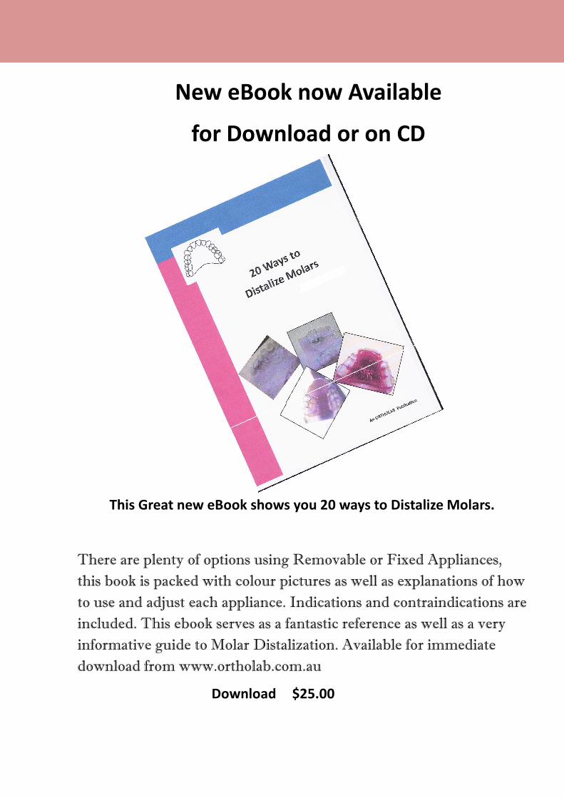
New from Ortholab
CLEARBOW Clear Labial Bow
Ortholab are proud to be among the first labs in Australia
to supply you with “Clearbow”, the new clear labial bow
that is virtually indestruc$ble!
Ask for it on your next appliance
Clearbow cases are just $20.00 extra.