Embed Size (px)
Citation preview
www.appliedradiology.com SUPPLEMENT TO APPLIED RADIOLOGY©
n 1July 2016
CONTRAST OPTIMIZATION IN LOW RADIATION DOSE IMAGING
THE JOURNAL OF PRACTICAL MEDICAL IMAGING AND MANAGEMENT
SUPPLEMENT TOJULY 2016
Contrast Optimization in Low Radiation Dose Imaging
Supported by an unrestricted educational grant from
CE Credits Available
Dushyant V. Sahani, MDDepartment of Radiology, Division of Abdominal Imaging and Interventional Radiology, Massachusetts General Hospital, Boston, MA
Lawrence N. Tanenbaum, MD, FACRVice President and Medical Director Eastern RegionDirector of MRI, CT and Advanced Imaging, RadNet, Inc.,New York, NY
Jeffrey C. Hellinger, MD, MBANew York Cardiovascular Institute, Lenox Hill Radiology and Medical Imaging, New York, NY
Contrast Optimization in Low Radiation Dose Imaging
Applied Radiology and this supplement, Contrast Optimization in Low Radiation Dose Imaging, are published by Anderson Publishing, Ltd. The journal does not warrant the expertise of any author in a particular field, nor is it responsible for any statements by such authors. The opin-ions expressed in this supplement are those of the authors. They do not imply endorsement of advertised products and do not necessarily reflect the opinions or recommendations of our sponsors or the editors and staff of Applied Radiology.
Copyright © 2016 by Anderson Publishing, Ltd.,
180 Glenside Avenue, Scotch Plains, NJ 07076
All rights reserved.
Publisher Kieran N. Anderson
Editor Joseph F. Jalkiewicz
Art and Production Barbara A. Shopiro
Dushyant V. Sahani, MDDepartment of Radiology,
Division of Abdominal Imaging and Interventional Radiology,
Massachusetts General Hospital
Boston, MA
Lawrence N. Tanenbaum, MD, FACRVice President and Medical Director Eastern Region
Director of MRI, CT and Advanced Imaging,
RadNet, Inc.,
New York, NY
Jeffrey C. Hellinger, MD, MBANew York Cardiovascular Institute,
Lenox Hill Radiology and Medical Imaging
New York, NY
Faculty
Program Information Target AudienceDate of Release: 07/01/16 Radiologic TechnologistsDate of Expiration: 06/30/18Estimated time to complete: 1 Hour
Learning ObjectivesAt the end of this educational activity, the participant (learner) will be able to:
• Review the need for strategies that minimize radiation exposure due to medical imaging in general, and to computed tomography (CT) examinations in particular;
• Define the key CT acquisition and postprocessing protocol parameters that impact radiation dose;
• Discuss the use of low kVp and high-concentration contrast media (HCCM) to maximize quality and consistency, and minimize radiation dose; and,
• Explain the synergistic impact of HCCM and iterative reconstruction (IR) post-processing techniques to increase CTA signal-to-noise ratios and image quality recovery.
AccreditationRadiologic TechnologistsThis course meets all criteria and has been approved by the AHRA, The Association for Medical Imaging Management, for one (1) ARRT Category A CE Credit.
Claim Credit Instructions 1. Visit: www.appliedradiology.org/cc9 2. Enter Course Code: ARSUP0716B 3. Follow promptsFor assistance, contact Kieran Anderson at (908) 301-1995 or email [email protected]
CE Accreditation InformationContrast Optimization in Low Radiation Dose Imaging
www.appliedradiology.com SUPPLEMENT TO APPLIED RADIOLOGY©
n 1July 2016
CONTRAST OPTIMIZATION IN LOW RADIATION DOSE IMAGING
Introduction
Medical exposure is currently one of the largest sources of radiation exposure in the United States, and computed tomography (CT) imaging is the largest
source of medical exposure.1,2 Governmental and regulatory agencies, as well as professional radiologic societies and equip-ment manufacturers, are all invested in balancing the estimated risk of radiation exposure with the potential diagnostic benefit of CT imaging. Radiation exposure reduction is important to consider in all patients, particularly in pediatric patients and in those who may have a diagnosis requiring multiple CT scans during their lifetime.
According to Image Wisely®, the American College of Radiology and the Radiological Society of North America Joint Task Force on Adult Radiation Protection, a stepwise approach to minimizing radiation dose is recommended.3,4 Preliminary steps include confirming the exam is indicated and has not been performed elsewhere recently, and also ensuring that equivalent information cannot be obtained from an alternative imaging exam associated with less expo-sure. Once these criteria have been met, there are several steps that can be taken for all exams, regardless of the spe-cific protocol. These include scanning only the requested and required region of interest, eliminating any unnecessary acquisition phases, and ensuring that the patient is carefully centered on the gantry table, all of which will reduce overall radiation exposure.
Optimizing specific CT protocol parameters can also contribute to lowering radiation dose exposure. For example, reducing both tube current (mA) and tube volt-age (kV) will result in reductions in radiation dose; how-
ever, it is important to note that small reductions in kV have a more substantial effect on radiation dose reduc-tion.5,6 Moreover, for iodinated contrast-enhanced exams, lower kVp values result not only in lower radiation dose exposure, but also higher contrast enhancement, especially when employed with high-iodine–concentration contrast media (HCCM).7-9 Modern postprocessing iterative recon-struction (IR) techniques start with the original filtered back-projection (FBP) and then iteratively refine the data, resulting in reduced noise within the CT images. These techniques help “recover” image quality at lower tube cur-rents and voltages, and in doing so help to substantially reduce net radiation doses while producing high-quality CT images.10
When implemented appropriately, the core radiation-dose reduction strategies discussed above can greatly reduce patient exposure while maintaining acceptable image qual-ity. Ensuring appropriate indications and scan coverage, as well as using the lowest possible mA and kV, should always be the cornerstones of a thoughtful and responsible approach to diagnostic CT; tube current modulation, low-kV imaging, and IR allow increased contrast enhancement at lower doses with acceptable noise levels. Using HCCM to achieve high-iodine flux contributes to improved diagnostic performance. Designing protocols for each indication in the context of acceptable radiation exposure and diagnostic image quality allows for overall radiation doses to be contained while CT use continues to advance. Here we present four cases that uti-lize HCCM protocols to produce high-quality images while reducing radiation dose exposure.
REFERENCES1. Brenner DJ. Should we be concerned about the rapid increase in CT usage? Rev Environ Health. 2010;25:63–68.2. Mettler FA Jr, Bhargavan M, Faulkner K, et al. Radiologic and nuclear medicine studies in the United States and worldwide: Frequency, radiation dose, and com-parison with other radiation sources--1950-2007. Radiology. 2009;253:520–531.3. Computed Tomography. Image Wisely® – Radiation Safety in Adult Medical Imaging. Available at: http://www.imagewisely.org/Imaging-Modalities/Computed-Tomography.4. Mayo-Smith WM. CT Protocol Design. November, 2010. Available at: http://www.imagewisely.org/imaging-modalities/computed-tomography/imaging-physi-cians/articles/ct-protocol-design.5. Corcuera-Solano I, McLellan AM, Doshi AH, Pawha PS, Tanenbaum LN. Whole-brain adaptive 70-kVp perfusion imaging with variable and extended sampling improves quality and consistency while reducing dose. Am J Neuroradiol. 2014;35:2045–2051.6. Li ZL, Li H, Zhang K, et al. Improvement of image quality and radiation dose of CT perfusion of the brain by means of low-tube voltage (70 KV). Eur Radiol. 2014;24:1906–1913.7. Furuta A, Ito K, Fujita T, Koike S, Shimizu A, Matsunaga N. Hepatic enhancement in multiphasic contrast-enhanced MDCT: Comparison of high- and low-iodine-concentration contrast medium in same patients with chronic liver disease. Am J Roentgenol. 2004;183:157–162.8. Rau MM, Setty BN, Blake MA, Ouellette-Piazzo K, Hahn PF, Sahani DV. Evaluation of renal transplant donors with 16-section multidetector CT angiography: com-parison of contrast media with low and high iodine concentrations. J Vasc Interv Radiol. 2007;18:603–609.9. Bae KT. Intravenous contrast medium administration and scan timing at CT: Considerations and approaches. Radiology. 2010;256:32–61.10. Corcuera-Solano I, Doshi AH, Noor A, Tanenbaum LN. Repeated head CT in the neurosurgical intensive care unit: feasibility of sinogram-affirmed iterative reconstruction-based ultra-low-dose CT for surveillance. Am J Neuroradiol. 2014;35:1281–1287.
2 n SUPPLEMENT TO APPLIED RADIOLOGY©
www.appliedradiology.com July 2016
CONTRAST OPTIMIZATION IN LOW RADIATION DOSE IMAGING
SUMMARYA 41-year-old male with a his-
tory of Crohn’s disease presented with abdominal cramps and discomfort. Physical examination revealed dis-tended abdomen. Labs showed CRP elevated to 61. Colonoscopy was per-formed and prematurely aborted due to a benign-appearing likely inflam-matory stricture in the mid sigmoid. Patient was admitted, and computed tomography enterography (CTE) with IV and oral contrast was ordered.
CT was performed with a 128-sec-tion multidetector row dual-energy CT (DECT) scanner (Somatom Defi-nition Flash, Siemens Medical Solu-tions; Forchheim, Germany). The patient was administered 90 mL of Isovue 370 (iopamidol, Bracco Diag-nostics) via 20 gauge IV catheter by power injection (Empower CTA) at a rate of 4 mL/s. The patient was also given 0.1% barium suspension (VoLu-men, E-Z-EM) as neutral oral contrast medium (total of 1350 mL prior to the scan; 450 mL at 60 minutes, 450 mL
at 40 minutes, 450 mL at 15 minutes followed by 8 oz of water at 5 min-utes). Dual energy data was acquired using the following parameters: tube potential of 140/100 kVp, mA ref 88/160 and 72/124, pitch of 0.95, rotation speed of 500msec, and a slice thickness of 5 mm. The images were reconstructed using SAFIRE level 3.
DIAGNOSISEnterocolonic fistula with exacer-
bation of Crohn’s disease
IMAGING FINDINGSThere is mucosal hyperenhance-
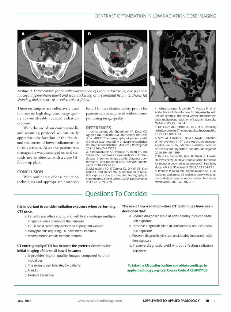
ment and mild wall thickening involv-ing a long segment of the terminal ileum (Figure 1). There is fat stranding within the deep pelvis between the terminal ileum and the sigmoid colon with adja-cent trace of free fluid (Figure 1). These findings likely represent active inflam-mation. In addition, there is an associ-ated enterocolonic fistula likely between the terminal ileum and cecum without the presence of an abscess (Figure 1).
DISCUSSIONCTE has become the preferred
method for initial imaging of the small bowel, as the image quality is superior to other modalities and the exam is well tolerated by patients.1,2 However, the prevalent use of CTE has become con-cerning in terms of radiation exposure, since patients are often young and will likely undergo multiple imaging studies to monitor their disease.3,4 The use of low-dose CT techniques have, therefore, been developed preserving diagnostic yield at considerably reduced radiation exposure.5 Low CT tube voltage and the administration of high-concentration contrast media (HCCM) yields greater contrast enhancement for a given injec-tion of contrast medium, lowering the iodinated contrast medium require-ments. Additionally, the ability to gen-erate a virtually nonenhanced scan with DECT removes the need for a separate nonenhanced scan. Several studies have also shown considerable radiation dose reduction using iterative reconstruc-tion algorithms in abdominal CT.6-8
Enterocolonic fistula with exacerbation of Crohn’s disease
Khalid W Shaqdan, MD; Manuel Patino, MD; Dushyant Sahani, MD
www.appliedradiology.com SUPPLEMENT TO APPLIED RADIOLOGY©
n 3July 2016
CONTRAST OPTIMIZATION IN LOW RADIATION DOSE IMAGING
FIGURE 1. Enterocolonic fistula with exacerbation of Crohn’s disease. (A) and (C) show mucosal hyperenhancement and wall thickening of the terminal ileum; (B) shows fat stranding and presence of an enterocolonic fistula.
These techniques are collectively used to maintain high diagnostic image qual-ity at considerably reduced radiation exposure.
With the use of our contrast media and scanning protocol we can easily appreciate the location of the fistula, and the extent of bowel inflammation in this patient. After the patient was managed he was discharged on oral ste-roids and antibiotics, with a close GI-follow up plan.
CONCLUSIONWith routine use of dose reduction
techniques and appropriate protocols
for CTE, the radiation safety profile for patients can be improved without com-promising image quality.
REFERENCES1. Kambadakone AR, Chaudhary NA, Desai GS, Nguyen DD, Kulkarni NM, and Sahani DV. Low-dose MDCT CT enterography of patients with Crohn disease: feasibility of adaptive statistical iterative reconstruction. AJR Am J Roentgenol. 2011;196:W743-W752.2. Kambadakone AR, Prakash P, Hahn PF, and Sahani DV. Low-dose CT examinations in Crohn’s disease: impact on image quality, diagnostic per-formance, and radiation dose. AJR Am J Roent-genol. 2010;195:78-88.3. McLaughlin PD, O’Connor OJ, O’Neill SB, Sha-nahan F, and Maher MM. Minimization of radia-tion exposure due to computed tomography in inflammatory bowel disease. ISRN Gastroenterol. 2012;2012:790279.
4. Wintersperger B, Jakobs T, Herzog P, et al. Aorto-iliac multidetector-row CT angiography with low kV settings: improved vessel enhancement and simultaneous reduction of radiation dose. Eur Radiol. 2005;15:334-341.5. Del Gaizo AJ, Fletcher JG, Yu L, et al. Reducing radiation dose in CT enterography. Radiographics. 2013;33:1109-1124.6. Silva AC, Lawder HJ, Hara A, Kujak J, Pavlicek W. Innovations in CT dose reduction strategy: Application of the adaptive statistical iterative reconstruction algorithm. AJR Am J Roentgenol. 2010;194:191-199.7. Hara AK, Paden RG, Silva AC, Kujak JL, Lawder HJ, Pavlicek W. Iterative reconstruction technique for reducing body radiation dose at CT: Feasibility study. AJR Am J Roentgenol. 2009;193:764-771.8. Prakash P, Kalra MK, Kambadakone AK, et al. Reducing abdominal CT radiation dose with adap-tive statistical iterative reconstruction technique. Invest Radiol. 2010;45:202-210
A B C
Questions To Consider
It is important to consider radiation exposure when performing
CTE since:
a. Patients are often young and will likely undergo multiple
imaging studies to monitor their disease
b. CTE is most commonly performed in pregnant women
c. Many patients requiring CTE have metal implants
d. Patient motion results in more artifacts
The use of low-radiation–dose CT techniques have been
developed that:
a. Reduce diagnostic yield at considerably reduced radia-
tion exposure
b. Preserve diagnostic yield at considerably reduced radia-
tion exposure
c. Preserve diagnostic yield at considerably increased radia-
tion exposure
d. Preserve diagnostic yield without affecting radiation
exposureCT enterography (CTE) has become the preferred method for
initial imaging of the small bowel because:
a. It provides higher quality images compared to other
modalities
b. The exam is well tolerated by patients
c. A and B
d. None of the above
To take the CE posttest online and obtain credit, go to
appliedradiology.org/cc9, Course Code: ARSUP0716B
4 n SUPPLEMENT TO APPLIED RADIOLOGY©
www.appliedradiology.com July 2016
CONTRAST OPTIMIZATION IN LOW RADIATION DOSE IMAGING
SUMMARYA 53-year-old male with a history
of alcoholism and chronic pancreatitis presented to the emergency depart-ment with an acute episode of abdomi-nal pain. Physical examination revealed a distended abdomen with epigastric tenderness radiating to the suprapubic region. Due to the uncertain nature of his clinical findings, the patient was admitted to the hospital, treated medi-cally for his pancreatitis, and further investigation with a computed tomog-raphy (CT) scan of the abdomen was ordered.
Dual phase CT was performed with a fast kVp switching 64-section multidetector row dual-energy CT scanner (DECT; Discovery 750 HD; GE healthcare, Milwaukee, WI). The patient was administered 90 mL of Iso-vue 370 (iopamidol, Bracco Diagnos-tics) via 18 gauge IV catheter by power injection (Empower CTA) at a rate of 3 mL/s. Arterial phase contrast-enhanced CT scanning was initiated after an enhancement threshold of 150 was reached. Dual energy data was acquired using the following parameters: tube potential of 140/80 kVp, tube current of 600 mA, pitch of 1.375:1, rotation
speed of 0.8, and a slice thickness of 2.5 mm. The images were reconstructed using ASiR 50%.
IMAGING FINDINGSThere is a hyperdense soft tis-
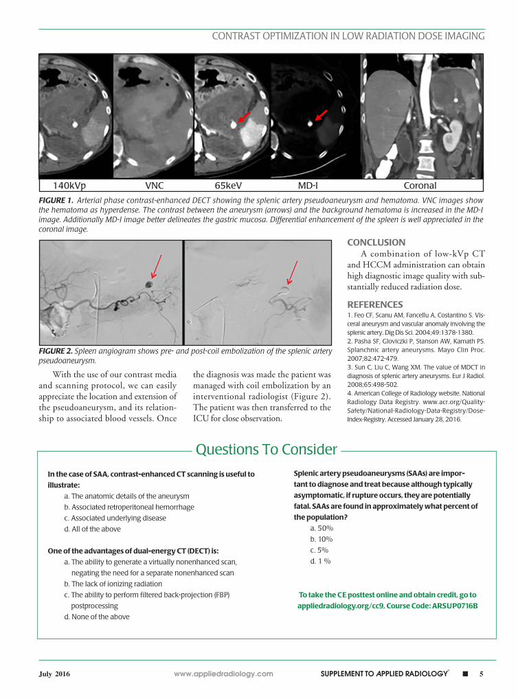
sue abnormality/collection in the left upper quadrant that extends along the paracolic gutters into the pelvis (Fig-ure 1). Within the collection, there is a well-defined 8 mm homogenous lesion showing enhancement that was isoattenuating to the splenic artery consistent with the appearance of a pseudoaneurysm (Figure 1). There is also a differential enhancement of the spleen, likely related to an evolv-ing splenic infarct from compromised splenic artery flow (Figure 1).
DIAGNOSISSplenic artery pseudoaneurysm
DISCUSSIONSplenic artery pseudoaneurysms
(SAAs) are found in approximately 1% of the population, and account for almost 60% of all visceral arterial aneurysms.1 The importance of diag-nosing and treating SAA lies in the pos-sible risk for fatal complications. SAAs
are usually asymptomatic, but when a rupture occurs, the patient commonly presents with epigastric pain and hypo-volemic shock. Ruptured SAA has a mortality rate of 25–70 %, but with current CT technology, patients can be imaged quickly to detect such lesions, preventing fatal complications.2,3
Contrast-enhanced CT scanning is useful to illustrate the anatomic details of the aneurysm, associated retro-peritoneal hemorrhage, and associated underlying disease. The administration of HCCM and low CT tube voltage yields greater contrast enhancement for a given injection of contrast medium, lowering the iodinated contrast medium requirements. Additionally, the abil-ity to generate a virtually nonenhanced scan with DECT removes the need for a separate nonenhanced scan. These techniques have been individually or jointly used to achieve the least radiation dose possible to the patient while main-taining high diagnostic image quality. According to the dose index registry (DIR), the median size-specific dose estimate (SSDE) for CT abdomenangio with IV contrast at all DIR sites is 17.6 mGy.4 The SSDE dose to the patient in this study was 11.7 mGy.
Splenic artery pseudoaneurysmKhalid W. Shaqdan, MD; Manuel Patino, MD; Dushyant Sahani, MD
www.appliedradiology.com SUPPLEMENT TO APPLIED RADIOLOGY©
n 5July 2016
CONTRAST OPTIMIZATION IN LOW RADIATION DOSE IMAGING
With the use of our contrast media and scanning protocol, we can easily appreciate the location and extension of the pseudoaneurysm, and its relation-ship to associated blood vessels. Once
the diagnosis was made the patient was managed with coil embolization by an interventional radiologist (Figure 2). The patient was then transferred to the ICU for close observation.
CONCLUSIONA combination of low-kVp CT
and HCCM administration can obtain high diagnostic image quality with sub-stantially reduced radiation dose.
REFERENCES1. Feo CF, Scanu AM, Fancellu A, Costantino S. Vis-ceral aneurysm and vascular anomaly involving the splenic artery. Dig Dis Sci. 2004;49:1378-1380.2. Pasha SF, Gloviczki P, Stanson AW, Kamath PS. Splanchnic artery aneurysms. Mayo Clin Proc. 2007;82:472-479. 3. Sun C, Liu C, Wang XM. The value of MDCT in diagnosis of splenic artery aneurysms. Eur J Radiol. 2008;65:498-502.4. American College of Radiology website. National Radiology Data Registry. www.acr.org/Quality-Safety/National-Radiology-Data-Registry/Dose-Index-Registry. Accessed January 28, 2016.
FIGURE 1. Arterial phase contrast-enhanced DECT showing the splenic artery pseudoaneurysm and hematoma. VNC images show the hematoma as hyperdense. The contrast between the aneurysm (arrows) and the background hematoma is increased in the MD-I image. Additionally MD-I image better delineates the gastric mucosa. Differential enhancement of the spleen is well appreciated in the coronal image.
FIGURE 2. Spleen angiogram shows pre- and post-coil embolization of the splenic artery pseudoaneurysm.
140kVp VNC 65keV MD-I Coronal
Questions To Consider
In the case of SAA, contrast-enhanced CT scanning is useful to
illustrate:
a. The anatomic details of the aneurysm
b. Associated retroperitoneal hemorrhage
c. Associated underlying disease
d. All of the above
One of the advantages of dual-energy CT (DECT) is:
a. The ability to generate a virtually nonenhanced scan,
negating the need for a separate nonenhanced scan
b. The lack of ionizing radiation
c. The ability to perform filtered back-projection (FBP)
postprocessing
d. None of the above
Splenic artery pseudoaneurysms (SAAs) are impor-
tant to diagnose and treat because although typically
asymptomatic, if rupture occurs, they are potentially
fatal. SAAs are found in approximately what percent of
the population?
a. 50%
b. 10%
c. 5%
d. 1 %
To take the CE posttest online and obtain credit, go to
appliedradiology.org/cc9, Course Code: ARSUP0716B
6 n SUPPLEMENT TO APPLIED RADIOLOGY©
www.appliedradiology.com July 2016
CONTRAST OPTIMIZATION IN LOW RADIATION DOSE IMAGING
CASE SUMMARYAn 80-year-old male presented to
the emergency room (ER) sometime after onset of right hemispheric stroke symptoms. An unenhanced CT of the head revealed no evidence of stroke or hemorrhage (Figure 1A); thus, he was a candidate for intravenous throm-bolytic therapy. Inspection of the intracranial vasculature led to detec-tion of a hyperdense (ie, denser than other arteries of similar or larger size) right internal carotid artery (ICA) and middle cerebral artery (MCA) – a find-ing strongly suggestive of thrombosis (Figure 1B). While eligible patients should still receive intravenous recom-binant tissue plasminogen activator (rTPA), endovascular therapy is now the standard of care for cardiovascular accident (CVA) caused by large vessel occlusion. Because of uncertainty as to the time since symptom onset, a com-bined time-resolved CT angiography (CTA) and perfusion CT (PCT) was performed.
IMAGING FINDINGSThe early and late phases of the
time-resolved CTA reveal occlusion of the right ICA, with markedly delayed filling and draining of the right MCA and its branches via collaterals (Figure 1C). The delayed phase from the time-
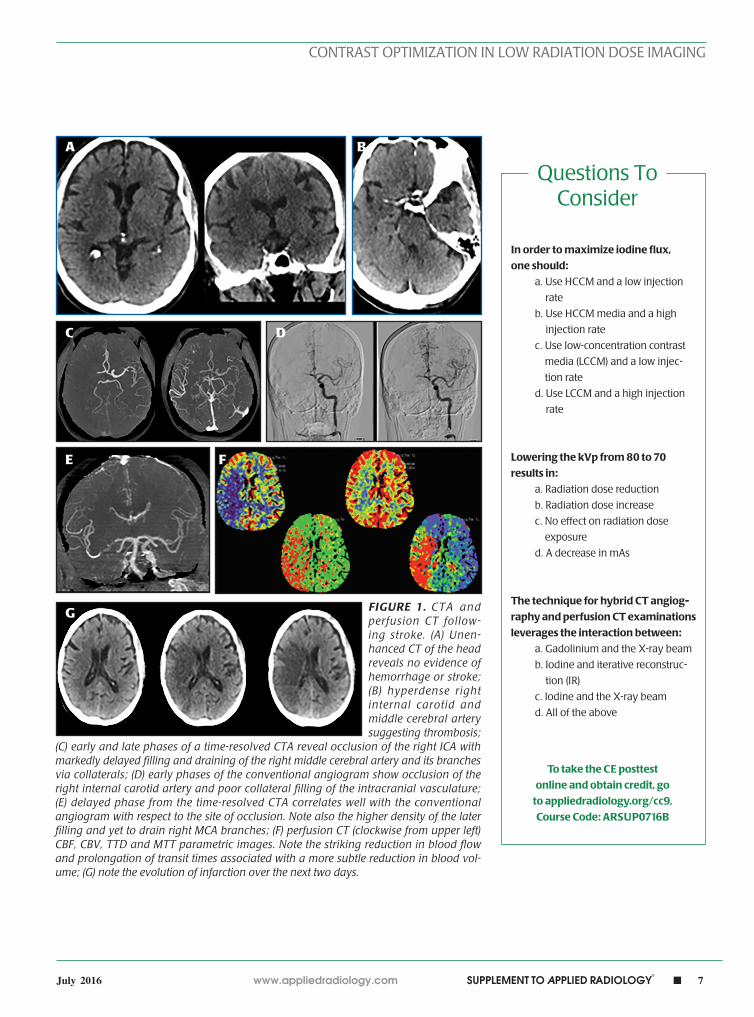
resolved CTA correlates well with the conventional angiogram (Figure 1D) with respect to the site of occlu-sion. Note also the higher density of the later filling and yet-to-drain right MCA branches, suggesting poor col-lateralization (Figure 1E). The con-comitant PCT parametric maps reveal reduced blood flow. There is a strik-ing prolongation of transit times evi-denced by mean transit time (MTT) and time to drain (TTD) images. In general, cerebral blood volumes increase in the setting of ischemia due to intact autoregulatory mechanisms. In this case, there is a subtle reduction in blood volume consistent with fail-ure of autoregulation. This suggests infarction within the right MCA ter-ritory (Figure 1F). Serial CT examina-tions over the next two days manifest progressive evidence of the infarction with no subsequent development of hemorrhage (Figure 1G).
DIAGNOSISOcclusion of the right ICA; infarc-
tion within the right MCA territory
DISCUSSIONThe technique for hybrid CTA-
PCT examinations leverages the inter-action between iodine and the X-ray beam. In general, when designing a
contrast-enhanced exam, optimizing the opacity created by iodine is critical to quality and consistency. Maximizing iodine flux by using an HCCM (370 mgI/mL in this example) and a high injection rate (4 mL/sec here) is a pow-erful way to accomplish this.1 Low-ering the energy of the X-ray to more closely match the unique characteris-tics (k-edge) of iodine is another way to maximize tissue and vascular contrast. By using 70 kVp, as opposed to the tra-ditional 80 kVp used for PCT or 100 kVp used for CTA, radiation dose is substantially reduced and image qual-ity is improved, even at low tube cur-rent (150 mAs).2
CONCLUSIONReadily available CT, CTA, and
PCT techniques facilitate the mod-ern treatment of acute stroke. Refined techniques such as low kVp and HCCM use maximize quality and con-sistency and minimize radiation dose.
REFERENCES1. Bae KT. Intravenous contrast medium adminis-tration and scan timing at CT: Considerations and approaches. Radiology. 2010;256:32–61.2. Corcuera-Solano I, McLellan AM, Doshi AH, Pawha PS, Tanenbaum LN. Whole-brain adap-tive 70-kVp perfusion imaging with variable and extended sampling improves quality and con-sistency while reducing dose. Am J Neuroradiol. 2014;35:2045–2051.
CTA and perfusion CT following strokeLawrence N. Tanenbaum, MD, FACR
www.appliedradiology.com SUPPLEMENT TO APPLIED RADIOLOGY©
n 7July 2016
CONTRAST OPTIMIZATION IN LOW RADIATION DOSE IMAGING
FIGURE 1. CTA and perfusion CT follow-ing stroke. (A) Unen-hanced CT of the head reveals no evidence of hemorrhage or stroke; (B) hyperdense right internal carotid and middle cerebral artery suggesting thrombosis;
(C) early and late phases of a time-resolved CTA reveal occlusion of the right ICA with markedly delayed filling and draining of the right middle cerebral artery and its branches via collaterals; (D) early phases of the conventional angiogram show occlusion of the right internal carotid artery and poor collateral filling of the intracranial vasculature; (E) delayed phase from the time-resolved CTA correlates well with the conventional angiogram with respect to the site of occlusion. Note also the higher density of the later filling and yet to drain right MCA branches; (F) perfusion CT (clockwise from upper left) CBF, CBV, TTD and MTT parametric images. Note the striking reduction in blood flow and prolongation of transit times associated with a more subtle reduction in blood vol-ume; (G) note the evolution of infarction over the next two days.
A
C
E
G
B
D
F
In order to maximize iodine flux,
one should:
a. Use HCCM and a low injection
rate
b. Use HCCM media and a high
injection rate
c. Use low-concentration contrast
media (LCCM) and a low injec-
tion rate
d. Use LCCM and a high injection
rate
Lowering the kVp from 80 to 70
results in:
a. Radiation dose reduction
b. Radiation dose increase
c. No effect on radiation dose
exposure
d. A decrease in mAs
The technique for hybrid CT angiog-
raphy and perfusion CT examinations
leverages the interaction between:
a. Gadolinium and the X-ray beam
b. Iodine and iterative reconstruc-
tion (IR)
c. Iodine and the X-ray beam
d. All of the above
To take the CE posttest
online and obtain credit, go
to appliedradiology.org/cc9,
Course Code: ARSUP0716B
Questions ToConsider
8 n SUPPLEMENT TO APPLIED RADIOLOGY©
www.appliedradiology.com July 2016
CONTRAST OPTIMIZATION IN LOW RADIATION DOSE IMAGING
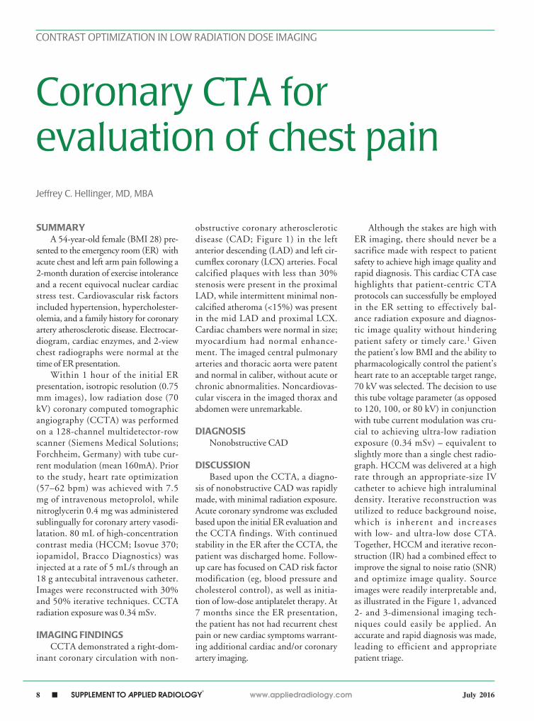
SUMMARYA 54-year-old female (BMI 28) pre-
sented to the emergency room (ER) with acute chest and left arm pain following a 2-month duration of exercise intolerance and a recent equivocal nuclear cardiac stress test. Cardiovascular risk factors included hypertension, hypercholester-olemia, and a family history for coronary artery atherosclerotic disease. Electrocar-diogram, cardiac enzymes, and 2-view chest radiographs were normal at the time of ER presentation.
Within 1 hour of the initial ER presentation, isotropic resolution (0.75 mm images), low radiation dose (70 kV) coronary computed tomographic angiography (CCTA) was performed on a 128-channel multidetector-row scanner (Siemens Medical Solutions; Forchheim, Germany) with tube cur-rent modulation (mean 160mA). Prior to the study, heart rate optimization (57–62 bpm) was achieved with 7.5 mg of intravenous metoprolol, while nitroglycerin 0.4 mg was administered sublingually for coronary artery vasodi-latation. 80 mL of high-concentration contrast media (HCCM; Isovue 370; iopamidol, Bracco Diagnostics) was injected at a rate of 5 mL/s through an 18 g antecubital intravenous catheter. Images were reconstructed with 30% and 50% iterative techniques. CCTA radiation exposure was 0.34 mSv.
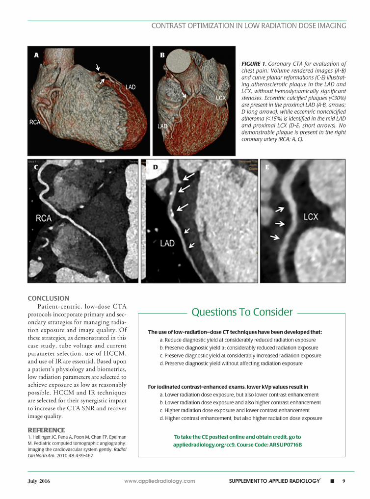
IMAGING FINDINGSCCTA demonstrated a right-dom-
inant coronary circulation with non-
obstructive coronary atherosclerotic disease (CAD; Figure 1) in the left anterior descending (LAD) and left cir-cumflex coronary (LCX) arteries. Focal calcified plaques with less than 30% stenosis were present in the proximal LAD, while intermittent minimal non-calcified atheroma (<15%) was present in the mid LAD and proximal LCX. Cardiac chambers were normal in size; myocardium had normal enhance-ment. The imaged central pulmonary arteries and thoracic aorta were patent and normal in caliber, without acute or chronic abnormalities. Noncardiovas-cular viscera in the imaged thorax and abdomen were unremarkable.
DIAGNOSISNonobstructive CAD
DISCUSSIONBased upon the CCTA, a diagno-
sis of nonobstructive CAD was rapidly made, with minimal radiation exposure. Acute coronary syndrome was excluded based upon the initial ER evaluation and the CCTA findings. With continued stability in the ER after the CCTA, the patient was discharged home. Follow-up care has focused on CAD risk factor modification (eg, blood pressure and cholesterol control), as well as initia-tion of low-dose antiplatelet therapy. At 7 months since the ER presentation, the patient has not had recurrent chest pain or new cardiac symptoms warrant-ing additional cardiac and/or coronary artery imaging.
Although the stakes are high with ER imaging, there should never be a sacrifice made with respect to patient safety to achieve high image quality and rapid diagnosis. This cardiac CTA case highlights that patient-centric CTA protocols can successfully be employed in the ER setting to effectively bal-ance radiation exposure and diagnos-tic image quality without hindering patient safety or timely care.1 Given the patient’s low BMI and the ability to pharmacologically control the patient’s heart rate to an acceptable target range, 70 kV was selected. The decision to use this tube voltage parameter (as opposed to 120, 100, or 80 kV) in conjunction with tube current modulation was cru-cial to achieving ultra-low radiation exposure (0.34 mSv) – equivalent to slightly more than a single chest radio-graph. HCCM was delivered at a high rate through an appropriate-size IV catheter to achieve high intraluminal density. Iterative reconstruction was utilized to reduce background noise, which is inherent and increases with low- and ultra-low dose CTA. Together, HCCM and iterative recon-struction (IR) had a combined effect to improve the signal to noise ratio (SNR) and optimize image quality. Source images were readily interpretable and, as illustrated in the Figure 1, advanced 2- and 3-dimensional imaging tech-niques could easily be applied. An accurate and rapid diagnosis was made, leading to efficient and appropriate patient triage.
Coronary CTA for evaluation of chest painJeffrey C. Hellinger, MD, MBA
www.appliedradiology.com SUPPLEMENT TO APPLIED RADIOLOGY©
n 9July 2016
CONTRAST OPTIMIZATION IN LOW RADIATION DOSE IMAGING
CONCLUSIONPatient-centric, low-dose CTA
protocols incorporate primary and sec-ondary strategies for managing radia-tion exposure and image quality. Of these strategies, as demonstrated in this case study, tube voltage and current parameter selection, use of HCCM, and use of IR are essential. Based upon a patient’s physiology and biometrics, low radiation parameters are selected to achieve exposure as low as reasonably possible. HCCM and IR techniques are selected for their synergistic impact to increase the CTA SNR and recover image quality.
REFERENCE1. Hellinger JC, Pena A, Poon M, Chan FP, Epelman M. Pediatric computed tomographic angiography: imaging the cardiovascular system gently. Radiol Clin North Am. 2010;48:439-467.
FIGURE 1. Coronary CTA for evaluation of chest pain: Volume rendered images (A-B) and curve planar reformations (C-E) illustrat-ing atherosclerotic plaque in the LAD and LCX, without hemodynamically significant stenoses. Eccentric calcified plaques (<30%) are present in the proximal LAD (A-B, arrows; D long arrows), while eccentric noncalcified atheroma (<15%) is identified in the mid LAD and proximal LCX (D-E, short arrows). No demonstrable plaque is present in the right coronary artery (RCA; A, C).
A
C E
B
D
Questions To Consider
The use of low-radiation–dose CT techniques have been developed that:
a. Reduce diagnostic yield at considerably reduced radiation exposure
b. Preserve diagnostic yield at considerably reduced radiation exposure
c. Preserve diagnostic yield at considerably increased radiation exposure
d. Preserve diagnostic yield without affecting radiation exposure
For iodinated contrast-enhanced exams, lower kVp values result in
a. Lower radiation dose exposure, but also lower contrast enhancement
b. Lower radiation dose exposure and also higher contrast enhancement
c. Higher radiation dose exposure and lower contrast enhancement
d. Higher contrast enhancement, but also higher radiation dose exposure
To take the CE posttest online and obtain credit, go to
appliedradiology.org/cc9, Course Code: ARSUP0716B

THIS PROGRAM IS SUPPORTED BY AN UNRESTRICTED EDUCATIONAL GRANT FROM BRACCO


![Zl evgZyrdheZ F;HM] BjdmlkdZKHR · Zl evgZyrdheZ F;HM] BjdmlkdZKHR ... x](https://img.pdfslide.net/doc/110x75/5e1d0c6018f9b34c47575318/zl-evgzyrdhez-fhm-bjdmlkdzkhr-zl-evgzyrdhez-fhm-bjdmlkdzkhr-x.jpg)