Embed Size (px)
Citation preview
The Level I Obstetrical Sonogram
Lori Strachowski, MDClinical Professor of Radiology
University of California, San FranciscoChief of Ultrasound, Zuckerberg San Francisco General
An Update on AIUM/ACR/ACOG/SRU Guidelines No disclosures.
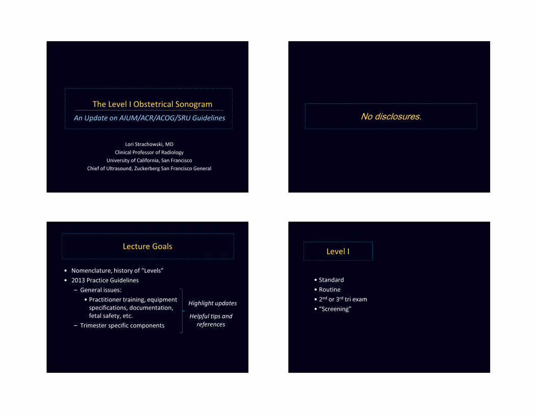
Lecture Goals
• Nomenclature, history of “Levels”• 2013 Practice Guidelines
– General issues: • Practitioner training, equipment
specifications, documentation, fetal safety, etc.
– Trimester specific components
Highlight updatesHelpful tips and
references
• Standard• Routine• 2nd or 3rd tri exam• “Screening”
Level I
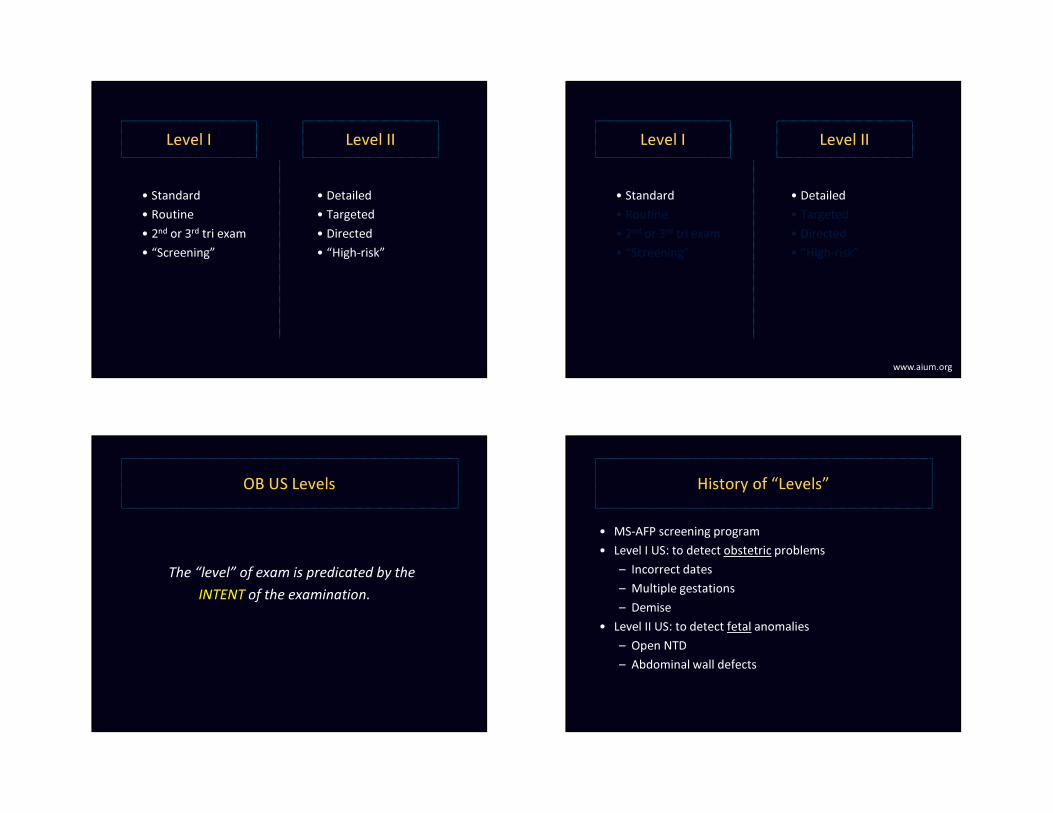
Level I
• Standard• Routine• 2nd or 3rd tri exam• “Screening”
• Detailed• Targeted• Directed• “High-risk”
Level II Level I
• Standard• Routine• 2nd or 3rd tri exam• “Screening”
• Detailed• Targeted• Directed• “High-risk”
Level II
www.aium.org
OB US Levels
The “level” of exam is predicated by the INTENT of the examination.
History of “Levels”
• MS-AFP screening program• Level I US: to detect obstetric problems
– Incorrect dates– Multiple gestations– Demise
• Level II US: to detect fetal anomalies– Open NTD– Abdominal wall defects
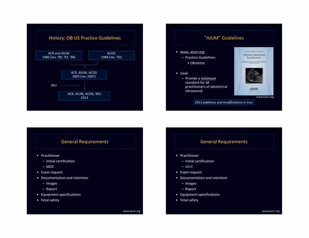
History: OB US Practice Guidelines
ACR and AIUM1986 (rev. ‘90, ‘93, ’96)
ACOG1988 (rev. ’93)
ACR, AIUM, ACOG2003 (rev. 2007)
ACR, AIUM, ACOG, SRU2013
SRU
“AIUM” Guidelines
• www. aium.org– Practice Guidelines
• Obstetric
• Goal: – Provide a minimum
standard for all practitioners of obstetrical ultrasound
2013 additions and modifications in bluewww.aium.org
General Requirements
• Practitioner– Initial certification– MOC
• Exam request• Documentation and retention
– Images– Report
• Equipment specifications• Fetal safety
www.aium.org
General Requirements
• Practitioner– Initial certification– MOC
• Exam request• Documentation and retention
– Images– Report
• Equipment specifications• Fetal safety
www.aium.org
General Requirements
• Practitioner– Initial certification– MOC - 170 exams/yr + 30 hrs Category 1 Credits/3 yrs
• Exam request• Documentation and retention
– Images– Report
• Equipment specifications• Fetal safety
www.aium.org
General Requirements
• Practitioner– Initial certification– MOC - 170 exams/yr + 30 hrs Category 1 Credits/3 yrs
• Exam request• Documentation and retention
– Images– Report
• Equipment specifications• Fetal safety
www.aium.org
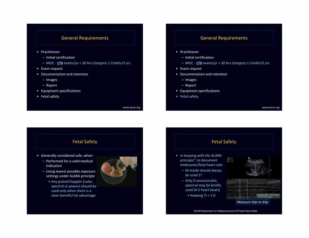
Fetal Safety
• Generally considered safe, when:– Performed for a valid medical
indication– Using lowest possible exposure
settings under ALARA principle• Any pulsed Doppler (color,
spectral or power) should be used only when there is a clear benefit/risk advantage
Fetal Safety
• In keeping with the ALARA principle”, to document embryonic/fetal heart rate:– M-mode should always
be used 1st
– Only if unsuccessful, spectral may be briefly used (4-5 heart beats)
• Keeping TI < 1.0
AIUM Statement on Measurement of Fetal Heart Rate
Measure blip to blip
Bioeffects of US
• Two major categories:– Mechanical:
• Energy imparted on gas particles to create movement → cavita on
– MI (mechanical index)– Thermal:
• Absorbed energy → heat– TI (thermal index)
Not an issuefor OB
BIG issuefor OB
Abramowicz, J, Lewin, P, et al, Glob. libr. women's med.,(1756-2228) 2011
Thermal Index
• Definition:• Ratio of power used to that required to produce a 1°C
increase• For obstetrics, subdivided as:
– TIS (soft tissues) < 10 wks– TIB (bone) ≥ 10 wks
• Value: should always be < 1.0– Doppler exposure time < 5-10 min (never > 60 min)
AIUM Statement on the Safe Use of Doppler Ultrasound During 11-14 week scans (or earlier in pregnancy)
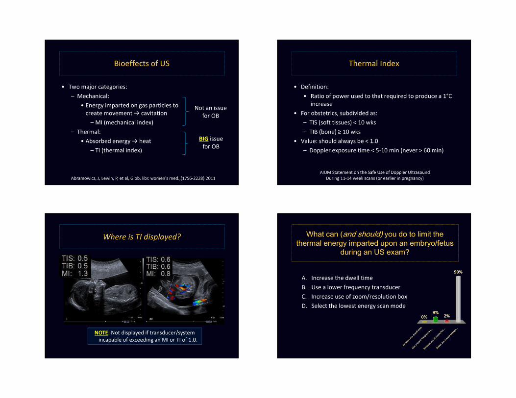
Where is TI displayed?
NOTE: Not displayed if transducer/system incapable of exceeding an MI or TI of 1.0.
What can (and should) you do to limit the thermal energy imparted upon an embryo/fetus
during an US exam?
A. Increase the dwell time B. Use a lower frequency transducerC. Increase use of zoom/resolution boxD. Select the lowest energy scan mode
I n c re a s
e t he d w
e l l ti m e
U s e a l o
w e r f r e
q u en c y
t . . .
I n c re a s
e u se o f
z o om / r
e s . ..
S e l ec t t h
e l ow e s
t e ne r g
y . . .
0%
90%
2%9%
What can (and should) you do to limit the thermal energy imparted upon an embryo/fetus
during an US exam?
A. Increase the dwell time B. Use a lower frequency transducerC. Increase use of zoom/resolution boxD. Select the lowest energy scan mode
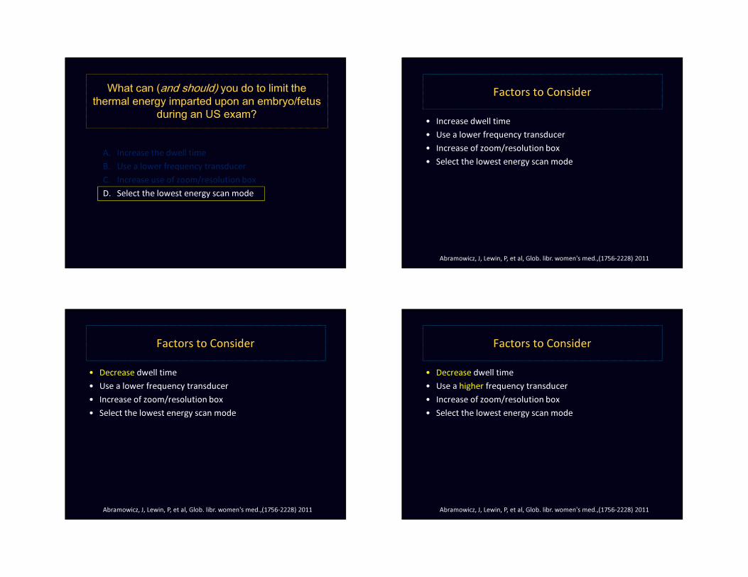
Factors to Consider• Increase dwell time • Use a lower frequency transducer• Increase of zoom/resolution box• Select the lowest energy scan mode
Abramowicz, J, Lewin, P, et al, Glob. libr. women's med.,(1756-2228) 2011
Factors to Consider• Decrease dwell time • Use a lower frequency transducer• Increase of zoom/resolution box• Select the lowest energy scan mode
Abramowicz, J, Lewin, P, et al, Glob. libr. women's med.,(1756-2228) 2011
Factors to Consider• Decrease dwell time • Use a higher frequency transducer• Increase of zoom/resolution box• Select the lowest energy scan mode
Abramowicz, J, Lewin, P, et al, Glob. libr. women's med.,(1756-2228) 2011
Factors to Consider• Decrease dwell time • Use a higher frequency transducer• Limit use of zoom/resolution box• Select the lowest energy scan mode
Abramowicz, J, Lewin, P, et al, Glob. libr. women's med.,(1756-2228) 2011
Factors to Consider• Decrease dwell time • Use a higher frequency transducer• Limit use of zoom/resolution box• Select the lowest energy scan mode
Abramowicz, J, Lewin, P, et al, Glob. libr. women's med.,(1756-2228) 2011
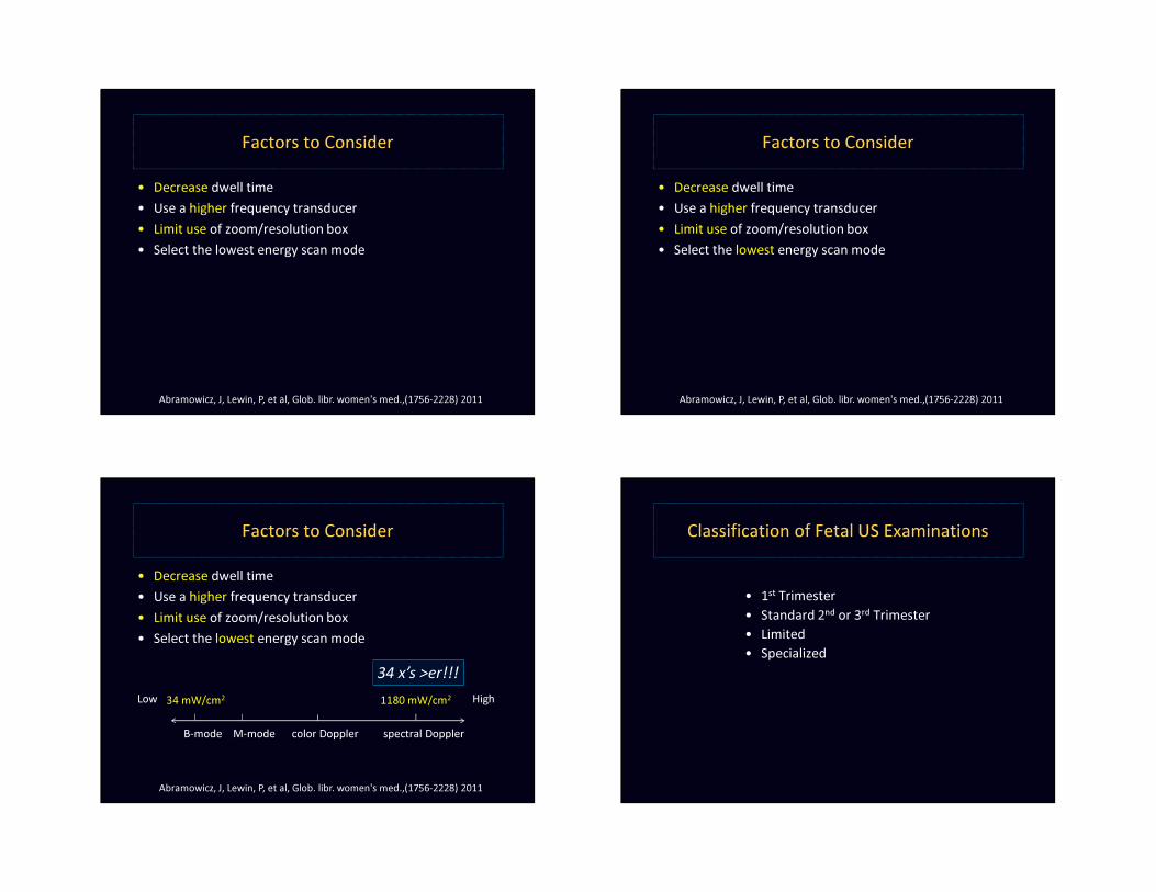
Factors to Consider• Decrease dwell time • Use a higher frequency transducer• Limit use of zoom/resolution box• Select the lowest energy scan mode
Low High
B-mode M-mode color Doppler spectral Doppler
1180 mW/cm234 mW/cm2
Abramowicz, J, Lewin, P, et al, Glob. libr. women's med.,(1756-2228) 2011
34 x’s >er!!!
Classification of Fetal US Examinations
• 1st Trimester • Standard 2nd or 3rd Trimester• Limited • Specialized
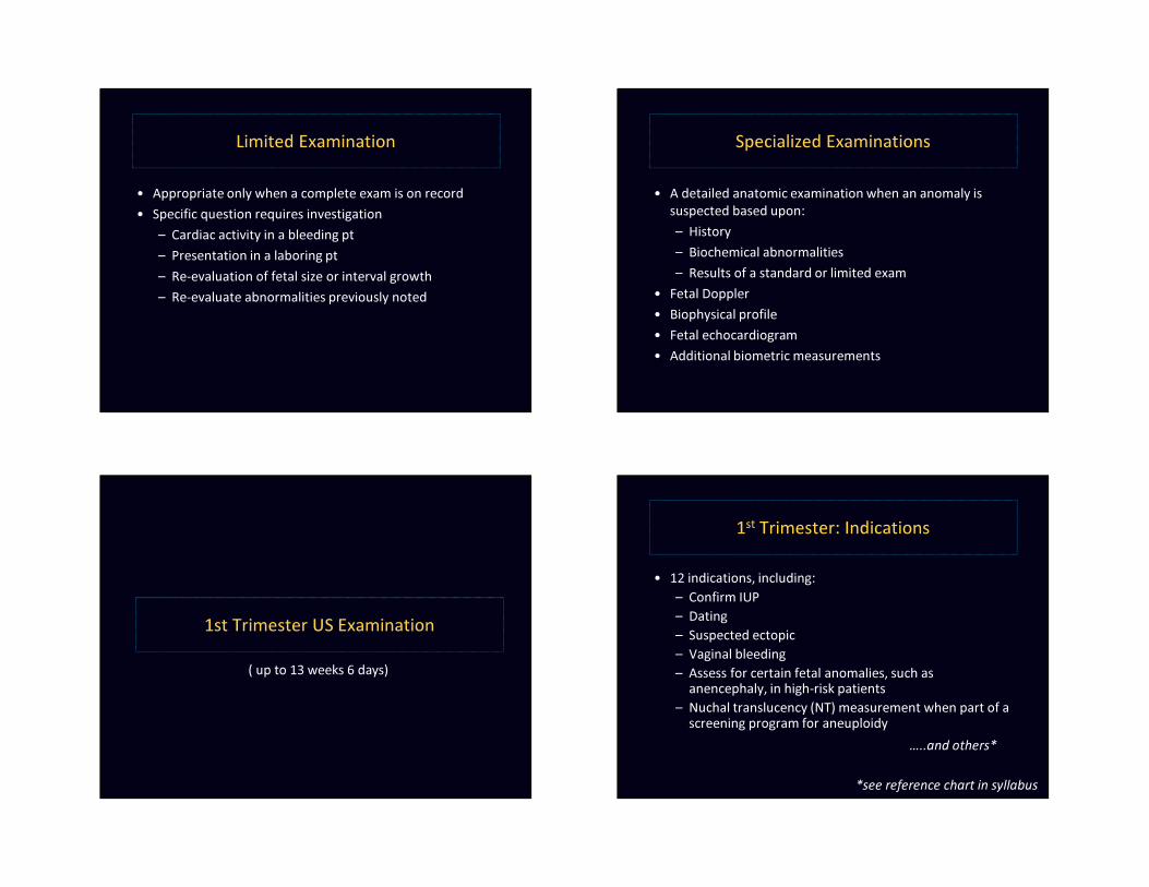
Limited Examination
• Appropriate only when a complete exam is on record• Specific question requires investigation
– Cardiac activity in a bleeding pt– Presentation in a laboring pt– Re-evaluation of fetal size or interval growth – Re-evaluate abnormalities previously noted
Specialized Examinations
• A detailed anatomic examination when an anomaly is suspected based upon:– History– Biochemical abnormalities– Results of a standard or limited exam
• Fetal Doppler• Biophysical profile• Fetal echocardiogram• Additional biometric measurements
1st Trimester US Examination( up to 13 weeks 6 days)
1st Trimester: Indications
• 12 indications, including:– Confirm IUP– Dating– Suspected ectopic– Vaginal bleeding– Assess for certain fetal anomalies, such as
anencephaly, in high-risk patients – Nuchal translucency (NT) measurement when part of a
screening program for aneuploidy …..and others*
*see reference chart in syllabus
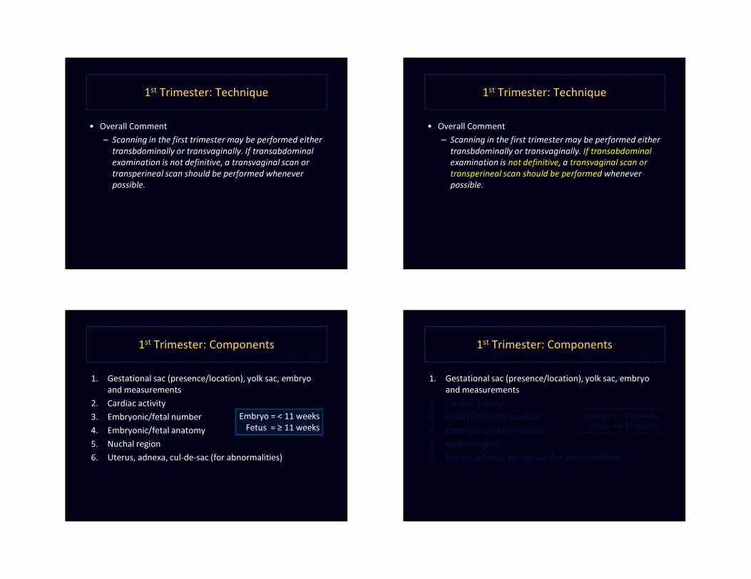
1st Trimester: Technique
• Overall Comment– Scanning in the first trimester may be performed either
transbdominally or transvaginally. If transabdominalexamination is not definitive, a transvaginal scan or transperineal scan should be performed whenever possible.
1st Trimester: Technique
• Overall Comment– Scanning in the first trimester may be performed either
transbdominally or transvaginally. If transabdominalexamination is not definitive, a transvaginal scan or transperineal scan should be performed whenever possible.
1st Trimester: Components
1. Gestational sac (presence/location), yolk sac, embryo and measurements
2. Cardiac activity3. Embryonic/fetal number 4. Embryonic/fetal anatomy5. Nuchal region6. Uterus, adnexa, cul-de-sac (for abnormalities)
Embryo = < 11 weeksFetus = ≥ 11 weeks
1st Trimester: Components
1. Gestational sac (presence/location), yolk sac, embryo and measurements
2. Cardiac activity3. Embryonic/fetal number 4. Embryonic/fetal anatomy5. Nuchal region6. Uterus, adnexa, cul-de-sac (for abnormalities)
Embryo = < 11 weeksFetus = ≥ 11 weeks
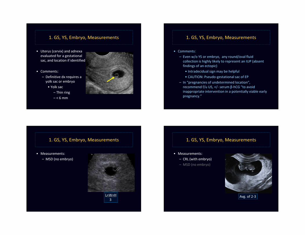
1. GS, YS, Embryo, Measurements
• Uterus (cervix) and adnexa evaluated for a gestational sac, and location if identified
• Comments:– Definitive dx requires a
yolk sac or embryo• Yolk sac
– Thin ring– < 6 mm
1. GS, YS, Embryo, Measurements
• Comments: – Even w/o YS or embryo, any round/oval fluid
collection is highly likely to represent an IUP (absent findings of an ectopic)
• Intradecidual sign may be helpful• CAUTION: Pseudo-gestational sac of EP
– In “pregnancies of undetermined location”, recommend f/u US, +/- serum β-hCG “to avoid inappropriate intervention in a potentially viable early pregnancy.”
1. GS, YS, Embryo, Measurements
• Measurements:– MSD (no embryo)
L+W+H3
1. GS, YS, Embryo, Measurements
• Measurements:– CRL (with embryo)– MSD (no embryo)
Avg. of 2-3
1. GS, YS, Embryo, Measurements
• Measurements:– Biometry (when possible)– CRL (with embryo)– MSD (no embryo)
Pregnancy “Dating”
• Clinical dates = how old the pregnancy is– AKA: Menstrual ↔ clinical ↔ LMP age– Based on LNMP (assumes a 28 day cycle)– Equal to conceptual (fetal) age + 14 days
• US dates = size = how big the pregnancy is – AKA: US age– Based on US measurements (MSD, CRL, biometry)– Standardized to equate with menstrual age
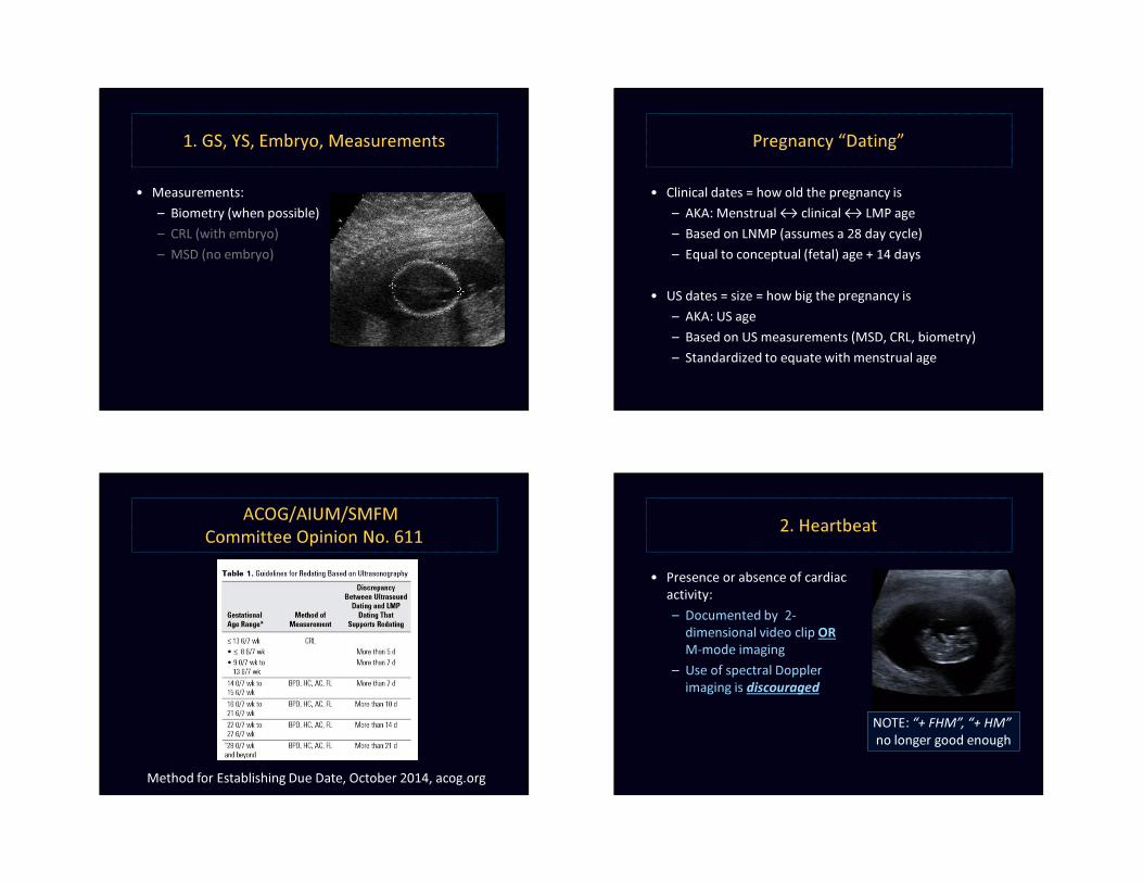
ACOG/AIUM/SMFM Committee Opinion No. 611
Method for Establishing Due Date, October 2014, acog.org
2. Heartbeat
• Presence or absence of cardiac activity:– Documented by 2-
dimensional video clip ORM-mode imaging
– Use of spectral Doppler imaging is discouraged
NOTE: “+ FHM”, “+ HM” no longer good enough
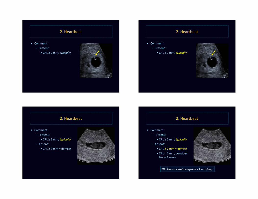
2. Heartbeat
• Comment:– Present:
• CRL ≥ 2 mm, typically
2. Heartbeat
• Comment:– Present:
• CRL ≥ 2 mm, typically
2. Heartbeat
• Comment:– Present:
• CRL ≥ 2 mm, typically– Absent:
• CRL ≥ 7 mm = demise
2. Heartbeat
• Comment:– Present:
• CRL ≥ 2 mm, typically– Absent:
• CRL ≥ 7 mm = demise• CRL < 7 mm, consider
f/u in 1 week
TIP: Normal embryo grows˜ 1 mm/day
2. Heartbeat
• Measured only if subjectively slow • M mode = safest
TIPS:- 100 –190: avg at 5-9 wks- < 85: poor outcome- < 70: 100% loss rate
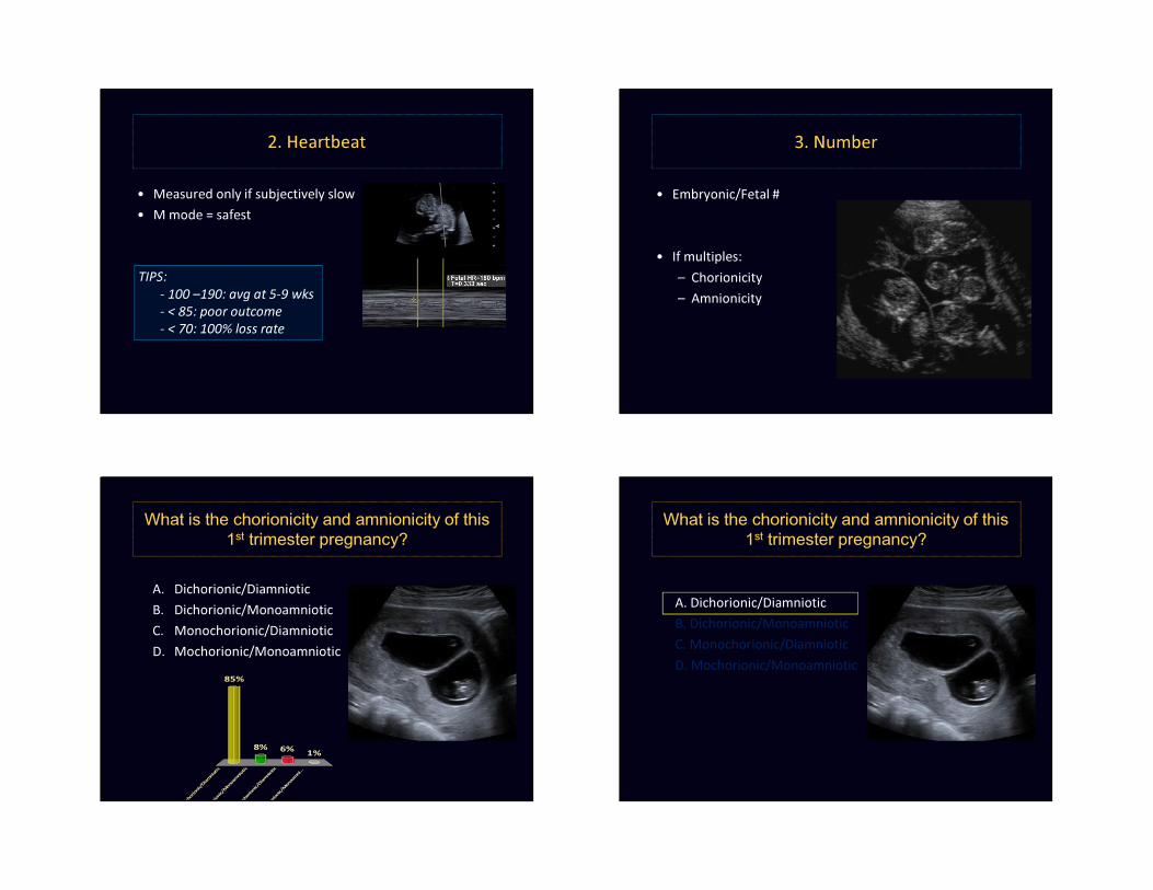
3. Number
• Embryonic/Fetal #
• If multiples:– Chorionicity– Amnionicity
What is the chorionicity and amnionicity of this 1st trimester pregnancy?
A. Dichorionic/DiamnioticB. Dichorionic/MonoamnioticC. Monochorionic/DiamnioticD. Mochorionic/Monoamniotic
D i ch o r
i o ni c / D
i a mn i o
t i c
D i ch o r
i o ni c / M
o n oa m
n i ot i c
M on o c
h o ri o n
i c / Di a m
n i ot i c
M oc h o
r i o ni c / M
o n oa m
n i . ..
85%
1%6%8%
What is the chorionicity and amnionicity of this 1st trimester pregnancy?
A. Dichorionic/DiamnioticB. Dichorionic/MonoamnioticC. Monochorionic/DiamnioticD. Mochorionic/Monoamniotic
Mo/Di
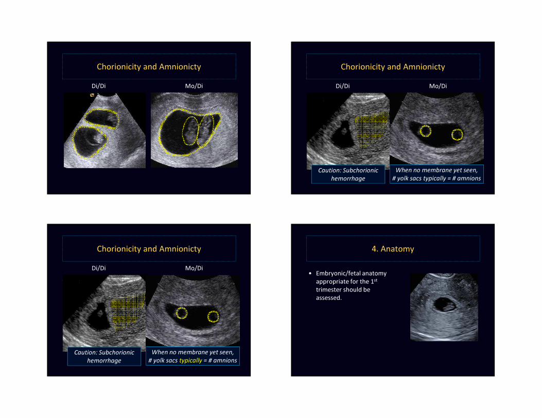
Chorionicity and AmnionictyDi/Di Mo/Di
Chorionicity and AmnionictyDi/Di
Caution: Subchorionichemorrhage
When no membrane yet seen,# yolk sacs typically = # amnions
Mo/Di
Chorionicity and AmnionictyDi/Di
Caution: Subchorionichemorrhage
When no membrane yet seen,# yolk sacs typically = # amnions
4. Anatomy
• Embryonic/fetal anatomy appropriate for the 1st
trimester should be assessed.
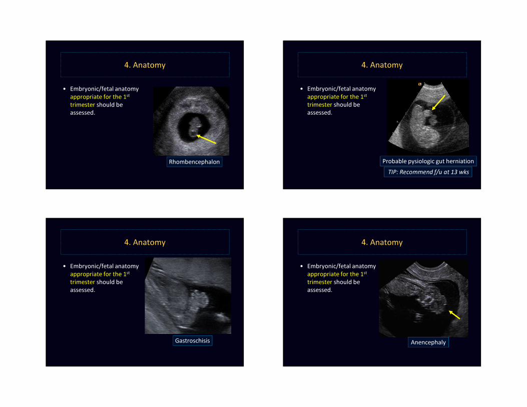
4. Anatomy
• Embryonic/fetal anatomy appropriate for the 1st
trimester should be assessed.
Rhombencephalon
4. Anatomy
• Embryonic/fetal anatomy appropriate for the 1st
trimester should be assessed.
Probable pysiologic gut herniationTIP: Recommend f/u at 13 wks
4. Anatomy
• Embryonic/fetal anatomy appropriate for the 1st
trimester should be assessed.
Gastroschisis
4. Anatomy
• Embryonic/fetal anatomy appropriate for the 1st
trimester should be assessed.
Anencephaly
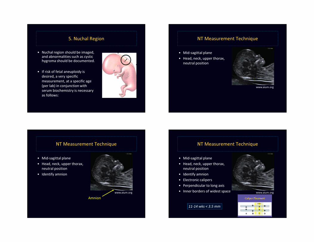
5. Nuchal Region
• Nuchal region should be imaged, and abnormalities such as cystic hygroma should be documented.
• If risk of fetal aneuploidy is desired, a very specific measurement, at a specific age (per lab) in conjunction with serum biochemistry is necessary as follows:
NT Measurement Technique
• Mid-sagittal plane• Head, neck, upper thorax,
neutral position
www.aium.org
NT Measurement Technique
• Mid-sagittal plane• Head, neck, upper thorax,
neutral position• Identify amnion
Amnionwww.aium.org
NT Measurement Technique
• Mid-sagittal plane• Head, neck, upper thorax,
neutral position• Identify amnion• Electronic calipers• Perpendicular to long axis• Inner borders of widest space
11-14 wks < 3.5 mm
www.aium.org
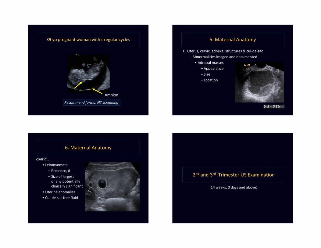
39 yo pregnant woman with irregular cycles
Recommend formal NT screeningAmnion
• Uterus, cervix, adnexal structures & cul-de-sac– Abnormalities imaged and documented
• Adnexal masses– Appearance– Size– Location
6. Maternal Anatomy
6. Maternal Anatomycont’d…
• Leiomyomata– Presence, #– Size of largest
or any potentially clinically significant
• Uterine anomalies• Cul-de-sac free fluid
2nd and 3rd Trimester US Examination(14 weeks, 0 days and above)
2nd & 3rd Trimester Indications
• 28 indications, including:– Estimation of gestational age– Evaluation of fetal growth– Vaginal bleeding– Pelvic pain– To assess for findings that may increase risk for
aneuploidy – Screening for fetal anomalies
…..and others*
*see reference chart in syllabus
2nd & 3rd Trimester: Components
1. Fetal cardiac activity, number, presentation2. Amniotic fluid volume 3. Placenta and cord 4. Gestational age assessment 5. Fetal weight estimation 6. Maternal anatomy 7. Fetal anatomy
2nd & 3rd Trimester: Components
1. Fetal cardiac activity, number, presentation2. Amniotic fluid volume 3. Placenta and cord 4. Gestational age assessment 5. Fetal weight estimation 6. Maternal anatomy 7. Fetal anatomy
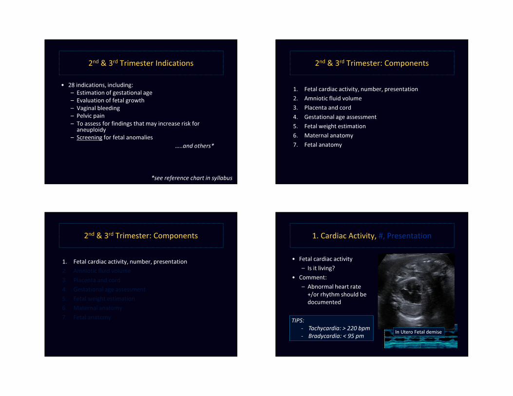
1. Cardiac Activity, #, Presentation
• Fetal cardiac activity– Is it living?
• Comment:– Abnormal heart rate
+/or rhythm should be documented
TIPS:- Tachycardia: > 220 bpm- Bradycardia: < 95 pm In Utero Fetal demise

1. Cardiac Activity, #, Presentation
• Fetal number– Multiple gestations:
• Chorionicity & amnionicity• Comparison of fetal size • Estimation of amniotic fluid volume on both sides
of the membrane• Fetal genitalia (when visualized)
Chorionicity and Amnionicity
In Order…..• Count placental masses
– Two: Dichorionic– One: Monochorionic or Dichorionic “fused”
• Evaluate genitalia– Opposite: Dichorionic– Same: Mono or Di
• Assess membrane• Thick, “twin-peak” sign: Di/Di• Thin, “T” sign”: Mono/Di• None: Mo/Di or Mo/Mo
Chorionicity and Amnionicity
In Order…..• Count placental masses
– Two: Dichorionic– One: Monochorionic or Dichorionic “fused”
• Evaluate genitalia– Opposite: Dichorionic– Same: Mono or Di
• Assess membrane• Thick, “twin-peak” sign: Di/Di• Thin, “T” sign”: Mono/Di• None: Mo/Di or Mo/Mo
Trickiest part!
Membrane Assessment
Thick – Di/Di Thin – Mo/Di
Membrane Assessment
• Membrane thickness– Subjective– Technical
considerations– Progressively thins
• Intersection w/ placenta may be more informative
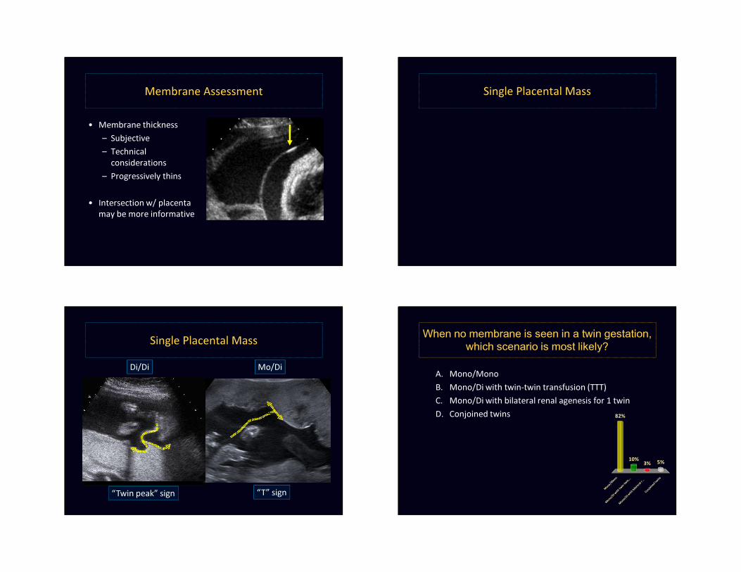
Single Placental Mass
Single Placental MassMo/DiDi/Di
“T” sign“Twin peak” sign
When no membrane is seen in a twin gestation, which scenario is most likely?
A. Mono/MonoB. Mono/Di with twin-twin transfusion (TTT) C. Mono/Di with bilateral renal agenesis for 1 twinD. Conjoined twins
M on o /
M on o
M on o /
D i wi t h
t w i n- t w
i n . . .
M on o /
D i wi t h
b i l at e r a
l r . ..
C o nj o i n
e d tw i n
s
82%
5%3%10%
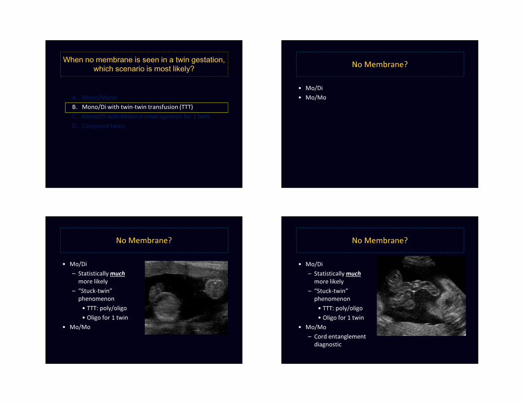
When no membrane is seen in a twin gestation, which scenario is most likely?
A. Mono/MonoB. Mono/Di with twin-twin transfusion (TTT) C. Mono/Di with bilateral renal agenesis for 1 twinD. Conjoined twins
No Membrane?
• Mo/Di• Mo/Mo
No Membrane?
• Mo/Di– Statistically much
more likely– “Stuck-twin”
phenomenon• TTT: poly/oligo • Oligo for 1 twin
• Mo/Mo
No Membrane?
• Mo/Di– Statistically much
more likely– “Stuck-twin”
phenomenon• TTT: poly/oligo• Oligo for 1 twin
• Mo/Mo– Cord entanglement
diagnostic
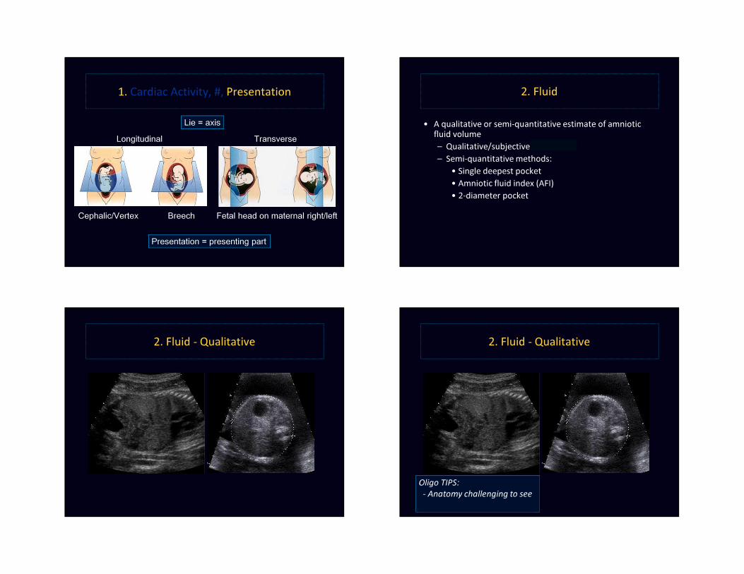
1. Cardiac Activity, #, Presentation
Lie = axisLongitudinal Transverse
Presentation = presenting part
Cephalic/Vertex Breech Fetal head on maternal right/left
2. Fluid
• A qualitative or semi-quantitative estimate of amniotic fluid volume– Qualitative/subjective – BEST!– Semi-quantitative methods:
• Single deepest pocket • Amniotic fluid index (AFI) • 2-diameter pocket
2. Fluid - Qualitative 2. Fluid - Qualitative
Oligo TIPS: - Anatomy challenging to see- Decreased fetal movement
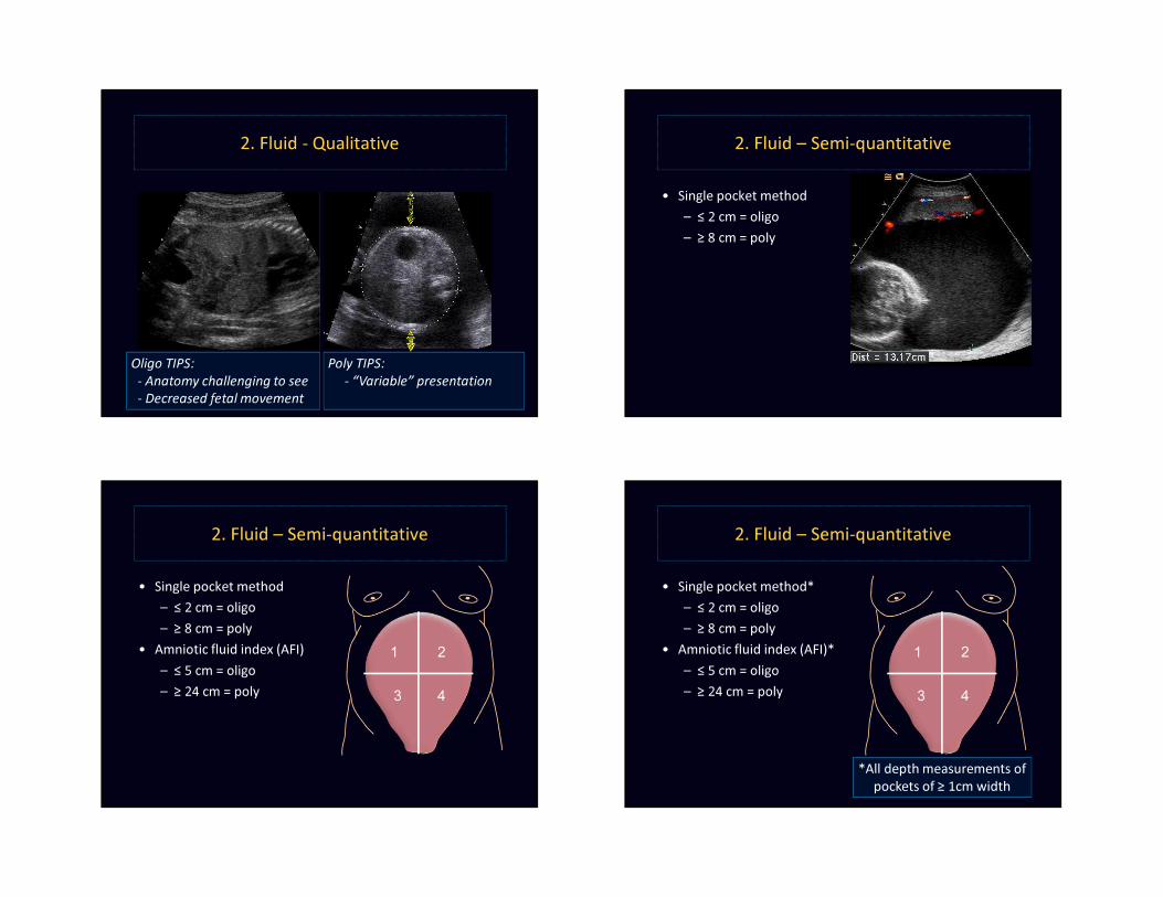
2. Fluid - Qualitative
Poly TIPS: - “Variable” presentation- AC doesn’t fill uterine cavity
Oligo TIPS: - Anatomy challenging to see- Decreased fetal movement
2. Fluid – Semi-quantitative
• Single pocket method– ≤ 2 cm = oligo– ≥ 8 cm = poly
1
43
2
2. Fluid – Semi-quantitative
• Single pocket method– ≤ 2 cm = oligo– ≥ 8 cm = poly
• Amniotic fluid index (AFI)– ≤ 5 cm = oligo– ≥ 24 cm = poly
1
43
2
2. Fluid – Semi-quantitative
• Single pocket method*– ≤ 2 cm = oligo– ≥ 8 cm = poly
• Amniotic fluid index (AFI)*– ≤ 5 cm = oligo– ≥ 24 cm = poly
1
43
2
*All depth measurements ofpockets of ≥ 1cm width
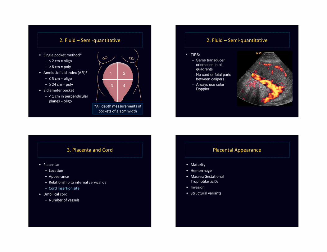
2. Fluid – Semi-quantitative
• Single pocket method*– ≤ 2 cm = oligo– ≥ 8 cm = poly
• Amniotic fluid index (AFI)*– ≤ 5 cm = oligo– ≥ 24 cm = poly
• 2 diameter pocket– < 1 cm in perpendicular
planes = oligo
1
43
2
*All depth measurements ofpockets of ≥ 1cm width
2. Fluid – Semi-quantitative
• TIPS:– Same transducer
orientation in all quadrants
– No cord or fetal parts between calipers
– Always use color Doppler
3. Placenta and Cord
• Placenta:– Location– Appearance– Relationship to internal cervical os– Cord Insertion site
• Umbilical cord: – Number of vessels
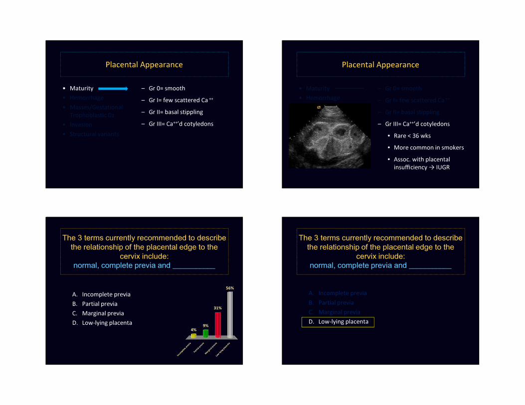
Placental Appearance
• Maturity• Hemorrhage• Masses/Gestational
Trophoblastic Dz• Invasion• Structural variants
Placental Appearance
• Maturity• Hemorrhage• Masses/Gestational
Trophoblastic Dz• Invasion• Structural variants
– Gr 0= smooth– Gr I= few scattered Ca ++
– Gr II= basal stippling – Gr III= Ca++’d cotyledons
Placental Appearance
• Maturity• Hemorrhage• Masses/Gestational
Trophoblastic Dz• Invasion• Structural variants
– Gr 0= smooth– Gr I= few scattered Ca ++
– Gr II= basal stippling – Gr III= Ca++’d cotyledons
• Rare < 36 wks• More common in smokers• Assoc. with placental
insufficiency → IUGR
The 3 terms currently recommended to describe the relationship of the placental edge to the
cervix include: normal, complete previa and ___________
A. Incomplete previaB. Partial previaC. Marginal previaD. Low-lying placenta
I n c om p
l e t e p r e
v i aP a r
t i a l p r e
v i aM a
r g i na l p
r e vi a
L o w- l y i
n g pl a c e
n t a
4%
56%
31%
9%
The 3 terms currently recommended to describe the relationship of the placental edge to the
cervix include: normal, complete previa and ___________
A. Incomplete previaB. Partial previaC. Marginal previaD. Low-lying placenta
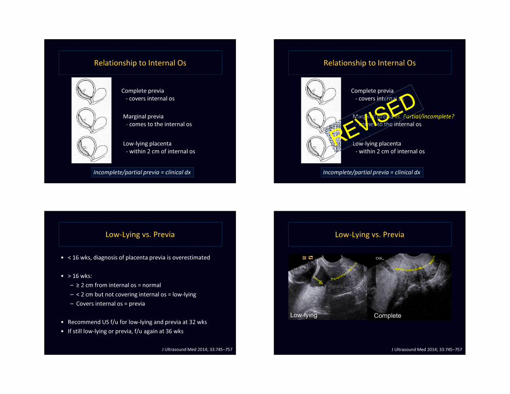
Relationship to Internal Os
Complete previa- covers internal os
Low-lying placenta- within 2 cm of internal os
Marginal previa- comes to the internal os
Incomplete/partial previa = clinical dx
Relationship to Internal Os
Complete previa- covers internal os
Low-lying placenta- within 2 cm of internal os
Marginal previa vs. Partial/incomplete?- comes to the internal os
Incomplete/partial previa = clinical dx
Low-Lying vs. Previa
• < 16 wks, diagnosis of placenta previa is overestimated
• > 16 wks:– ≥ 2 cm from internal os = normal– < 2 cm but not covering internal os = low-lying– Covers internal os = previa
• Recommend US f/u for low-lying and previa at 32 wks• If still low-lying or previa, f/u again at 36 wks
J Ultrasound Med 2014; 33:745–757
Low-Lying vs. Previa
J Ultrasound Med 2014; 33:745–757
CompleteLow-lying
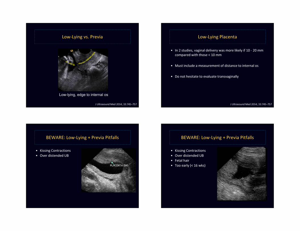
Low-Lying vs. Previa
J Ultrasound Med 2014; 33:745–757
Low-lying, edge to internal os
Low-Lying Placenta
• In 2 studies, vaginal delivery was more likely if 10 - 20 mm compared with those < 10 mm
• Must include a measurement of distance to internal os
• Do not hesitate to evaluate transvaginally
J Ultrasound Med 2014; 33:745–757
BEWARE: Low-Lying + Previa Pitfalls
• Kissing Contractions• Over distended UB
BEWARE: Low-Lying + Previa Pitfalls
• Kissing Contractions• Over distended UB• Fetal hair• Too early (< 16 wks)
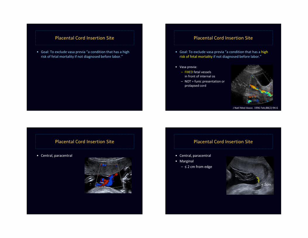
Placental Cord Insertion Site
• Goal: To exclude vasa previa “a condition that has a high risk of fetal mortality if not diagnosed before labor.”
Placental Cord Insertion Site
• Goal: To exclude vasa previa “a condition that has a high risk of fetal mortality if not diagnosed before labor.”
• Vasa previa:– FIXED fetal vessels
in front of internal os– NOT = funic presentation or
prolapsed cord
J Natl Med Assoc. 1996 Feb;88(2):94-6
Placental Cord Insertion Site
• Central, paracentral
Placental Cord Insertion Site
• Central, paracentral• Marginal
– ≤ 2 cm from edge
< 2cm
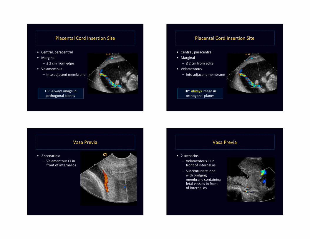
Placental Cord Insertion Site
• Central, paracentral• Marginal
– ≤ 2 cm from edge• Velamentous
– Into adjacent membrane< 2cm
TIP: Always image in orthogonal planes
Placental Cord Insertion Site
• Central, paracentral• Marginal
– ≤ 2 cm from edge• Velamentous
– Into adjacent membrane< 2cm
TIP: Always image in orthogonal planes
Vasa Previa
• 2 scenarios:– Velamentous CI in
front of internal os
Vasa Previa
• 2 scenarios:– Velamentous CI in
front of internal os– Succenturiate lobe
with bridging membrane containing fetal vessels in front of internal os
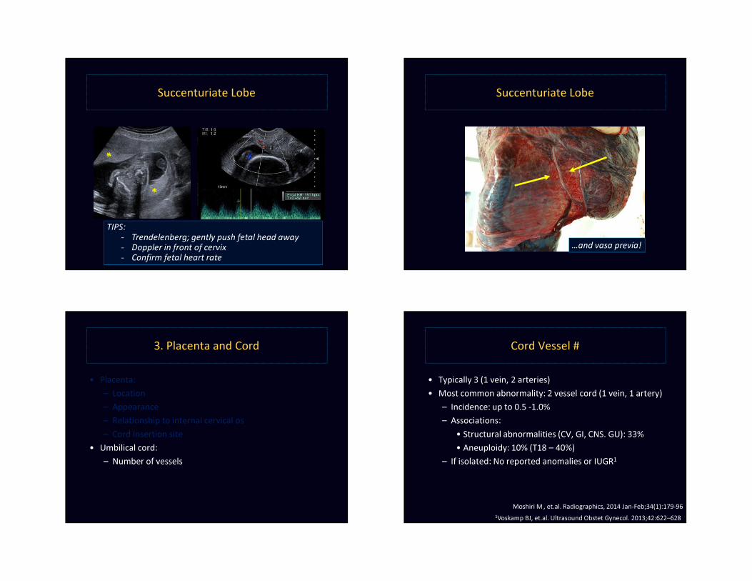
Succenturiate Lobe
*
*
TIPS:- Trendelenberg; gently push fetal head away - Doppler in front of cervix- Confirm fetal heart rate
Succenturiate Lobe
…and vasa previa!
3. Placenta and Cord
• Placenta:– Location– Appearance– Relationship to internal cervical os– Cord Insertion site
• Umbilical cord: – Number of vessels
Cord Vessel #
• Typically 3 (1 vein, 2 arteries)• Most common abnormality: 2 vessel cord (1 vein, 1 artery)
– Incidence: up to 0.5 -1.0%– Associations:
• Structural abnormalities (CV, GI, CNS. GU): 33%• Aneuploidy: 10% (T18 – 40%)
– If isolated: No reported anomalies or IUGR1
1Voskamp BJ, et.al. Ultrasound Obstet Gynecol. 2013;42:622–628Moshiri M , et.al. Radiographics, 2014 Jan-Feb;34(1):179-96
Cord Vessel #
CAUTION: UB view may miss up to 14% of 2 VC’s1
1Bornemeier S, et.al. JDMS. November 1996 vol. 12 no. 6 260-265
3VC 2VC
Cord Vessel #
Mickey Mouse view
3VC 2VC
TIP: May consider an axial view.
4. Assessment of Mean Gestational Age
• Biometry evaluated:– Biparietal diameter– Head circumference– Femural diaphysis length– Abdominal circumference
• “Average Abdominal Diameter”
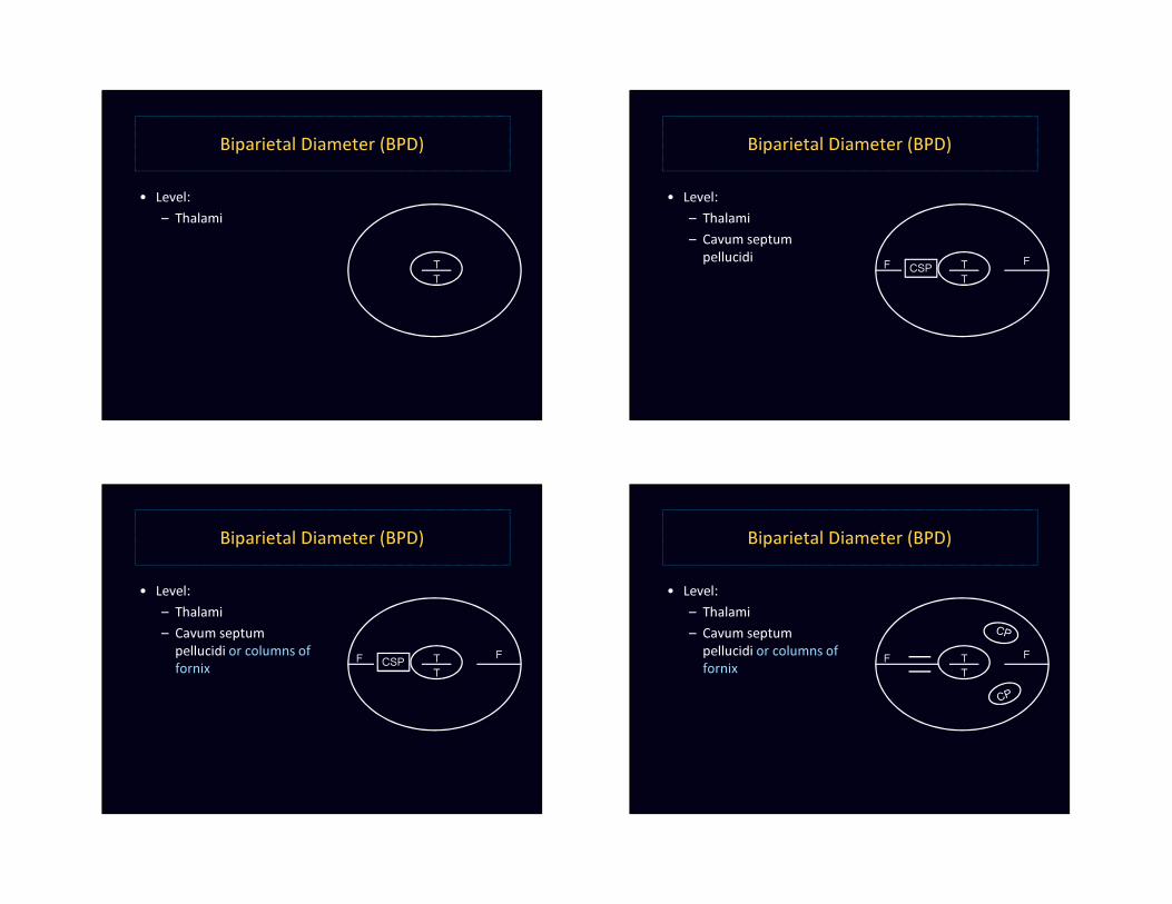
Biparietal Diameter (BPD)
• Level:– Thalami
TT
Biparietal Diameter (BPD)
• Level:– Thalami– Cavum septum
pellucidi TT
CSP FF
Biparietal Diameter (BPD)
• Level:– Thalami– Cavum septum
pellucidi or columns of fornix T
TCSP FF
Biparietal Diameter (BPD)
• Level:– Thalami– Cavum septum
pellucidi or columns of fornix T
TFF
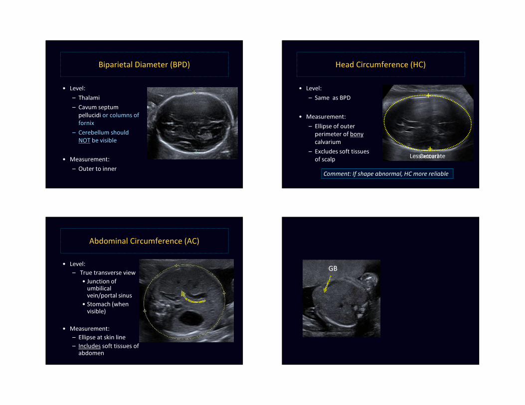
Biparietal Diameter (BPD)
• Level:– Thalami– Cavum septum
pellucidi or columns of fornix
– Cerebellum should NOT be visible
• Measurement: – Outer to inner
TT
FF
+
+
Head Circumference (HC)
• Level:– Same as BPD
• Measurement:– Ellipse of outer
perimeter of bony calvarium
– Excludes soft tissues of scalp
TT
CSP FF
Comment: If shape abnormal, HC more reliable
+
+Less accurateBetter!
Abdominal Circumference (AC)
• Level:– True transverse view
• Junction of umbilical vein/portal sinus
• Stomach (when visible)
• Measurement: – Ellipse at skin line– Includes soft tissues of
abdomen
St
SpPV
UV
StomachGB
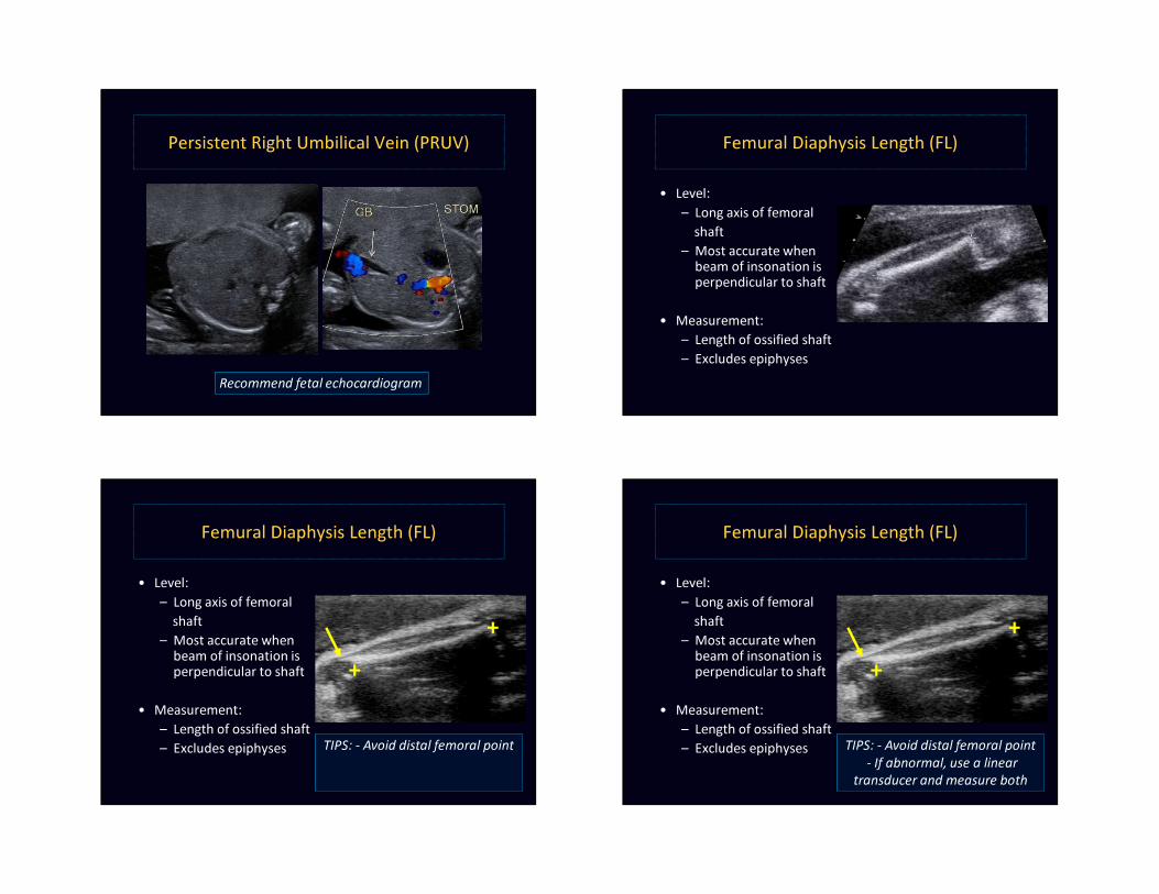
Persistent Right Umbilical Vein (PRUV)
Recommend fetal echocardiogram
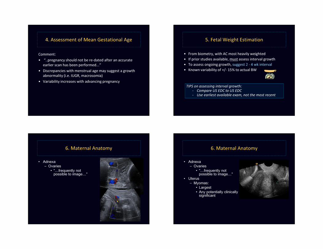
Femural Diaphysis Length (FL)
• Level: – Long axis of femoral
shaft– Most accurate when
beam of insonation is perpendicular to shaft
• Measurement: – Length of ossified shaft– Excludes epiphyses
++
Femural Diaphysis Length (FL)
• Level: – Long axis of femoral
shaft– Most accurate when
beam of insonation is perpendicular to shaft
• Measurement: – Length of ossified shaft– Excludes epiphyses
+
TIPS: - Avoid distal femoral point
++
+
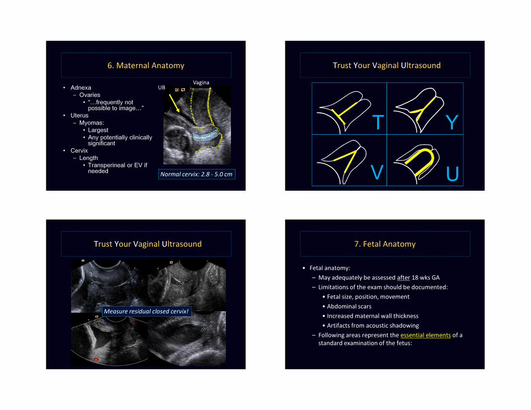
Femural Diaphysis Length (FL)
• Level: – Long axis of femoral
shaft– Most accurate when
beam of insonation is perpendicular to shaft
• Measurement: – Length of ossified shaft– Excludes epiphyses
+
TIPS: - Avoid distal femoral point- If abnormal, use a linear
transducer and measure both
++
+
4. Assessment of Mean Gestational Age
Comment:• “..pregnancy should not be re-dated after an accurate
earlier scan has been performed...”• Discrepancies with menstrual age may suggest a growth
abnormality (i.e. IUGR, macrosomia)• Variability increases with advancing pregnancy
5. Fetal Weight Estimation
• From biometry, with AC most heavily weighted• If prior studies available, must assess interval growth • To assess ongoing growth, suggest 2 - 4 wk interval• Known variability of +/- 15% to actual BW
TIPS on assessing interval growth:- Compare US EDC to US EDC- Use earliest available exam, not the most recent
6. Maternal Anatomy
• Adnexa– Ovaries
• “…frequently not possible to image…”
6. Maternal Anatomy
• Adnexa– Ovaries
• “…frequently not possible to image…”
• Uterus– Myomas:
• Largest• Any potentially clinically
significant
• Adnexa– Ovaries
• “…frequently not possible to image…”
• Uterus– Myomas:
• Largest• Any potentially clinically
significant • Cervix
– Length• Transperineal or EV if
needed Normal cervix: 2.8 - 5.0 cm
UB Vagina
6. Maternal Anatomy Trust Your Vaginal Ultrasound
U
YT
V
Trust Your Vaginal Ultrasound
U
YT
V
Measure residual closed cervix!
7. Fetal Anatomy
• Fetal anatomy:– May adequately be assessed after 18 wks GA – Limitations of the exam should be documented:
• Fetal size, position, movement• Abdominal scars• Increased maternal wall thickness• Artifacts from acoustic shadowing
– Following areas represent the essential elements of a standard examination of the fetus:
Fetal Anatomy
• Head, face and neck– Cerebellum– Cisterna magna– Choroid plexus – Lateral ventricles – Midline falx– Cavum septum pellucidi– Upper lip
• Chest– 4 chamber heart– Outflow tracts
• Abdomen– Stomach– Kidneys – Urinary bladder– Cord insertion site– Umbilical cord vessel #
• Spine• Extremities• Sex
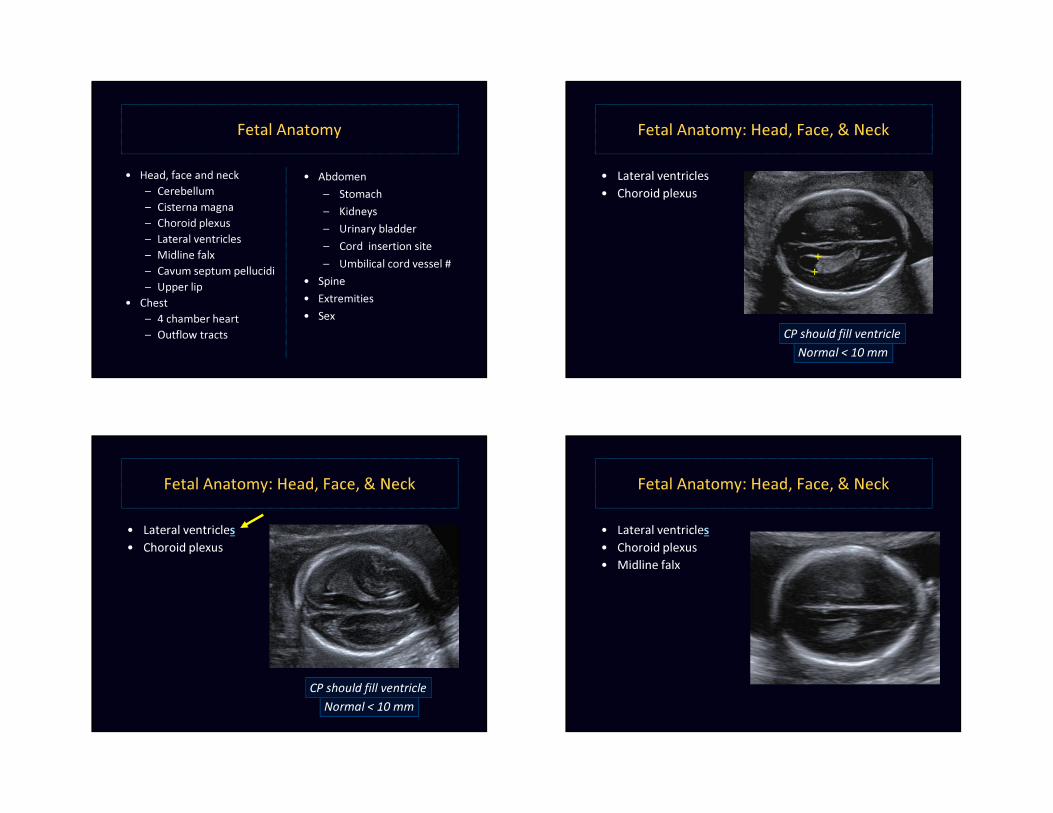
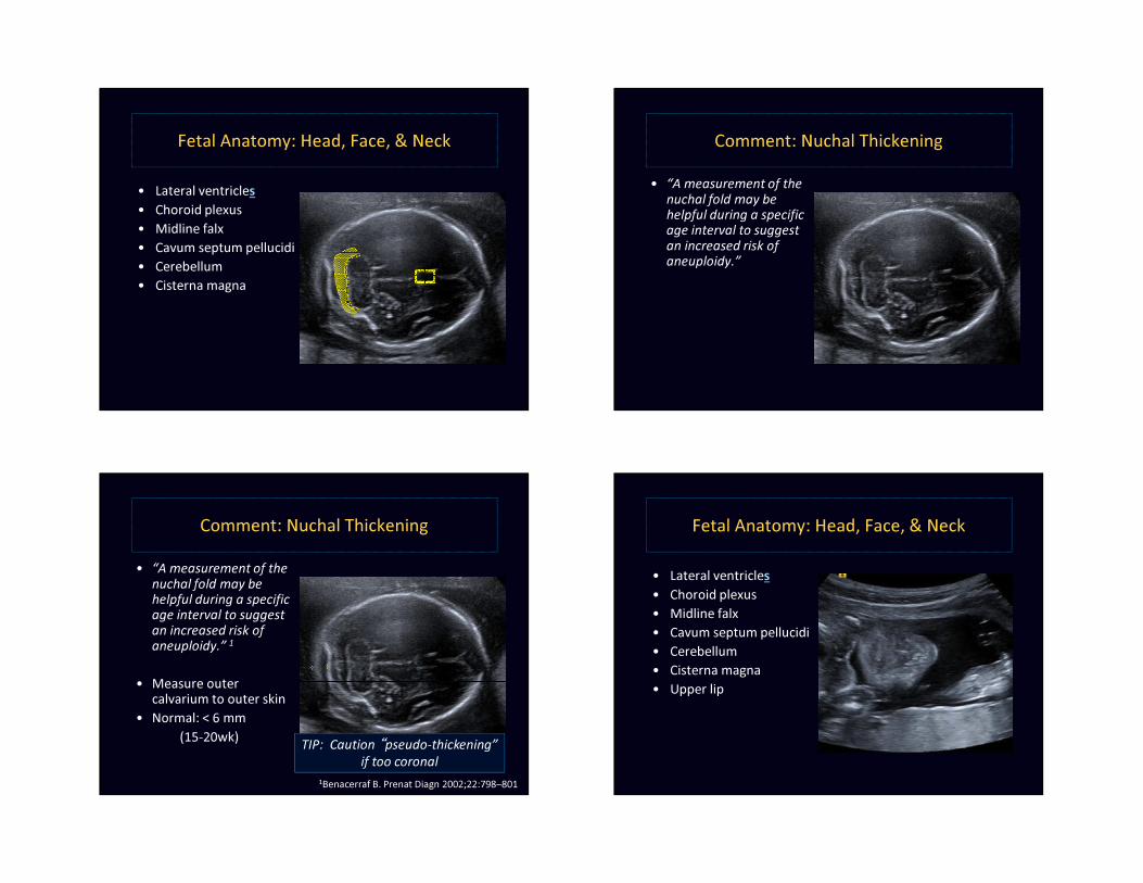
Fetal Anatomy: Head, Face, & Neck
++
Normal < 10 mmCP should fill ventricle
• Lateral ventricles• Choroid plexus
++
Fetal Anatomy: Head, Face, & Neck
Normal < 10 mmCP should fill ventricle
• Lateral ventricles• Choroid plexus
Fetal Anatomy: Head, Face, & Neck
• Lateral ventricles• Choroid plexus • Midline falx
Fetal Anatomy: Head, Face, & Neck
• Lateral ventricles• Choroid plexus • Midline falx• Cavum septum pellucidi• Cerebellum• Cisterna magna
Comment: Nuchal Thickening • “A measurement of the
nuchal fold may be helpful during a specific age interval to suggest an increased risk of aneuploidy.”
Comment: Nuchal Thickening • “A measurement of the
nuchal fold may be helpful during a specific age interval to suggest an increased risk of aneuploidy.” 1
• Measure outer calvarium to outer skin
• Normal: < 6 mm(15-20wk)
1Benacerraf B. Prenat Diagn 2002;22:798–801
TIP: Caution “pseudo-thickening” if too coronal
Fetal Anatomy: Head, Face, & Neck
• Lateral ventricles• Choroid plexus • Midline falx• Cavum septum pellucidi• Cerebellum• Cisterna magna• Upper lip
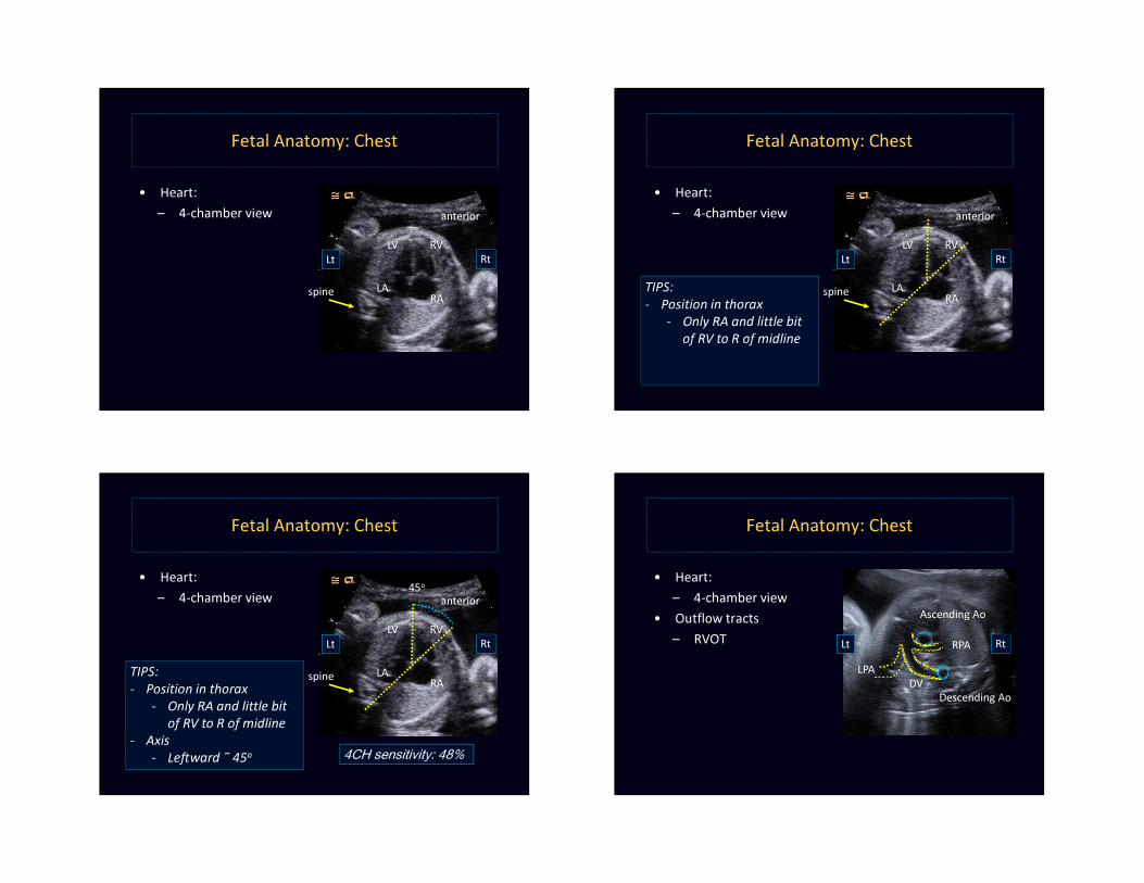
Fetal Anatomy: Chest
• Heart:– 4-chamber view
RA
RVLV
LAspine
anterior
Lt Rt
Fetal Anatomy: Chest
• Heart:– 4-chamber view
RA
RVLV
LAspine
anterior
Lt Rt
TIPS:- Position in thorax
- Only RA and little bit of RV to R of midline
Fetal Anatomy: Chest
• Heart:– 4-chamber view
RA
RVLV
LAspine
anterior
Lt Rt
TIPS:- Position in thorax
- Only RA and little bit of RV to R of midline
- Axis- Le�ward ˜ 45o
45o
4CH sensitivity: 48%
• Heart:– 4-chamber view
• Outflow tracts– RVOT
Fetal Anatomy: Chest
Ascending Ao
Descending Ao
RPALPA
DV
Lt Rt
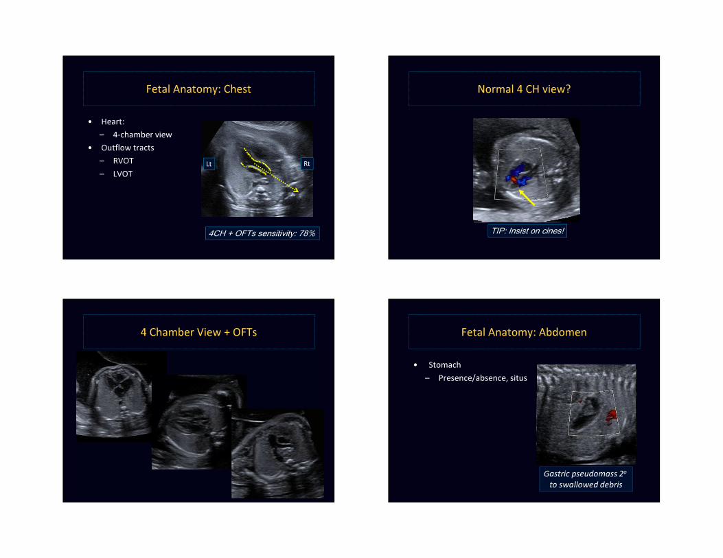
• Heart:– 4-chamber view
• Outflow tracts– RVOT– LVOT
Fetal Anatomy: Chest
Ascending Ao
Descending Ao
Lt Rt
4CH + OFTs sensitivity: 78%
Normal 4 CH view?
TIP: Insist on cines!
4 Chamber View + OFTs Fetal Anatomy: Abdomen
Lt
Gastric pseudomass 2o
to swallowed debris
• Stomach– Presence/absence, situs
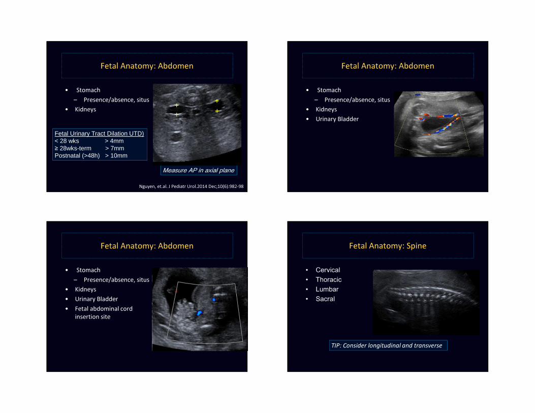
Fetal Anatomy: Abdomen
Measure AP in axial plane
++
++
• Stomach– Presence/absence, situs
• Kidneys
Fetal Urinary Tract Dilation UTD)< 28 wks > 4mm≥ 28wks-term > 7mmPostnatal (>48h) > 10mm
Nguyen, et.al. J Pediatr Urol.2014 Dec;10(6):982-98
• Stomach– Presence/absence, situs
• Kidneys • Urinary Bladder
Fetal Anatomy: Abdomen
Fetal Anatomy: Abdomen
++
++
• Stomach– Presence/absence, situs
• Kidneys • Urinary Bladder• Fetal abdominal cord
insertion site
Fetal Anatomy: Spine
TIP: Consider longitudinal and transverse
• Cervical• Thoracic• Lumbar• Sacral
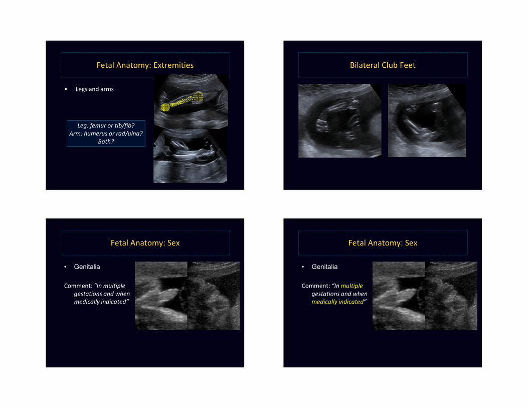
• Legs and arms
Fetal Anatomy: Extremities
Leg: femur or tib/fib?Arm: humerus or rad/ulna?
Both?
Bilateral Club Feet
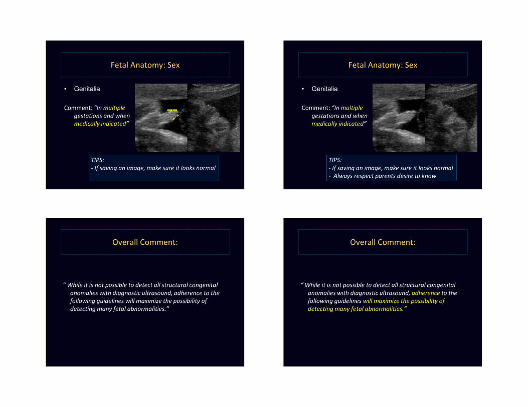
Fetal Anatomy: Sex
• Genitalia
Comment: “In multiple gestations and when medically indicated”
Fetal Anatomy: Sex
• Genitalia
Comment: “In multiplegestations and when medically indicated”
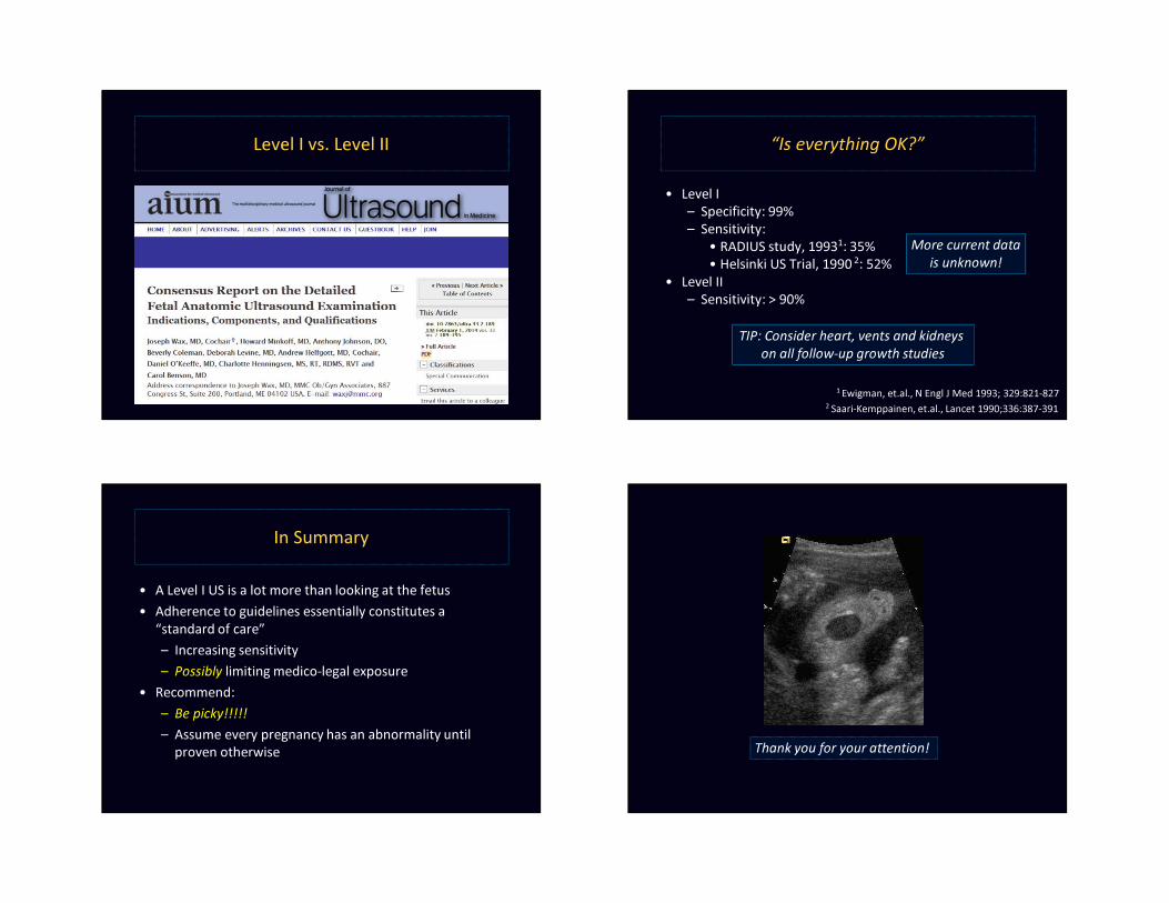
• Genitalia
Comment: “In multiplegestations and when medically indicated”
TIPS: - If saving an image, make sure it looks normal
Fetal Anatomy: Sex
• Genitalia
Comment: “In multiplegestations and when medically indicated”
TIPS: - If saving an image, make sure it looks normal- Always respect parents desire to know
Fetal Anatomy: Sex
Overall Comment:
“ While it is not possible to detect all structural congenital anomalies with diagnostic ultrasound, adherence to the following guidelines will maximize the possibility of detecting many fetal abnormalities.”
Overall Comment:
“ While it is not possible to detect all structural congenital anomalies with diagnostic ultrasound, adherence to the following guidelines will maximize the possibility of detecting many fetal abnormalities.”

Hard to miss.
Overall Comment:
“…a more detailed anatomic examination of the fetus may be necessary in some cases, such as when an abnormality is found or suspected on the standard examination or in pregnancies at high risk for fetal anomalies.”
“Level 2” or “Detailed” sonogram
CPT Codes: Level I vs. Level II
• 76805– Ultrasound, pregnant uterus, real time with image
documentation, fetal and maternal evaluation, after first trimester (> or = 14 weeks 0 days), transabdominal approach; single or first gestation.
• 76811– Ultrasound, pregnant uterus, real time with image
documentation, fetal and maternal evaluation plus detailed fetal anatomic examination, transabdominal approach; single or first gestation.
Level I vs. Level II “Is everything OK?”
• Level I– Specificity: 99%– Sensitivity:
• RADIUS study, 19931: 35%• Helsinki US Trial, 1990 2: 52%
• Level II– Sensitivity: > 90%
1 Ewigman, et.al., N Engl J Med 1993; 329:821-8272 Saari-Kemppainen, et.al., Lancet 1990;336:387-391
More current datais unknown!
TIP: Consider heart, vents and kidneys on all follow-up growth studies
In Summary
• A Level I US is a lot more than looking at the fetus• Adherence to guidelines essentially constitutes a
“standard of care”– Increasing sensitivity– Possibly limiting medico-legal exposure
• Recommend:– Be picky!!!!!– Assume every pregnancy has an abnormality until
proven otherwise Thank you for your attention!


![Bassin obstetrical [enregistrement automatique]](https://img.pdfslide.net/doc/110x75/58ed40f31a28ab9f298b45d3/bassin-obstetrical-enregistrement-automatique.jpg)