Embed Size (px)
Citation preview
10/22/13
1
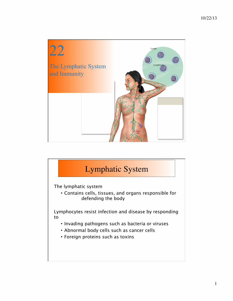
22 The Lymphatic System and Immunity
Lymph
Lymphocyte
Lymphatic System
The lymphatic system • Contains cells, tissues, and organs responsible for defending the body
Lymphocytes resist infection and disease by responding to • Invading pathogens such as bacteria or viruses • Abnormal body cells such as cancer cells • Foreign proteins such as toxins

10/22/13
2
• Our body is constantly being attacked by possible Pathogens = Microscopic organisms that cause disease
• Includes Viruses – Bacteria – Fungi – Parasites
• Each attacks in a specific way
• Lymphatic system protects us against disease causing organisms, including our own malfunctioning cells.
Lymphatic System
• The Lymphatic System
• Protects us against disease
• Lymphatic system cells respond to:
• Environmental pathogens – Toxins - Abnormal body cells, such as cancers
• Defense mechanisms include:
• Specific Defenses : cell mediated immunity (they identify, attack and develop immunity with ‘memory’)
• Non Specific : block or attack any potential infectious organism but cannot distinguish one attack from another.
Lymphatic System

10/22/13
3
Lymphatic System Organization
Lymphatic System
• Lymph • Lymphatic vessels • Lymphoid tissues and organs • Lymphocytes and supporting phagocytic cells
Lymphatic System consist out of

10/22/13
4
Lymphatic System
Functions of Lymphatic System
• Drainage system of interstitial fluid
• Re-directs extra fluid from in between cells back to the venous blood system
• Primary function is production, maintenance, and distribution of lymphocytes
• Lymphocytes must: • Detect where problems exist • Be able to reach the site of injury or infection
Lymphatic Vessels
• Lymphatic capillaries • Small lymphatic vessels (veins) • Major lymph-collecting vessels
The lymphatic system has neither a heart nor arteries. Its microscopic dead-end capillaries extend into most tissues, paralleling the blood capillaries.

10/22/13
5
Lymphatic Vessels
The lymphatic circulation is a one-directional drainage system. Its job in maintaining fluid balance is to:
• collect excess interstitial fluid and return it to the blood (approximately 3 liters daily). • return plasma proteins to the blood.
Once interstitial fluid enters a lymph capillary, it is referred to as lymph.
Lymphatic Capillaries

10/22/13
6
Lymphatic Capillaries The wall of a lymph capillary is constructed of endothelial cells that overlap one another.
When fluid outside the capillary pushes against the overlapping cells, they swing slightly inward. Fluid inside the capillary cannot flow out through these openings.
The arrows represent the direction of flow of the lymph. Note the internal valve which allows the lymph to flow in one direction only.
Lymphatic Capillaries
In the villi of the small intestines, the lymphatic capillaries form special structures called LACTEALS.
Fatty material (lipids etc..) absorbed in the small intestines will first enter the lacteals after which it is directed towards the blood stream.

10/22/13
7
Lymphatic Veins
Lymph capillaries join to form larger vessels called lymphatics or lymph veins.
These resemble blood-conducting veins but have thinner walls and relatively larger lumen, and they have more valves.
In the skin, lymphatics are located in subcutaneous tissue and follow same paths as veins.
Deeper in the body lymphatics generally follow arteries and form plexuses (networks) around them.
Lymphatic Veins

10/22/13
8
Lymphatic Nodes
At certain locations lymphatic veins enter lymph nodes. These are structures that consist of lymphatic tissue.
As the lymph flows slowly through the lymph sinuses within the tissue of the lymph node, it is filtered. Macrophages remove bacteria and other foreign matter as well as debris.
Lymphocytes are added to the lymph as it flows through the sinuses of a lymph node. Thus the lymph leaving the node is both cleaner of debris and richer in lymphocytes.
Lymph nodes are distributed to monitor peripheral infections. They respond before infections reach vital organs of trunk
• Large lymph nodes are found for example at groin, axillary region and base of neck
• They typically swell in response to inflammation (due to action of increased number of lymphocytes)
Lymph nodes are also present in gut, trachea, lungs, and thoracic duct
• Protect against pathogens in digestive and respiratory systems
Lymphatic Nodes

10/22/13
9
Major Lymphatic Vessels
Large lymphatics that drain groups of lymph nodes are often called lymph trunks.
Lymphatics from the lower portion of the body converge to form a dilated lymph vessel, the cisterna chyli, in the lumbar region of the abdominal cavity.
The cisterna chyli extends for about 6 centimetres just to the right of the abdominal aorta.
At the level of the twelfth thoracic vertebra, the cisterna chyli narrows and becomes the thoracic duct.
Lymphatic System

10/22/13
10
Major Lymphatic Vessels
Lymphatic vessels from all over the body, except the upper right quadrant, drain into the thoracic duct.
This vessel delivers the lymph into the base of the left subcIavian vein at the junction of the left subcIavian and internal jugular veins. In this way lymph is continuously emptied into the blood where it mixes with the plasma.
Only about 1 centimeter in length, the right lymphatic duct receives lymph from the lymphatic vessels in the upper right quadrant of the body. The right lymphatic duct empties lymph into the base of the right subclavian vein (at the point where it unites with the internal jugular vein to form the brachiocephalic)
Lymphatic System Lymphatic vessels from all over the body, except the upper right quadrant, drain into the thoracic duct.
This vessel delivers the lymph into the base of the left subcIavian vein at the junction of the left subcIavian and internal jugular veins.
In this way lymph is continuously emptied into the blood where it mixes with the plasma.

10/22/13
11
Lymphatic System
Only about 1 centimeter in length, the right lymphatic duct receives lymph from the lymphatic vessels in the upper right quadrant of the body.
The right lymphatic duct empties lymph into the base of the right subclavian vein (at the point where it unites with the internal jugular vein to form the brachiocephalic).
Lymphoid Tissues
Lymphoid Tissue refers to connective tissue dominated by lymphocytes. Typical example are Lymph nodules.
Lymph nodules are small masses of lymph tissue (up to a millimeter or so in diameter) in which lymphocytes are produced. Lymph nodules are scattered throughout loose connective tissue ( e.g. areolar CT) , especially beneath moist epithelial membranes such as those that line the upper respiratory tract, intestine, and urinary tract.
Lymph nodules appear to be strategically distributed to defend the body against disease organisms that penetrate the lining of passageways that communicate with the outside of the body.

10/22/13
12
Lymphoid Tissues
A lymph nodule consists mainly of large numbers of lymphocytes enmeshed within reticular fibers. Lymph nodules do not have vessels bringing lymph to them. The periphery of the nodule is not sharply defined.
Some lymph nodules develop germinal centers, central areas filled with immature lymphocytes. Here new lymphocytes proliferate from stem cells that originate in the bone marrow. The lighter-staining germinal centre is surrounded by a darker-staining region called the cortex.
Figure 22-7a Lymphoid Nodules (Part 1 of 2)
Intestinal lumen
Aggregated���lymphoid nodule
Underlying���connective tissue
LM × 40 Aggregated lymphoid nodules in large intestine
Aggregated lymphoid nodules in section

10/22/13
13
Figure 22-7a Lymphoid Nodules (Part 2 of 2)
Aggregated lymphoid nodules in section
Intestinal lumen
Mucous ���membrane
Germinal center
Aggregated���lymphoid nodule
Underlying���connective tissue
Figure 22-7b Lymphoid Nodules
Pharyngeal���epithelium
Germinal centers ���within nodules
Pharyngeal tonsil
Palate
Palatine tonsil
Lingual tonsil
The positions of the tonsils and a tonsil in section. Notice the pale germinal centers, where lymphocyte cell divisions occur.
Pharyngeal tonsil LM × 20

10/22/13
14
Lymphoid Tissues Most lymphatic nodules are small and solitary. However, some are found in large clusters. They are located strategically to defend against invading bacteria.
For example, the tonsils are aggregates of lymph nodules.
Large aggregates of lymph nodules occur in the wall of the lower portion (ileum) of the small intestine as well. These large masses of lymph nodules are known as Peyer's patches.
Collectively this tissue in the intestine is referred to as Mucosa associated lymph tissue (MALT).
Along with the spleen and thymus (which are lymphatic organs), lymph nodules are involved in the development of immunity. However, unlike lymph nodes, lymph nodules have no afferent lymph vessels and therefore does not filter lymph.
Lymphoid Tissues To summarize, we have
• solitary lymphatic follicles
• palatine (2) and lingual tonsils(2) : between the mouth and the oral part of the pharynx.
• pharyngeal tonsil or adenoids : on the wall of the nasal part of the pharynx
• aggregated lymphatic follicles (Peyer's patches) : in the wall of the small intestine
• vermiform appendix : an outgrowth from the caecum

10/22/13
15
Lymphoid Tissues
Peyer’s Patch
Germinal Center
Vermiform Appendix
In a secondary nodule the lymphocytes have encountered antigen and are rapidly dividing. This rapid division is taking place with in the Germinal Center, shown by the red arrows. This is what determines the difference between a primary and a secondary nodule. If there is a germinal center present it is a secondary nodule, if not than it is a primary nodule.
Lymphoid Organs Lymphoid organs are separated by a fibrous connective tissue capsule from the surrounding tissues. They include :
• Lymph Nodes
• Thymus
• Spleen

10/22/13
16
Lymphoid Nodes Lymph nodes have a surrounding capsule of fibrous tissue which dips down into the node substance forming partitions, or trabeculae. The main substance of the node consists of reticular and lymphatic tissue containing many lymphocytes and macrophages.
As many as four or five afferent Iymph vessels may enter a lymph node while only one efferent vessel carries lymph away from the node. Each node has a concave surface called the hilum where an artery enters and vein and the efferent lymph vessel leave.
There are large numbers of lymph nodes situated in strategic positions throughout the body in deep and superficial groups.
Lymphoid Nodes Lymph nodes are found throughout the body the picture shows the positioning of some of the major groups of lymph nodes.
[1] Mastoid and Sub occipital nodes of the head
[2] Cervical lymph nodes of the neck
[3] Axillary lymph nodes under the arms
[4] Inguinal lymph nodes of the groin area
[5] Popliteal nodes behind the knee
An enlargement of these nodes is common in inflammation and malignant disease. As a result palpation (feeling) of the neck, armpits and the groin area is an important part of clinical investigation.

10/22/13
17
Lymphoid Nodes Functions of lymph nodes Filtering and phagocytosis
Lymph is filtered by the reticular and lymphoid tissue as it passes through lymph nodes. Material not filtered off and dealt with in one lymph node passes on to the next and so on. Thus by the time the lymph reaches the blood it has usually been cleaned of all impurities such as cell debris and foreign bodies. In some instances where phagocytation is incomplete the node may swell. Swelling of lymph nodes is often an indication of an infection.
Proliferation of lymphocytes
Activated T- and B- lymphocytes multiply in the lymph nodes. T- and B- Lymphocytes are added to the lymph as it flows through the sinuses of a lymph node. Thus the lymph leaving the node is richer in lymphocytes. Antibodies produced by the B- lymphocytes enter the lymph and the blood draining the node.
Lymphoid Nodes

10/22/13
18
Lymphoid Nodes
Lymphoid Thymus • Located behind sternum in anterior mediastinum
– Capsule – Two lobes
• Divided into lobules, each with a dense packed outer cortex and medulla
– T-lymphocytes divide in cortex – Mature Lymphocute move to medulla and leave via medullary
bloodvessels • Cortical lymphocytes are surrounded by reticular
endothelial cells (REC) – REC Maintain blood–thymus barrier (keeps foreign antigens
from getting into cortex) – Secretes thymic hormones: thymosins, thymopoietins, and
thymulin

10/22/13
19
Lymphoid Thymus
Lymphoid Thymus Thymus lobule
Yellow arrow - Trabeculae
Red arrow - Cortex
Blue arrow -Medulla
White arrow - Hassell's Corpuscle
Hassell’corpuscle is a concentric aggregation of reticular endothelial cells in the nedulla

10/22/13
20
Lymphoid Spleen Largest mass of lymphoid tissue
– Cellular components form pulp • Red pulp contains RBC • White pulp similar to lymphoid nodules
Spleen functions include – Removal of abnormal blood cells and other blood
components – Storage of iron, platelets – Initiation of the specific immune response
Lymphoid Spleen

10/22/13
21
Lymphoid Spleen Low power image of the Spleen.
Capsule, indicated by the black arrows
There are two main organizations of the rest of the spleen
Red Pulp: • The red arrows • The Red Pulp is the blood filtering component
of the spleen. It is made up of Splenic Cords and Splenic Sinuses made up of a meshwork Reticular fibers involved in the filtering process.
White Pulp: Blue arrows • The White Pulp is composed of mainly
Lymphocytes (White Blood Cells), hence the name white pulp.
Lymphocytes
Three classes of lymphocytes • T (thymus dependent) cells • B (bone marrow-derived) cells • NK (natural killer) cells

10/22/13
22
T-Lymphocytes
• Make up 80% of circulating lymphocytes • Main Types of T Cells
• Cytotoxic T (TC) cells • Memory T cells • Helper T (TH) cells • Suppressor T (TS) cells
T-Lymphocytes • Cytotoxic T Cells
• Attack cells infected by viruses • Produce cell-mediated immunity
• Memory T Cells • Formed in response to foreign substance • Remain in body to give “immunity”
• Helper T Cells • Stimulate function of T cells and B cells
• Suppressor T Cells • Inhibit function of T cells and B cells

10/22/13
23
Figure 22-5 Classes of Lymphocytes (Part 1 of 2)
Classes of Lymphocytes
subdivided into
can differentiate into
T Cells
Approximately 80% of���circulating lymphocytes are���classified as T cells.
Cytotoxic���T Cells
Helper ���T Cells
Cytotoxic T cells ���attack foreign cells ���or body cells ���infected by viruses.
Helper T cells ���stimulate the���activation and���function of���both T cells ���and B cells.
Suppressor T���cells inhibit���the activation���and function���of both T cells and B ���cells.
Memory T cells ���are a subset of���T cells that���respond to a ���previously���encountered ���antigen.
Memory ���T Cells
Suppressor���T Cells
B-Lymphocytes/NK Cells
B Cells • Make up 10–15% of circulating lymphocytes • Differentiate (change) into plasma cells • Plasma cells
• Produce and secrete antibodies (immunoglobulin proteins)
Natural Killer (NK) Cells Also called large granular lymphocytes Make up 5–10% of circulating lymphocytes Responsible for immunological surveillance Attack foreign cells, virus-infected cells, and cancer cell

10/22/13
24
Figure 22-5 Classes of Lymphocytes (Part 2 of 2)
subdivided into
B Cells
Plasma Cells
When stimulated,���B cells can���differentiate into���plasma cells, which���produce and secrete���antibodies.
B cells make up���10-15% of circulating���lymphocytes.
NK cells make���up the remaining���5-10% of���circulating���lymphocytes.
NK Cells
Classes of Lymphocytes
NK Cells Constant monitoring of normal tissue occurs by NK cells. Function of NK cells is to :
• Recognize cell surface markers on foreign cells • Destroy cells with foreign antigens • Virus invaded cells and cancer cells usually display different cell surface markers and thus become targets for NK cells

10/22/13
25
Lymphocyte Distribution
• Tissues maintain different T cell and B cell populations • Lymphocytes wander through tissues • They enter blood vessels or lymphatics for transport and can survive many years
Process of lymphocyte production = lymphopoiesis. Involves :
• Bone marrow • Thymus • Peripheral Lymphoid tissues
Lymphopoeisis • Process starts with Hemocytoblast in bone marrow • Results in two types of Lymphoid stem cells
Group 1 • Remains in bone marrow and develop with help of stromal cells • Produces B cells and natural killer cells when exposed to
interleukin-7
Group 2 • Migrates to Thymus • Produces T cells in an environment isolated by blood–thymus
barrier • Maturation of T cells requires thymic hormones

10/22/13
26
Figure 22-6a The Derivation and Distribution of Lymphocytes
Red Bone Marrow
One group of lymphoid stem���cells remains in the bone���marrow, producing daughter���cells that mature into B���cells and NK cells that���enter peripheral tissues.
Hemocytoblasts
Transported���in the���
bloodstream
Mature T cell B cells
Interleukin-7
NK cells
Lymphoid stem cells Lymphoid stem cells
Lymphopoiesis

10/22/13
27
Functions of Lymphocytes
• Antigen is usually any particle that triggers an immune response in your body.
• Phagocytotic cells for example ‘eat’ invading bacteria and present pieces of the bacteria (the antigens) on their cell surface.
• This will activate T-cells when they attack this antigen on the phagocytotic cell
• They (the T-cells) in turn will stimulate B cells to make antibodies against the antigen
• Activated B cells mature and produce specific antibodies • Antibodies attacks antigen… neutralizing the foreign ‘invasion’.
Functions of Lymphocytes