Embed Size (px)
Citation preview
Edited by
Kateryna KonMahendra Rai
AMSTERDAM • BOSTON • HEIDELBERG • LONDONNEW YORK • OXFORD • PARIS • SAN DIEGO
SAN FRANCISCO • SINGAPORE • SYDNEY • TOKYO
Academic Press is an imprint of Elsevier
The Microbiology of Respiratory
System Infections

Academic Press is an imprint of Elsevier125 London Wall, London EC2Y 5AS, United Kingdom525 B Street, Suite 1800, San Diego, CA 92101-4495, United States50 Hampshire Street, 5th Floor, Cambridge, MA 02139, United StatesThe Boulevard, Langford Lane, Kidlington, Oxford OX5 1GB, UK
Copyright © 2016 Elsevier Inc. All rights reserved.
No part of this publication may be reproduced or transmitted in any form or by any means, electronic or mechanical, including photocopying, recording, or any information storage and retrieval system, without permission in writing from the publisher. Details on how to seek permission, further information about the Publisher’s permissions policies and our arrangements with organizations such as the Copyright Clearance Center and the Copyright Licensing Agency, can be found at our website: www.elsevier.com/permissions.
This book and the individual contributions contained in it are protected under copyright by the Publisher (other than as may be noted herein).
NoticesKnowledge and best practice in this field are constantly changing. As new research and experience broaden our understanding, changes in research methods, professional practices, or medical treatment may become necessary.
Practitioners and researchers must always rely on their own experience and knowledge in evaluating and using any information, methods, compounds, or experiments described herein. In using such information or methods they should be mindful of their own safety and the safety of others, including parties for whom they have a professional responsibility.
To the fullest extent of the law, neither the Publisher nor the authors, contributors, or editors, assume any liability for any injury and/or damage to persons or property as a matter of products liability, negligence or otherwise, or from any use or operation of any methods, products, instructions, or ideas contained in the material herein.
Library of Congress Cataloging-in-Publication DataA catalog record for this book is available from the Library of Congress
British Library Cataloguing-in-Publication DataA catalogue record for this book is available from the British Library
ISBN: 978-0-12-804543-5
For information on all Academic Press publications visit our website at https://www.elsevier.com/
Publisher: Sara TenneyAcquisition Editor: Linda Versteeg-buschmanEditorial Project Manager: Halima WilliamsProduction Project Manager: Julia HaynesDesigner: Matt Limbert
Typeset by Thomson Digital
xiii
List of Contributors
T. AdamUniversiti Malaysia Perlis, Institute of Nano Electronic Engineering (INEE); Universiti Malaysia Perlis (UniMAP), School of Electronic Engineering Technology, Faculty of Engineering Technology, Kangar, Perlis, Malaysia
J.A. Al-TawfiqSpeciality Internal Medicine Department, Johns Hopkins Aramco Healthcare, Dhahran, Kingdom of Saudi Arabia; Department of Medicine, Indiana University School of Medicine, Indianapolis, IN, United States
M.K. Md ArshadUniversiti Malaysia Perlis, Institute of Nano Electronic Engineering (INEE), Kangar, Perlis, Malaysia
R.M. AyubUniversiti Malaysia Perlis, Institute of Nano Electronic Engineering (INEE), Kangar, Perlis, Malaysia
S. BenkouitenAix Marseille University, Research Unit on Emerging Infectious and Tropical Diseases (URMITE); University Hospital Institute for Infectious Diseases (Méditerranée Infection), Marseille, France
V. D’OrianoUniversity of Naples Federico II, Department of Experimental Medicine, Naples, Italy
V. de Carvalho Santos-EbinumaSão Paulo State University, Araraquara, SP, Brazil
F.S. Del FiolUniversity of Sorocaba, Sorocaba, SP, Brazil
T. DubeyTBON-LAB, Investment Blvd. Hayward, CA, United States
F. EstevesDepartment of Genetics, Toxicogenomics & Human Health (ToxOmics), NOVA Medical School/Faculdade de Ciências Médicas, Universidade Nova de Lisboa, Portugal
A. FalangaUniversity of Naples Federico II, Department of Pharmacy, Naples, Italy
G. FranciUniversity of Naples Federico II, Department of Experimental Medicine, Naples, Italy
M. GaldieroUniversity of Naples Federico II, Department of Experimental Medicine, Naples, Italy
S. GaldieroUniversity of Naples Federico II, Department of Pharmacy, Naples, Italy

xiv List of Contributors
P. GautretAix Marseille University, Research Unit on Emerging Infectious and Tropical Diseases (URMITE); University Hospital Institute for Infectious Diseases (Méditerranée Infection), Marseille, France
M. GerenuttiUniversity of Sorocaba, Sorocaba, SP, Brazil
A.L.S. GonçalvesMSc at Universidade Federal do Rio Grande do Sul, Porto Alegre, Rio Grande do Sul, Brazil
S.C.B. GopinathUniversiti Malaysia Perlis, Institute of Nano Electronic Engineering (INEE), Kangar; Universiti Malaysia Perlis, School of Bioprocess Engineering, Arau, Perlis, Malaysia
D. GrottoUniversity of Sorocaba, Sorocaba, SP, Brazil
J.A. GuisantesUniversity of The Basque Country, Department of Immunology, Microbiology and Parasitology, Faculty of Pharmacy and Laboratory of Parasitology and Allergy, Research Center Lascaray, Paseo University, Vitoria, Spain
U. HashimUniversiti Malaysia Perlis, Institute of Nano Electronic Engineering (INEE), Kangar, Perlis, Malaysia
A.P. IngleNanobiotechnology Laboratory, Department of Biotechnology, SGB Amravati University, Amravati, Maharashtra, India
A.F. JozalaUniversity of Sorocaba, Sorocaba, SP, Brazil
K. KonDepartment of Microbiology, Virology and Immunology, Kharkiv National Medical University, Kharkiv, Ukraine
S.R. KonduriWayne State University School of Medicine, Division of Pulmonary, Critical Care and Sleep Medicine, Detroit, Michigan, United States
A. KrishnamurthySwinburne University of Technology, Department of Chemistry and Biotechnology, Faculty of Science, Engineering and Technology, Hawthorn, Victoria, Melbourne, Australia
T. LakshmipriyaUniversiti Malaysia Perlis, Institute of Nano Electronic Engineering (INEE), Kangar, Perlis, Malaysia
J. MartínezUniversity of The Basque Country, Department of Immunology, Microbiology and Parasitology, Faculty of Pharmacy and Laboratory of Parasitology and Allergy, Research Center Lascaray, Paseo University, Vitoria, Spain

xvList of Contributors
O. MatosMedical Parasitology Unit, Group of Opportunistic Protozoa/HIV and Other Protozoa, Global Health and Tropical Medicine, Instituto de Higiene e Medicina Tropical, Universidade Nova de Lisboa, Portugal
Z.A. MemishMinistry of Health; Alfaisal University, College of Medicine, Riyadh, Kingdom of Saudi Arabia
L.C.L. NovaesRWTH Aachen University, Aachen, Germany
L. PalombaUniversity of Naples Federico II, Department of Experimental Medicine, Naples, Italy
E. PalomboSwinburne University of Technology, Department of Chemistry and Biotechnology, Faculty of Science, Engineering and Technology, Hawthorn, Victoria, Melbourne, Australia
R. PanditNanobiotechnology Laboratory, Department of Biotechnology, SGB Amravati University, Amravati, Maharashtra, India
P. ParalikarNanobiotechnology Laboratory, Department of Biotechnology, SGB Amravati University, Amravati, Maharashtra, India
A. PasdaranPhytochemistry Research Center, Shahid Beheshti University of Medical Sciences, Tehran, Iran
A. PasdaranGuilan University of Medical Sciences, Department of Pharmacognosy, School of Pharmacy, Research and Development Center of Plants and Medicinal Chemistry, Rasht; Shiraz University of Medical Sciences, Medicinal Plants Processing Research Center, Shiraz; Phytochemistry Research Center, Shahid Beheshti University of Medical Sciences, Tehran, Iran
N. PetrovskyVaxine Pty Ltd, Department of Endocrinology, Flinders Medical Centre; Flinders University, Faculty of Medicine, Adelaide, Australia
S.U. PicoliUniversidade Feevale, Novo Hamburgo, Rio Grande do Sul, Brazil
I. PostigoUniversity of The Basque Country, Department of Immunology, Microbiology and Parasitology, Faculty of Pharmacy and Laboratory of Parasitology and Allergy, Research Center Lascaray, Paseo University, Vitoria, Spain
S. QuereshiDepartment of Microbiology and Biotechnology, Indira Priyadarshini College, Chhindwara, Madhya Pradesh, India
M. RaiNanobiotechnology Laboratory, Department of Biotechnology, SGB Amravati University, Amravati, Maharashtra, India
xvi List of Contributors
R.Y. Ramírez-RuedaPedagogical and Technological University of Colombia, Faculty of Health Sciences, School of Nursing, Tunja, Colombia
M. Razzaghi-AbyanehDepartment of Mycology, Pasteur Institute of Iran, Tehran, Iran
L. RinaldiSecond University of Naples, Internal Medicine of Clinic Hospital of Marcianise, Department of Medicine, Surgery, Neurology, Geriatric and Metabolic Diseases, ASL Caserta, Italy
O. SchildgenWitten/Herdecke University, Department of Pathology, gGmbH clinics of Cologne, Cologne, Germany
V. SchildgenWitten/Herdecke University, Department of Pathology, gGmbH clinics of Cologne, Cologne, Germany
D. SheikhiRegulations (GCP/ICH), Pharmaceuticals, Denmark
S. ShendeNanobiotechnology Laboratory, Department of Biotechnology, SGB Amravati University, Amravati, Maharashtra, India
A.O. SoubaniWayne State University School of Medicine, Division of Pulmonary, Critical Care and Sleep Medicine, Detroit, Michigan, United States
S. TikarNanobiotechnology Laboratory, Department of Biotechnology, SGB Amravati University, Amravati, Maharashtra, India
C. ZannellaUniversity of Naples Federico II, Department of Experimental Medicine, Naples, Italy
xvii
Preface
Respiratory infections include a diverse group of bacterial, viral, and fungal infections of upper and lower respiratory systems. Some of them have been known for a long time, such as tuberculosis and influenza, whereas others have recently emerged, such as coronoviral infections SARS, MERS, and human bocaviruses. Despite the presence of advanced hospital techniques and discovery of new an-timicrobial drugs, respiratory infections are still associated with significant mortality and morbidity worldwide. The high healthcare importance of this group of diseases makes it necessary to have a well-structured source of up-to-date scientific information, discussing existent clinical and diagnostic guidelines as well as new and perspective trends in the diagnosis, treatment, and prophylaxis of respira-tory system infections.
The present book has been divided into three sections according to the types of respiratory patho-gens. The first section contain reviews on the most common and epidemiologically important respira-tory viruses, such as influenza virus, severe acute respiratory system coronavirus, recently discovered Middle East respiratory syndrome coronavirus and human bocavirus.
The second section is devoted to the respiratory infections caused by bacterial and fungal pathogens, including Mycobacterium tuberculosis, multidrug resistant bacteria, such as metallo beta lactamase producing Pseudomonas aeruginosa, and fungal pathogens including Aspergillus spp., Pneumocystis jirovecii, and other fungi. Special attention has been paid to the questions of circulation of respiratory pathogens during mass gatherings, connection between indoor air pollution and respiratory diseases, association of allergic respiratory diseases with the presence of parasites, and to respiratory infections in patients with hematological malignancies.
The third section of this book discusses treatment approaches against different types of bacterial infections of lower respiratory tract. This section reviews classical antimicrobial and phytomedical ap-proaches as well as application of nanotechnology against respiratory pathogens.
This book would be very useful for graduate and postgraduate students, researchers, university teachers, scientists, medical practitioners, and specialists from pharmaceutical and laboratory diagnos-tic companies.
1
CHAPTER
The Microbiology of Respiratory System Infections. http://dx.doi.org/10.1016/B978-0-12-804543-5.00001-4Copyright © 2016 Elsevier Inc. All rights reserved.
INFLUENZA VIRUS INFECTIONS: CLINICAL UPDATE, MOLECULAR BIOLOGY, AND THERAPEUTIC OPTIONS
G. Franci*, L. Palomba*, A. Falanga**, C. Zannella*, V. D’Oriano*, L. Rinaldi†, S. Galdiero**, M. Galdiero*
*University of Naples Federico II, Department of Experimental Medicine, Naples, Italy; **University of Naples Federico II, Department of Pharmacy, Naples, Italy; †Second University
of Naples, Internal Medicine of Clinic Hospital of Marcianise, Department of Medicine,
Surgery, Neurology, Geriatric and Metabolic Diseases, ASL Caserta, Italy
1 INTRODUCTIONInfluenza is an ancient and deadly disease which has sickened and killed millions of people in local epi-demics and global pandemics. Nowadays, it is common knowledge that influenza is a highly infectious viral illness, but before the discovery of viruses the etiological factor of influenza was not known and, therefore, we had to relay solely on the clinical picture characterized by a sudden onset of high fever, cough, headache, muscle and joint pain, unwell feeling, sore throat, and runny nose. These symptoms were clearly described by Hippocrates roughly 2400 years ago, but historical data on influenza were of difficult interpretation, since these symptoms can be similar to those of other respiratory diseases, therefore not distinctive enough.
The word Influenza originated in the 15th century from the Italian language, meaning “influence” since the disease was ascribed to unfavorable astrological influences. A different origin could be the word “influsso” for describing the sweating characteristic of the illness or meaning “influence of the cold.” It was not until 1703 when J. Hugger’s thesis submitted at the University of Edinburgh and named “De Catarrho epidemio, vel Influenza, prout in India occidentali sese ostendit” that the English-spoken world directly associated “influenza” with the disease and its symptoms. After that the name influenza and its shorthand “flu” came into more general use.1
The influenza virus was first isolated from pigs in 1930 by Shope and Lewis.2 This seminal discov-ery was followed by the isolation in ferrets of influenza A virus by Smith, Andrewes, and Laidlaw.3 In 1936, Burnet demonstrated that influenza virus could be grown in chicken embryonated eggs,4 opening the path for the study of the characteristics of the virus.
It is estimated that influenza virus infects every year 5–10% of the adult population worldwide and 20–30% of the children. Even though most patients recover from flu symptoms within a short period
1
2 CHAPTER 1 INFLUENZA VIRUS INFECTIONS
and without serious sequelae, the estimates indicate from 3–5 million cases of serious illness and over 250,000 deaths per year. Therefore, due to its medical importance, influenza viruses have been the focus of extensive research to decipher the molecular mechanisms that dominate cell invasion and pathogenesis.
2 CLASSIFICATIONInfluenza virus belongs to the Orthomyxoviridae family, represented by negative-strand RNA viruses whose genome is divided into six to eight individual RNA segments. The orthomyxovirus family name is derived from the Greek words “orthos” which means “correct” while “myxa” stands for “mucus”. The family is subdivided into four genera: Influenzavirus, Isavirus, Thogotovirus, and Quaranjavirus. The first genera contain viruses that cause disease in vertebrates, including birds, humans, and other mam-mals, while isaviruses are fish-infecting viruses (infectious salmon anemia virus).5 Thogotoviruses have been primarily associated with either hard or soft ticks and they have a wide geographic distribution. Moreover, they can infect several mammals but only a few cases have been reported of human infections and a novel thogotovirus (Bourbon virus) has only recently been associated with a febrile illness and death of a human patient in the United States in 2014.6 Quaranjaviruses predominantly infect arthropods and birds.7 Here we focus our attention on the influenza viruses, including influenza A, B, and C, which are of greater medical importance. The most common and also the most medically important of the in-fluenza viruses are those designated type A, which infect humans and a wide array of other mammals, and predominantly birds. In particular, aquatic birds represent the wildlife reservoirs of the virus and play a role of paramount importance in the creation of human epidemic and pandemic influenza strains. Influenza B is not known to give rise to pandemics, and has a more limited host spectrum, in fact it has only recently been found to cause infections in seals, apart from man.8 Type C influenza viruses infect humans and swine, but only cause mild respiratory illness or no symptoms at all. More importantly, type C influenza viruses are not able to produce epidemics and, therefore, are of limited medical interest.
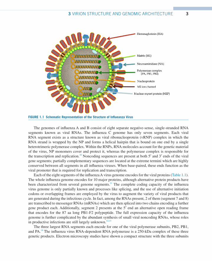
3 VIRION STRUCTURE AND GENOMIC ARCHITECTUREInfluenza viruses are pleomorphic, spherical particles (about 100 nm in diameter), although filamen-tous forms can occur. The virions are relatively unstable in the environment and influenza viruses are inactivated by heat, dryness, extremes of pH, and detergents. Virions are enveloped and their lipid membrane is derived from the host cell. The envelope lodges different glycosylated proteins that proj-ect from the surface of the virus. These proteins are the hemagglutinin (HA), the neuraminidase (NA), and the M2 ion channel proteins. The morphology of influenza A virus particles is, therefore, charac-terized by distinctive spikes, with lengths from 10 to 14 nm, which are readily observable in electron micrographs of virus particles. The approximate ratio between HA and NA is 4:1.
The matrix protein (M1) is situated just beneath the envelope, and underlying the M1 layer a helical superstructure, representing the core of the virus particle which is made of the ribonucleoprotein (RNP) complex, is observed. The RNP complex consists of the viral RNA segments, which are coated with the nucleoprotein (NP) and associated with the heterotrimeric polymerase complex (PB1, PB2, and PA) (Fig. 1.1). The chemical composition of virus particles is approximately 1% RNA, 5–8% carbohydrate, 20% lipid, and approximately 70% protein.9
33 VIRION STRUCTURE AND GENOMIC ARCHITECTURE
The genomes of influenza A and B consist of eight separate negative-sense, single-stranded RNA segments known as viral RNAs. The influenza C genome has only seven segments. Each viral RNA segment exists as a structure known as viral ribonucleoprotein (vRNP) complex in which the RNA strand is wrapped by the NP and forms a helical hairpin that is bound on one end by a single heterotrimeric polymerase complex. Within the RNPs, RNA molecules account for the genetic material of the virus, NP monomers cover and protect it, whereas the polymerase complex is responsible for the transcription and replication.10 Noncoding sequences are present at both 5′ and 3′ ends of the viral gene segments; partially complementary sequences are located at the extreme termini which are highly conserved between all segments in all influenza viruses. When base-paired, these ends function as the viral promoter that is required for replication and transcription.
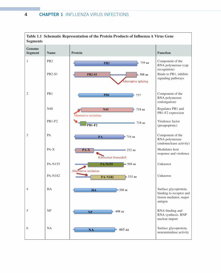
Each of the eight segments of the influenza A virus genome encodes for the viral proteins (Table 1.1). The whole influenza genome encodes for 10 major proteins, although alternative protein products have been characterized from several genome segments.11 The complete coding capacity of the influenza virus genome is only partially known and processes like splicing, and the use of alternative initiation codons or overlapping frames are employed by the virus to augment the variety of viral products that are generated during the infectious cycle. In fact, among the RNAs present, 2 of them (segment 7 and 8) are transcribed to messenger RNAs (mRNAs) which are then spliced into two chains encoding a further gene product each. Additionally, segment 2 presents at the 5′ end an alternative open reading frame that encodes for the 87 aa long PB1-F2 polypeptide. The full expression capacity of the influenza genome is further complicated by the abundant synthesis of small viral noncoding RNAs, whose roles in productive infections are still largely unknown.12,13
The three largest RNA segments each encode for one of the viral polymerase subunits, PB2, PB1, and PA.14 The influenza virus RNA-dependent RNA polymerase is a 250-kDa complex of these three genetic products. Electron microscopy studies have shown a compact structure with the three subunits
FIGURE 1.1 Schematic Representation of the Structure of Influeanza Virus
4 CHAPTER 1 INFLUENZA VIRUS INFECTIONS
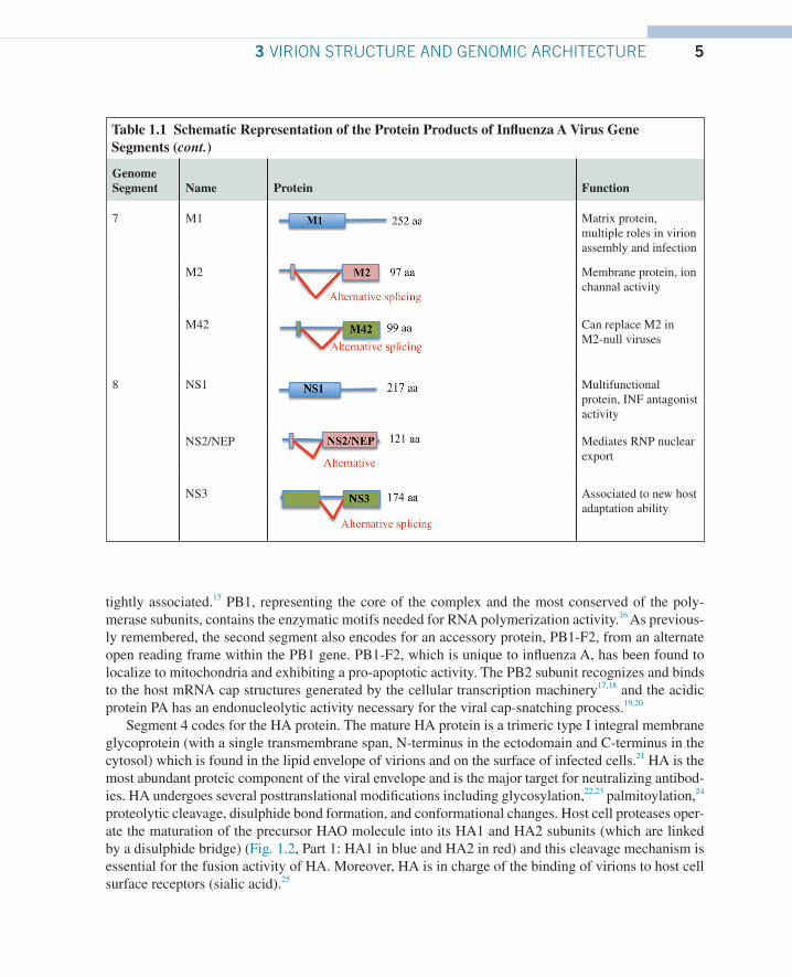
Table 1.1 Schematic Representation of the Protein Products of Influenza A Virus Gene Segments
Genome Segment Name Protein Function
1 PB2 Component of the RNA polymerase (cap recognition)
PB2-S1 Binds to PB1, inhibits signaling pathways
2 PB1 Component of the RNA polymerase (enlongation)
N40 Regulates PB1 and PB1-F2 expression
PB1-F2 Virulence factor (proapoptotic)
3 PA Component of the RNA polymerase (endonuclease activity)
PA-X Modulates host response and virulence
PA-N155 Unknown
PA-N182 Unknown
4 HA Surface glycoprotein, binding to receptor and fusion mediator, major antigen
5 NP RNA-binding and RNA synthesis. RNP nuclear import
6 NA Surface glycoprotein, neuraminidase activity
53 VIRION STRUCTURE AND GENOMIC ARCHITECTURE
tightly associated.15 PB1, representing the core of the complex and the most conserved of the poly-merase subunits, contains the enzymatic motifs needed for RNA polymerization activity.16 As previous-ly remembered, the second segment also encodes for an accessory protein, PB1-F2, from an alternate open reading frame within the PB1 gene. PB1-F2, which is unique to influenza A, has been found to localize to mitochondria and exhibiting a pro-apoptotic activity. The PB2 subunit recognizes and binds to the host mRNA cap structures generated by the cellular transcription machinery17,18 and the acidic protein PA has an endonucleolytic activity necessary for the viral cap-snatching process.19,20
Segment 4 codes for the HA protein. The mature HA protein is a trimeric type I integral membrane glycoprotein (with a single transmembrane span, N-terminus in the ectodomain and C-terminus in the cytosol) which is found in the lipid envelope of virions and on the surface of infected cells.21 HA is the most abundant proteic component of the viral envelope and is the major target for neutralizing antibod-ies. HA undergoes several posttranslational modifications including glycosylation,22,23 palmitoylation,24 proteolytic cleavage, disulphide bond formation, and conformational changes. Host cell proteases oper-ate the maturation of the precursor HAO molecule into its HA1 and HA2 subunits (which are linked by a disulphide bridge) (Fig. 1.2, Part 1: HA1 in blue and HA2 in red) and this cleavage mechanism is essential for the fusion activity of HA. Moreover, HA is in charge of the binding of virions to host cell surface receptors (sialic acid).25
Genome Segment Name Protein Function
7 M1 Matrix protein, multiple roles in virion assembly and infection
M2 Membrane protein, ion channal activity
M42 Can replace M2 in M2-null viruses
8 NS1 Multifunctional protein, INF antagonist activity
NS2/NEP Mediates RNP nuclear export
NS3 Associated to new host adaptation ability
Table 1.1 Schematic Representation of the Protein Products of Influenza A Virus Gene Segments (cont.)

6 CHAPTER 1 INFLUENZA VIRUS INFECTIONS
RNA segment 5 of influenza encodes RNA-binding protein NP.26 This is a highly basic protein whose main function is encapsidation of the viral RNA (an NP monomer of 56 kD binds 24 bases of RNA)27 leaving accessibility to the polymerase as a template for transcription.28
NP also plays a crucial role in transporting the viral RNPs into the nucleus.29,30 In fact, RNPs are too large to passively diffuse through the 9 Å nuclear pores.31 In order to shuttle RNPs in and out of nuclei and from the cytoplasm to the cell periphery, NP makes multiple viral and cellular protein associations involving distinct NP recognition sequences (NLSs, nuclear localization signals, and NES, nuclear export signals). During the late stages of infection, NP also associates with the cytoskeleton32 and NP has been found to bind to filamentous-actin.33
Segment 6 of influenza A encodes the NA protein which is the second major viral glycoprotein. This type II integral membrane protein (single transmembrane span, C-terminus in the ectodomain and N-terminus in the cytosol) is a tetramer and possesses critical sialidase (NA) activity that is needed for a productive release of viral particles from the infected cell.34
Segment 7 of influenza A encodes two proteins, the M1 and M2 protein. M1 is expressed from a collinear transcript, while M2 is derived from an alternative spliced mRNA. M1 associates with lipid membranes and plays an essential role in viral budding. It also regulates the movement of RNPs out of the nucleus and inhibits viral RNA synthesis at later stages of viral replication. M2 is a tetrameric type III membrane protein that has ion channel activity. It functions primarily during virus entry where it is responsible for acidifying the core of the particle which triggers dissociation of M1 from the viral RNPs (uncoating).
FIGURE 1.2 Influenza Virus HA Mediated Entry Mechanism
(1) Influenza HA binds to sialic acids. (2) Low pH conformational change of HA releases the fusion peptide at the N-terminus of HA2 and a conformational change locates the fusion peptide on the distal part of the extended helix and insertion of the fusion peptide into the cell membrane occurs. The transmembrane domain links the HA2 with the viral envelope. (3) At final low pH, a further conformational change drives a refolding mechanism which leads to the formation of a trimeric coiled-coil (the six-helix bundle) that positionates both the transmembrane domains and the fusion peptides in the same fused membrane.
74 VIRAL REPLICATION
The shortest RNA (segment 8 in influenza A) encodes the NS1 protein from a collinear transcript and the NEP/NS2 protein from an alternatively spliced transcript. NS1 is a RNA-binding protein that is expressed at high levels in infected cells and is useful to inhibit host antiviral response, besides in-terfering with the host mRNA processing. The NEP/NS2 protein mediates the nuclear export of newly synthesized RNPs, corresponding with its expression at later times during viral infections.
Recently, several additions have been made to the list of gene products coded by the influenza A genome.35 As just described earlier in the paragraph, the PB1-F2 protein was discovered in 2001 and is encoded by an alternative open translation initiation sites near the 5′ end of the PB1 gene.36 More-over, a third protein is made by the same mechanism by the PB1 gene, named PB1-N40. PB1-N40 is an N-terminal 39 aa polypeptide which is translated from the fifth AUG codon in frame with the PB1 start.37 In addition, segment 1, coding for PB2, produces a newly discovered viral protein, termed PB2-S1, encoded by a novel spliced mRNA in which the region corresponding to nucleotides 1513–1894 of the PB2 mRNA is deleted.38 Novel polypeptides are also encoded by genomic segment 3 and are named PA-X, PA-N155, and PA-N182. The first of them is derived from a second open reading frame ( X-ORF), accessed via ribosomal frameshifting39 and modulates host response.40 The other two (PA-N155 and PA-N182) do not have a polymerase activity, and have been found to be important for virulence and pathogenesis.41 Expression of specific spliced viral products throughout infection is also applied for two of the smallest segments, M1 and NS1. It was already known for a while that protein products from segment 7 included the matrix (M1) and ion channel (M2) proteins, made from a spliced transcript, but a further protein product with an alternative ectodomain and again by a splicing mecha-nism, has been recently identified, the M42.42 Finally, segment 8, besides the nonstructural (NS) protein NS1, also encodes a nuclear export protein NS2/NEP, and NS3 by alternative mRNA splicing.43
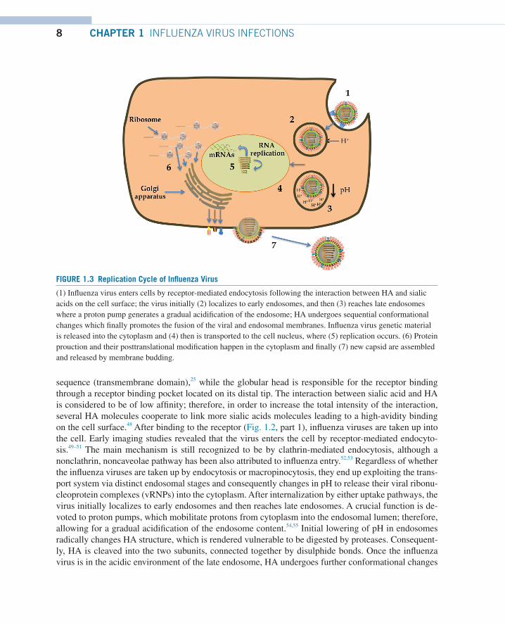
4 VIRAL REPLICATIONInfluenza viruses bind to neuraminic acids (sialic acids) on the surface of cells to initiate the entry pro-cess25 (Fig. 1.3). Notwithstanding the ubiquitous nature of sialic acids, HAs of influenza viruses infect-ing different animal species show some preference for particular glycosidic linkages of the receptor. Human viruses preferentially bind to sialic acids linked to galactose by alfa 2,6 glycosidic bonds (SAα2,6Gal). On the other hand, avian viruses show a preference for alfa 2,3 glycosidic bonds (SAα2,3Gal).44,45 The preference of different influenza subtypes for diverse host-species is explained by the fact that SAα2,6Gal is found mostly in human trachea, while SAα2,3Gal is abundant in the gut epithelium of several bird species, but it should be noted that this represents a preference for receptor-antireceptor linkage and not an absolute specificity. In fact, avian and human cells contain sialic acids with both linkages, though differently expressed and distributed. In addition, high viral inoculum or adaptive point mutations in the HA gene may circumvent this problem.46 HA is able to perform both action of binding and mediation of fusion of juxtaposing membranes. HA is expressed as a trimeric rod-shaped molecule on the virion surface and is produced as a HA0 precursor which is then cleaved into two subunits HA1 (the globular more external part of the molecule) and HA2 (holding a transmem-brane domain) by host cell proteases. The cleavage is needed for the full activity of the molecule. The major structural features of the HA trimer are: a long stem of triple-stranded coiled-coil of α-helices and a globular ectodomain exposed to the environment and derived from the HA1 portion of the mono-mers.47 The stalk region of HA connects the molecule to the virion envelope by a short hydrophobic
8 CHAPTER 1 INFLUENZA VIRUS INFECTIONS
sequence (transmembrane domain),25 while the globular head is responsible for the receptor binding through a receptor binding pocket located on its distal tip. The interaction between sialic acid and HA is considered to be of low affinity; therefore, in order to increase the total intensity of the interaction, several HA molecules cooperate to link more sialic acids molecules leading to a high-avidity binding on the cell surface.48 After binding to the receptor (Fig. 1.2, part 1), influenza viruses are taken up into the cell. Early imaging studies revealed that the virus enters the cell by receptor-mediated endocyto-sis.49–51 The main mechanism is still recognized to be by clathrin-mediated endocytosis, although a nonclathrin, noncaveolae pathway has been also attributed to influenza entry.52,53 Regardless of whether the influenza viruses are taken up by endocytosis or macropinocytosis, they end up exploiting the trans-port system via distinct endosomal stages and consequently changes in pH to release their viral ribonu-cleoprotein complexes (vRNPs) into the cytoplasm. After internalization by either uptake pathways, the virus initially localizes to early endosomes and then reaches late endosomes. A crucial function is de-voted to proton pumps, which mobilitate protons from cytoplasm into the endosomal lumen; therefore, allowing for a gradual acidification of the endosome content.54,55 Initial lowering of pH in endosomes radically changes HA structure, which is rendered vulnerable to be digested by proteases. Consequent-ly, HA is cleaved into the two subunits, connected together by disulphide bonds. Once the influenza virus is in the acidic environment of the late endosome, HA undergoes further conformational changes
FIGURE 1.3 Replication Cycle of Influenza Virus
(1) Influenza virus enters cells by receptor-mediated endocytosis following the interaction between HA and sialic acids on the cell surface; the virus initially (2) localizes to early endosomes, and then (3) reaches late endosomes where a proton pump generates a gradual acidification of the endosome; HA undergoes sequential conformational changes which finally promotes the fusion of the viral and endosomal membranes. Influenza virus genetic material is released into the cytoplasm and (4) then is transported to the cell nucleus, where (5) replication occurs. (6) Protein prouction and their posttranslational modification happen in the cytoplasm and finally (7) new capsid are assembled and released by membrane budding.