Embed Size (px)
Citation preview
Vol.:(0123456789)1 3
Pediatric Surgery International (2019) 35:97–105 https://doi.org/10.1007/s00383-018-4380-8
ORIGINAL ARTICLE
The multi-disciplinary management of complex congenital and acquired tracheo-oesophageal fistulae
H. S. Thakkar1 · R. Hewitt2,3 · K. Cross1 · E. Hannon1 · F. De Bie1,4 · S. Blackburn1 · S. Eaton5,6 · C. A. McLaren3,5,6 · D. J. Roebuck3,5,6 · M. J. Elliott3,7 · J. I. Curry1 · N. Muthialu3,7 · P. De Coppi1,3,5
Accepted: 18 October 2018 / Published online: 3 November 2018 © The Author(s) 2018
AbstractAim of the study Complex tracheo-oesophageal fistulae (TOF) are rare congenital or acquired conditions in children. We discuss here a multidisciplinary (MDT) approach adopted over the past 5 years.Methods We retrospectively collected data on all patients with recurrent or acquired TOF managed at a single institution. All cases were investigated with neck and thorax CT scan. Other investigations included flexible bronchoscopy and broncho-gram (B&B), microlaryngobronchoscopy (MLB) and oesophagoscopy. All cases were subsequently discussed in an MDT meeting on an emergent basis if necessary.Main results 14 patients were referred during this study period of which half had a congenital aetiology and the other half were acquired. The latter included button battery ingestions (5/7) and iatrogenic injuries during oesophageal atresia (OA) repair. Surgical repair was performed on cardiac bypass in 3/7 cases of recurrent congenital fistulae and all cases of acquired fistulae. Post-operatively, 9/14 (64%) patients suffered complications including anastomotic leak (1), bilateral vocal cord paresis (1), further recurrence (1), and mortality (1). Ten patients continue to receive surgical input encompassing tracheal/oesophageal stents and dilatations.Conclusions MDT approach to complex cases is becoming increasingly common across all specialties and is important in making decisions in these difficult cases. The benefits include shared experience of rare cases and full access to multidis-ciplinary expertise.
Keywords Recurrent tracheo-oesophageal fistula · Button battery · Bronchoscopy · Thoracotomy · Cardio-pulmonary bypass
Introduction
Complex tracheo-oesophageal fistulae (TOF) are rare condi-tions in children [1]. Most often these occur after repair of congenital oesophageal atresia (OA) with a distal TOF (C type). Primary repair has a re-fistulisation rate of 3–5% [1, 2]. Complex TOFs can also be a result of oesophageal injury by ingestion of caustic fluids or button batteries [3, 4].
The surgical repair of complex recurrent TOF or acquired lesions is challenging for a number of reasons. First, the diagnosis itself can be difficult and often requires oesopha-geal contrast studies and endoscopies to confirm and intubate the fistula. The literature suggests that a prone oesophago-gram whilst withdrawing an NG tube is the most sensi-tive investigation with the fewest false negatives [5]. The most sensitive test in our experience has been to perform
* P. De Coppi [email protected]
1 Neonatal and Paediatric Surgery, Great Ormond Street Hospital, London, UK
2 Department of Otolaryngology, Great Ormond Street Hospital, London, UK
3 Tracheal Team, Great Ormond Street Hospital, London, UK4 General Surgery Resident, KU Leuven, Leuven, Belgium5 Stem Cells and Regenerative Medicine Section, DBC,
University College London, London, UK6 Department of Radiology, Great Ormond Street Children’s
Hospital, London, UK7 Department of Cardiothoracic Surgery, Great Ormond Street
Hospital, London, UK
98 Pediatric Surgery International (2019) 35:97–105
1 3
bronchography with bronchoscopic (B&B) probing of the fistula pit.
The quality of tissues may present a surgical challenge, particularly in acquired injuries where the defects tend to be larger and the tissues friable with areas of ischaemia or even frank necrosis. In recurrent TOF the scarring due to previous surgery also makes the access and safe mobilisation of the trachea and oesophagus more difficult, increasing the risk of intra- and post-operative complications.
Different approaches for managing these fistulae have been described: endoscopic use of glue for small defects [6], standard thoracotomy with a variety of adjuncts such as interposition flaps [7] (pericardium, pleura), early aban-donment of the oesophagus in preference of tracheal pres-ervation and median sternotomy with or without the use of cardiopulmonary bypass (CPB) for the more complex cases [8, 9].
Following a recent increase in tertiary referrals of com-plex TOF cases to our institution, a multidisciplinary (MDT) approach has been adopted so as to treat every patient on an individual basis. In this paper, we discuss our MDT approach based on the case series of complex TOFs treated in our institution over the past 5 years.
Methods
We retrospectively collected data on all patients with recur-rent or acquired TOF managed at Great Ormond Street Hos-pital from January 2013 till July 2018. All patients were tertiary referrals from other UK or international surgical centres. All cases were investigated with contrast CT scan of the neck and thorax. Other investigations included B&B, MLB and oesophagoscopy.
All cases were subsequently discussed in our MDT meet-ing (Table 1). We followed a 3 “Ds” approach—Diagnosis, Discussion and Decision-making. Those present at our MDT include a cardiothoracic surgeon, diagnostic radiolo-gist, ENT surgeon, general paediatric surgeon, intensive care physician, interventional radiologist and radiographer, respiratory physician and specialist nurses. We have specifi-cally incorporated the experience of our specialist nurses into this MDT setting to create a complex aero-digestive team. Decisions on the approach to be used in each case were based on several factors. Size, position of defect in the trachea and aetiology were the most important factors when considering whether CPB was required for repair or whether an endotracheal tube could be safely placed distal to the TOF allowing conventional ventilation. Size of defect also determined whether complete circumferential control of the trachea may be needed for division and repair of the trachea—necessitating a sternotomy approach. Surgical his-tory of repeated thoracotomy was also a relative indication for sternotomy.
Fisher’s test and chi-squared tests were used for statistical analysis with P < 0.05 considered statistically significant.
Results
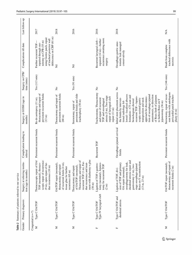
14 patients were referred during this study period of which half had a congenital aetiology (C-TOF) and the other half acquired (A-TOF). Two patients were referred from out-side the UK, with the remaining referred from other UK tertiary paediatric surgical centres. Table 2 summarises the patient demographics, medical history, reasons for referral to our institution, the nature of surgery undertaken at our centre and their outcomes. The aetiology for the majority
Table 1 MDT approach to complex/acquired TOF
Diagnosis Tertiary referral received History reviewedExternal imaging reviewedTransfer to appropriate ward/intensive care
Discussion Investigations Contrast CT thorax and neckFlexible bronchoscopy and bronchography and oesophagogramAirway endoscopy +/- oesophagoscopy
MDT discussionCardiothoracic surgeryDiagnostic radiologistENTGeneral surgeryIntensive care physicianInterventional radiologistRadiographerRespiratory physicianSpecialist nurses
Questions consideredHistory—previous surgery, approach, etc.Co-morbidities—especially cardiacDefect Size Position in trachea Quality of tissue Possible repair possible—primary repair, slide or patch tracheoplasty
Decision Repair of cardiopulmonary bypass Repair via thoracotomy Tissue engineering/experimental
99Pediatric Surgery International (2019) 35:97–105
1 3
Tabl
e 2
Sum
mar
y of
pat
ient
s ref
erre
d to
our
serv
ice
Gen
der
Prim
ary
diag
nosi
sSu
rger
y at
refe
rrin
g in
stitu
-tio
n (a
ge in
mon
ths)
Com
plic
atio
n le
adin
g to
re
ferr
alSu
rger
y at
GO
SH (a
ge in
m
onth
s)Su
rger
y on
CPB
/tim
e (m
ins)
Com
plic
atio
ns ti
ll da
teLa
st fo
llow
-up
Con
geni
tal (n =
7) M
Type
C O
A/T
OF
Thor
acos
copi
c re
pair
of O
A/
TOF
(neo
nata
l), th
orac
o-sc
opic
repa
ir of
recu
rren
ce
(9 m
), op
en re
pair
of fu
r-th
er re
curr
ence
(10
m)
Pers
isten
t rec
urre
nt fi
stula
Re-d
o ao
rtope
xy (1
1 m
), th
orac
otom
y an
d re
pair
of
pers
isten
t rec
urre
nt fi
stula
(1
3 m
)
Yes (
113
min
)Fu
rther
recu
rren
t TO
F—re
paire
d th
roug
h ste
r-no
tom
y on
CPB
(55
m)
usin
g G
orte
x pa
tch,
repa
ir of
trac
heal
pos
terio
r wal
l de
hisc
ence
on
CB
P (6
1 m
)
2017
MTy
pe C
OA
/TO
FO
A/T
OF
repa
ir (n
eona
tal),
th
orac
otom
y an
d re
pair
of re
curr
ent fi
stula
(4 m
), tis
sue
glue
for f
urth
er
recu
rren
ce (6
5 m
)
Pers
isten
t rec
urre
nt fi
stula
Thor
acot
omy
and
repa
ir of
pe
rsist
ent r
ecur
rent
fistu
la
(68
m)
No
Nil
2018
MTy
pe C
OA
/TO
FTh
orac
otom
y an
d re
pair
of O
A/T
OF
(neo
nata
l),
Thor
acot
omy
and
repa
ir of
re
curr
ent T
OF
(1 m
), Fu
r-th
er re
curr
ence
and
trea
t-m
ent w
ith d
iath
erm
y + gl
ue
(38
m)
Pers
isten
t rec
urre
nt fi
stula
Ster
noto
my,
repa
ir of
pe
rsist
ent fi
stula
and
slid
e tra
cheo
plas
ty (3
8 m
)
Yes (
91 m
in)
Nil
2016
FTy
pe C
OA
/TO
FTy
pe IV
lary
ngea
l cle
ftLi
gatio
n of
TO
F +
gastr
os-
tom
y (n
eona
tal),
Jeju
nos-
tom
y fo
r rec
urre
nt T
OF
(2 m
)
Recu
rren
t TO
FTr
ache
osto
my,
Tho
raco
tom
y an
d re
pair
of re
curr
ent
TOF
and
oeso
phag
eal
atre
sia
(5 m
), Tw
o-st
age
repa
ir of
lary
ngea
l cle
ft re
pair
(11
m)
No
Recu
rren
t lar
ynge
al c
left—
repa
ired
(14
m)—
furth
er
recu
rren
ce aw
aitin
g m
ore
surg
ery
2016
FTy
pe C
OA
/TO
F an
d du
oden
al a
tresi
aLi
gatio
n of
TO
F, d
ivi-
sion
of T
OF
and
prim
ary
OA
repa
ir (n
eona
tal),
oe
soph
agos
tom
y fo
r lea
k,
duod
enal
atre
sia
repa
ir an
d ga
stros
tom
y (1
m),
two-
stag
e oe
soph
ago-
jeju
nal
Roux
-en-
Y re
cons
truct
ion
(13
m, 2
3 m
)
Oes
opha
go-je
juna
l cer
vica
l fis
tula
Initi
al st
ent p
lace
men
t acr
oss
the
fistu
la b
y in
terv
en-
tiona
l rad
iolo
gy fo
l-lo
wed
—lo
calis
ed a
bsce
ss
form
atio
n—C
T sc
an a
nd
bron
chog
ram
show
ed
recu
rren
t TO
F—la
paro
-sc
opic
-ass
isted
gas
tric
trans
posi
tion
and
neck
ap
proa
ch fo
r dis
conn
ec-
tion
of o
esop
hago
-jeju
nal
anas
tom
osis
, re-
dire
ctio
n of
Rou
x lim
b of
jeju
num
to
fash
ion
Roux
-en-
Y
jeju
nosto
my
(36
m)
No
Oes
opha
go-g
astri
c an
as-
tom
otic
leak
man
aged
co
nser
vativ
ely
2018
MTy
pe C
OA
/TO
FO
A/T
OF
repa
ir (n
eona
tal),
th
orac
otom
y an
d re
pair
of
recu
rren
t fistu
la (2
m)
Pers
isten
t rec
urre
nt fi
stula
Ster
noto
my,
repa
ir of
recu
r-re
nt fi
stula
with
aut
olog
ous
peric
ardi
al p
atch
trac
heo-
plas
ty (6
m)
Yes (
146
min
)D
eath
from
com
plet
e tra
chea
l deh
isce
nce
with
ne
cros
is
N/A
100 Pediatric Surgery International (2019) 35:97–105
1 3
Tabl
e 2
(con
tinue
d)
Gen
der
Prim
ary
diag
nosi
sSu
rger
y at
refe
rrin
g in
stitu
-tio
n (a
ge in
mon
ths)
Com
plic
atio
n le
adin
g to
re
ferr
alSu
rger
y at
GO
SH (a
ge in
m
onth
s)Su
rger
y on
CPB
/tim
e (m
ins)
Com
plic
atio
ns ti
ll da
teLa
st fo
llow
-up
MTy
pe D
OA
/TO
FTh
orac
otom
y an
d re
pair
of
OA
/TO
F (n
eona
tal)
Recu
rren
t TO
F, le
ft vo
cal
cord
pal
syTh
orac
otom
y an
d re
pair
of re
curr
ent T
OF
(5 m
), th
orac
osco
pic
aorto
pexy
(6
m)
No
Bila
tera
l poo
r voc
al c
ord
mov
emen
t20
18
Gen
der
Age
at p
rese
ntat
ion
to
loca
l (m
onth
s)Pr
imar
y di
agno
sis
Com
plic
atio
n le
adin
g to
re
ferr
alSu
rger
y at
GO
SH (a
ge in
m
onth
s)Su
rger
y on
byp
ass
Com
plic
atio
ns ti
ll da
teLa
st fo
llow
-up
Acq
uire
d (n
= 7)
F23
But
ton
batte
ry in
gesti
onA
cqui
red
TOF
Ster
noto
my,
dire
ct re
pair
of
oeso
phag
us, g
astro
stom
y an
d sl
ide
trach
eopl
asty
(2
3 m
), la
paro
scop
ic-
assi
sted
gastr
ic tr
ansp
osi-
tion
(45
m),
stem
-cel
l tra
chea
l tra
nspl
ant (
47 m
)
Yes (
124
min
)Th
orac
otom
y fo
r rec
ur-
rent
fistu
la, e
xcis
ion
of
mid
-oes
opha
gus a
nd
oeso
phag
osto
my
(29
m),
re-d
o ste
rnot
omy
and
trach
eal r
epai
r on
CPB
(4
6 m
)
2018
M20
But
ton
batte
ry in
gesti
onA
cqui
red
TOF
Ster
noto
my,
dire
ct re
pair
of
the
oeso
phag
us a
nd sl
ide
trach
eopl
asty
(20
m)
Yes (
101
min
)Po
st-op
erat
ive
med
iasti
no-
cuta
neou
s fistu
la, r
ecur
rent
fis
tula
repa
ired
with
au
tolo
gous
per
icar
dial
pa
tch
trach
eopl
asty
and
di
rect
repa
ir of
the
oeso
ph-
agus
(26
m)
2018
M3
Long
-gap
OA
Iatro
geni
c tra
chea
l inj
ury
at in
itial
surg
ery
repa
ired
durin
g th
orac
otom
y fo
r de
laye
d pr
imar
y re
pair
of
OA
but
late
r dev
elop
men
t of
acq
uire
d TO
F
Exci
sion
of t
rach
ea-o
esop
h-ag
eal fi
stula
, aut
olo-
gous
per
icar
dial
pat
ch
trach
eopl
asty
, clo
sure
of
pro
xim
al a
nd d
istal
oe
soph
agea
l stu
mps
(3 m
), la
paro
scop
ic- a
ssist
ed
gastr
ic tr
ansp
ositi
on a
nd
jeju
nosto
my
(16
m)
Yes (
135
min
)La
paro
scop
ic li
gatio
n of
di
stal
oes
opha
gus f
or
recu
rren
t fistu
la, g
as-
trosto
my
(4 m
), ai
r lea
k re
quiri
ng tr
ache
al re
pair
unde
r byp
ass (
4 m
)
2018
F12
But
ton
batte
ry in
gesti
onA
cqui
red
TOF
Nea
r tot
al o
esop
hage
ctom
y,
cerv
ical
oes
opha
gosto
my,
ga
stros
tom
y, a
utol
o-go
us p
eric
ardi
al p
atch
tra
cheo
plas
ty (1
2 m
), la
paro
scop
ic-a
ssist
ed g
as-
tric
trans
posi
tion
(22
m)
Yes (
110
min
)Tr
ansi
ent b
ilate
ral v
ocal
co
rd p
alsy
–tra
cheo
stom
y (2
3 m
) now
dec
annu
late
d
2018
101Pediatric Surgery International (2019) 35:97–105
1 3
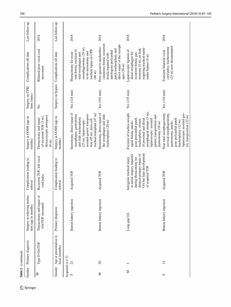
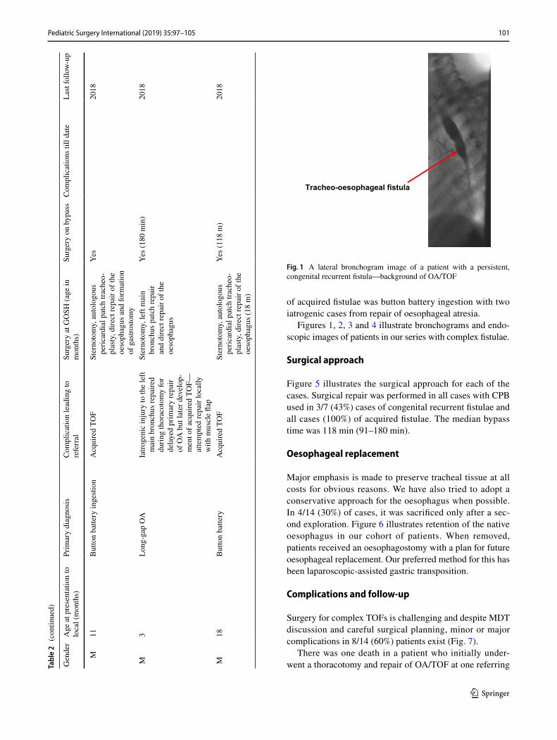
of acquired fistulae was button battery ingestion with two iatrogenic cases from repair of oesophageal atresia.
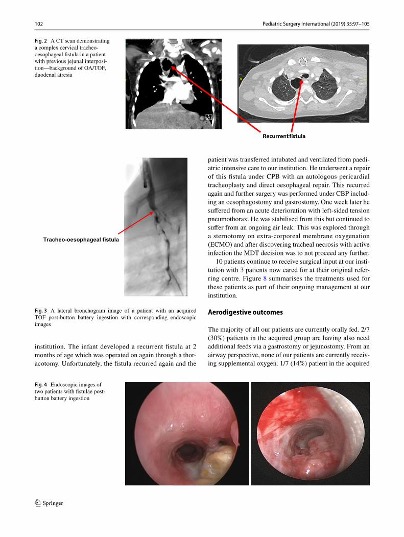
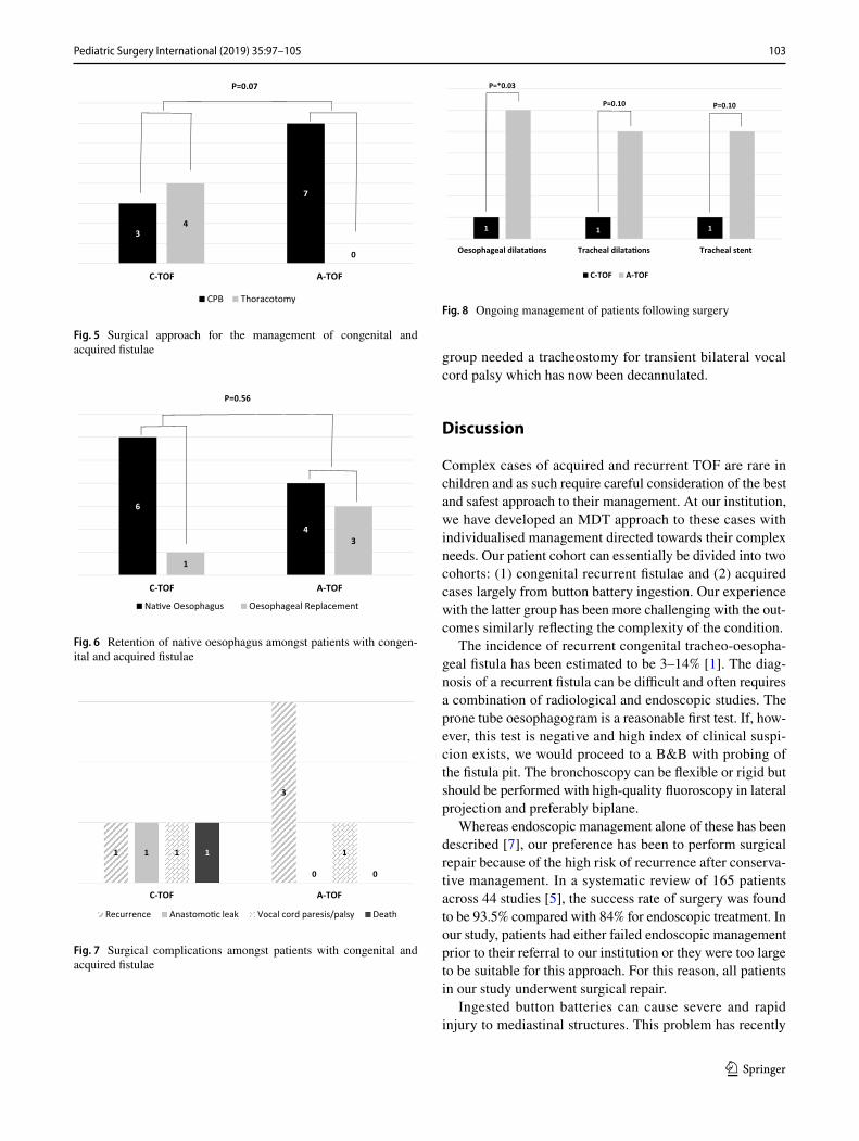
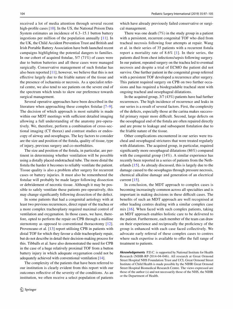
Figures 1, 2, 3 and 4 illustrate bronchograms and endo-scopic images of patients in our series with complex fistulae.
Surgical approach
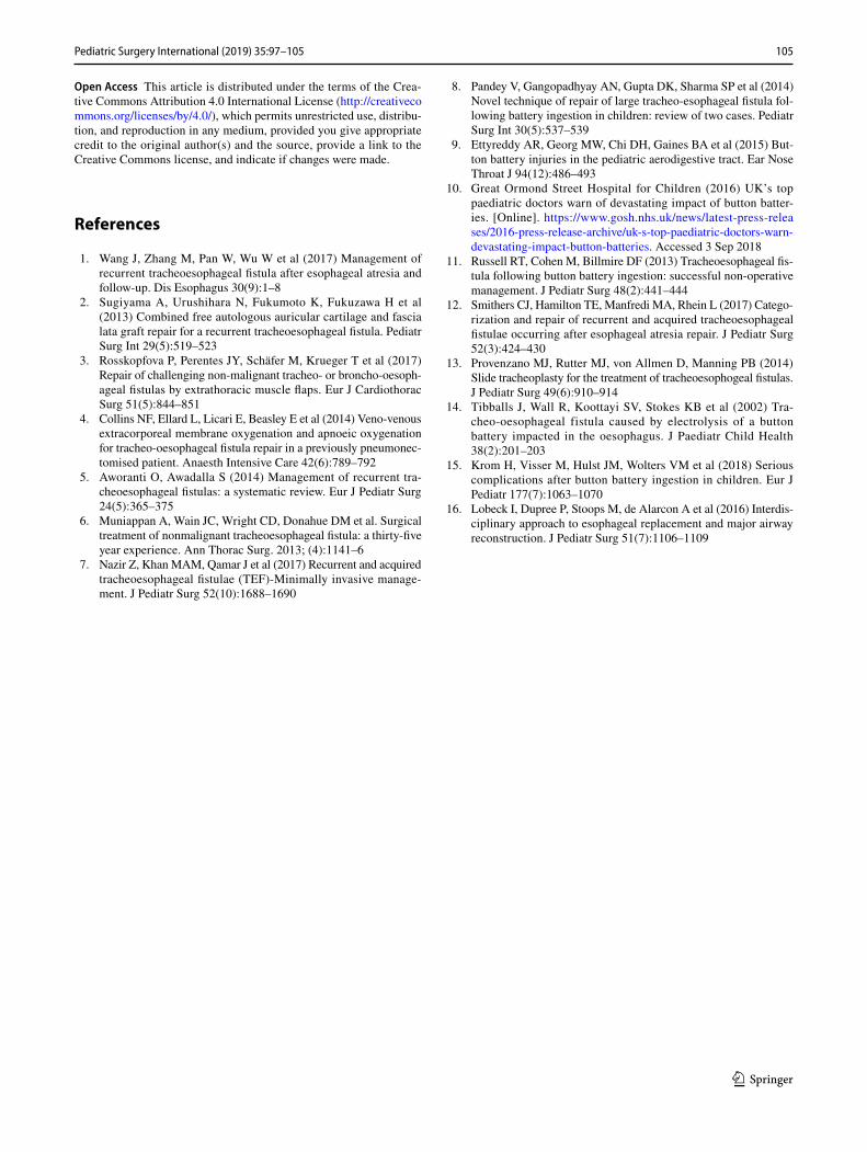
Figure 5 illustrates the surgical approach for each of the cases. Surgical repair was performed in all cases with CPB used in 3/7 (43%) cases of congenital recurrent fistulae and all cases (100%) of acquired fistulae. The median bypass time was 118 min (91–180 min).
Oesophageal replacement
Major emphasis is made to preserve tracheal tissue at all costs for obvious reasons. We have also tried to adopt a conservative approach for the oesophagus when possible. In 4/14 (30%) of cases, it was sacrificed only after a sec-ond exploration. Figure 6 illustrates retention of the native oesophagus in our cohort of patients. When removed, patients received an oesophagostomy with a plan for future oesophageal replacement. Our preferred method for this has been laparoscopic-assisted gastric transposition.
Complications and follow‑up
Surgery for complex TOFs is challenging and despite MDT discussion and careful surgical planning, minor or major complications in 8/14 (60%) patients exist (Fig. 7).
There was one death in a patient who initially under-went a thoracotomy and repair of OA/TOF at one referring Ta
ble
2 (c
ontin
ued)
Gen
der
Age
at p
rese
ntat
ion
to
loca
l (m
onth
s)Pr
imar
y di
agno
sis
Com
plic
atio
n le
adin
g to
re
ferr
alSu
rger
y at
GO
SH (a
ge in
m
onth
s)Su
rger
y on
byp
ass
Com
plic
atio
ns ti
ll da
teLa
st fo
llow
-up
M11
But
ton
batte
ry in
gesti
onA
cqui
red
TOF
Ster
noto
my,
aut
olog
ous
peric
ardi
al p
atch
trac
heo-
plas
ty, d
irect
repa
ir of
the
oeso
phag
us a
nd fo
rmat
ion
of g
astro
stom
y
Yes
2018
M3
Long
-gap
OA
Iatro
geni
c in
jury
to th
e le
ft m
ain
bron
chus
repa
ired
durin
g th
orac
otom
y fo
r de
laye
d pr
imar
y re
pair
of O
A b
ut la
ter d
evel
op-
men
t of a
cqui
red
TOF—
atte
mpt
ed re
pair
loca
lly
with
mus
cle
flap
Ster
noto
my,
left
mai
n br
onch
us p
atch
repa
ir an
d di
rect
repa
ir of
the
oeso
phag
us
Yes (
180
min
)20
18
M18
But
ton
batte
ryA
cqui
red
TOF
Ster
noto
my,
aut
olog
ous
peric
ardi
al p
atch
trac
heo-
plas
ty, d
irect
repa
ir of
the
oeso
phag
us (1
8 m
)
Yes (
118
m)
2018
Tracheo-oesophageal fistula
Fig. 1 A lateral bronchogram image of a patient with a persistent, congenital recurrent fistula—background of OA/TOF
102 Pediatric Surgery International (2019) 35:97–105
1 3
institution. The infant developed a recurrent fistula at 2 months of age which was operated on again through a thor-acotomy. Unfortunately, the fistula recurred again and the
patient was transferred intubated and ventilated from paedi-atric intensive care to our institution. He underwent a repair of this fistula under CPB with an autologous pericardial tracheoplasty and direct oesophageal repair. This recurred again and further surgery was performed under CBP includ-ing an oesophagostomy and gastrostomy. One week later he suffered from an acute deterioration with left-sided tension pneumothorax. He was stabilised from this but continued to suffer from an ongoing air leak. This was explored through a sternotomy on extra-corporeal membrane oxygenation (ECMO) and after discovering tracheal necrosis with active infection the MDT decision was to not proceed any further.
10 patients continue to receive surgical input at our insti-tution with 3 patients now cared for at their original refer-ring centre. Figure 8 summarises the treatments used for these patients as part of their ongoing management at our institution.
Aerodigestive outcomes
The majority of all our patients are currently orally fed. 2/7 (30%) patients in the acquired group are having also need additional feeds via a gastrostomy or jejunostomy. From an airway perspective, none of our patients are currently receiv-ing supplemental oxygen. 1/7 (14%) patient in the acquired
Fig. 2 A CT scan demonstrating a complex cervical tracheo-oesophageal fistula in a patient with previous jejunal interposi-tion—background of OA/TOF, duodenal atresia
Tracheo-oesophageal fistula
Fig. 3 A lateral bronchogram image of a patient with an acquired TOF post-button battery ingestion with corresponding endoscopic images
Fig. 4 Endoscopic images of two patients with fistulae post-button battery ingestion
103Pediatric Surgery International (2019) 35:97–105
1 3
group needed a tracheostomy for transient bilateral vocal cord palsy which has now been decannulated.
Discussion
Complex cases of acquired and recurrent TOF are rare in children and as such require careful consideration of the best and safest approach to their management. At our institution, we have developed an MDT approach to these cases with individualised management directed towards their complex needs. Our patient cohort can essentially be divided into two cohorts: (1) congenital recurrent fistulae and (2) acquired cases largely from button battery ingestion. Our experience with the latter group has been more challenging with the out-comes similarly reflecting the complexity of the condition.
The incidence of recurrent congenital tracheo-oesopha-geal fistula has been estimated to be 3–14% [1]. The diag-nosis of a recurrent fistula can be difficult and often requires a combination of radiological and endoscopic studies. The prone tube oesophagogram is a reasonable first test. If, how-ever, this test is negative and high index of clinical suspi-cion exists, we would proceed to a B&B with probing of the fistula pit. The bronchoscopy can be flexible or rigid but should be performed with high-quality fluoroscopy in lateral projection and preferably biplane.
Whereas endoscopic management alone of these has been described [7], our preference has been to perform surgical repair because of the high risk of recurrence after conserva-tive management. In a systematic review of 165 patients across 44 studies [5], the success rate of surgery was found to be 93.5% compared with 84% for endoscopic treatment. In our study, patients had either failed endoscopic management prior to their referral to our institution or they were too large to be suitable for this approach. For this reason, all patients in our study underwent surgical repair.
Ingested button batteries can cause severe and rapid injury to mediastinal structures. This problem has recently
3
7
4
0
A-TOFC-TOF
CPB Thoracotomy
P=0.07
Fig. 5 Surgical approach for the management of congenital and acquired fistulae
6
4
1
3
FOT-AFOT-C
Na�ve Oesophagus Oesophageal Replacement
P=0.56
Fig. 6 Retention of native oesophagus amongst patients with congen-ital and acquired fistulae
1
3
1
0
1 11
0
C-TOF A-TOF
Recurrence Anastomo�c leak Vocal cord paresis/palsy Death
Fig. 7 Surgical complications amongst patients with congenital and acquired fistulae
1 1 1
Oesophageal dilata ons Tracheal s Tracheal stent
C-TOF A-TOF
P=*0.03
P=0.10 P=0.10
Fig. 8 Ongoing management of patients following surgery
104 Pediatric Surgery International (2019) 35:97–105
1 3
received a lot of media attention through several recent high-profile cases [10]. In the US, the National Poison Data System estimates an incidence of 6.3–15.1 button battery ingestions per million of the population annually [11]. In the UK, the Child Accident Prevention Trust and British and Irish Portable Battery Association have both launched recent campaigns highlighting the potential dangers to families. In our cohort of acquired fistulae, 5/7 (71%) of cases were due to button batteries and all these cases were managed surgically. Conservative management of such fistulae has also been reported [11]; however, we believe that this is not effective largely due to the friable nature of the tissue and the presence of ischaemia or necrosis. As a specialist refer-ral centre, we also tend to see patients on the severe end of the spectrum which tends to skew our preference towards surgical management.
Several operative approaches have been described in the literature when approaching these complex fistulae [7–9]. The decision of which approach is most suitable is made within our MDT meetings with sufficient detailed imaging allowing a full understanding of the anatomy pre-opera-tively. We, therefore, perform a combination of cross-sec-tional imaging (CT thorax) and contrast studies or endos-copy of airway and oesophagus. The key factors to consider are the size and position of the fistula, quality of tissue, type of injury, previous surgery and co-morbidities.
The size and position of the fistula, in particular, are per-tinent in determining whether ventilation will be possible using a distally placed endotracheal tube. The more distal the fistula the harder it becomes to reliably ventilate the patient. Tissue quality is also a problem after surgery for recurrent cases or battery injuries. It must also be remembered the fistulae will probably be made larger following dissection or debridement of necrotic tissue. Although it may be pos-sible to safely ventilate these patients pre-operatively, this may change significantly following dissection of the defect.
In some patients that had a congenital aetiology with at least two previous recurrences, direct repair of the trachea or a more complex tracheoplasty required maximal control of ventilation and oxygenation. In those cases, we have, there-fore, opted to perform the repair on CPB through a midline sternotomy as opposed to conventional thoracotomy [12]. Provenzano et al. [13] report utilising CPB in patients with distal TOF for which they favour a slide tracheoplasty repair, but do not describe in detail their decision-making process for this. Tibballs et al. have also demonstrated the need for CPB in the case of a huge relatively proximal TOF from a button battery injury in which adequate oxygenation could not be adequately achieved with conventional ventilation [14].
The complexity of the patients that have been referred to our institution is clearly evident from this report with our outcomes reflective of the severity of the conditions. As an institution, we often receive a select population of patients
which have already previously failed conservative or surgi-cal management.
There was one death (7%) in the study group in a patient with a persistent, recurrent congenital TOF who died from tracheal necrosis following four attempts at repair. Wang et al. in their series of 35 patients with a recurrent fistula report a mortality rate of 8.6% [1]. In their series, the patients died from chest infections/sepsis following surgery. In our patient, repeated surgery on the trachea led to eventual necrosis and despite a trial of ECMO the patient did not survive. One further patient in the congenital group referred with a persistent TOF developed a recurrence after surgery. This patient required surgery on CPB on two further occa-sions and has required a biodegradable tracheal stent with ongoing tracheal and oesophageal dilatations.
In the acquired group, 3/7 (43%) patients have had further recurrences. The high incidence of recurrence and leaks in our series is a result of several factors. First, the complexity of the defects, especially those at the carina makes success-ful primary repair more difficult. Second, large defects on the oesophageal end of the fistula are often repaired directly and are prone to leakage and subsequent fistulation due to the friable nature of the tissue.
Other complications encountered in our series were tra-cheal and oesophageal strictures that were treated regularly with dilatations. The acquired group, in particular, required significantly more oesophageal dilatations (86%) compared with the congenital group (14%). A similar experience has recently been reported in a series of patients from the Neth-erlands [15]. As already discussed, this is largely due to the damage caused to the oesophagus through pressure necrosis, chemical alkaline damage and generation of an electrical current [15].
In conclusion, the MDT approach to complex cases is becoming increasingly common across all specialties and is important in making decisions in these difficult cases. The benefits of such an MDT approach are well recognised at other leading centres dealing with a similar complex case mix [16]. When faced with such complex patients, taking an MDT approach enables holistic care to be delivered to the patient. Furthermore, each member of the team can draw on their experience and reciprocally the proficiency of the group is enhanced with each case faced collectively. We advocate early referral of these complex cases to centres where such expertise is available to offer the full range of treatment to patients.
Aknowledgements P.D.C. is supported by National Institute for Health Research (NIHR-RP-2014-04-046). All research at Great Ormond Street Hospital NHS Foundation Trust and UCL Great Ormond Street Institute of Child Health is made possible by the NIHR Great Ormond Street Hospital Biomedical Research Centre. The views expressed are those of the author (s) and not necessarily those of the NHS, the NIHR or the Department of Health.
105Pediatric Surgery International (2019) 35:97–105
1 3
Open Access This article is distributed under the terms of the Crea-tive Commons Attribution 4.0 International License (http://creat iveco mmons .org/licen ses/by/4.0/), which permits unrestricted use, distribu-tion, and reproduction in any medium, provided you give appropriate credit to the original author(s) and the source, provide a link to the Creative Commons license, and indicate if changes were made.
References
1. Wang J, Zhang M, Pan W, Wu W et al (2017) Management of recurrent tracheoesophageal fistula after esophageal atresia and follow-up. Dis Esophagus 30(9):1–8
2. Sugiyama A, Urushihara N, Fukumoto K, Fukuzawa H et al (2013) Combined free autologous auricular cartilage and fascia lata graft repair for a recurrent tracheoesophageal fistula. Pediatr Surg Int 29(5):519–523
3. Rosskopfova P, Perentes JY, Schäfer M, Krueger T et al (2017) Repair of challenging non-malignant tracheo- or broncho-oesoph-ageal fistulas by extrathoracic muscle flaps. Eur J Cardiothorac Surg 51(5):844–851
4. Collins NF, Ellard L, Licari E, Beasley E et al (2014) Veno-venous extracorporeal membrane oxygenation and apnoeic oxygenation for tracheo-oesophageal fistula repair in a previously pneumonec-tomised patient. Anaesth Intensive Care 42(6):789–792
5. Aworanti O, Awadalla S (2014) Management of recurrent tra-cheoesophageal fistulas: a systematic review. Eur J Pediatr Surg 24(5):365–375
6. Muniappan A, Wain JC, Wright CD, Donahue DM et al. Surgical treatment of nonmalignant tracheoesophageal fistula: a thirty-five year experience. Ann Thorac Surg. 2013; (4):1141–6
7. Nazir Z, Khan MAM, Qamar J et al (2017) Recurrent and acquired tracheoesophageal fistulae (TEF)-Minimally invasive manage-ment. J Pediatr Surg 52(10):1688–1690
8. Pandey V, Gangopadhyay AN, Gupta DK, Sharma SP et al (2014) Novel technique of repair of large tracheo-esophageal fistula fol-lowing battery ingestion in children: review of two cases. Pediatr Surg Int 30(5):537–539
9. Ettyreddy AR, Georg MW, Chi DH, Gaines BA et al (2015) But-ton battery injuries in the pediatric aerodigestive tract. Ear Nose Throat J 94(12):486–493
10. Great Ormond Street Hospital for Children (2016) UK’s top paediatric doctors warn of devastating impact of button batter-ies. [Online]. https ://www.gosh.nhs.uk/news/lates t-press -relea ses/2016-press -relea se-archi ve/uk-s-top-paedi atric -docto rs-warn-devas tatin g-impac t-butto n-batte ries. Accessed 3 Sep 2018
11. Russell RT, Cohen M, Billmire DF (2013) Tracheoesophageal fis-tula following button battery ingestion: successful non-operative management. J Pediatr Surg 48(2):441–444
12. Smithers CJ, Hamilton TE, Manfredi MA, Rhein L (2017) Catego-rization and repair of recurrent and acquired tracheoesophageal fistulae occurring after esophageal atresia repair. J Pediatr Surg 52(3):424–430
13. Provenzano MJ, Rutter MJ, von Allmen D, Manning PB (2014) Slide tracheoplasty for the treatment of tracheoesophogeal fistulas. J Pediatr Surg 49(6):910–914
14. Tibballs J, Wall R, Koottayi SV, Stokes KB et al (2002) Tra-cheo-oesophageal fistula caused by electrolysis of a button battery impacted in the oesophagus. J Paediatr Child Health 38(2):201–203
15. Krom H, Visser M, Hulst JM, Wolters VM et al (2018) Serious complications after button battery ingestion in children. Eur J Pediatr 177(7):1063–1070
16. Lobeck I, Dupree P, Stoops M, de Alarcon A et al (2016) Interdis-ciplinary approach to esophageal replacement and major airway reconstruction. J Pediatr Surg 51(7):1106–1109