Embed Size (px)
Citation preview
The Muscular System
Copyright © The McGraw-Hill Companies, Inc. Permission required for reproduction or display.
Functions and Types of Muscles
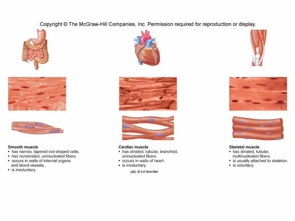
o Smooth Muscle• Located in the walls of hollow organs
and blood vessels• Involuntary contraction• Moves materials through organs and
regulates blood flow• Cylindrical cells with pointed ends• Each cell is uninucleate
Functions and Types of Muscles
o Cardiac Muscle• Forms the heart wall• Fibers are uninucleated, striated,
tubular, and branched• Fibers interlock at intercalated disks,
which permit contractions to spread quickly throughout the heart
• Contraction does not require outside nervous stimulation
• Nerves do affect heart rate and strength of contraction
Functions and Types of Muscles
o Skeletal Muscle• Fibers are tubular, multinucleated,
and striated• Make up muscles attached to the
skeleton• Contraction is voluntary
Functions and Types of Muscles
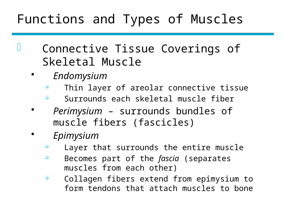
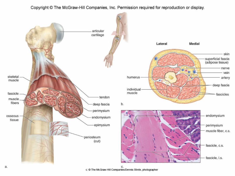
o Connective Tissue Coverings of Skeletal Muscle
• Endomysium Thin layer of areolar connective tissue Surrounds each skeletal muscle fiber
• Perimysium – surrounds bundles of muscle fibers (fascicles)
• Epimysium Layer that surrounds the entire muscle Becomes part of the fascia (separates muscles
from each other) Collagen fibers extend from epimysium to form
tendons that attach muscles to bone
Functions and Types of Muscles
o Functions of Skeletal Muscles• Support the body• Make bones and other body parts
move• Help maintain a constant body
temperature• Assists movement in cardiovascular
and lymphatic vessels• Help protect bones and internal
organs, and stabilize joints
Microscopic Anatomy
o Muscle fiber components• Sarcolemma – plasma membrane• Sarcoplasm – cytoplasm
Contains glycogen that provides energy for muscle contraction
Contains myoglobin which binds oxygen until needed
• Sarcoplasmic reticulum – endoplasmic reticulum
• T (transverse) tubules Formed by the sarcolemma penetrating into the
cell Come into contact with expanded portions of the
sarcoplasmic reticulum
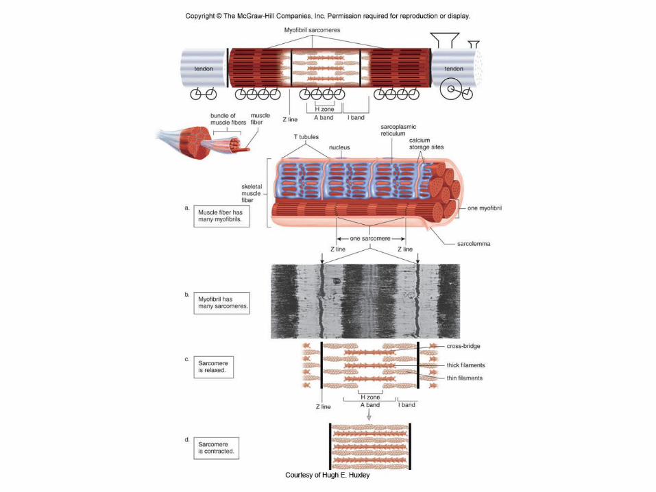
Microscopic Anatomy
o Myofibrils and Sarcomeres• Myofibrils run the length of the muscle fiber• Composed of numerous sarcomeres
Extends between two vertical Z lines Contains two types of protein myofilaments
Thick filaments – made up of myosin Thin filaments – made up of actin, tropomyosin, and
troponin I band contains only thin filaments A band in the center of the sarcomere contains
thick and thin filaments H zone in the center of the A band has only
myosin filaments
Microscopic Anatomy
o Myofilaments• Thick filaments
Composed of several hundred of molecules of myosin
Myosin molecules end in a cross-bridge • Thin filaments
Two strands of actin Double strands of tropomyosin coil of
each actin strand Troponin occurs at intervals on the
tropomyosin strand
Microscopic Anatomy
• Sliding filaments Occurs when sarcomeres shorten (during
muscle contraction) Actin filaments slide past the myosin
filaments Thick and thin filaments remain the same
length
Contraction of Skeletal Muscle
o Neuromuscular junction• Axon terminals
Come into close proximity to the sarcolemma
Have vesicles that contain acetylcholine (Ach)
• Synaptic cleft – a small gap that separates the axon from the sarcolemma
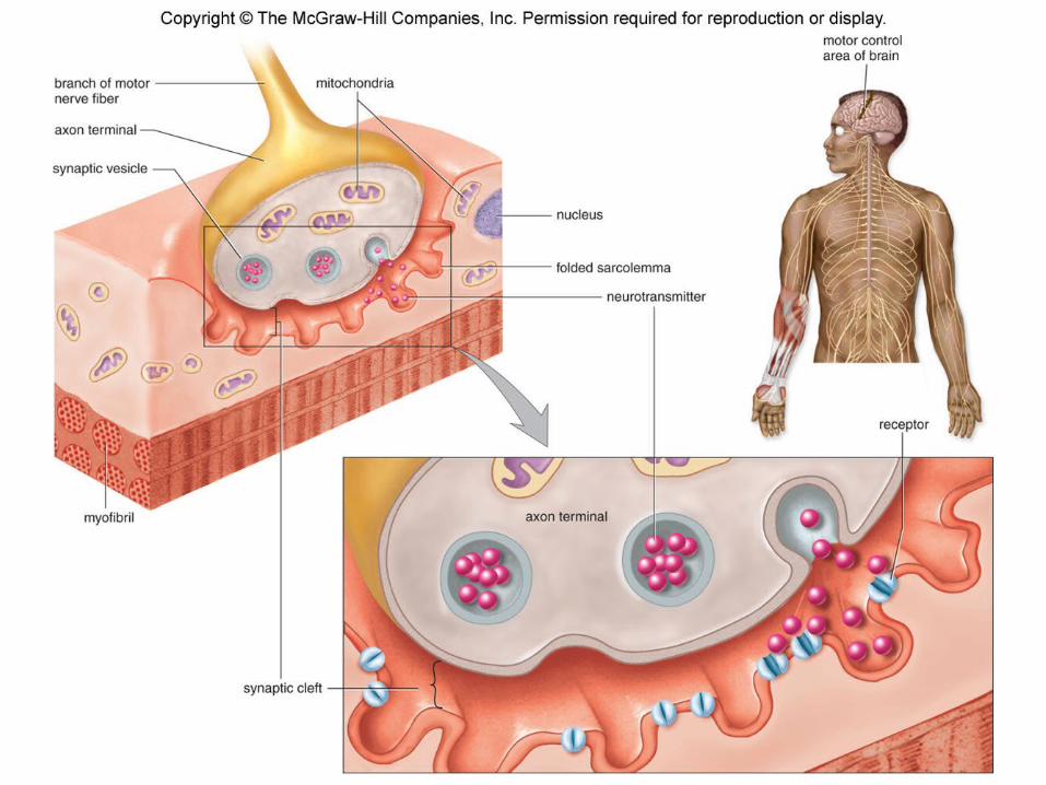
Fig 7.4
Contraction of Skeletal Muscle
o Steps involved in skeletal muscle contraction
• Nerve signal arrives at the axon terminal• The synaptic vesicles release Ach• Ach binds to receptors on the sarcolemma• The sarcolemma generates a signal that
travels down the T tubules to the SR• The SR releases calcium• Calcium from the SR causes the filaments to
slide past one another
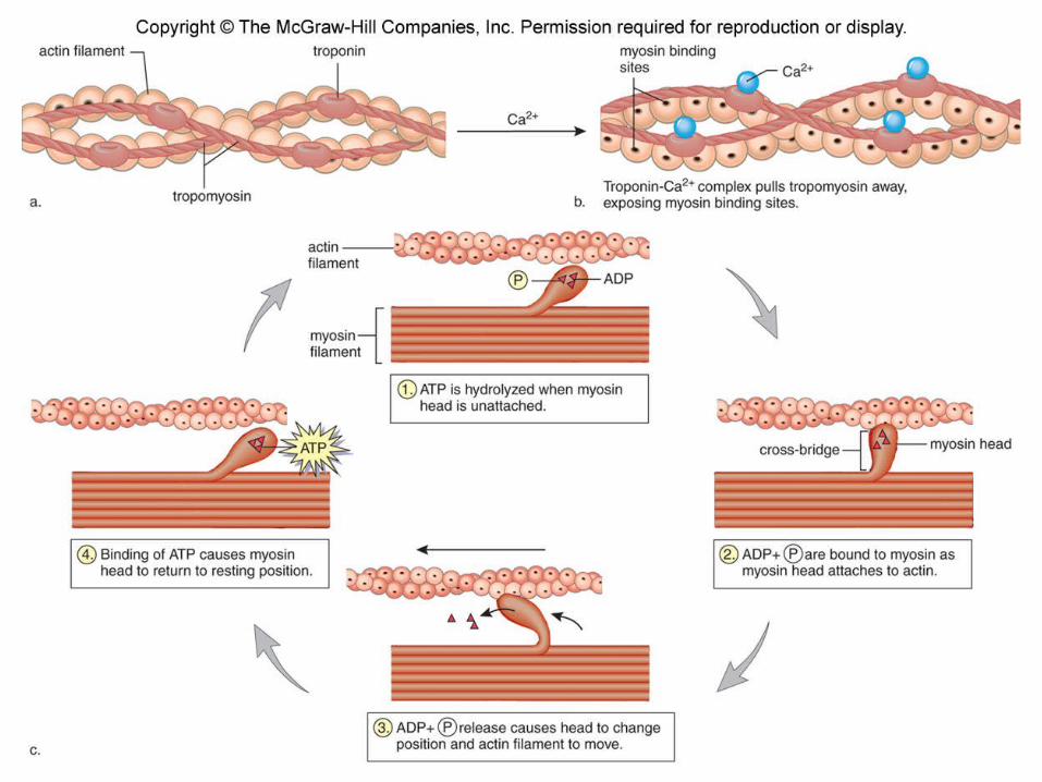
Contraction of Skeletal Muscle
o The Role of Actin and Myosin• Myosin binding sites on actin
molecules Covered by tropomyosin when muscle is
relaxed Released calcium combines with troponin
and myosin binding sites are exposed• Cross-bridges of myosin have two
binding sites One site binds to ATP Second binding site binds to actin
Contraction of Skeletal Muscle
o Energy for Muscle Contraction• ATP present before strenuous exercise
only lasts a few seconds• Muscles acquire new ATP in three
ways Creatine phosphate breakdown Cellular respiration Fermentation
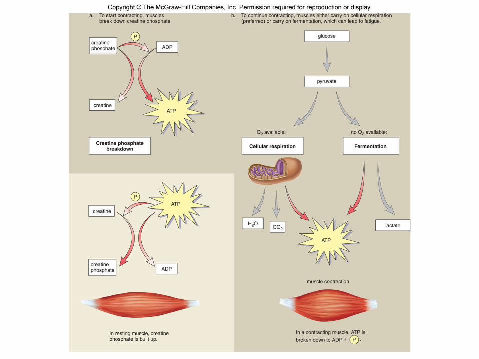
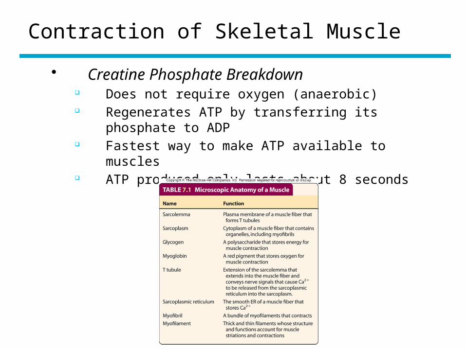
Contraction of Skeletal Muscle
• Creatine Phosphate Breakdown Does not require oxygen (anaerobic) Regenerates ATP by transferring its phosphate to
ADP Fastest way to make ATP available to muscles ATP produced only lasts about 8 seconds
TA 7.1
Contraction of Skeletal Muscle
• Cellular Respiration Usually provides most of a muscle’s ATP Uses glucose from stored glycogen and fatty acids from
stored fats Required oxygen Myoglobin can make oxygen available to muscle
mitochondria Carbon dioxide and water are end products Heat is a by-product
Contraction of Skeletal Muscle
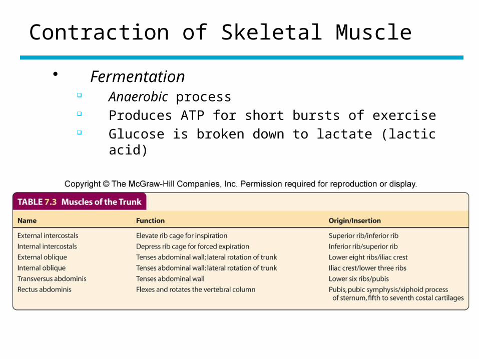
• Fermentation Anaerobic process Produces ATP for short bursts of exercise Glucose is broken down to lactate (lactic acid)
Contraction of Skeletal Muscle
o Oxygen Debt• Occurs when muscles use
fermentation to supply ATP• Requires replenishing creatine
phosphate supplies and disposing of lactic acid
Contraction of Smooth Muscle
o Smooth muscle fibers contain thick and thin filaments
• Filaments are not arranged into myofibrils that create striations
• Thin filaments are anchored to the sarcolemma or dense bodies
o When contracted, the elongated cells become shorter and wider
o Contraction occurs very slowlyo Contractions can last for long
periods of time without fatigue
Muscle Responses in the Laboratory
o All-or-none law – a muscle fiber contracts completely or not at all
o A whole muscle shows degrees of contraction
• Muscle twitch – a single contraction that lasts only a fraction of a second
Latent period Contraction period Relaxation period
• Summation – increased muscle contraction• Tetanic contraction – maximal sustained
contraction
Muscle Responses in the Laboratory
o Fatigue• Muscle relaxes even though
stimulation continues• Reasons for fatigue
ATP is depleted Accumulation of lactic acid in the
sarcoplasm inhibits muscle function ACh may become depleted
Muscle Responses in the Body
o Motor unit• A nerve fiber together with all of the muscle
fibers it innervates• Obeys the all-or-none law
o Recruitment• As the intensity of nervous stimulation
increases, more motor units are activated• Results in stronger muscle contractions
o Tone• Some muscle fibers are always contracting• Important in maintaining posture
Muscle Responses in the Body
o Athletics and muscle contraction• Size of muscles
Atrophy – a decrease in muscle size Hypertrophy – an increase in muscle size
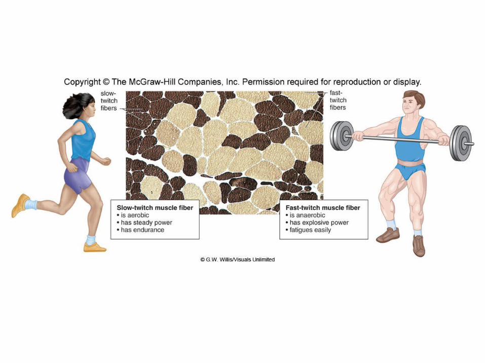
• Slow-twitch fibers (Type I fibers) Tend to be aerobic Have more endurance Have many mitochondria Dark in color because they contain
myoglobin Highly resistant to fatigue
Muscle Responses In the Body
• Fast-twitch fibers (Type II fibers) Tend to be anaerobic Designed for strength Light in color Have fewer mitochondria, little or no
myoglobin, and fewer blood vessels than fast-twitch fibers
Vulnerable to accumulation of lactic acid and can fatigue easily
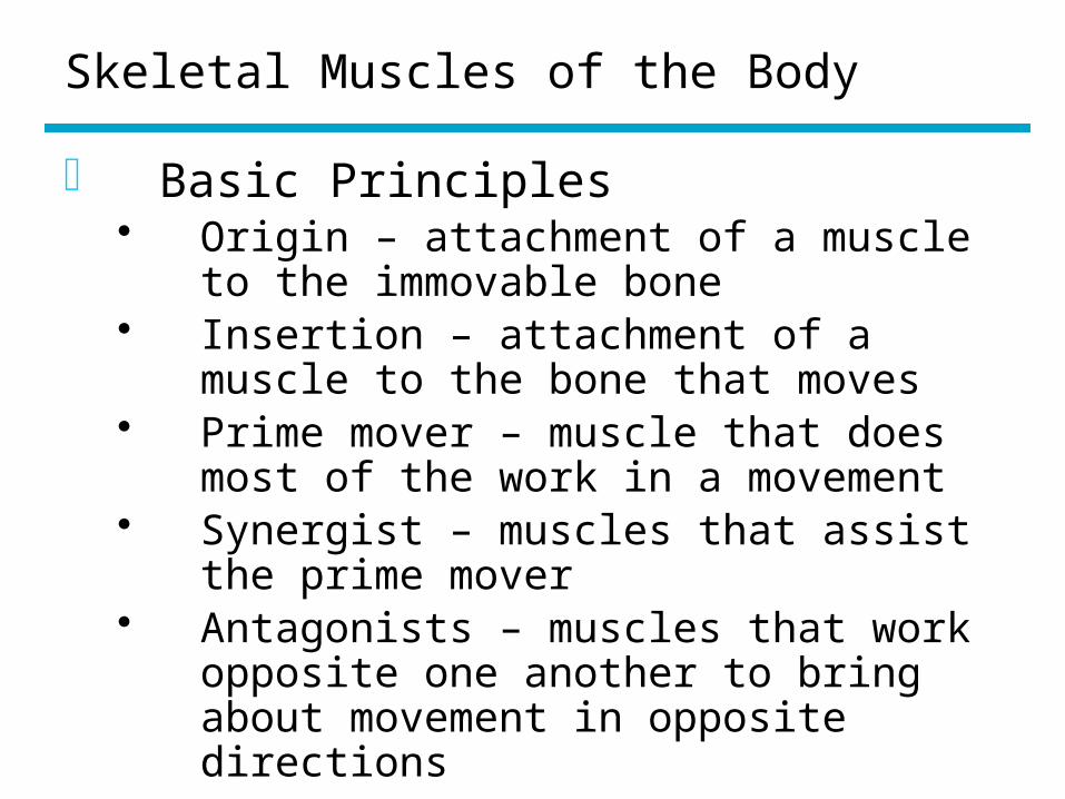
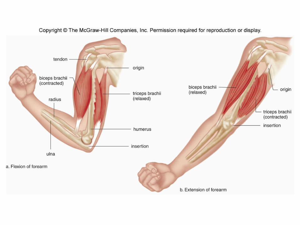
Skeletal Muscles of the Body
o Basic Principles• Origin – attachment of a muscle to the
immovable bone• Insertion – attachment of a muscle to
the bone that moves• Prime mover – muscle that does most
of the work in a movement• Synergist – muscles that assist the
prime mover• Antagonists – muscles that work
opposite one another to bring about movement in opposite directions
Skeletal Muscles of the Body
o Naming Muscles• Size• Shape• Direction of fibers• Location• Attachment• Number of attachments• Action
Skeletal Muscles of the Body
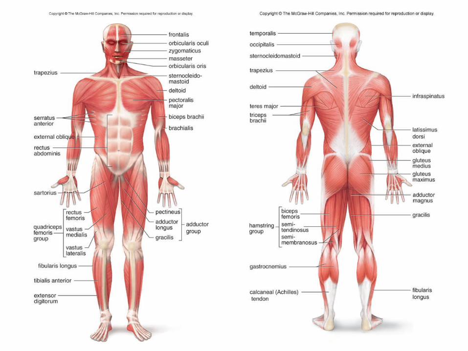
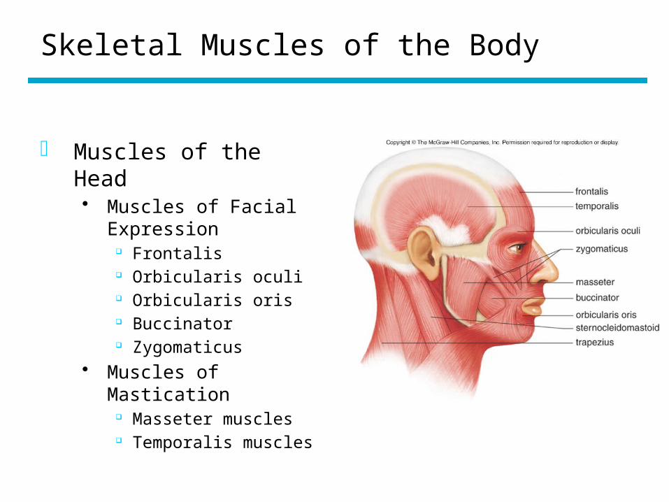
o Muscles of the Head• Muscles of Facial
Expression Frontalis Orbicularis oculi Orbicularis oris Buccinator Zygomaticus
• Muscles of Mastication Masseter muscles Temporalis muscles
Fig 7.13
Skeletal Muscles of the Body
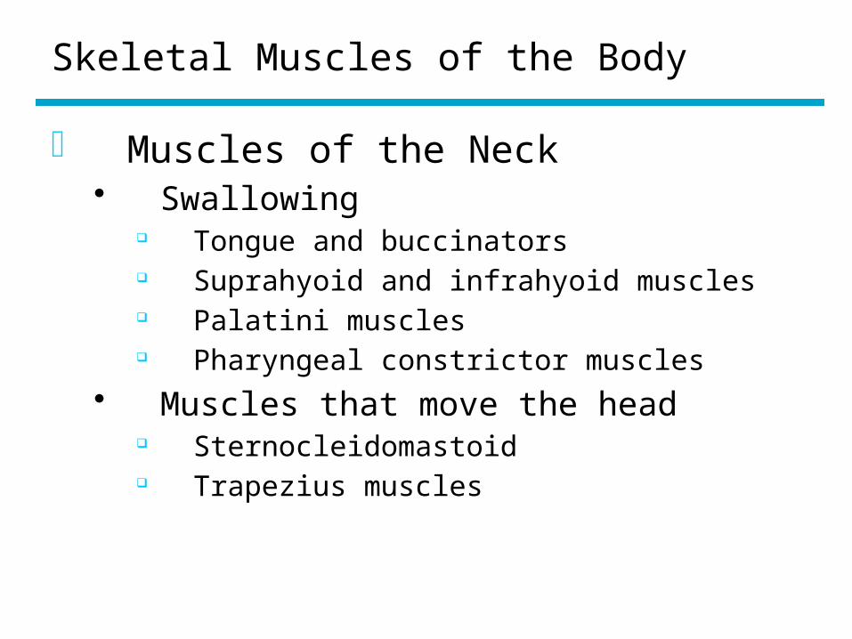
o Muscles of the Neck• Swallowing
Tongue and buccinators Suprahyoid and infrahyoid muscles Palatini muscles Pharyngeal constrictor muscles
• Muscles that move the head Sternocleidomastoid Trapezius muscles
Skeletal Muscles of the Body
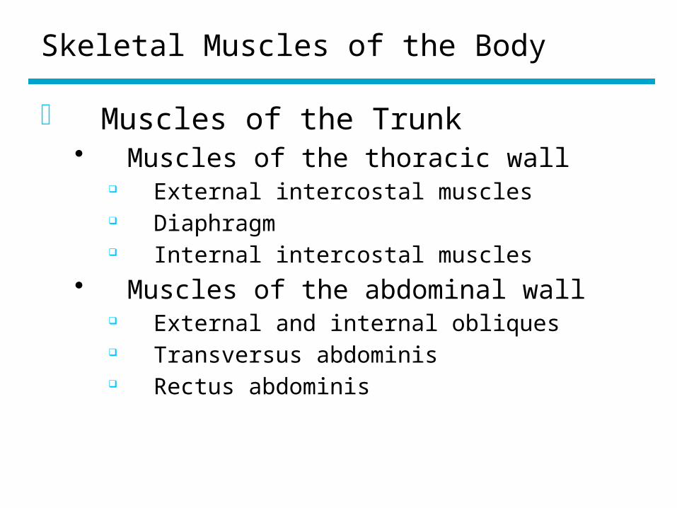
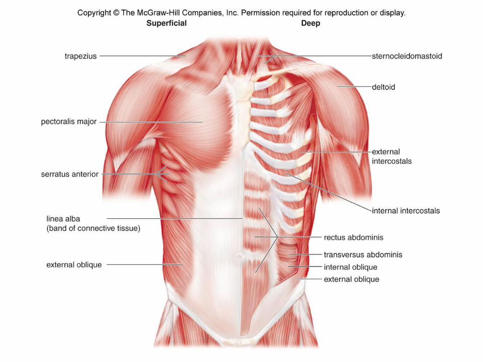
o Muscles of the Trunk• Muscles of the thoracic wall
External intercostal muscles Diaphragm Internal intercostal muscles
• Muscles of the abdominal wall External and internal obliques Transversus abdominis Rectus abdominis
Skeletal Muscles of the Body
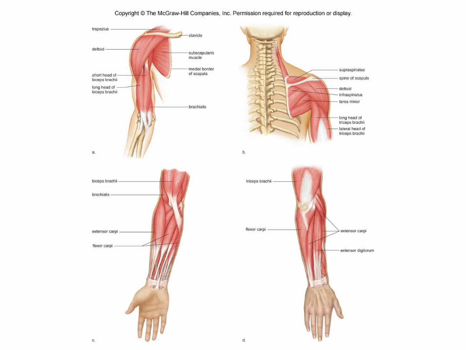
o Muscles of the Shoulder• Muscles that move the scapula
Trapezius Serratus anterior
• Muscles that move the arm Deltoid Pectoralis major Latissimus dorsi Rotator cuff muscles
Supraspinatus Infraspinatus Teres minor Subscapularis
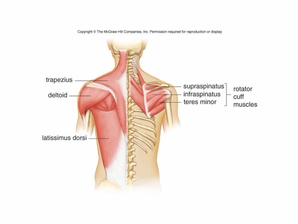
Fig 7.15
Skeletal Muscles of the Body
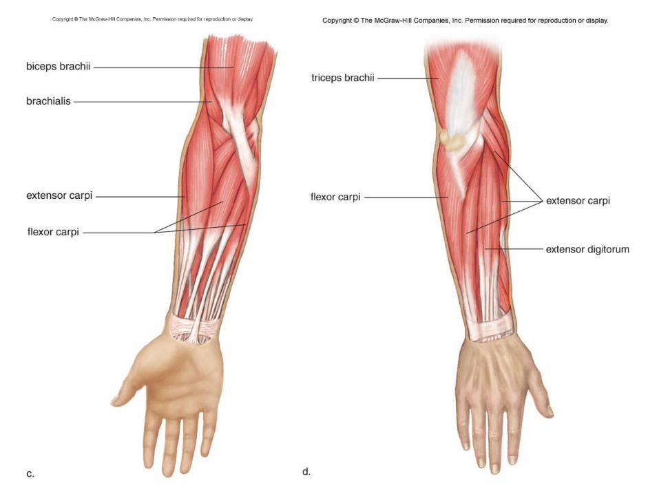
o Muscles of the Arm• Biceps brachii• Brachialis• Triceps brachii
o Muscles of the Forearm• Flexor carpi and extensor carpi• Flexor digitorum and extensor
digitorum
Skeletal Muscles of the Body
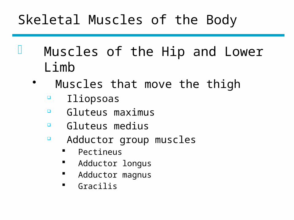
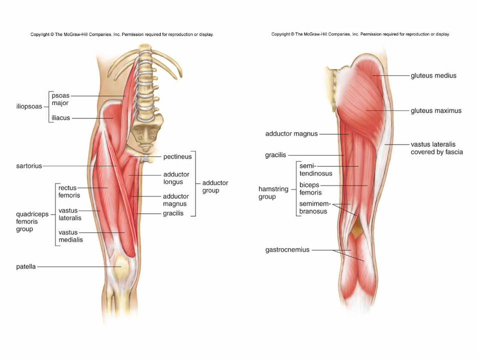
o Muscles of the Hip and Lower Limb• Muscles that move the thigh
Iliopsoas Gluteus maximus Gluteus medius Adductor group muscles
Pectineus Adductor longus Adductor magnus Gracilis
Skeletal Muscles of the Body
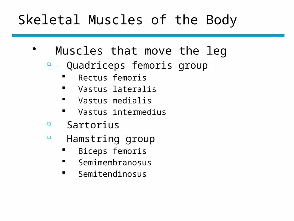
• Muscles that move the leg Quadriceps femoris group
Rectus femoris Vastus lateralis Vastus medialis Vastus intermedius
Sartorius Hamstring group
Biceps femoris Semimembranosus Semitendinosus
Skeletal Muscles of the Body
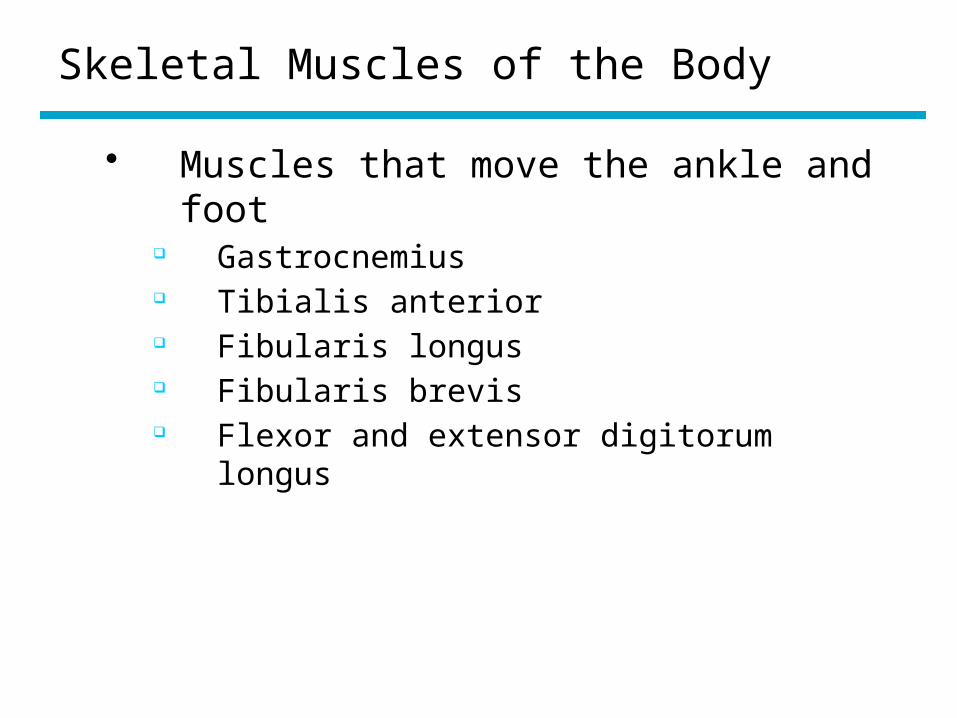
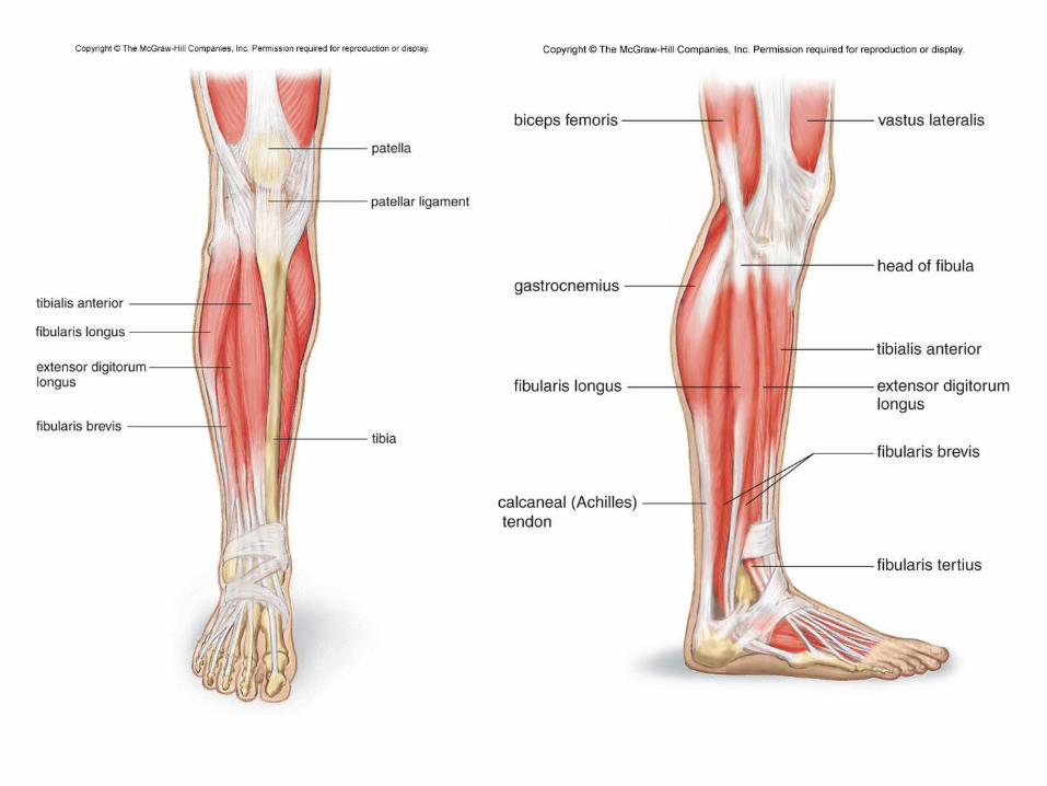
• Muscles that move the ankle and foot Gastrocnemius Tibialis anterior Fibularis longus Fibularis brevis Flexor and extensor digitorum longus
Effects of Aging
o Muscle mass and strength tend to decrease
o Endurance decreaseso Exercise at any age can stimulate
muscle buildup
Homeostasis
o Cardiac muscle contraction forces blood into the arteries and arterioles
o Smooth muscle in arteries and arterioles help maintain blood pressure
o Smooth muscle contraction moves food along the digestive tract and assists in the voiding of urine
o Skeletal muscles protect internal organs and stabilizes joints
o Skeletal muscles are active during breathingo Heat produced by skeletal muscle contraction
helps maintain normal body temperatureo Skeletal muscle contraction allows us to
relocate our bodies