Embed Size (px)
Citation preview
The Nervous System ReviewPsTL 1082-1/Fall 08
Modified slides from
Murray Jensen’s Originals
for PsTL 1135Images from..
Loyola University Medical CenterLumen Histology Site
Anatomy TVJayDoc HistoWeb
McGraw Hill
QuickTime™ and aTIFF (Uncompressed) decompressor
are needed to see this picture.

What is removed in a lobotomy?
• What are some of the prefrontal cortex functions?
What is a lobotomy?
• It is a surgery that destroys the prefrontal cortex--common changes include a major personality change
• Prefrontal cortex functions – planning, moral judgement, and emotional
control
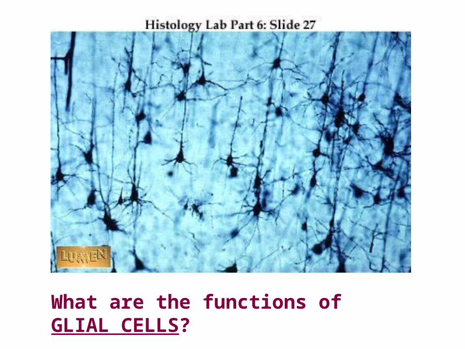
What are the functions of GLIAL CELLS?
Functions of Glial Cells
• (1) to surround neurons and hold them in place,
• (2) to supply nutrients and oxygen to neurons,
• (3) to insulate one neuron from another, and
• (4) to destroy pathogens and remove dead neurons.
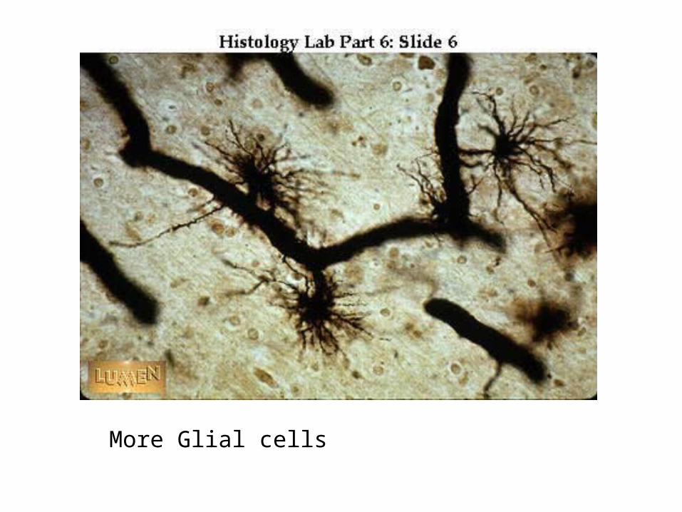
More Glial cells
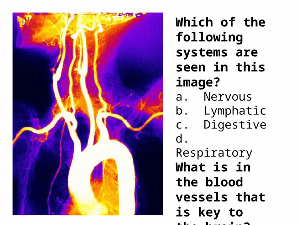
Which of the following systems are seen in this image?a. Nervousb. Lymphaticc. Digestived. RespiratoryWhat is in the blood vessels that is key to the brain?

What is in the blood vessels that is key to the brain?
Oxygen!!!

Blood transports oxygen and other nutrients necessary for the health of neurons, so a constant flow of blood to the brain must be maintained.
The brain uses approximately twenty percent of the body's blood and needs twenty-five percent of the body's oxygen supply to function optimally.
Approximately 46 milliliters of oxygen are used by the entire brain in one minute.
During sleep, blood flow to the brain is increased, but the rate of oxygen consumption remains the same.
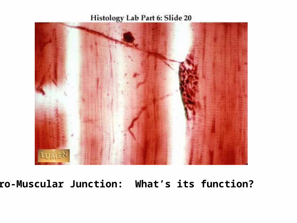
Neuro-Muscular Junction: What’s its function?
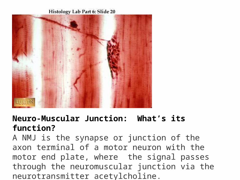
Neuro-Muscular Junction: What’s its function?A NMJ is the synapse or junction of the axon terminal of a motor neuron with the motor end plate, where the signal passes through the neuromuscular junction via the neurotransmitter acetylcholine.
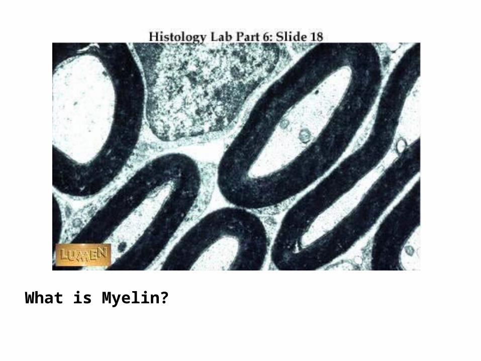
What is Myelin?
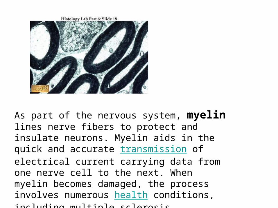
As part of the nervous system, myelin lines nerve fibers to protect and insulate neurons. Myelin aids in the quick and accurate transmission of electrical current carrying data from one nerve cell to the next. When myelin becomes damaged, the process involves numerous health conditions, including multiple sclerosis.
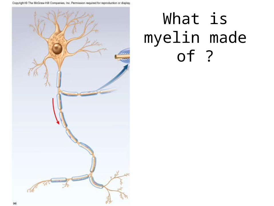
What is myelin made of ?
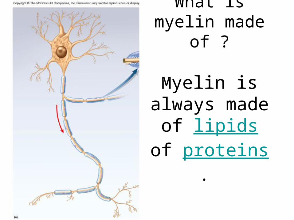
What is myelin made of ?
Myelin is always made of lipids of proteins.
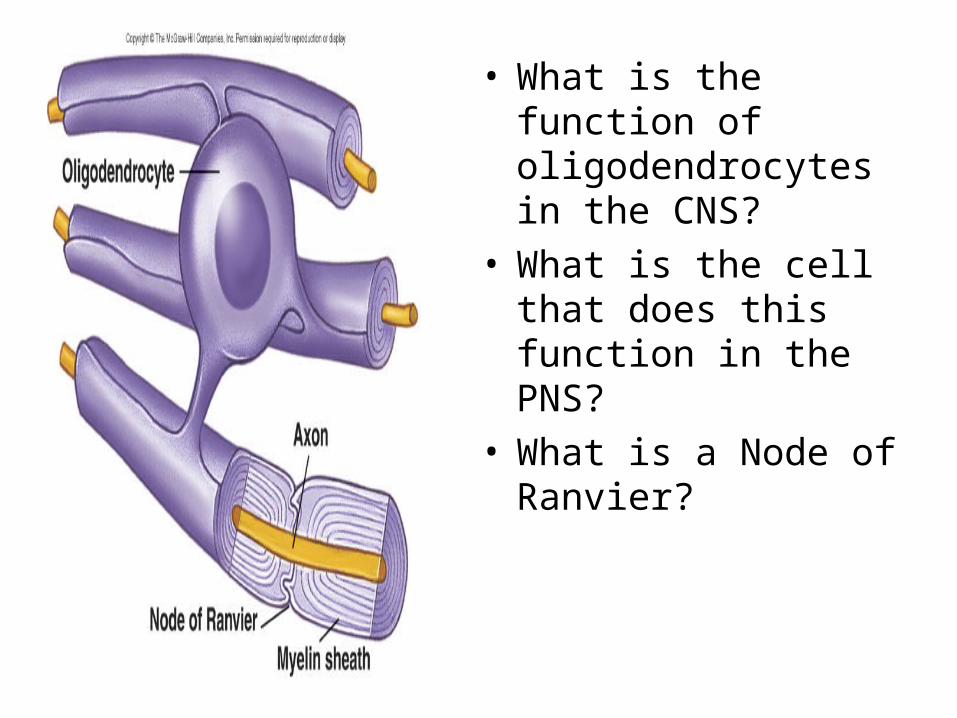
• What is the function of oligodendrocytes in the CNS?
• What is the cell that does this function in the PNS?
• What is a Node of Ranvier?
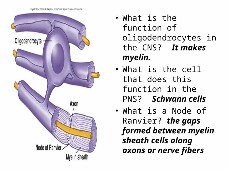
• What is the function of oligodendrocytes in the CNS? It makes myelin.
• What is the cell that does this function in the PNS? Schwann cells
• What is a Node of Ranvier? the gaps formed between myelin sheath cells along axons or nerve fibers

Subdivisions of Nervous System
Two major anatomical subdivisions• Central nervous system (CNS)
– brain and spinal cord enclosed in bony coverings
• Peripheral nervous system (PNS)– nerve = bundle of axons in connective tissue– ganglion = swelling of cell bodies in a nerve

Fundamental Types of Neurons
• What are the functions of Sensory (afferent) neurons?
• Where are Interneurons found? • What are the functions of interneurons?• There are more interneurons than sensory and motor
neurons. True/False
• What is the function of Motor (efferent) neurons?• What are effectors?
Fundamental Types of Neurons
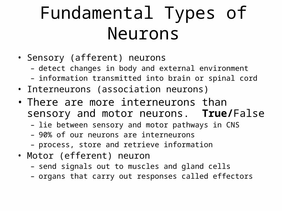
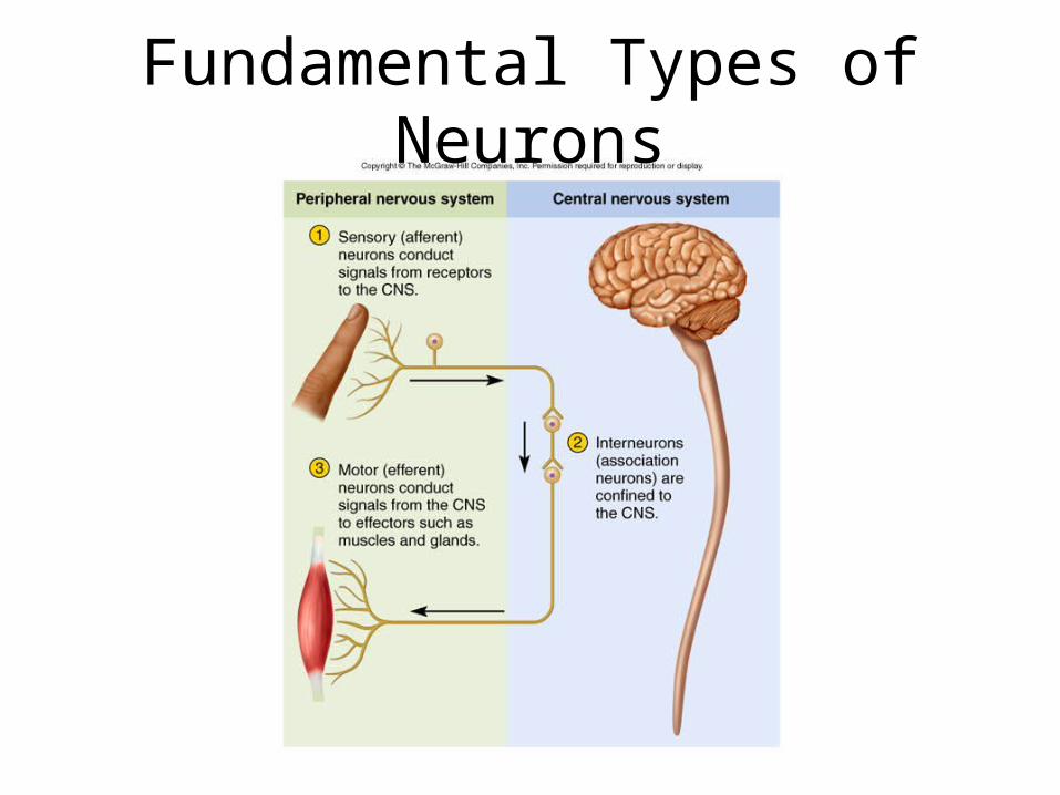
• Sensory (afferent) neurons– detect changes in body and external environment– information transmitted into brain or spinal cord
• Interneurons (association neurons)
• There are more interneurons than sensory and motor neurons. True/False– lie between sensory and motor pathways in CNS– 90% of our neurons are interneurons– process, store and retrieve information
• Motor (efferent) neuron– send signals out to muscles and gland cells– organs that carry out responses called effectors
Fundamental Types of Neurons
Meninges of the BrainWhat are the functions of the
meninges?
What are the names of the meninges in order from
superficial to deep?

Meninges of the BrainWhat are the functions of the meninges? Their major function is to
protect the CNS.
What are the names of the meninges in order from
superficial to deep? The dura mater, arachnoid mater, and pia mater.

Meninges of the Brain
What is cerebrospinal fluid?
Where is it located in the meninges?
What is cerebrospinal fluid? Where is it located in the
meninges?
• Cerebrospinal fluid is a clear bodily fluid that occupies the subarachnoid space and the ventricular system around and inside the brain. Essentially, the brain "floats" in it.
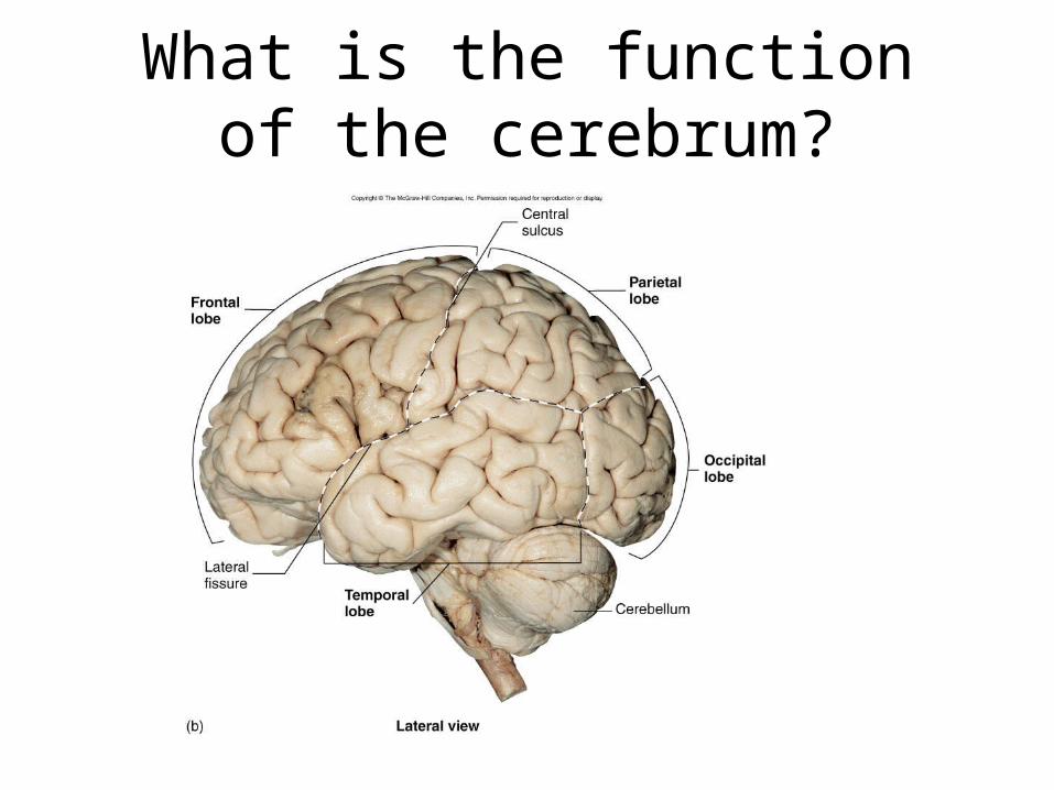
What is the function of the cerebrum?

What is the function of the cerebrum?
• Movement
• Sensory Processing
• Olfaction
• Language and communication
• Learning and Memory
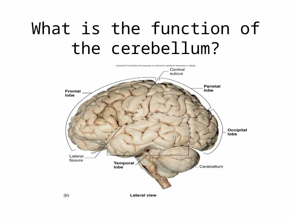
What is the function of the cerebellum?
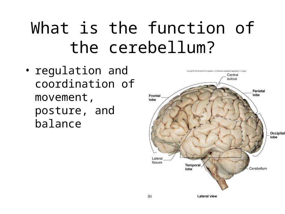
What is the function of the cerebellum?
• regulation and coordination of movement, posture, and balance
What is the difference between a sulcus and a
gyrus?
• A sulcus is a depression or fissure in the surface of the brain.
• A gyrus is a convoluted ridge between anatomical grooves.
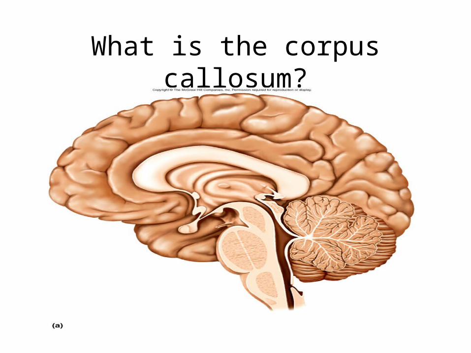
What is the corpus callosum?
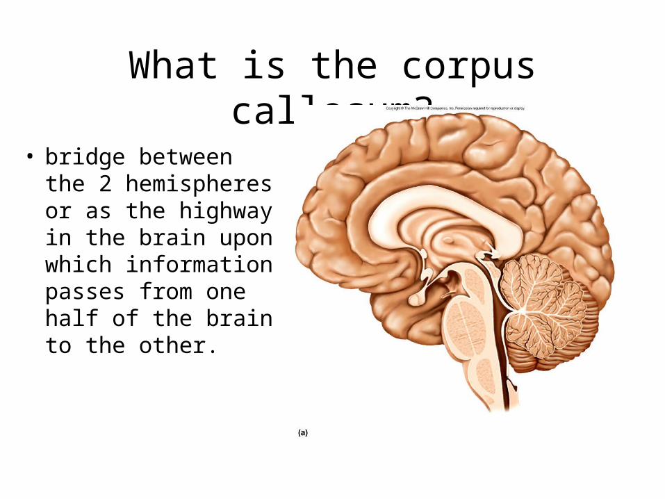
What is the corpus callosum?
• bridge between the 2 hemispheres or as the highway in the brain upon which information passes from one half of the brain to the other.
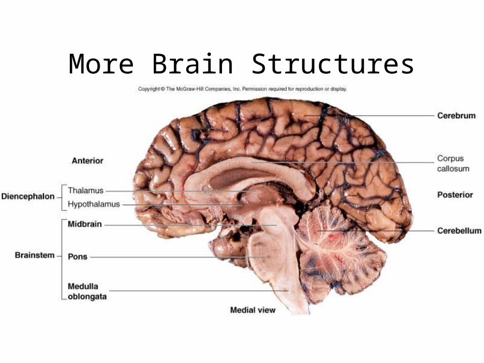
More Brain Structures
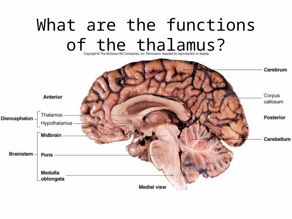
What are the functions of the thalamus?
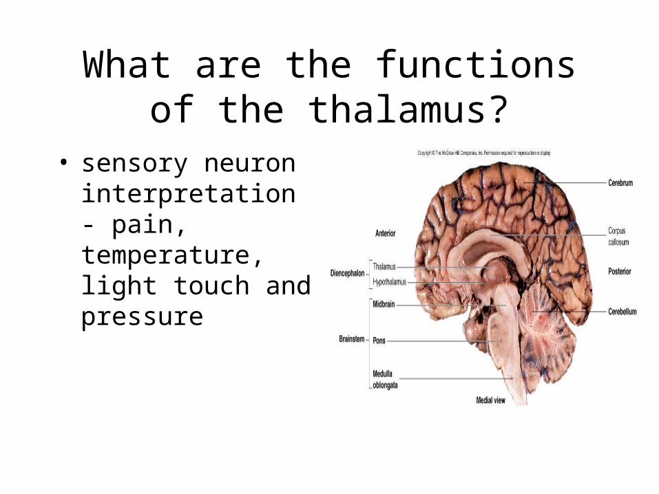
What are the functions of the thalamus?
• sensory neuron interpretation - pain, temperature, light touch and pressure

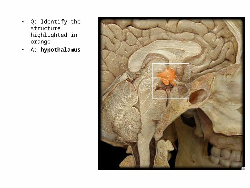
What are the functions of the hypothalamus?
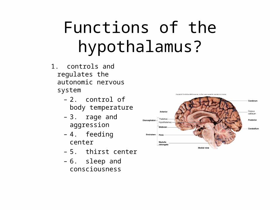
Functions of the hypothalamus?
1. controls and regulates the autonomic nervous system
– 2. control of body temperature
– 3. rage and aggression
– 4. feeding center– 5. thirst center– 6. sleep and
consciousness

More Brain Structures

More Brain Structures
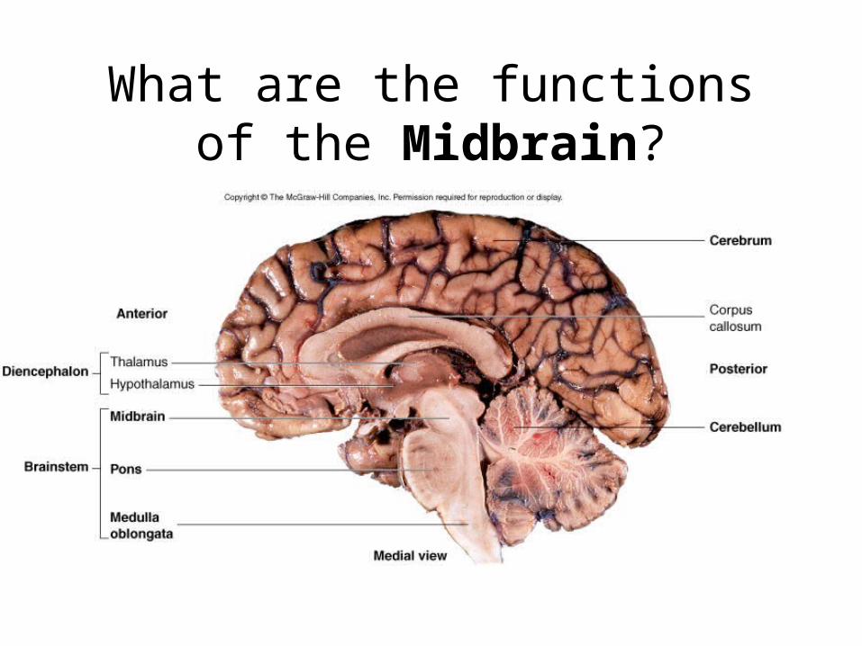
What are the functions of the Midbrain?
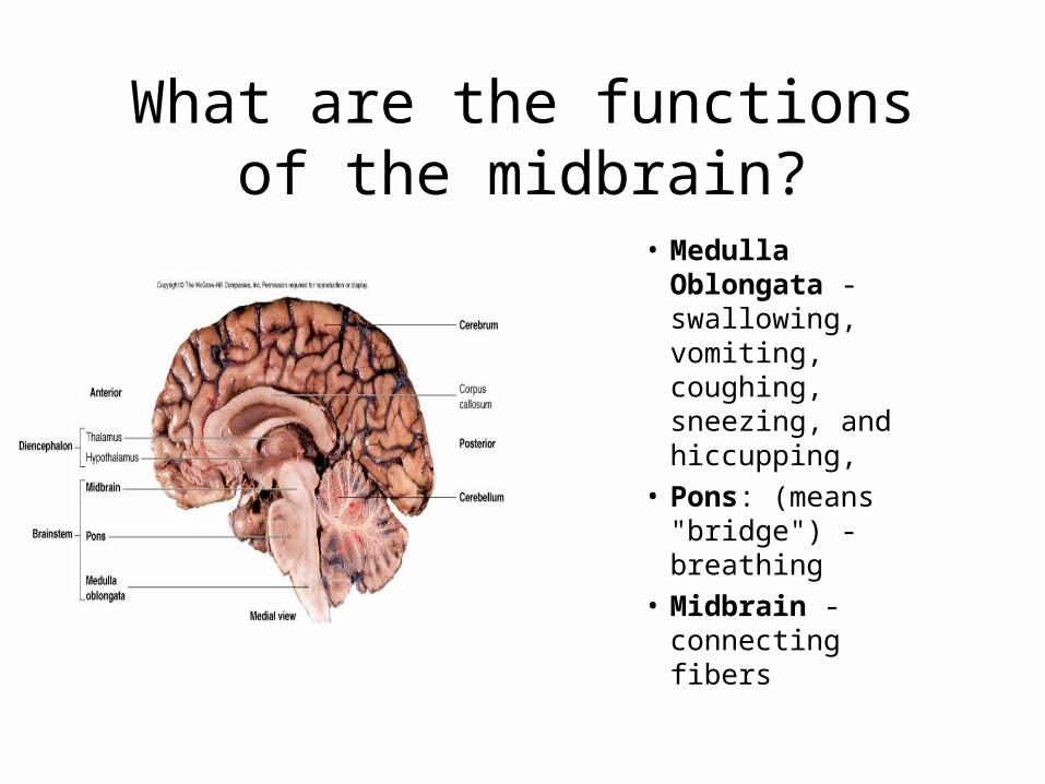
What are the functions of the midbrain?
• Medulla Oblongata - swallowing, vomiting, coughing, sneezing, and hiccupping,
• Pons: (means "bridge") - breathing
• Midbrain - connecting fibers
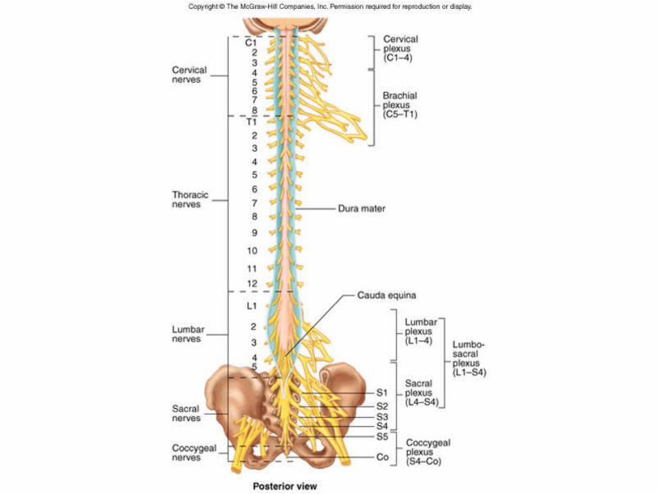
Figure 8.21
Figure 12.12

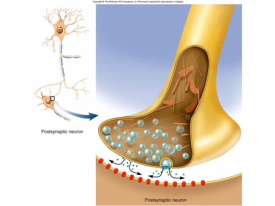
Figure 12.20
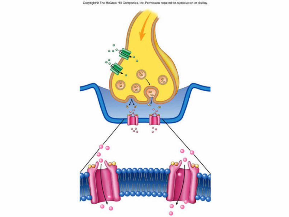
Figure 8.13
Figure 12.13a

Meningitis
• Inflammation of the meninges
• Disease of infancy and childhood– between 3 months and 2 years of age
• Bacterial and virus invasion of the CNS by way of the nose and throat
• Signs include high fever, stiff neck, drowsiness and intense headache and may progress to coma
• Diagnose by examining the CSF– lumbar puncture (spinal tap)
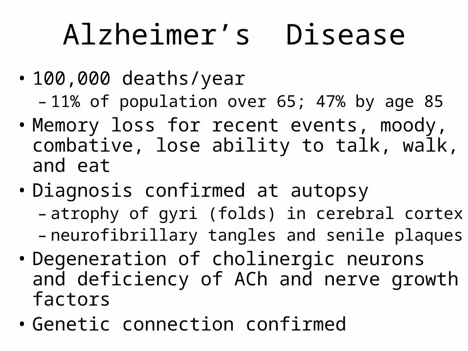
Alzheimer’s Disease
• 100,000 deaths/year– 11% of population over 65; 47% by age 85
• Memory loss for recent events, moody, combative, lose ability to talk, walk, and eat
• Diagnosis confirmed at autopsy– atrophy of gyri (folds) in cerebral cortex– neurofibrillary tangles and senile plaques
• Degeneration of cholinergic neurons and deficiency of ACh and nerve growth factors
• Genetic connection confirmed

Effects of Alzheimer’s Disease
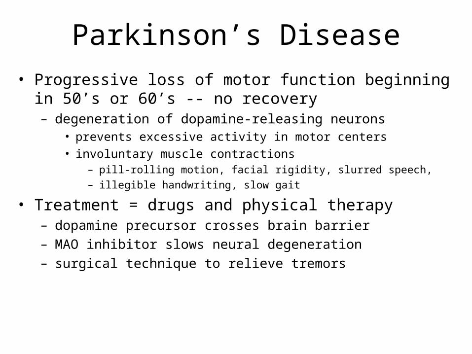
Parkinson’s Disease• Progressive loss of motor function beginning in 50’s or 60’s --
no recovery– degeneration of dopamine-releasing neurons
• prevents excessive activity in motor centers
• involuntary muscle contractions– pill-rolling motion, facial rigidity, slurred speech, – illegible handwriting, slow gait
• Treatment = drugs and physical therapy– dopamine precursor crosses brain barrier– MAO inhibitor slows neural degeneration– surgical technique to relieve tremors
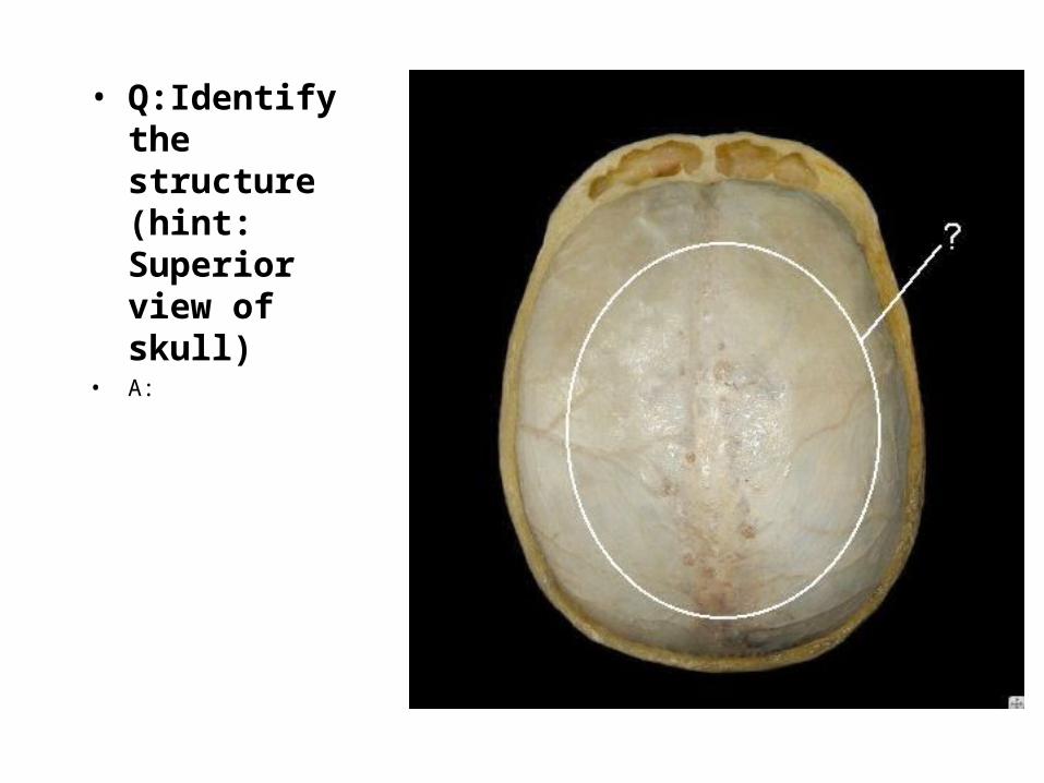
• Q:Identify the structure (hint: Superior view of skull)
• A:
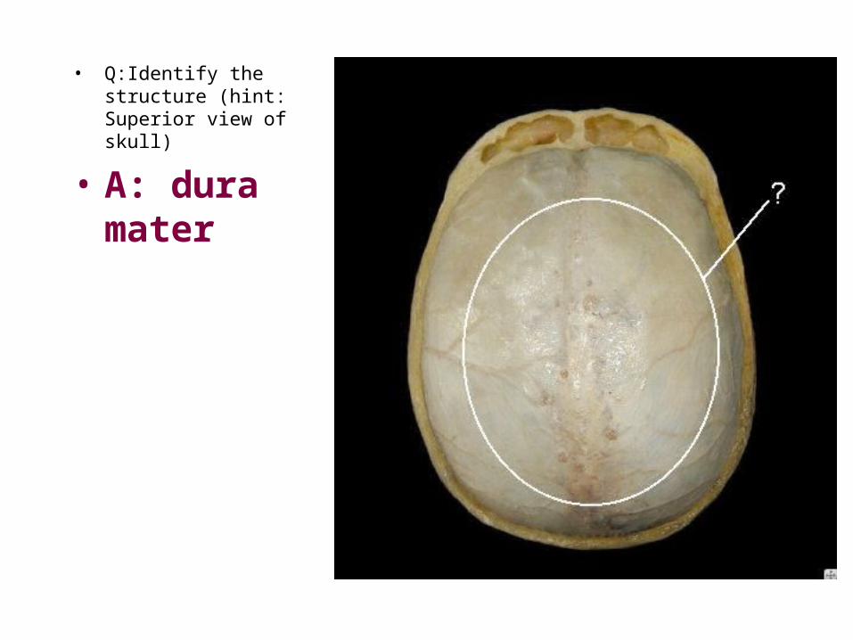
• Q:Identify the structure (hint: Superior view of skull)
• A: dura mater
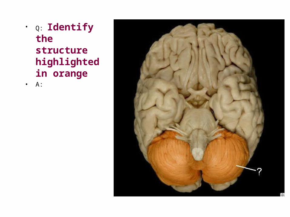
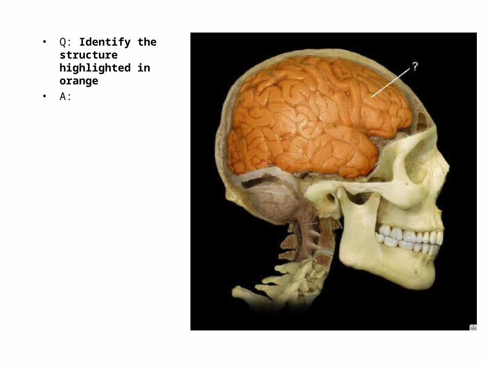
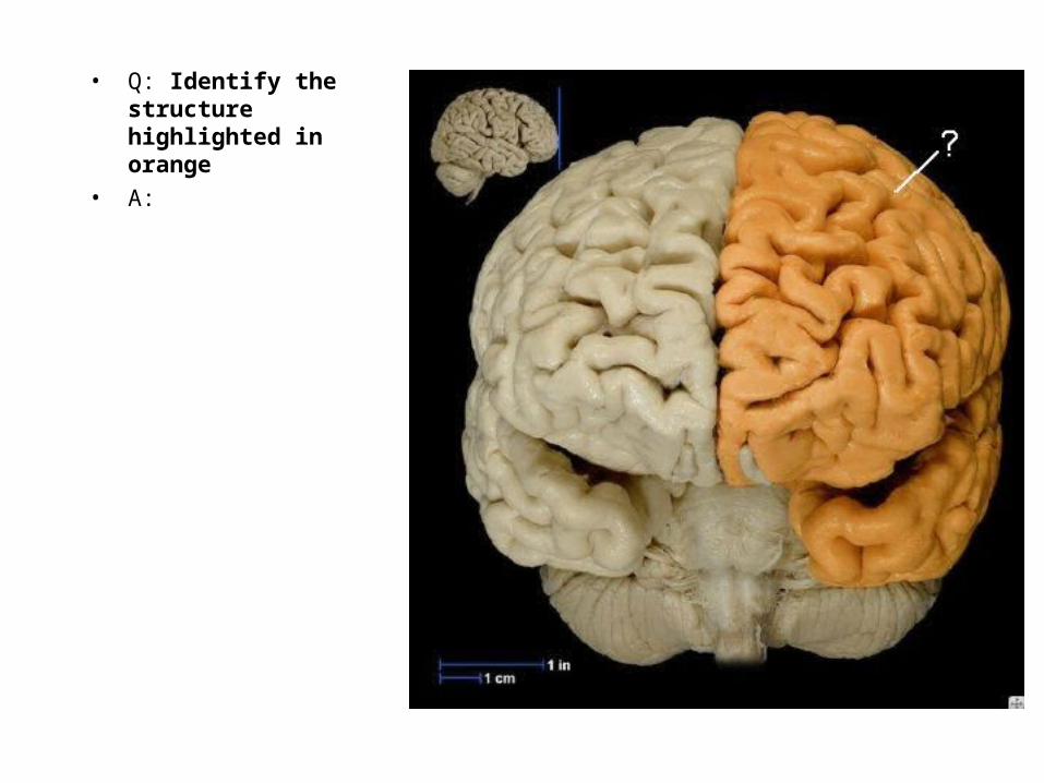
• Q: Identify the structure highlighted in orange
• A:
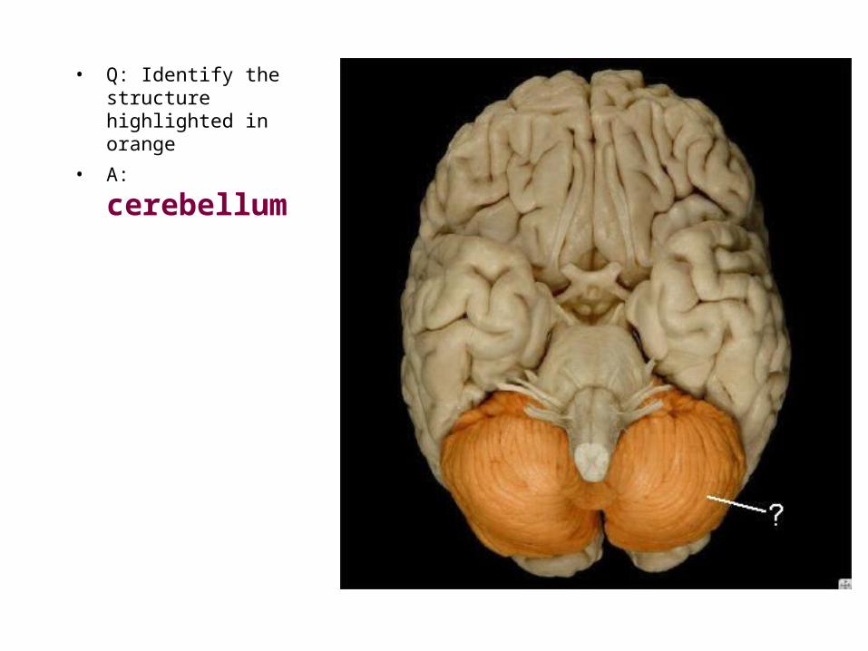
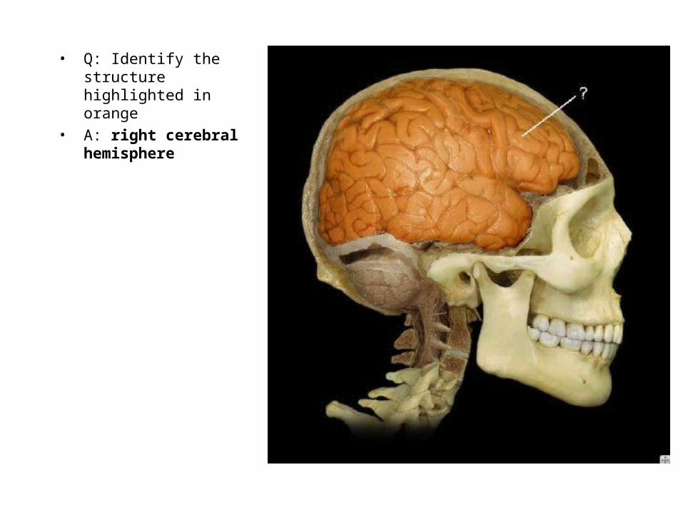
• Q: Identify the structure highlighted in orange
• A: cerebellum

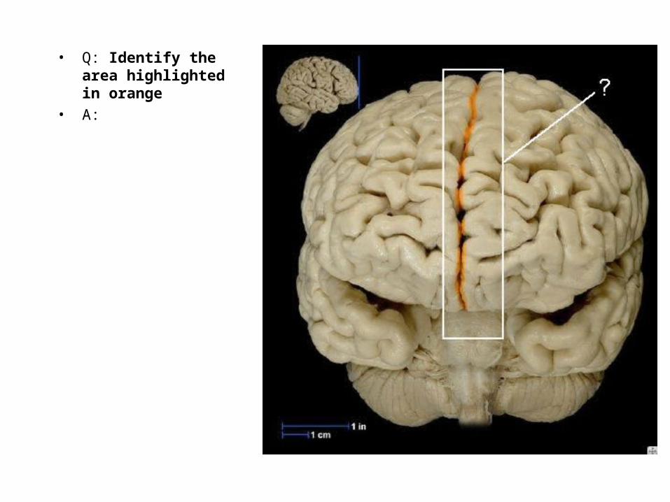
• Q: Identify the area highlighted in orange
• A:

• Q: Identify the area highlighted in orange
• A:
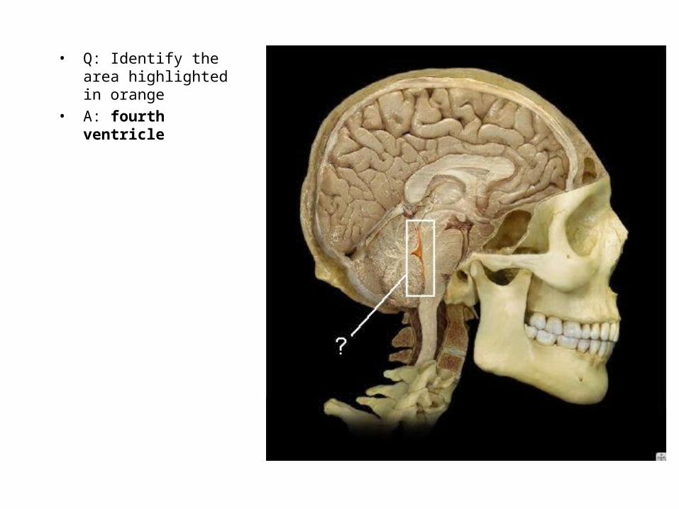
• Q: Identify the area highlighted in orange
• A: fourth ventricle
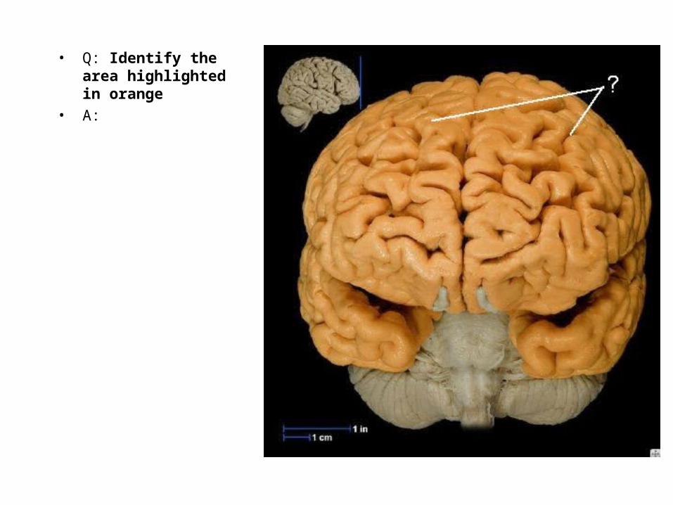
• Q: Identify the area highlighted in orange
• A:
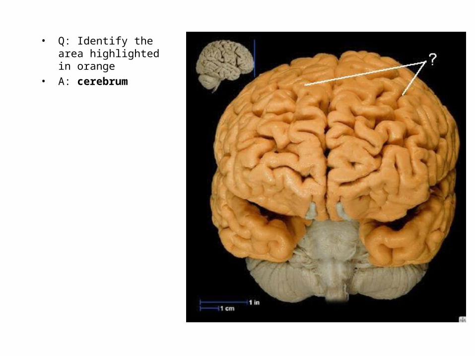
• Q: Identify the area highlighted in orange
• A: cerebrum
• Q: Identify the structure highlighted in orange
• A:
• Q: Identify the structure highlighted in orange
• A: right cerebral hemisphere
• Q: Identify the structure highlighted in orange
• A:
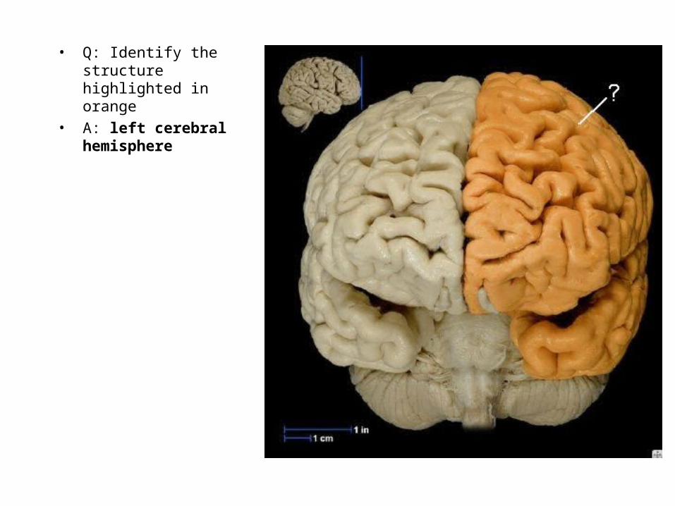
• Q: Identify the structure highlighted in orange
• A: left cerebral hemisphere

• Q: Identify the structure highlighted in orange
• A:
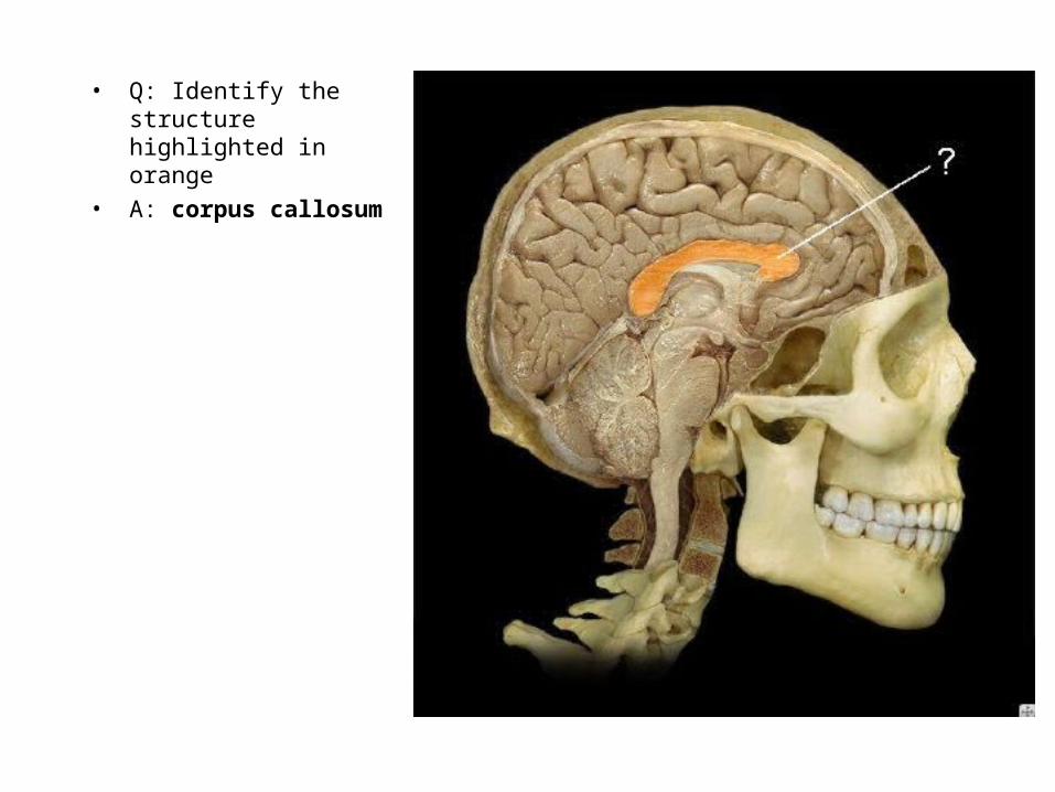
• Q: Identify the structure highlighted in orange
• A: corpus callosum

• Q: Identify the structure highlighted in orange
• A:

• Q: Identify the structure highlighted in orange
• A: fornix

• Q: Identify the structure highlighted in orange
• A:

• Q: Identify the structure highlighted in orange
• A: precentral gyrus

• Q: Identify the structure highlighted in orange
• A:

• Q: Identify the structure highlighted in orange
• A: postcentral
gyrus
• Q: Identify the area highlighted in orange
• A:
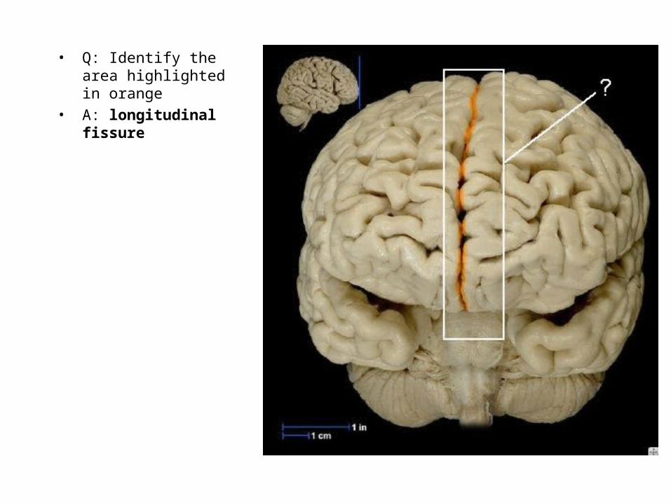
• Q: Identify the area highlighted in orange
• A: longitudinal fissure

• Q: Identify the structure highlighted in orange
• A:

• Q: Identify the structure highlighted in orange
• A: frontal lobe
• Q: Identify the structure highlighted in orange
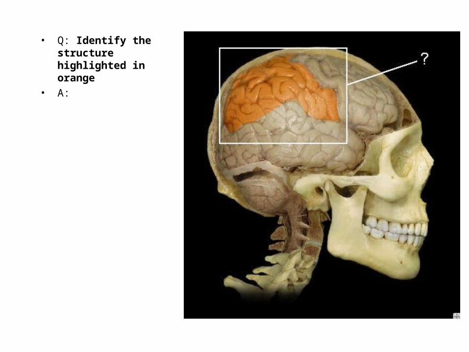
• A:
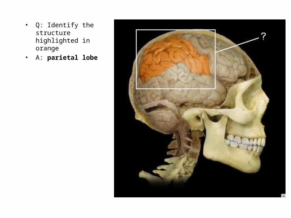
• Q: Identify the structure highlighted in orange
• A: parietal lobe

• Q: Identify the structure highlighted in orange
• A:

• Q: Identify the structure highlighted in orange
• A: occipital lobe

• Q: Identify the structure highlighted in orange
• A:
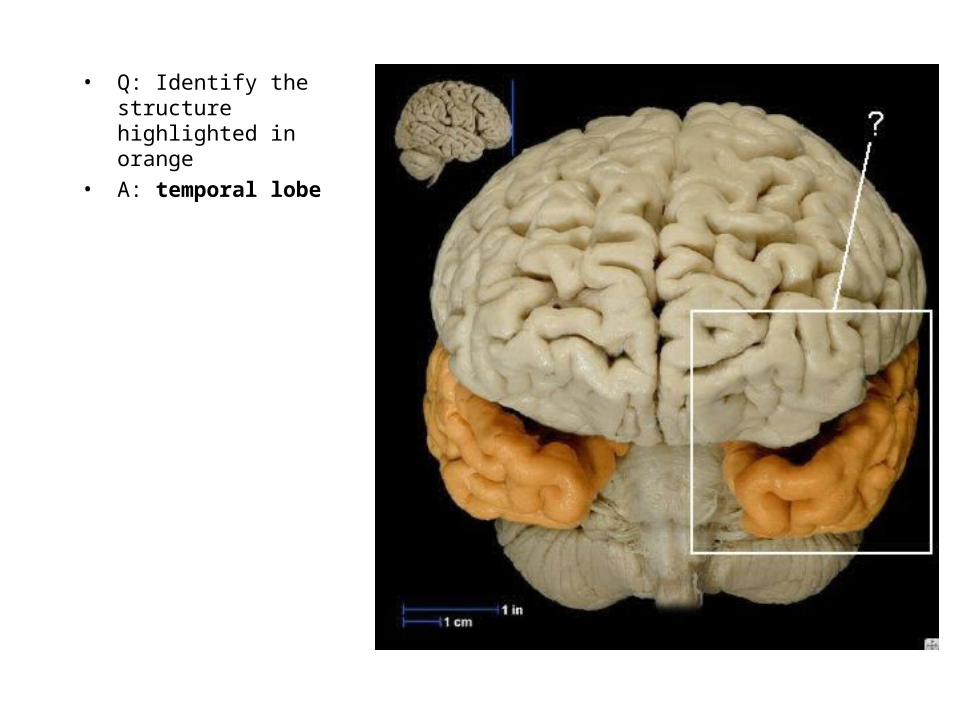
• Q: Identify the structure highlighted in orange
• A: temporal lobe
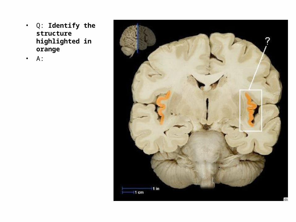
• Q: Identify the structure highlighted in orange
• A:
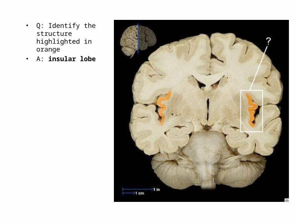
• Q: Identify the structure highlighted in orange
• A: insular lobe
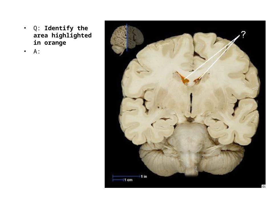
• Q: Identify the area highlighted in orange
• A:
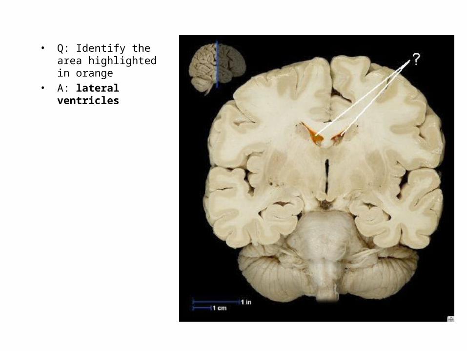
• Q: Identify the area highlighted in orange
• A: lateral ventricles

• Q: Identify the area highlighted in orange
• A:

• Q: Identify the area highlighted in orange
• A: third ventricle

• Q: Identify the area shaded in pink
• A:
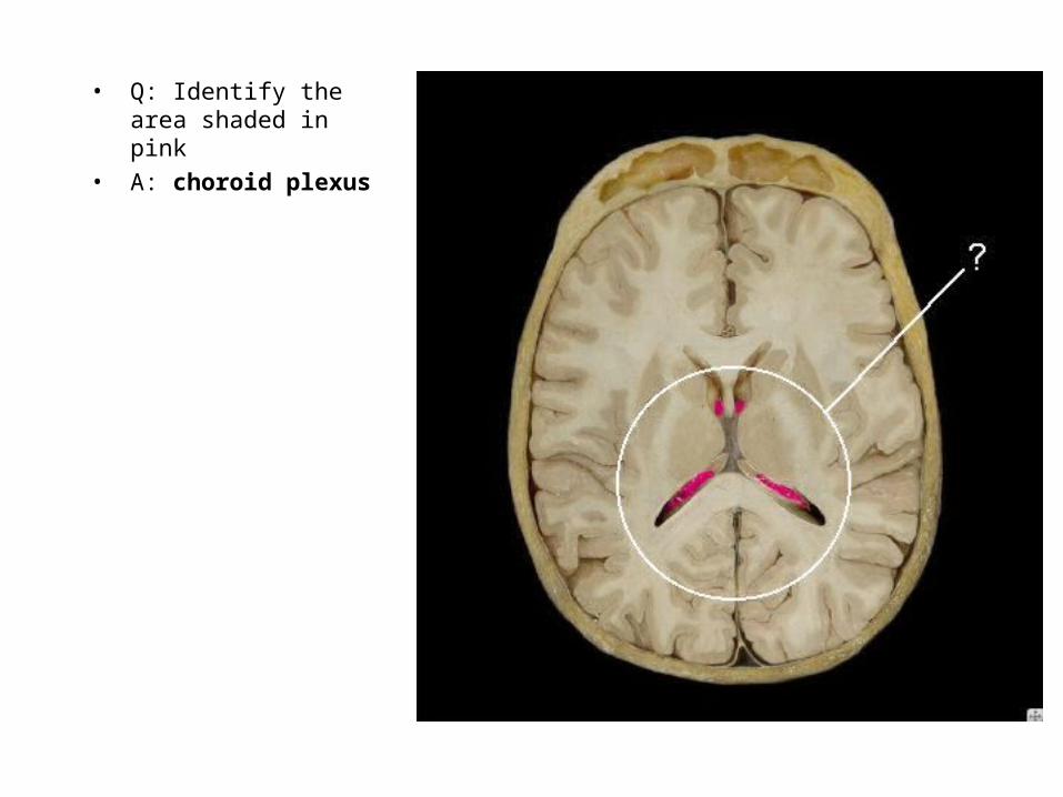
• Q: Identify the area shaded in pink
• A: choroid plexus
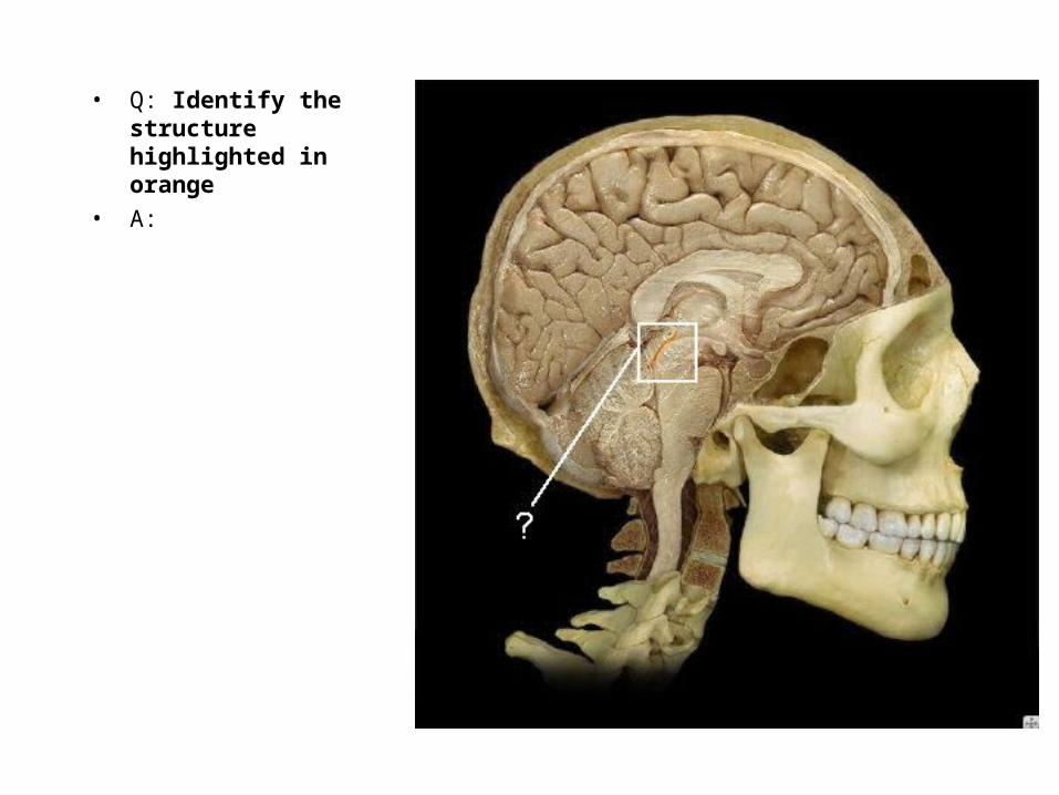
• Q: Identify the structure highlighted in orange
• A:
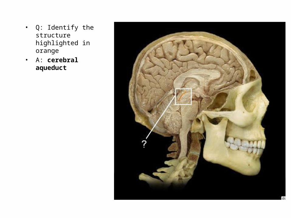
• Q: Identify the structure highlighted in orange
• A: cerebral aqueduct
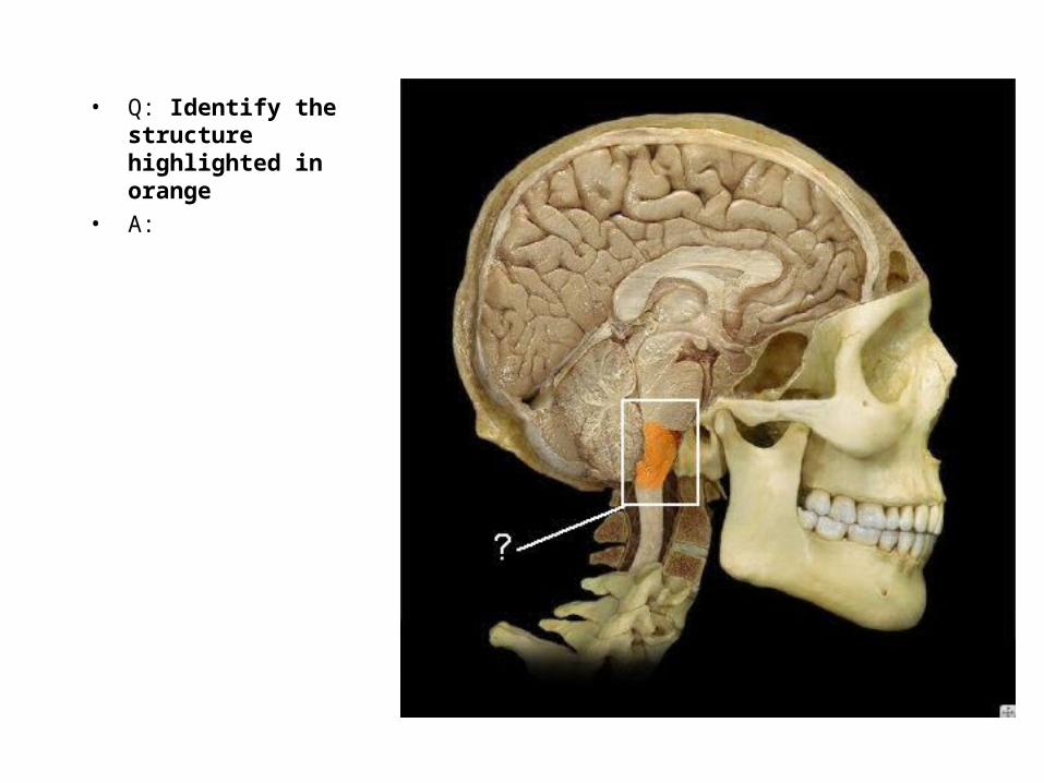
• Q: Identify the structure highlighted in orange
• A:

• Q: Identify the structure highlighted in orange
• A: medulla oblongata
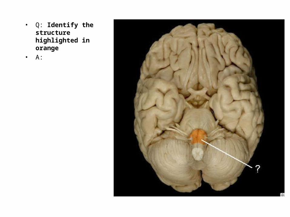
• Q: Identify the structure highlighted in orange
• A:
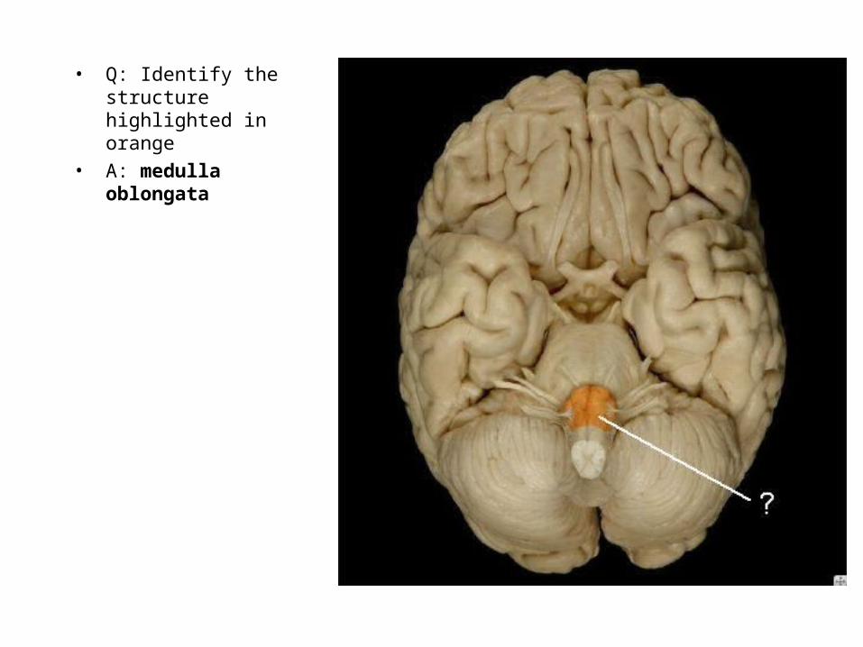
• Q: Identify the structure highlighted in orange
• A: medulla oblongata

• Q: Identify the structure highlighted in orange
• A:
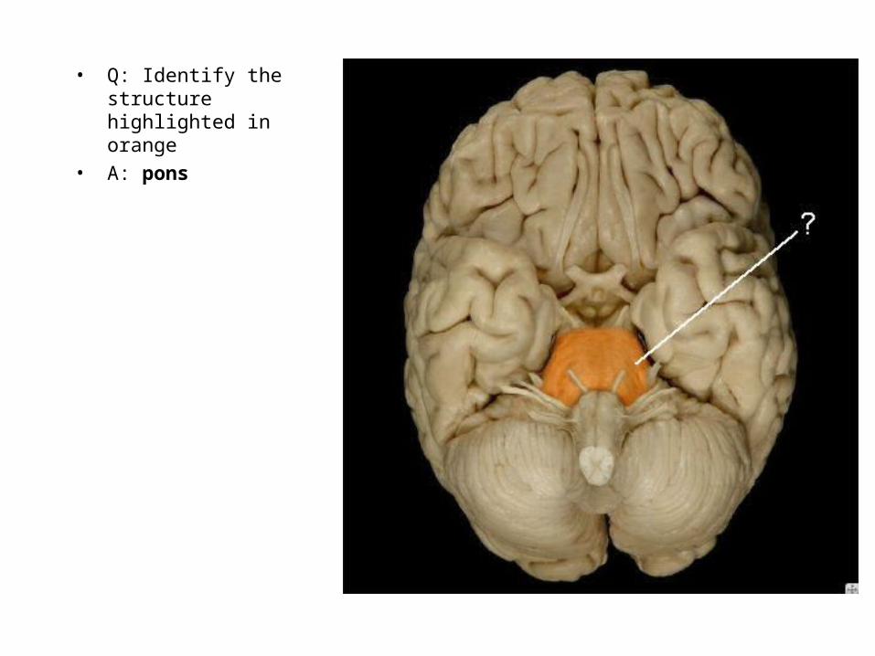
• Q: Identify the structure highlighted in orange
• A: pons

• Q: Identify the structure highlighted in orange
• A:

• Q: Identify the structure highlighted in orange
• A: pons

• Q: Identify the area highlighted in orange
• A:

• Q: Identify the area highlighted in orange
• A: midbrain
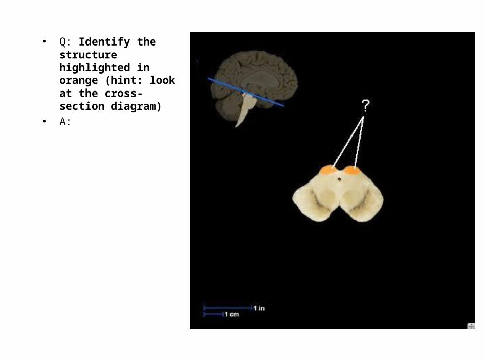
• Q: Identify the structure highlighted in orange (hint: look at the cross-section diagram)
• A:
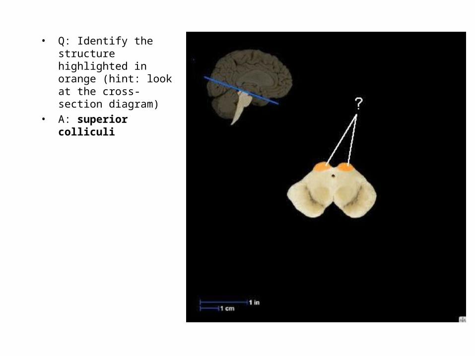
• Q: Identify the structure highlighted in orange (hint: look at the cross-section diagram)
• A: superior colliculi
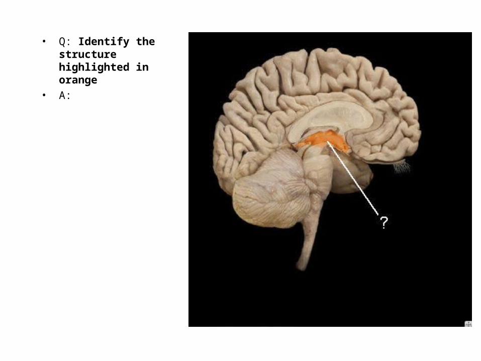
• Q: Identify the structure highlighted in orange
• A:
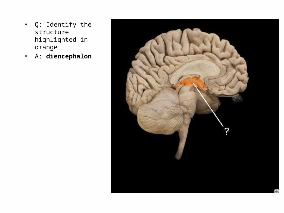
• Q: Identify the structure highlighted in orange
• A: diencephalon
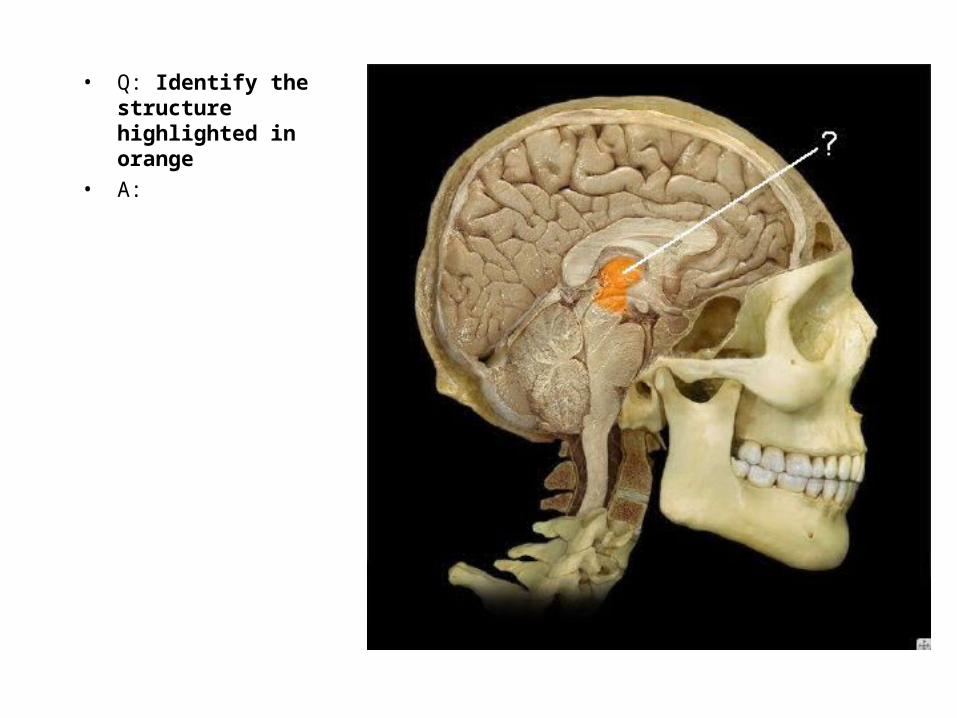
• Q: Identify the structure highlighted in orange
• A:
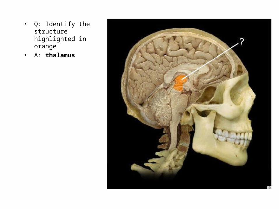
• Q: Identify the structure highlighted in orange
• A: thalamus
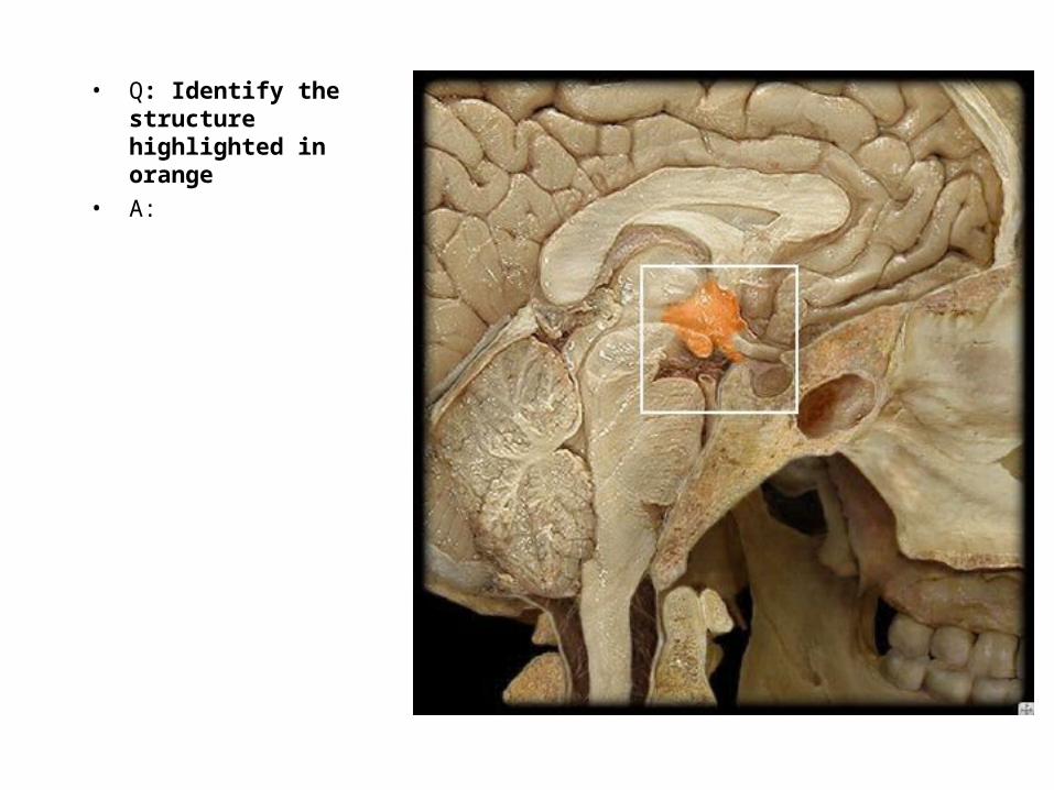
• Q: Identify the structure highlighted in orange
• A:
• Q: Identify the structure highlighted in orange
• A: hypothalamus

• Q: Identify the structure highlighted in orange
• A:

• Q: Identify the structure highlighted in orange
• A: thalamus

• Q: Identify the structure highlighted in purple
• A:
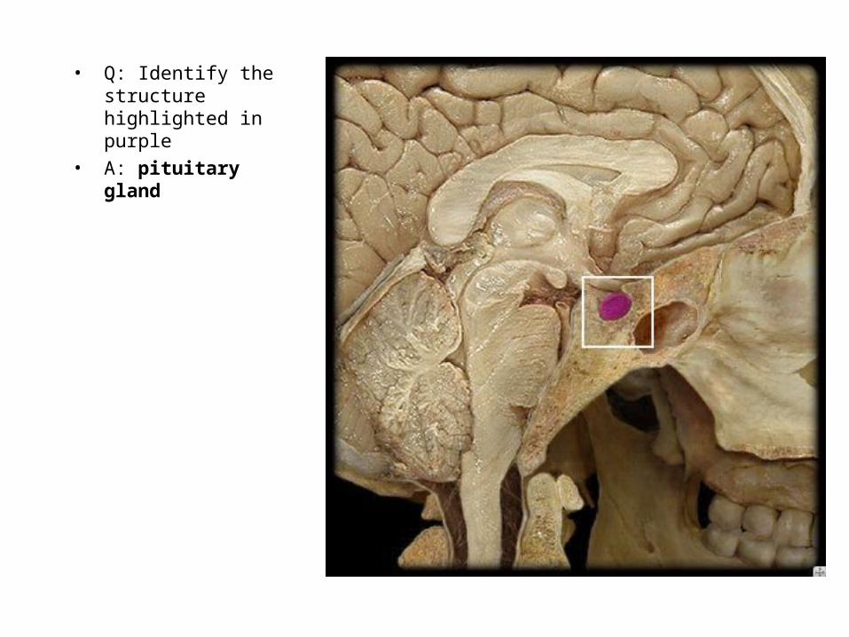
• Q: Identify the structure highlighted in purple
• A: pituitary gland
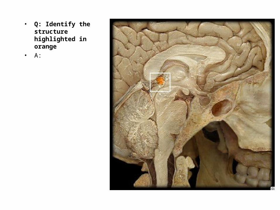
• Q: Identify the structure highlighted in orange
• A:
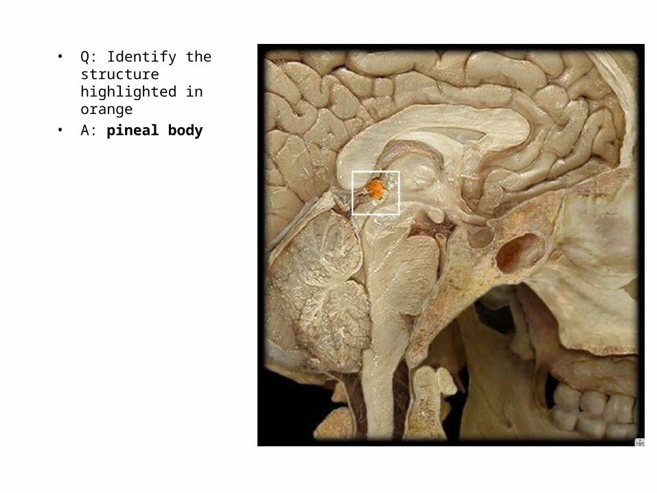
• Q: Identify the structure highlighted in orange
• A: pineal body

• Q: Identify the structure highlighted in yellow
• A:

• Q: Identify the structure highlighted in yellow
• A: spinal nerve

• Q: Identify the structure highlighted in yellow
• A:
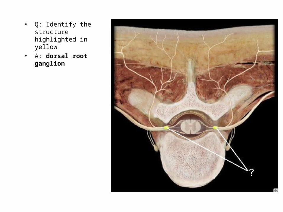
• Q: Identify the structure highlighted in yellow
• A: dorsal root ganglion

• Q: Identify the structure highlighted in yellow
• A:

• Q: Identify the structure highlighted in yellow
• A: dorsal root

• Q: Identify the structure highlighted in yellow
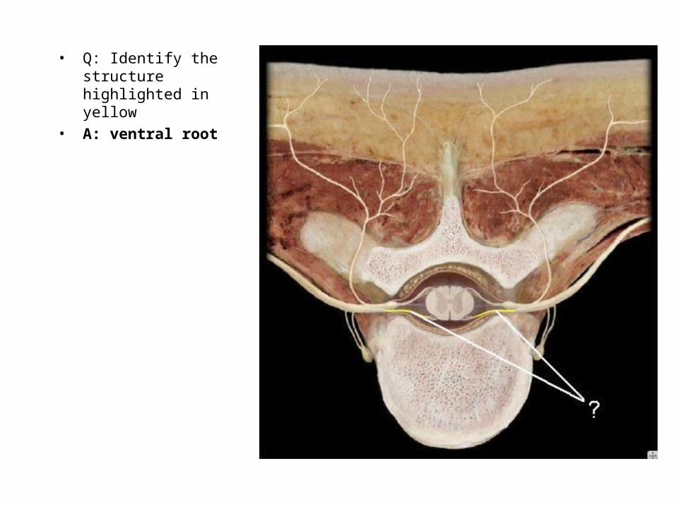
• Q: Identify the structure highlighted in yellow
• A: ventral root
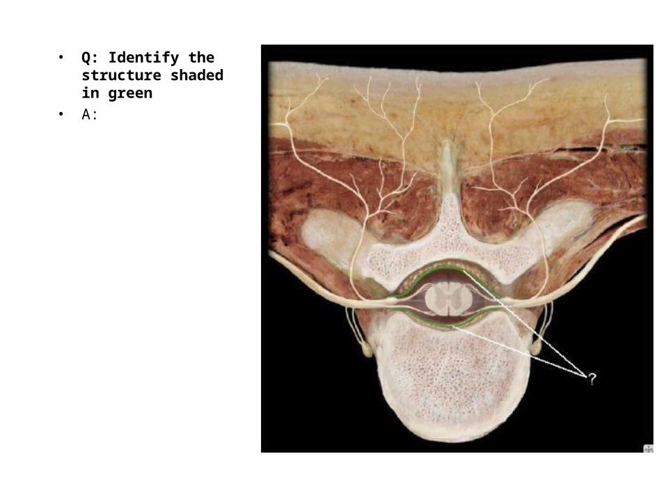
• Q: Identify the structure shaded in green
• A:

• Q: Identify the structure shaded in green
• A: dura mater
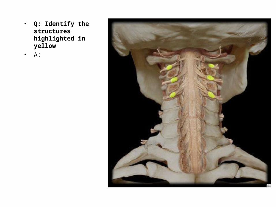
• Q: Identify the structures highlighted in yellow
• A:
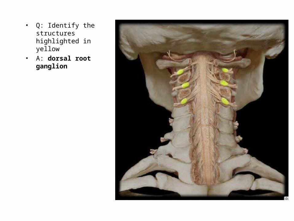
• Q: Identify the structures highlighted in yellow
• A: dorsal root ganglion

• Q: Identify the structures highlighted in yellow
• A:
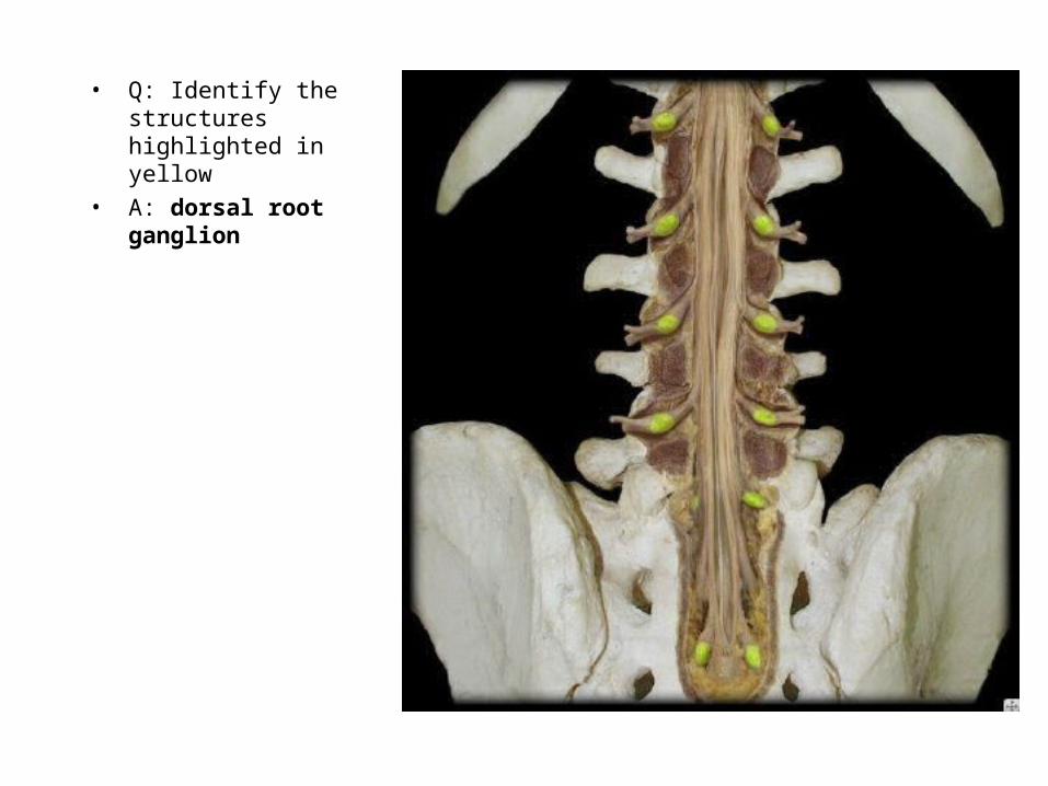
• Q: Identify the structures highlighted in yellow
• A: dorsal root ganglion
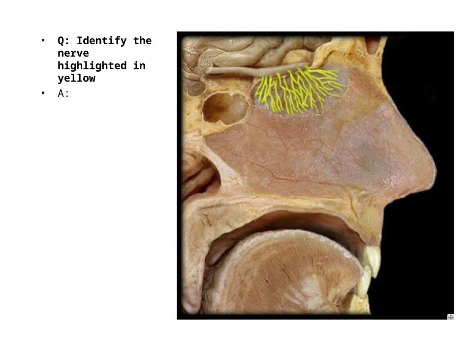
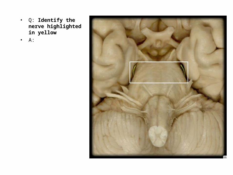
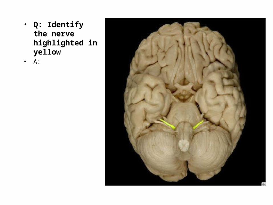
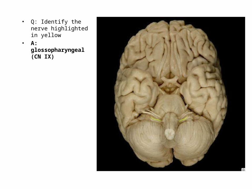
• Q: Identify the nerve highlighted in yellow
• A:

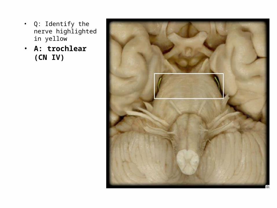
• Q: Identify the nerve highlighted in yellow
• A: olfactory (CN I)
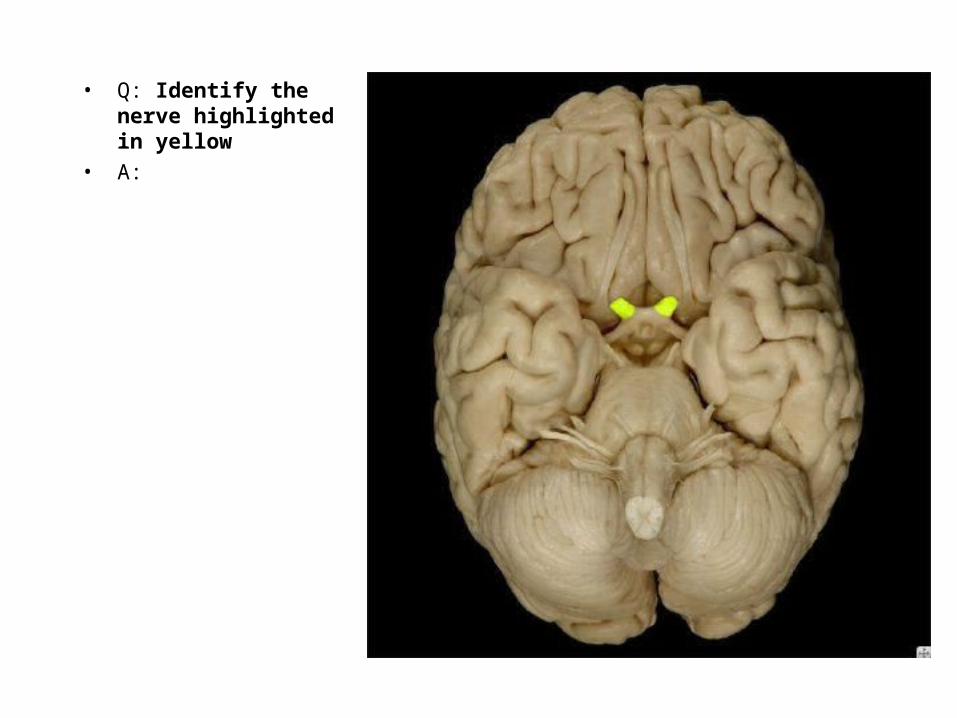
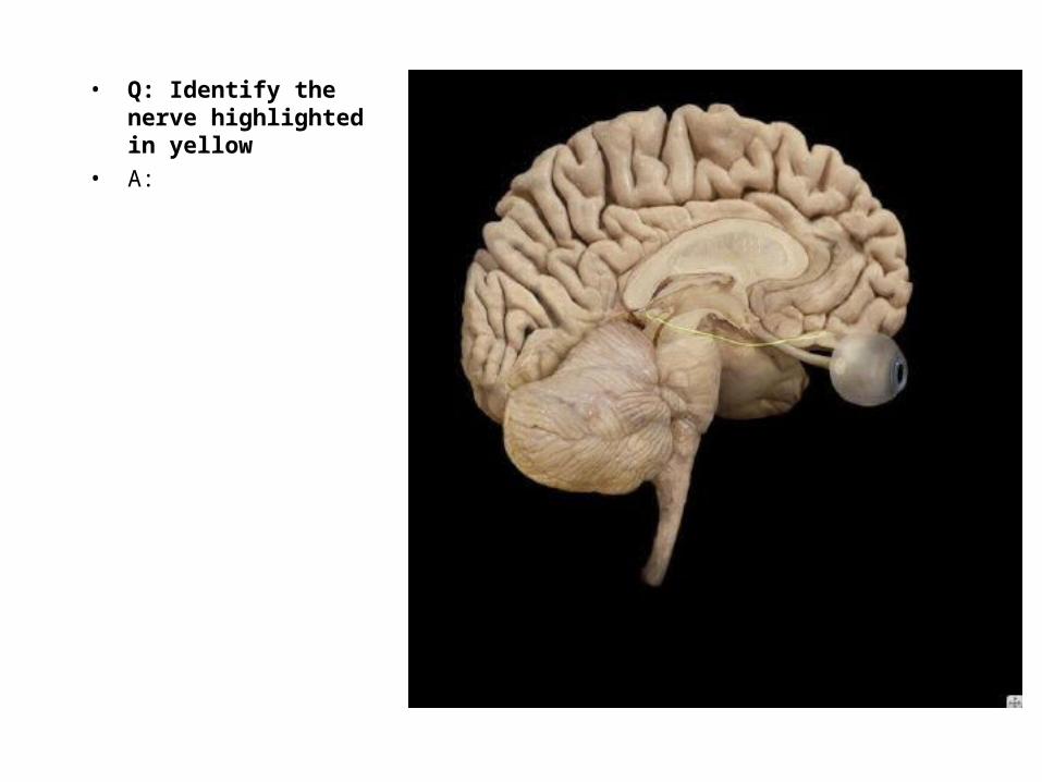
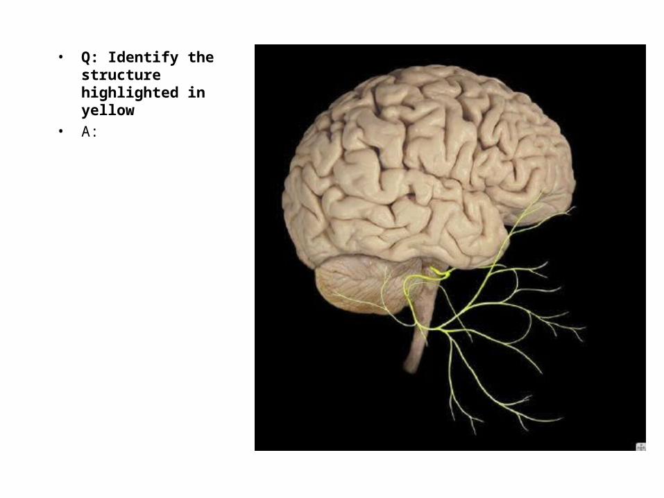
• Q: Identify the nerve highlighted in yellow
• A:
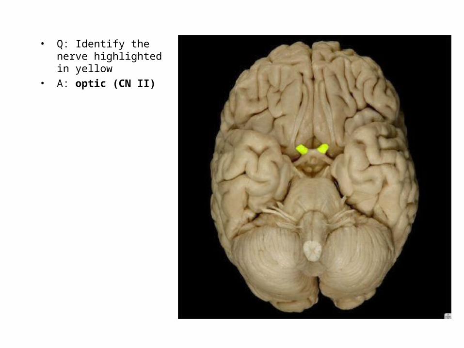
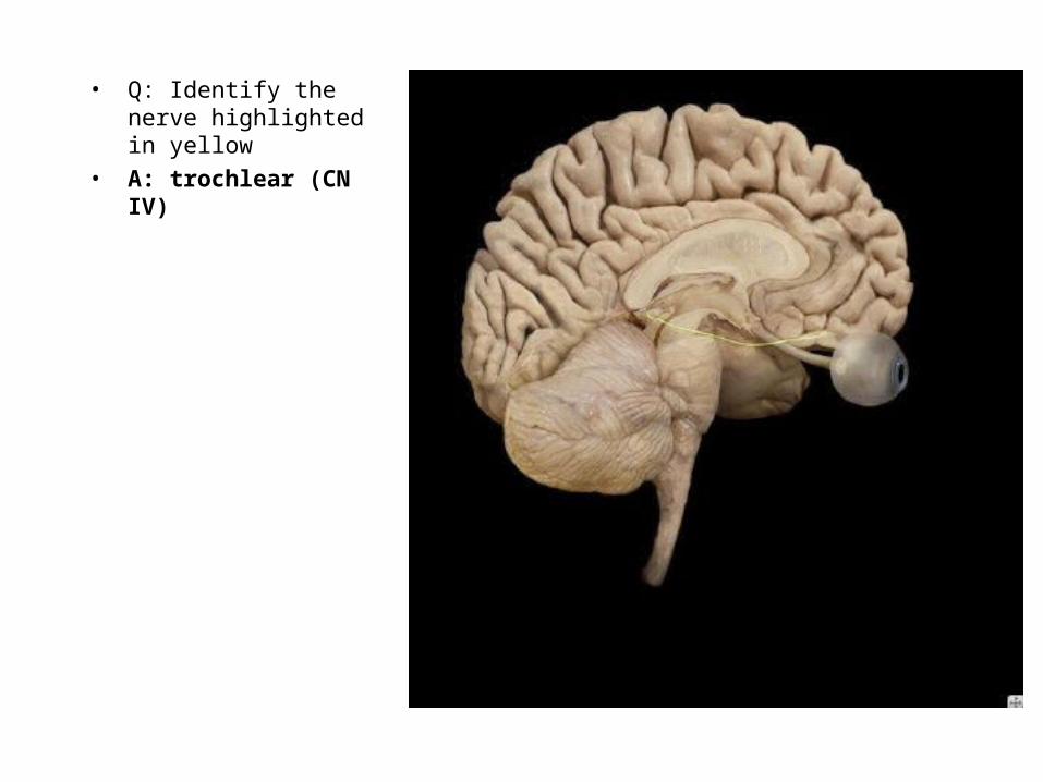
• Q: Identify the nerve highlighted in yellow
• A: optic (CN II)

• Q: Identify the nerve highlighted in yellow
• A:
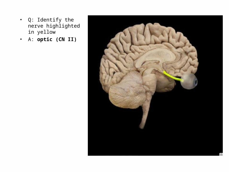
• Q: Identify the nerve highlighted in yellow
• A: optic (CN II)

• Q: Identify the structure highlighted in yellow
• A:

• Q: Identify the structure highlighted in yellow
• A: oculomotor (CN III)
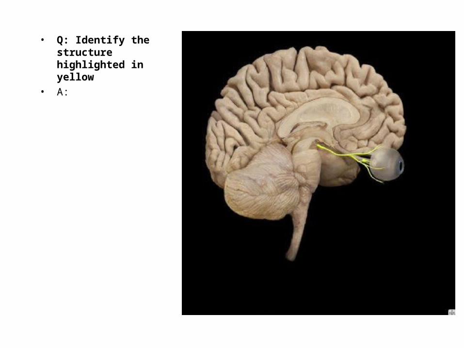
• Q: Identify the structure highlighted in yellow
• A:
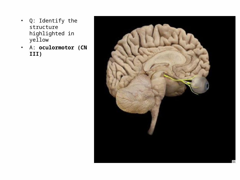
• Q: Identify the structure highlighted in yellow
• A: oculormotor (CN III)
• Q: Identify the nerve highlighted in yellow
• A:
• Q: Identify the nerve highlighted in yellow
• A: trochlear (CN IV)
• Q: Identify the nerve highlighted in yellow
• A:
• Q: Identify the nerve highlighted in yellow
• A: trochlear (CN IV)

• Q: Identify the nerve highlighted in yellow
• A:
• Q: Identify the nerve highlighted in yellow
• A: trigeminal (CN V)

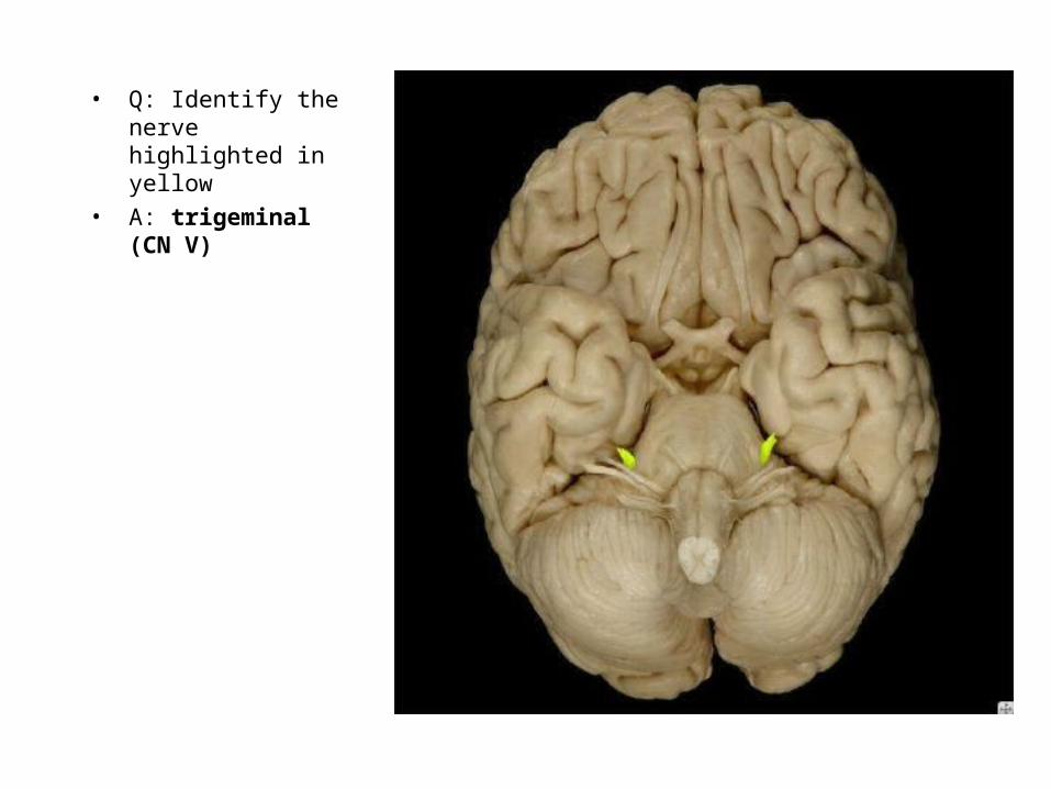
• Q: Identify the nerve highlighted in yellow
• A:
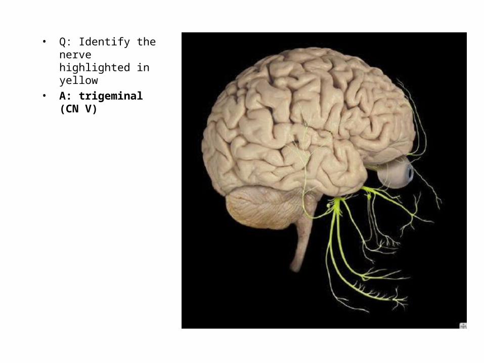
• Q: Identify the nerve highlighted in yellow
• A: trigeminal (CN V)
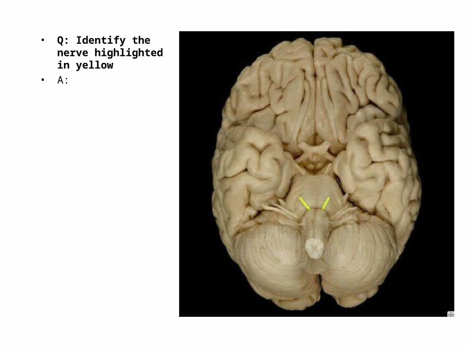
• Q: Identify the nerve highlighted in yellow
• A:
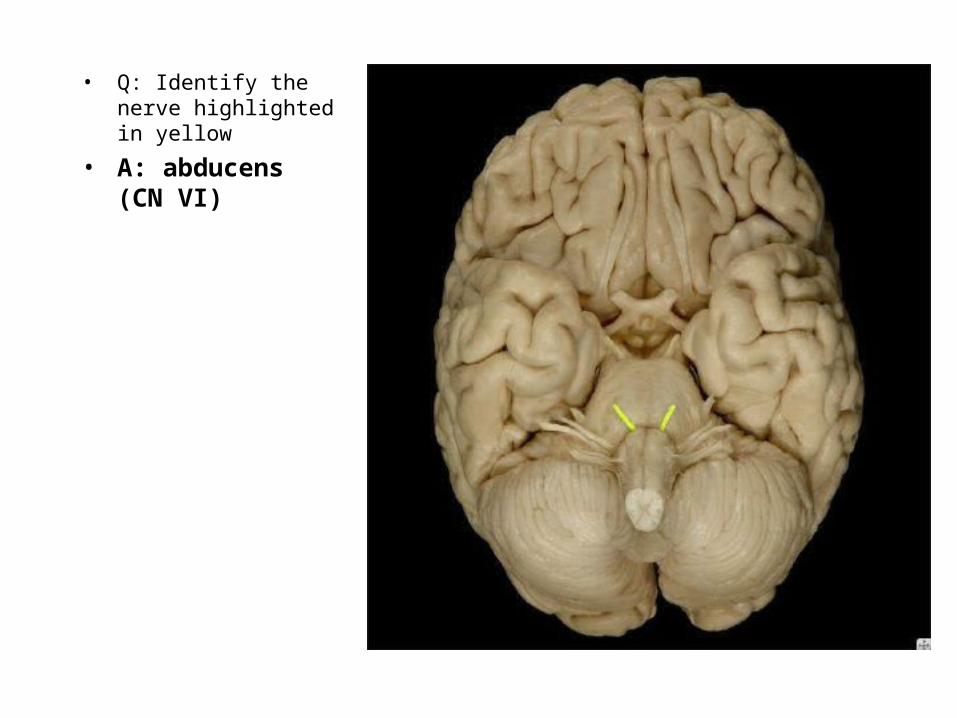
• Q: Identify the nerve highlighted in yellow
• A: abducens (CN VI)
• Q: Identify the nerve highlighted in yellow
• A:

• Q: Identify the nerve highlighted in yellow
• A: facial (CN VII)
• Q: Identify the structure highlighted in yellow
• A:

• Q: Identify the structure highlighted in yellow
• A: facial (CV VII)
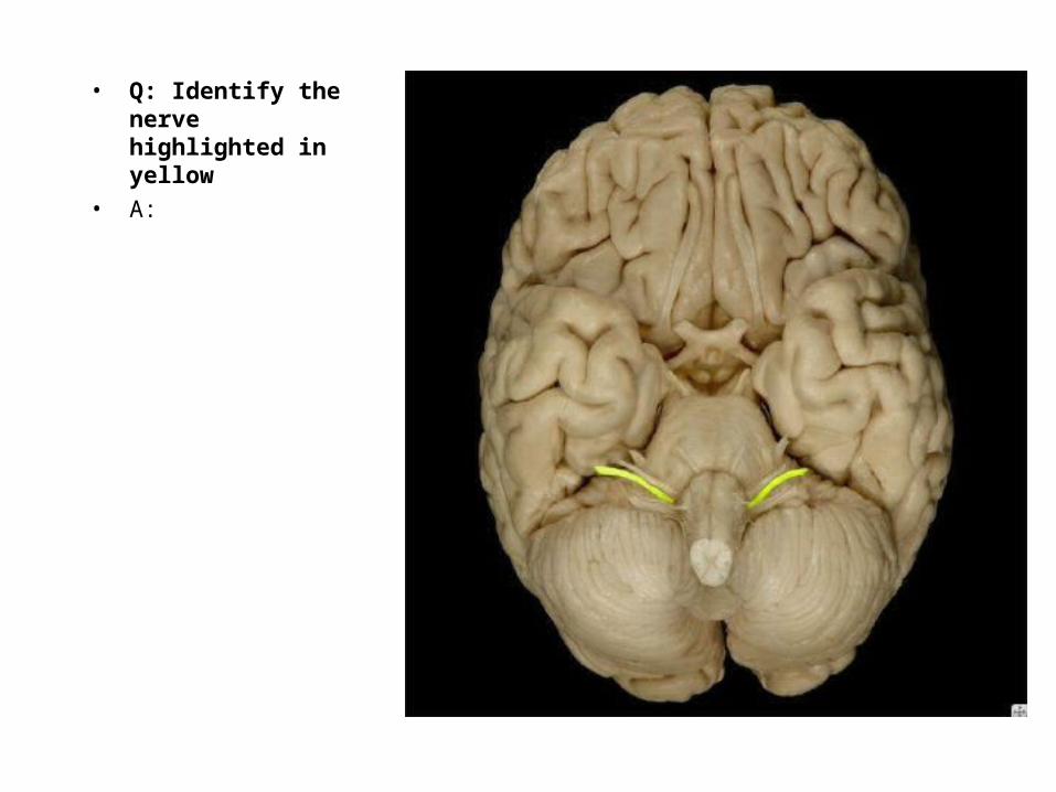
• Q: Identify the nerve highlighted in yellow
• A:
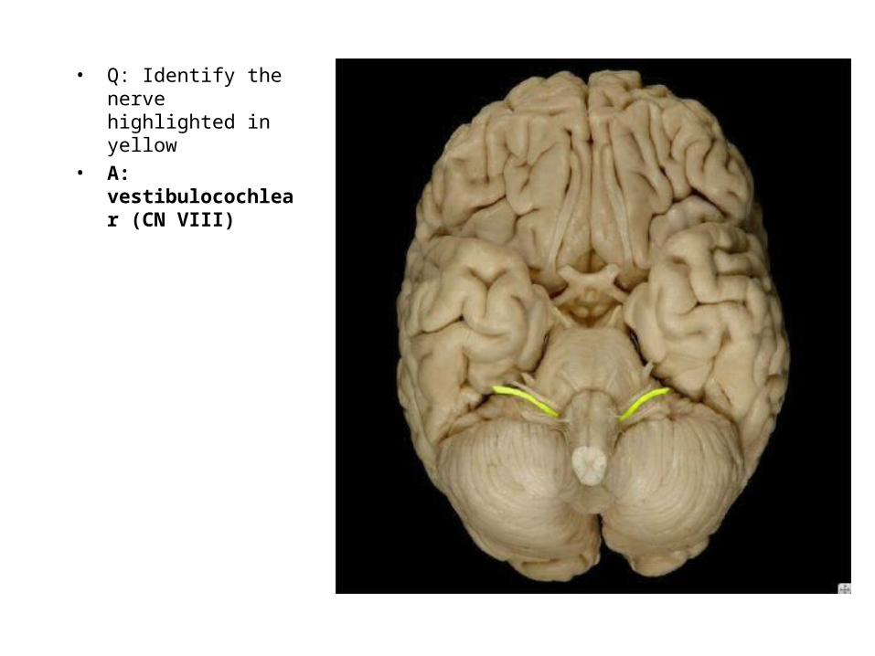
• Q: Identify the nerve highlighted in yellow
• A: vestibulocochlear (CN VIII)

• Q: Identify the nerve highlighted in yellow
• A:
• Q: Identify the nerve highlighted in yellow
• A: glossopharyngeal (CN IX)

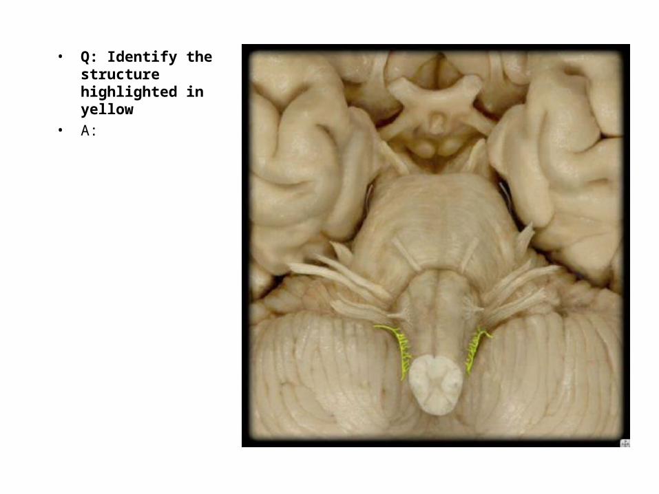
• Q: Identify the structure highlighted in yellow
• A:

• Q: Identify the structure highlighted in yellow
• A: glossopharyngeal (CN IX)

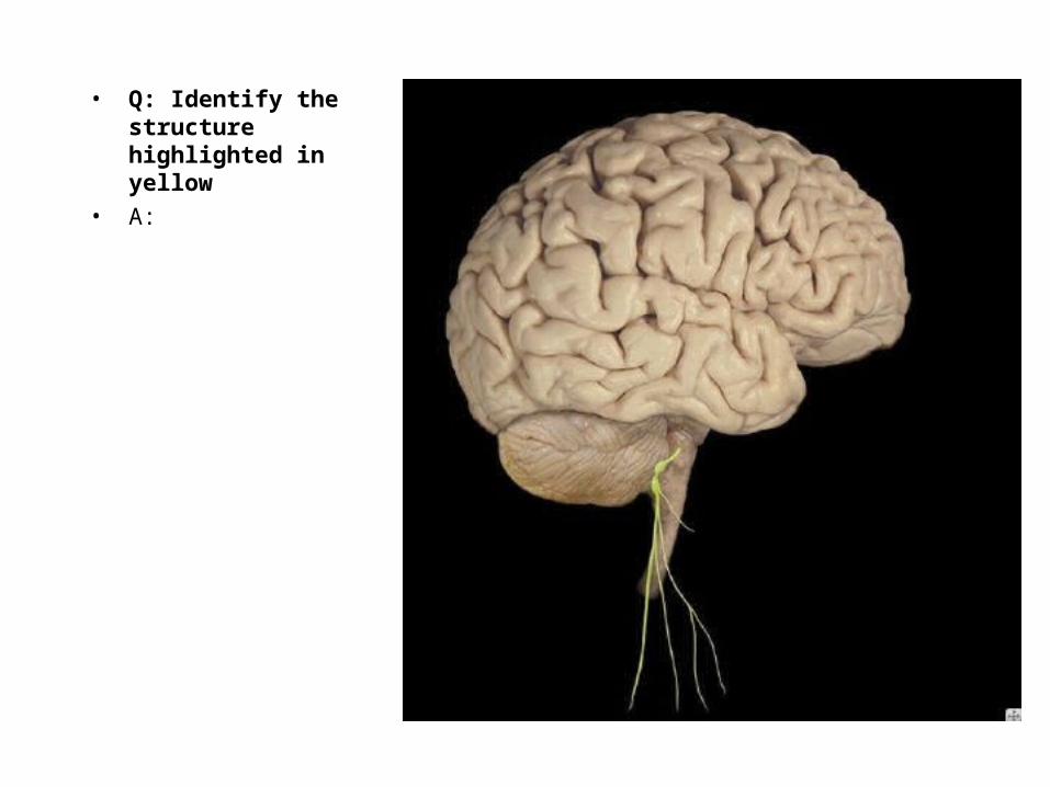
• Q: Identify the structure highlighted in yellow
•

• Q: Identify the structure highlighted in yellow
• A: vagus nerve (CN X)
• Q: Identify the structure highlighted in yellow
• A:
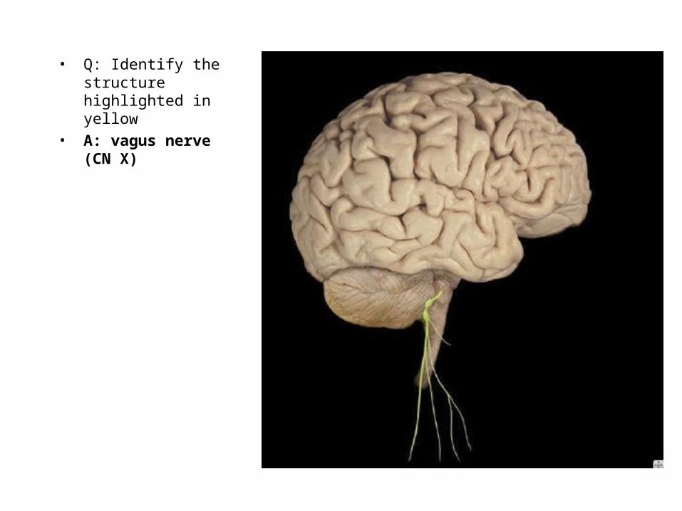
• Q: Identify the structure highlighted in yellow
• A: vagus nerve (CN X)
• Q: Identify the structure highlighted in yellow
• A:

• Q: Identify the structure highlighted in yellow
• A: accessory (CN XI)
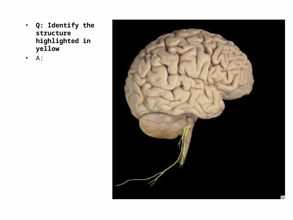
• Q: Identify the structure highlighted in yellow
• A:
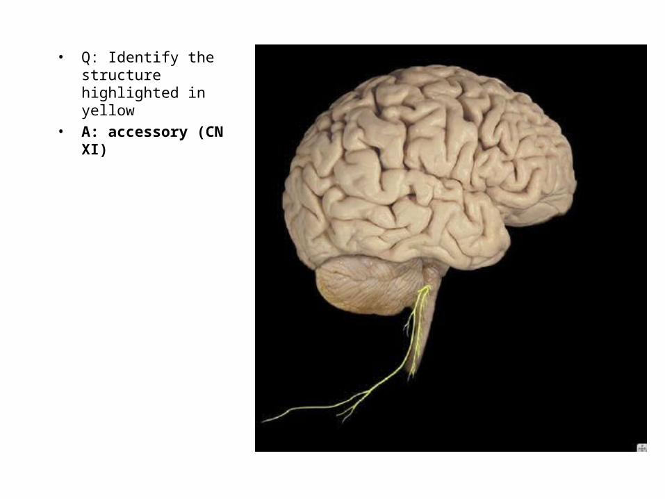
• Q: Identify the structure highlighted in yellow
• A: accessory (CN XI)
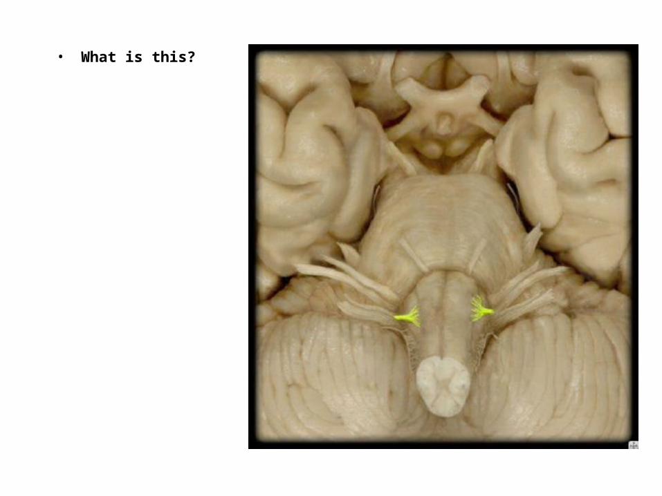
• What is this?
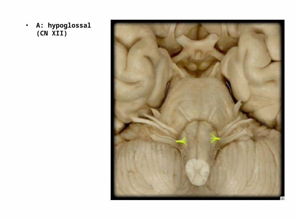
• A: hypoglossal (CN XII)
Concussion
Bruised Brain!
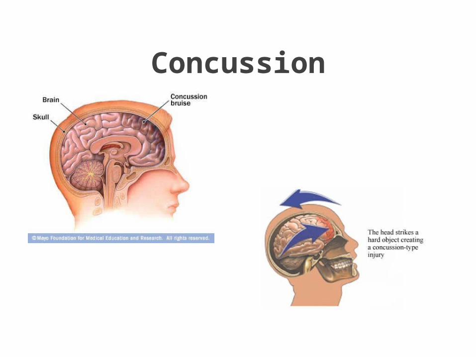
Concussion
Concussion
Exam: checking your memory and concentration, vision, hearing, balance,
coordination and reflexes.
More severe: bleeding or swelling in your skull
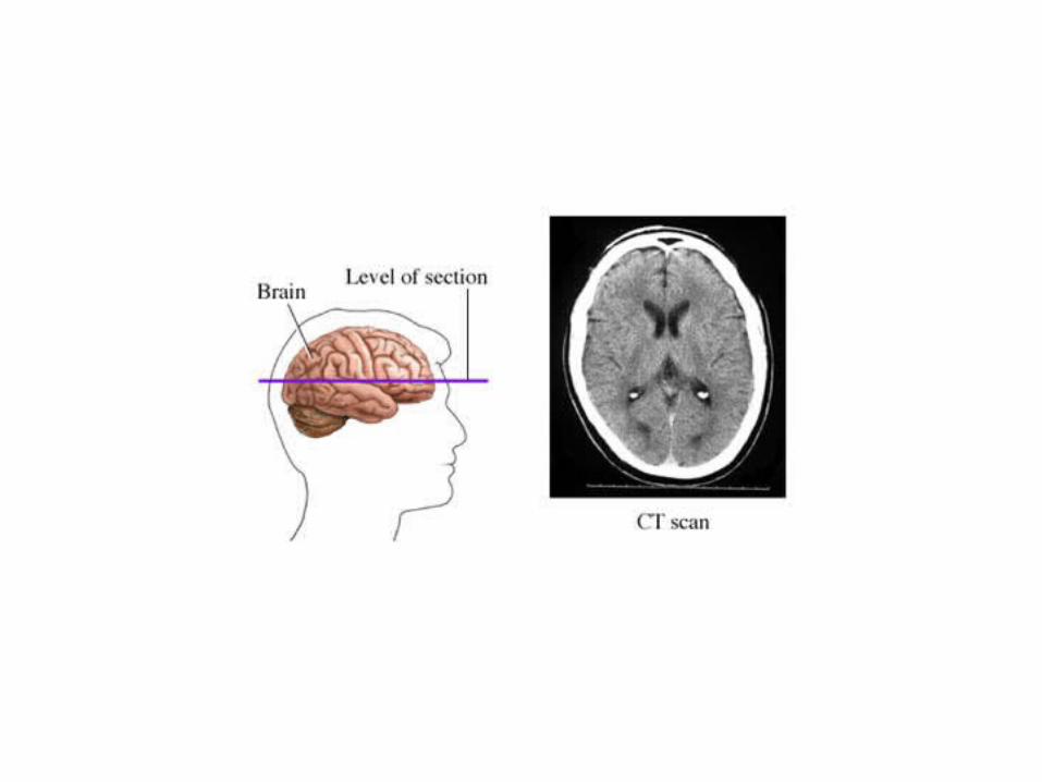
CT scan if necessary
A CT scanner takes multiple cross-sectional X-rays and combines all the resulting images to produce detailed,
two-dimensional images of your skull and brain.

The End
• Quick tip on studying for your next coop quiz and Test #3: Answer the questions in the PsTL 1135 Studyguide, review your notes, and then look up the concepts in the textbook after Murray covers them in lecture!!!
• Remember to prepare for the exam on Monday, November 17 in lecture!!