Embed Size (px)
Citation preview
The Nervous System
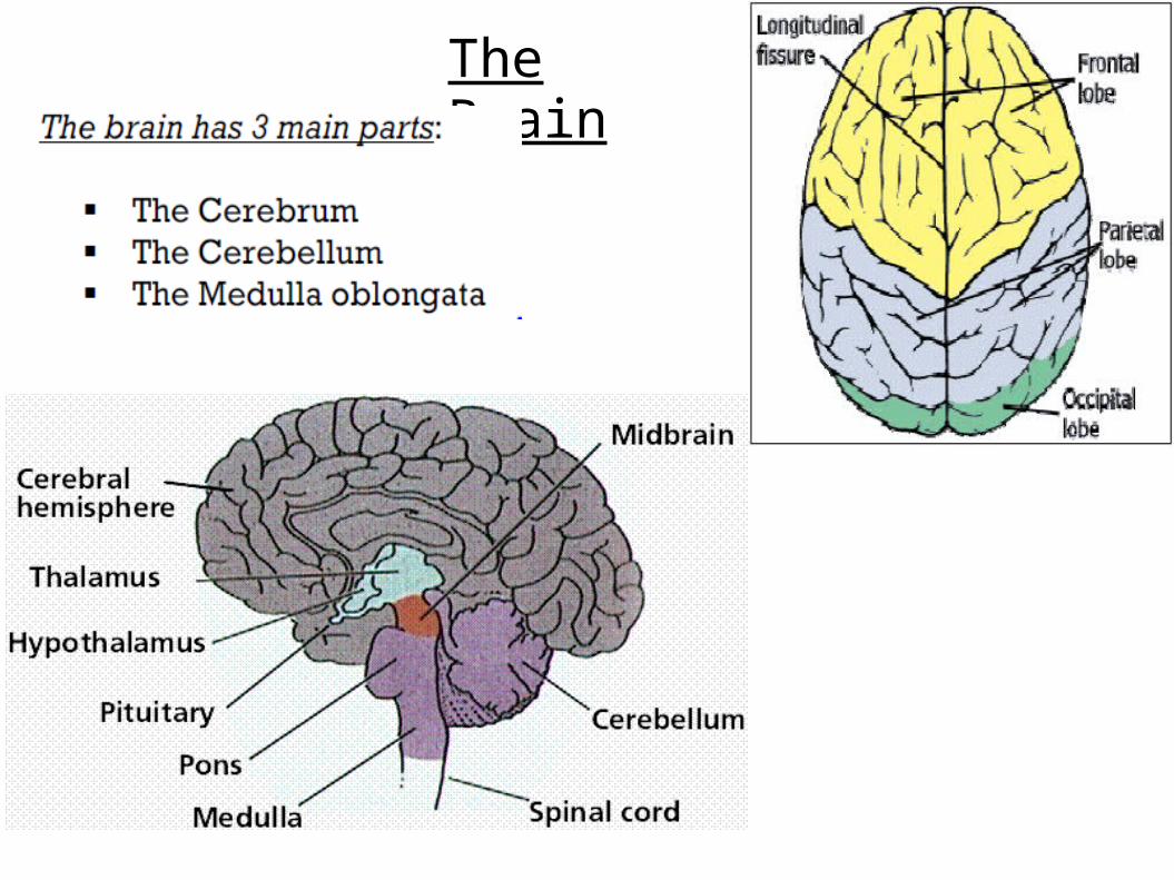
The Brain
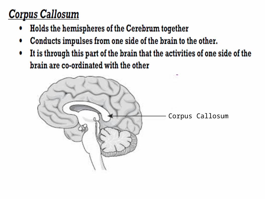
Corpus Callosum
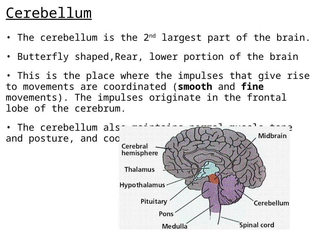
Cerebellum
• The cerebellum is the 2nd largest part of the brain.
• Butterfly shaped,Rear, lower portion of the brain
• This is the place where the impulses that give rise to movements are coordinated (smooth and fine movements). The impulses originate in the frontal lobe of the cerebrum.
• The cerebellum also maintains normal muscle tone and posture, and coordinates balance.
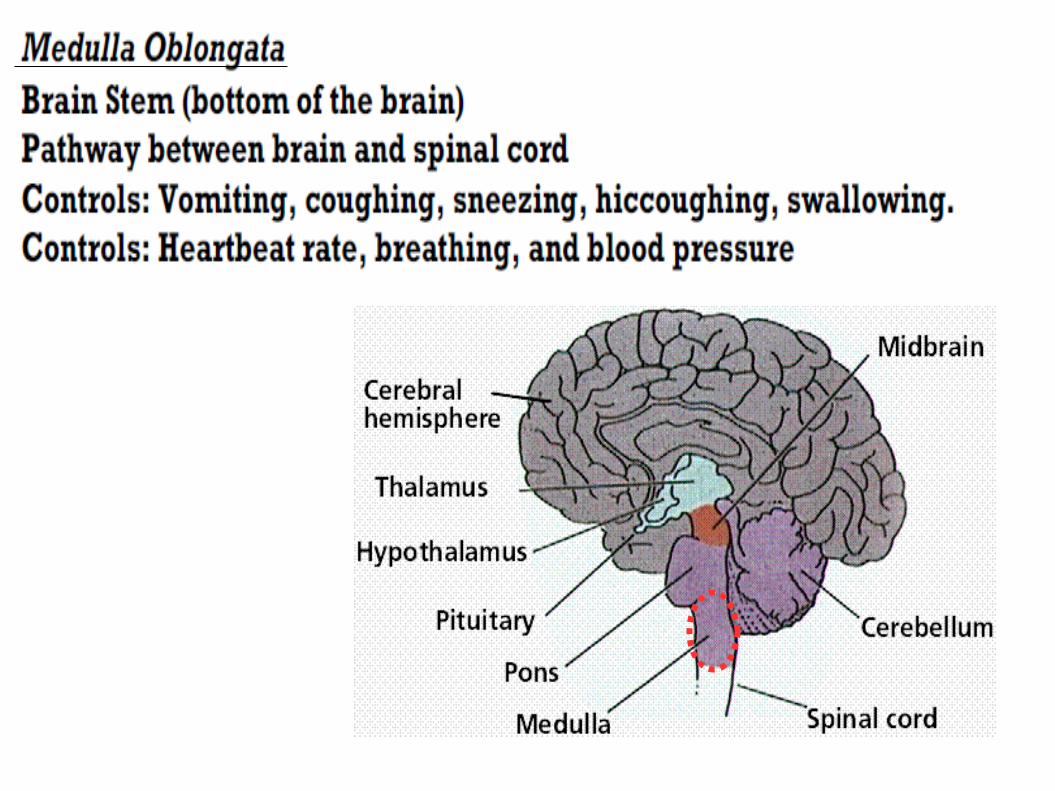
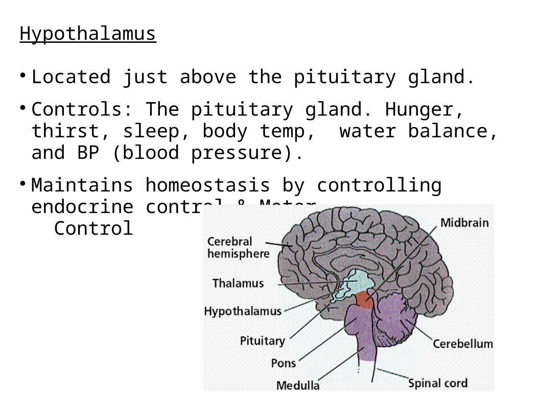
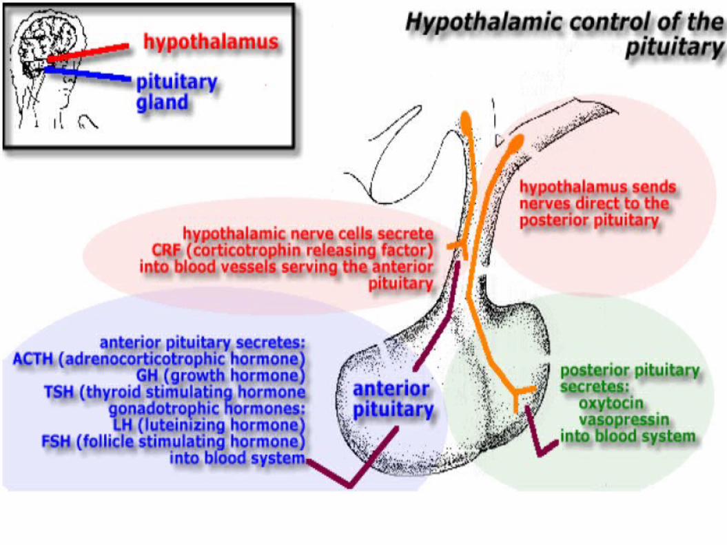
Hypothalamus
Located just above the pituitary gland.
Controls: The pituitary gland. Hunger, thirst, sleep, body temp, water balance, and BP (blood pressure).
Maintains homeostasis by controlling endocrine control & Motor Control
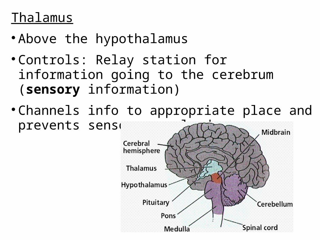
ThalamusAbove the hypothalamusControls: Relay station for information going to the
cerebrum (sensory information)Channels info to appropriate place and prevents sensory
overload.
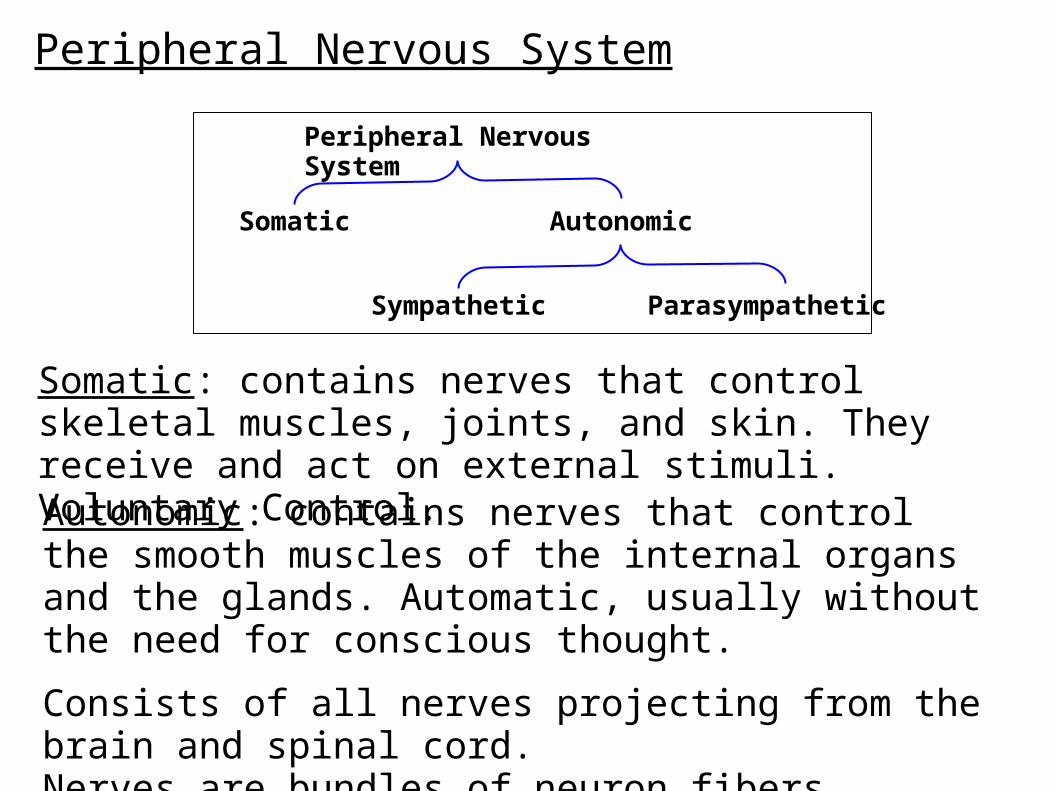
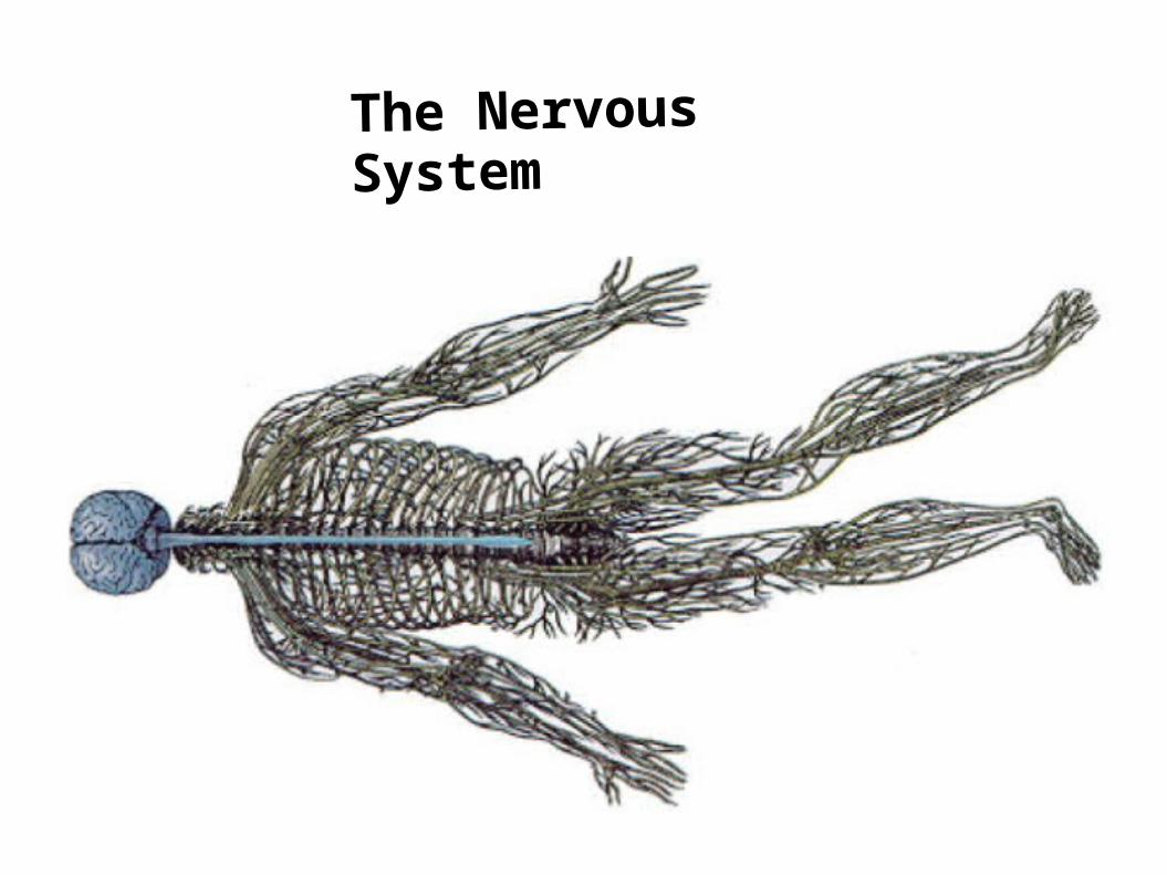
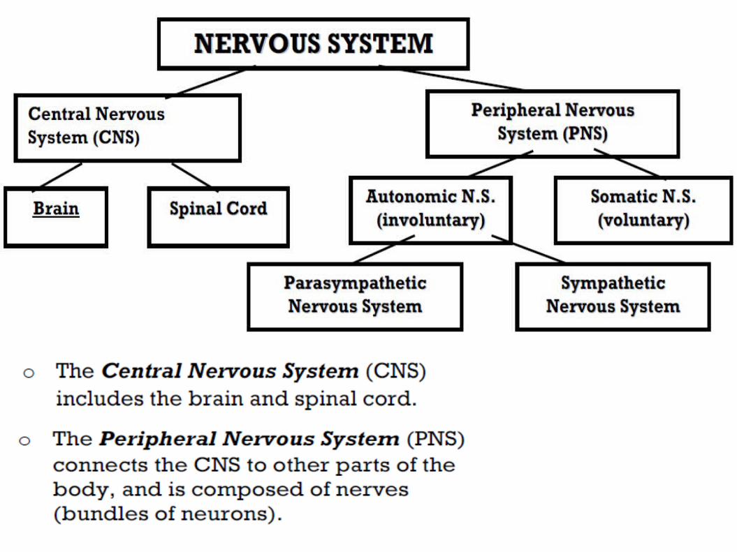
Peripheral Nervous System
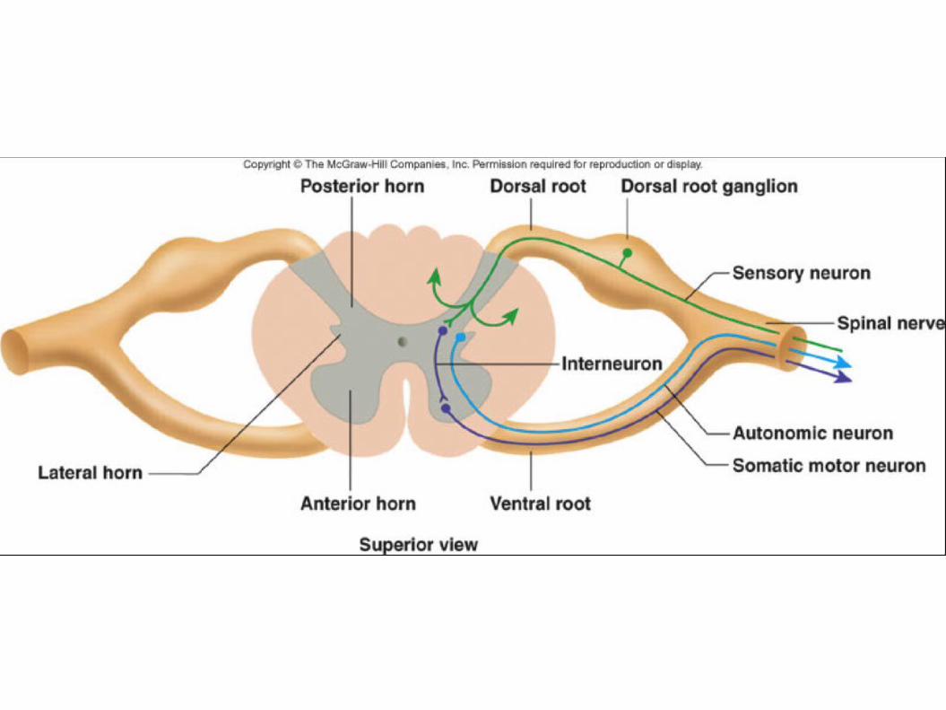
Somatic: contains nerves that control skeletal muscles, joints, and skin. They receive and act on external stimuli. Voluntary Control.
Autonomic: contains nerves that control the smooth muscles of the internal organs and the glands. Automatic, usually without the need for conscious thought.
Consists of all nerves projecting from the brain and spinal cord.Nerves are bundles of neuron fibers.
Peripheral Nervous System
Somatic Autonomic
Sympathetic Parasympathetic

Sensory Nerves: Bundles of dendrites from sensory neurons.
Motor Nerves: Bundles of axons from motor neurons.
Mixed Nerves: Dendrites of sensory neurons and axons of motor neurons running together.
Cranial Nerves: Arise from the Brain
Spinal Nerves: Arise from the spinal cord
Ganglion: A collection of cell bodies from many neurons. Appears as an enlarged portion of the nerve.
Types of Nerves
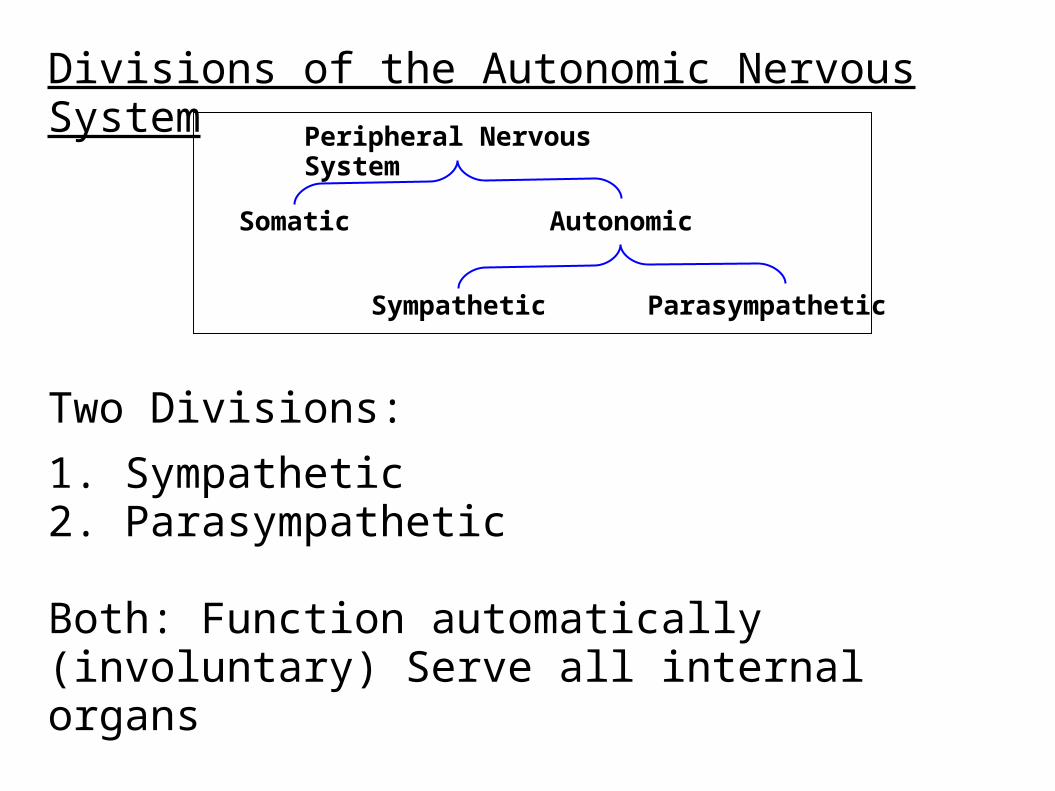
Divisions of the Autonomic Nervous System
Two Divisions:
1. Sympathetic2. Parasympathetic
Both: Function automatically (involuntary) Serve all internal organs
Have two motor neurons with a ganglion between
Peripheral Nervous System
Somatic Autonomic
Sympathetic Parasympathetic
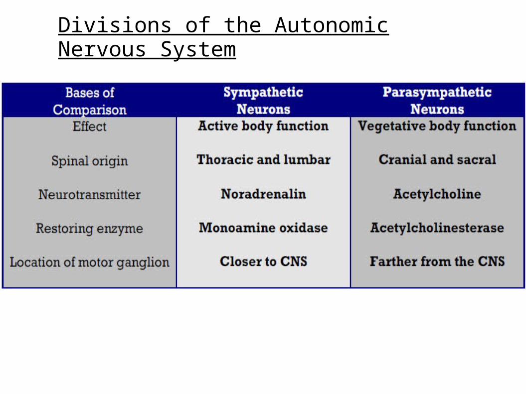
Divisions of the Autonomic Nervous System
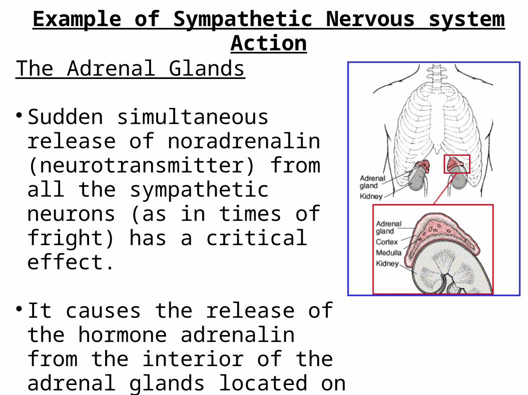
Example of Sympathetic Nervous system Action
The Adrenal Glands
Sudden simultaneous release of noradrenalin (neurotransmitter) from all the sympathetic neurons (as in times of fright) has a critical effect.
It causes the release of the hormone adrenalin from the interior of the adrenal glands located on top of the kidneys.
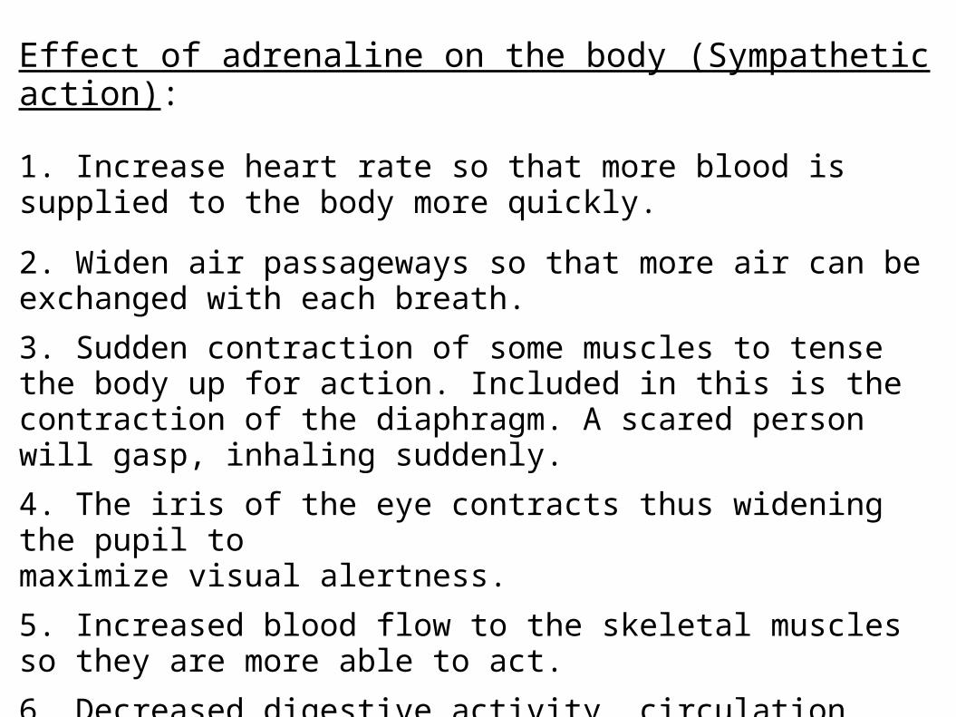
Effect of adrenaline on the body (Sympathetic action):
1. Increase heart rate so that more blood is supplied to the body more quickly.
2. Widen air passageways so that more air can be exchanged with each breath.
3. Sudden contraction of some muscles to tense the body up for action. Included in this is the contraction of the diaphragm. A scared person will gasp, inhaling suddenly.
4. The iris of the eye contracts thus widening the pupil tomaximize visual alertness.
5. Increased blood flow to the skeletal muscles so they are more able to act.
6. Decreased digestive activity, circulation.
* The opposite is true for parasympathetic action.
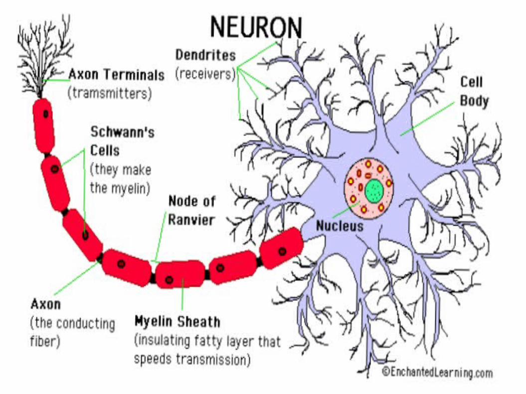
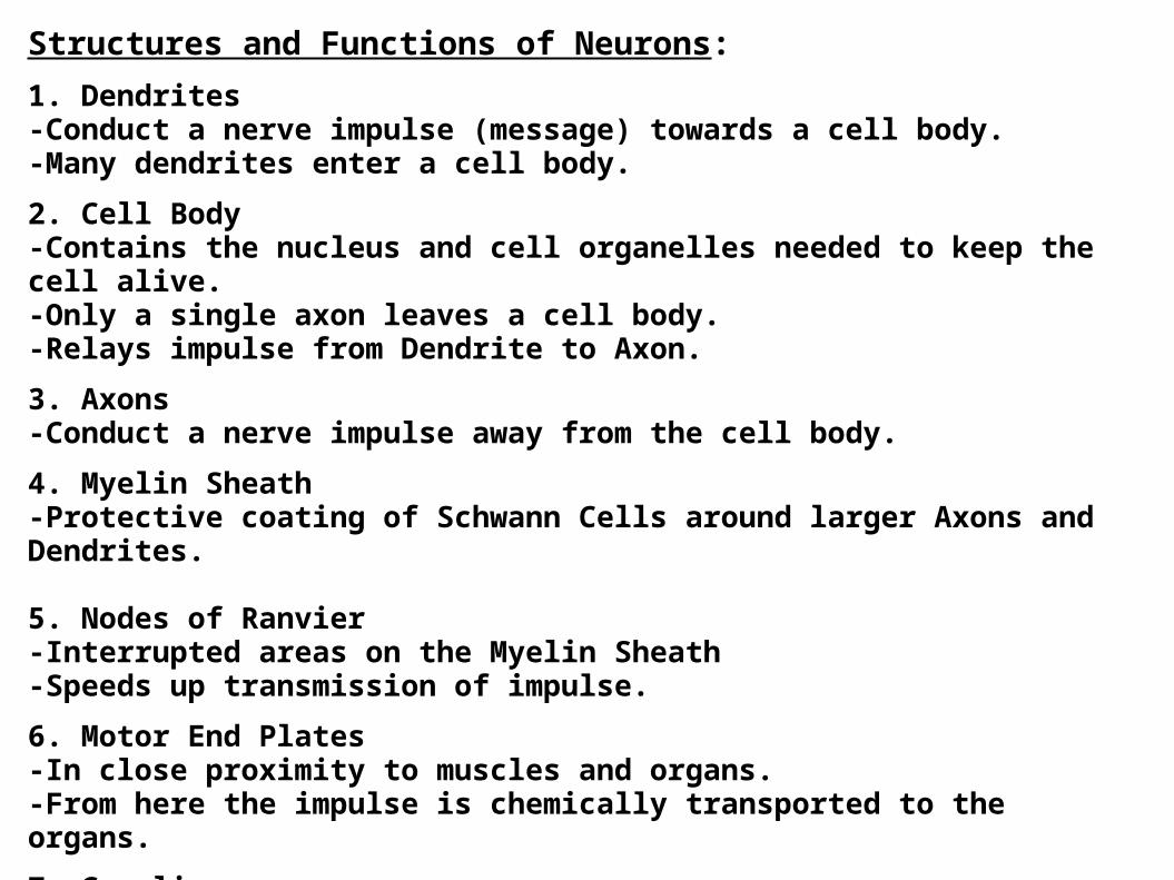
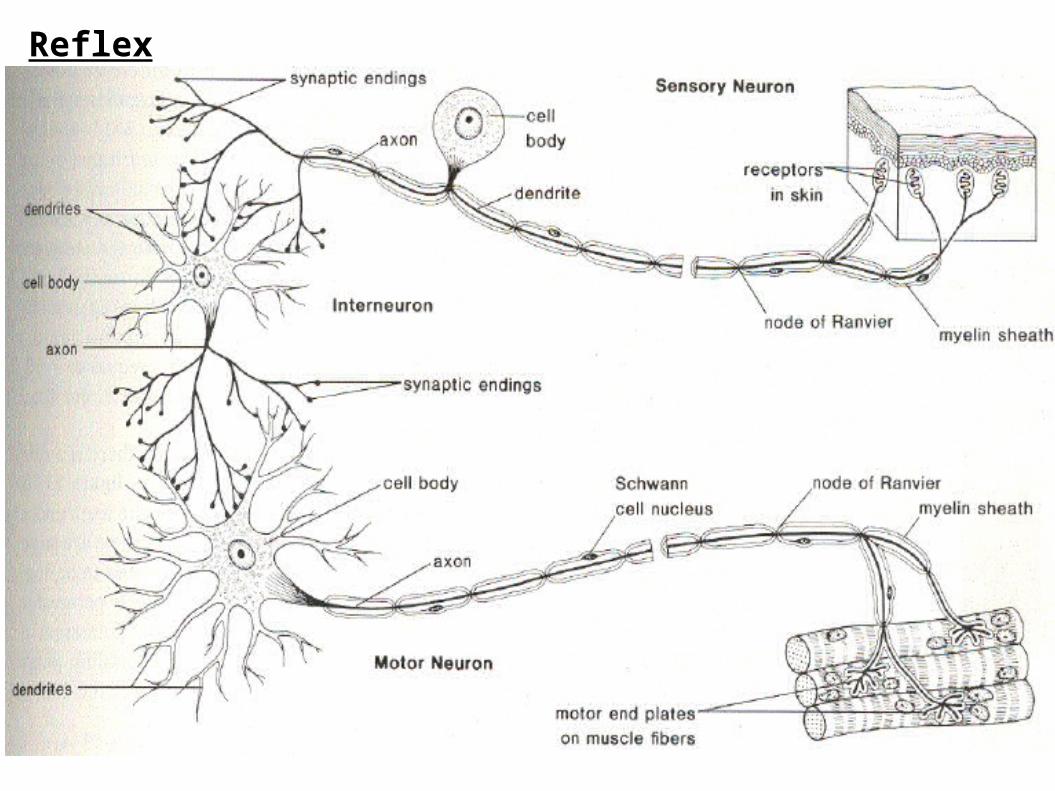
Structures and Functions of Neurons:
1. Dendrites-Conduct a nerve impulse (message) towards a cell body.-Many dendrites enter a cell body.
2. Cell Body-Contains the nucleus and cell organelles needed to keep the cell alive.-Only a single axon leaves a cell body.-Relays impulse from Dendrite to Axon.
3. Axons-Conduct a nerve impulse away from the cell body.
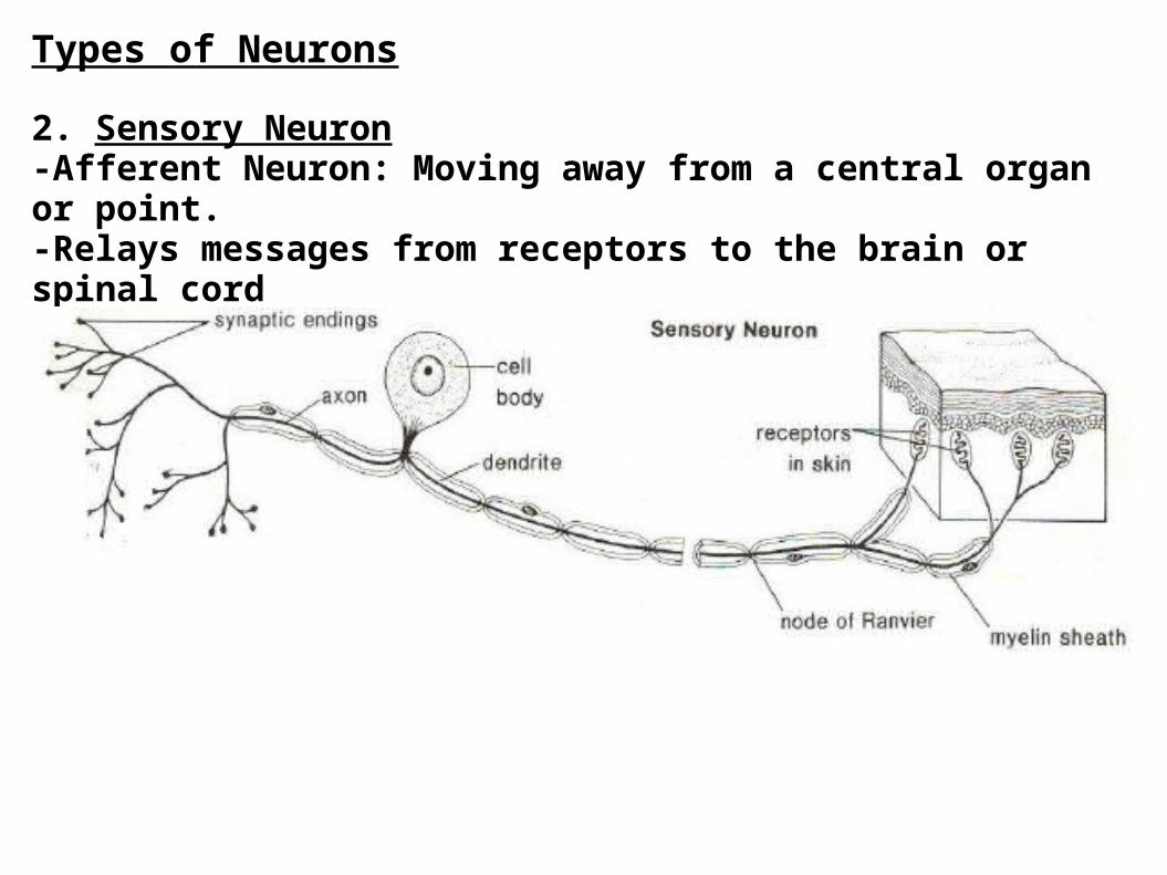
4. Myelin Sheath-Protective coating of Schwann Cells around larger Axons and Dendrites.
5. Nodes of Ranvier-Interrupted areas on the Myelin Sheath-Speeds up transmission of impulse.
6. Motor End Plates-In close proximity to muscles and organs.-From here the impulse is chemically transported to the organs.
7. Ganglia-A collection of cell bodies outside of the Central NervousSystem.
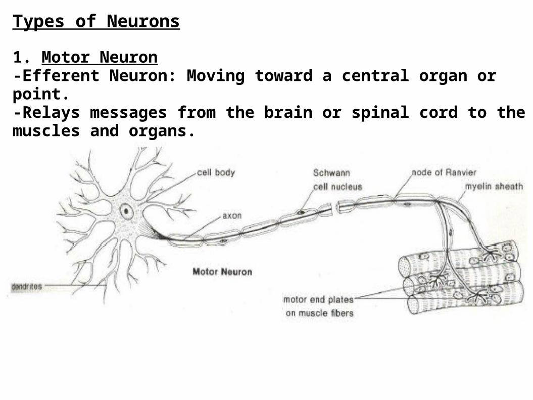
Types of Neurons
1. Motor Neuron-Efferent Neuron: Moving toward a central organ or point.-Relays messages from the brain or spinal cord to the muscles and organs.
Types of Neurons
2. Sensory Neuron-Afferent Neuron: Moving away from a central organ or point.-Relays messages from receptors to the brain or spinal cord
Types of Neurons
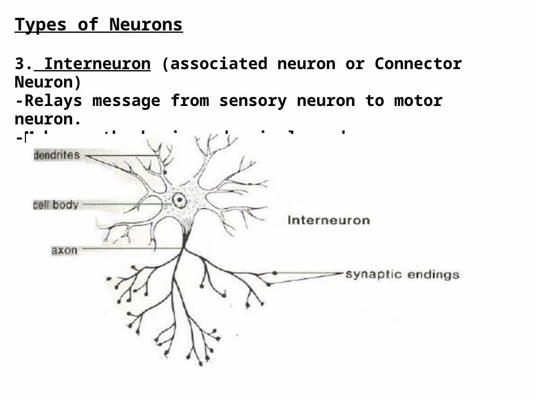
3. Interneuron (associated neuron or Connector Neuron)-Relays message from sensory neuron to motor neuron.-Make up the brain and spinal cord.
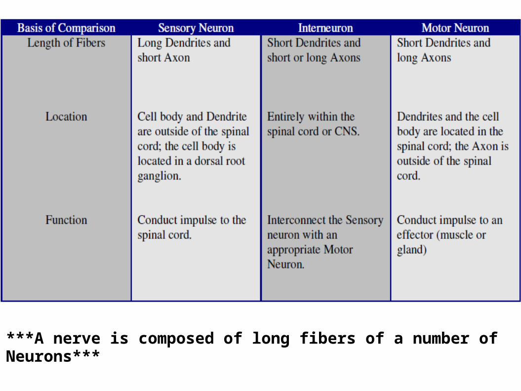
***A nerve is composed of long fibers of a number of Neurons***
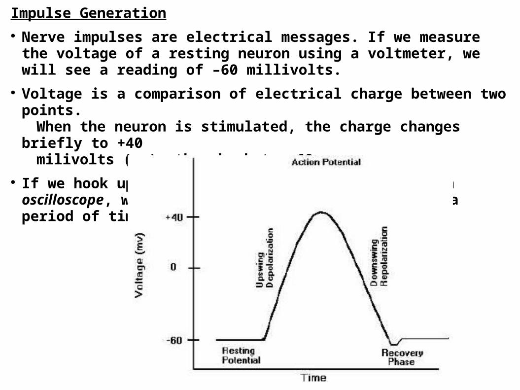
Impulse Generation Nerve impulses are electrical messages. If we measure the
voltage of a resting neuron using a voltmeter, we will see a reading of –60 millivolts.
Voltage is a comparison of electrical charge between two points.
When the neuron is stimulated, the charge changes briefly to +40
milivolts (mv), then back to –60mv. If we hook up our voltmeter to a machine called an oscilloscope, we can see the change in voltage over a period of time.
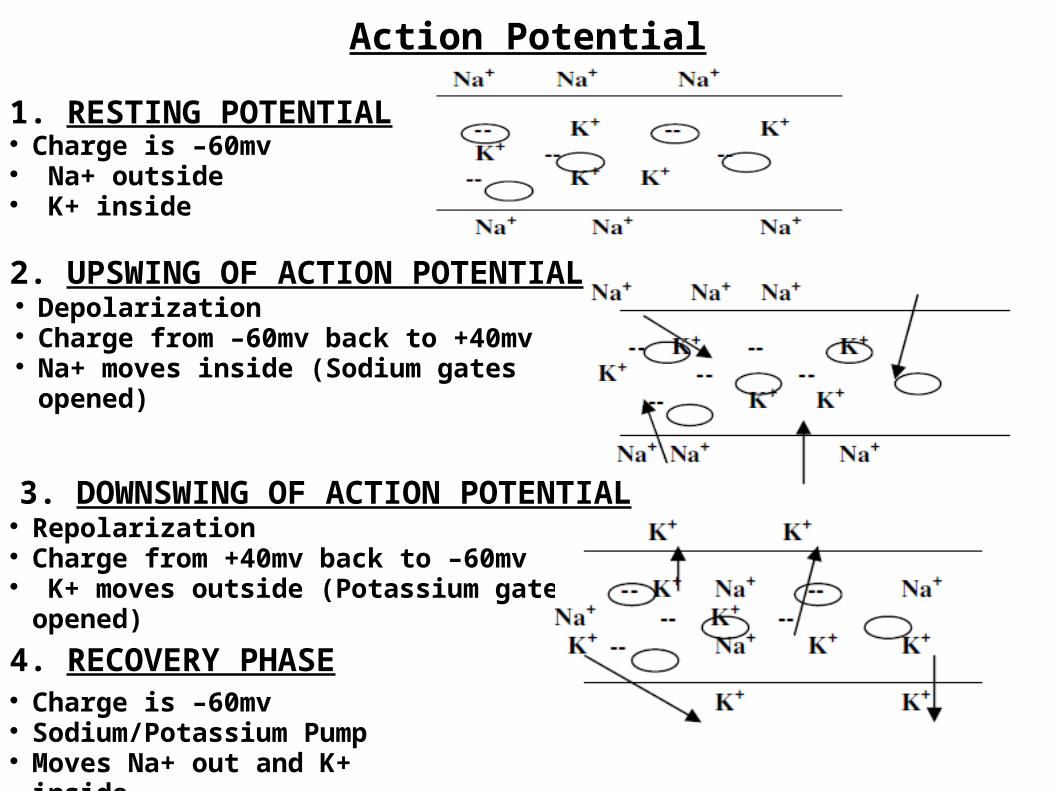
Action Potential
1. RESTING POTENTIAL Charge is –60mv Na+ outside K+ inside
2. UPSWING OF ACTION POTENTIAL Depolarization Charge from –60mv back to +40mv Na+ moves inside (Sodium gates opened)
3. DOWNSWING OF ACTION POTENTIAL Repolarization Charge from +40mv back to –60mv K+ moves outside (Potassium gates opened)
4. RECOVERY PHASE Charge is –60mv Sodium/Potassium Pump Moves Na+ out and K+ inside

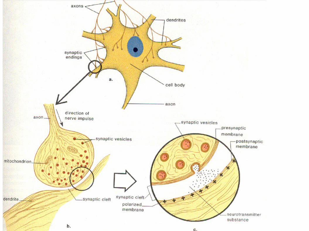
Synapse Each axon branches off and ends with a swelled tip or terminal knob lies close to but not touching the dendrite of another neuron. (or an organ). The entire region is called a synapse. Transmission of nerve impulses across a Synaptic cleft is carried out by chemicals called Neurotransmitters substances. These substances are stored in vesicles at the end of the Axon. Noradrenalin (speeds up activity) and acetylcholine (slows down activity) are examples of Neurotransmitters. When an impulse reaches the end of the axon like it usually would, not only does Na+
come into the axon, but Ca+2 as well. This calcium binds with contractile proteins that pull the Neurotransmitter vesicles to the membrane surface. The vesicles join with the cell membrane, forcing the neurotransmitter into the cleft (exocytosis)
Neurotransmitter’s job is to increase the permeability of the sodium ions on the postsynaptic membrane.
The Neurotransmitter binds to specific receptor sites on the dendrite of the next neuron. If enough transmitter substance is received, the neuron will “fire” and continue the impulse.
A neurotransmitter only has a short period to work once it has been released into the synaptic cleft. Enzymes rapidly break down the transmitter substance to clear the synapse so the next impulse can be transmitted. Monoamine oxidase breaks down noradrenaline and Acetylcholinesterase breaks down acetylcholine. An impulse can only travel across a synapse in one direction. Only the axon contains neurotransmitter vesicles, so the impulse can only travel AXON DENDRITE across a synapse.**** ALL OR NONE LAW (threshold): If enough neurotransmitter is received by the postsynapticfiber, it will fire 100% (all). If not enough substance is received, it will not fire at all (none).

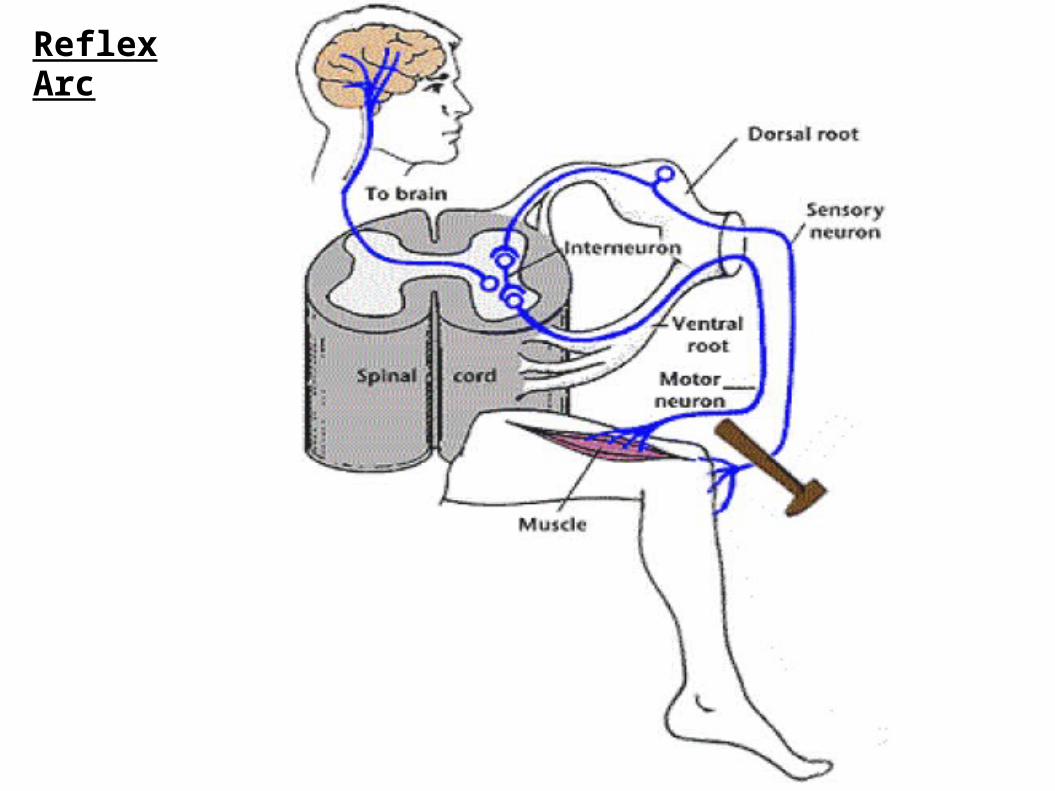
Reflex Arc Reflexes are Automatic, involuntary responses to changes occurring
inside or outside the body. Some involve the brain (such as blinking the eye), while others do not (such as moving your hand away from a hot object).
Why does the brain not have to be involved? If it were, by the time the impulse traveled to the brain, the brain figured out what was happening, and sent a response to the body, serious damage might occur. So the body evolved a method of by passing the brain.
Stages of Reflex Arc
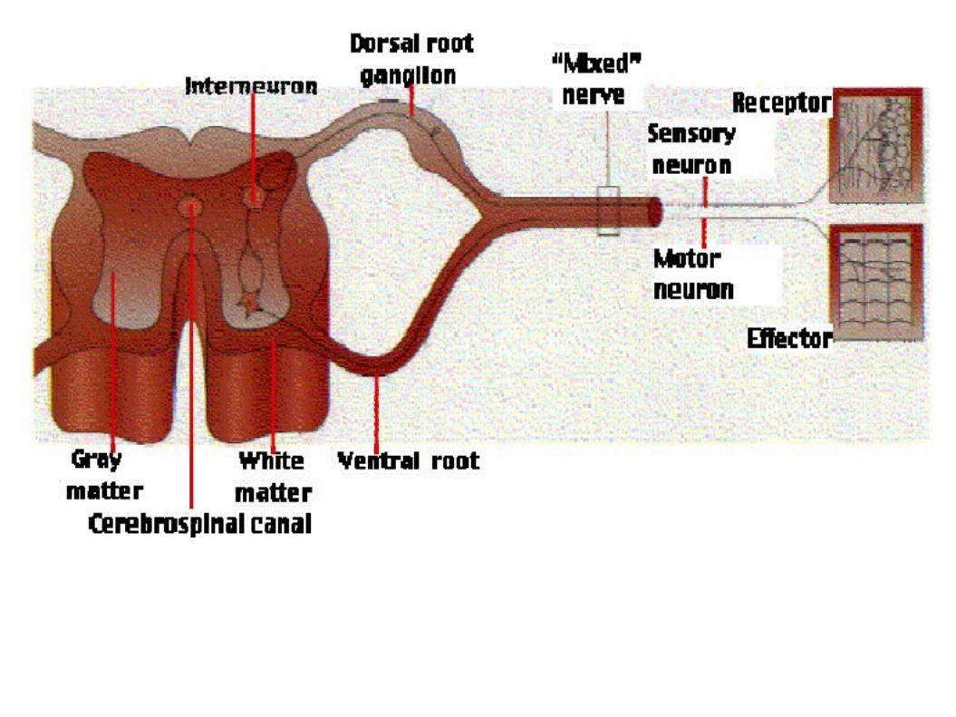
1. Receptor is stimulated and formulate message. ie. nerve impulse2. Sensory neuron takes the message to the Central Nervous System.(spinal cord)3. Interneuron passes the message to a motor neuron.4. Motor Neuron takes the message away from the C.N.S. to the effector(muscle/organ)5. The muscle receives the message and contracts.***The brain finds out later what had happened***
Reflex Arc
Reflex Arc