Embed Size (px)
Citation preview

THE JOURNAL OF BIOLOGICAL CHEMISTRY 0 1992 by The American Society for Biochemistry and Molecular Biology, Inc
Vol. 267, No. 32, Issue of November 15, pp. 23409-23417,1992 Printed in U. S. A.
G Protein Subunits Synthesized in Sf9 Cells FUNCTIONAL CHARACTERIZATION AND THE SIGNIFICANCE OF PRENYLATION OF y*
(Received for publication, June 25, 1992)
Jorge A. Iiiiguez-LluhiSg, Melvin I. Simonq, Janet D. RobishawI)**, and Alfred G. GilmanS $3 From the $Department of Pharmacology, University of Texas Southwestern Medical Center, Dallas, Texas 75235, the VDepartrnent of Biology, California Institute of Technology, Pasadena, California 91125, and the 11 Weis Center for Research, Geisinger Clinic, Danvilk, Pennsylvania 17822
Heterotrimeric guanine nucleotide-binding regula- tory proteins (G proteins) consist of a nucleotide-bind- ing (Y subunit and a high-affinity complex of @ and y subunits. There is molecular heterogeneity of @ and y, but the significance of this diversity is poorly under- stood. Different G protein @ and y subunits have been expressed both singly and in combinations in Sf9 cells. Although expression of individual subunits is achieved in all cases, By subunit activity (support of pertussis toxin-catalyzed ADP-ribosylation of rGial) is detected only when j3 and y are expressed concurrently. Of the six combinations of By tested (Dl or with yl, yz, or y3), only one, Bzyl, failed to generate a functional com- plex. Each of the other five complexes has been purified by subunit exchange chromatography using G,-aga- rose as the chromatographic matrix. We have detected differences in the abilities of the purified proteins to support ADP-ribosylation of Gial; these differences are attributable to the y component of the complex. When assayed for their ability to inhibit calmodulin-stimu- lated type-I adenylylcyclase activity or to potentiate G,-stimulated type-I1 adenylylcyclase, recombinant Btyl and transducin @y are approximately 10 and 20 times less potent, respectively, than the other com- plexes examined. Prenylation andlor further carboxyl- terminal processing of y are not required for assembly of the By subunit complex but are indispensable for high affinity interactions of @y with either G protein a subunits or adenylylcyclases.
Guanine nucleotide-binding regulatory proteins (G pro- teins)’ transmit signals that are generated by a large number of plasma membrane-bound receptors to intracellular effector
* This work was supported in part by National Institutes of Health Grants GM34497, GM34236, and GM39867, American Cancer Soci- ety Grant BE30N, the Perot Family Foundation, the Lucille P. Markey Charitable Trust, and the Raymond and Ellen Willie Chair of Molecular Neuropharmacology. The costs of publication of this article were defrayed in part by the payment of page charges. This article must therefore be hereby marked “advertisement” in accord- ance with 18 U.S.C. Section 1734 solely to indicate this fact.
5 Recipient of a Howard Hughes Medical Institute predoctoral training award.
** Established Investigator of the American Heart Association. $$ To whom correspondence should be addressed Dept. of Phar-
macology, University of Texas Southwestern Medical Center, 5323 Harry Hines Blvd., Dallas, TX 75235.
The abbreviations used are: G protein, guanine nucleotide-bind- ing regulatory protein; the short form of G,, expressed in E. coli; rG,,,,, the a subunit of Gi expressed in E. coli; Go,, the 01 subunit of the major 39-kDa G protein from brain; DTT, dithiothreitol; GTP-yS, guanosine 5’-(3-O-thio)triphosphate; PAGE, polyacrylamide gel electrophoresis; kb, kilobase pair(s).
enzymes and ion channels (reviewed in Refs. 1-3). G proteins are heterotrimers, composed of a guanine nucleotide-binding a subunit (molecular mass of 39-46 kDa) and a tight complex of P (37 kDa) and y (8-10 kDa) subunits. The signaling process involves the sequential formation and dissociation of complexes between G protein OL and Py subunits and between G protein subunits and both upstream (receptor) and down- stream (effector) elements. This process is driven by the binding of agonist ligands to receptors and by the binding and hydrolysis of GTP by the G protein a subunit (1-4). Receptors catalyze exchange of GDP for GTP on G protein a subunits; activated GTP-bound a then dissociates from the receptor and By, and both appear capable of regulating downstream effector molecules. This active state is transient, however, and decays because of the intrinsic GTPase activity of the a subunit. GDP-bound a has a high affinity for fly, reassociates to form a heterotrimer, and is thus available for further stimulation by receptors.
Most interest in G proteins has been focused on their a subunits, since these proteins bind and hydrolyze GTP and most obviously regulate the activity of the best-studied effec- tors (cyclic GMP phosphodiesterase and adenylylcyclase). Much less is known about the structure and function of by. The Py subunit complex is essential for functional interac- tions of G protein LY subunits with receptors (5-7). Recent data also suggest that the interaction of By with receptors is important for agonist-dependent receptor phosphorylation, suggesting an important role for Py in desensitization (8). By subunits can also regulate effector molecules. Genetic studies of Saccharomyces cerevisiae indicate that Py carries the signal from pheromone receptors to an as yet unidentified down- stream effector (9-11). Py can also regulate adenylylcyclase activity, and the effects of the protein complex differ for individual forms of the enzyme. Whereas by inhibits type-I adenylylcyclase when stimulated by either GB, or calmodulin, Py greatly potentiates the stimulatory effect of G,, on either type-I1 or type-IV adenylylcyclase (12, 13). Although contro- versial, By may also be involved in the direct or indirect regulation of ion channels (14-16).
There is substantial molecular diversity in both P and y. Sequences of four distinct mammalian ,f3 (17-21) and five y (22-25) subunits have been reported to date. Although Py subunit complexes are, to at least a certain extent, inter- changeable among different a subunits, the molecular diver- sity of p and y demands examination of functional signifi- cance. However, it has been very difficult to search for such differences, since preparations of Py are often heterogeneous and their composition is poorly defined. Nevertheless, some distinctions have been made with purified proteins (26-29). In addition, little is known about the functional importance of the fact that y subunits are prenylated and processed at
23409
at CA
LIFO
RN
IA IN
ST
ITU
TE
OF
TE
CH
NO
LOG
Y on D
ecember 25, 2006
ww
w.jbc.org
Dow
nloaded from
23410 G Protein Pr Subunits
their carboxyl termini in a manner analogous to that described in particular for p21'"" (30, 31). To approach these questions, we have synthesized f i and y subunits singly and in combi- nations in Sf9 cells using recombinant baculovirus. Individual ,By subunit complexes have been purified from this source, and we have initiated their characterization.
MATERIALS AND METHODS
Plasmid Construction-cDNAs encoding G protein PI, P2, yl, y,, r2C68S, and y:j subunits were transferred to a baculovirus expression vector (pVL1392 or pVL1393) as follows. For the P1 subunit (B), a 3.0-kb cDNA ligated at the EcoRI site of pBluescript (+) was digested with Asp700 and NcoI. The resulting 1.1-kb fragment containing the 8, coding sequence was isolated after filling the 3' recessed terminus generated by NcoI. This fragment was transferred to pVL1393 at the SmaI site. For the 0, subunit, the 1.06-kb ApaI fragment from the Okayama and Berg vector containing the P2 cDNA (32) was ligated a t the ApaI site of pGEM-llZf(-). The resultingplasmid was digested with BamHI and NotI. The 1.08-kb fragment containing the entire coding sequence was then transferred to pVL1393 that had been digested with the same enzymes. For the yl subunit (24), the 0.26-kb AuaI/Asp7OO fragment containing the entire y1 sequence was gener- ated from pNG-2-1. This fragment was transferred to pVL1392 that had been digested with SmaI and NotI. The termini generated by AuaI and NotI were made complementary by partially filling the AvaI site with dTTP and dCTP and the NotI site with dGTP. For the yz subunit (25), a 0.425-kb BglII/EcoRI fragment containing the entire coding sequence was isolated from a pBluescript based vector. This fragment was transferred to pVL1392 that had been digested with the same enzymes. The y,C68S sequence, in which cysteine 68 has been replaced by serine (33), was subcloned into pVL1392 as was y2.
The coding region of the bovine y3 cDNA (23) was prepared by polymerase chain reaction amplification of reverse transcribed mRNA with the introduction of PstI and XbaI sites at the 5' and 3' ends, respectively. This fragment was cloned into pVL1392.
Cell Culture and Cloning of Recombinant Baculouiruses-Fall ar- myworm ovary (Sf9) cells were propagated in IPL-41 medium supple- mented with 10% fetal calf serum as described (34). Recombinant baculoviruses were generated by cotransfection of Sf9 cells with the expression vectors described above and with linearized AcRP23-lacZ viral DNA by the lipofection method (35). Positive viral clones were isolated by plaque assay and were identified by their ability to direct the expression of the appropriate proteins as revealed by immuno- blotting.
Gel Electrophoresis and Zmmunoblotting-To resolve the y sub- units, SDS-PAGE was carried out in linear gradient gels (10-20% acrylamide; acry1amide:bisacrylamide 26:l) containing 4 M urea. Sam- ples were reduced and alkylated with N-ethylmaleimide as described (36). In some cases samples were precipitated with 15% trichloroacetic acid and the pellets washed with cold acetone before reduction and alkylation. Gels were either stained with silver (37) or processed for immunoblotting as described (38). The following antisera were used a t the indicated dilutions: P-specific, U-49 1:10,000 (38) and affinity- purified K-521 1:400 (32); y-specific, affinity-purified NG-1 (23), affinity-purified X-263 (33), and B-53, all at 1:500. B-53 antiserum was generated using a peptide corresponding to the amino-terminal 17 residues of the bovine y3 sequence. Detection was achieved using donkey anti-rabbit IgG F(ab'), conjugated to horseradish peroxidase and the enhanced chemiluminescence detection system according to the manufacturer's instructions (Amersham Corp.).
Expression of Py Subunits in Sf9 Cells and Cell Fractionation- Typically, Sf9 cells (100-250 ml at 2 X 10' cells/ml) were infected at a multiplicity of infection of one for each of the indicated viruses. Cells were collected by centrifugation 60 h after infection and were resuspended in a solution containing 20 mM NaHepes (pH 8.0), 2 mM MgCI,, 1 mM EDTA, 2 mM DTT, and protease inhibitors' at a density of 25 X 10 ' cells/ml. After 5 min on ice, cells were homogenized by forcing the cell suspension (six times) through a 25-gauge needle attached to a disposable syringe. The lysate was then centrifuged in a TL-100.3 rotor (Beckman) at 200,000 X g for 15 min at 4 "C. The supernatant or cytosolic fraction was removed, and the crude partic-
16 pg/ml each 1-1-tosylamido-2-phenylethyl chloromethyl ke- tone, l-chloro-3-tosylamido-7-amino-2-heptanone, and phenylmeth- ylsulfonyl fluoride; 3.2 pg/ml each leupeptin and soybean trypsin inhibitor; and 1 pg/ml aprotinin.
ulate fraction was resuspended in 0.25-0.5 ml of the same buffer. Detergent extracts were made by diluting particulate fractions to a final protein concentration of 5 mg/ml and addition of 10% Lubrol PX or 20% sodium cholate to final concentrations of 0.6 or 1%, respectively. The mixtures were then homogenized by forcing them through a 25-gauge needle (six times). The detergent extracts were collected after centrifugation at 200,000 X g for 15 min at 4 "C in a
with Lubrol PX and 50% with sodium cholate. TL-100.3 rotor. Solubilization of total protein approximated 20%
Purification of Py"Sf9 cells (1 liter) were grown to a density of 1 X lo6 cells/ml in IPL 41 medium supplemented with 1% fetal calf serum and lipid concentrate (GIBCO). Cultures were coinfected with the appropriate recombinant baculoviruses at a multiplicity of infec- tion of 1 for each virus, and cells were collected 60 h after infection. Unless otherwise indicated all subsequent manipulations were carried out at 4 "C. Preparation of Sf9 cell membranes was carried out essentially as described (34). The cells were lysed in 20 mM NaHepes (pH 8.0), 1 mM EDTA, 1 mM EGTA, 5 mM MgCl,, 2 mM DTT, 150 mM NaCI, and protease inhibitors, and the membrane pellets were washed and resuspended in 20 mM NaHepes (pH 8.0), 2 mM MgCI2, 1 mM EDTA, 2 mM DTT, and protease inhibitors. Membranes (typically 30 mg) were solubilized in the same buffer containing 1% sodium cholate at a protein concentration of 5 mg/ml. Functional By complexes were purified by affinity chromatography using a 10-ml column of bovine brain Go,,-agarose. Purified bovine brain Go, (20 mg) was coupled to 10 ml of w-aminobutyl-agarose by the method of Sternweis et al. (30). The concentration of Py binding sites on the resulting resin was approximately 0.6 p ~ . (We have observed, how- ever, that the binding capacity of the column is lower when crude detergent extracts are loaded.) The resin was packed in a Pharmacia XK-26-20 column fitted with flow adapters and was used in a fast protein liquid chromatography system. Approximately 6 nmol of By activity (determined by support of ADP-ribosylation (see below), using purified bovine brain by as a standard) was diluted to 10 ml with 20 mM NaHepes (pH 8.0), 400 mM NaCl, 1 mM EDTA, 2 mM DTT, 5 p M GDP (buffer A), and 0.5% Lubrol PX and applied at a rate of 0.3 ml/min to the Go,,-agarose, which had been equilibrated in the same buffer. After washing the column with 20 ml of buffer A containing 0.5% Lubrol PX, the flow was increased to 3 ml/min and the concentration of Lubrol PX in the wash buffer was reduced to 0.05% over 20 ml. The resin was washed with 200 ml of buffer A containing 0.05% Lubrol PX. Elution was then achieved with 20 mM NaHepes (pH 8.0), 400 mM NaCl, 2 mM DTT, 5 p~ GDP, 30 p M A1CI3, 20 mM MgCl,, 10 mM NaF (buffer B), and 0.05% Lubrol PX. Fractions (10 ml) were collected at a rate of 0.3 ml/min. To increase the rate of dissociation of By during the elution, the resin was warmed to approximately 15 "C with a water jacket. Fractions containing By (judged by SDS-PAGE and silver stain) from two or three chromat- ographic runs were pooled (100-200 ml) and concentrated in a PM- 10 Amicon ultrafiltration device to a volume of 10-20 ml. This material was loaded onto a 1-ml hydroxylapatite column previously equilibrated with 20 mM NaHepes (pH 8.0), 100 mM NaC1, 2 mM DTT, and 0.05% Lubrol PX. The column was then washed with 5 ml of the same buffer and Py was eluted in 1-ml fractions with 5 ml of the same buffer containing 50 mM potassium phosphate. Fractions containing 07 were pooled and subjected again to chromatography on Go,,-agarose. The pooled fractions were concentrated and subse- quently adsorbed to hydroxylapatite and eluted as described above. The final preparations were diluted appropriately to achieve a Py concentration of 0.5 p~ (based on an Amido Black protein assay (39) using bovine serum albumin as standard) and a Lubrol PX concen- tration of 0.025%. The preparations were stored at -80 "C. The final yields varied from 50 to 200 pg of By, depending on the particular complex that was synthesized. Endogenous Py from Sf9 cells was purified as described above from membranes of cells infected with control (Lac-Z) virus encoding @-galactosidase. Bovine brain By was purified as described by Sternweis et al. (36) and corresponds to a pool from the second heptylamine-Sepharose column in the presence of AI3+, M e , and F-. Transducin Py was a generous gift of Dr. Susanne Mumby. Both bovine brain and transducin By were subjected to Go,-agarose chromatography, followed by hydroxylapatite chro- matography, as described.
Partial Purification of Ply2C68S-A cytosolic fraction (10 ml, 2 mg/ml) from Sf9 cells infected with recombinant baculoviruses en- coding P I and y2C68S was loaded onto a Pharmacia Mono-Q HR 5/5 column equilibrated with 50 mM Tris-HC1 (pH 8.0), 1 mM EDTA, and 2 mM DTT. After washing the column with 5 ml of the same buffer, proteins were eluted with a linear gradient of NaCl (0-350
at CA
LIFO
RN
IA IN
ST
ITU
TE
OF
TE
CH
NO
LOG
Y on D
ecember 25, 2006
ww
w.jbc.org
Dow
nloaded from
G Protein P-y Subunits 23411
mM). Fractions containing 81~2C68S (eluting at approximately 150 mM NaC1) were pooled and concentrated. The amount of By in the pool was estimated by quantitative immunoblotting, using purified &y2 as a standard. The expressed protein constituted approximately 20% of the total protein in the pool.
Support of ADP-ribosylation and Adenylylcyclase Assays-The ca- pacity of By to support the ADP-ribosylation of rGi,,l was assessed as described, with some modifications (40). The final lipid concentration was increased to 1 mM, and purified rGi,, (20 pmol/assay) was used as a substrate. The use of a high concentration of substrate devoid of any endogenous By allowed the reliable quantitation of 50 fmol of By activity (1.2 nM final concentration in the assay), even in crude detergent extracts. To assay column fractions containing Al"+, M e , and F-, the MgCI, in the reaction mixture was replaced with EDTA to yield a final concentration of EDTA that was at least 1 mM in excess of the concentration of MgC12 contributed by the sample. This change has essentially no effect on the kinetics of the reaction (26). For adenylylcyclase assays, membranes from Sf9 cells infected with recombinant baculoviruses encoding type-I or type-I1 adenylylcyclase were prepared as described (12). Purified recombinant Drosophila calmodulin and rGan.* were provided by Dr. Wei-Jen Tang. rGIR.* was activated with GTPyS for 30 min at 30 "C in 20 mM NaHepes (pH 8.0), 1 mM EDTA, 1 mM DTT, 10 mM MgS04, and 100 p M GTPyS. Free nucleotide was removed by gel filtration. Membranes containing type-I or type-I1 adenylylcyclase were incubated with Ca2+/calmodu- lin or GTPyS-rG,.., respectively, for 10 min at 30 "C. Aliquots (IO pl) were then distributed to tubes containing the indicated amounts of purified Py (10 pl). The reactions were initiated by the addition of 80 pl of the remaining reaction components (12). After 10 min at 30 "C, the reactions were terminated and cyclic AMP was quantified by the method of Salomon (41). The final concentrations of activators were 50 p M CaCI2 and 100 nM calmodulin for type-I adenylylcyclase and 50 nM GTPyS-rG,, for type-I1 adenylylcyclase.
Gel Filtration Chromatography-Cytosolic fractions and cholate extracts of membranes were prepared as described above from Sf9 cells independently expressing or ~2C68S as well as from cells expressing both subunits. Before chromatography, cytosolic fractions were adjusted to 1% sodium cholate. Aliquots (200 pl; 0.5-1 mg of protein) of samples containing PI (cholate extract), y2C68S (cytosol), or 81y2C68S (cytosol) were injected onto a Pharmacia Superose 12 HR 10/30 gel filtration column that had been equilibrated with 20 mM NaHepes (pH 8.0), 2 mM MgCI2, 150 mM NaCI, 1 mM EDTA, 2 mM DTT, and 1% sodium cholate. Proteins were eluted at a rate of 0.3 ml/min, and 0.5-ml fractions were collected (starting 2 ml after injection of the sample). Aliquots (100 pl) of selected fractions were precipitated with 15% trichloroacetic acid, and 25% of each sample was subjected to SDS-PAGE and immunoblotting as described above. fi and y subunits were identified with antisera U-49 and X-263, respectively. The intensities of bands were quantified with a laser densitometer.
RESULTS
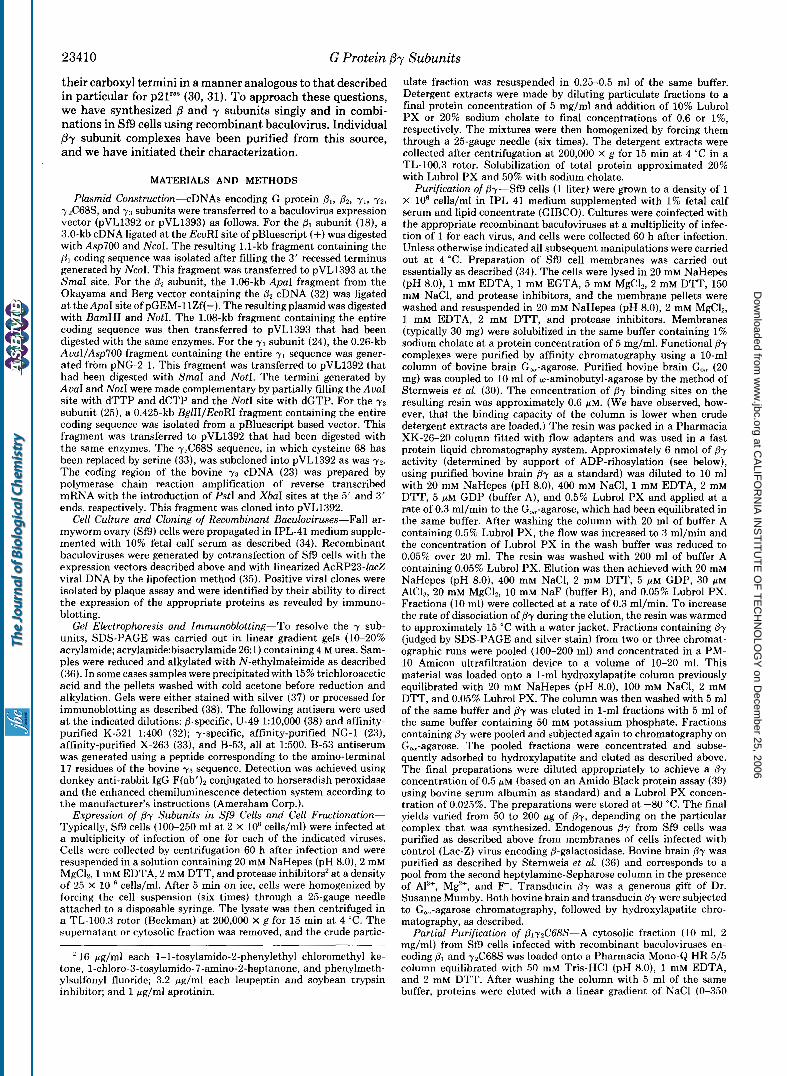
Expression of G Protein @y Subunits in Sf9 Cells-Concur- rent infection of Sf9 cells with recombinant baculoviruses encoding the G protein p2 and y2 subunits leads to a substan- tial, time-dependent increase in By subunit activity as as- sessed by the capacity of detergent extracts of cell membranes to support the ADP-ribosylation of rGial (Fig. 1). No such activity is detected when either subunit is expressed alone or when the cells are infected with a control virus encoding @- galactosidase (Lac-Z). The lack of activity when each subunit is expressed independently is not due to a failure to synthesize the polypeptides, since expression of the subunits was con- firmed by SDS-PAGE and immunoblotting (Fig. 1, inset). It thus appears that this characteristic /3y activity is dependent on both subunits. We have not been successful in attempts to reconstitute activity by mixing detergent extracts from cells expressing each subunit independently (data not shown).
Little is known about the selectivity of interactions between different B and y subunits. However, by coinfecting Sf9 cells with pl- or &-encoding viruses and either yl, 7 2 , or 7 3 viruses we have tested the ability of these six combinations to yield a complex capable of supporting the ADP-ribosylation of
1.2
1.0 t
;; 0.8 E
5 0.6
E,
(u
0) - c 2 0 n g 0.4 a 0 Q
0.2
0.0
110 - r' 84- 47 - 33 - 24 - /
e - B /
I I I I I I I 0 12 24 36 48 60 72
Time post infection (hr)
FIG. 1. Expression of B and 7 subunits in Sf9 cells. Cultures (100 ml) of Sf9 cells (1 X IO6 cells/ml) were infected at a multiplicity of infection of 1 with control (Lac-Z) virus (V), with recombinant baculoviruses encoding G protein p2 (0) or y2 (A) subunits, or with both and y2 viruses (m). A t the indicated times after infection, 10- ml aliquots were withdrawn and 0.6% Lubrol PX extracts of crude particulate fractions were obtained and assayed (0.5 pg of protein) for support of ADP-ribosylation activity as described under "Mate- rials and Methods." Inset, 20 pg of crude particulate fractions from cells collected 72 h after infection were subjected to SDS-PAGE and immunoblotting as described under "Materials and Methods." The upper and lower sections of the blot were separated and probed with affinity-purified K-521 and X-263 antisera, respectively. The migra- tions of molecular weight standards and of the and y subunits are indicated at the left and right, respectively.
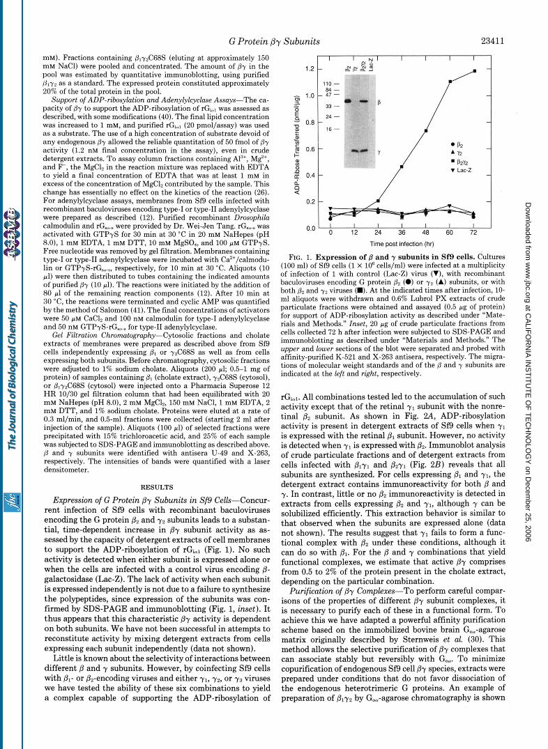
rGiol. All combinations tested led to the accumulation of such activity except that of the retinal y1 subunit with the nonre- tinal p2 subunit. As shown in Fig. 2A, ADP-ribosylation activity is present in detergent extracts of Sf9 cells when y1 is expressed with the retinal @, subunit. However, no activity is detected when y1 is expressed with &. Immunoblot analysis of crude particulate fractions and of detergent extracts from cells infected with Plyl and @2y1 (Fig. 2B) reveals that all subunits are synthesized. For cells expressing and yl, the detergent extract contains immunoreactivity for both @ and y. In contrast, little or no O2 immunoreactivity is detected in extracts from cells expressing pZ and yl , although y can be solubilized efficiently. This extraction behavior is similar to that observed when the subunits are expressed alone (data not shown). The results suggest that y1 fails to form a func- tional complex with p2 under these conditions, although it can do so with Dl. For the /3 and y combinations that yield functional complexes, we estimate that active @y comprises from 0.5 to 2% of the protein present in the cholate extract, depending on the particular combination.
Purification of @y Complexes-To perform careful compar- isons of the properties of different Py subunit complexes, it is necessary to purify each of these in a functional form. To achieve this we have adapted a powerful affinity purification scheme based on the immobilized bovine brain Goo-agarose matrix originally described by Sternweis et al. (30). This method allows the selective purification of @y complexes that can associate stably but reversibly with Goa. To minimize copurification of endogenous Sf9 cell @y species, extracts were prepared under conditions that do not favor dissociation of the endogenous heterotrimeric G proteins. An example of preparation of ply2 by Go,-agarose chromatography is shown
at CA
LIFO
RN
IA IN
ST
ITU
TE
OF
TE
CH
NO
LOG
Y on D
ecember 25, 2006
ww
w.jbc.org
Dow
nloaded from
G Protein Pr Subunits 23412 0.8
7 ’
0.0 0.1 0.2 0.3 0.4 0.5 pg of protein
6.
M E M E M E “
a
u-49
NG-1
FIG. 2. Concurrent expression of y1 with B1 or B2 subunits. A, the indicated amounts of cholate extract protein derived from membranes of Sf9 cells were assayed for support of ADP-ribosylation activity as described under “Materials and Methods.” Cells were infected with control (Lac-Z) virus (0) or were infected concurrently with PI and y1 viruses (0) or with 82 and y1 viruses (0). B, membranes (M) (1 pg, upper panel; 10 pg, lower panel) and cholate extracts ( E ) (5 pg, both panels) of membranes from Sf9 cells infected with the indicated viruses were subjected to SDS-PAGE and immunoblotting as described under “Materials and Methods.” The upper and lower panels were probed with affinity-purified K-521 and NG-1 antisera, respectively. Only the regions of the blot showing immunoreactivity are shown. The amount of membranes used in the upper panel was reduced because of the large amount of 0 immunoreactivity.
- LO&- 10 20 30 40 50 60 70 80 Fraction Number
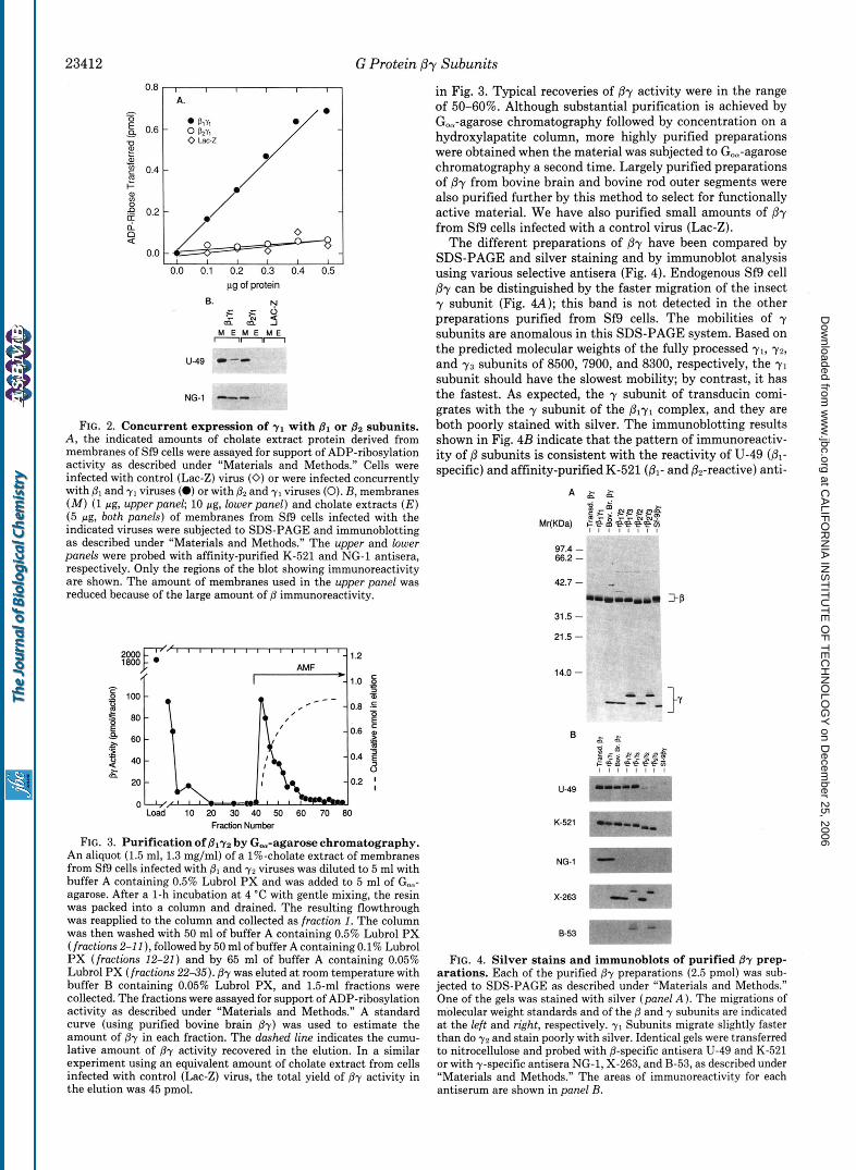
FIG. 3. Purification of Bly2 by G,-agarose chromatography. An aliquot (1.5 ml, 1.3 mg/ml) of a 1%-cholate extract of membranes from Sf9 cells infected with @, and y2 viruses was diluted to 5 ml with buffer A containing 0.5% Lubrol PX and was added to 5 ml of Go,- agarose. After a 1-h incubation at 4 “C with gentle mixing, the resin was packed into a column and drained. The resulting flowthrough was reapplied to the column and collected as fraction 1. The column was then washed with 50 ml of buffer A containing 0.5% Lubrol PX (fractions 2-1 1 ), followed by 50 ml of buffer A containing 0.1% Lubrol PX (fractions 12-21) and by 65 ml of buffer A containing 0.05% Lubrol PX (fractions 22-35). By was eluted at room temperature with buffer B containing 0.05% Lubrol PX, and 1.5-ml fractions were collected. The fractions were assayed for support of ADP-ribosylation activity as described under ”Materials and Methods.” A standard curve (using purified bovine brain @y) was used to estimate the amount of By in each fraction. The dashed line indicates the cumu- lative amount of @y activity recovered in the elution. In a similar experiment using an equivalent amount of cholate extract from cells infected with control (Lac-Z) virus, the total yield of By activity in the elution was 45 pmol.
in Fig. 3. Typical recoveries of by activity were in the range of 50-60%. Although substantial purification is achieved by Go,-agarose chromatography followed by concentration on a hydroxylapatite column, more highly purified preparations were obtained when the material was subjected to Go,-agarose chromatography a second time. Largely purified preparations of /3y from bovine brain and bovine rod outer segments were also purified further by this method to select for functionally active material. We have also purified small amounts of ,8r from Sf9 cells infected with a control virus (Lac-Z).
The different preparations of /3y have been compared by SDS-PAGE and silver staining and by immunoblot analysis using various selective antisera (Fig. 4). Endogenous Sf9 cell Py can be distinguished by the faster migration of the insect y subunit (Fig. 4A); this band is not detected in the other preparations purified from Sf9 cells. The mobilities of y subunits are anomalous in this SDS-PAGE system. Based on the predicted molecular weights of the fully processed yl, 7 2 ,
and 7 3 subunits of 8500, 7900, and 8300, respectively, the y1 subunit should have the slowest mobility; by contrast, it has the fastest. As expected, the y subunit of transducin comi- grates with the y subunit of the plyl complex, and they are both poorly stained with silver. The immunoblotting results shown in Fig. 4B indicate that the pattern of immunoreactiv- ity of /3 subunits is consistent with the reactivity of U-49 (01-
specific) and affinity-purified K-521(&- and &reactive) anti-
97.4 - 66.2 -
42.7 -
31.5 -
21.5 -
14.0 -
B
u-49
K-521
NG-1
x-263
553 1
FIG. 4. Silver stains and immunoblots of purified By prep-
jected to SDS-PAGE as described under “Materials and Methods.” arations. Each of the purified @y preparations (2.5 pmol) was sub-
One of the gels was stained with silver (panel A ) . The migrations of molecular weight standards and of the B and y subunits are indicated at the left and right, respectively. y1 Subunits migrate slightly faster than do y2 and stain poorly with silver. Identical gels were transferred to nitrocellulose and probed with @-specific antisera U-49 and K-521 or with y-specific antisera NG-1, X-263, and B-53, as described under “Materials and Methods.” The areas of immunoreactivity for each antiserum are shown in panel B.
at CA
LIFO
RN
IA IN
ST
ITU
TE
OF
TE
CH
NO
LOG
Y on D
ecember 25, 2006
ww
w.jbc.org
Dow
nloaded from

G Protein P-y Subunits 23413
sera. The Sf9 cell py preparation reacts only weakly with both antisera. The affinity-purified NG-1 antiserum raised against a peptide sequence unique to y1 recognizes only the y subunits of transducin and ply1. As anticipated, based on the amino acid sequence of the peptide used as the antigen (30), affinity- purified antiserum X-263 reacts with both y2 and y3 subunits. Antiserum B-53 recognizes only 7 3 . None of the antibodies recognize the Sf9 cell y polypeptide. Based on silver staining and immunoblotting, the bovine brain By preparation is most probably composed largely of p1 and y2 subunits.
Support of ADP-ribosylution by Purified Py Preparatiom- ADP-ribosylation of G protein a subunits by pertussis toxin is a sensitive means to monitor interactions between a and By, since the heterotrimeric G protein is the preferred sub- strate for the toxin. As an initial screen to evaluate potential functional differences between individual /?y subunits, we have determined the ability of the purified By preparations to support catalytically the ADP-ribosylation of rGial and bovine brain When rGial is used as substrate, the rate of ADP- ribosylation is substantially different for the different Py complexes. Irrespective of the p subunit, the highest rates are observed for the complexes containing y2, while the lowest rates are observed with y1 (including transducin By). Inter- mediate rates are observed for p1y3 and p2y3. The largest difference observed was approximately 6-fold. When bovine brain Go,, is used as substrate (Fig. 5 B ) , the pattern is differ- ent, and the largest difference in rates is only 2-fold. ADP- ribosylation is most rapid with transducin by, and the lowest rate is observed with &y3. There is no clear grouping based on y subunit composition. It thus appears that G protein a subunits can discriminate between different complexes. However, we have not yet addressed whether the differences observed are due to intrinsic differences between the two CY subunits or to the fact that the recombinant protein (Giml) is
rGlul I I I
n
8 -
6 -
4 -
2 -
n- - I I I I I
0.0 0.1 0.2 0.3 0.4 Pmol pr
GOU I , I
2.5 - g % 2.0
2 1.5-
2 2 1.0-
E
- E - a
0.5 -
0.0 - 8 8 I ,
0.0 0.1 0.2 0.3 0.4 Pmol &u
FIG. 5. Support of ADP-ribosylation of rGiml and bovine brain G, by purified 67 preparations. The indicated amounts of By were assayed for support of ADP-ribosylation activity as described under “Materials and Methods” using 20 pmol of rGiml (panel A ) or bovine brain G,,, (panel B ) as substrates.
not myristoylated. In addition, although this assay is simple technically and very sensitive, it is complex mechanistically and does not allow a straightforward assessment of the rela- tive affinities of a for j3-y (see below).
Effect of Different By Complexes on Type-I and Type-II Adenylylcyclase Activity-Type-I adenylylcyclase is activated by either Ca’+/calmodulin or GBm; such activation can be partially reversed by Py in a noncompetitive fashion (12). By contrast, when type-I1 adenylylcyclase is stimulated by GBa, further addition of fly causes a large potentiation of Gsm- stimulated activity (12). We examined the ability of the different Py complexes to inhibit Ca*+/calmodulin-stimulated type-I adenylylcyclase (Fig. 6). All preparations can inhibit type-I adenylylcyclase activity. However, plyl and transducin Py are approximately 10 and 20 times, respectively, less potent than the other fly preparations. The difference is thus attrib- utable to the y subunit. The bovine brain preparation behaves essentially as does its recombinant counterpart, ply2. The pattern was very similar when we examined the effect of the different Py preparations on rG,,.,-stimulated type-I1 adeny- lylcyclase (Fig. 7); again, transducin by and plyl are approx- imately 10 and 20 times less potent than the other complexes, and ply2 is very similar to bovine brain By.
Effects of Prenylation on Py Function-We have compared the properties of the wild type y2 subunit with those of a mutant yz in which the cysteine four amino acid residues from the carboxyl terminus has been changed to serine (y2C68S). This cysteine residue is the presumed site of pren- ylation of y, and this mutant protein cannot be prenylated in vivo (33, 42). When both wild type and mutant y z subunits are coexpressed with pl, ADP-ribosylation activity is detected only in detergent extracts of Sf9 cells infected with wild type yz (Fig. &I). In a mammalian cell expression system, coexpression of y2C68S with PI causes distribution of to the cytosolic fraction. SDS-PAGE and immunoblot analysis of Sf9 cell fractions (Fig. 8C) indicates that substantial y2 im- munoreactivity is found in the cholate extract of membranes, with little or no reactivity in the cytosolic fraction. By con- trast, little or no rzC68S is found in the cholate extract, whereas substantial immunoreactivity is present in the cyto- sol. Furthermore, coinfection of yzC68S with P1 leads to a
[Pr I (M) FIG. 6. Inhibition of Ca’+/calmodulin-stimulated type-I ad-
enylylcyclase by purified fly preparations. Aliquots (10 pg) of Sf9 cell membranes containing type-I adenylylcyclase were assayed for 10 min in the presence of 100 nM calmodulin, 50 pM CaCl,, and the indicated concentrations of the purified preparations of Pr. Values are expressed as a percent of the maximal calmodulin-stimulated activity in the absence of Py. Data from duplicate determinations of two to four independent experiments are included. The average basal and calmodulin-stimulated activities were 0.4 and 5.1 nmol of cyclic AMP X min” X mg”, respectively. The curues shown are the nonlin- ear least-squares fits to the four parameter logistic equation Y = (A/ (1 + ( C / X ) ’ ) ) + D, and the derived ICsovalues are indicated in the inset.
at CA
LIFO
RN
IA IN
ST
ITU
TE
OF
TE
CH
NO
LOG
Y on D
ecember 25, 2006
ww
w.jbc.org
Dow
nloaded from
23414 G Protein Pr Subunits
40
3[ 20 10
[& 1 (M)
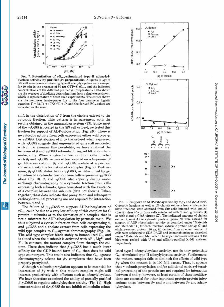
FIG. 7. Potentiation of rG,..-stimulated type-I1 adenylyl- cyclase activity by purified By preparations. Aliquots (1 pg) of Sf9 cell membranes containing type-I1 adenylylcyclase were assayed for 10 min in the presence of 50 nM GTP+-rG,.. and the indicated concentrations of the different purified By preparations. Data shown are the averages of duplicate determinations from a single experiment, which is representative of three such experiments. The curues shown are the nonlinear least-squares fits to the four parameter logistic equation Y = ( A / ( I + ( C / X ) H ) ) + D, and the derived ECsovalues are indicated in the inset.
shift in the distribution of B from the cholate extract to the cytosolic fraction. This pattern is in agreement with the results obtained in the mammalian system (33). Since most of the y2C68S is located in the Sf9 cell cytosol, we tested this fraction for support of ADP-ribosylation (Fig. BB). There is no cytosolic activity from cells expressing either wild type 7 2
or 72C68S. Distribution of /3 to the cytosol when expressed with y2C68S suggests that unprenylated y 2 is still associated with 8. To examine this possibility, we have analyzed the behavior of 0 and 72C68S subunits during gel filtration chro- matography. When a cytosolic fraction from cells infected with p1 and y2C68S viruses is fractionated on a Superose 12 gel filtration column, B1 and y2C68S coelute at a position consistent with the formation of a complex (Fig. 9). Further- more, p172C68S elutes before y2C68S, as determined by gel filtration of a cytosolic fraction from cells expressing 72C68S alone (Fig. 9). Dl and y2C68S also copurify during anion exchange chromatography of a cytosolic fraction from cells expressing both subunits, again consistent with the existence of a complex between the subunits (data not shown). Taken together, these data indicate that prenylation and subsequent carboxyl-terminal processing are not required for interaction between /3 and y.
The failure of BlyzC68S to support ADP-ribosylation of rGic,l could be due to a very low affinity of this complex for G protein (Y subunits or to the formation of a complex that is not a substrate for ADP-ribosylation by pertussis toxin. We thus subjected a cytosolic fraction from cells coexpressing and y2C68S and a cholate extract from cells expressing the wild type complex to Go,-agarose chromatography (Fig. 10). The wild type complex binds stably to immobilized Go, and is eluted when the CY subunit is activated with Al", MF, and F-. In contrast, the mutant complex flows through the col- umn. These data indicate that 8172C68S has a much lower affinity for the GDP-bound form of Go, than does its wild type counterpart. This result also indicates that Go,-agarose chromatography selects for By complexes that have been properly prenylated.
Although y subunit prenylation appears to be essential for interaction of By with a, this mutant complex might still interact productively with effectors such as adenylylcyclase. We have therefore examined the ability of partially purified P1~2C68S to regulate adenylylcyclase activity (Fig. 11). High concentrations of &y2C68S do not inhibit calmodulin-stimu-
I I I I I
c g 0.1
Q 0.0
0.0 0.1 0.2 0.3 0.4
1 I
pg of protein - 8 0.2
B. Cytosol
0 PlY2C68S 0 Lac-z -0
a,
0 I 1 a
0.0 0.2 0.4 0.6 0.8 1.0 pg of protein
c. r 2 ? *
Finn C E C E C E . .
110- e.4-
"-
47 -
33-
24 -
16-
>e
FIG. 8. Support of ADP-ribosylation by Blyz and B1r2C68S. Cytosolic fractions as well as 1%-cholate extracts from crude partic- ulate fractions were obtained from Sf9 cells infected with control (Lac-Z) virus (0) or from cells coinfected with 8, and y2 viruses (m) or with @ and r2C68S viruses (0). The indicated amounts of cholate extract (panel A ) or cytosolic protein (panel B ) were assayed for support of ADP-ribosylation activity as described under "Materials and Methods." C, for each infection, cytosolic protein (30 pg, C) and cholate-extract protein (20 pg, E ) derived from an equal number of cells were subjected to SDS-PAGE and immunoblotting as described under "Materials and Methods." The upper and lower sections of the blot were probed with U-49 and affinity-purified X-263 antisera, respectively.
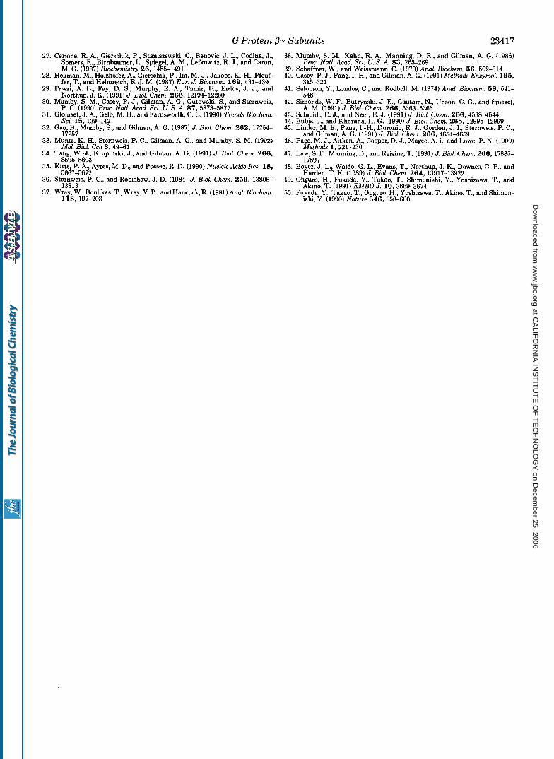
lated type-I adenylylcyclase activity, nor do they potentiate G,-stimulated type-I1 adenylylcyclase activity. Furthermore, the mutant complex fails to diminish the effects of wild type By when the mutant is in 100-fold excess. Thus, it appears that y subunit prenylation and/or additional carboxyl-termi- nal processing of the protein are not required for interaction between p and y; however, at least certain of these modifica- tions are essential for other important protein-protein inter- actions: those between Py and (Y and between By and adeny- lylcyclase.
at CA
LIFO
RN
IA IN
ST
ITU
TE
OF
TE
CH
NO
LOG
Y on D
ecember 25, 2006
ww
w.jbc.org
Dow
nloaded from
G Protein Py Subunits 23415
Fraction Number i o 14 18 20 22 24 26 28
p region
I7 y region
1 ° L 4 0 b+ 10 15 20 25 30
Fraction Number
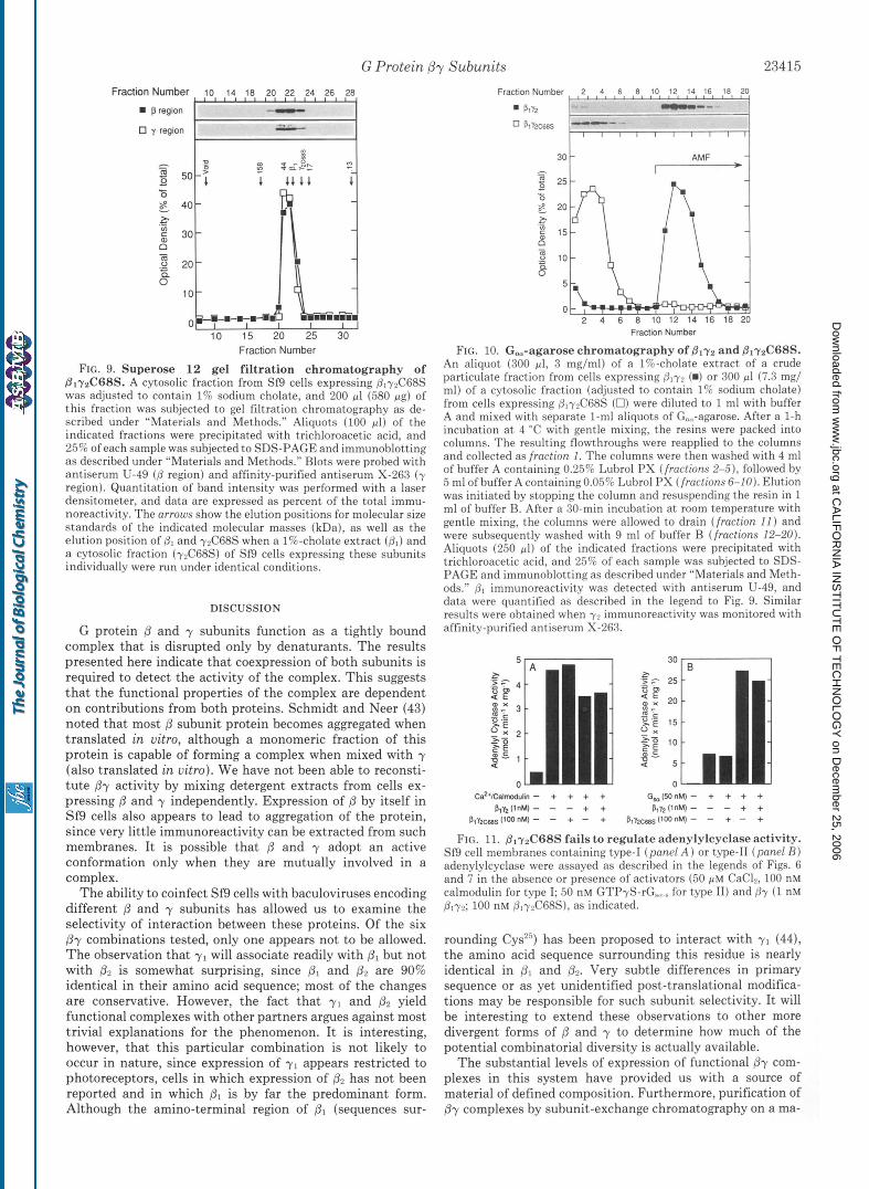
FIG. 9. Superose 12 gel filtration chromatography of PlyaC68S. A cytosolic fraction from Sf9 cells expressing p1-y2C68S was adjusted to contain 1% sodium cholate, and 200 pl (580 pg) of this fraction was subjected to gel filtration chromatography as de- scribed under “Materials and Methods.” Aliquots (100 pl) of the indicated fractions were precipitated with trichloroacetic acid, and 25% of each sample was subjected to SDS-PAGE and immunohlotting as described under “Materials and Methods.” Blots were probed with antiserum U-49 ( B region) and affinity-purified antiserum X-263 ( y region). Quantitation of band intensity was performed with a laser densitometer, and data are expressed as percent of the total immu- noreactivity. The arrows show the elution positions for molecular size standards of the indicated molecular masses (kDa), as well as the elution position of p1 and 7 4 6 8 s when a 1%-cholate extract (Dl ) and a cytosolic fraction (yrC68S) of Sf9 cells expressing these subunits individually were run under identical conditions.
DISCUSSION
G protein p and y subunits function as a tightly bound complex that is disrupted only by denaturants. The results presented here indicate that coexpression of both subunits is required to detect the activity of the complex. This suggests that the functional properties of the complex are dependent on contributions from both proteins. Schmidt and Neer (43) noted that most /3 subunit protein becomes aggregated when translated in uitro, although a monomeric fraction of this protein is capable of forming a complex when mixed with y (also translated in uitro). We have not been able to reconsti- tute by activity by mixing detergent extracts from cells ex- pressing p and y independently. Expression of @ by itself in Sf9 cells also appears to lead to aggregation of the protein, since very little immunoreactivity can be extracted from such membranes. It is possible that p and y adopt an active conformation only when they are mutually involved in a complex.
The ability to coinfect Sf9 cells with baculoviruses encoding different p and y subunits has allowed us to examine the selectivity of interaction between these proteins. Of the six Py combinations tested, only one appears not to be allowed. The observation that y1 will associate readily with p1 but not with 0. is somewhat surprising, since p1 and p2 are 90% identical in their amino acid sequence; most of the changes are conservative. However, the fact that y1 and p2 yield functional complexes with other partners argues against most trivial explanations for the phenomenon. It is interesting, however, that this particular combination is not likely to occur in nature, since expression of y1 appears restricted to photoreceptors, cells in which expression of has not been reported and in which p1 is by far the predominant form. Although the amino-terminal region of p1 (sequences sur-
30 t - 1
Fraction Number
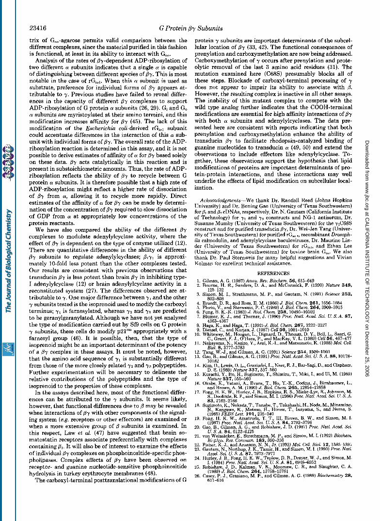
FIG. 10. G,-agarose chromatography of Blyz and B1yzC68S. An aliquot (300 pl, 3 mg/ml) of a 1%-cholate extract of a crude particulate fraction from cells expressing Slyy (m) or 300 11 (7.3 mg/ ml) of a cytosolic fraction (adjusted to contain 1% sodium cholate) from cells expressing @,yyC68S (0) were diluted to 1 ml with buffer A and mixed with separate 1-ml aliquots of Go,,-agarose. After a 1-h incubation a t 4 “C with gentle mixing, the resins were packed into columns. The resulting flowthroughs were reapplied to the columns and collected as fraction 1. The columns were then washed with 4 ml of buffer A containing 0.25% Lubrol PX (fractions 2-S), followed by 5 ml of buffer A containing0.059; Luhrol PX (fractions 6-10). Elution was initiated by stopping the column and resuspending the resin in 1 ml of buffer €3. After a 30-min incubation at room temperature with gentle mixing, the columns were allowed to drain (fraction I 1 ) and were subsequently washed with 9 ml of buffer B (fractions 12-20). Aliquots (250 pl) of the indicated fractions were precipitated with trichloroacetic acid, and 25% of each sample was subjected to SDS- PAGE and immunohlotting as described under “Materials and Meth- ods.” Dl immunoreactivity was detected with antiserum U-49, and data were quantified as described in the legend to Fig. 9. Similar results were ohtained when yr immunoreactivity was monitored with affinity-purified antiserum X-263.
FIG. 11. B1yzC68S fails to regulate adenylylcyclase activity. Sf9 cell membranes containing type-I (panel A ) or t-ype-I1 (panel R ) adenylylcyclase were assayed as described in the legends of Figs. 6 and 7 in the absence or presence of activators (50 p M Ca& 100 nM calmodulin for type I; 50 nM GTPyS-rG,,.., for type 11) and Py (1 nM B,y?; 100 nM @,-yyC68S), as indicated.
rounding Cys’”) has been proposed to interact with y1 (44), the amino acid sequence surrounding this residue is nearly identical in p, and (3’. Very subtle differences in primary sequence or as yet unidentified post-translational modifica- tions may be responsible for such subunit selectivity. It will be interesting to extend these observations to other more divergent forms of /3 and y to determine how much of the potential combinatorial diversity is actually available.
The substantial levels of expression of functional Py com- plexes in this system have provided us with a source of material of defined composition. Furthermore, purification of Py complexes by subunit-exchange chromatography on a ma-
at CA
LIFO
RN
IA IN
ST
ITU
TE
OF
TE
CH
NO
LOG
Y on D
ecember 25, 2006
ww
w.jbc.org
Dow
nloaded from
23416 G Protein ,By Subunits
trix of Go,-agarose permits valid comparison between the different complexes, since the material purified in this fashion is functional, at least in its ability to interact with Goa.
Analysis of the rates of &-dependent ADP-ribosylation of two different a subunits indicates that a single a is capable of distinguishing between different species of By. This is most notable in the case of rGial. When this a subunit is used as substrate, preference for individual forms of /3y appears at- tributable to y. Previous studies have failed to reveal differ- ences in the capacity of different Py complexes to support ADP-ribosylation of G protein (Y subunits (26, 29). Gi and Go a subunits are myristoylated at their amino termini, and this modification increases affinity for Py (45). The lack of this modification of the Escherichia coli-derived rGial subunit could accentuate differences in the interaction of this a sub- unit with individual forms of &. The overall rate of the ADP- ribosylation reaction is determined in this assay, and it is not possible to derive estimates of affinity of a for Py based solely on these data. Py acts catalytically in this reaction and is present in substoichiometric amounts. Thus, the rate of ADP- ribosylation reflects the ability of By to recycle between G protein a subunits. It is therefore possible that a high rate of ADP-ribosylation might reflect a higher rate of dissociation of by from a, allowing it to recycle more rapidly. Direct estimates of the affinity of a for Py can be made by determi- nation of the concentration of @y required to slow dissociation of GDP from a at appropriately low concentrations of the protein reactants.
We have also compared the ability of the different /3y complexes to modulate adenylylcyclase activity, where the effect of ,f3y is dependent on the type of enzyme utilized (12). There are quantitative differences in the ability of different By subunits to regulate adenylylcyclases; plyl is approxi- mately 10-fold less potent than the other complexes tested. Our results are consistent with previous observations that transducin Py is less potent than brain By in inhibiting type- I adenylylcyclase (12) or brain adenylylcyclase activity in a reconstituted system (27). The differences observed are at- tributable to y. One major difference between y1 and the other y subunits tested is the isoprenoid used to modify the carboxyl terminus; y1 is farnesylated, whereas y 2 and 7 3 are predicted to be geranylgeranylated. Although we have not yet analyzed the type of modification carried out by Sf9 cells on G protein y subunits, these cells do modify p21'"" appropriately with a farnesyl group (46). It is possible, then, that the type of isoprenoid might be an important determinant of the potency of a /3y complex in these assays. It must be noted, however, that the amino acid sequence of y1 is substantially different from those of the more closely related 7 2 and 7 3 polypeptides. Further experimentation will be necessary to delineate the relative contributions of the polypeptides and the type of isoprenoid to the properties of these complexes.
In the assays described here, most of the functional differ- ences can be attributed to the y subunits. It seems likely, however, that functional differences due to p may be revealed when interactions of Py with other components of the signal- ing system (e.g. receptors or other effectors) are examined or when a more extensive group of p subunits is examined. In this respect, Law et al. (47) have suggested that brain so- matostatin receptors associate preferentially with complexes containing Bl. It will also be of interest to examine the effects of individual /3y complexes on phosphoinositide-specific phos- pholipases. Complex effects of By have been observed on receptor- and guanine nucleotide-sensitive phosphoinositide hydrolysis in turkey erythrocyte membranes (48).
The carboxyl-terminal posttranslational modifications of G
protein y subunits are important determinants of the subcel- lular location of By (33, 42). The functional consequences of prenylation and carboxymethylation are now being addressed. Carboxymethylation of y occurs after prenylation and prote- olytic removal of the last 3 amino acid residues (31). The mutation examined here (C68S) presumably blocks all of these steps. Blockade of carboxyl-terminal processing of y does not appear to impair its ability to associate with /3. However, the resulting complex is inactive in all other assays. The inability of this mutant complex to compete with the wild type analog further indicates that the COOH-terminal modifications are essential for high affinity interactions of ,8y with both a subunits and adenylylcyclases. The data pre- sented here are consistent with reports indicating that both prenylation and carboxymethylation enhance the ability of transducin Py to facilitate rhodopsin-catalyzed binding of guanine nucleotides to transducin a (49, 50) and extend the observations to include effectors like adenylylcyclase. To- gether, these observations support the hypothesis that lipid modifications of proteins are important determinants of pro- tein-protein interactions, and these interactions may well underlie the effects of lipid modification on subcellular local- ization.
Acknowledgments-We thank Dr. Randall Reed (Johns Hopkins University) and Dr. Boning Gao (University of Texas Southwestern) for PI and 0 2 cDNAs, respectively, Dr. N. Gautam (California Institute of Technology) for y1 and y2 constructs and NG-1 antiserum, Dr. Susanne Mumby (University of Texas Southwestern) for the y2C68S construct and for purified transducin By, Dr. Wei-Jen Tang (Univer- sity of Texas Southwestern) for purified rG.,.8, recombinant Drosoph- ih calmodulin, and adenylylcyclase baculoviruses, Dr. Maurine Lin- der (University of Texas Southwestern) for r G 1 , and Ethan Lee (University of Texas Southwestern) for bovine brain Goa. We also thank Dr. Paul Sternweis for many helpful suggestions and Vivian Kolman for excellent technical assistance.
REFERENCES 1. Gilman, A. G. (1987) Annu. Rev. Biochem. 56,615-649 2. Bourne, H. R., Sanders, D. A., and McCormick, F. (1990) Nature 3 4 8 ,
3. Simon, M. I., Strathmann, M. P., and Gautam, N. (1991) Science 2 5 2 ,
4. Brandt, D. R., and Ross, E. M. (1986) J. Biol. Chem. 261,1656-1664 5. Florio, V., and Sternweis, P. C. (1989) J. Biol. Chem. 264,3909-3915 6. Fung, B. K.-K. (1983) J. Biol. Chem. 258,10495-10502 7. Blumer, K. J., and Thorner, J. (1990) Proc. Natl. Acad. Sci. U. S. A. 8 7 ,
8. Haga, K., and Haga, T. (1992) J. Biol. Chem. 267,2222-2227 9. Dietzel, C., and Kurjan, J. (1987) Cell 5 0 , 1001-1010
125-132
802-808
4363-4367
10. Whiteway, M., Hougan, L., Dignard, D., Thomas, D. Y., Bell, L., Saari, G.
11. Nakayama, N., Kaziro, Y., Arai, K.-I., and Matsumoto, K. (1988) Mol. Cell. C., Grant, F. J., O'Hara, P., and MacKay, V. L. (1989) Cell 56,467-477
12. Tang, W.-J., and Gilman, A. G. (1991) Science 2 6 4 , 1500-1503 13. Gao, B., and Gilman, A. G. (1991) Proc. Natl. Acad. Sci. U. S. A. 88,10178-
Bhl. 8,3777-3783
14. Kim, D., Lewis, D. L., Graziadei, L., Neer, E. J., Bar-Sagi, D., and Clapham,
15. Kurachi. Y.. Ito. H.. Sueimoto. T.. Shimizu. T.. Miki. I.. and Ui. M. (1989)
10182
D. E. (1989) Nature 337,557-560 . , . .
16. Okabe, K., Yatani, A,, Evans, T., Ho, Y.-K., Codina, J., Birnbaumer, L., Nature 337,5551566 ' '
and Brown, A. M. (1990) J. Bcol. Chem. 2 6 5 , 12854-12858 17. Fong, H. K. W., Hurley, J. B., Hopkins, R. S., Miake-Lye, R., Johnson, M.
S.. Doolittle, R. F.. and Simon, M. I. (1986) Proc. Natl. Acad. Scr. U. S. A.
18.
19.
20.
21.
22. 23.
24.
25.
26.
83,2162-2166 '
Sugimoto, K., Nukada, T., Tanabe, T., Takahashi, H., Noda, M., Minamino, N., Kangawa, K., Matsuo, H., Hirose, T., Inayama, S., and Numa, S.
Fong, H. K. W., Amatruda, T. T., 111, Birren, B. W., and Simon, M. I.
Gao, B., Gilman, A. G., and Robishaw, J. D. (1987) Proc. Natl. Acad. Sci.
von Weizsacker, E., Strathmann, M. P., and Simon, M. I. (1992) Biochem.
Fisher, K. J., and Aronson, N. N., Jr. (1992) Mol. Cell. Biol. 12,1585-1591 Gautam, N., Northup, J. K., Tamir, H., and Simon, M. I. (1990) Proc. Natl.
Hurley, J. B., Fong, H. K. W., Teplow, D. B., Dreyer. W. J., and Simon, M.
Robishaw, J. ,D., Kalman, V. K., Moomaw, C. R., and Slaughter, C. A.
Casey, P. J., Graziano, M. P., and Gilman, A. G . (1989) Biochemistry 2 8 ,
(1985) FEES Lett. 191,235-240
(1987) Proc. Natl. Acad. Sci. U. S. A. 8 4 , 3792-3796
U. S. A. 8 4 , 6122-6125
Biopkys. Res. Commun. 183,350-356
Acad. Sci. U. S. A. 87,7913-7977
I. (1984) Proc. Natl. Acad. Sci. U. S. A. 81,6948-6952
(1989) J. Blol. Chem. 2 6 4 , 15758-15761
611-616
at CA
LIFO
RN
IA IN
ST
ITU
TE
OF
TE
CH
NO
LOG
Y on D
ecember 25, 2006
ww
w.jbc.org
Dow
nloaded from
G Protein P-y Subunits 23417 27. Cerione, R. A,, Gierschik, P., Staniszewski, C., Benovic, J. L., Codina, J., 38. Mumby, S. M., Kahn, R. A., Manning, D. R., and Gilman, A. G. (1986)
Somers, R., Birnbaumer, L., Spiegel, A. M., Leikowitz, R. J., and Caron, Proc. Natl. Acad. Sei. U. S. A . 8 3 , 265-269 M. G. (1987) Biochemistry 26,1485-1491
fer, T., and Helmreich, E. J. M. (1987) Eur. J. Biochern. 169,431-439 315-321
Northup, J. K. (1991) J. Biol. Chem. 266,12194-12200
P. C. (1990) Proc. Natl. Acad. Sci. U. S. A. 87,5873-5877 A. M. (1991) J. Biol. Chem. 266,5363-5366
Sci. 15,139-142
17257 and Gilman, A. G. (1991) J. Biol. Chern. 266,4654-4659
Mol. Biol. Cell 3 , 49-61
85954603 17897
5667-5672
13813 Akino, T. (1991) EMBO J. 10, 3669-3674
118, 197-203 rshr, Y. (1990) Nature 346,658-660
28. Hekman, M., Holzhofer, A., Gierschik, P., Im, M.-J., Jakobs, K.-H., Pfeuf- 40. Casey, P. J., Pang, I.-H., and Gilman, A. G. (1991) Methods Enzymol. 195 ,
29. Fawzi, A. B., Fay, D. S., Murphy, E. A,, Tamir, H., Erdos, J. J., and 41. Salomon, Y., Londos, C., and Rodbell, M. (1974) Anal. Biochern. 58 , 541-
30. Mumby, S. M., Casey, P. J., Gilman, A. G., Gutowski, S., and Sternweis, 42. Simonds, W. F., Butrynski, J. E., Gautam, N., Unson, C. G., and Spiegel,
31. Glomset, J. A., Gelb, M. H., and Farnsworth, C. C. (1990) Trends Biochern. 43. Schmidt, C. J., and Neer, E. J. (1991) J. Bml. Chern. 266 , 4538-4544
32. Gao, B., Mumby, S., and Gilman, A. G. (1987) J. Biol. Chern. 262,17254- 45. Linder, M. E., Pang, I.-H., Duronio, R. J., Gordon, J. I., Sternweis, P. C.,
33. Muntz, K. H., Sternweis, P. C., Gilman, A. G., and Mumby, S. M. (1992) 46. Page, M. J., Aitken, A., Cooper, D. J., Magee, A. I., and Lowe, P. N. (1990)
34. Tang, W.-J., Krupinski, J., and Gilman, A. G. (1991) J. B i d . Chern. 266 , 47. Law, S. F., Manning, D., and Reisine, T. (1991) J. Biol. Chem. 266,17885-
35. Kitts, P. A., Ayres, M. D., and Possee, R. D. (1990) Nucleic Acids Res. 1 8 , 48. Boyer, J. L., Waldo, G. L., Evans, T., Northup, J. K., Downes, C. P., and
36. Sternweis, P. C., and Robishaw, J. D. (1984) J. Biol. Chern. 2 5 9 , 13806- 49. Ohguro, H., Fukada, Y., Takao, T., Shimonishi, Y., Yoshizawa, T., and
37. Wray, W., Boulikas, T., Wray, V. P., and Hancock, R. (1981) Anal. Biochem. 50. Fukae , Y., Takao, T., Ohguro, H., Yoshizawa, T., Akino, T., and Shimon-
39. Schaffner, W., and Weissmann, C. (1973) Anal. Biochem. 56,502-514
548
44. Bubis, J., and Khorana, H. G. (1990) J. Biol. Chern. 265,12995-12999
Methods 1,221-230
Harden, T. K. (1989) J. Biol. Chern. 264,13917-13922
at CA
LIFO
RN
IA IN
ST
ITU
TE
OF
TE
CH
NO
LOG
Y on D
ecember 25, 2006
ww
w.jbc.org
Dow
nloaded from