Embed Size (px)
Citation preview
The optical properties of rat abdominal wall muscle
Luís Oliveira1,2,3, Maria Inês Carvalho4, Elisabete Nogueira1,3,
Valery Tuchin5,6,7
1 Physics Department – Polytechnic of Porto, School of Engineering, Rua Dr. António Bernardino de Almeida, 431, 4200-072 Porto, Portugal.
2 FEUP – University of Porto, Rua Dr. Roberto Frias, 4200-465 Porto, Portugal.
3 CIETI – Centre of Innovation in Engineering and Industrial Technology, ISEP, Rua Dr. António Bernardino de Almeida, 431, 4200-072 Porto, Portugal.
4 DEEC/FEUP and INESC TEC, University of Porto, Rua Dr. Roberto Frias, 4200-465 Porto, Portugal.
5 Research-Educational Institute of Optics and Biophotonics, Saratov State University, 83 Astrakhanskaya Str., Saratov 410012, Russia.
6 Laboratory of Laser Diagnostics of Technical and Living Systems, Institute of Precise Mechanics and Control RAS, Saratov, 410028, Russia.
7 Optoelectronics and Measurement Techniques Laboratory, University of Oulu, PO Box 4500 FI-90014, Oulu, Finland.
1. IntroductionThe knowledge of the optical properties of biological tissue and their wavelength dependency is very important for clinical applications and related research where optical technologies are to be used [1][2].
The basic optical properties are the absorption coefficient (µa), the scattering coefficient (µs), the reduced scattering coefficient (µs’) and the anisotropy factor (g) [3].
The absorption coefficient quantifies the amount of absorbed photons per unit length inside the biological tissue [4].
The optical
properties of rat
abdominal wall
muscle
Luís Oliveira
Maria Inês Carvalho
Elisabete Nogueira
Valery Tuchin
Saratov Fall Meeting
2013
1
The scattering coefficient quantifies the amount of scattered photons per unit length inside the biological tissue [4].
The reduced scattering coefficient quantifies both the amount of scattered photons and directionality of scattering per unit length inside the biological tissue [4].
The anisotropy factor quantifies the directionality of the scattering events inside the biological tissue [4].
The determination of such optical properties can be made using direct and indirect methods, depending on the instrumentation available and desired precision [3].
The optical
properties of rat
abdominal wall
muscle
Luís Oliveira
Maria Inês Carvalho
Elisabete Nogueira
Valery Tuchin
Saratov Fall Meeting
2013
2
Among the indirect methods, computer simulations using the Adding-Doubling and Monte Carlo methods are commonly used and generated results present high precision [3].
Some computer codes based on these methods are available. They are used to perform inverse simulations and estimate the optical properties of biological tissues [5] [6] [7] [8].
The abdominal wall muscle from rat is a fibrous tissue that contains a collection of fiber bundles distributed over the interstitial fluid [9].
The muscle fiber bundles contain several muscle fibers, which are chains of proteins (actin and myosin) [10].
The optical
properties of rat
abdominal wall
muscle
Luís Oliveira
Maria Inês Carvalho
Elisabete Nogueira
Valery Tuchin
Saratov Fall Meeting
2013
3
Due to the composition of the muscle, we expect to observe much higher values for the scattering coefficient than for the absorption coefficient [3].
The interstitial fluid is a background liquid that contains mainly water and some dissolved salts and minerals [10].
Such fact is due to the refractive index profile observed between the scatterers (muscle fibers) and the background material (interstitial fluid) [10].
To study the optical properties of the abdominal wall muscle from rat – species Wistar Han, we have adopted an indirect technique and estimated the optical properties from inverse simulations using the Monte Carlo and the Adding Doubling codes.
The optical
properties of rat
abdominal wall
muscle
Luís Oliveira
Maria Inês Carvalho
Elisabete Nogueira
Valery Tuchin
Saratov Fall Meeting
2013
4
As we have observed with this study, our experimental measurements present errors that induce bad wavelength dependencies for the estimated optical properties.
We have used some experimental assemblies to measure transmittance and reflectance spectra from muscle samples. Afterwards, we have retrieved experimental values from these spectra between 400 and 1000 nm to use as input in the inverse simulations [9].
By comparing the wavelength dependencies obtained from our study with the ones published in literature [11]
[12], we could correct our results and identify the source of those errors in the experimental set-ups that we have used.
The optical
properties of rat
abdominal wall
muscle
Luís Oliveira
Maria Inês Carvalho
Elisabete Nogueira
Valery Tuchin
Saratov Fall Meeting
2013
5
2. Experimental MethodologyWe have developed an inverse MC simulation code based on the code developed for the forward problem. Such forward code is designated as Monte Carlo for Multi-Layered media (MCML) and was developed by Lihong Wang and Steven Jacques in 1992 [5].
Similarly, we have also used an IAD calculator developed by Scott Prahl [6]. This calculator was used to estimate the optical properties of the muscle. The calculator can be found at: http://omlc.ogi.edu/calc/iad_calc.html.
Such forward code is freeware and can be found on the internet, at: http://omlc.ogi.edu/software/mc/.
The optical
properties of rat
abdominal wall
muscle
Luís Oliveira
Maria Inês Carvalho
Elisabete Nogueira
Valery Tuchin
Saratov Fall Meeting
2013
6
The measurements of Tt and Rt were made using an integrating sphere. The measuring set-ups used to measure Tc and Rs were specially constructed for this study.
Based on the input parameters for the IMC code and the IAD calculator, we have chosen to perform experimental measurements of Total transmittance (Tt), Total reflectance (Rt), Collimated transmittance (Tc) and Specular reflectance (Rs).
In the Tc measuring set-up we have used a light beam from a tungsten-halogen lamp to illuminate the sample – see figure 1:
The optical
properties of rat
abdominal wall
muscle
Luís Oliveira
Maria Inês Carvalho
Elisabete Nogueira
Valery Tuchin
Saratov Fall Meeting
2013
7
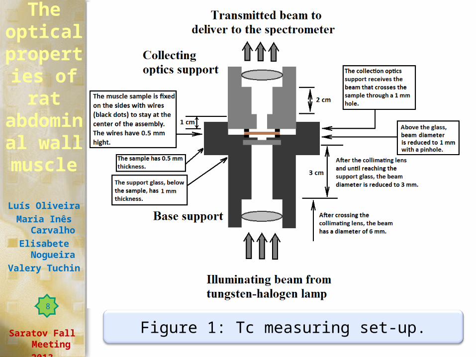
Figure 1: Tc measuring set-up.
The optical
properties of rat
abdominal wall
muscle
Luís Oliveira
Maria Inês Carvalho
Elisabete Nogueira
Valery Tuchin
Saratov Fall Meeting
2013
8
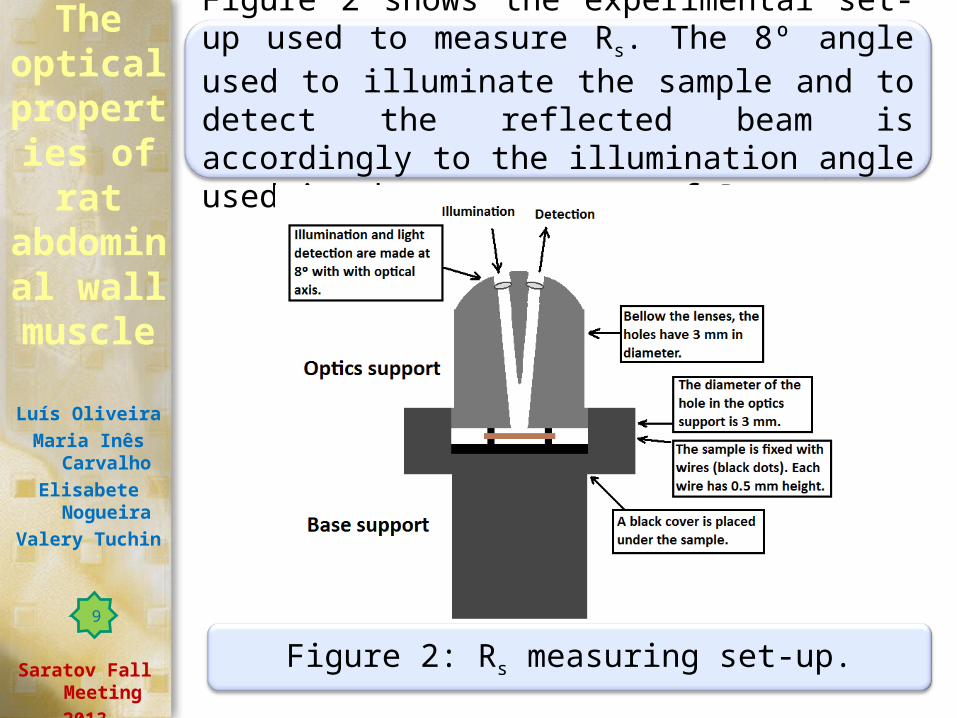
Figure 2 shows the experimental set-up used to measure Rs. The 8º angle used to illuminate the sample and to detect the reflected beam is accordingly to the illumination angle used in the measurement of Rt.
Figure 2: Rs measuring set-up.
The optical
properties of rat
abdominal wall
muscle
Luís Oliveira
Maria Inês Carvalho
Elisabete Nogueira
Valery Tuchin
Saratov Fall Meeting
2013
9
From the spectra measured with each experimental assembly, we considered all wavelengths separated by 25 nm between 400 and 1000 nm.
We will explain below how to calculate both A and Rd from the other measurements.
To perform the inverse MC simulations, we need the same measurements as used for IAD simulations. Additionally, IMC code needs the absorbance (A) and the diffuse reflectance (Rd).
The measurements of Tt, Rt and Ts are sufficient to use in the IAD calculator.
The optical
properties of rat
abdominal wall
muscle
Luís Oliveira
Maria Inês Carvalho
Elisabete Nogueira
Valery Tuchin
Saratov Fall Meeting
2013
10
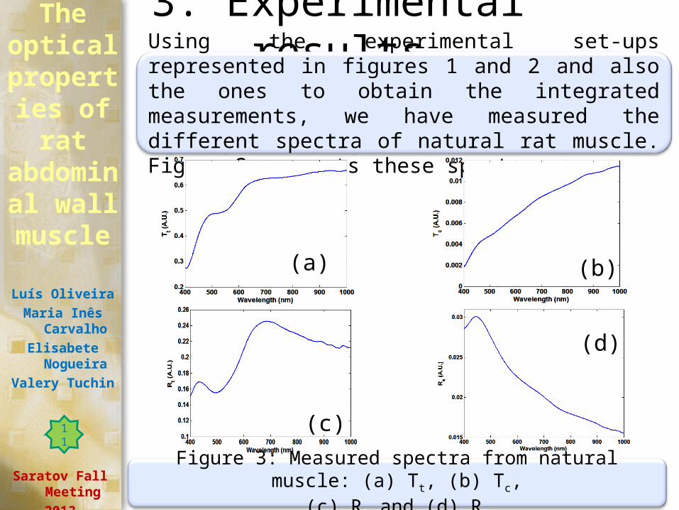
3. Experimental resultsUsing the experimental set-ups represented in figures 1 and 2 and also the ones to obtain the integrated measurements, we have measured the different spectra of natural rat muscle. Figure 3 presents these spectra:
(a) (b)
(c)
(d)
The optical
properties of rat
abdominal wall
muscle
Luís Oliveira
Maria Inês Carvalho
Elisabete Nogueira
Valery Tuchin
Saratov Fall Meeting
2013
Figure 3: Measured spectra from natural muscle: (a) Tt, (b) Tc, (c) Rt and (d) Rs.
11
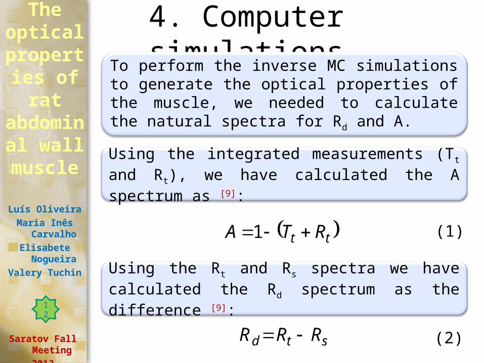
4. Computer simulationsTo perform the inverse MC simulations to generate the optical properties of the muscle, we needed to calculate the natural spectra for Rd and A.
Using the integrated measurements (Tt and Rt), we have calculated the A spectrum as [9]:
tt RTA 1
Using the Rt and Rs spectra we have calculated the Rd spectrum as the difference [9]:
std RRR
(1)
(2)
The optical
properties of rat
abdominal wall
muscle
Luís Oliveira
Maria Inês Carvalho
Elisabete Nogueira
Valery Tuchin
Saratov Fall Meeting
2013
12
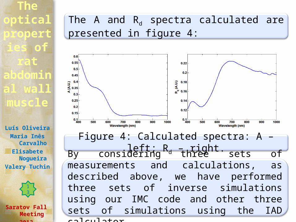
The A and Rd spectra calculated are presented in figure 4:
Figure 4: Calculated spectra: A – left; Rd – right.
By considering three sets of measurements and calculations, as described above, we have performed three sets of inverse simulations using our IMC code and other three sets of simulations using the IAD calculator.
The optical
properties of rat
abdominal wall
muscle
Luís Oliveira
Maria Inês Carvalho
Elisabete Nogueira
Valery Tuchin
Saratov Fall Meeting
2013
13
The optical
properties of rat
abdominal wall
muscle
Luís Oliveira
Maria Inês Carvalho
Elisabete Nogueira
Valery Tuchin
Saratov Fall Meeting
2013
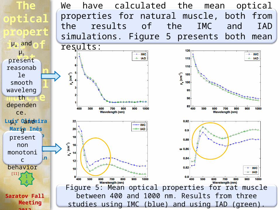
We have calculated the mean optical properties for natural muscle, both from the results of the IMC and IAD simulations. Figure 5 presents both mean results:
Figure 5: Mean optical properties for rat muscle between 400 and 1000 nm. Results from three studies using IMC (blue) and using IAD (green).
µa and µs present
reasonable smooth
wavelength dependence
.
µs’ and g present non monotonic behavior [11]
[12].
14
5. Corrections to wavelength dependency
The non monotonic behavior seen for µs’ and g on figure 5 are a result of erroneous measurements for Tc and Rs.
By considering the experimental set-up to measure Tc (see figure 1), more than unscattered transmitted photons are detected. Considering the Rs set-up presented in figure 2, we see that a great amount of diffused reflected photons are also collected by the detector [13].
Such errors in our measurement set-ups produce wrong spectra that will generate erroneous optical properties. Such errors are more evident for lower wavelengths.
The optical
properties of rat
abdominal wall
muscle
Luís Oliveira
Maria Inês Carvalho
Elisabete Nogueira
Valery Tuchin
Saratov Fall Meeting
2013
15
To correct these errors, we have considered the typical wavelength dependencies for µs and µs’ that are presented in literature [11] [12].
We have observed that for the case of µa, we have similar wavelength dependency as indicated in literature [11] [14].
In the case of µs, our results show also a wavelength dependence that is similar to typical behavior seen in literature [11] [14].
The major difference between our results and literature is seen for µs’ [11] [12]. In this case, to correct our results, we had to neglect the values obtained for wavelengths between 450 and 650 nm and then adjust the remaining values with an exponential decay.
The optical
properties of rat
abdominal wall
muscle
Luís Oliveira
Maria Inês Carvalho
Elisabete Nogueira
Valery Tuchin
Saratov Fall Meeting
2013
16
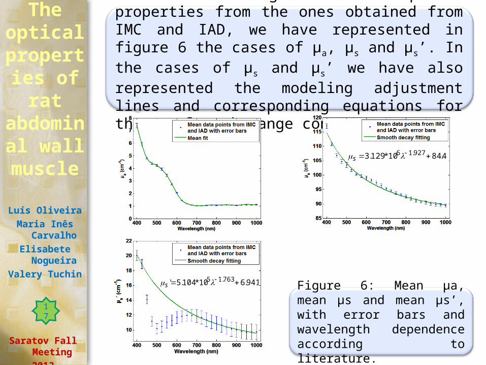
After calculating the mean optical properties from the ones obtained from IMC and IAD, we have represented in figure 6 the cases of µa, µs and µs’. In the cases of µs and µs’ we have also represented the modeling adjustment lines and corresponding equations for the wavelength range considered.
Figure 6: Mean µa, mean µs and mean µs’, with error bars and wavelength dependence according to literature.
The optical
properties of rat
abdominal wall
muscle
Luís Oliveira
Maria Inês Carvalho
Elisabete Nogueira
Valery Tuchin
Saratov Fall Meeting
2013
941.610*104.5' 763.15 s
4.8410*129.3 927.16 s
17
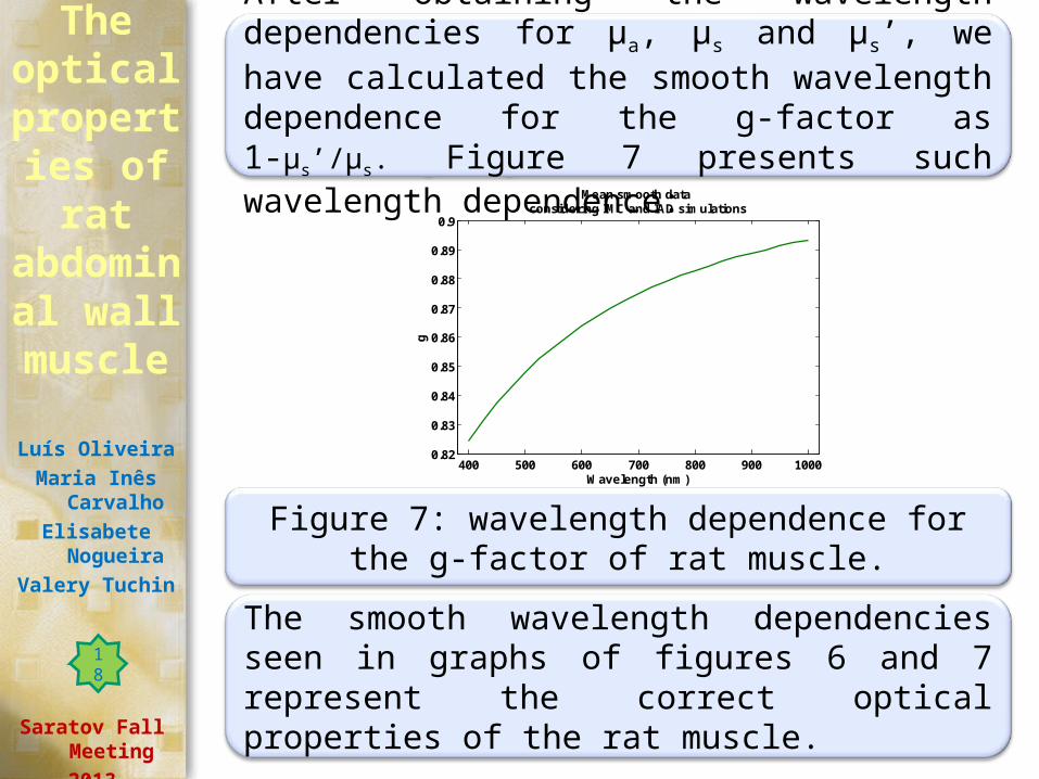
After obtaining the wavelength dependencies for µa, µs and µs’, we have calculated the smooth wavelength dependence for the g-factor as 1-µs’/µs. Figure 7 presents such wavelength dependence:
The smooth wavelength dependencies seen in graphs of figures 6 and 7 represent the correct optical properties of the rat muscle.
Figure 7: wavelength dependence for the g-factor of rat muscle.
The optical
properties of rat
abdominal wall
muscle
Luís Oliveira
Maria Inês Carvalho
Elisabete Nogueira
Valery Tuchin
Saratov Fall Meeting
2013
400 500 600 700 800 900 10000.82
0.83
0.84
0.85
0.86
0.87
0.88
0.89
0.9
Wavelength (nm)
g
Mean smooth data considering IMC and IAD simulations
18
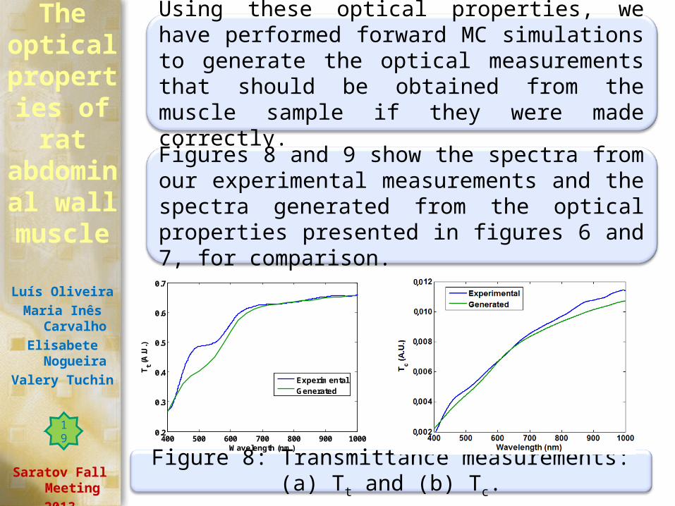
Using these optical properties, we have performed forward MC simulations to generate the optical measurements that should be obtained from the muscle sample if they were made correctly.
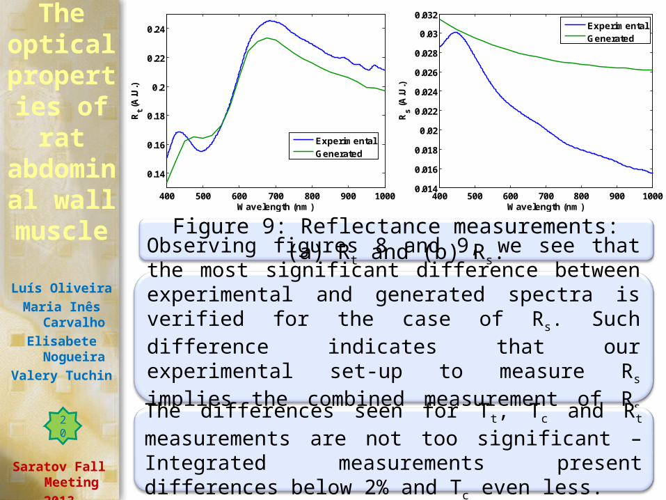
Figures 8 and 9 show the spectra from our experimental measurements and the spectra generated from the optical properties presented in figures 6 and 7, for comparison.
The optical
properties of rat
abdominal wall
muscle
Luís Oliveira
Maria Inês Carvalho
Elisabete Nogueira
Valery Tuchin
Saratov Fall Meeting
2013Figure 8: Transmittance measurements: (a) Tt and (b) Tc.
400 500 600 700 800 900 10000.2
0.3
0.4
0.5
0.6
0.7
Wavelength (nm)
Tt (
A.U
.)
ExperimentalGenerated
19
The optical
properties of rat
abdominal wall
muscle
Luís Oliveira
Maria Inês Carvalho
Elisabete Nogueira
Valery Tuchin
Saratov Fall Meeting
2013
Figure 9: Reflectance measurements: (a) Rt and (b) Rs.
400 500 600 700 800 900 1000
0.14
0.16
0.18
0.2
0.22
0.24
Wavelength (nm)
Rt (
A.U
.)
ExperimentalGenerated
400 500 600 700 800 900 10000.014
0.016
0.018
0.02
0.022
0.024
0.026
0.028
0.03
0.032
Wavelength (nm)
Rs (
A.U
.)
ExperimentalGenerated
Observing figures 8 and 9, we see that the most significant difference between experimental and generated spectra is verified for the case of Rs. Such difference indicates that our experimental set-up to measure Rs implies the combined measurement of Rs and Rd light.
The differences seen for Tt, Tc and Rt measurements are not too significant – Integrated measurements present differences below 2% and Tc even less.
20
The optical
properties of rat
abdominal wall
muscle
Luís Oliveira
Maria Inês Carvalho
Elisabete Nogueira
Valery Tuchin
Saratov Fall Meeting
2013
6. ConclusionsWith the research that we have developed, we have obtained the optical properties for skeletal muscle of the Wistar Han rat for the wavelength range of 400 to 1000 nm.
Such optical properties were obtained not only from optical measurements. We needed to model the wavelength dependence of μs and μs’, accordingly to what is presented in literature.
By performing such modeling of wavelength dependece for the scattering and reduced scattering coefficients, we were abble to identify errors in our experimental measurements.
The knowledge of the optical properties of natural rat muscle allow us to develop or improve clinical applications for the muscle in the wavelength range of 400 to 1000 nm.
21
Another application that can make use of the optical properties that we have estimated concerns the study of optical clearing of the muscle.
The optical
properties of rat
abdominal wall
muscle
Luís Oliveira
Maria Inês Carvalho
Elisabete Nogueira
Valery Tuchin
Saratov Fall Meeting
2013
Such is in fact a great motivation to proceed with our research and we expect to confirm that the optical clearing treatment produces a decrease in the scattering coefficient as indicated in literature [15].
With the optical properties for the natural muscle, we can now calculate their variations and also estimate the variations in the refractive index of the tissue under treatment with an optical clearing agent.
22
The optical
properties of rat
abdominal wall
muscle
Luís Oliveira
Maria Inês Carvalho
Elisabete Nogueira
Valery Tuchin
Saratov Fall Meeting
2013
AcknowledgementsThe authors would like to acknowledge the help given in the preparation of tissue samples by LAIMM – Laboratório de Apoio à Investigação em Medicina Molecular, Departamento de Biologia Experimental, Faculdade de Medicina da Universidade do Porto, Portugal.
For the resources and instruments made available to perform the present research, the authors would also like to thank CIETI – Centre of Innovation in Engineering and Industrial Technology and the Physics Department of Polytechnic of Porto – School of Engineering, Portugal.
23
[1] A. N. Bashkatov, E. A. Genina, V. I. Kochubey, V. V. Tuchin, “Optical properties of human skin, subcutaneous and mucous tissues in the wavelength range from 400 to 2000 nm”, J. Phys. D: Appl Phys, 38, pp. 2543 – 2555, (2005).
[2] R. Cicchi, F. S. Pavone, D. Massi, D. D. Sampson, “Contrast and Depth enhancement in two-photon microscopy of human skin ex vivo by use of optical clearing agents”, Optics Express 13(7), (2005).
[3] V. V. Tuchin, “Tissue Optics: Light Scattering Methods and Instruments for Medical Diagnosis”, 2nd ed., SPIE Press, Bellingham, 2007.
[4] Tuan Vo-Dinh, “Biomedical Photonics Handbook”, CRC Press LLC, 2003.
ReferencesThe
optical properties
of rat abdominal
wall muscle
Luís Oliveira
Maria Inês Carvalho
Elisabete Nogueira
Valery Tuchin
Saratov Fall Meeting
2013
24
[5] L.-H. Wang, S. L. Jacques, L.-Q. Zheng, “MCML – Monte Carlo modeling of photon transport in multi-layered tissues”, Computer Methods and Programs in Biomedicine, 47, 131 – 146, 1995.
[6] S. A. Prahl. “The adding-doubling method”. In A. J. Welch and M. J. C. van Gemert, editors, Optical-Thermal Response of Laser Irradiated Tissue, chapter 5, pages 101-129. Plenum Press, 1995.
[7] V. V. Tuchin, “Tissue Optics: Light Scattering Methods and Instruments for Medical Diagnosis”, 2nd ed., SPIE Press, Bellingham, 2007.
[8] [8] E. Zamora-Rojas, B. Aernouts, A. Garrido-Varo, D. Pérez-Marín, J. E. Guerrero-Ginel, W. Saeys, “Double integrating sphere measurements for estimating optical properties of pig subcutaneous adipose tissue”, Innovative Food Science and Emerging Technologies, 19, pp. 218-226, (2013).
The optical
properties of rat
abdominal wall
muscle
Luís Oliveira
Maria Inês Carvalho
Elisabete Nogueira
Valery Tuchin
Saratov Fall Meeting
2013
25
[9] L. M. Oliveira, M. I. Carvalho, E. M. Nogueira, V. V. Tuchin, “Optical measurements of rat muscle samples under treatment with ethylene glycol and glucose”, Journal of Innovative Optical Health Sciences, 6(2), 1350012-1 – 1350012-15, (2013).
[10] L. M. Oliveira, M. I. Carvalho, E. M. Nogueira, V. V. Tuchin, “The characteristic time of glucose diffusion measured for muscle tissue at optical clearing”, Laser Phys. 23(7), 075606 (6pp), 2013.
[11] S. L. Jacques, “Optical properties of biological tissues: a review”, Phys. Med. Biol. 58, pp. R37 – R61, (2013).
[12] A. N. Bashkatov, E. A. Genina, V. V. Tuchin, “Optical properties of skin, subcutaneous, and muscle tissues: a review”, Journal of Innovative Optical Health Sciences, 4(1), pp. 9 – 38, 2011.
The optical
properties of rat
abdominal wall
muscle
Luís Oliveira
Maria Inês Carvalho
Elisabete Nogueira
Valery Tuchin
Saratov Fall Meeting
2013
26
[13] E. Vitkin, V. Turzhitsky, L. Qiu, L. Guo, I. Itzkan, E. B. Hanlon, L. T. Perelman, “Photon diffusion near the point-of-entry in anisotropically scattering turbid media”, Nature communications, 2011. Corrigir depois de ver citação na net.
[14] A. N. Bashkatov, E. A. Genina, V. I. Kochubey, A. A. Gavrilova, S. V. Kapralov, V. A. Grishaev, V. V. Tuchin, “Optical properties of human stomach mucosa in the spectral range from 400 to 2000 nm: prognosis for gastroenterology”, Medical Laser Applications, 22, pp. 95 – 104, (2007).
[15] V. V. Tuchin, “Optical Clearing of Tissues and Blood”, SPIE Press, Bellingham, WA (2006).
The optical
properties of rat
abdominal wall
muscle
Luís Oliveira
Maria Inês Carvalho
Elisabete Nogueira
Valery Tuchin
Saratov Fall Meeting
2013
27