Embed Size (px)
Citation preview

The Peripheral Nervous System
Chapter 14
Introduction The CNS would be useless without a
means of sensing our own internal as well as the external environments
In addition, we need a means by which we can effect our external environment
The peripheral nervous system provides these links to the CNS
Introduction The peripheral nervous system includes
all the neural structures outside the brain and spinal cord – Sensory receptors– Peripheral nerves and their ganglia– Efferent motor endings
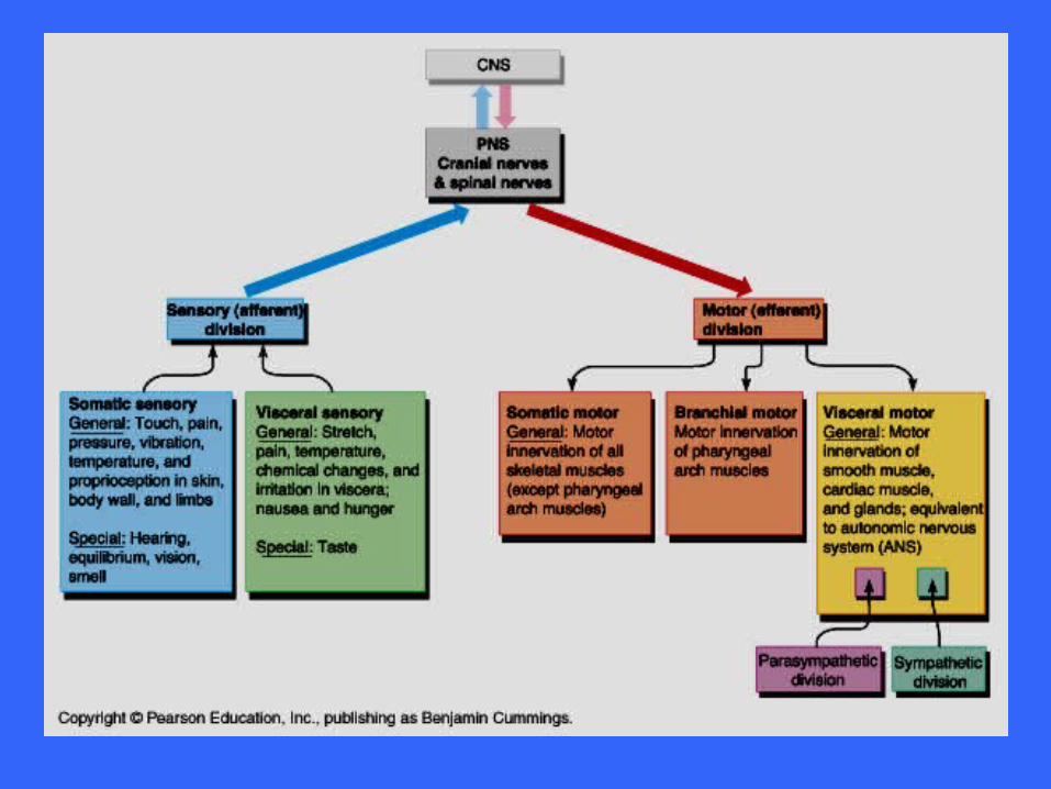
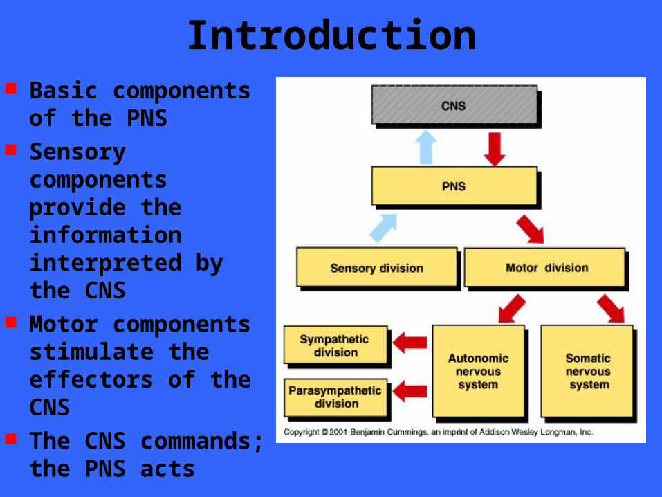
Introduction Basic components of
the PNS Sensory components
provide the information interpreted by the CNS
Motor components stimulate the effectors of the CNS
The CNS commands; the PNS acts
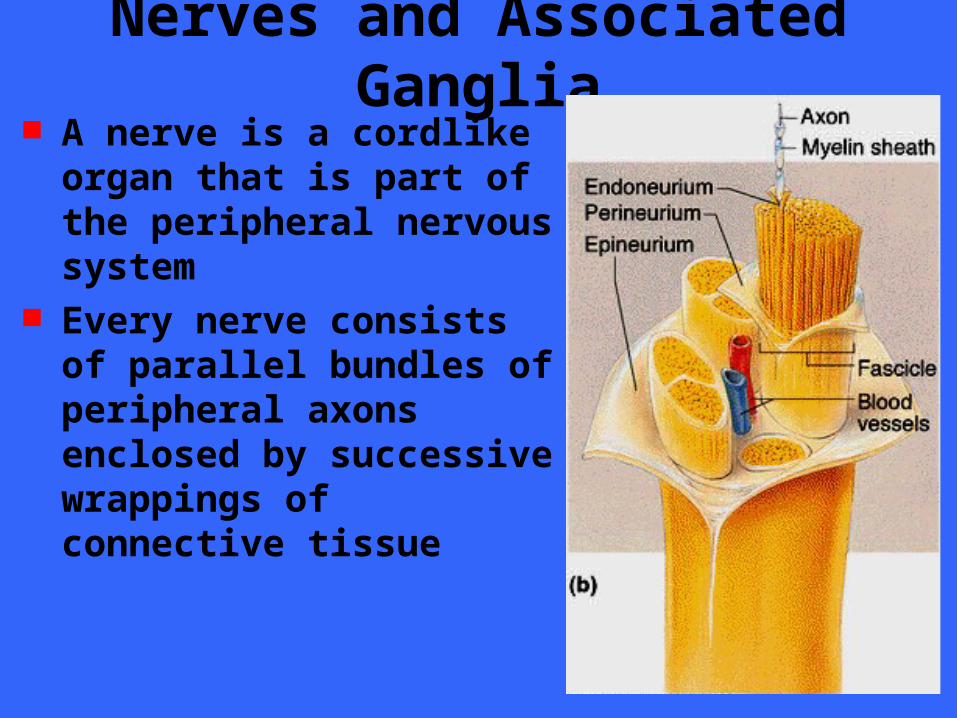
Nerves and Associated Ganglia A nerve is a cordlike organ
that is part of the peripheral nervous system
Every nerve consists of parallel bundles of peripheral axons enclosed by successive wrappings of connective tissue
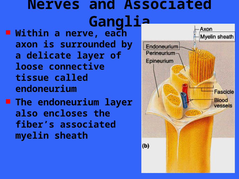
Nerves and Associated Ganglia Within a nerve, each axon is
surrounded by a delicate layer of loose connective tissue called endoneurium
The endoneurium layer also encloses the fiber’s associated myelin sheath
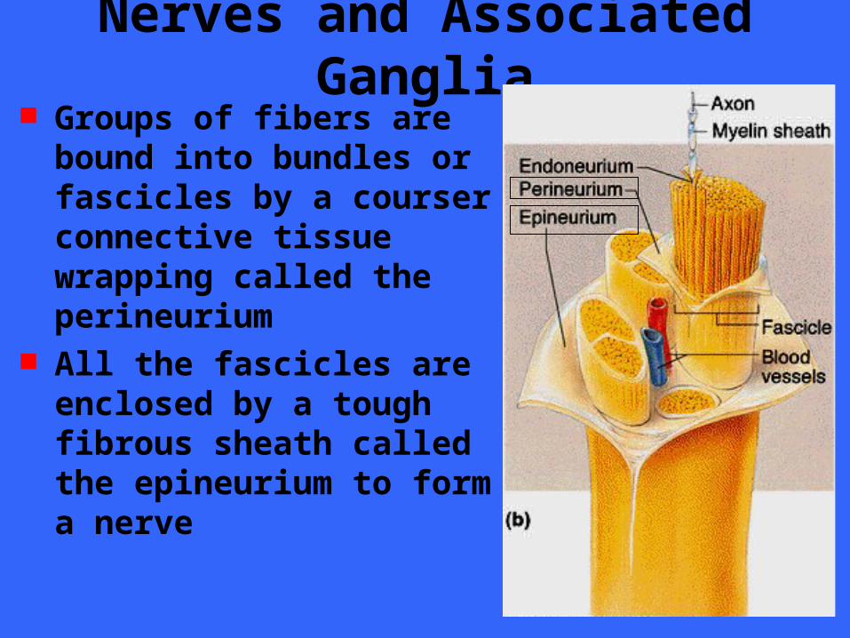
Nerves and Associated Ganglia Groups of fibers are bound
into bundles or fascicles by a courser connective tissue wrapping called the perineurium
All the fascicles are enclosed by a tough fibrous sheath called the epineurium to form a nerve
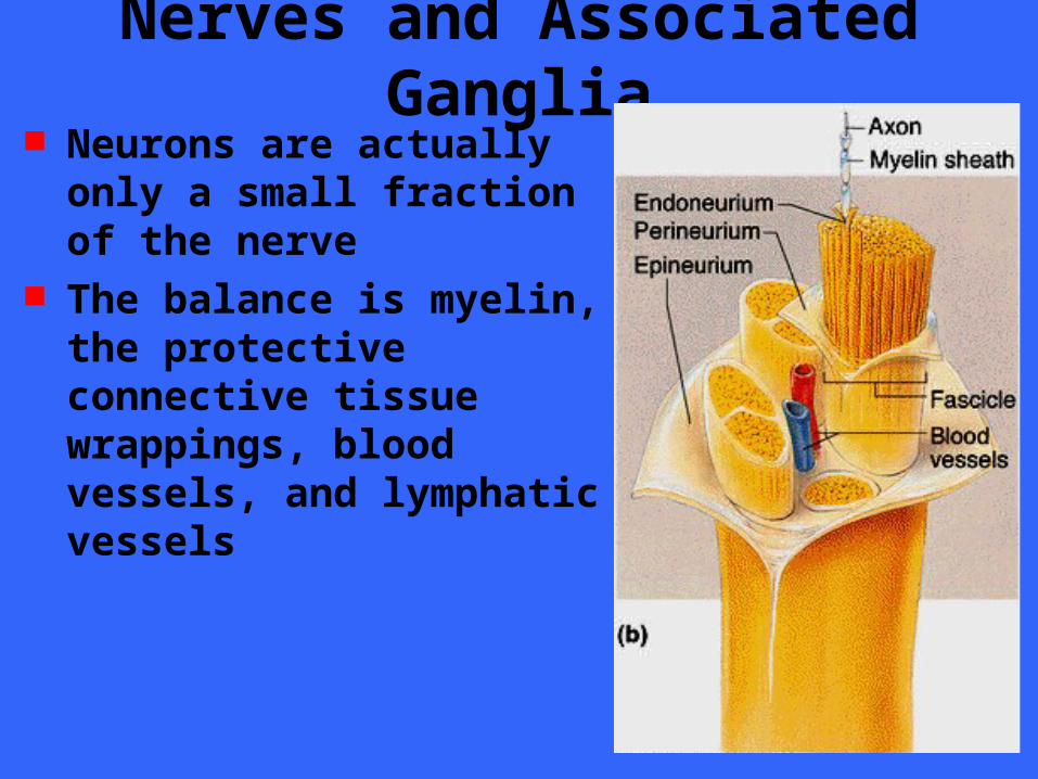
Nerves and Associated Ganglia Neurons are actually only a
small fraction of the nerve The balance is myelin, the
protective connective tissue wrappings, blood vessels, and lymphatic vessels
Nerves and Associated Ganglia Nerves are classified according to the
direction in which they transmit impulses– Nerves containing both sensory and motor
fibers are called mixed nerves– Nerves that carry impulses toward the
CNS only are sensory (afferent) nerves– Nerves that carry impulses only away from
the CNS are motor (efferent) nerves Most nerves are mixed as purely sensory
or motor nerves are extremely rare
Nerves and Associated Ganglia Since mixed nerves often carry both
somatic and autonomic (visceral) nervous system fibers, the fibers within them may be classified further according to the region they innervate as– Somatic afferent– Somatic efferent– Visceral afferent– Visceral efferent
Nerves and Associated Ganglia Peripheral nerves are generally classified
on whether they arise from the brain or spinal cord as– Cranial nerves / brain and brain stem– Spinal nerves / spinal cord
Ganglia are collections of neuron cell bodies associated with nerves in the PNS– Ganglia associated with afferent nerve fibers
contain cell bodies of sensory neurons– Ganglia associated with efferent nerve fibers
contain cell bodies of autonomic neurons, as well as a variety of integrative neurons
Sensory Receptors Sensory receptors are structures that are
specialized to respond to changes in their environment
Such environmental changes are called stimuli
Typically activation of a sensory receptor by an adequate stimulus results in depolarization or graded potentials that trigger nerve impulses along the afferent fibers coursing to the CNS
Peripheral Sensory Receptors Peripheral sensory receptors are
structures that pick up sensory stimuli and then initiate signals in the sensory axons
Most receptors fit into two main categories;– Dendritic endings of sensory neurons– Complete receptor cells
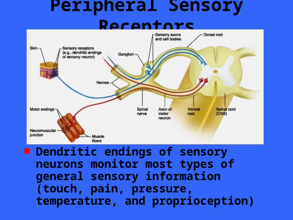
Peripheral Sensory Receptors
Dendritic endings of sensory neurons monitor most types of general sensory information (touch, pain, pressure, temperature, and proprioception)
Peripheral Sensory Receptors Complete receptor cells are specialized
epithelial cells or small neurons that transfer sensory information to sensory neurons
Specialized receptor cells monitor most types of special sensory information (taste, vision, hearing, and equilibrium)
Sensory Receptors Sensory receptors are classified by
– The type of stimulus they detect– Their location in the body– Their structure
Classification by Location Receptors are recognized according to
their location or the location of the stimuli to which they respond– Externoceptors– Internoceptors or visceroceptors– Proprioceptors
Classification by Location Externoceptors
– Sensitive to stimuli arising from outside of the body
– Typically located near the surface of the body– Include receptors for
• Touch
• Pressure
• Pain
• Temperature
• Special sense receptors
Classification by Location Internoceptors or visceroceptors
– Respond to stimuli arising from within the internal viscera and body organs,
– Internoceptors monitor a variety of internal stimuli
• Changes in chemical concentration• Taste stimuli• The stretching of tissues• Temperature
– Their activation causes us to feel visceral pain, nausea, hunger, or fullness
Classification by Location Proprioceptors
– Located in the musculoskeletal organs such as skeletal muscles, tendons, joints and ligaments
– Proprioceptors monitor the degree of stretch of these locomotor organs and send input to the CNS
Classification by Stimulus Detected Mechanoreceptors
– general nerve impulses when they, or adjacent tissues, are deformed by mechanical forces
• Touch
• Pressure
• Vibration
• Stretch
• Itch
Thermoreceptors – Sensitive to temperature changes
Classification by Stimulus Detected Photoreceptors
– Respond to light energy Chemoreceptors
– Respond to chemicals in solution• Smell
• Taste
• Blood chemistry
Nociceptors– Respond to potentially damaging stimuli that
result in pain
Classification by Stimulus Detected Note that the over-stimulation of any of
the aforementioned receptors is painful and thus virtually all receptors can function as nociceptors at one time or another
Classification by Structure General sensory receptors are divided
into two broad groups– Free (naked) endings– Encapsulated dendritic endings
It should be pointed out that there is no one receptor - one function relationship
Rather, one receptor type can respond to several different kinds of stimuli, and different receptor types can respond to similar stimuli
Adaptation of Sensory Receptors Adaptation occurs in certain sensory
receptors when they are subjected to an unchanging stimulus
As a result, the receptor potentials decline in frequency or stop
Some receptors adapt quickly (pressure, touch and smell)
Nocioceptors and proprioceptors adapt slowly or not at all as they serve a protective function
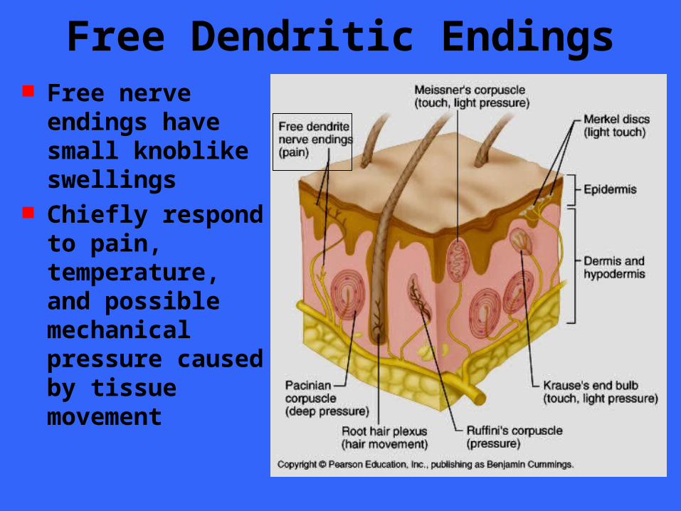
Free Dendritic Endings Free nerve endings
have small knoblike swellings
Chiefly respond to pain, temperature, and possible mechanical pressure caused by tissue movement
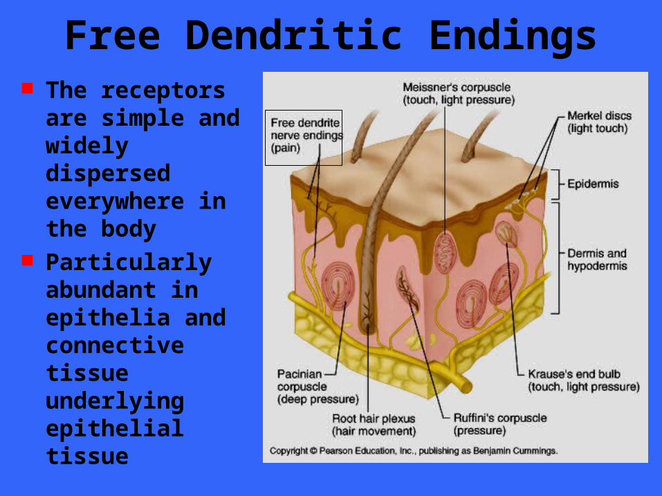
Free Dendritic Endings The receptors are
simple and widely dispersed everywhere in the body
Particularly abundant in epithelia and connective tissue underlying epithelial tissue
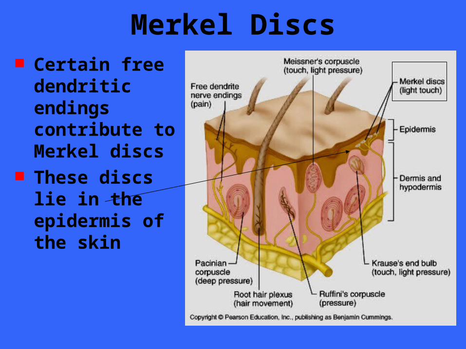
Merkel Discs Certain free
dendritic endings contribute to Merkel discs
These discs lie in the epidermis of the skin
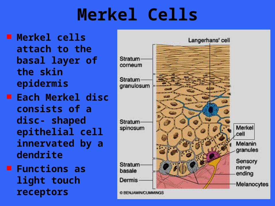
Merkel Cells Merkel cells attach
to the basal layer of the skin epidermis
Each Merkel disc consists of a disc- shaped epithelial cell innervated by a dendrite
Functions as light touch receptors
Merkel Discs Merkel cells seem to be slowly adapting
receptors for light touch Slowly adapting means that they
continue to respond to stimuli present and send out action potentials even long after a period of continual stimulation
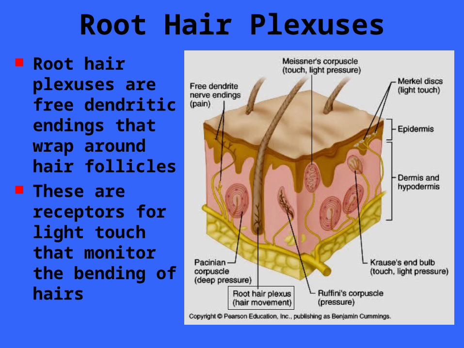
Root Hair Plexuses Root hair plexuses
are free dendritic endings that wrap around hair follicles
These are receptors for light touch that monitor the bending of hairs
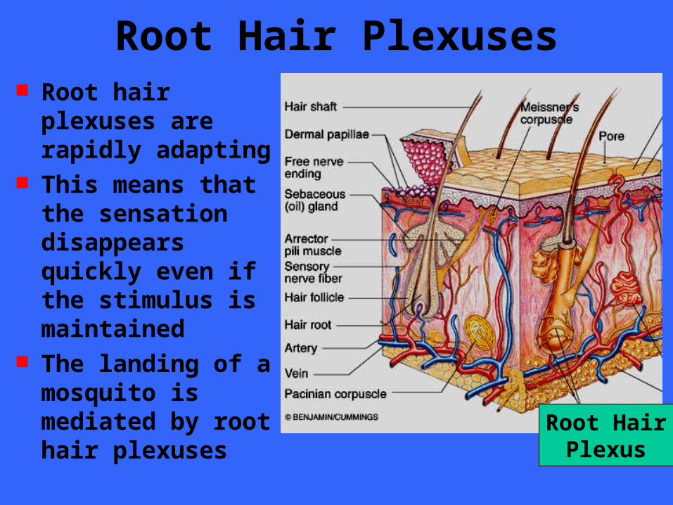
Root Hair Plexuses Root hair plexuses
are rapidly adapting This means that the
sensation disappears quickly even if the stimulus is maintained
The landing of a mosquito is mediated by root hair plexuses Root Hair
Plexus
Encapsulated Dendritic Endings All encapsulated dendritic endings
consist of one or more end fibers of sensory neurons enclosed in a capsule of connective tissue
All seem to be mechanoreceptors, and their capsules serve to either amplify the stimulus or to filter out the wrong types of stimuli
Encapsulated Dendritic Endings Encapsulated receptors vary widely in
shape, size, and distribution in the body The main types are
– Meissner’s corpuscles– Krause’s end bulbs– Pacinian corpuscles– Ruffini’s corpuscles – Proprioceptors
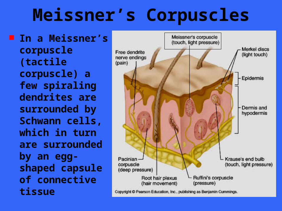
Meissner’s Corpuscles In a Meissner’s
corpuscle (tactile corpuscle) a few spiraling dendrites are surrounded by Schwann cells, which in turn are surrounded by an egg-shaped capsule of connective tissue
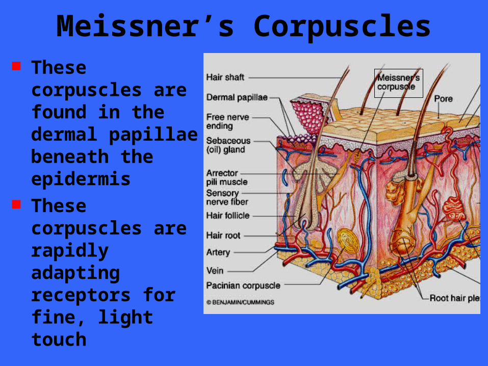
Meissner’s Corpuscles These corpuscles
are found in the dermal papillae beneath the epidermis
These corpuscles are rapidly adapting receptors for fine, light touch
Meissner’s Corpuscles Meissner’s corpuscles occur in sensitive
and hairless areas of the skin, such as the soles of the feet, palms, fingertips, nipples, and lips
Apparently, Meissner’s corpuscles perform the same “light touch” function in hairless skill that root hair plexuses perform in hairy skin
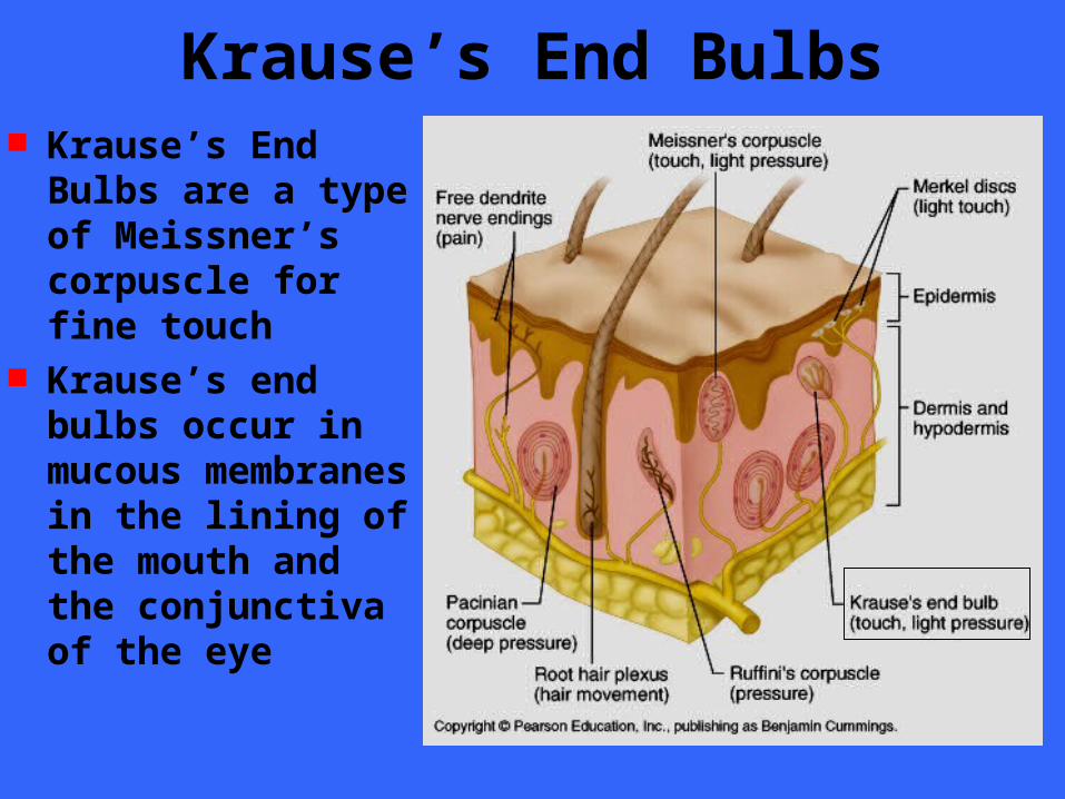
Krause’s End Bulbs Krause’s End Bulbs
are a type of Meissner’s corpuscle for fine touch
Krause’s end bulbs occur in mucous membranes in the lining of the mouth and the conjunctiva of the eye

Pacinian Corpuscle Pacinian corpuscle
are scattered throughout the deep connective tissues of the body
Occur in the hypodermis of the skin
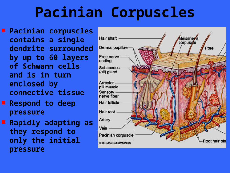
Pacinian Corpuscles Pacinian corpuscles
contains a single dendrite surrounded by up to 60 layers of Schwann cells and is in turn enclosed by connective tissue
Respond to deep pressure
Rapidly adapting as they respond to only the initial pressure
Pacinian Corpuscles Pacinian corpuscles are rapidly adapting
receptors and are best suited to monitor vibrations which is an on-off stimulus
These corpuscles are large enough to be visible to the naked eye
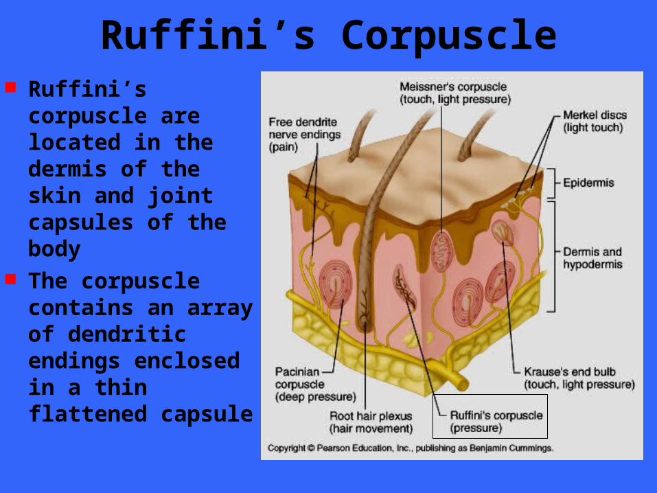
Ruffini’s Corpuscle Ruffini’s corpuscle
are located in the dermis of the skin and joint capsules of the body
The corpuscle contains an array of dendritic endings enclosed in a thin flattened capsule
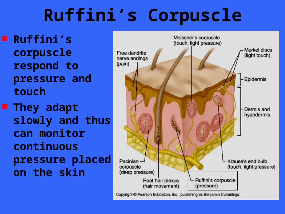
Ruffini’s Corpuscle Ruffini’s corpuscle
respond to pressure and touch
They adapt slowly and thus can monitor continuous pressure placed on the skin
Proprioceptors
Proprioceptors Virtually all proprioceptors are
encapsulated dendritic endings that monitor stretch in the locomotor organs
Proprioceptors include…– Muscle spindles– Golgi tendon organs– Joint kinesthetic receptors
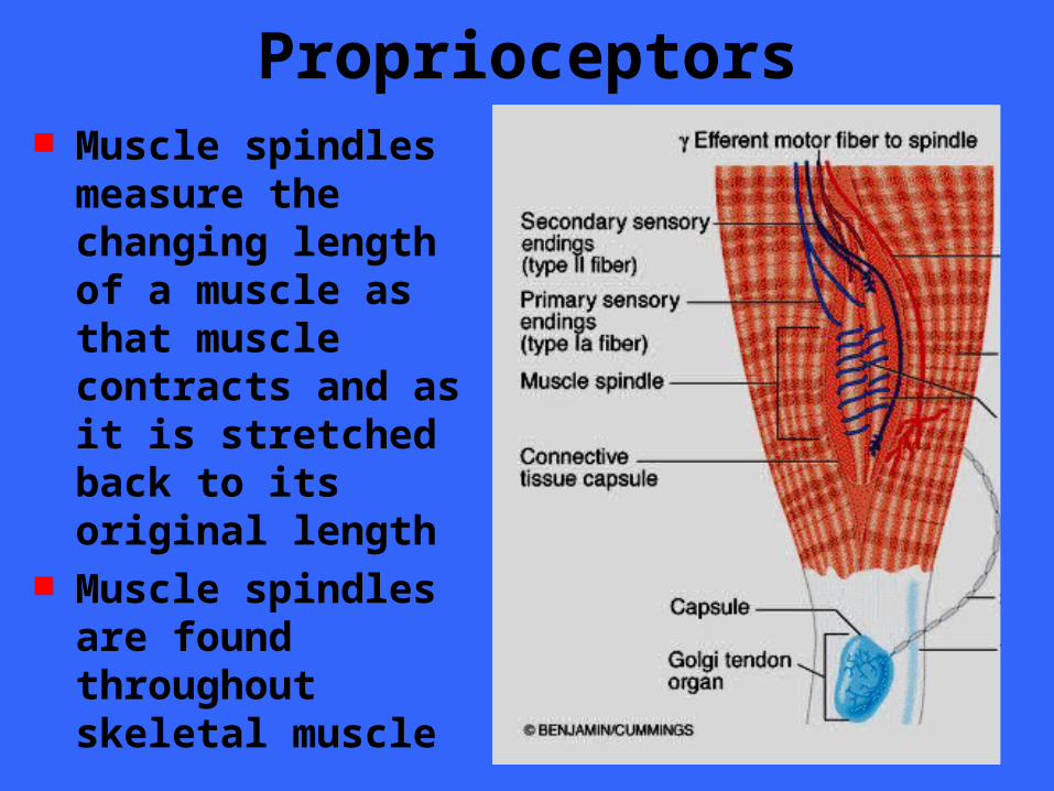
Proprioceptors Muscle spindles
measure the changing length of a muscle as that muscle contracts and as it is stretched back to its original length
Muscle spindles are found throughout skeletal muscle
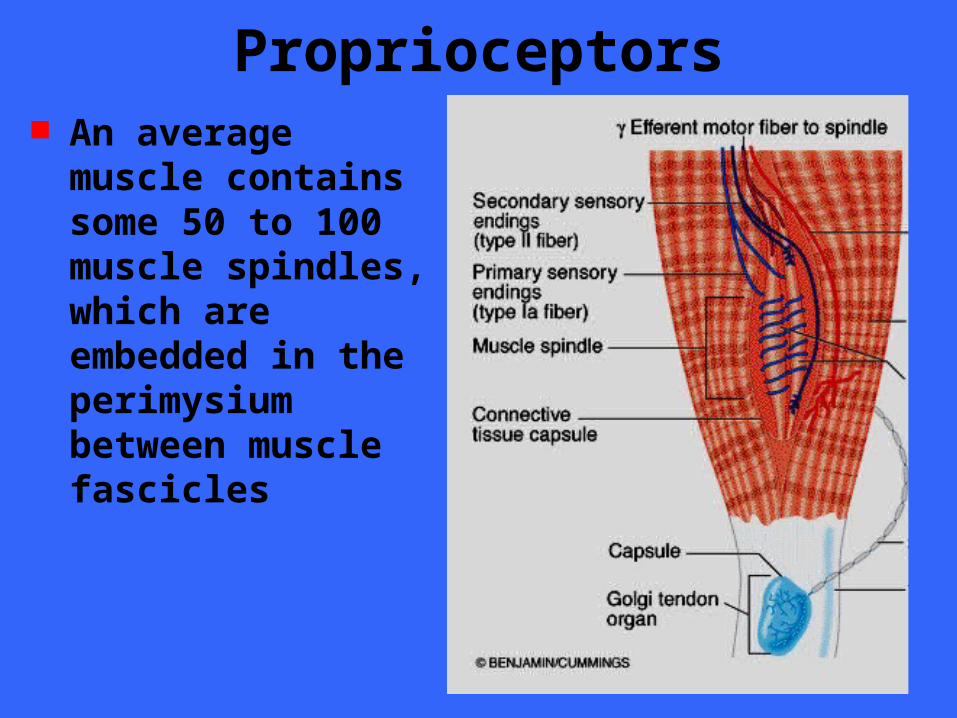
Proprioceptors An average muscle
contains some 50 to 100 muscle spindles, which are embedded in the perimysium between muscle fascicles
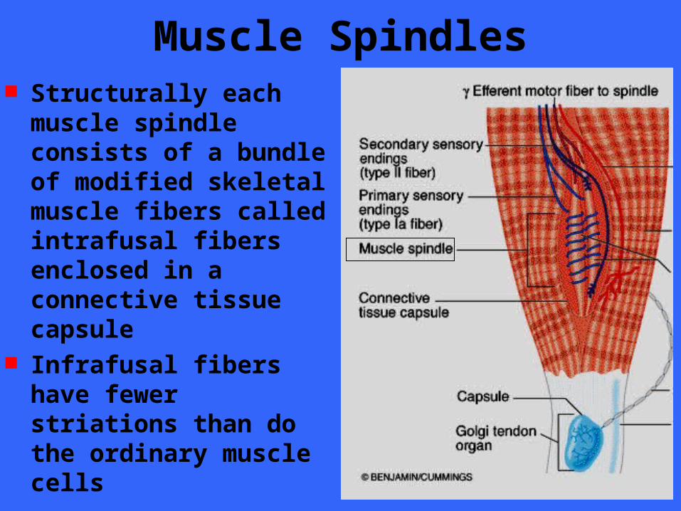
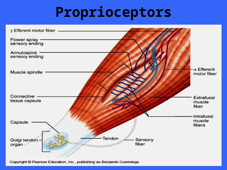
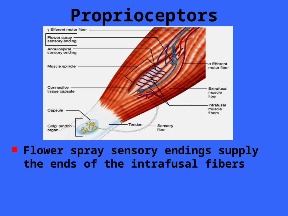
Muscle Spindles Structurally each muscle
spindle consists of a bundle of modified skeletal muscle fibers called intrafusal fibers enclosed in a connective tissue capsule
Infrafusal fibers have fewer striations than do the ordinary muscle cells
Proprioceptors
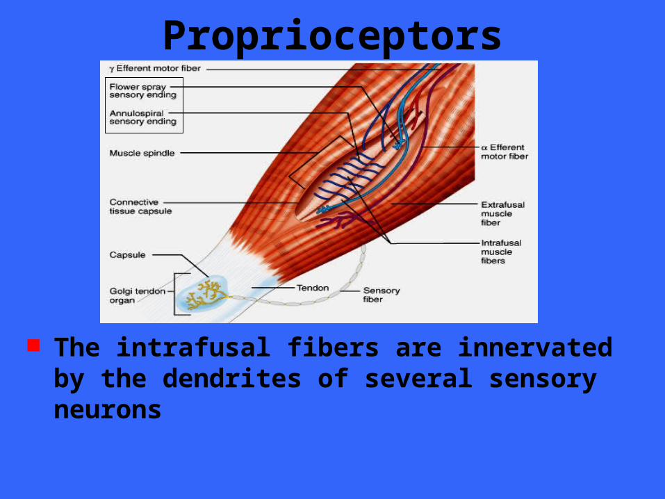
The intrafusal fibers are innervated by the dendrites of several sensory neurons
Proprioceptors
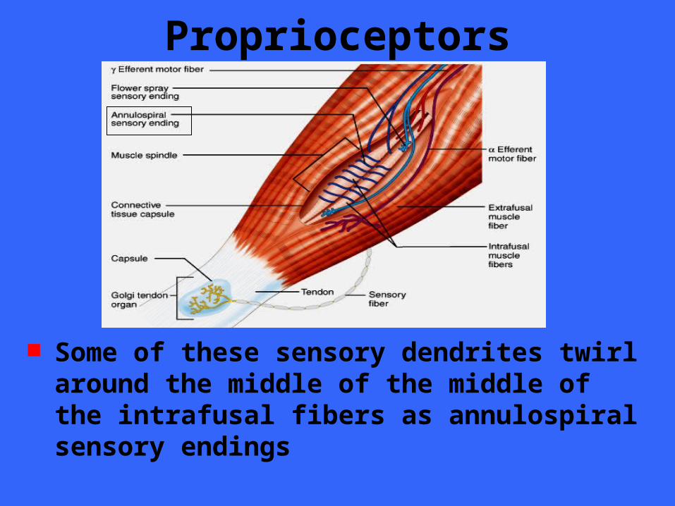
Some of these sensory dendrites twirl around the middle of the middle of the intrafusal fibers as annulospiral sensory endings
Proprioceptors
Flower spray sensory endings supply the ends of the intrafusal fibers
Proprioceptors Muscles are stretched by the contraction
of antagonist muscles and also by the movements that occur when we lose our balance
The muscle spindles sense these changes and compensate for the stretch
Proprioceptors Muscle spindles sense changes in muscle
length by the simple fact that as the muscle is stretched the muscle spindle is also stretched
The stretching activates the sensory neurons that innervate the spindle, causing them to signal the spinal cord and brain
Proprioceptors
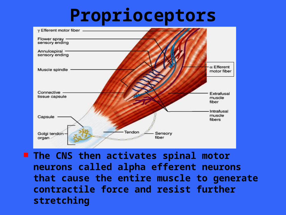
The CNS then activates spinal motor neurons called alpha efferent neurons that cause the entire muscle to generate contractile force and resist further stretching
Proprioceptors This response to stretching can take the
form of a monosynapatic spinal reflex that makes a rapid adjustment to prevent a fall
Alternatively, the stretch response can be controlled by the cerebellum, in which case it is involved in the regulation of muscle tone – The steady force generated by non-
contracting muscle to resist stretching
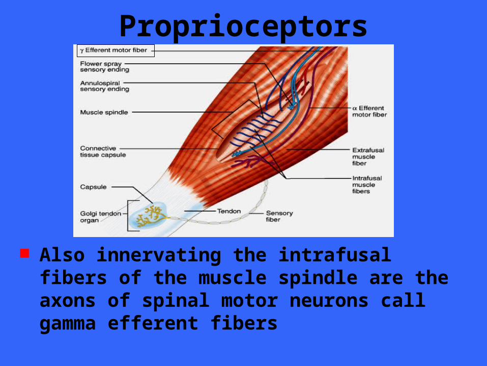
Proprioceptors
Also innervating the intrafusal fibers of the muscle spindle are the axons of spinal motor neurons call gamma efferent fibers
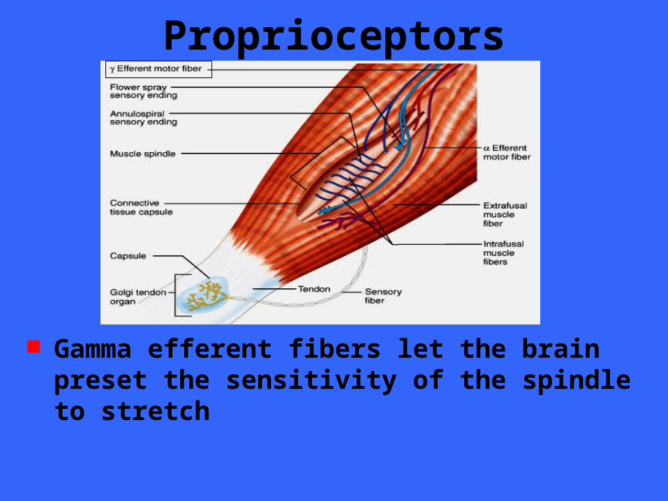
Proprioceptors
Gamma efferent fibers let the brain preset the sensitivity of the spindle to stretch
Proprioceptors When the brain signals gamma motor
neurons to fire, the intrafusal muscle fibers contract and become tense so that very little stretch is needed to stimulate the sensory dendrites
Making the spindles highly sensitive to stretch is advantageous when balance reflexes have little margin for error
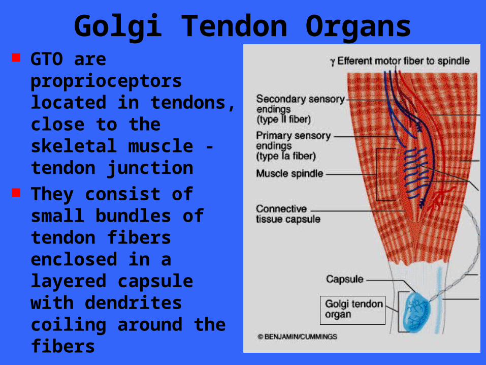
Golgi Tendon Organs GTO are proprioceptors
located in tendons, close to the skeletal muscle - tendon junction
They consist of small bundles of tendon fibers enclosed in a layered capsule with dendrites coiling around the fibers
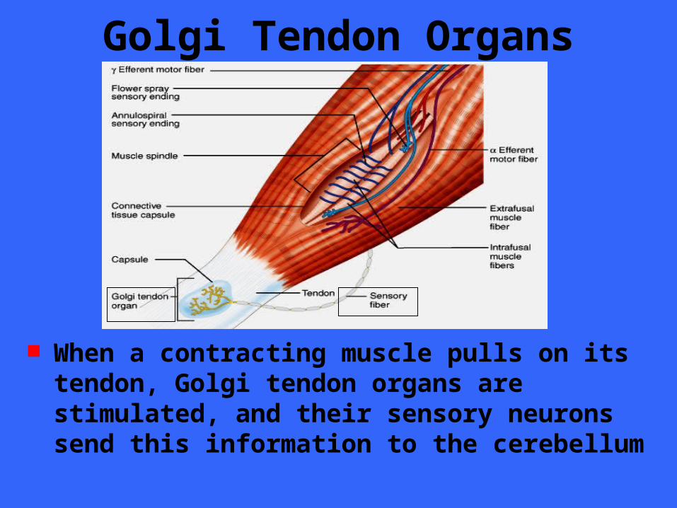
Golgi Tendon Organs
When a contracting muscle pulls on its tendon, Golgi tendon organs are stimulated, and their sensory neurons send this information to the cerebellum
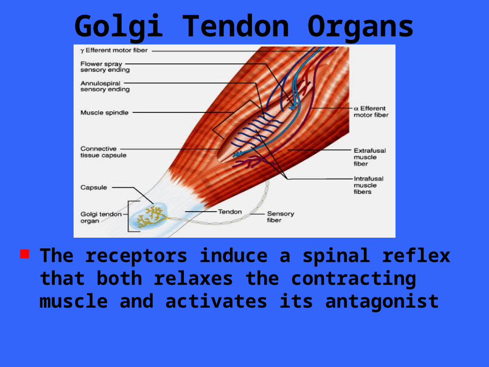
Golgi Tendon Organs
The receptors induce a spinal reflex that both relaxes the contracting muscle and activates its antagonist
Golgi Tendon Organs Relaxation reflex is important in motor
activities that involve the rapid alternation between flexion and extension such as in sprinting
Joint Kinesthetic Receptors These proprioceptors monitor stretch in
the synovial joints Specifically, they are sensory dendritic
endings within the joint capsules Four types of receptors are present
within each joint capsule– Pacinian corpuscles– Ruffini corpuscles– Free dendritic endings– Golgi tendon organs (kinda?)
Joint Kinesthetic Receptors Pacinian corpuscles are rapidly adapting
stretch receptors that are ideal for measuring acceleration and rapid movement of the joints
Ruffini corpuscles are slowly adapting stretch receptors that are ideal for measuring the positions of non-moving joints and the stretch of joints that undergo slow, sustained movements
Joint Kinesthetic Receptors Free dendritic endings in joint may serve
as pain receptors Receptors resembling Golgi tendon
organs have been identified in joints but their function is not yet known
Joint Kinesthetic Receptors Joint receptors, like the other two classes
of proprioceptors, send information on body movements to the cerebellum and cerebrum, as well as to spinal reflex arcs
Innervation of Skeletal Muscle
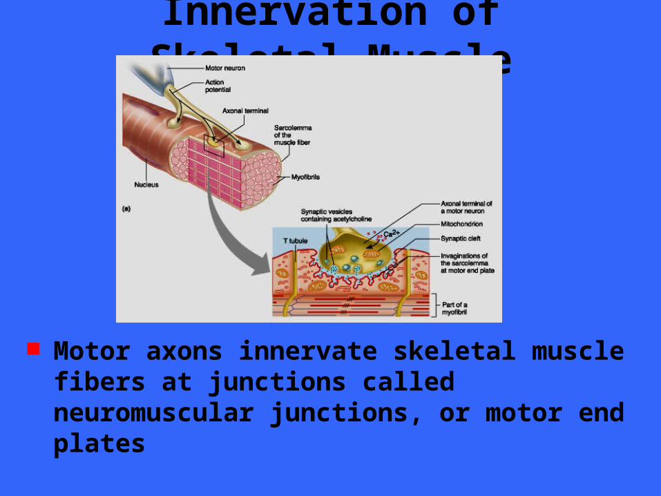
Motor axons innervate skeletal muscle fibers at junctions called neuromuscular junctions, or motor end plates
Innervation of Skeletal Muscle
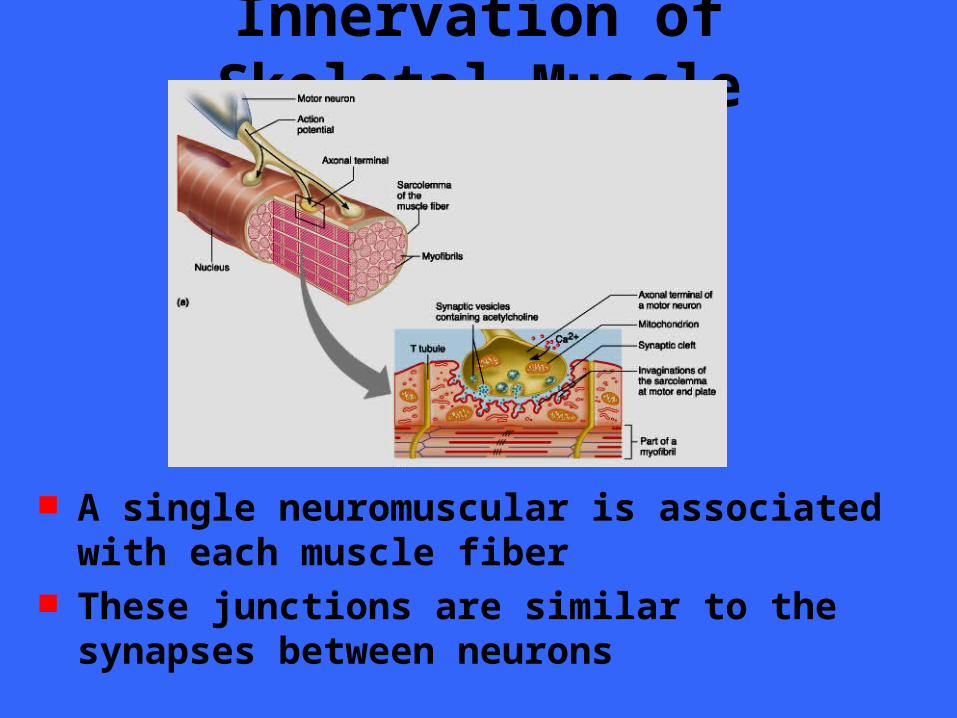
A single neuromuscular is associated with each muscle fiber
These junctions are similar to the synapses between neurons
Innervation of Skeletal Muscle
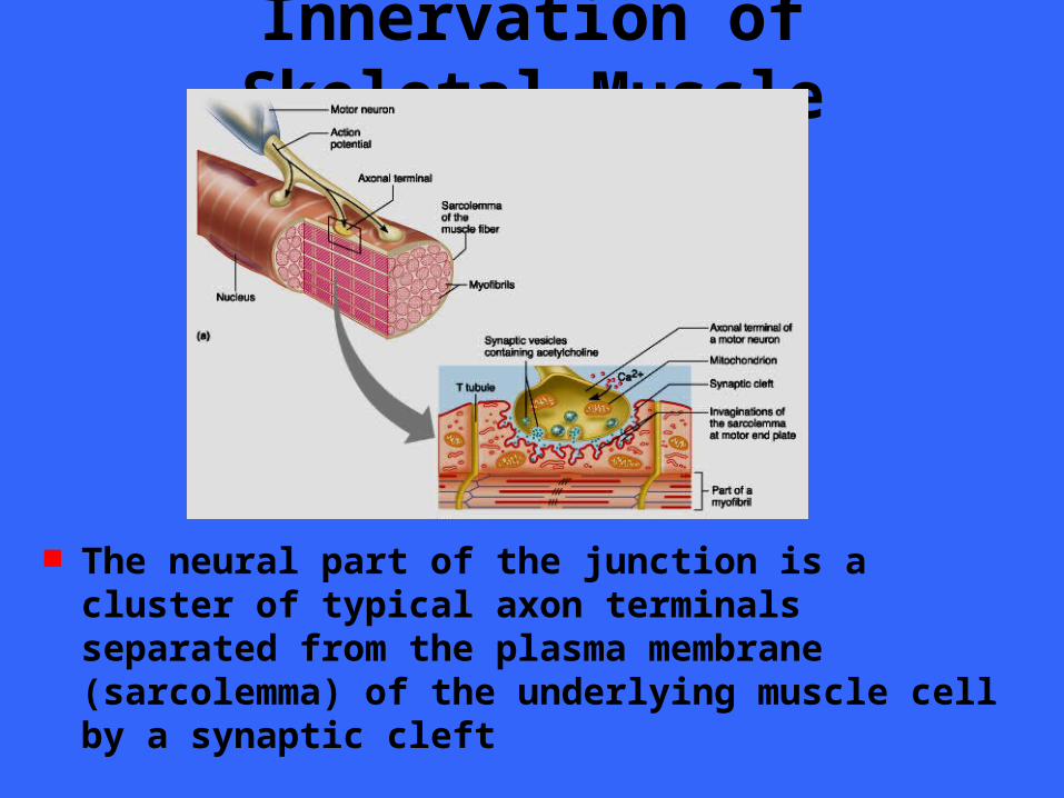
The neural part of the junction is a cluster of typical axon terminals separated from the plasma membrane (sarcolemma) of the underlying muscle cell by a synaptic cleft
Innervation of Skeletal Muscle As in typical synapses, the axon terminals
contain synaptic vesicles that release a neurotransmitter when a nerve impulse reaches the terminals
The neurotransmitter (acetylcholine) diffuses across the synaptic cleft and binds to receptor molecules on the sarcolemma, where it induces an impulse that signals the muscle cell to contract
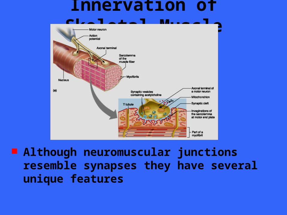
Innervation of Skeletal Muscle
Although neuromuscular junctions resemble synapses they have several unique features
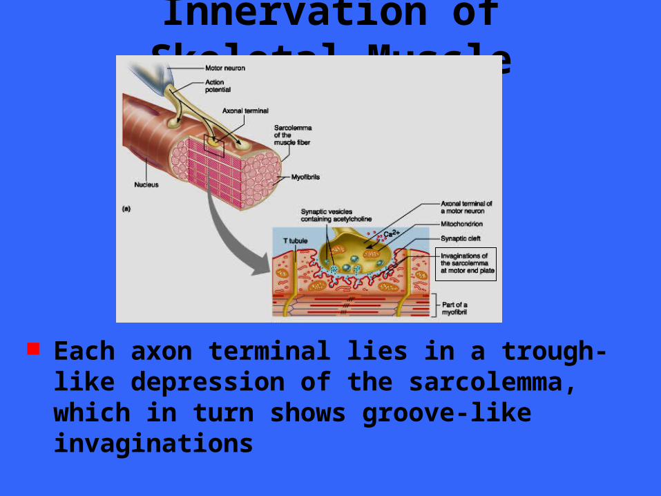
Innervation of Skeletal Muscle
Each axon terminal lies in a trough-like depression of the sarcolemma, which in turn shows groove-like invaginations
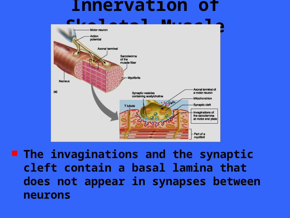
Innervation of Skeletal Muscle
The invaginations and the synaptic cleft contain a basal lamina that does not appear in synapses between neurons
Innervation of Skeletal Muscle This basal lamina contains the enzyme
acetylcholinesterase which breaks down acetylcholine immediately after the neurotransmitter signals a single contraction
This assures that each nerve impulse in the motor axon produces just one twitch of the muscle cell, preventing any undersireable additional twitches that would occur acetylcholine lingered in the synaptic cleft
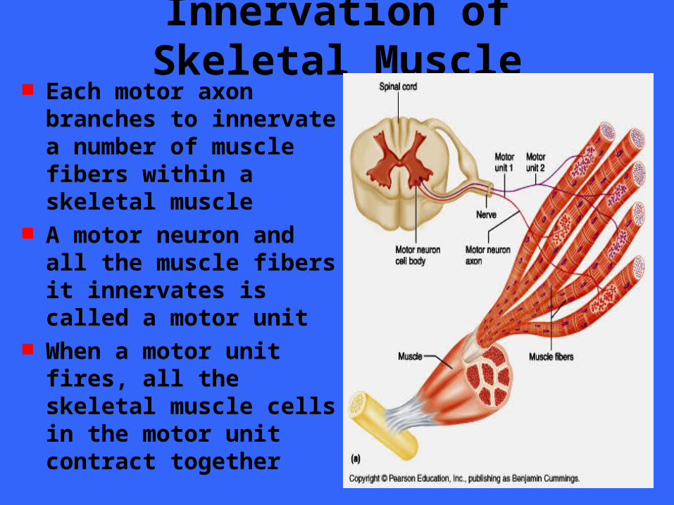
Innervation of Skeletal Muscle Each motor axon
branches to innervate a number of muscle fibers within a skeletal muscle
A motor neuron and all the muscle fibers it innervates is called a motor unit
When a motor unit fires, all the skeletal muscle cells in the motor unit contract together
Innervation of Skeletal Muscle Although the average number of muscle
fibers in a motor unit is 150, a motor unit may contain as many as several hundred fibers or as few as four muscle fibers
Muscles that require very fine control, such as the muscles moving the fingers and eyes have few muscle fibers per motor unit, whereas weight-bearing muscles whose movements are less precise have many muscle fibers per unit

Innervation of Skeletal Muscle The muscle fibers of a single motor unit
are not clustered together but spread throughout the muscle
As a result, stimulation of a single motor unit causes a weak contraction of the entire muscle
Innervation of Visceral Muscle The contacts between visceral motor
endings and the visceral effectors are much simpler than the elaborate neuromuscular junctions present on skeletal muscle
Near the smooth muscle of gland cells it innervates, a visceral motor axon swells into a row of knobs (varicosities) resembling the beads on a necklace
Innervation of Visceral Muscle Varicosities are the presynaptic terminals
which contain synaptic vesicles filled with neruotransmitter
Some of the axon terminals form shallow indentations on the membrane of the effector cell, but many axon terminals remain a considerable distance from any cell
Innervation of Visceral Muscle Because it takes time for neurotransmitters
to diffuse across these wide synaptic clefts, visceral motor responses tend to be slower that somatic motor reflexes
Innervation of Cardiac Muscle The motor innervation of cardiac muscle
cells resembles that of smooth muscle fibers and glands
However, the axon terminals are of a uniform diameter and do not include varicosities at the sites where they release their neurotransmitters
Cranial Nerves Twelve pair of cranial nerves are
associated with the brain and pass through various foramina of the skull
The first two attach to the forebrain, while the rest originate from the brain stem
Cranial nerves serve only the head and neck structures with the exception of the vagus nerves
In most cases, the nerve are named for the structures they serve or their primary functions
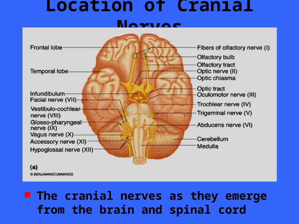
Location of Cranial Nerves
The cranial nerves as they emerge from the brain and spinal cord
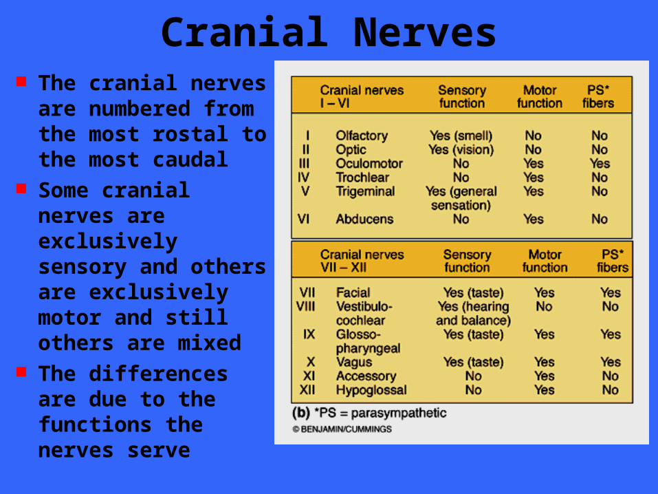
Cranial Nerves The cranial nerves
are numbered from the most rostal to the most caudal
Some cranial nerves are exclusively sensory and others are exclusively motor and still others are mixed
The differences are due to the functions the nerves serve
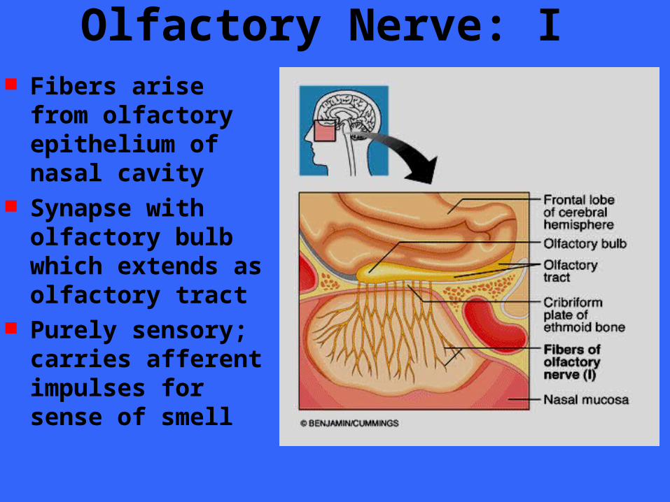
Olfactory Nerve: I Fibers arise from
olfactory epithelium of nasal cavity
Synapse with olfactory bulb which extends as olfactory tract
Purely sensory; carries afferent impulses for sense of smell
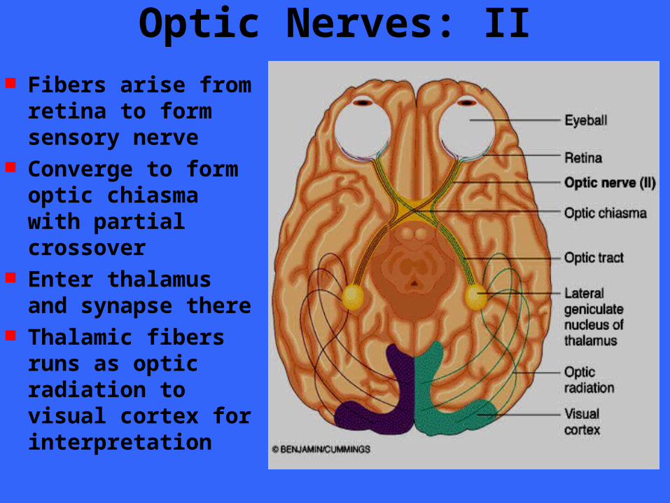
Optic Nerves: II Fibers arise from
retina to form sensory nerve
Converge to form optic chiasma with partial crossover
Enter thalamus and synapse there
Thalamic fibers runs as optic radiation to visual cortex for interpretation
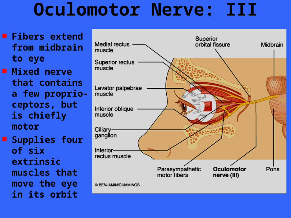
Oculomotor Nerve: III Fibers extend
from midbrain to eye
Mixed nerve that contains a few proprio- ceptors, but is chiefly motor
Supplies four of six extrinsic muscles that move the eye in its orbit

Trochlear Nerves: IV Fibers emerge
from midbrain to enter orbits
Mixed nerve; primarily motor
Innervates extrinsic muscles in the orbit
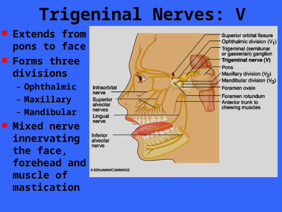
Trigeninal Nerves: V Extends from
pons to face Forms three
divisions– Ophthalmic
– Maxillary
– Mandibular Mixed nerve
innervating the face, forehead and muscle of mastication
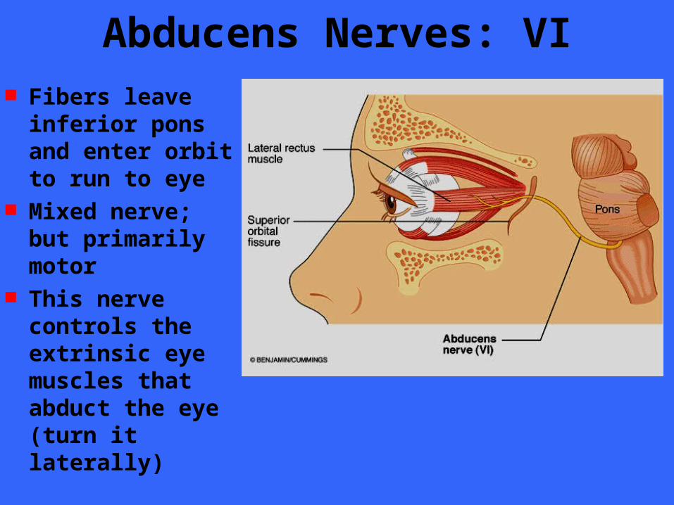
Abducens Nerves: VI Fibers leave
inferior pons and enter orbit to run to eye
Mixed nerve; but primarily motor
This nerve controls the extrinsic eye muscles that abduct the eye (turn it laterally)
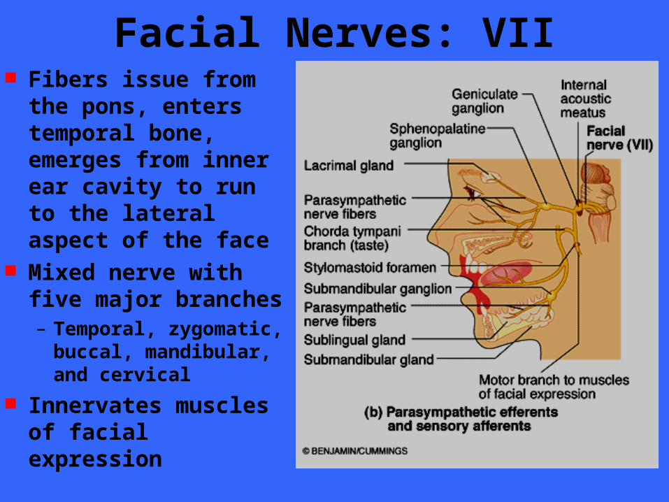
Facial Nerves: VII Fibers issue from the
pons, enters temporal bone, emerges from inner ear cavity to run to the lateral aspect of the face
Mixed nerve with five major branches– Temporal, zygomatic,
buccal, mandibular, and cervical
Innervates muscles of facial expression
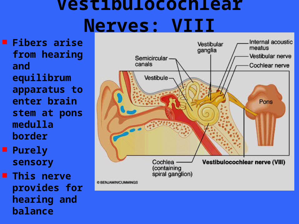
Vestibulocochlear Nerves: VIII Fibers arise
from hearing and equilibrum apparatus to enter brain stem at pons medulla border
Purely sensory This nerve
provides for hearing and balance
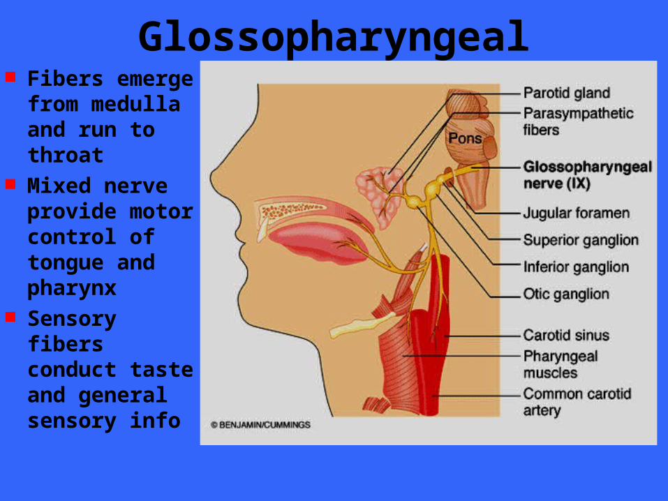
Glossopharyngeal Fibers emerge
from medulla and run to throat
Mixed nerve provide motor control of tongue and pharynx
Sensory fibers conduct taste and general sensory info
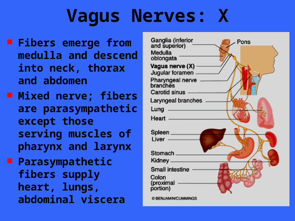
Vagus Nerves: X Fibers emerge from
medulla and descend into neck, thorax and abdomen
Mixed nerve; fibers are parasympathetic except those serving muscles of pharynx and larynx
Parasympathetic fibers supply heart, lungs, abdominal viscera
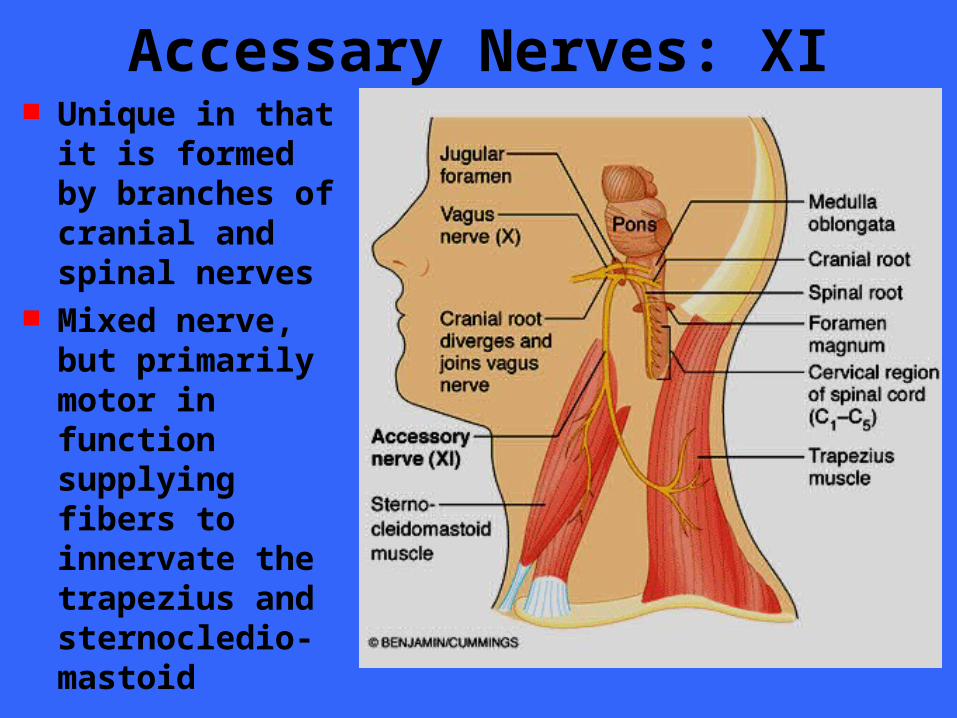
Accessary Nerves: XI Unique in that it
is formed by branches of cranial and spinal nerves
Mixed nerve, but primarily motor in function supplying fibers to innervate the trapezius and sternocledio- mastoid
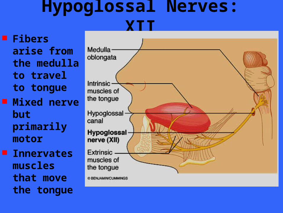
Hypoglossal Nerves: XII Fibers arise
from the medulla to travel to tongue
Mixed nerve but primarily motor
Innervates muscles that move the tongue
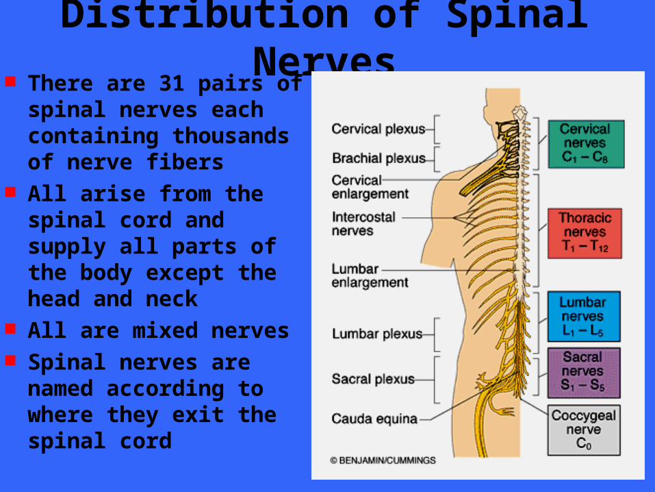
Distribution of Spinal Nerves There are 31 pairs of
spinal nerves each containing thousands of nerve fibers
All arise from the spinal cord and supply all parts of the body except the head and neck
All are mixed nerves Spinal nerves are named
according to where they exit the spinal cord
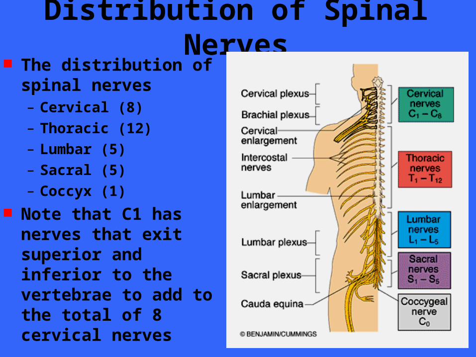
Distribution of Spinal Nerves The distribution of
spinal nerves – Cervical (8)
– Thoracic (12)
– Lumbar (5)
– Sacral (5)
– Coccyx (1) Note that C1 has nerves
that exit superior and inferior to the vertebrae to add to the total of 8 cervical nerves
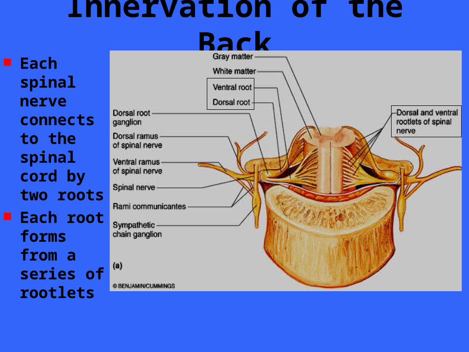
Innervation of the Back Each
spinal nerve connects to the spinal cord by two roots
Each root forms from a series of rootlets
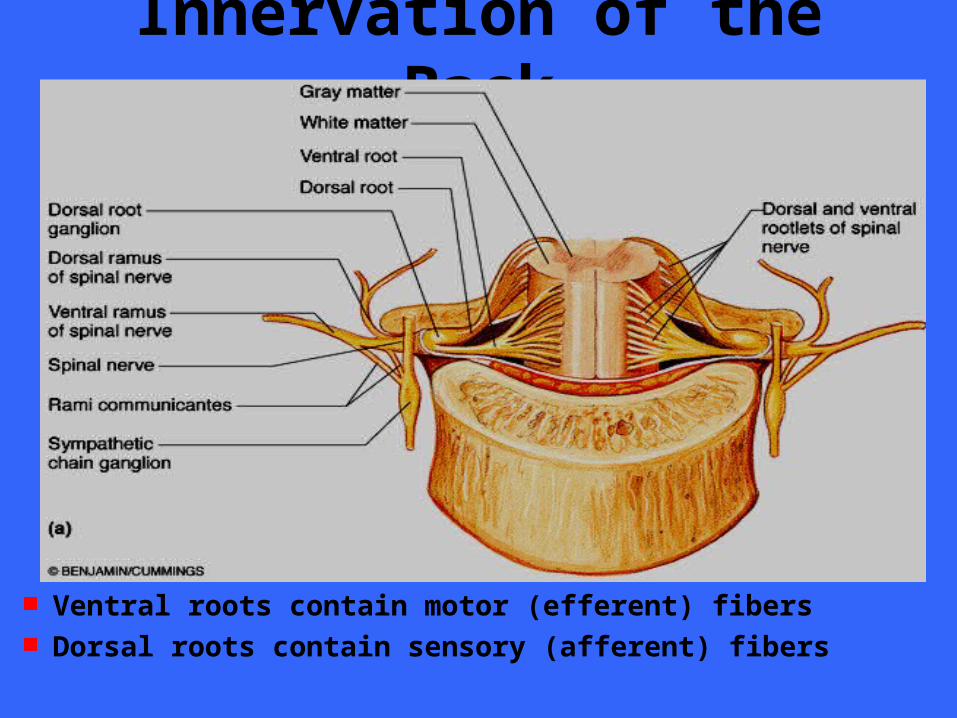
Innervation of the Back
Ventral roots contain motor (efferent) fibers Dorsal roots contain sensory (afferent) fibers
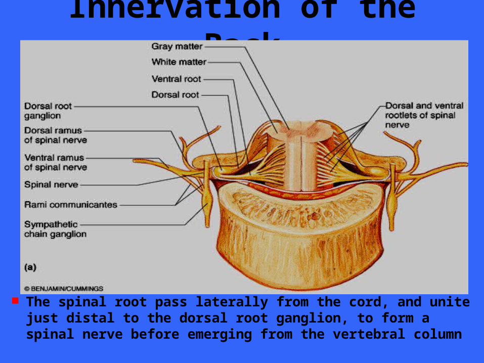
Innervation of the Back
The spinal root pass laterally from the cord, and unite just distal to the dorsal root ganglion, to form a spinal nerve before emerging from the vertebral column
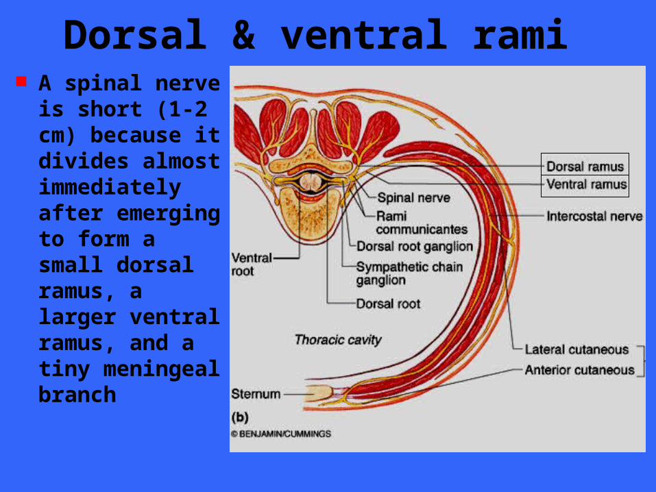
Dorsal & ventral rami A spinal nerve is
short (1-2 cm) because it divides almost immediately after emerging to form a small dorsal ramus, a larger ventral ramus, and a tiny meningeal branch
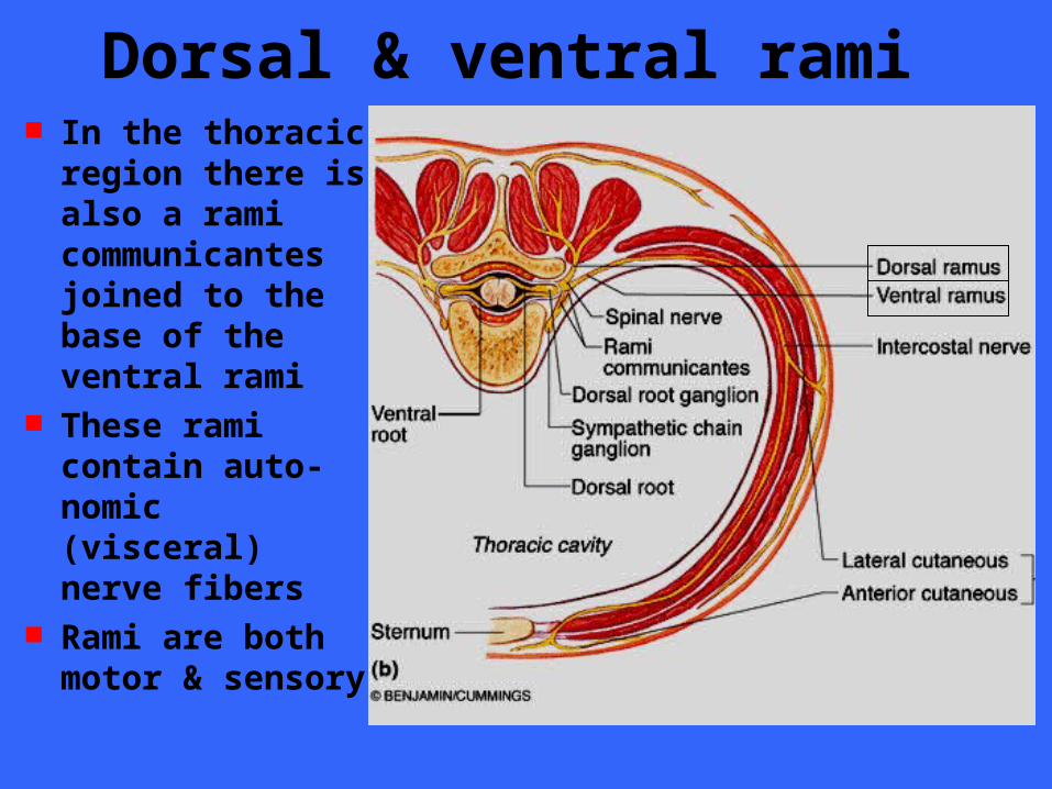
Dorsal & ventral rami In the thoracic
region there is also a rami communicantes joined to the base of the ventral rami
These rami contain auto-nomic (visceral) nerve fibers
Rami are both motor & sensory
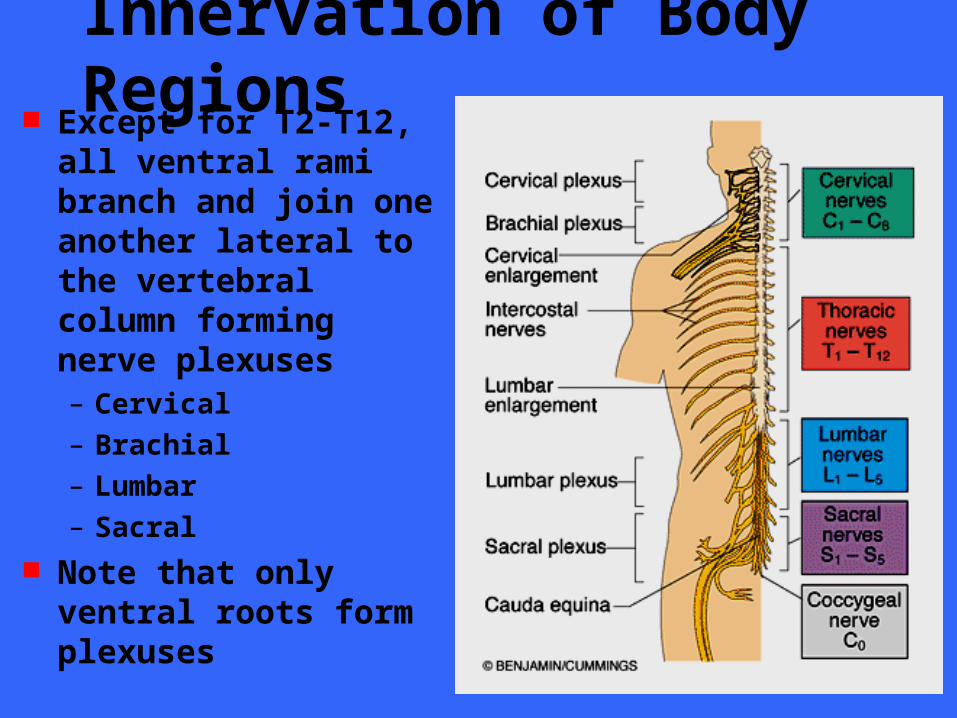
Innervation of Body Regions Except for T2-T12, all
ventral rami branch and join one another lateral to the vertebral column forming nerve plexuses– Cervical
– Brachial
– Lumbar
– Sacral Note that only ventral
roots form plexuses
Innervation of Body Regions Within plexuses the different ventral rami
crisscross each other and become redistributed so that– Each branch of the plexus contains fibers from
several different spinal nerves– Fibers from each ventral ramus travel to the body
periphery via several different routes or branches Thus, each muscle in a limb receives its nerve
supply from more than one spinal nerve Damage to a single root cannot completely
paralyze any limb muscle
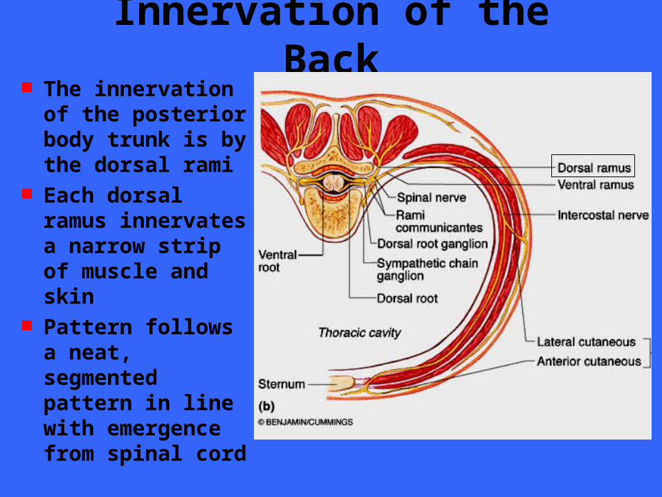
Innervation of the Back The innervation
of the posterior body trunk is by the dorsal rami
Each dorsal ramus innervates a narrow strip of muscle and skin
Pattern follows a neat, segmented pattern in line with emergence from spinal cord
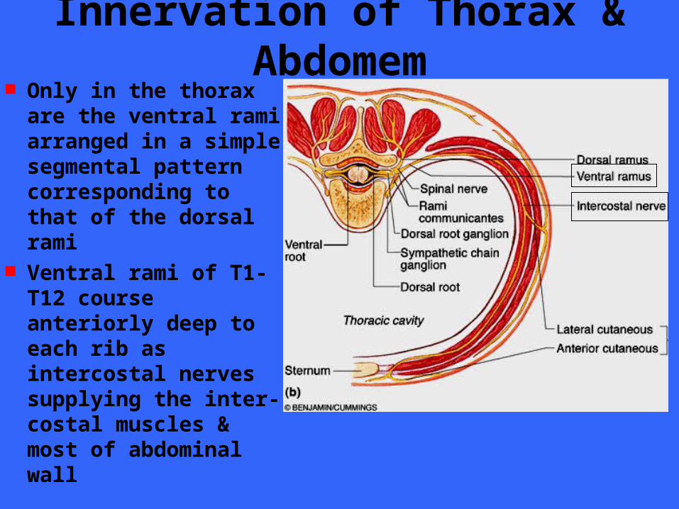
Innervation of Thorax & Abdomem Only in the thorax
are the ventral rami arranged in a simple segmental pattern corresponding to that of the dorsal rami
Ventral rami of T1-T12 course anteriorly deep to each rib as intercostal nerves supplying the inter- costal muscles & most of abdominal wall
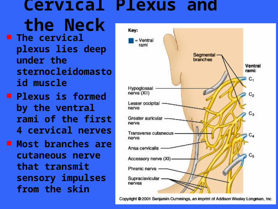
Cervical Plexus and the Neck The cervical plexus
lies deep under the sternocleidomastoid muscle
Plexus is formed by the ventral rami of the first 4 cervical nerves
Most branches are cutaneous nerve that transmit sensory impulses from the skin
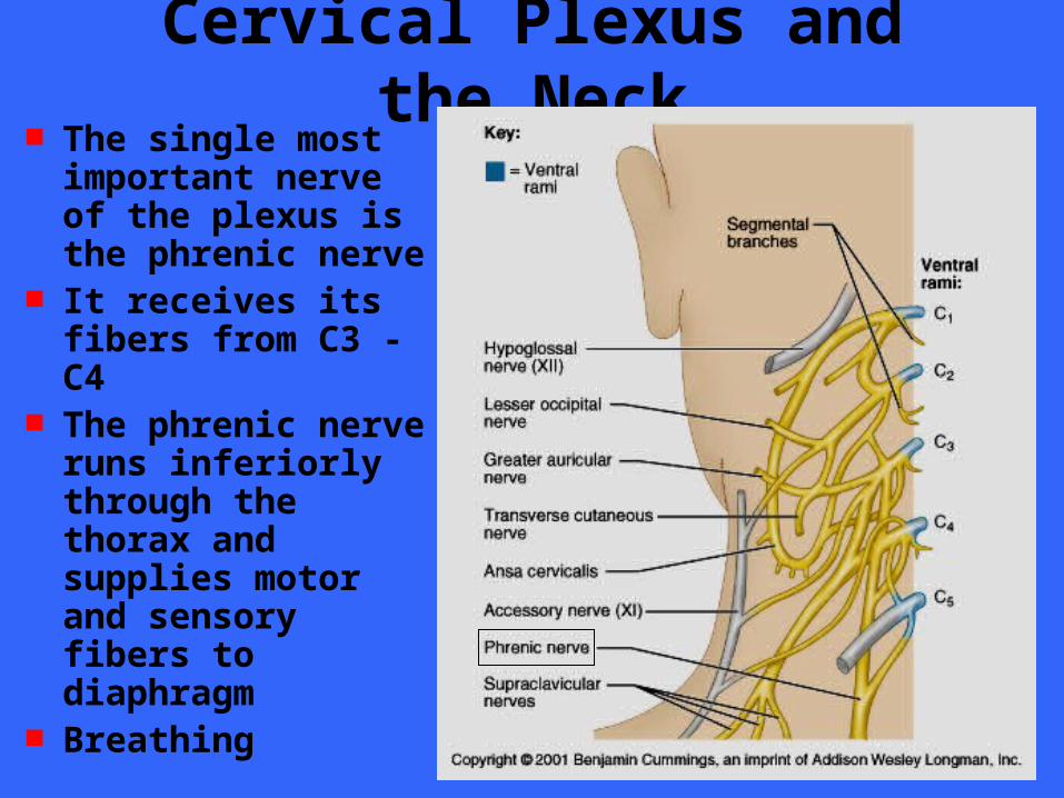
Cervical Plexus and the Neck The single most
important nerve of the plexus is the phrenic nerve
It receives its fibers from C3 - C4
The phrenic nerve runs inferiorly through the thorax and supplies motor and sensory fibers to diaphragm
Breathing
Brachial Plexus and Upper Limb The large important brachial plexus is
situated partly in the neck and partly in the axilla
It gives rise to virtually all the nerves that innervate the upper limb
The brachial plexus is very complex and is often referred to as the anatomy student’s nightmare
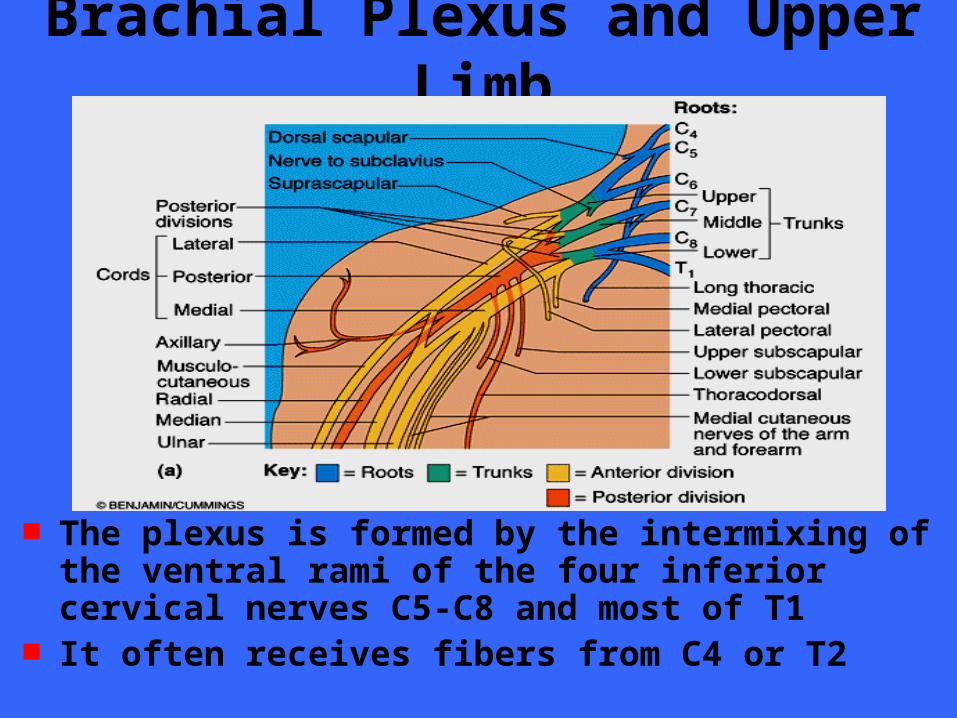
Brachial Plexus and Upper Limb
The plexus is formed by the intermixing of the ventral rami of the four inferior cervical nerves C5-C8 and most of T1
It often receives fibers from C4 or T2
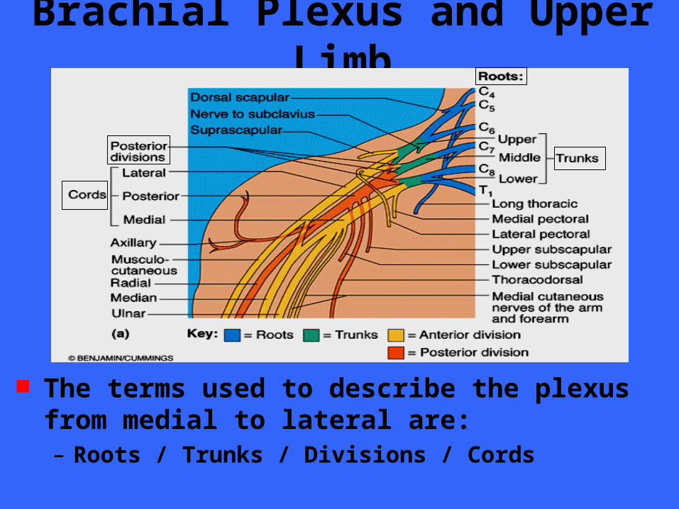
Brachial Plexus and Upper Limb
The terms used to describe the plexus from medial to lateral are:– Roots / Trunks / Divisions / Cords
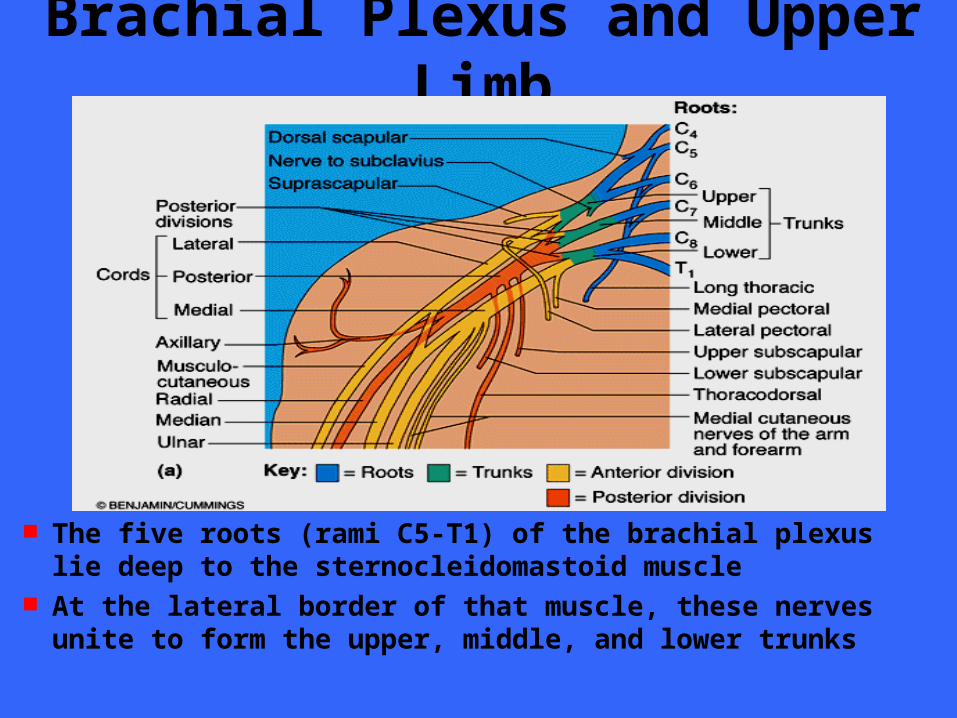
Brachial Plexus and Upper Limb
The five roots (rami C5-T1) of the brachial plexus lie deep to the sternocleidomastoid muscle
At the lateral border of that muscle, these nerves unite to form the upper, middle, and lower trunks
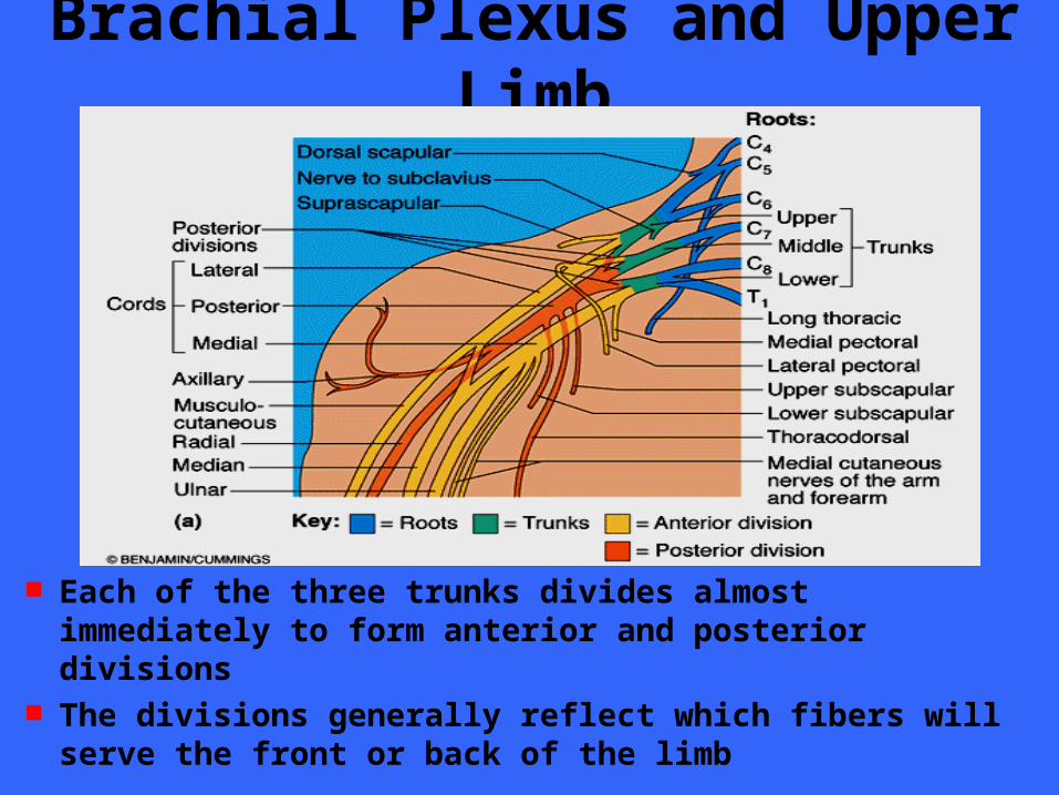
Brachial Plexus and Upper Limb
Each of the three trunks divides almost immediately to form anterior and posterior divisions
The divisions generally reflect which fibers will serve the front or back of the limb
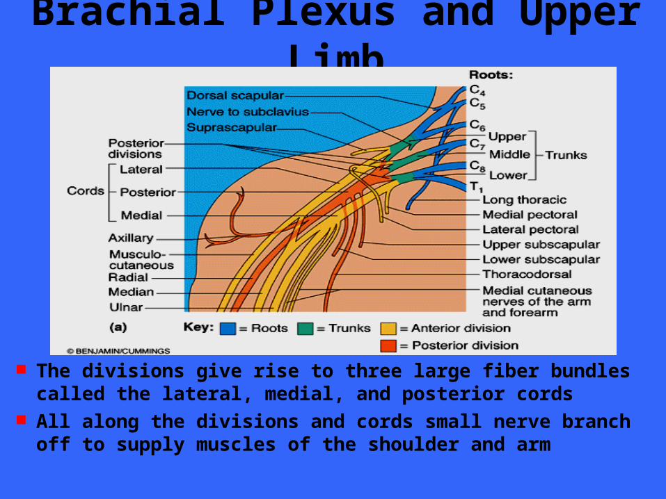
Brachial Plexus and Upper Limb
The divisions give rise to three large fiber bundles called the lateral, medial, and posterior cords
All along the divisions and cords small nerve branch off to supply muscles of the shoulder and arm
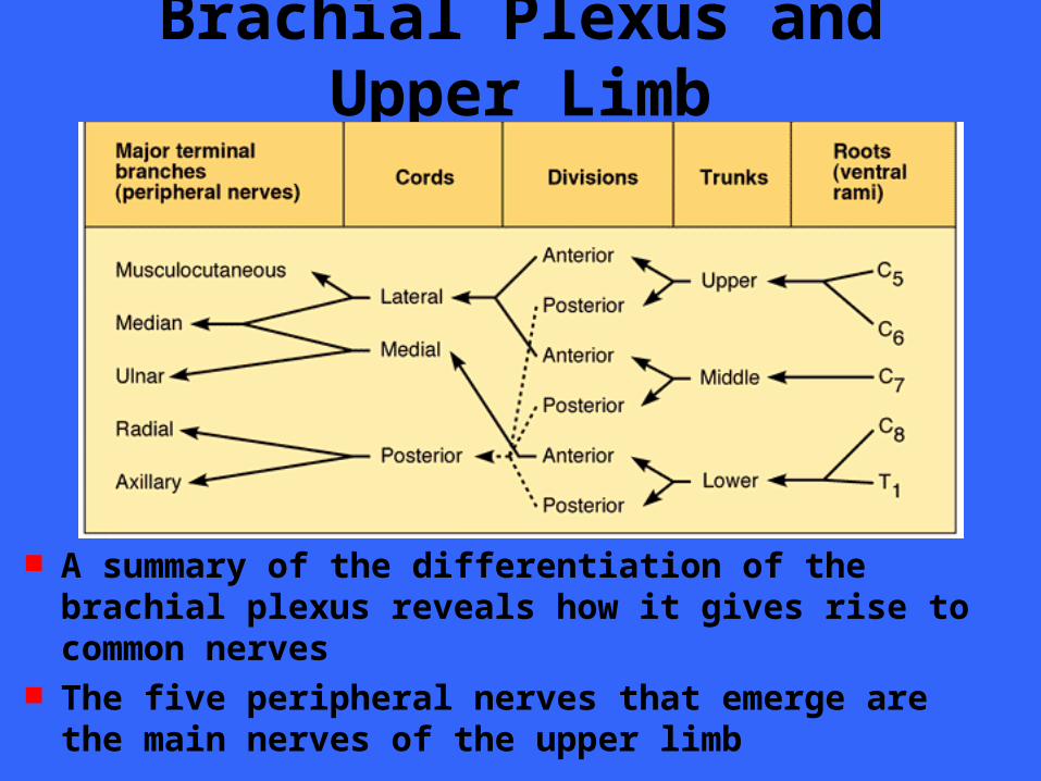
Brachial Plexus and Upper Limb
A summary of the differentiation of the brachial plexus reveals how it gives rise to common nerves
The five peripheral nerves that emerge are the main nerves of the upper limb
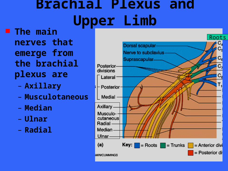
Brachial Plexus and Upper Limb The main nerves
that emerge from the brachial plexus are– Axillary
– Musculotaneous
– Median
– Ulnar
– Radial
Roots
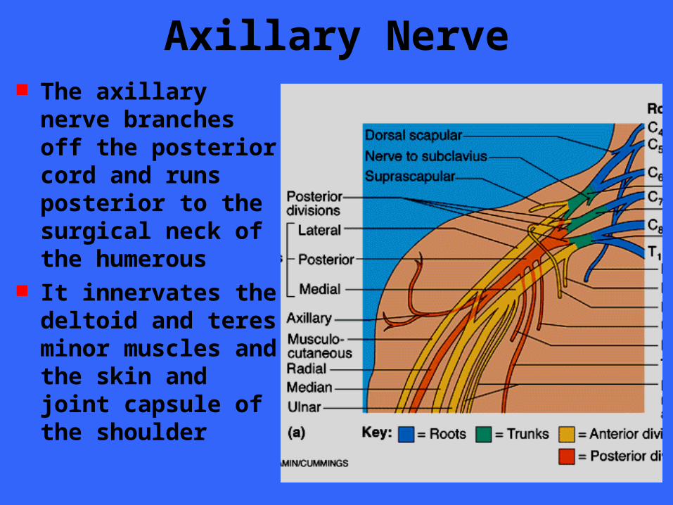
Axillary Nerve The axillary nerve
branches off the posterior cord and runs posterior to the surgical neck of the humerous
It innervates the deltoid and teres minor muscles and the skin and joint capsule of the shoulder
Axillary Nerve Muscular branches
– Deltoid – Teres minor
Cutaneous branches– Some of the skin of shoulder region
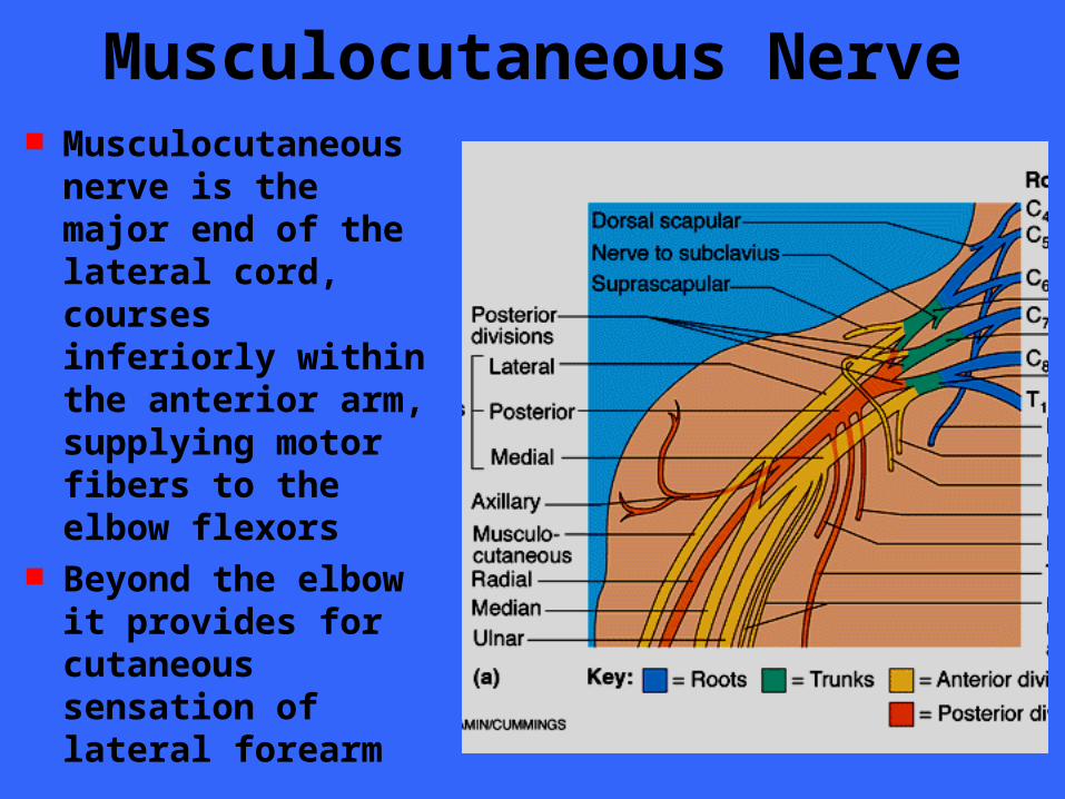
Musculocutaneous Nerve Musculocutaneous
nerve is the major end of the lateral cord, courses inferiorly within the anterior arm, supplying motor fibers to the elbow flexors
Beyond the elbow it provides for cutaneous sensation of lateral forearm
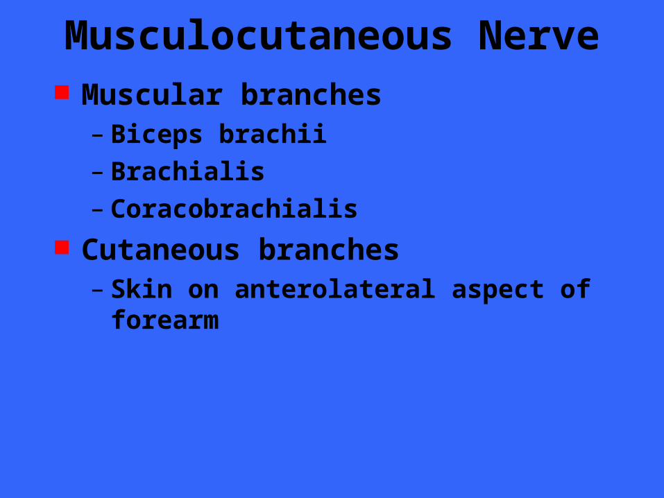
Musculocutaneous Nerve Muscular branches
– Biceps brachii– Brachialis– Coracobrachialis
Cutaneous branches– Skin on anterolateral aspect of forearm

Median Nerve The median nerve
descends through the arm without branching
In the anterior forearm, it gives off branches to the skin and most of the flexor muscles
It innervates the five intrinsic muscles of the lateral palm
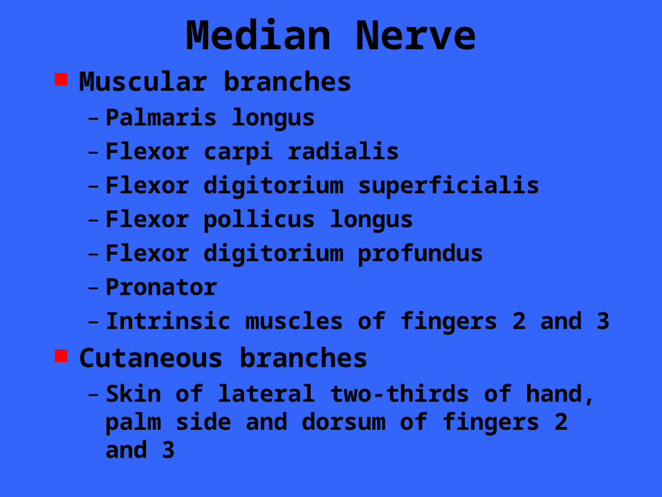
Median Nerve Muscular branches
– Palmaris longus– Flexor carpi radialis– Flexor digitorium superficialis– Flexor pollicus longus– Flexor digitorium profundus– Pronator– Intrinsic muscles of fingers 2 and 3
Cutaneous branches– Skin of lateral two-thirds of hand, palm side
and dorsum of fingers 2 and 3
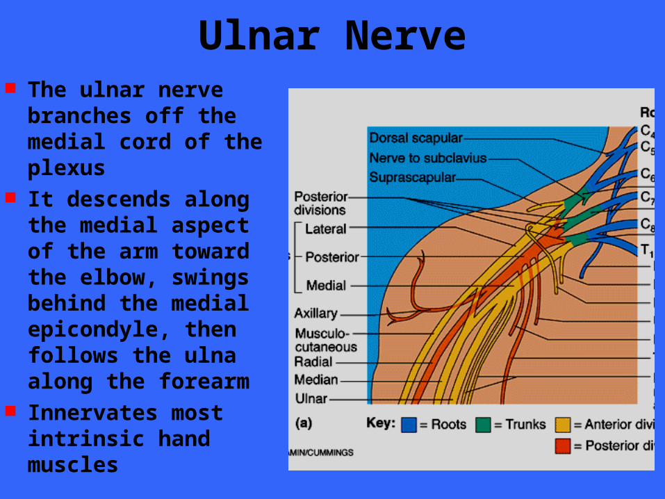
Ulnar Nerve The ulnar nerve
branches off the medial cord of the plexus
It descends along the medial aspect of the arm toward the elbow, swings behind the medial epicondyle, then follows the ulna along the forearm
Innervates most intrinsic hand muscles
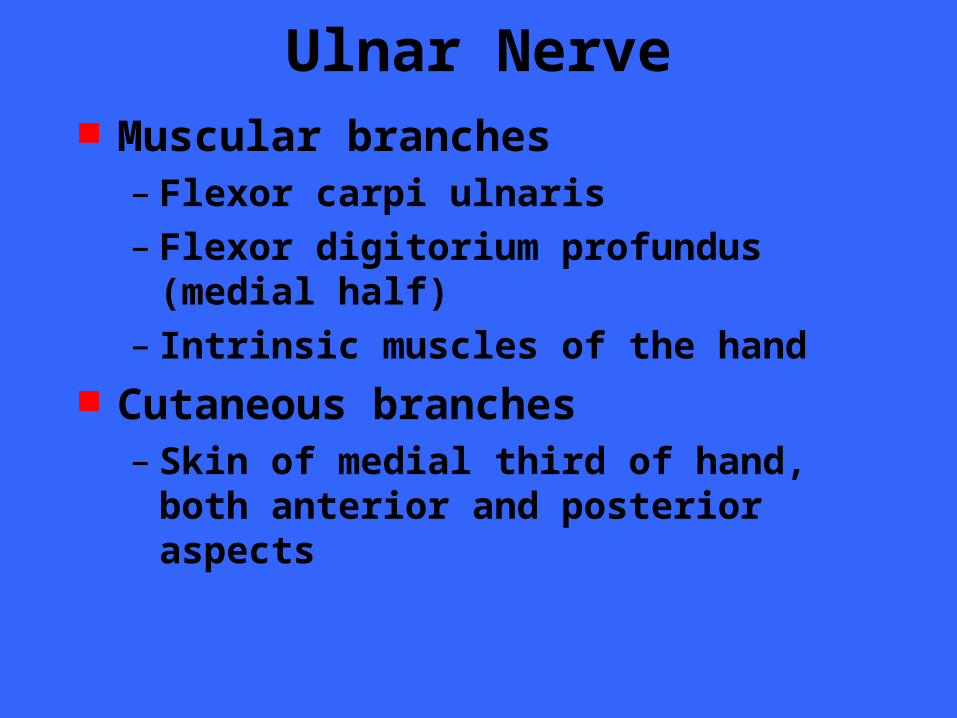
Ulnar Nerve Muscular branches
– Flexor carpi ulnaris– Flexor digitorium profundus (medial half)– Intrinsic muscles of the hand
Cutaneous branches– Skin of medial third of hand, both anterior
and posterior aspects
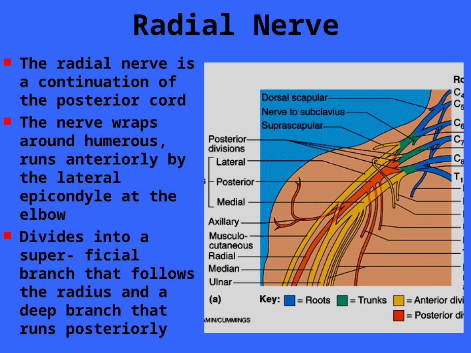
Radial Nerve The radial nerve is a
continuation of the posterior cord
The nerve wraps around humerous, runs anteriorly by the lateral epicondyle at the elbow
Divides into a super- ficial branch that follows the radius and a deep branch that runs posteriorly
Radial Nerve Muscular branches
– Triceps brachii– Anconeus– Supinator– Brachioradialis– Extensor capri radialis– Extensor carpi brevis– Extensor carpi ulnaris– Muscles that extend fingers
Cutaneous branches– Skin of posterior surface of entire limb
Lumbosacral Plexus The sacral and lumbar plexuses overlap
substantially Since many of the fibers of the lumbar
plexus contribute to the sacral plexus via the lumbosacral trunk, the two plexuses are often referred to as the lumbosacral plexus
Although the lumbosacral plexus mainly serves the lower limb, it also sends some branches to the abdomen, pelvis and buttocks
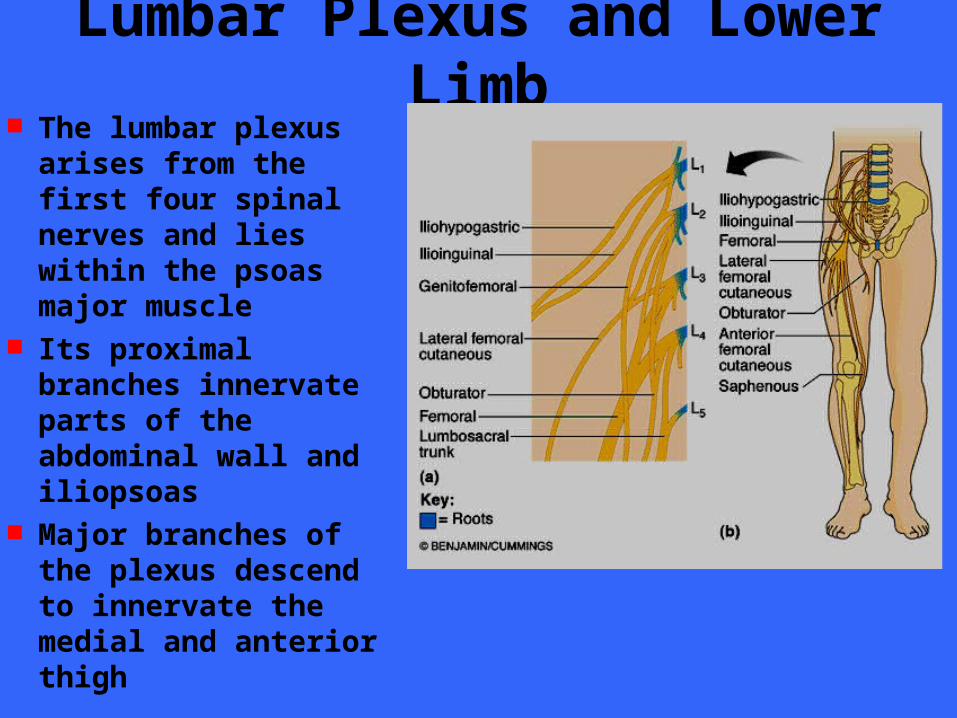
Lumbar Plexus and Lower Limb The lumbar plexus
arises from the first four spinal nerves and lies within the psoas major muscle
Its proximal branches innervate parts of the abdominal wall and iliopsoas
Major branches of the plexus descend to innervate the medial and anterior thigh
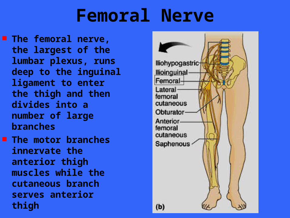
Femoral Nerve The femoral nerve, the
largest of the lumbar plexus, runs deep to the inguinal ligament to enter the thigh and then divides into a number of large branches
The motor branches innervate the anterior thigh muscles while the cutaneous branch serves anterior thigh
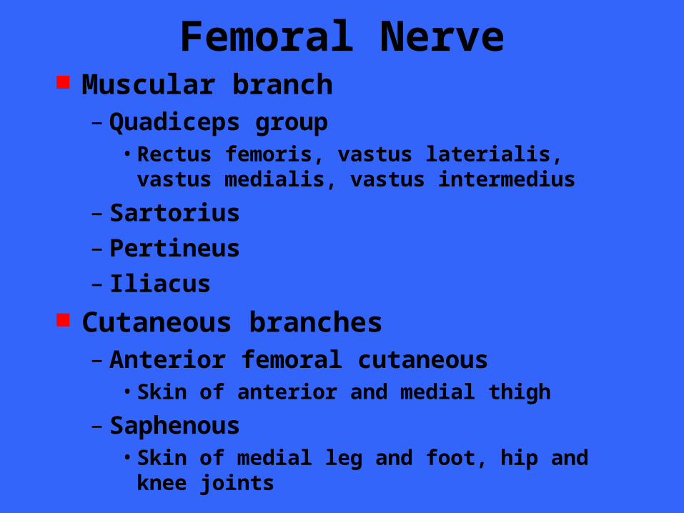
Femoral Nerve Muscular branch
– Quadiceps group• Rectus femoris, vastus laterialis, vastus medialis,
vastus intermedius
– Sartorius– Pertineus– Iliacus
Cutaneous branches– Anterior femoral cutaneous
• Skin of anterior and medial thigh
– Saphenous• Skin of medial leg and foot, hip and knee joints
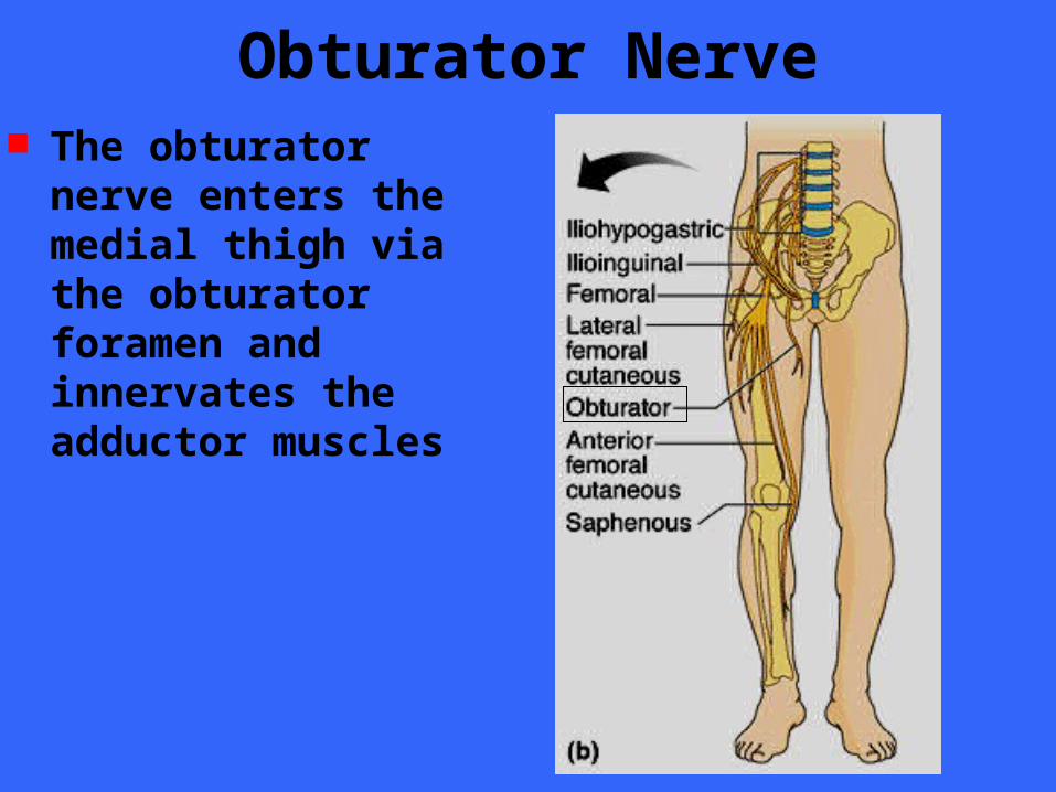
Obturator Nerve The obturator nerve
enters the medial thigh via the obturator foramen and innervates the adductor muscles
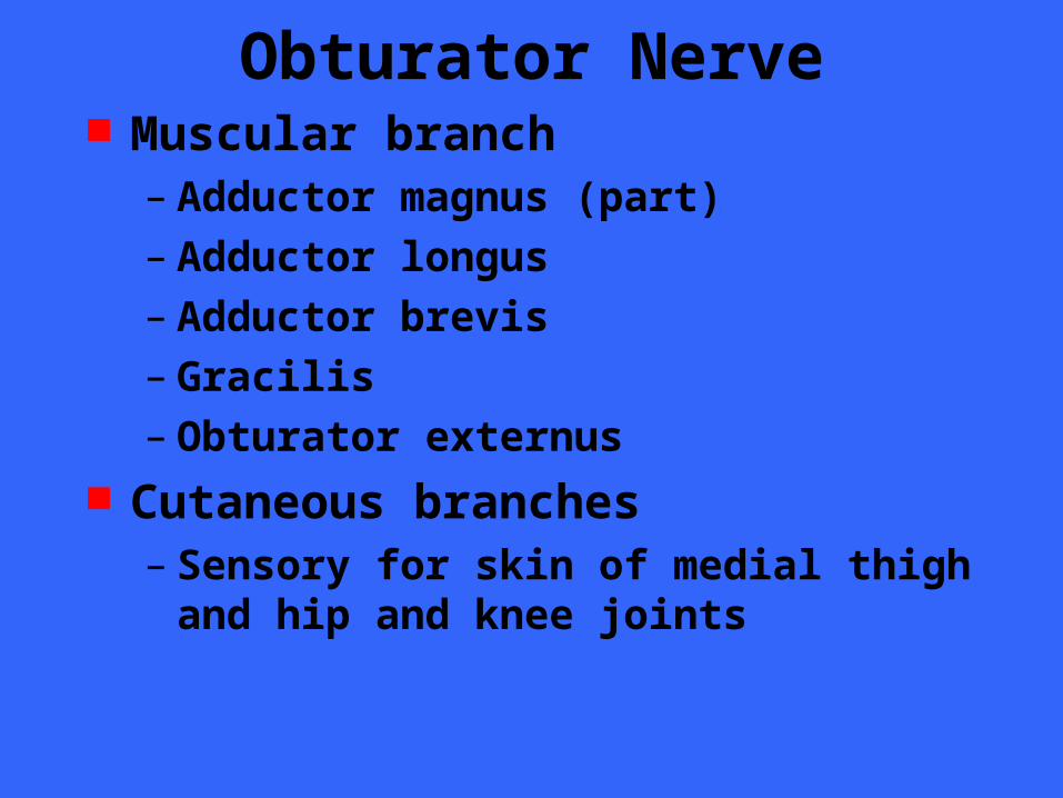
Obturator Nerve Muscular branch
– Adductor magnus (part)– Adductor longus– Adductor brevis– Gracilis– Obturator externus
Cutaneous branches– Sensory for skin of medial thigh and hip and
knee joints
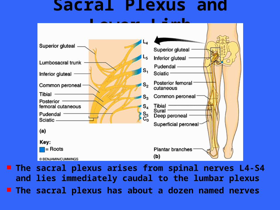
Sacral Plexus and Lower Limb
The sacral plexus arises from spinal nerves L4-S4 and lies immediately caudal to the lumbar plexus
The sacral plexus has about a dozen named nerves
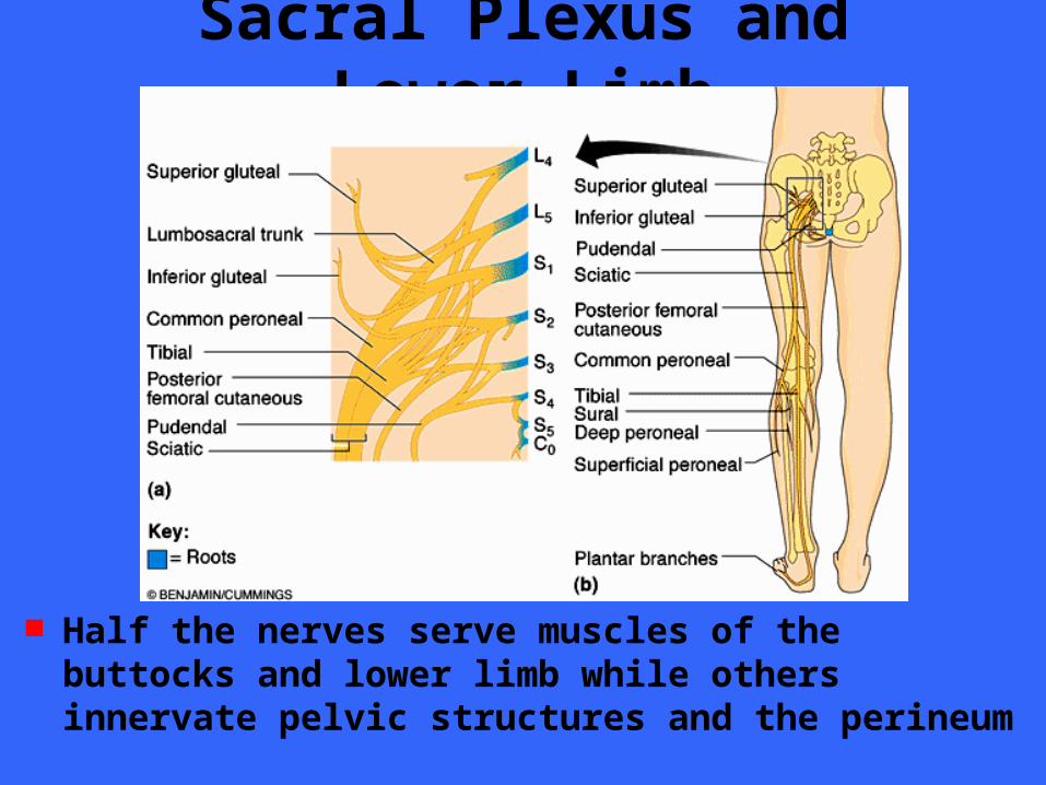
Sacral Plexus and Lower Limb
Half the nerves serve muscles of the buttocks and lower limb while others innervate pelvic structures and the perineum
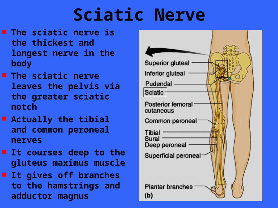
Sciatic Nerve The sciatic nerve is the
thickest and longest nerve in the body
The sciatic nerve leaves the pelvis via the greater sciatic notch
Actually the tibial and common peroneal nerves
It courses deep to the gluteus maximus muscle
It gives off branches to the hamstrings and adductor magnus
Sciatic Nerve Muscular branch
– Bicep femoris– Semitendinous– Semimembranous– Adductor magnus
Cutaneous branches– Posterior thigh
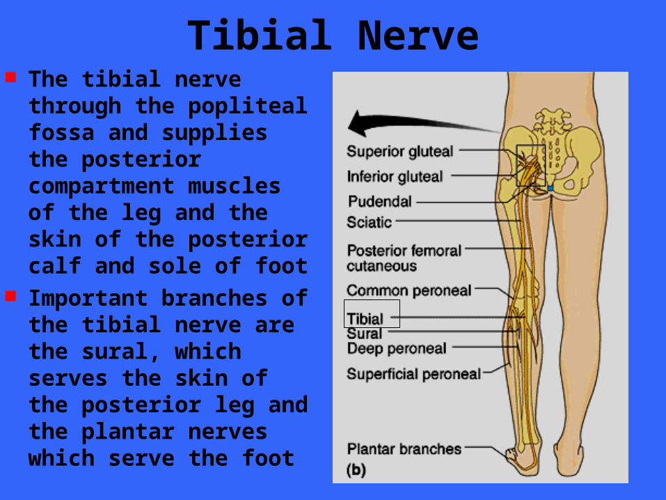
Tibial Nerve The tibial nerve through
the popliteal fossa and supplies the posterior compartment muscles of the leg and the skin of the posterior calf and sole of foot
Important branches of the tibial nerve are the sural, which serves the skin of the posterior leg and the plantar nerves which serve the foot
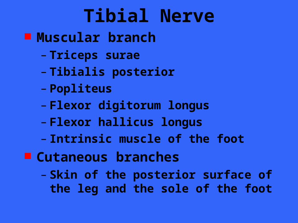
Tibial Nerve Muscular branch
– Triceps surae– Tibialis posterior– Popliteus– Flexor digitorum longus– Flexor hallicus longus – Intrinsic muscle of the foot
Cutaneous branches– Skin of the posterior surface of the leg and
the sole of the foot
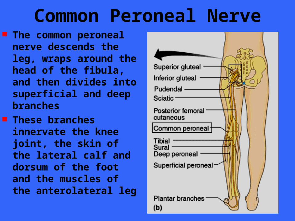
Common Peroneal Nerve The common peroneal
nerve descends the leg, wraps around the head of the fibula, and then divides into superficial and deep branches
These branches innervate the knee joint, the skin of the lateral calf and dorsum of the foot and the muscles of the anterolateral leg
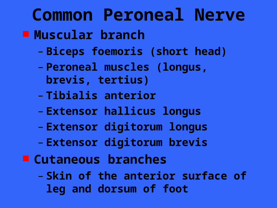
Common Peroneal Nerve Muscular branch
– Biceps foemoris (short head)– Peroneal muscles (longus, brevis, tertius)– Tibialis anterior– Extensor hallicus longus– Extensor digitorum longus– Extensor digitorum brevis
Cutaneous branches– Skin of the anterior surface of leg and
dorsum of foot
Sarcal Plexus Nerves Superior and inferior gluteal
– Innervate the gluteal muscles and tensor fasciae latae
Pudendal– Innervates the muscles of the skin of the
perineum– Mediates the act of erection– Voluntary control of urination– External anal sphinter
Innervation of the Joints Hilton’s law “. . . any nerve serving a
muscle producing movement at a joint also innervates the joint itself and the skin over the joint”
Innervation of Skin: Desmatomes The are of skin that is innervated by the
cutaneous branch of a spinal nerve is called a dermatome
All spinal nerves except C1 participate in dermatomes
Adjacent dermatomes on the body trunk are fairly uniform in width, almost horizontal, and in direct line with their spinal nerves
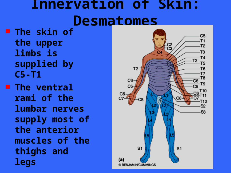
Innervation of Skin: Desmatomes The skin of the
upper limbs is supplied by C5-T1
The ventral rami of the lumbar nerves supply most of the anterior muscles of the thighs and legs
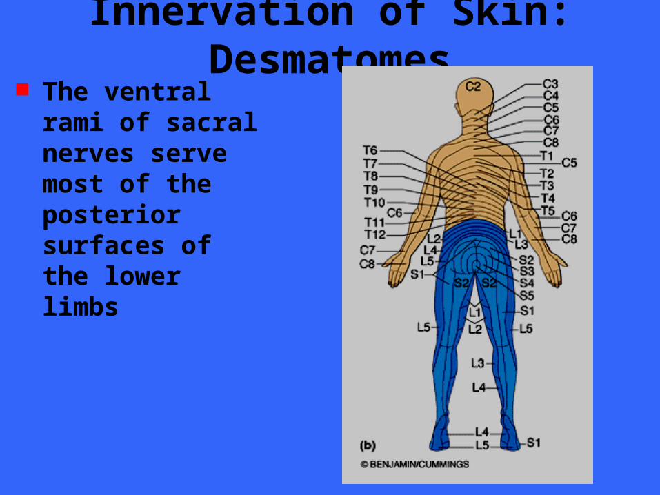
Innervation of Skin: Desmatomes The ventral rami of
sacral nerves serve most of the posterior surfaces of the lower limbs

End of Chapter
Chapter 14
Reflex Activity Many of the body’s control systems
belong to the general category of stimulus response consequences known as reflexes
A reflex is a rapid, predictable motor response to a stimulus
It is unlearned, unpremeditated, and involuntary
Basic reflexes may be considered to be built into our neural anatomy
Reflex Activity In addition to these basic, inborn types of
reflexes, there are many learned, or acquired reflexes that result from practice of repetition
There is no clear cut distinction between basic and learned reflexes
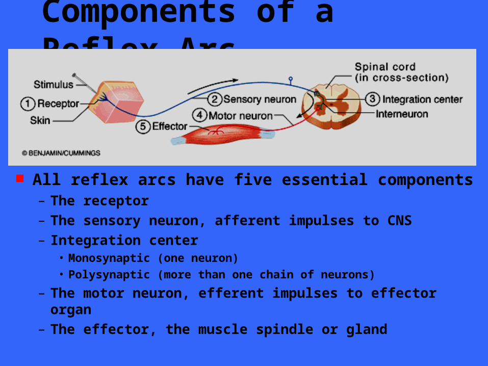
Components of a Reflex Arc
All reflex arcs have five essential components– The receptor
– The sensory neuron, afferent impulses to CNS
– Integration center• Monosynaptic (one neuron)
• Polysynaptic (more than one chain of neurons)
– The motor neuron, efferent impulses to effector organ
– The effector, the muscle spindle or gland
Components of a Reflex Arc Reflexes are classified functionally as
– Somatic reflexes • (activate skeletal muscle)
– Visceral reflexes (autonomic reflexes) • (activate smooth, cardiac muscle or visceral organs

Spinal Reflexes Somatic reflexes mediated by the spinal
cord are called spinal reflexes These reflexes may occur without the
involvement of higher brain centers Other reflexes may require the activity of
the brain for their successful completion Additionally, the brain is “advised” of
most types of spinal cord reflex activity and can facilitate or inhibit them
Stretch and Deep Tendon Reflexes If skeletal muscles are to perform
normally – The brain must be continually informed of
the current state of the muscles• Depends on information from muscle spindles
and Golgi tendon organs
– The muscles must exhibit healthy tone• Depends on stretch reflexes initiated by the
muscle spindles
These processes are important to normal skeletal muscle function, posture and locomotion
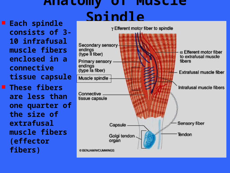
Anatomy of Muscle Spindle Each spindle
consists of 3-10 infrafusal muscle fibers enclosed in a connective tissue capsule
These fibers are less than one quarter of the size of extrafusal muscle fibers (effector fibers)
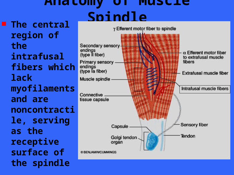
Anatomy of Muscle Spindle The central
region of the intrafusal fibers which lack myofilaments and are noncontractile, serving as the receptive surface of the spindle

Anatomy of Muscle Spindle Intrafusal fibers
are wrapped by two types of afferent endings that send sensory inputs to the CNS
Primary sensory endings – Type Ia fibers
Secondary sensory endings– Type II fibers

Anatomy of Muscle Spindle Primary sensory
endings – Type Ia fibers
Stimulated by both the rate and amount of stretch
Innervate the center of the spindle
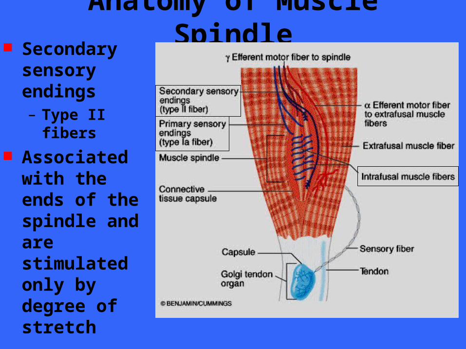
Anatomy of Muscle Spindle Secondary
sensory endings – Type II fibers
Associated with the ends of the spindle and are stimulated only by degree of stretch
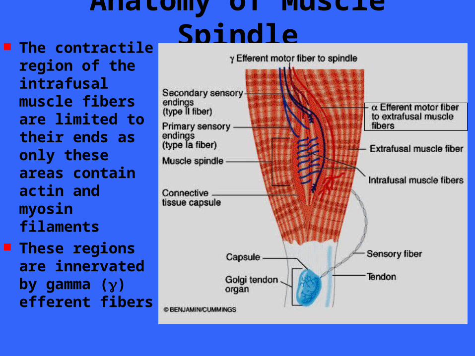
Anatomy of Muscle Spindle The contractile
region of the intrafusal muscle fibers are limited to their ends as only these areas contain actin and myosin filaments
These regions are innervated by gamma () efferent fibers
The Stretch Reflex Exciting a muscle spindle occurs in two
ways– Applying a force that lengthens the entire
muscle (external stretch - either by weight or by the action of an antagonist)
– Activing the motor neurons that stimulate the distal ends of the intrafusal fibers to contact, thus stretching the mid-portion of the spindle (internal stretch)
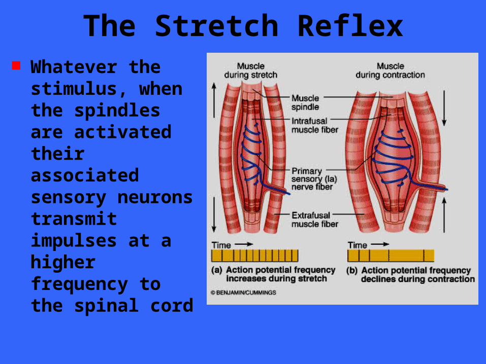
The Stretch Reflex Whatever the
stimulus, when the spindles are activated their associated sensory neurons transmit impulses at a higher frequency to the spinal cord
The Stretch Reflex
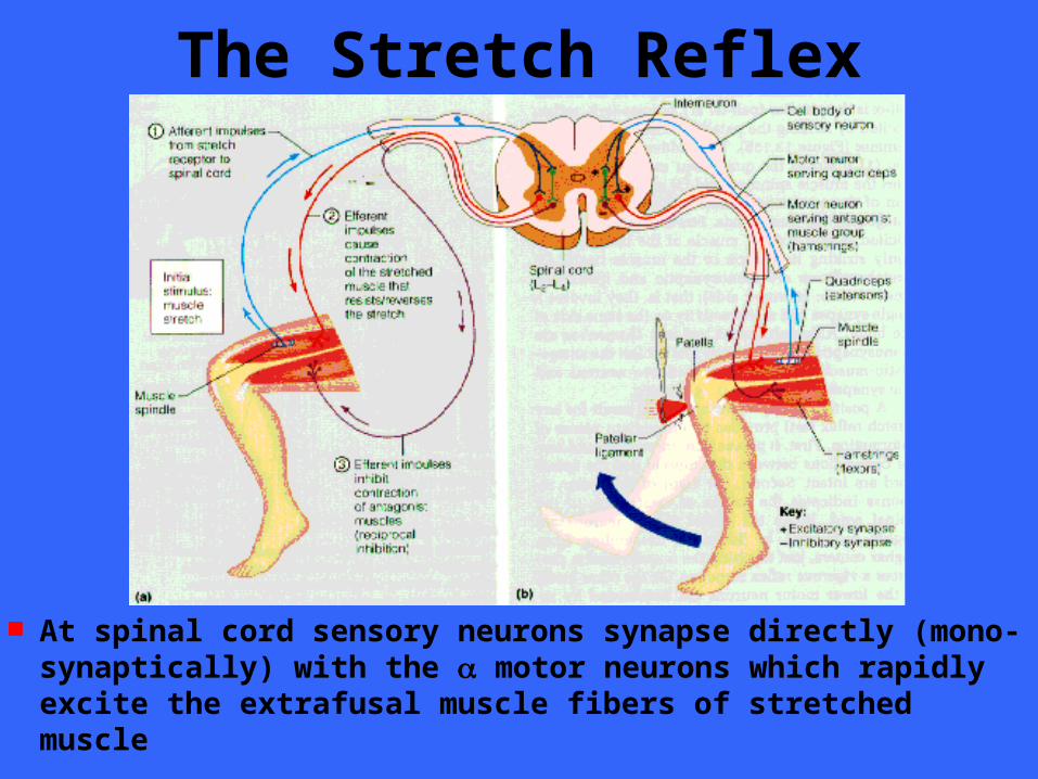
At spinal cord sensory neurons synapse directly (mono- synaptically) with the motor neurons which rapidly excite the extrafusal muscle fibers of stretched muscle
The Stretch Reflex
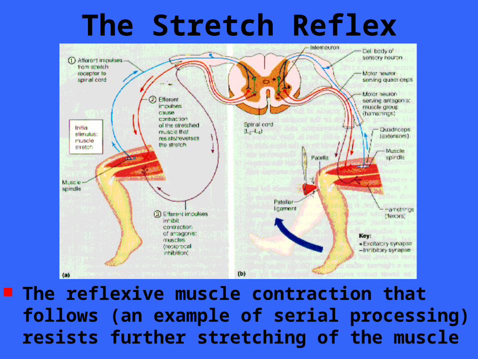
The reflexive muscle contraction that follows (an example of serial processing) resists further stretching of the muscle
The Stretch Reflex
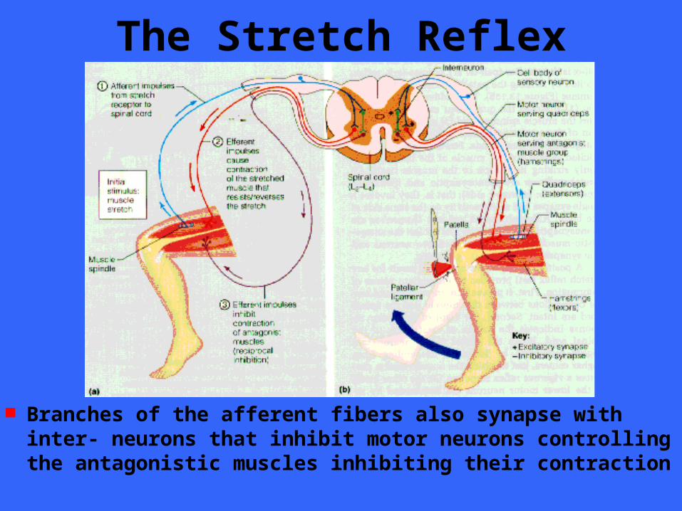
Branches of the afferent fibers also synapse with inter- neurons that inhibit motor neurons controlling the antagonistic muscles inhibiting their contraction
The Stretch Reflex Inhibition of the antagonistic muscles is
called reciprocal inhibition In essence, the stretch stimulus causes the
antagonists to relax so that they cannot resist the shortening of the “stretched” muscle caused by the main reflex arc
While this spinal reflex is occurring, impulses providing information on muscle length and the velocity of shortening are also being relayed to the brain
The Stretch Reflex The stretch reflex is most important in
large extensor muscles which sustain upright posture
Contractions of the postural muscles of the spine are almost continuously regulated by stretch reflexes initiated first on one side of the spine and then the other
The Deep Tendon Reflex Deep tendon reflexes cause muscle
relaxation and lengthening in response to the muscle’s contraction
This effect is opposite of those elicited by stretch reflexes
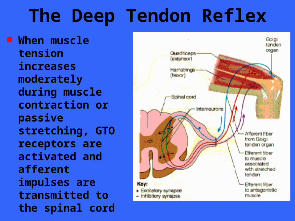
The Deep Tendon Reflex When muscle tension
increases moderately during muscle contraction or passive stretching, GTO receptors are activated and afferent impulses are transmitted to the spinal cord
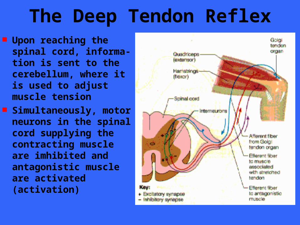
The Deep Tendon Reflex Upon reaching the
spinal cord, informa- tion is sent to the cerebellum, where it is used to adjust muscle tension
Simultaneously, motor neurons in the spinal cord supplying the contracting muscle are imhibited and antagonistic muscle are activated (activation)
The Deep Tendon Reflex Golgi tendon organs help ensure smooth
onset and termination of muscle contraction and are particularly important in activities involving rapid switching between flexion and extension such as in running
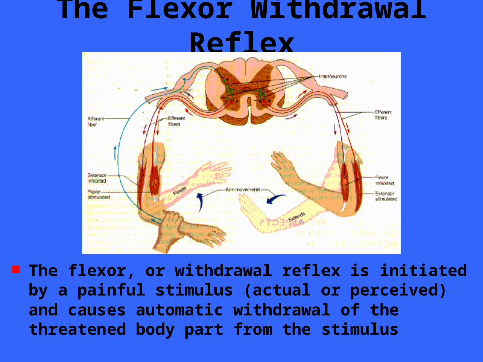
The Flexor Withdrawal Reflex
The flexor, or withdrawal reflex is initiated by a painful stimulus (actual or perceived) and causes automatic withdrawal of the threatened body part from the stimulus
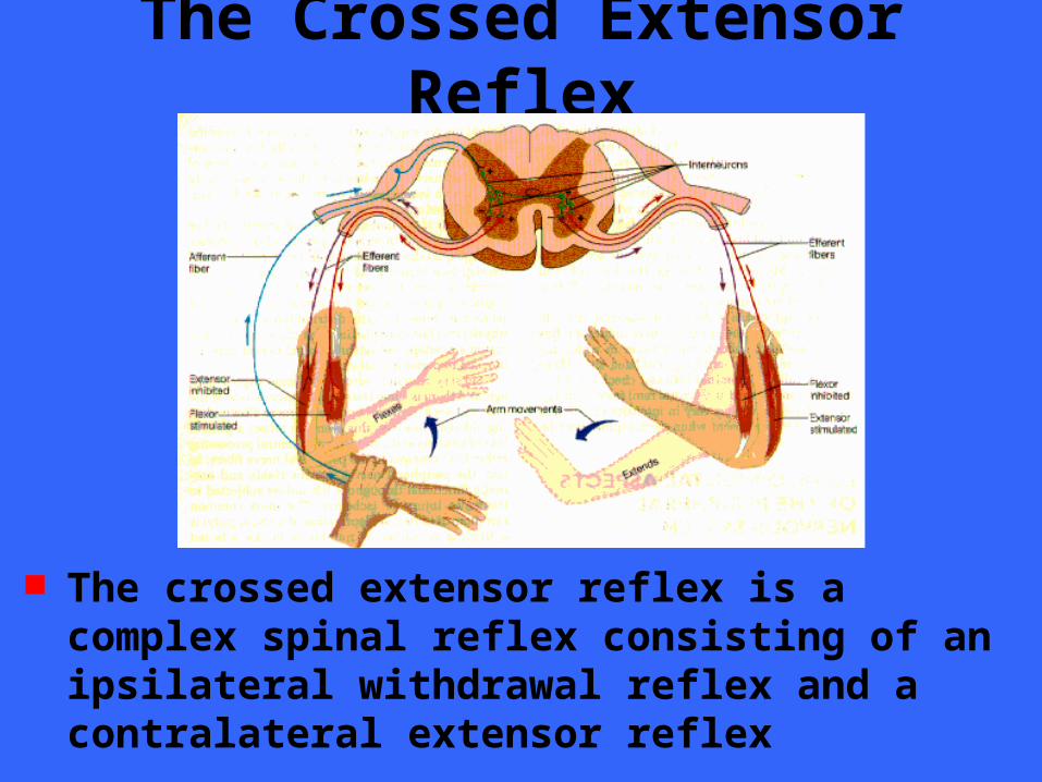
The Crossed Extensor Reflex
The crossed extensor reflex is a complex spinal reflex consisting of an ipsilateral withdrawal reflex and a contralateral extensor reflex
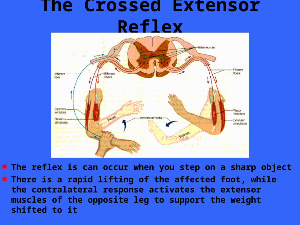
The Crossed Extensor Reflex
The reflex is can occur when you step on a sharp object There is a rapid lifting of the affected foot, while the
contralateral response activates the extensor muscles of the opposite leg to support the weight shifted to it
Superficial Reflexes Superficial reflexes are elicited by
cutaneous stimulation These reflexes are dependent upon
functional upper motor pathways and spinal cord reflex arcs
Babinski reflex
Classification by Structure Based on structural complexity there
simple and complex receptors– Simple are equivalent to modified dendritic
endings of sensory neurons• Found in skin, mucous membranes, muscles and
connective tissue– Monitor general sensory information
– Complex receptors are associated with the special senses
• Located in the special sensory organs– Specific sensory information (sight, hearing, etc)

End of Chapter
Regeneration of Nerve Fibers Damage to nervous tissue is serious
because mature neurons do not divide If the damage is severe or close to the cell
body, the entire neuron may die, and other neurons that are normally stimulated by its axon may die as well
However, in certain cases, cut or compressed axons on peripheral nerves can regenerate successfully
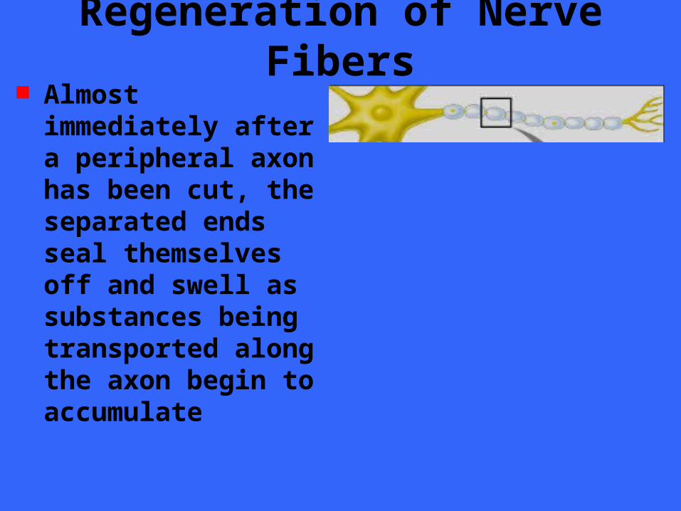
Regeneration of Nerve Fibers Almost immediately
after a peripheral axon has been cut, the separated ends seal themselves off and swell as substances being transported along the axon begin to accumulate
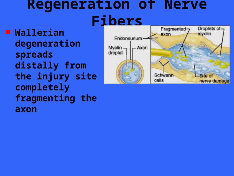
Regeneration of Nerve Fibers Wallerian
degeneration spreads distally from the injury site completely fragmenting the axon
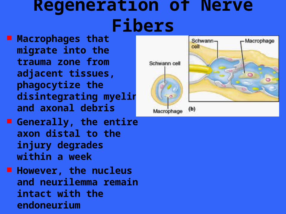
Regeneration of Nerve Fibers Macrophages that
migrate into the trauma zone from adjacent tissues, phagocytize the disintegrating myelin and axonal debris
Generally, the entire axon distal to the injury degrades within a week
However, the nucleus and neurilemma remain intact with the endoneurium
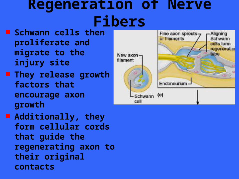
Regeneration of Nerve Fibers Schwann cells then
proliferate and migrate to the injury site
They release growth factors that encourage axon growth
Additionally, they form cellular cords that guide the regenerating axon to their original contacts
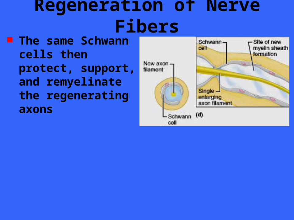
Regeneration of Nerve Fibers The same Schwann cells
then protect, support, and remyelinate the regenerating axons
Regeneration of Nerve Fibers Axons regenerate at a rate of 1 to 5 mm a
day The greater the distance between the
severed nerve endings, the greater the time for regeneration
Greater distances also lessen the chance of successful regeneration because adjacent tissues often block growth by protruding into larger gaps
Regeneration of Nerve Fibers CNS nerve fibers never regenerate under
normal circumstances Brain and spinal cord damage is
considered as irreversible The difference in regenerative capacity is
largely due to the support cells of the CNS Macrophage invasion in the CNS is much
slower than in the PNS Oligodendrocytes surrounding the
damaged axon die and thus cannot guide axon regeneration and growth

Sensory Receptor Potentials Sensory stimuli reaches us as many
different forms of energy Sensory receptors associated with sensory
neurons convert the energy of the stimulus into electrical energy
The energy changes the action potential of the receptor
Action potentials are generated as long as the stimulus is applied
Stimulus strength is determined by the frequency of impulse transmission