Embed Size (px)
Citation preview
The Peripheral Nervous System
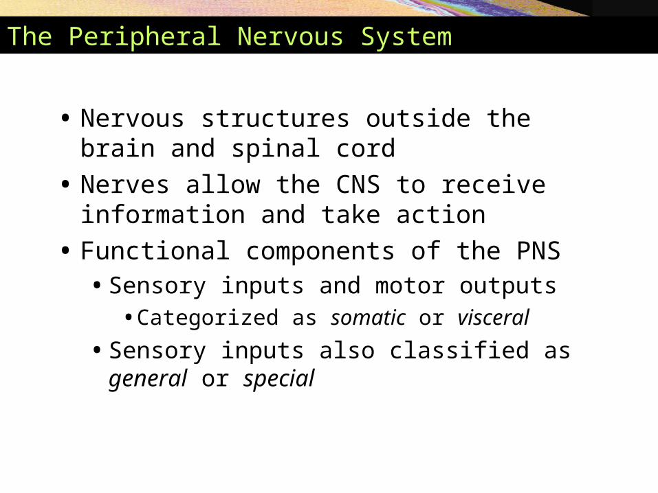
• Nervous structures outside the brain and spinal cord
• Nerves allow the CNS to receive information and take action
• Functional components of the PNS• Sensory inputs and motor outputs
• Categorized as somatic or visceral
• Sensory inputs also classified as general or special
The Peripheral Nervous System
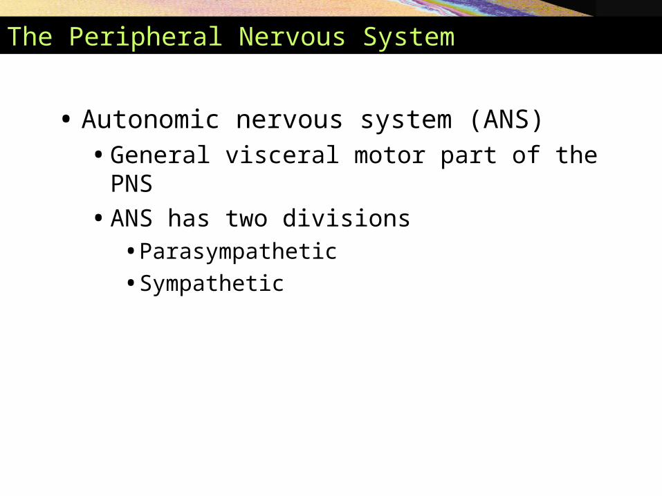
• Autonomic nervous system (ANS) • General visceral motor part of the PNS
• ANS has two divisions• Parasympathetic
• Sympathetic
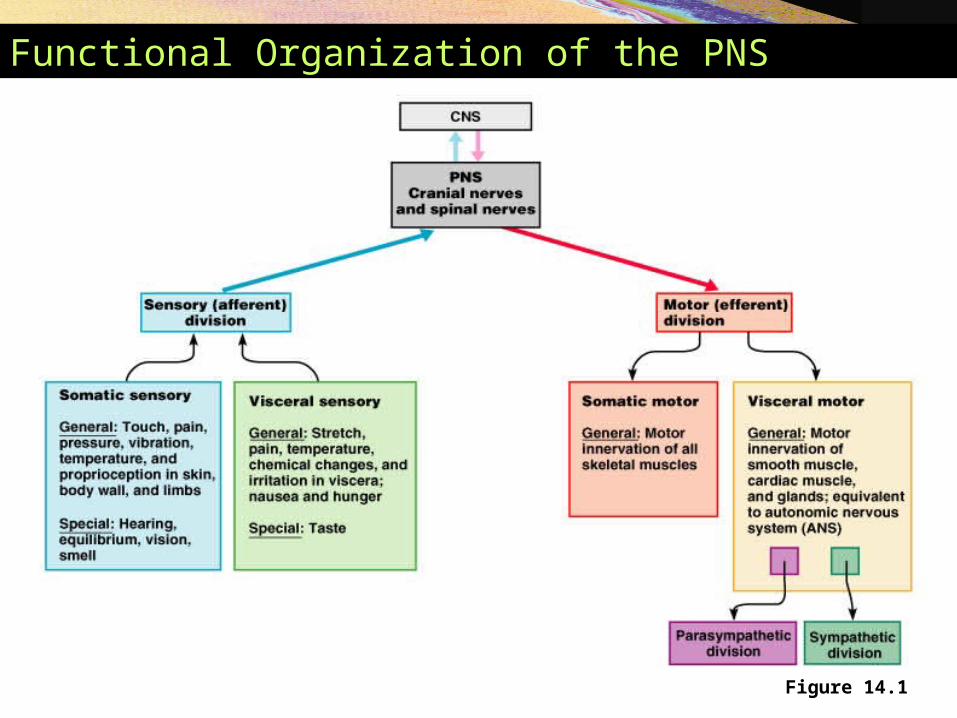
Functional Organization of the PNS
Figure 14.1
Basic Structural Components of the PNS
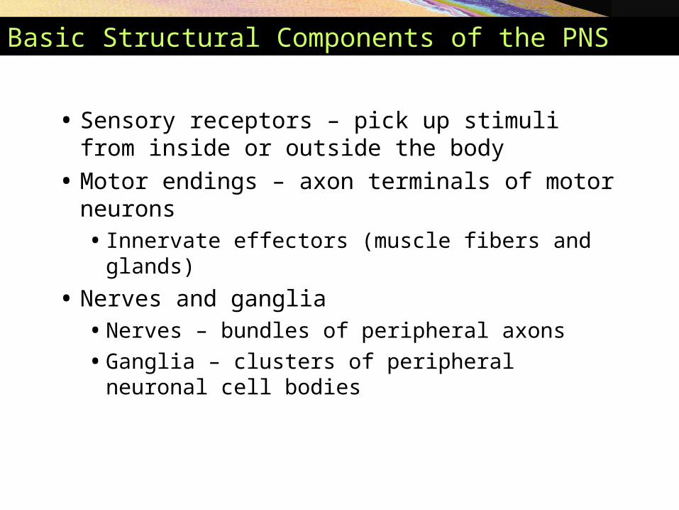
• Sensory receptors – pick up stimuli from inside or outside the body
• Motor endings – axon terminals of motor neurons• Innervate effectors (muscle fibers and glands)
• Nerves and ganglia • Nerves – bundles of peripheral axons
• Ganglia – clusters of peripheral neuronal cell bodies
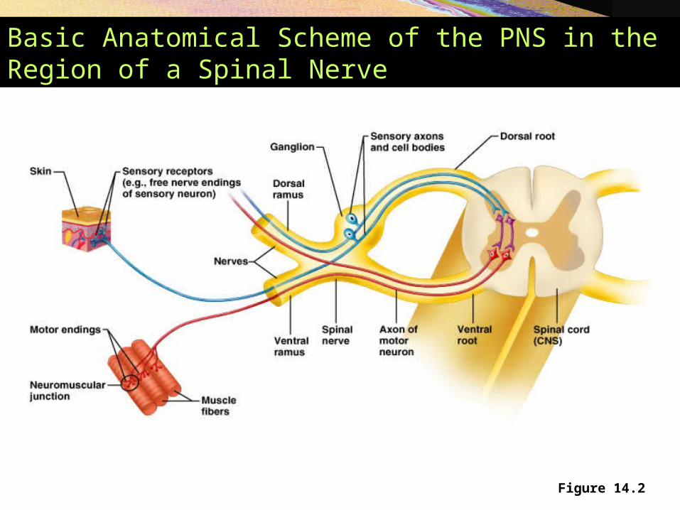
Basic Anatomical Scheme of the PNS in the Region of a Spinal Nerve
Figure 14.2

Peripheral Sensory Receptors
• Structures that pick up sensory stimuli• Initiate signals in sensory axons
• Two main categories of sensory receptors• Free nerve endings of sensory neurons
• Monitor general sensory information
• Complete receptor cells – specialized epithelial cells or small neurons• Monitor most types of special sensory information
Peripheral Sensory Receptors
• Sensory receptors also classified according to: • Location
• Type of stimulus detected
• Structure
Classification by Location
• Exteroceptors – sensitive to stimuli arising from outside the body• Located at or near body surfaces• Include receptors for touch, pressure, pain, and
temperature
• Interoceptors – (visceroceptors) receive stimuli from internal viscera• Monitor a variety of stimuli
• Proprioceptors – monitor degree of stretch• Located in musculoskeletal organs
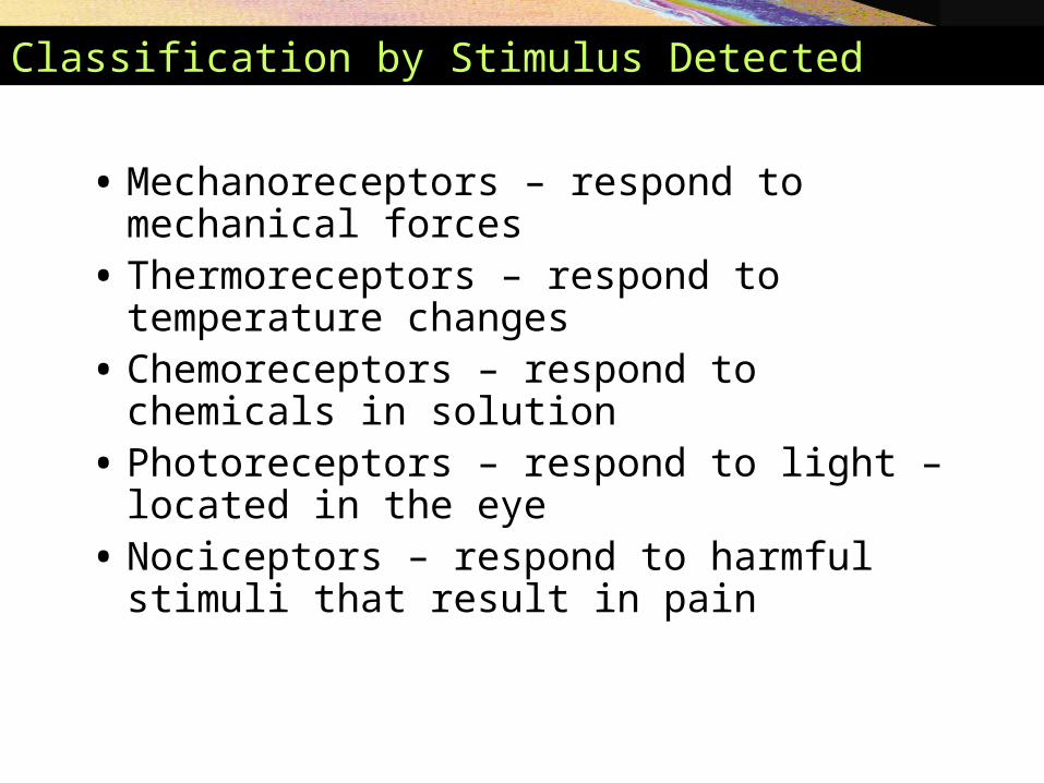
Classification by Stimulus Detected
• Mechanoreceptors – respond to mechanical forces• Thermoreceptors – respond to temperature
changes• Chemoreceptors – respond to chemicals in
solution• Photoreceptors – respond to light – located in the
eye • Nociceptors – respond to harmful stimuli that
result in pain
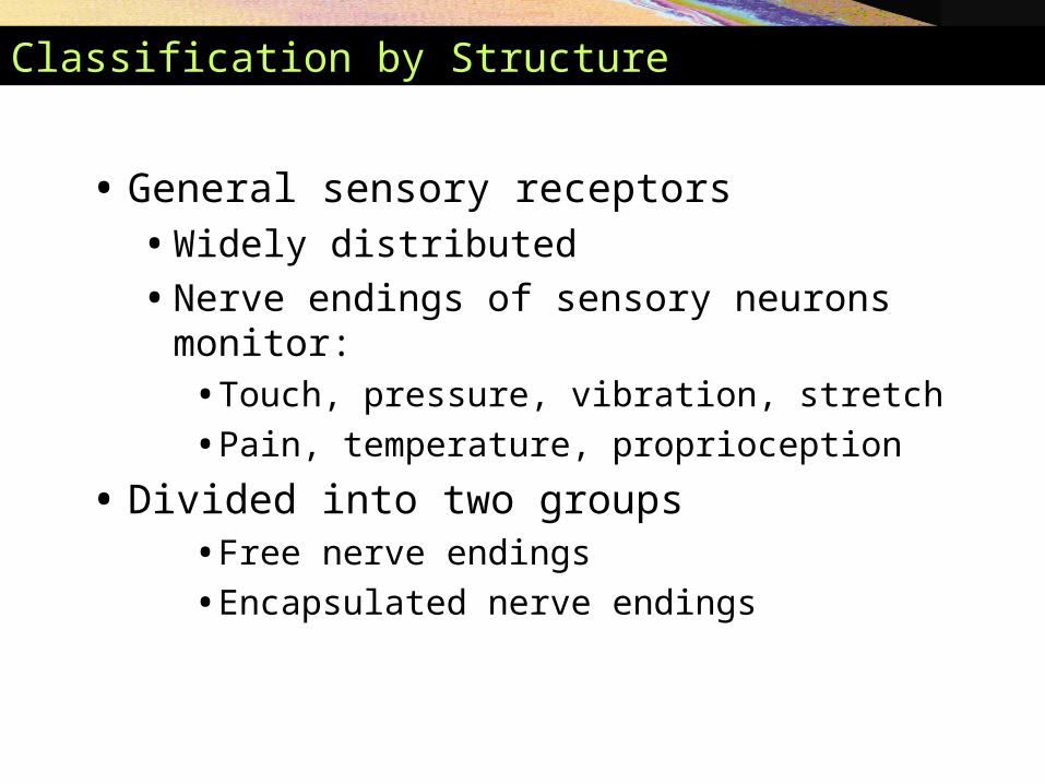
Classification by Structure
• General sensory receptors• Widely distributed
• Nerve endings of sensory neurons monitor:• Touch, pressure, vibration, stretch
• Pain, temperature, proprioception
• Divided into two groups• Free nerve endings
• Encapsulated nerve endings
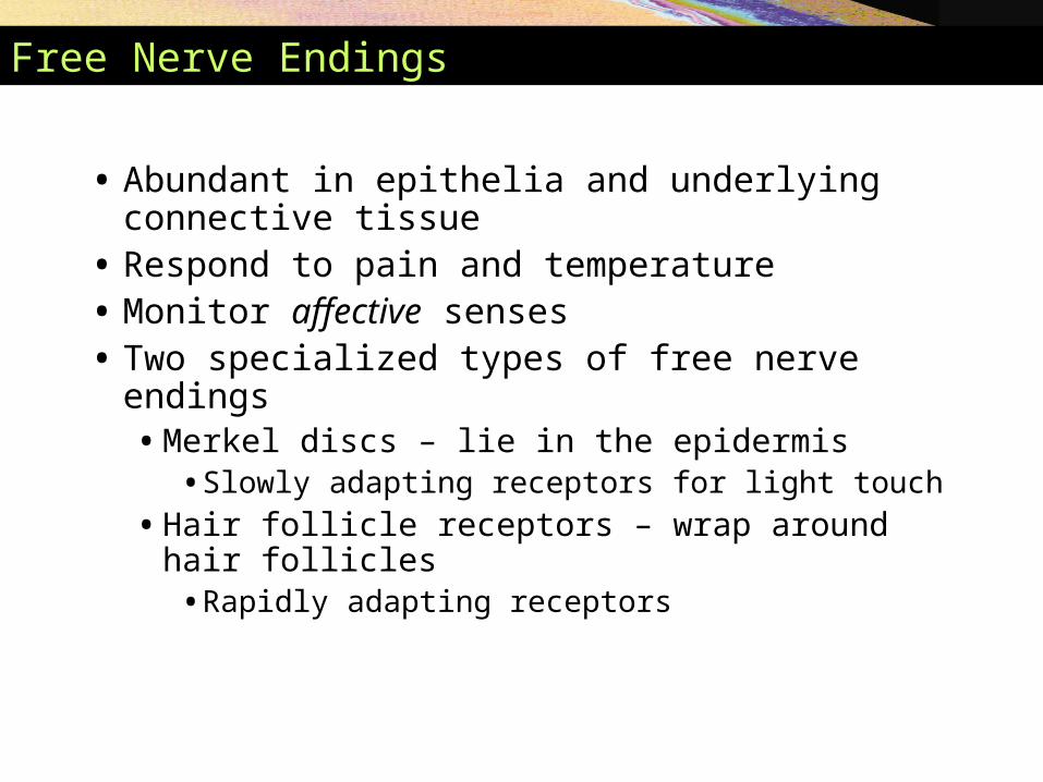
Free Nerve Endings
• Abundant in epithelia and underlying connective tissue
• Respond to pain and temperature• Monitor affective senses• Two specialized types of free nerve endings
• Merkel discs – lie in the epidermis• Slowly adapting receptors for light touch
• Hair follicle receptors – wrap around hair follicles• Rapidly adapting receptors
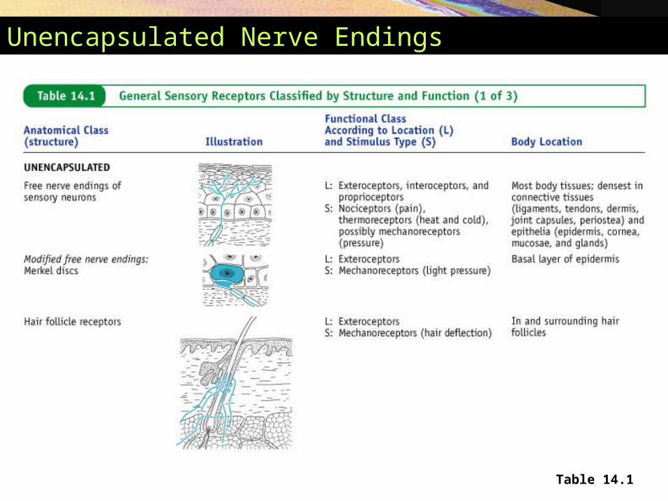
Unencapsulated Nerve Endings
Table 14.1
Encapsulated Nerve Endings
• Consist of one or more end fibers of sensory neurons
• Enclosed in connective tissue
• Mechanoreceptors
• Include four main types
Encapsulated Nerve Endings
• Meissner’s corpuscles • Spiraling nerve ending surrounded by Schwann
cells
• Occur in the dermal papillae
• Rapidly adapting receptors for discriminative touch
• Occur in sensitive, hairless areas of the skin
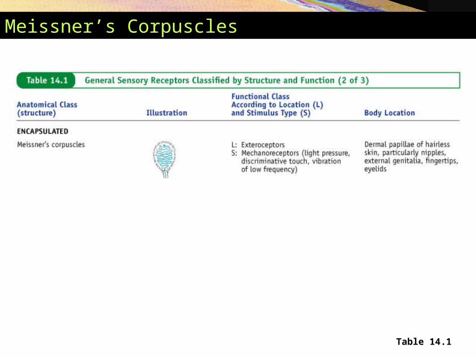
Meissner’s Corpuscles
Table 14.1
Encapsulated Nerve Endings
• Pacinian corpuscles • Single nerve ending surrounded by layers of
flattened Schwann cells
• Occur in the hypodermis
• Sensitive to deep pressure – rapidly adapting receptors
• Ruffini’s corpuscles• Located in the dermis and respond to pressure
• Monitor continuous pressure on the skin – adapt slowly
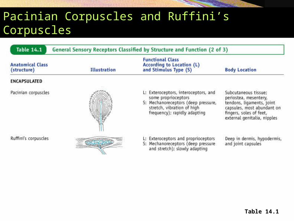
Pacinian Corpuscles and Ruffini’s Corpuscles
Table 14.1
Encapsulated Nerve Endings
• Proprioceptors • Monitor stretch in locomotory organs
• Three types of proprioceptors
Three Types of Proprioceptors
• Muscle spindles – measure the changing length of a muscle• Imbedded in the perimysium between muscle
fascicles
• Golgi tendon organs – located near the muscle-tendon junction• Monitor tension within tendons
• Joint kinesthetic receptors • Sensory nerve endings within the joint capsules
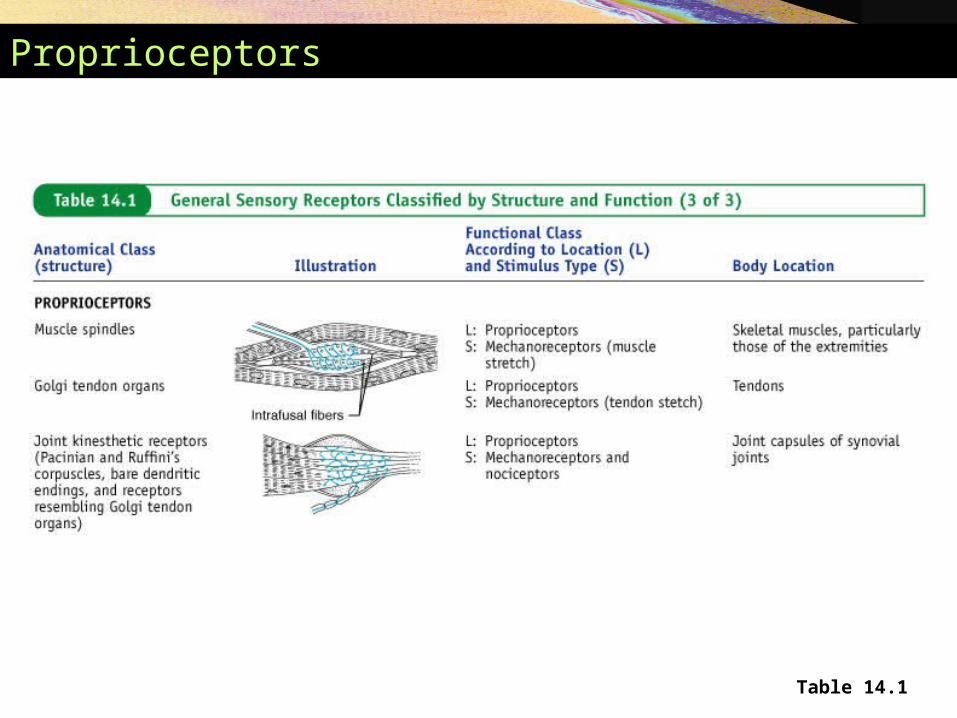
Proprioceptors
Table 14.1
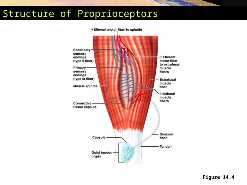
Structure of Proprioceptors
Figure 14.4


![The Nervous System. Divisions of the Nervous System Central Nervous System [CNS] = Spinal Cord Brain Peripheral Nervous System [PNS]= Spinal Nerves](https://img.pdfslide.net/doc/110x75/56649d6c5503460f94a4c71d/the-nervous-system-divisions-of-the-nervous-system-central-nervous-system.jpg)