Embed Size (px)
DESCRIPTION
Biology of plant cell
Citation preview
Levetin−McMahon: Plants and Society, Fifth Edition
II. Introduction to Plant Life: Botanical Principles
2. The Plant Cell © The McGraw−Hill Companies, 2008
19
2The Plant Cell
CHAPTER OUTLINE Early Studies of Cells 20 The Cell Wall 22 The Protoplast 22
Membranes 22 Moving Into and Out of Cells 22 Organelles 23
A CLOSER LOOK 2.1 Origin of Chloroplasts and Mitochondria 25The Nucleus 26
Cell Division 26The Cell Cycle 26 Prophase 27 Metaphase 27 Anaphase 27 Telophase 27 Cytokinesis 27
Chapter Summary 30 Review Questions 30 Further Reading 30
KEY CONCEPTS 1. The Cell Theory establishes that the cell
is the basic unit of life, that all living organisms are composed of cells, and that cells arise from preexisting cells.
2. Plant cells are eukaryotic, having an organized nucleus and membrane-bound organelles.
3. Substances can move into and out of cells by diffusion and osmosis.
4. Mitosis, followed by cytokinesis, results in two genetically identical daughter cells. Growth, replacement of cells, and asexual reproduction all depend on the process of cell division.
C H A P T E R
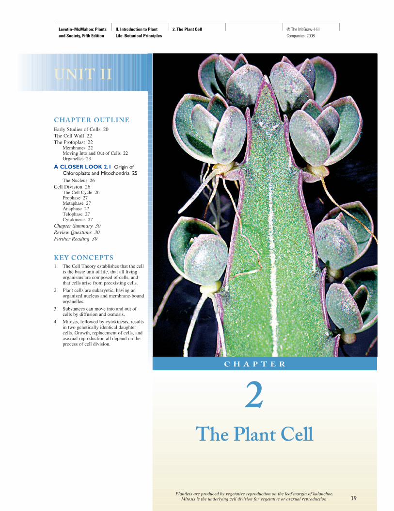
Plantlets are produced by vegetative reproduction on the leaf margin of kalanchoe. Mitosis is the underlying cell division for vegetative or asexual reproduction.
UNIT II
Levetin−McMahon: Plants and Society, Fifth Edition
II. Introduction to Plant Life: Botanical Principles
2. The Plant Cell © The McGraw−Hill Companies, 2008
20 U N I T I I Introduction to Plant Life: Botanical Principles
All plants (and every other living organism) are com-posed of cells. In some algae and fungi, the whole organism consists of a single cell, but angiosperms
are complex multicellular organisms composed of many dif-ferent types of cells. Plant cells are microscopic and typically range from 10 to 100 μm in length. This means that there would be between 254 and 2,540 of these cells to an inch ( fig. 2.1 ). In Chapter 3 we will be looking at the variety of cells, but in this chapter we will focus on a composite angio-sperm plant cell.
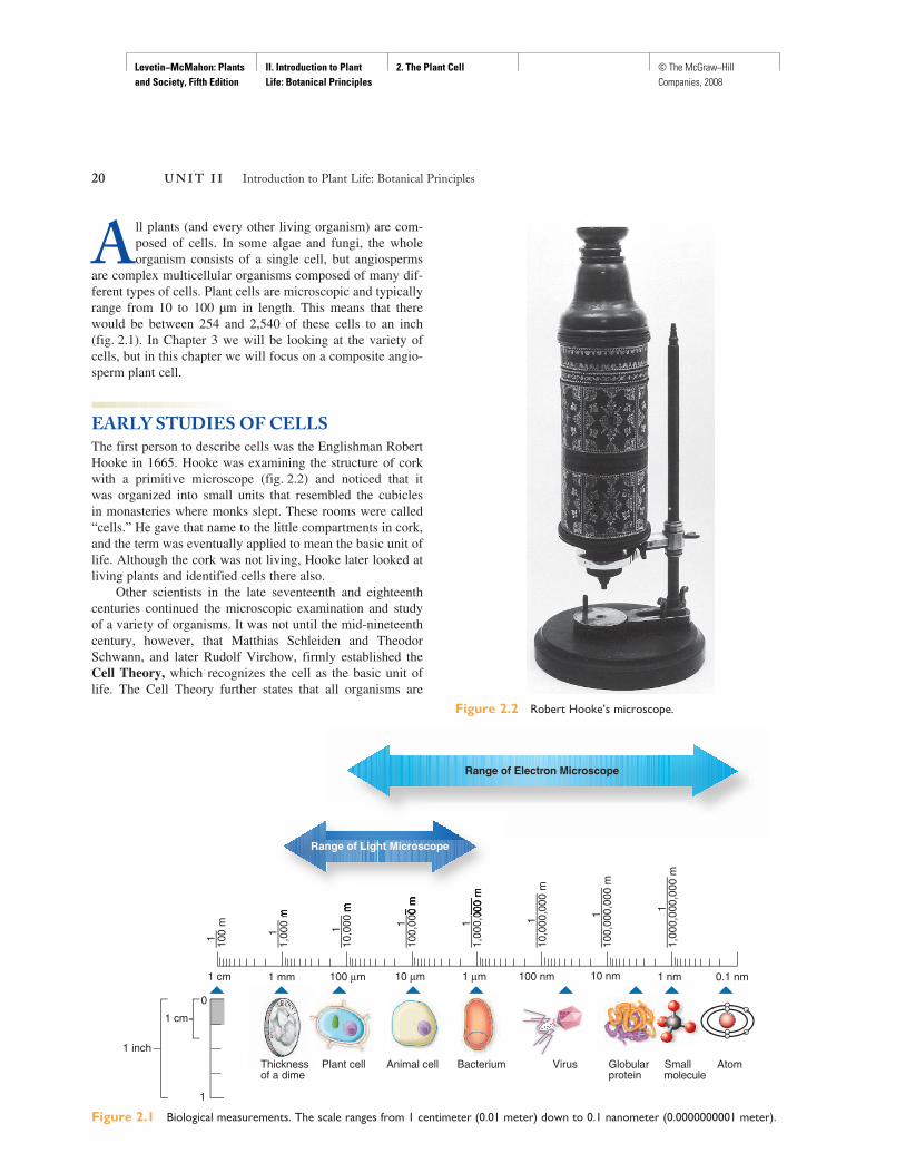
EARLY STUDIES OF CELLS The first person to describe cells was the Englishman Robert Hooke in 1665. Hooke was examining the structure of cork with a primitive microscope ( fig. 2.2 ) and noticed that it was organized into small units that resembled the cubicles in monasteries where monks slept. These rooms were called “cells.” He gave that name to the little compartments in cork, and the term was eventually applied to mean the basic unit of life. Although the cork was not living, Hooke later looked at living plants and identified cells there also.
Other scientists in the late seventeenth and eighteenth centuries continued the microscopic examination and study of a variety of organisms. It was not until the mid-nineteenth century, however, that Matthias Schleiden and Theodor Schwann, and later Rudolf Virchow, firmly established the Cell Theory, which recognizes the cell as the basic unit of life. The Cell Theory further states that all organisms are
Figure 2.1 Biological measurements. The scale ranges from 1 centimeter (0.01 meter) down to 0.1 nanometer (0.0000000001 meter).
1
——
——
—–
100,
000,
000
m
1—
——
——
10,0
00,0
00 m
1—
——
—–
1,00
0,00
0 m
1—
——
–10
0,00
0 m
1
——
—10
,000
m
1 cm 1 mm 100 μm 1 μm 100 nm 10 nm 1 nm 0.1 nm10 μm
1—
——
——
—–
1,00
0,00
0,00
0 m
1
——
–1,
000
m
1 —–
100
m
Thicknessof a dime
Plant cell Animal cell Bacterium Virus Globularprotein
Smallmolecule
Atom
1 inch
1 cm
0
1
Range of Light Microscope
Range of Electron Microscope
Figure 2.2 Robert Hooke’s microscope.
Levetin−McMahon: Plants and Society, Fifth Edition
II. Introduction to Plant Life: Botanical Principles
2. The Plant Cell © The McGraw−Hill Companies, 2008
C H A P T E R 2 The Plant Cell 21
composed of cells and all cells arise from preexisting cells. This theory is one of the major principles in biology.
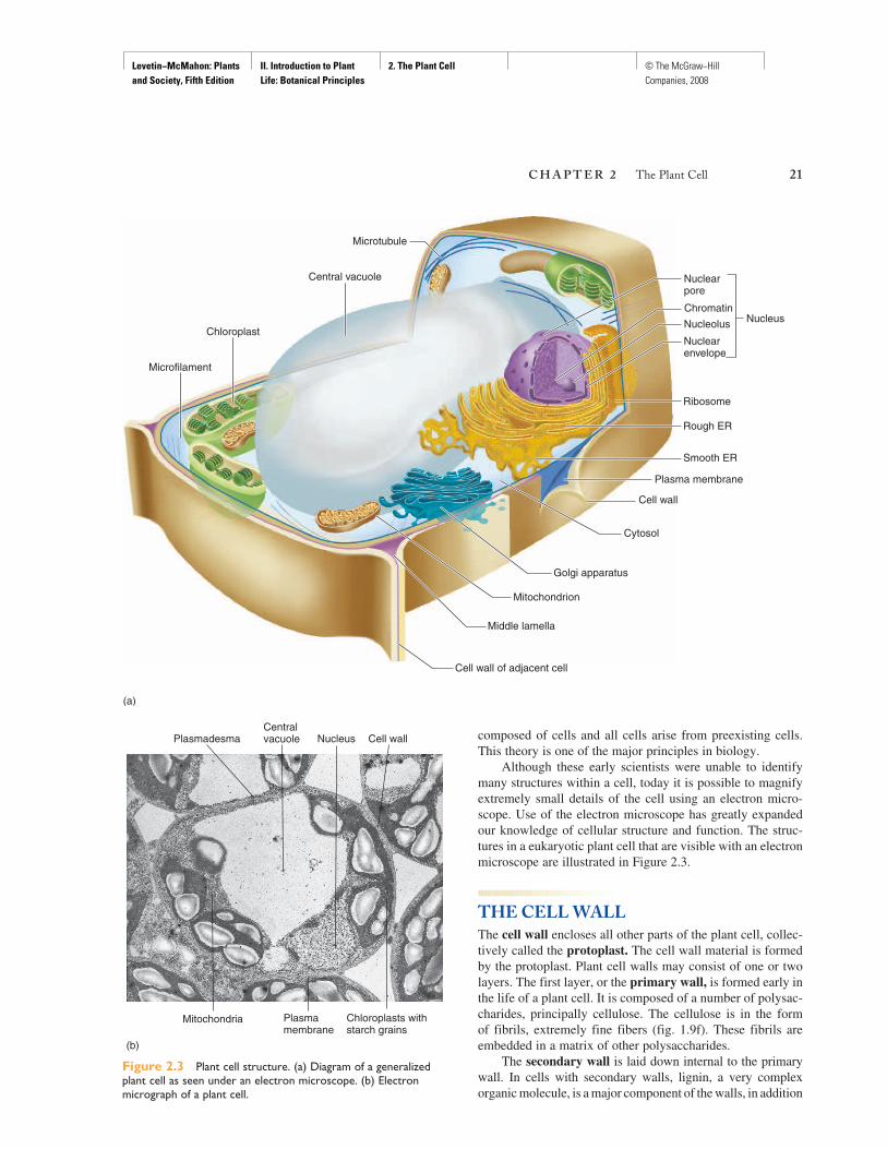
Although these early scientists were unable to identify many structures within a cell, today it is possible to magnify extremely small details of the cell using an electron micro-scope. Use of the electron microscope has greatly expanded our knowledge of cellular structure and function. The struc-tures in a eukaryotic plant cell that are visible with an electron microscope are illustrated in Figure 2.3 .
THE CELL WALL The cell wall encloses all other parts of the plant cell, collec-tively called the protoplast. The cell wall material is formed by the protoplast. Plant cell walls may consist of one or two layers. The first layer, or the primary wall, is formed early in the life of a plant cell. It is composed of a number of polysac-charides, principally cellulose. The cellulose is in the form of fibrils, extremely fine fibers (fig. 1.9f). These fibrils are embedded in a matrix of other polysaccharides.
The secondary wall is laid down internal to the primary wall. In cells with secondary walls, lignin, a very complex organic molecule, is a major component of the walls, in addition
Chromatin
Nucleolus
Nuclearpore
Nuclearenvelope
Rough ER
Smooth ER
Central vacuole
Cytosol
Golgi apparatus
Plasma membrane
Microtubule
Ribosome
Mitochondrion
Microfilament
Cell wall
Middle lamella
Chloroplast
Cell wall of adjacent cell
Nucleus
(a)
Figure 2.3 Plant cell structure. (a) Diagram of a generalized plant cell as seen under an electron microscope. (b) Electron micrograph of a plant cell.
(b)
Cell wall Centralvacuole
Chloroplasts withstarch grains
Nucleus
Mitochondria
Plasmadesma
Plasmamembrane
Levetin−McMahon: Plants and Society, Fifth Edition
II. Introduction to Plant Life: Botanical Principles
2. The Plant Cell © The McGraw−Hill Companies, 2008
22 U N I T I I Introduction to Plant Life: Botanical Principles
to the cellulose and other polysaccharides. Considering all the plant material on Earth, it is not surprising that cellulose is the most abundant organic compound, with lignin a close second.
Only certain types of plant cells have secondary walls, usually just those specialized for support, protection, or water conduction. Lignin is known for its toughness; it gives wood its characteristic strength and also provides protection against attack by pathogens (disease-causing agents) and consump-tion by herbivores (although certain species of wood-rotting fungi have the ability to break down lignin—see A Closer Look 23.2—Dry Rot and Other Wood Decay Fungi). To compare the characteristics of primary and secondary walls, imagine a chair made of lettuce leaves instead of wood!
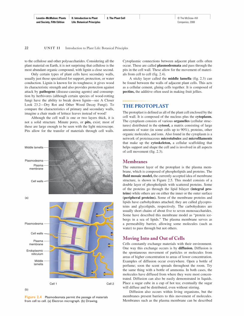
Although the cell wall is one or two layers thick, it is not a solid structure. Minute pores, or pits, exist; most of these are large enough to be seen with the light microscope. Pits allow for the transfer of materials through cell walls.
Cytoplasmic connections between adjacent plant cells often occur. These are called plasmodesmata and pass through the pits in the cell wall. These allow for the movement of materi-als from cell to cell ( fig. 2.4 ).
A sticky layer called the middle lamella ( fig. 2.3 ) can be found between the walls of adjacent plant cells. This acts as a cellular cement, gluing cells together. It is composed of pectins, the additive often used in making fruit jellies.
THE PROTOPLAST The protoplast is defined as all of the plant cell enclosed by the cell wall. It is composed of the nucleus plus the cytoplasm.The cytoplasm consists of various organelles (cellular struc-tures) distributed in the cytosol, a matrix consisting of large amounts of water (in some cells up to 90%), proteins, other organic molecules, and ions. Also found in the cytoplasm is a network of proteinaceous microtubules and microfilamentsthat make up the cytoskeleton, a cellular scaffolding that helps support and shape the cell and is involved in all aspects of cell movement ( fig. 2.3 ).
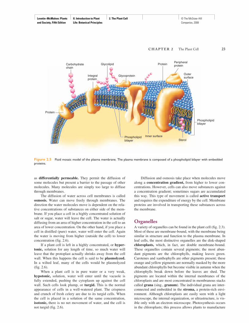
MembranesThe outermost layer of the protoplast is the plasma mem-brane, which is composed of phospholipids and proteins. The fluid mosaic model, the currently accepted idea of membrane structure, is shown in Figure 2.5 . This model consists of a double layer of phospholipids with scattered proteins. Some of the proteins go through the lipid bilayer (integral pro-teins) while others are on either the inner or the outer surface (peripheral proteins). Some of the membrane proteins and lipids have carbohydrates attached; they are called glycopro-teins and glycolipids, respectively. The carbohydrates are usually short chains of about five to seven monosaccharides. Some have described this membrane model as “protein ice-bergs in a sea of lipids.” The plasma membrane serves as a permeability barrier, allowing some molecules (such as water) to pass through but not others.
Moving Into and Out of Cells Cells constantly exchange materials with their environment. One way this exchange occurs is by diffusion. Diffusion is the spontaneous movement of particles or molecules from areas of higher concentration to areas of lower concentration. Examples of diffusion occur everywhere. Open a bottle of perfume; soon the scent spreads throughout the room. Try the same thing with a bottle of ammonia. In both cases, the molecules have diffused from where they were most concen-trated. Diffusion can also be easily demonstrated in liquids. Place a sugar cube in a cup of hot tea; eventually the sugar will diffuse and be distributed, even without stirring.
Diffusion also occurs within living organisms, but the membranes present barriers to this movement of molecules. Membranes such as the plasma membrane can be described
(a)
Middle lamella
Plasmodesma
Plasmamembrane
Cell walls
Figure 2.4 Plasmodesmata permit the passage of materials from cell to cell. (a) Electron micrograph. (b) Drawing.
Cell 1 Cell 2
(b)
Plasmodesma
Cell walls
Plasmamembrane
Endoplasmicreticulum
Middlelamella
Levetin−McMahon: Plants and Society, Fifth Edition
II. Introduction to Plant Life: Botanical Principles
2. The Plant Cell © The McGraw−Hill Companies, 2008
C H A P T E R 2 The Plant Cell 23
as differentially permeable. They permit the diffusion of some molecules but present a barrier to the passage of other molecules. Many molecules are simply too large to diffuse through membranes.
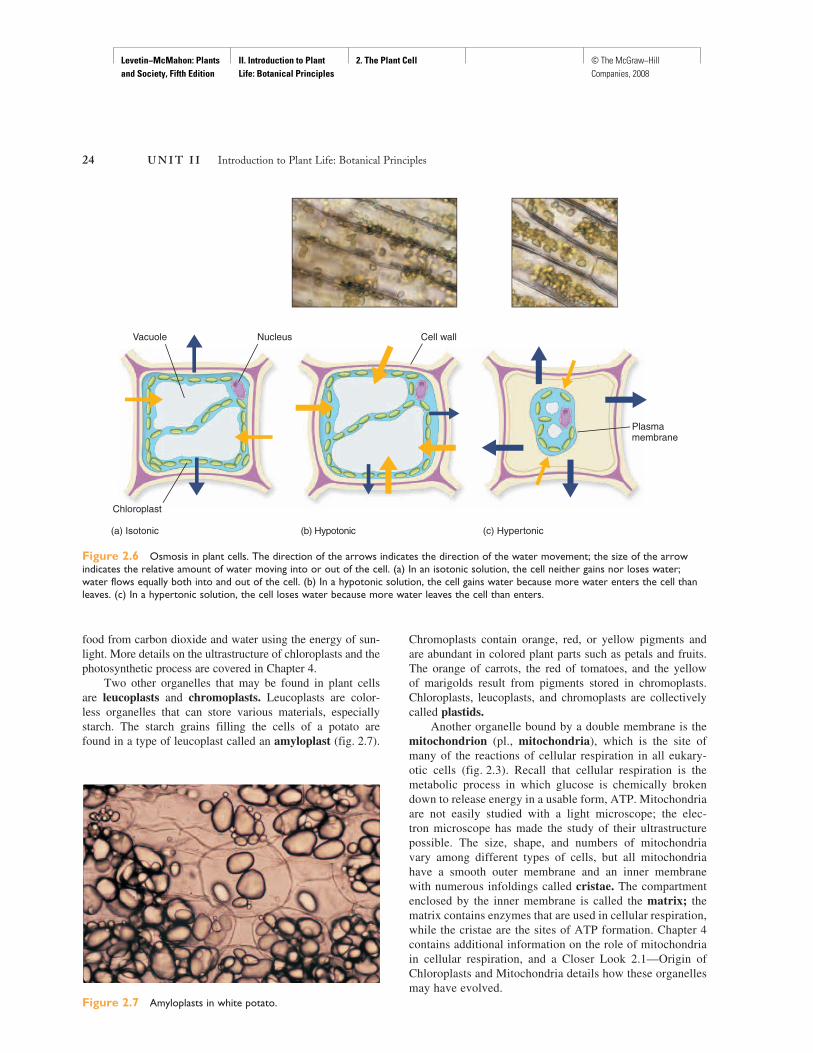
The diffusion of water across cell membranes is called osmosis. Water can move freely through membranes. The direction the water molecules move is dependent on the rela-tive concentrations of substances on either side of the mem-brane. If you place a cell in a highly concentrated solution of salt or sugar, water will leave the cell. The water is actually diffusing from an area of higher concentration in the cell to an area of lower concentration. On the other hand, if you place a cell in distilled (pure) water, water will enter the cell. Again the water is moving from higher (outside the cell) to lower concentration ( fig. 2.6 ).
If a plant cell is left in a highly concentrated, or hyper-tonic, solution for any length of time, so much water will leave that the protoplast actually shrinks away from the cell wall. When this happens the cell is said to be plasmolyzed.In a wilted leaf, many of the cells would be plasmolyzed ( fig. 2.6 ).
When a plant cell is in pure water or a very weak, hypotonic, solution, water will enter until the vacuole is fully extended, pushing the cytoplasm up against the cell wall. Such cells look plump, or turgid. This is the normal appearance of cells in a well-watered plant. The crispness and crunch of fresh celery are due to its turgid cells. When the cell is placed in a solution of the same concentration, isotonic, there is no net movement of water, and the cell is not turgid ( fig. 2.6 ).
Diffusion and osmosis take place when molecules move along a concentration gradient, from higher to lower con-centrations. However, cells can also move substances against a concentration gradient; sometimes sugars are accumulated this way. This type of movement is called active transportand requires the expenditure of energy by the cell. Membrane proteins are involved in transporting these substances across the membrane.
OrganellesA variety of organelles can be found in the plant cell ( fig. 2.3 ). Most of these are membrane-bound, with the membrane being similar in structure and function to the plasma membrane. In leaf cells, the most distinctive organelles are the disk-shaped chloroplasts, which, in fact, are double membrane-bound. These organelles contain several pigments; the most abun-dant pigments are the chlorophylls, making leaves green. Carotenes and xanthophylls are other pigments present; these orange and yellow pigments are normally masked by the more abundant chlorophylls but become visible in autumn when the chlorophylls break down before the leaves are shed. The pigments are located within the internal membranes of the chloroplasts and are most concentrated in membranous stacks called grana (sing., granum). The individual grana are inter-connected and embedded in the stroma, a protein-rich envi-ronment. Although chloroplasts are easily seen with a light microscope, the internal organization, or ultrastructure, is vis-ible only with an electron microscope. Photosynthesis occurs in the chloroplasts; this process allows plants to manufacture
Figure 2.5 Fluid mosaic model of the plasma membrane. The plasma membrane is composed of a phospholipid bilayer with embedded proteins.
Carbohydratechain
Integralprotein
Peripheralprotein
Outersurface
Phospholipidbilayer
Phospholipidbilayer
Protein
Glycoprotein
Glycolipid
Sterol
Protein
lnner surface
Levetin−McMahon: Plants and Society, Fifth Edition
II. Introduction to Plant Life: Botanical Principles
2. The Plant Cell © The McGraw−Hill Companies, 2008
24 U N I T I I Introduction to Plant Life: Botanical Principles
food from carbon dioxide and water using the energy of sun-light. More details on the ultrastructure of chloroplasts and the photosynthetic process are covered in Chapter 4.
Two other organelles that may be found in plant cells are leucoplasts and chromoplasts. Leucoplasts are color-less organelles that can store various materials, especially starch. The starch grains filling the cells of a potato are found in a type of leucoplast called an amyloplast ( fig. 2.7 ).
Chromoplasts contain orange, red, or yellow pigments and are abundant in colored plant parts such as petals and fruits. The orange of carrots, the red of tomatoes, and the yellow of marigolds result from pigments stored in chromoplasts. Chloroplasts, leucoplasts, and chromoplasts are collectively called plastids.
Another organelle bound by a double membrane is the mitochondrion (pl., mitochondria), which is the site of many of the reactions of cellular respiration in all eukary-otic cells ( fig. 2.3 ). Recall that cellular respiration is the metabolic process in which glucose is chemically broken down to release energy in a usable form, ATP. Mitochondria are not easily studied with a light microscope; the elec-tron microscope has made the study of their ultrastructure possible. The size, shape, and numbers of mitochondria vary among different types of cells, but all mitochondria have a smooth outer membrane and an inner membrane with numerous infoldings called cristae. The compartment enclosed by the inner membrane is called the matrix; the matrix contains enzymes that are used in cellular respiration, while the cristae are the sites of ATP formation. Chapter 4 contains additional information on the role of mitochondria in cellular respiration, and a Closer Look 2.1—Origin of Chloroplasts and Mitochondria details how these organelles may have evolved.
Figure 2.6 Osmosis in plant cells. The direction of the arrows indicates the direction of the water movement; the size of the arrow indicates the relative amount of water moving into or out of the cell. (a) In an isotonic solution, the cell neither gains nor loses water; water flows equally both into and out of the cell. (b) In a hypotonic solution, the cell gains water because more water enters the cell than leaves. (c) In a hypertonic solution, the cell loses water because more water leaves the cell than enters.
Nucleus
Chloroplast
Vacuole Cell wall
Plasmamembrane
(a) Isotonic (b) Hypotonic (c) Hypertonic
Figure 2.7 Amyloplasts in white potato.
Levetin−McMahon: Plants and Society, Fifth Edition
II. Introduction to Plant Life: Botanical Principles
2. The Plant Cell © The McGraw−Hill Companies, 2008
25
A CLOSER LOOK 2.1
As stated in Chapter 1, prokaryotes were the first organisms on Earth. Evidence indicates that prokaryotes first appeared approximately 3.5 billion years ago whereas eukaryotes appeared only around 1.5 billion years ago. One question that has intrigued biologists for many years is, How did the eukaryotic cell evolve? Dr. Lynn Margulis of the University of Massachusetts is one of the main proponents of a possible answer to this question, the Endosymbiont Theory. This theory states that the organelles of eukaryotic cells are the descendants of once free-living prokaryotes that took up residence in a larger cell, establishing a symbiotic relation-ship (symbiosis: two or more organisms living together). This association evolved into the well-studied eukaryotic cell.
Chloroplasts and mitochondria provide the best examples of this theory. Both organelles resemble free-living prokary-otes. In fact, as long ago as the 1880s some biologists observed that chloroplasts of eukaryotic cells resembled cyanobacteria (then called blue-green algae). Both chloroplasts and mito-chondria have structures that are associated with free-living cells. For example, they contain both DNA and ribosomes, which are bacterial in size and nature, allowing them to syn-thesize some of their own proteins. Both chloroplasts and mitochondria can divide to produce new chloroplasts and mitochondria in a manner very similar to prokaryotic cell division. The inner membranes of both organelles closely resemble the plasma membrane of prokaryotes. These fea-tures, as well as additional biochemical similarities, provide support for the validity of the Endosymbiont Theory.
Recent research has discovered certain bacteria that appear to be in the process of evolving into organelles as predicted by the Endosymbiont Theory. Approximately 10% of insect species house bacterial endosymbionts. Some of the best studied are bacteria which live inside specialized gut cells of sap-sucking pests. The sugary sap of plants is deficient in amino acids, and apparently the bacterial endosymbionts produce needed amino acids and other essential nutrients for their insect hosts. In return, bacterial endosymbionts have been passed from generation to generation in insect hosts for over hundreds of millions of years. During this time, the bacterial endosymbionts have lost most of the genes that are necessary for bacteria to be self-sufficient. They no longer possess the genes to make the outer plasma membrane, to metabolize lipids and nucleotides, to transport materials into a cell, or for cell division. There is evidence that some of these bacterial genes may have been trans-ferred to the nucleus of the host cell that now supports the endosymbiont.
Carsonella ruddii, an endosymbiont found in the gut cells of psyllids, a type of agricultural pest also known as jumping plant lice, has the smallest genome known for any bacterium with only 160,000 base pairs of DNA. Its genome size is similar to that of the mitochondria (<600,000 base pairs) and chloroplasts (220,000 base pairs) found in terrestrial plants. Perhaps this endosymbiont will one day evolve into an organelle.
Origin of Chloroplasts and Mitochondria
Most mature plant cells ( fig. 2.3 ) are characterized by a large central vacuole that is separated from the rest of the cytoplasm by its own membrane. In some cells, the vacuole takes up 90% of the cell volume, pushing the cytoplasm into a thin layer against the plasma membrane. The vacuole contains the cell sap, a watery solution of sugars, salts, amino acids, proteins, and crystals, all separated from the cytoplasm by the vacuolar membrane. The cell sap is often acidic; the tartness of lemons and limes is due to their very acidic cell sap. Some of the substances in the vacuole are waste prod-ucts; others can be drawn upon when needed by the cell. The concentrations of these materials in the vacuole may become so great that they precipitate out as crystals. The leaves of the common house plant dumb cane ( Dieffenbachia spp.)are poisonous because of the presence of large amounts of calcium oxalate crystals (see Chapter 21). If consumed, the crystals can injure the tissues of the mouth and throat, caus-ing a temporary inability to speak—hence the common name
dumb cane. Pigments can also be found in the vacuole; these are called anthocyanins and are responsible for deep red, blue, and purple colors of many plant organs, such as red onions and red cabbage. Unlike the pigments of the chloroplasts and chromoplasts, the anthocyanins are water soluble and are dis-tributed uniformly in the cell sap.
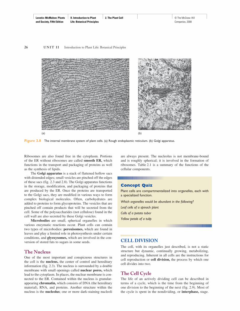
An internal membrane system also occurs in plant cells ( fig. 2.3 and 2.8 ). This consists of the endoplasmic reticu-lum (ER), the Golgi apparatus, and microbodies. Thesestructures are all involved in the synthesizing, packaging, and transporting of materials within the cell. The ER is a network of membranous channels throughout the cytoplasm. In some places the cytoplasmic side of the ER is studded with minute bodies called ribosomes. Ribosomes, composed of RNA and protein, are not membrane-bound and are the sites of protein synthesis. Portions of the ER with ribosomes attached are referred to as rough ER. Owing to the pres-ence of ribosomes, rough ER is active in protein synthesis.
Levetin−McMahon: Plants and Society, Fifth Edition
II. Introduction to Plant Life: Botanical Principles
2. The Plant Cell © The McGraw−Hill Companies, 2008
26 U N I T I I Introduction to Plant Life: Botanical Principles
Ribosomes are also found free in the cytoplasm. Portions of the ER without ribosomes are called smooth ER, whichfunctions in the transport and packaging of proteins as well as the synthesis of lipids.
The Golgi apparatus is a stack of flattened hollow sacs with distended edges; small vesicles are pinched off the edges of these sacs ( fig. 2.3 and 2.8 ). The Golgi apparatus functions in the storage, modification, and packaging of proteins that are produced by the ER. Once the proteins are transported to the Golgi sacs, they are modified in various ways to form complex biological molecules. Often, carbohydrates are added to proteins to form glycoproteins. The vesicles that are pinched off contain products that will be secreted from the cell. Some of the polysaccharides (not cellulose) found in the cell wall are also secreted by these Golgi vesicles.
Microbodies are small, spherical organelles in which various enzymatic reactions occur. Plant cells can contain two types of microbodies: peroxisomes, which are found in leaves and play a limited role in photosynthesis under certain conditions, and glyoxysomes, which are involved in the con-version of stored fats to sugars in some seeds.
The Nucleus One of the most important and conspicuous structures in the cell is the nucleus, the center of control and hereditary information ( fig. 2.3 ). The nucleus is surrounded by a double membrane with small openings called nuclear pores, whichlead to the cytoplasm. In places, the nuclear membrane is con-nected to the ER. Contained within the nucleus is granular-appearing chromatin, which consists of DNA (the hereditary material), RNA, and proteins. Another structure within the nucleus is the nucleolus; one or more dark-staining nucleoli
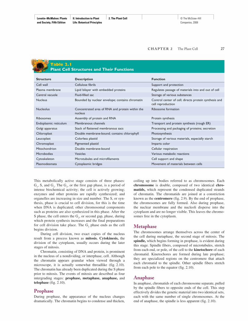
are always present. The nucleolus is not membrane-bound and is roughly spherical; it is involved in the formation of ribosomes. Table 2.1 is a summary of the functions of the cellular components.
Concept QuizPlant cells are compartmentalized into organelles, each with a specialized function.
Which organelles would be abundant in the following?
Leaf cells of a spinach plant
Cells of a potato tuber
Yellow petals of a tulip
CELL DIVISION The cell, with its organelles just described, is not a static structure but dynamic, continually growing, metabolizing, and reproducing. Inherent in all cells are the instructions for cell reproduction or cell division, the process by which one cell divides into two.
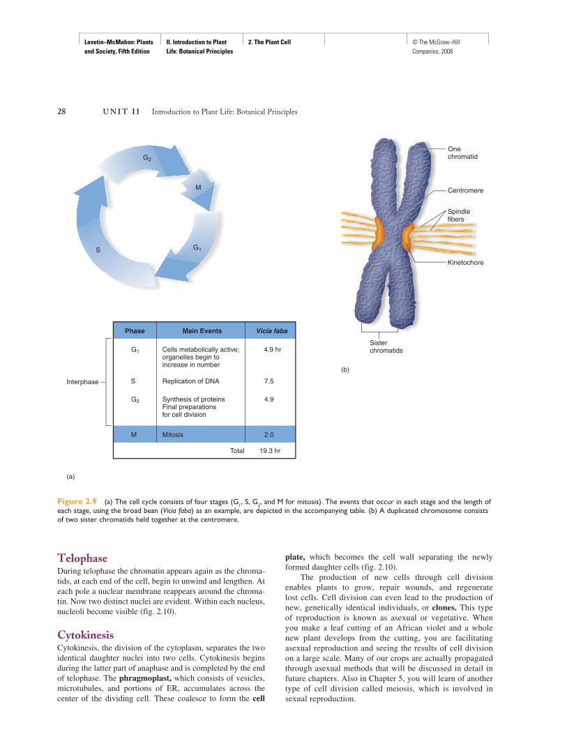
The Cell Cycle The life of an actively dividing cell can be described in terms of a cycle, which is the time from the beginning of one division to the beginning of the next ( fig. 2.9 ). Most of the cycle is spent in the nondividing, or interphase, stage.
Figure 2.8 The internal membrane system of plant cells. (a) Rough endoplasmic reticulum. (b) Golgi apparatus.
(a) (b)
Levetin−McMahon: Plants and Society, Fifth Edition
II. Introduction to Plant Life: Botanical Principles
2. The Plant Cell © The McGraw−Hill Companies, 2008
C H A P T E R 2 The Plant Cell 27
This metabolically active stage consists of three phases: G
1, S, and G
2. The G
1, or the first gap phase, is a period of
intense biochemical activity; the cell is actively growing; enzymes and other proteins are rapidly synthesized; and organelles are increasing in size and number. The S, or syn-thesis, phase is crucial to cell division, for this is the time when DNA is duplicated; other chromosomal components such as proteins are also synthesized in this phase. After the S phase, the cell enters the G
2, or second gap, phase, during
which protein synthesis increases and the final preparations for cell division take place. The G
2phase ends as the cell
begins division. During cell division, two exact copies of the nucleus
result from a process known as mitosis. Cytokinesis, thedivision of the cytoplasm, usually occurs during the later stages of mitosis.
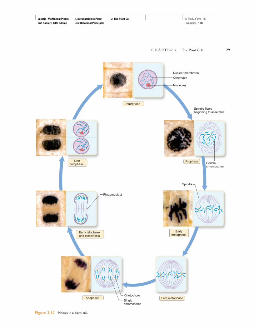
Chromatin, consisting of DNA and protein, is prominent in the nucleus of a nondividing, or interphase, cell. Although the chromatin appears granular when viewed through a microscope, it is actually somewhat threadlike (fig. 2.10). The chromatin has already been duplicated during the S phase prior to mitosis. The events of mitosis are described as four intergrading stages: prophase, metaphase, anaphase, andtelophase (fig. 2.10).
ProphaseDuring prophase, the appearance of the nucleus changes dramatically. The chromatin begins to condense and thicken,
coiling up into bodies referred to as chromosomes. Each chromosome is double, composed of two identical chro-matids, which represent the condensed duplicated strands of chromatin. The chromatids are joined at a constriction known as the centromere ( fig. 2.9 ). By the end of prophase, the chromosomes are fully formed. Also during prophase, the nuclear membrane and the nucleoli disperse into the cytoplasm and are no longer visible. This leaves the chromo-somes free in the cytoplasm.
MetaphaseThe chromosomes arrange themselves across the center of the cell during metaphase, the second stage of mitosis. The spindle, which begins forming in prophase, is evident during this stage. Spindle fibers, composed of microtubules, stretch from each end, or pole, of the cell to the kinetochore of each chromatid. Kinetochores are formed during late prophase; they are specialized regions on the centromere that attach each chromatid to the spindle. Other spindle fibers stretch from each pole to the equator (fig. 2.10).
AnaphaseIn anaphase, chromatids of each chromosome separate, pulled by the spindle fibers to opposite ends of the cell. This step effectively divides the genetic material into two identical sets, each with the same number of single chromosomes. At the end of anaphase, the spindle is less apparent (fig. 2.10).
Structure Description Function
Cell wall Cellulose fibrils Support and protection
Plasma membrane Lipid bilayer with embedded proteins Regulates passage of materials into and out of cell
Central vacuole Fluid-filled sac Storage of various substances
Nucleus Bounded by nuclear envelope; contains chromatin Control center of cell; directs protein synthesis and cell reproduction
Nucleolus Concentrated area of RNA and protein within the nucleus
Ribosome formation
Ribosomes Assembly of protein and RNA Protein synthesis
Endoplasmic reticulum Membranous channels Transport and protein synthesis (rough ER)
Golgi apparatus Stack of flattened membranous sacs Processing and packaging of proteins; secretion
Chloroplast Double membrane-bound; contains chlorophyll Photosynthesis
Leucoplast Colorless plastid Storage of various materials, especially starch
Chromoplast Pigmented plastid Imparts color
Mitochondrion Double membrane-bound Cellular respiration
Microbodies Vesicles Various metabolic reactions
Cytoskeleton Microtubules and microfilaments Cell support and shape
Plasmodesmata Cytoplasmic bridges Movement of materials between cells
Table 2.1Plant Cell Structures and Their Functions
Levetin−McMahon: Plants and Society, Fifth Edition
II. Introduction to Plant Life: Botanical Principles
2. The Plant Cell © The McGraw−Hill Companies, 2008
28 U N I T I I Introduction to Plant Life: Botanical Principles
TelophaseDuring telophase the chromatin appears again as the chroma-tids, at each end of the cell, begin to unwind and lengthen. At each pole a nuclear membrane reappears around the chroma-tin. Now two distinct nuclei are evident. Within each nucleus, nucleoli become visible (fig. 2.10).
CytokinesisCytokinesis, the division of the cytoplasm, separates the two identical daughter nuclei into two cells. Cytokinesis begins during the latter part of anaphase and is completed by the end of telophase. The phragmoplast, which consists of vesicles, microtubules, and portions of ER, accumulates across the center of the dividing cell. These coalesce to form the cell
plate, which becomes the cell wall separating the newly formed daughter cells (fig. 2.10).
The production of new cells through cell division enables plants to grow, repair wounds, and regenerate lost cells. Cell division can even lead to the production of new, genetically identical individuals, or clones. This type of reproduction is known as asexual or vegetative. When you make a leaf cutting of an African violet and a whole new plant develops from the cutting, you are facilitating asexual reproduction and seeing the results of cell division on a large scale. Many of our crops are actually propagated through asexual methods that will be discussed in detail in future chapters. Also in Chapter 5, you will learn of another type of cell division called meiosis, which is involved in sexual reproduction.
S
(a)
(b)
M
G2
G1
Kinetochore
Sisterchromatids
Spindlefibers
Centromere
Onechromatid
S
G2
G1
M
Phase Main Events Vicia faba
Cells metabolically active;organelles begin to increase in number
Replication of DNA
Synthesis of proteinsFinal preparationsfor cell division
4.9 hr
7.5
4.9
2.0
Total 19.3 hr
Mitosis
Interphase
Figure 2.9 (a) The cell cycle consists of four stages (G1, S, G2, and M for mitosis). The events that occur in each stage and the length of each stage, using the broad bean (Vicia faba) as an example, are depicted in the accompanying table. (b) A duplicated chromosome consists of two sister chromatids held together at the centromere.
Levetin−McMahon: Plants and Society, Fifth Edition
II. Introduction to Plant Life: Botanical Principles
2. The Plant Cell © The McGraw−Hill Companies, 2008
C H A P T E R 2 The Plant Cell 29
Figure 2.10 Mitosis in a plant cell.
Late metaphase
Latetelophase
Earlymetaphase
Spindle
Interphase
Anaphase
Prophase
Spindle fibersbeginning to assemble
Doublechromosome
Kinetochore
Singlechromosome
Phragmoplast
Early telophaseand cytokinesis
Nuclear membrane
Chromatin
Nucleolus
Levetin−McMahon: Plants and Society, Fifth Edition
II. Introduction to Plant Life: Botanical Principles
2. The Plant Cell © The McGraw−Hill Companies, 2008
30 U N I T I I Introduction to Plant Life: Botanical Principles
Concept QuizCloning based on cell division has become a major issue for discussions since researchers have cloned sheep, cattle, pigs, goats, cats, and other animals.
Is there a biological difference between cloning plants and animals? an ethical difference?
CHAPTER SUMMARY 1. All life on Earth, including plant life, has a cellular orga-
nization. The plant cell shares many characteristics in common with other eukaryotic cells. The plant protoplast includes the cytoplasm with the embedded organelles and nucleus. Within the nucleus is DNA, the genetic blueprint of all cells.
2. The plasma membrane, composed of phospholipids and proteins according to the fluid mosaic model, regulates the passage of materials into and out of the cell. Numerous mitochondria can be found within the cytoplasm; they are the sites of cellular respiration. The endoplasmic reticu-lum, Golgi apparatus, and microbodies make up an inter-nal membrane system that functions in the synthesizing, packaging, and transporting of materials.
3. Some features of a plant cell are unique. The primary cell wall containing cellulose surrounds a plant protoplast pro-viding protection and support. In certain specialized plant cells, a secondary cell wall, impregnated with the tough-ening agent lignin, imparts extra strength. Chloroplasts are the site for photosynthesis; they are one of several types of plastids. Other plastids are the food-storing leucoplasts and the pigment-containing chromoplasts. A large central vacuole may take up approximately 90% of the mature plant cell and act as a storage site for many substances.
4. The life of a cell can be described in terms of a cycle. Most cells spend the majority of the time in interphase, a nondividing stage. But at certain times in its life, a cell may undergo division whereby one cell divides into two. Mitosis is the duplication of the nucleus into two exact cop-ies. There are four intergrading stages in mitosis: prophase, metaphase, anaphase, and telophase. The division process is complete when, in the process of cytokinesis the cyto-plasm is split, and two identical daughter cells are formed.
REVIEW QUESTIONS 1. What is the significance of the Cell Theory to biology?
2. List the parts of a plant cell, and for each part describe its structure and function.
3. Describe the events occurring during the G 1, S, and G
2
stages of interphase.
4. Describe the stages of mitosis.
5. Describe the similarities and differences between chloro-plasts and mitochondria.
6. Differentiate between osmosis and diffusion.
7. Cancer cells are abnormal cells undergoing repeated cell divisions. Vincristine is a drug obtained from the Madagascar periwinkle that has been highly effective in treating certain cancers. Vincristine disrupts microtu-bules, preventing spindle formation. Explain the success of vincristine on the cellular level.
FURTHER READING Goldberg, Alfred L., Stephen J. Elledge, and J. Wade
Harper. 2001. The Cellular Chamber of Doom. ScientificAmerican 284(1): 68–73.
Mauseth, James D. 2003. Botany: An Introduction to Plant Biology, 3rd Edition. Jones and Bartlett, Sudbury, MA.
Netting, Jessica. 2001. A More Perfect Union. Science News159(20): 314–316.
Scott, John D., and Tony Pawson. 2000. Cell Communication: The Inside Story. Scientific American 282(6): 72–80.
Uno, Gordon, Richard Storey, and Randy Moore. 2001. Principles of Botany. McGraw-Hill, New York.
Wallace, Douglas C. 1997. Mitochondrial DNA in Aging and Disease. Scientific American 277(2): 40–47.
Wilmut, Ian. 1998. Cloning for Medicine. Scientific American279(6): 58–64.
ONLINE LEARNING CENTER
Visit www.mhhe.com/levetin5e for online quizzing, web links to chapter-related material, and more!


![Monitoring Polysaccharide Dynamics in the Plant Cell Wall1[OPEN] · Update on Plant Cell Wall Dynamics Monitoring Polysaccharide Dynamics in the Plant Cell Wall1[OPEN] Catalin Voiniciuc,](https://img.pdfslide.net/doc/110x75/5e57dc422f31c166d63f94c4/monitoring-polysaccharide-dynamics-in-the-plant-cell-wall1open-update-on-plant.jpg)