Embed Size (px)
Citation preview

The Post Kala-azar Dermal Leishmaniasis (PKDL) AtlasA Manual for Health Workers

WHO Library Cataloguing-in-Publication Data
The Post Kala-azar Dermal Leishmaniasis (PKDL) atlas: a manual for health workers.
1. Leishmaniasis, Visceral - diagnosis. 2. Leishmaniasis, Cutaneous – diagnosis. 3. Health personnel – education. 4. Handbooks. I. World Health Organization.
ISBN 978 92 4 150410 2 (NLM classification: WC 715)
WHO/HTM/NTD/IDM/2012.4
© World Health Organization 2012
All rights reserved. Publications of the World Health Organization are available on the WHO web site (www.who.int) or can be purchased from WHO Press, World Health Organization, 20 Avenue Appia, 1211 Geneva 27, Switzerland (tel.: +41 22 791 3264; fax: +41 22 791 4857; e-mail: [email protected]). Requests for permission to reproduce or translate WHO publications – whether for sale or for noncommercial distribution – should be addressed to WHO Press through the WHO web site (http://www.who.int/about/licensing/copyright_form/en/index.html).The designations employed and the presentation of the material in this publication do not imply the expression of any opinion whatsoever on the part of the World Health Organization concerning the legal status of any country, territory, city or area or of its authorities, or concerning the delimitation of its frontiers or boundaries. Dotted lines on maps represent approximate border lines for which there may not yet be full agreement.
The mention of specific companies or of certain manufacturers’ products does not imply that they are endorsed or recommended by the World Health Organization in preference to others of a similar nature that are not mentioned. Errors and omissions excepted, the names of proprietary products are distinguished by initial capital letters.
All reasonable precautions have been taken by the World Health Organization to verify the information contained in this publication. However, the published material is being distributed without warranty of any kind, either expressed or implied. The responsibility for the interpretation and use of the material lies with the reader. In no event shall the World Health Organization be liable for damages arising from its use.
Printed in Spain Printed by: BSDDesign: José Mª Ropero

The Post Kala-azar Dermal Leishmaniasis (PKDL) AtlasA Manual for Health Workers
Eduard E. ZijlstraRotterdam Centre for Tropical Medicine
Jorge AlvarWHO/NTD/IDM-Leishmaniasis Programme

5
Tabl
e of
con
tent
s
Table of contents
Preface
1. Introduction [pag. 9]
2. PKDL in Africa: clinical presentation and differential diagnosis [pag. 13] a. PKDL in Sudan [pag. 17] • Macular PKDL [pag. 17] • Macular PKDL and differential diagnosis [pag. 21] • Papular and nodular PKDL [pag. 33] • PKDL grading system [pag. 54] • Severe PKDL [pag. 68] • Differential diagnosis of papular and nodular PKDL [pag. 80] • Chronic PKDL [pag. 106] • Other post-kala-azar manifestations [pag. 109] • Evolution [pag. 113] b. PKDL in Ethiopia [pag. 117] •Various examples [pag. 117] • Differential diagnosis [pag. 123]
3. PKDL in Asia: clinical presentation and differential diagnosis [pag. 127] a. PKDL in India: hospital-based experience [pag. 130] b. PKDL in Bangladesh: community-based experience [pag. 148]
4. PKDL in other areas [pag. 171]
a. PKDL in China [pag. 173] b. PKDL in Brazil [pag. 174]
5. PKDL in immunocompromised patients and other skin manifestations of Leishmania in HIV-positive patients [pag. 175]
6. Other forms of leishmaniasis that resemble PKDL or that may be found in the same endemic area [pag. 187]
7. Literature [pag. 207]
8. Acknowledgements/List of contributors [pag. 211]


Pref
ace
Preface
Post Kala-azar dermal leishmaniasis (PKDL) is a well-recognized compli-cation of visceral leishmaniasis (VL) or kala-azar. It has been described since the beginning of the 20th century both in Asia and Africa, in areas where Leishmania donovani is the causative parasite. Its potential role in the transmission of kala-azar in particular in the interepidemic periods has been suggested many years ago and this was supported by feeding expe-riments in sandflies. Yet, PKDL has been neglected both from a clinical and an epidemiological point of view. This is partly caused by the difficulty in recognizing PKDL and making a firm diagnosis. The clinical spectrum varies and the list of differential diagnoses is extensive. We believe a reasonable diagnosis of PKDL can be made on clinical grounds only on the basis of a good clinical assessment by which differential diagnoses can be excluded.
This manual aims to be a guide to better and earlier recognition of PKDL by those who work in the field in remote areas. It may also be of use in the teaching of health workers at all levels. .
Geneva, August 2012
NoteWhile the diagnosis of PKDL and the conditions shown in the differential diagnosis was often confirmed, in others it is a clinical diagnosis based on experience. Despite this, we feel that this atlas reflects current clinical practice. There is a great need for further studies to deve-lop and to evaluate a clinical algorithm for PKDL and to develop simple and accurate tools that can be used under field conditions. The same would of course apply to the differential diagnosis.As PKDL is common in Sudan and has been well described, the differential diagnosis of ma-cular and papular/nodular PKDL is discussed extensively in the chapter on PKDL in Sudan. For other areas only the most common conditions encountered or conditions specific for that area are presented.
7


1. Introduction
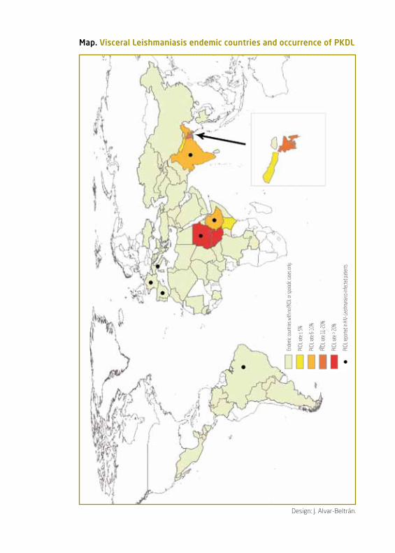
Map. Visceral Leishmaniasis endemic countries and occurrence of PKDL
Ende
mic c
ount
ries w
ith no
PKDL
or sp
oradic
case
s only
.
PKDL
rate
≤ 5%
PKDL
rate
6-10
%
PKDL
rate
11-2
0%
PKDL
rate
> 20
%
PKDL
repo
rted i
n HIV
- Leish
mania
co-in
fecte
d pat
ients
Design: J. Alvar-Beltrán.

11
1. I
ntro
duct
ion
Post-kala-azar dermal leishmaniasis (PKDL) is a complication of visceral leishmaniasis (VL) or kala-azar. It is common in areas endemic for VL cau-sed by L. donovani. These include countries in Africa in particular Sudan and in Asia, Bangladesh and to a lesser extent India. PKDL may also spora-dically occur in L. infantum or L. chagasi endemic areas, mainly the Medite-rranean countries and Latin America.
The condition is characterized by the occurrence of a skin rash after an episode of VL; the interval varies according to the endemic area. The rash is usually in the face, from which it may or may not spread to other parts of the body. In contrast to VL, the patient is not ill and PKDL is not fatal. In the Sudanese type, self cure is the rule while in Bangladesh and India, all cases are treated.
Risk factors for PKDL are not well known; previous treatment of VL with inadequate dosage of drug and the drug used, malnutrition, HIV in-fection and young age may play a role.
The importance of PKDL is twofold: • Clinical: patients develop a rash that may last for weeks or months;
in particular in small children, the rash may become generalized and severe with mucosal lesions in the mouth, causing general dis-comfort.
• Epidemiological: smears or biopsies taken from the lesions may show Leishmania parasites and there is evidence that the sandfly vector may take up these parasites while taking a blood meal and thus PKDL patients may play an important role in transmission (anthroponotic transmission). It is thought that VL occurs in cy-cles with epidemics of thousands of cases, followed by a period of seemingly low transmission. It is likely that chronic PKDL patients who harbour parasites may play an important role in subsequent upsurges in VL cases.
Diagnosis is usually clinical by the triad of the typical rash, its distribution and the previous episode of VL. There are however, often difficulties and exceptions: many patients do not have a previous episode of VL and the rash may mimic other common skin conditions. In addition, the presentation in Africa and Asia is quite different with the maculopapular form and typical spread being the most common in Sudan and the macular form being much more common in Bangladesh, often with a more atypical distribution.
Parasites may be found in the lesions but this requires a skin smear or biopsy; in papular or nodular PKDL the parasites can usually be demons-trated but in the macular type they are scanty. Serological diagnosis is not very helpful as most patients will have a previous history of VL and antibodies may persist as a result and therefore a positive test may be difficult to interpret.
ThE
PK
DL
ATLA
s. A
Man
ual f
or H
ealt
h W
orke
rs
12
Introduction
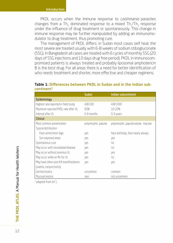
PKDL occurs when the immune response to Leishmania parasites changes from a Th2 dominated response to a mixed Th1/Th2 response under the influence of drug treatment or spontaneously. This change in immune response may be further manipulated by adding an immunomo-dulator to drug treatment, thus promoting cure.
The management of PKDL differs: in Sudan most cases self heal; the most severe are treated usually with 6-8 weeks of sodium stibogluconate (SSG). In Bangladesh all cases are treated with 6 cycles of monthly SSG (20 days of SSG injections and 10 days drug free period). PKDL in immunocom-promised patients is always treated and probably liposomal amphotericin B is the best drug. For all areas there is a need for better identification of who needs treatment and shorter, more effective and cheaper regimens.
Table 1: Differences between PKDL in sudan and in the Indian sub-continent1
sudan Indian subcontinentEpidemiologyHighest rate reported in field study 4.8/100 4.8/1000
Maximum reported PKDL rate after VL 60% 10-20%
Interval after VL 0-6 months 0-3 years
ClinicalMost common presentation polymorphic, papular polymorphic, papulonodular, macular
Typical distribution
Face-arms/chest-legs yes face and body, face nearly always
Sun-exposed areas yes yes
Spontaneous cure yes no
May occur with visceralized disease yes no
May occur without previous VL yes yes
May occur while on Rx for VL yes no
May have other post KA manifestations yes yes
(uveitis, conjunctivitis)
Genital lesions uncommon common
Mucosal lesions rare not uncommon1 adapted from ref 1.

2. PKDL in Africa: clinical presentation and differential diagnosisa. PKDL in Sudan
• Macular PKDL • Macular PKDL and differential diagnosis • Papular and nodular PKDL • PKDL grading system • Severe PKDL • Differential diagnosis of papular and nodular PKDL • Chronic PKDL • Other post-kala-azar manifestations • Evolution
b. PKDL in Ethiopia • Papular rash • Differential diagnosis


2. P
KD
L in
Afr
ica:
clin
ical
pre
sent
atio
n an
d di
ffer
enti
al d
iagn
osis
15
In Africa, PKDL by far mostly occurs in Sudan. It is much less common in Ethiopia, Kenya or Uganda. The reason for this is not clear; differences in the parasite or the genetic background of the population may be of im-portance. Up to 50-60% of VL cases develop PKDL, usually within 0-6 months after treatment. Some patients do not have a previous history of VL and probably had subclinical VL infection.
Clinical presentationIn contrast with VL, the patient is generally well, except in severe cases. The initial presentation is usually with some papules around the mouth; these increase in number and size and spread further to cover most of the face. The most common presentation is a maculopapular rash with papules occurring on a macular background. The papules may be small resembling measles; others increase in size and may be called nodules; these may beco-me confluent. PKDL is often described in 3 grades of density and spread of lesions. Patients may present with a macular rash only, but this is much less common as e.g. in Bangladesh. The macular rash seems not to follow the classical spread as in the papulonodular form. Other more uncommon pre-sentations include a patchy distribution of plaques and the verrucous type. Ulceration is not a feature, but there may be sloughing of heavily affected parts of the skin; in case of mucosal involvement ulcers may form. The skin may become quite dry with scaling.
Table 2: Differential diagnosis of PKDL in Africa
Papular/nodular rash Cutaneous leishmaniasisLeishmaniasis recidivansDiffuse Cutaneous Leishmaniasis (DCL)Mucosal leishmaniasisMiliaria rubra (prickly heat) LeprosyLupus vulgarisMeasles and other viral infections AcneNeurofibromatosisUrticaria pigmentosa/ mastocytosisDarier’ diseaseScabiesDiscoid lupus erythematosusGranuloma multiformeGranuloma annulareLichen planusMollusca contagiosaAfrican histoplasmosisKeloidsTuberous sclerosisMycosis fungoidesInfantile eczemaPsoriasis
Macular rashLeprosyVitiligoPityriasis versicolorTinea corporisTinea barbaePityriasis albaDiscoid lupus erythematosusOnchocerciasisBurn scarsBirth marksPellagraChloasma
ThE
PK
DL
ATLA
s. A
Man
ual f
or H
ealt
h W
orke
rs
16
As a rule, after treatment or spontaneous cure, the skin fully recovers without scarring. In those who have chronic PKDL, often for many years, depressed scars may develop or the skin may become fibrotic .
PKDL may develop while still on treatment for VL or patients may pre-sent with visceralized disease. This is called para-kala-azar dermal leish-maniasis. These cases may be clinically ill, with fever, splenomegaly etc. Similarly, PKDL may coincide with leishmanioma.
There are other post-kala-azar manifestations that may occur conco-mitantly with PKDL; of these uveitis and conjunctivitis are the most com-mon. These conditions are often not recognized and may lead to blind-ness. As in PKDL, parasites persist in the eye for unknown reasons and the developing immune response causes inflammation and destruction. Simi-larly, post-kala-azar mucosal leishmaniasis in the nose has been described.

2. P
KD
L in
Afr
ica:
clin
ical
pre
sent
atio
n an
d di
ffer
enti
al d
iagn
osis
17
1-5 Macular PKDL mainly around the mouth and spread to other parts of the face.
PKDL in Sudan
3
2

ThE
PK
DL
ATLA
s. A
Man
ual f
or H
ealt
h W
orke
rs
18
PKDL in sudan Macular PKDL
54

2. P
KD
L in
Afr
ica:
clin
ical
pre
sent
atio
n an
d di
ffer
enti
al d
iagn
osis
19
Macular PKDL
6 Macular rash affecting the “butterfly” area.
7 Symmetrical hypopigmented patches resembling lepromatous leprosy (see also fig. 131).

ThE
PK
DL
ATLA
s. A
Man
ual f
or H
ealt
h W
orke
rs
20
PKDL in sudan Macular PKDL
8 Macular lesions mainly on the trunk.
10 Same patient; lesions on the upper legs.
9 Close-up of the abdomen.

2. P
KD
L in
Afr
ica:
clin
ical
pre
sent
atio
n an
d di
ffer
enti
al d
iagn
osis
21
Macular PKDL and differential diagnosis
11 Pityriasis alba.
12 Tinea corporis.Scattered hypopigmented patches.
13 Pityriasis versicolor.Usually more common in the
trunk than in the face. This patient also had VL and
the rash disappeared with stibogluconate treatment
for VL only, suggesting an increased susceptibility for
this fungal skin infection during VL.

ThE
PK
DL
ATLA
s. A
Man
ual f
or H
ealt
h W
orke
rs
22
PKDL in sudan
16
Discoid lupus erythematosus;
healed scars on both cheeks and upper lip
(14, 15, 16)
14
15
Macular PKDL and differential diagnosis

2. P
KD
L in
Afr
ica:
clin
ical
pre
sent
atio
n an
d di
ffer
enti
al d
iagn
osis
23
17
Discoid Lupus Erythematosus.Symmetrical depigmented inflammatory lesions in the butterfly area and arms (17, 18).
18
Macular PKDL and differential diagnosis
ThE
PK
DL
ATLA
s. A
Man
ual f
or H
ealt
h W
orke
rs
24
PKDL in sudan
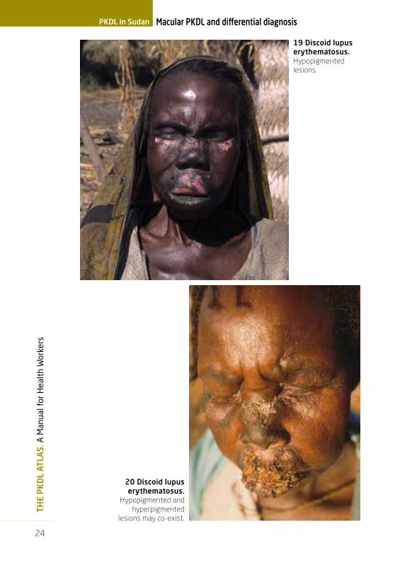
19 Discoid lupus erythematosus. Hypopigmented lesions.
20 Discoid lupus erythematosus.
Hypopigmented and hyperpigmented
lesions may co-exist.
Macular PKDL and differential diagnosis

2. P
KD
L in
Afr
ica:
clin
ical
pre
sent
atio
n an
d di
ffer
enti
al d
iagn
osis
25
21 Lupus vulgaris.Violaceous
infiltrated ulcerating plaque with atrophic
scarring in the center.
22 Burn scars.Depigmented irregular scars caused by previous burn injury.
Macular PKDL and differential diagnosis

ThE
PK
DL
ATLA
s. A
Man
ual f
or H
ealt
h W
orke
rs
26
PKDL in sudan
segmental or zosteriform vitiligo.Clinical clues: Depigmented lesions, asymmetrical in the distribution of a dermatome(s). This type may occur in younger patients. The dark vertical lines on both cheeks are tribal markings.
23
24
Table 3: Differential diagnosis of macular PKDL and vitiligo
Macular PKDL VitiligoFamily members yes yes
similar exposure to VL genetic
Predilection
face yes yes
acra no yes
central back affected sparing central back
Appearance hypopigmented depigmented
Bordering skin normal sometimes hyperpigmented
Other skin abnormalities macules may be erythema-tous; papules, nodules
none
Sparing of most pigmented areas(axillae, inguinal area)
yes no
Macular PKDL and differential diagnosis

2. P
KD
L in
Afr
ica:
clin
ical
pre
sent
atio
n an
d di
ffer
enti
al d
iagn
osis
27
Vitiligo.Clinical clues:The macules are
depigmented and not hypopigmented as in PKDL (see Table 3). The age of the
patient also suggests vitiligo, rather than PKDL. Note that some of hairs in the
beard and moustache are white.
25
27
26
28
Macular PKDL and differential diagnosis

ThE
PK
DL
ATLA
s. A
Man
ual f
or H
ealt
h W
orke
rs
28
PKDL in sudan
scleroderma. Clinical clues:
Symmetrical hypopigmented lesions, taut skin, reduced mouth opening,
impairment in movement of the fingers.
29
31
30
Macular PKDL and differential diagnosis

2. P
KD
L in
Afr
ica:
clin
ical
pre
sent
atio
n an
d di
ffer
enti
al d
iagn
osis
29
32
Onchocerciasis.Clinical clues:
leopard skin with hypopigmented
macules (32, 33); look for onchocercomata
(34, arrow) and scratch marks (35).
34 35
33
Macular PKDL and differential diagnosis

ThE
PK
DL
ATLA
s. A
Man
ual f
or H
ealt
h W
orke
rs
30
PKDL in sudan
36 Borderline leprosy.Note the raised edge, the area of hypopigmentation with central repigmentation. Clinical clues: look for other signs of leprosy: anaesthetic patches, thickened nerves. See Table 4.
Table 4: Differential diagnosis of PKDL and leprosy
PKDL LeprosyEpidemiologyMost important age group young children older individuals
Frequency in endemic areas common uncommon
ClinicalLesions M, P, N, plaques M, P, N, plaques
Symmetrical lesions yes indeterminate, tuberculoid: no lepromatous: yes
Uniform in size yes no
Single lesion uncommon common in undetermined and tuberculoid leprosy
Neurological features- clinical none anaesthetic patches, thickened
nerves, nerve palsies, loss ofsweating
- pathological neuritis in cutaneous nerves id
Lobulation of ears yes yes
Madarosis no yes
Predilection sun exposed parts yes no (cooler parts)
DiagnosisSkin slit smear Leishmania amastigotes acid-fast bacilli
(Giemsa stain) (modified ZN stain)
ManagementSelf-cure yes ( the rule in Sudan) no
no (Asia)
M macules, P papules, N nodules.
Macular PKDL and differential diagnosis

2. P
KD
L in
Afr
ica:
clin
ical
pre
sent
atio
n an
d di
ffer
enti
al d
iagn
osis
31
37 Cutaneous leishmaniasis scars.Hyperpigmented depressed scars of previous cutaneous leishmaniasis ulcers.The longitudinal scars on the cheek are tribal markings.
38 Prayer marks in a muslim man.Note the hyperpigmentation on the forehead caused
by frequent pressure exerted while praying.
Macular PKDL and differential diagnosis

ThE
PK
DL
ATLA
s. A
Man
ual f
or H
ealt
h W
orke
rs
32
PKDL in sudan
Pseudomelanosis.Although kala-azar means the
“black disease”, which refers to hyperpigmentation of the skin
found in Indian kala-azar, this hyperpigmentation is not found in Sudanese patients. In these 2
confirmed kala-azar cases, the black discoloration was caused by dirt and
could be removed with water and soap.
39
40
Macular PKDL and differential diagnosis
2. P
KD
L in
Afr
ica:
clin
ical
pre
sent
atio
n an
d di
ffer
enti
al d
iagn
osis
33
42
43
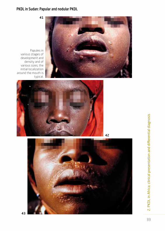
PKDL in sudan: Papular and nodular PKDL
Papules in various stages of development and
density and of various sizes; the initial localization
around the mouth is typical.
41

ThE
PK
DL
ATLA
s. A
Man
ual f
or H
ealt
h W
orke
rs
34
PKDL in sudan Papular and nodular PKDL
44 Micropapular rash.

2. P
KD
L in
Afr
ica:
clin
ical
pre
sent
atio
n an
d di
ffer
enti
al d
iagn
osis
35
Papular and nodular PKDL
45 Macropapular rash.

ThE
PK
DL
ATLA
s. A
Man
ual f
or H
ealt
h W
orke
rs
36
PKDL in sudan
46
47
Further spread of the lesions to the nose, around the eyes and the forehead.
Papular and nodular PKDL

2. P
KD
L in
Afr
ica:
clin
ical
pre
sent
atio
n an
d di
ffer
enti
al d
iagn
osis
37
48 Further spread of the lesions to the nose, around the eyes and the forehead.
Papular and nodular PKDL

ThE
PK
DL
ATLA
s. A
Man
ual f
or H
ealt
h W
orke
rs
38
PKDL in sudan
Papular rash with increasing density.
49
50
51
52
Papular and nodular PKDL

2. P
KD
L in
Afr
ica:
clin
ical
pre
sent
atio
n an
d di
ffer
enti
al d
iagn
osis
39
53
55
56
54
Maculopapular rash.
Papular and nodular PKDL

ThE
PK
DL
ATLA
s. A
Man
ual f
or H
ealt
h W
orke
rs
40
PKDL in sudan
57 Maculopapular rash.
Papular and nodular PKDL

2. P
KD
L in
Afr
ica:
clin
ical
pre
sent
atio
n an
d di
ffer
enti
al d
iagn
osis
41
Papular and nodular PKDL
58 Papular PKDL, covering the whole face.

ThE
PK
DL
ATLA
s. A
Man
ual f
or H
ealt
h W
orke
rs
42
PKDL in sudan
61 Nuer tribesman from South Sudan.Note absence of lesions on forehead: the horizontal lines are tribal markings and fibrotic changes may prevent PKDL papules to develop.
60
59
Papular PKDL, covering the whole face.Note verrucous plaques over eyebrows (60).
Papular and nodular PKDL

2. P
KD
L in
Afr
ica:
clin
ical
pre
sent
atio
n an
d di
ffer
enti
al d
iagn
osis
43
62
Micropapular rash, measles-like.
63
Papular and nodular PKDL

ThE
PK
DL
ATLA
s. A
Man
ual f
or H
ealt
h W
orke
rs
44
PKDL in sudan
Hyperpigmented papules.
64
65
Papular and nodular PKDL

2. P
KD
L in
Afr
ica:
clin
ical
pre
sent
atio
n an
d di
ffer
enti
al d
iagn
osis
45
Papular and nodular PKDL
66 Nodular lesions.

ThE
PK
DL
ATLA
s. A
Man
ual f
or H
ealt
h W
orke
rs
46
PKDL in sudan
Nodular lesions.
67
69
68
Papular and nodular PKDL

2. P
KD
L in
Afr
ica:
clin
ical
pre
sent
atio
n an
d di
ffer
enti
al d
iagn
osis
47
Papular and nodular PKDL
Papules and nodules become confluent
to form plaques.
70
72
71

ThE
PK
DL
ATLA
s. A
Man
ual f
or H
ealt
h W
orke
rs
48
PKDL in sudan
Patient with three solitary plaques on forehead (73), chin (74) and earlobe (121) .
73
74
Papular and nodular PKDL
2. P
KD
L in
Afr
ica:
clin
ical
pre
sent
atio
n an
d di
ffer
enti
al d
iagn
osis
49
Papular and nodular PKDL
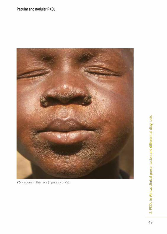
75 Plaques in the face (Figures 75-79).

ThE
PK
DL
ATLA
s. A
Man
ual f
or H
ealt
h W
orke
rs
50
PKDL in sudan Papular and nodular PKDL
76

2. P
KD
L in
Afr
ica:
clin
ical
pre
sent
atio
n an
d di
ffer
enti
al d
iagn
osis
51
Papular and nodular PKDL
77

ThE
PK
DL
ATLA
s. A
Man
ual f
or H
ealt
h W
orke
rs
52
PKDL in sudan
78
Papular and nodular PKDL

2. P
KD
L in
Afr
ica:
clin
ical
pre
sent
atio
n an
d di
ffer
enti
al d
iagn
osis
53
79
Papular and nodular PKDL

ThE
PK
DL
ATLA
s. A
Man
ual f
or H
ealt
h W
orke
rs
54
PKDL in sudan PKDL grading system
80 Grade 1.1 Lesions only in the face and restricted to area around the nose and mouth with normal skin in between.
Table 5: Grading system of PKDL in sudanDistribution Density
Grade 1 face mainly with some lesions on trunk and arms
scattered lesions
Grade 2 face, upper parts of trunk, arms and legs affected, gradually becoming less distally; hands and feet free
moderate density with normal skin in between
Grade 3 all over body; including hands and feet

2. P
KD
L in
Afr
ica:
clin
ical
pre
sent
atio
n an
d di
ffer
enti
al d
iagn
osis
55
PKDL grading system
81
82
Grade 1.3.Dense maculopapular rash, but mainly on the face.
Grade 1.1.Transition into Grade 1.2 (Figures 81-82).
83

ThE
PK
DL
ATLA
s. A
Man
ual f
or H
ealt
h W
orke
rs
56
PKDL in sudan PKDL grading system
84
85
Grade 1.3 The whole face is affected with a dense papular rash (84) and macular rash (85) with few papules in other areas

2. P
KD
L in
Afr
ica:
clin
ical
pre
sent
atio
n an
d di
ffer
enti
al d
iagn
osis
57
PKDL grading system
86
Grade 2.1 Papular rash and leishmaniomas (arrows). In this patient there was no previous history of VL.
Grade 2.2. Papular rash. Most parts of the body are affected; the rash is dense but
still normal skin can be seen.
87

ThE
PK
DL
ATLA
s. A
Man
ual f
or H
ealt
h W
orke
rs
58
PKDL in sudan PKDL grading system
Grade 2.2. Most of the trunk is also
involved.
88
89

2. P
KD
L in
Afr
ica:
clin
ical
pre
sent
atio
n an
d di
ffer
enti
al d
iagn
osis
59
PKDL grading system
Grade 3.2. Macular rash.
90
91
Grade 3.2. Combined macular and papular rash.

ThE
PK
DL
ATLA
s. A
Man
ual f
or H
ealt
h W
orke
rs
60
PKDL in sudan PKDL grading system
92 Grade 3.2 Maculopapular rash .

2. P
KD
L in
Afr
ica:
clin
ical
pre
sent
atio
n an
d di
ffer
enti
al d
iagn
osis
61
93
94
Grade 3.3.Mainly hyperpigmented papules.
PKDL grading system

ThE
PK
DL
ATLA
s. A
Man
ual f
or H
ealt
h W
orke
rs
62
PKDL in sudan
Grade 3 macular PKDL.In macular PKDL, the rash
does not follow the normal distribution as in papular/
nodular PKDL. The face and lower arms may be affected
with little involvement of the trunk (95, 96).
PKDL grading system
95
96
2. P
KD
L in
Afr
ica:
clin
ical
pre
sent
atio
n an
d di
ffer
enti
al d
iagn
osis
63
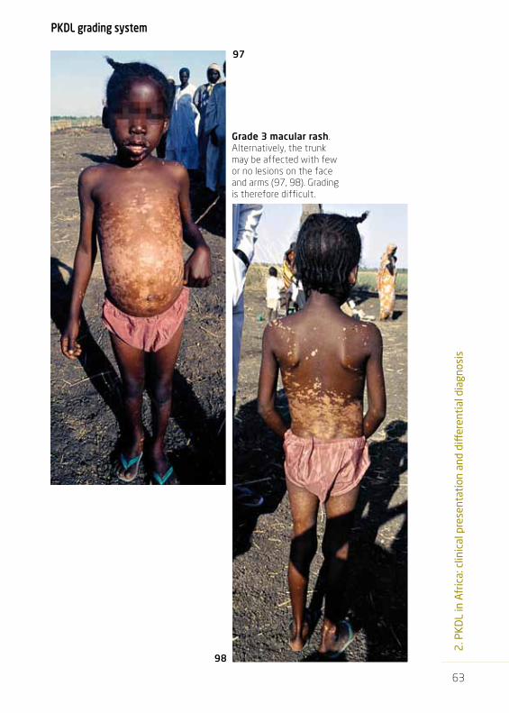
Grade 3 macular rash.Alternatively, the trunk may be affected with few or no lesions on the face and arms (97, 98). Grading is therefore difficult.
98
97
PKDL grading system

ThE
PK
DL
ATLA
s. A
Man
ual f
or H
ealt
h W
orke
rs
64
PKDL in sudan
99
Grade 3.3Maculopapular rash.
PKDL grading system

2. P
KD
L in
Afr
ica:
clin
ical
pre
sent
atio
n an
d di
ffer
enti
al d
iagn
osis
65
100 Grade 3.3. Papulonodular rash.
PKDL grading system
ThE
PK
DL
ATLA
s. A
Man
ual f
or H
ealt
h W
orke
rs
66
PKDL in sudan PKDL grading system
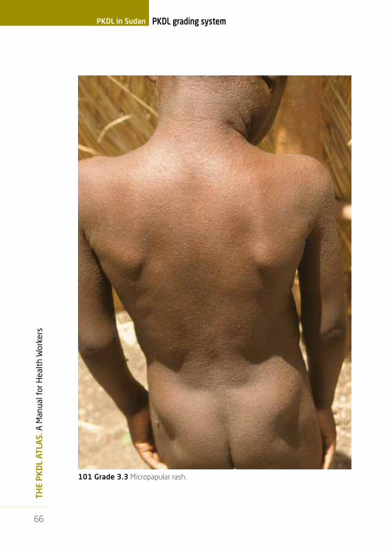
101 Grade 3.3 Micropapular rash.

2. P
KD
L in
Afr
ica:
clin
ical
pre
sent
atio
n an
d di
ffer
enti
al d
iagn
osis
67
102
Grade 3.3 Close-up: dense maculopapular rash.
103
Grade 3.3Papulonodular rash.
All parts of the body are involved with plaques
around the mouth and on eyebrows.
PKDL grading system

ThE
PK
DL
ATLA
s. A
Man
ual f
or H
ealt
h W
orke
rs
68
PKDL in sudan
104 Severe PKDL grade 3 in the face with crusts and sloughing of the skin.
severe PKDL

2. P
KD
L in
Afr
ica:
clin
ical
pre
sent
atio
n an
d di
ffer
enti
al d
iagn
osis
69
106
105
severe PKDL
Severe PKDL grade 3 in the face
with crusts and sloughing
of the skin.

ThE
PK
DL
ATLA
s. A
Man
ual f
or H
ealt
h W
orke
rs
70
PKDL in sudan
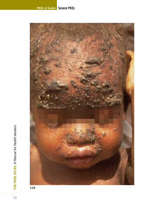
107 Severe PKDL grade 3. (Figures 107-112).
severe PKDL

2. P
KD
L in
Afr
ica:
clin
ical
pre
sent
atio
n an
d di
ffer
enti
al d
iagn
osis
71
108
severe PKDL
109
ThE
PK
DL
ATLA
s. A
Man
ual f
or H
ealt
h W
orke
rs
72
PKDL in sudan
110
severe PKDL

2. P
KD
L in
Afr
ica:
clin
ical
pre
sent
atio
n an
d di
ffer
enti
al d
iagn
osis
73
111
severe PKDL

ThE
PK
DL
ATLA
s. A
Man
ual f
or H
ealt
h W
orke
rs
74
PKDL in sudan
112
severe PKDL

2. P
KD
L in
Afr
ica:
clin
ical
pre
sent
atio
n an
d di
ffer
enti
al d
iagn
osis
75
severe PKDL
Grade 3. Desquamation of the skin.
113
114
115

ThE
PK
DL
ATLA
s. A
Man
ual f
or H
ealt
h W
orke
rs
76
PKDL in sudan
Grade 3. PKDL also
affects the hands
(back and palm).
117
116
118
severe PKDL

2. P
KD
L in
Afr
ica:
clin
ical
pre
sent
atio
n an
d di
ffer
enti
al d
iagn
osis
77
severe PKDL
119PKDL affecting the earlobe.

ThE
PK
DL
ATLA
s. A
Man
ual f
or H
ealt
h W
orke
rs
78
PKDL in sudan
PKDL affecting the earlobe; see also fig. 85.
120
121
122
123
severe PKDL

2. P
KD
L in
Afr
ica:
clin
ical
pre
sent
atio
n an
d di
ffer
enti
al d
iagn
osis
79
PKDL in the genital area. The ulcerative lesions may be leishmaniomas.
124
125
126
severe PKDL

ThE
PK
DL
ATLA
s. A
Man
ual f
or H
ealt
h W
orke
rs
80
PKDL in sudan Differential diagnosis of papular and nodular PKDL*
127
Miliaria rubra.Clinical clues: young child,
wrapped in many layers of cloth, despite hot weather conditions;
typically tiny papules on the forehead and not around
the mouth as in PKDL; may be itchy.
Acne.Clinical clues: adolescent age, different stages of development, greasy skin, comedones (papules with white head).
128
129
*Note that some photographs included in this chapter are from other countries than Sudan for comparison

2. P
KD
L in
Afr
ica:
clin
ical
pre
sent
atio
n an
d di
ffer
enti
al d
iagn
osis
81
130, 131, 132, 133, 134, 135. Leprosy.Clinical clues: anaesthetic skin patches, thickened nerves, e.g. greater auricular nerve, peripheral nerve palsies, such as claw hand (ulnar nerve) and wrist drop (radial nerve) with destruction of phalanges.Top left: the variation in size of papules, nodules and plaques is not seen in PKDL. Top middle: lepromatous leprosy (cf. Fig 7). Top right: tuberculoid leprosy: the great auricular nerve is clearly visible and palpable.Middle left: wrist drop and destruction of phalanges. Middle right: wrist drop and claw hand. Bottom: collapse of the nose; not seen in PKDL.See also Table 4.
Differential diagnosis of papular and nodular PKDL

ThE
PK
DL
ATLA
s. A
Man
ual f
or H
ealt
h W
orke
rs
82
PKDL in sudan Differential diagnosis of papular and nodular PKDL
136 Lepromatous leprosy.Clinical clues:Elderly person, madarosis, lesions in different stages of development, preference for cooler body parts: upper arms rather than lower arms that are exposed to sunlight.

2. P
KD
L in
Afr
ica:
clin
ical
pre
sent
atio
n an
d di
ffer
enti
al d
iagn
osis
83
Differential diagnosis of papular and nodular PKDL
Lepromatous leprosy.Details of Fig 136.
137
138

ThE
PK
DL
ATLA
s. A
Man
ual f
or H
ealt
h W
orke
rs
84
PKDL in sudan
139 Lues stage II.Clinical clues:History of genital ulcer; as in PKDL, palms and soles may be involved.
Differential diagnosis of papular and nodular PKDL

2. P
KD
L in
Afr
ica:
clin
ical
pre
sent
atio
n an
d di
ffer
enti
al d
iagn
osis
85
Differential diagnosis of papular and nodular PKDL
Keloids. Clinical clues:
There is thickening of the skin due to fibrosis, typically in a
scar. Surgical removal usually results in a (more severe)
relapse.
140 141
142
ThE
PK
DL
ATLA
s. A
Man
ual f
or H
ealt
h W
orke
rs
86
PKDL in sudan
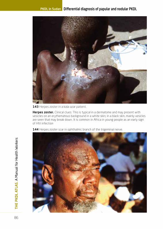
herpes zoster. Clinical clues: This is typical in a dermatome and may present with vesicles on an erythematous background in a white skin; in a black skin, mainly vesicles are seen that may break down. It is common in Africa in young people as an early sign of HIV infection
Differential diagnosis of papular and nodular PKDL
143 Herpes zoster in a kala-azar patient.
144 Herpes zoster scar in ophthalmic branch of the trigeminal nerve.

2. P
KD
L in
Afr
ica:
clin
ical
pre
sent
atio
n an
d di
ffer
enti
al d
iagn
osis
87
Differential diagnosis of papular and nodular PKDL
Herpes zoster in the black skin; because of the different
skin structure, the vesicles are less likely to break down. (147, 148).
145 Herpes zoster in white skin; vesicles on erythematous background.
146
147
148 Varicella (chickenpox).Clinical clues:Vesicles in various stages of development; evolves over days.

ThE
PK
DL
ATLA
s. A
Man
ual f
or H
ealt
h W
orke
rs
88
PKDL in sudan
150Multiple
tricho-epitheliomas (presumed diagnosis; differential diagnosis:
cylindroma, syringoma, adenoma sebaceum).
Predilection for central part of face; may run in
families: the patient’s brother had the same
condition.
149Dermatosis papulosa nigra. Common in the black skin, mostly in upper part of face, cheeks and temples. Numbers of papules increase with age; starts during adolescence.
151 Keloid after varicella in
childhood; now presents with herpes zoster scar.
Differential diagnosis of papular and nodular PKDL

2. P
KD
L in
Afr
ica:
clin
ical
pre
sent
atio
n an
d di
ffer
enti
al d
iagn
osis
89
Differential diagnosis of papular and nodular PKDL
152Mollusca contagiosa in an HIV-positive patient.

ThE
PK
DL
ATLA
s. A
Man
ual f
or H
ealt
h W
orke
rs
90
PKDL in sudan
Noma or cancrum oris in a VL patient who had relapsed after treatment; note the splenomegaly.
Differential diagnosis of papular and nodular PKDL
153
154

2. P
KD
L in
Afr
ica:
clin
ical
pre
sent
atio
n an
d di
ffer
enti
al d
iagn
osis
91
Differential diagnosis of papular and nodular PKDL
Measles.The micropapular rash is
difficult to distinguish from PKDL.
157 Measles-like PKDL.
155 156

ThE
PK
DL
ATLA
s. A
Man
ual f
or H
ealt
h W
orke
rs
92
PKDL in sudan
Measles.In measles, the rash can be
virtually indistinguishable from the micropapular rash in PKDL. The rash may also
desquamate (159) (see also 113-115).
Clinical clues: look for other signs of measles such as
fever, cough, conjunctivitis (158), Koplik’s spots (160)
and otitis media (discharge of pus probably as a result of
a perforated eardrum, 161).
Differential diagnosis of papular and nodular PKDL
158
159
161
160

2. P
KD
L in
Afr
ica:
clin
ical
pre
sent
atio
n an
d di
ffer
enti
al d
iagn
osis
93
Differential diagnosis of papular and nodular PKDL
scabies Typical interdigital lesions (162); may also be more widespread (163).162
163

ThE
PK
DL
ATLA
s. A
Man
ual f
or H
ealt
h W
orke
rs
94
PKDL in sudan Differential diagnosis of papular and nodular PKDL
164 African histoplasmosis.

2. P
KD
L in
Afr
ica:
clin
ical
pre
sent
atio
n an
d di
ffer
enti
al d
iagn
osis
95
Differential diagnosis of papular and nodular PKDL
Neurofibromatosis.Clinical clues:
Family history: in this case, his grandmother, father and his 2 siblings also had the disease compatible with
autosomal dominant inheritance.Chronic slowly progressing. Nodules in
different sizes, also affecting the scalp. The nose and earlobes remain free. Note
the multiple and giant nodules on the abdomen; this is not seen in PKDL.
165
166 167

ThE
PK
DL
ATLA
s. A
Man
ual f
or H
ealt
h W
orke
rs
96
PKDL in sudan Differential diagnosis of papular and nodular PKDL
168
171
169
170
168 Endemic syphilis, macular lesions with raised edge.
169, 170 Yaws, nodular (from the Democratic Republic of Congo) Note atypical distribution on face and extremities, leaving chest clear.
171 Late yaws: gangosa.
2. P
KD
L in
Afr
ica:
clin
ical
pre
sent
atio
n an
d di
ffer
enti
al d
iagn
osis
97
Differential diagnosis of papular and nodular PKDL
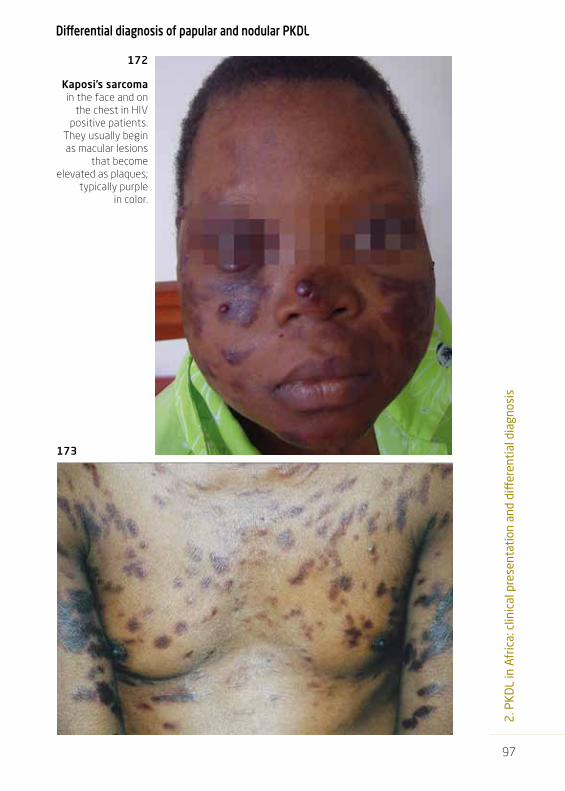
Kaposi’s sarcoma in the face and on
the chest in HIV positive patients.
They usually begin as macular lesions
that become elevated as plaques;
typically purple in color.
173
172

ThE
PK
DL
ATLA
s. A
Man
ual f
or H
ealt
h W
orke
rs
98
PKDL in sudan Differential diagnosis of papular and nodular PKDL
175
174
sporotrichosis. Spread along the lymphatics. While this is also seen in cutaneous leishmaniasis, it is not a feature of PKDL.

2. P
KD
L in
Afr
ica:
clin
ical
pre
sent
atio
n an
d di
ffer
enti
al d
iagn
osis
99
Differential diagnosis of papular and nodular PKDL
Tribal markings.
Nuer tribe.
177
176
Shilluk tribe.

ThE
PK
DL
ATLA
s. A
Man
ual f
or H
ealt
h W
orke
rs
100
PKDL in sudan Differential diagnosis of papular and nodular PKDL
178
179
Nuer tribal markings, with PKDL papules occurring between the markings.

2. P
KD
L in
Afr
ica:
clin
ical
pre
sent
atio
n an
d di
ffer
enti
al d
iagn
osis
101
Differential diagnosis of papular and nodular PKDL
181
Nuer tribe. Here the papules are also found on the forehead despite the scarring, but with less density than on the cheeks and nose.
180 Nuer tribe. The papules do not
appear on the forehead, perhaps because of fibrosis caused by
scarring.
182

ThE
PK
DL
ATLA
s. A
Man
ual f
or H
ealt
h W
orke
rs
102
PKDL in sudan Differential diagnosis of papular and nodular PKDL
183, 184 Psoriasis.

2. P
KD
L in
Afr
ica:
clin
ical
pre
sent
atio
n an
d di
ffer
enti
al d
iagn
osis
103
Differential diagnosis of papular and nodular PKDL
PKDL.Swelling of the face is not a feature of PKDL; no diagnosis was made in this
case, but the appearance and the patient’s age may suggest Burkitt’s lymphoma.
185
ThE
PK
DL
ATLA
s. A
Man
ual f
or H
ealt
h W
orke
rs
104
PKDL in sudan Differential diagnosis of papular and nodular PKDL
186
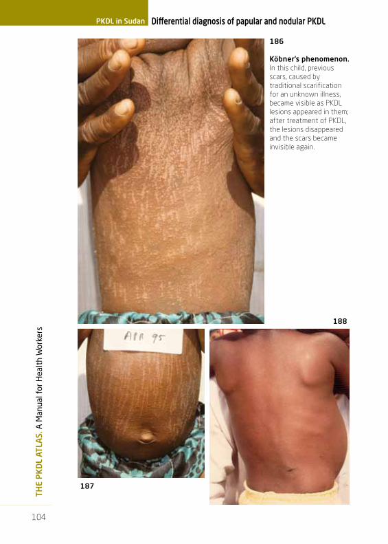
Köbner’s phenomenon.In this child, previous scars, caused by traditional scarification for an unknown illness, became visible as PKDL lesions appeared in them; after treatment of PKDL, the lesions disappeared and the scars became invisible again.
188
187

2. P
KD
L in
Afr
ica:
clin
ical
pre
sent
atio
n an
d di
ffer
enti
al d
iagn
osis
105
Differential diagnosis of papular and nodular PKDL
189 Köbner’s phenomenon.Note the preferential localization of the papules in the scars causing an asymmetrical distribution which is otherwise unusual in PKDL.

ThE
PK
DL
ATLA
s. A
Man
ual f
or H
ealt
h W
orke
rs
106
PKDL in sudan Chronic PKDL
190 Depressed scars developing after longstanding PKDL.

2. P
KD
L in
Afr
ica:
clin
ical
pre
sent
atio
n an
d di
ffer
enti
al d
iagn
osis
107
Chronic PKDL
Depressed scars developing after longstanding PKDL.
191
192

ThE
PK
DL
ATLA
s. A
Man
ual f
or H
ealt
h W
orke
rs
108
PKDL in sudan
Fibrosis of the skin in longstanding PKDL.
Chronic PKDL
193
195194
196

2. P
KD
L in
Afr
ica:
clin
ical
pre
sent
atio
n an
d di
ffer
enti
al d
iagn
osis
109
Post kala-azar manifestations
197
199
198
Post-kala-azar conjunctivitis
and uveitis.The patient was treated for VL in
the recent past but developed
increasing swelling of the
eyelids and loss of vision.
These were not diagnosed
as related to persistent
Leishmanial infection in the
eyes and led to complete
blindness.

ThE
PK
DL
ATLA
s. A
Man
ual f
or H
ealt
h W
orke
rs
110
PKDL in sudan Post kala-azar manifestations
200
Concomitant PKDL and post-kala-azar conjunctivitis (top)
and blepharitis (bottom).
201

2. P
KD
L in
Afr
ica:
clin
ical
pre
sent
atio
n an
d di
ffer
enti
al d
iagn
osis
111
Post kala-azar manifestations
202 Post-kala-azar uveitis. Note the irregular pupil and nodules.
Differential diagnosis:
203
Onchocerciasis (middle). Left: beginning of overgrowth of cornea (pannus).Right: uveitis; irregular pupil.
Trachoma (below).Left: inversion of eyelids. Right: after eyelid surgery; the cornea is opaque.
205204

ThE
PK
DL
ATLA
s. A
Man
ual f
or H
ealt
h W
orke
rs
112
PKDL in sudan
206 Mucosal lesions in severe PKDL.
Post kala-azar manifestations

2. P
KD
L in
Afr
ica:
clin
ical
pre
sent
atio
n an
d di
ffer
enti
al d
iagn
osis
113
207 Para-kala-azar dermal leishmaniasis.VL (confirmed in a lymph node aspirate) and concomitant micropapular PKDL.
Other post kala-azar manifestations (conjunctivitis, uveitis, mucosal)

ThE
PK
DL
ATLA
s. A
Man
ual f
or H
ealt
h W
orke
rs
114
PKDL in sudan Evolution of PKDL
Nuer tribesman before (208, 209) and after 30 days of stibogluconate treatment (210, 211). In spite of the difference in brightness of the figures, his skin indeed became lighter after treatment.
208
210
209
211

2. P
KD
L in
Afr
ica:
clin
ical
pre
sent
atio
n an
d di
ffer
enti
al d
iagn
osis
115
Evolution of PKDL
Before treatment (212, 213), after 30
days of treatment with SSG (214) and
after 6 weeks (215).
212 213
214
215

ThE
PK
DL
ATLA
s. A
Man
ual f
or H
ealt
h W
orke
rs
116
PKDL in sudan Evolution of PKDL
216
218
217
Initial presentation
(216), 2 weeks later (217) and
after treatment (218).

2. P
KD
L in
Afr
ica:
clin
ical
pre
sent
atio
n an
d di
ffer
enti
al d
iagn
osis
117
PKDL in Ethiopia. Various examples
219
221
Various examples of papular rash, mainly around the mouth.
220
PKDL in Ethiopia

ThE
PK
DL
ATLA
s. A
Man
ual f
or H
ealt
h W
orke
rs
118
PKDL in Ethiopia PKDL in Ethiopia. Various examples
222 Papular rash, covering most of the face.

2. P
KD
L in
Afr
ica:
clin
ical
pre
sent
atio
n an
d di
ffer
enti
al d
iagn
osis
119
223
224
Papular rash.
PKDL in Ethiopia. Various examples

ThE
PK
DL
ATLA
s. A
Man
ual f
or H
ealt
h W
orke
rs
120
PKDL in Ethiopia PKDL in Ethiopia. Various examples
225
226
Papular rash.

2. P
KD
L in
Afr
ica:
clin
ical
pre
sent
atio
n an
d di
ffer
enti
al d
iagn
osis
121
Papular rash with plaques (228).
227
228
PKDL in Ethiopia. Various examples
ThE
PK
DL
ATLA
s. A
Man
ual f
or H
ealt
h W
orke
rs
122
PKDL in Ethiopia. Various examplesPKDL in Ethiopia
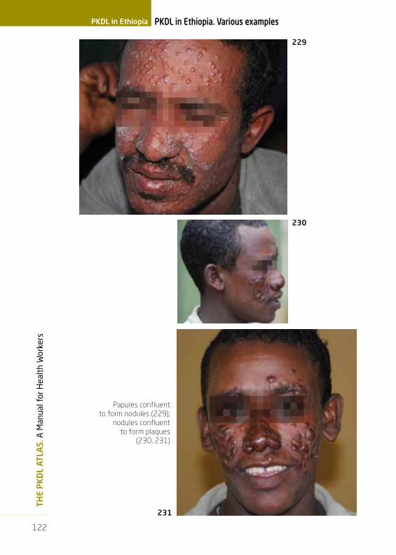
Papules confluent to form nodules (229);
nodules confluent to form plaques
(230, 231)
231
229
230

2. P
KD
L in
Afr
ica:
clin
ical
pre
sent
atio
n an
d di
ffer
enti
al d
iagn
osis
123
Differential diagnosis
233 234
232Subcutaneous nodular lesions in an HIV co-infected VL patient: para-kala-azar dermal leishmaniasis. The same strain was isolated from the spleen and from the skin. The lesions resemble those of Kaposi’s sarcoma.
Kaposi’s sarcoma in an HIV+ve patient.

ThE
PK
DL
ATLA
s. A
Man
ual f
or H
ealt
h W
orke
rs
124
Differential diagnosisPKDL in Ethiopia
Diffuse Cutaneous Leishmaniasis; the lesions had been there for 3 years and were never treated.
235
236
2. P
KD
L in
Afr
ica:
clin
ical
pre
sent
atio
n an
d di
ffer
enti
al d
iagn
osis
125
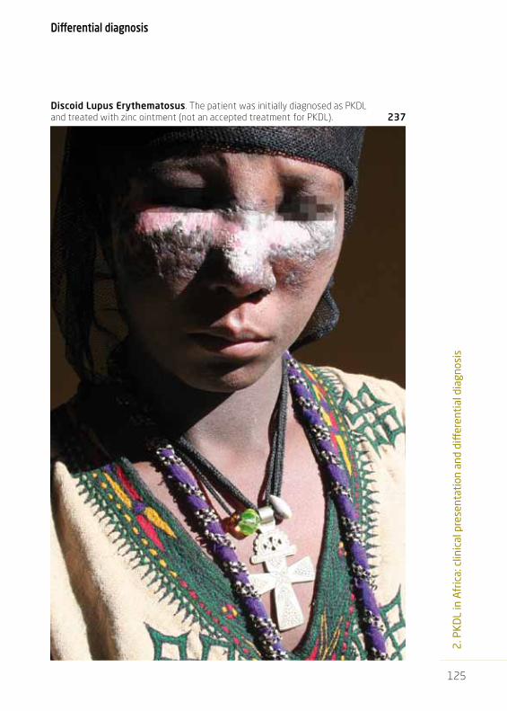
Discoid Lupus Erythematosus. The patient was initially diagnosed as PKDL and treated with zinc ointment (not an accepted treatment for PKDL). 237
Differential diagnosis


a. PKDL in India: hospital-based experience
b. PKDL in Bangladesh: community-based experience
3. PKDL in Asia: clinical presentation and differential diagnosis


129
3. P
KD
L in
Asi
a: c
linic
al p
rese
ntat
ion
and
diff
eren
tial
dia
gnos
is
PKDL in Asia is much different from Africa. There are few longitudinal stu-dies with active follow-up of VL cases and most reports are on cases that present with chronic PKDL. PKDL is increasingly reported from Bangladesh as more field studies are conducted.
The interval between VL and PKDL is usually 2-3 years; however, pa-tients may report a shorter interval. In one study with active follow-up 40% of cases developed PKDL within 12 months of VL.
Clinical presentationThe currently available data do not permit a general description that is applicable to all areas (India, Bangladesh, Nepal); data from field studies are different from hospital –based studies; whether these differences are the result of patient delay, reporting bias or true differences related to pa-rasite involved, genetic background, treatment received etc. is unknown.
Hospital-based studies in India: The polymorphic form showing hypo-pigmented or erythematous macules with papules and/or nodules is the commonest. A monomorphic presentation with only papulonodules may be seen, the monomorphic form macular form being uncommon. Both of these monomorphic forms can mimic leprosy. In addition vitiligo is also an important differential diagnosis for macular PKDL. Verrucous lesions may be seen, though uncommon. A generalized redness of the face and body with scattered papules and plaques indicates the rare erythematous form of PKDL.
Field studies in Bangladesh: The macular form is by far the most com-mon presentation. While the face is usually involved the spread to other parts of the body does not always follow the classical pattern described for Sudan. Most cases in Bangladesh present with longstanding lesions that seem to spread and remain macular; sometimes concomitant papules may be found in the face. Up to 10% of cases present without a previous history of VL.
In the Indian subcontinent all cases are treated as they are considered chronic cases also given the long interval after VL; while the skin returns to normal, repigmentation may take time and cannot be taken as a para-meter for cure.
Other post-kala-azar manifestations have been described such as post-kala-azar uveitis.
The main differential diagnosis for the macular form is vitiligo (Table 3) and for all forms leprosy (Table 4).

ThE
PK
DL
ATLA
s. A
Man
ual f
or H
ealt
h W
orke
rs
130
PKDL in India
238 Macular lesions on the legs.
PKDL in India

131
3. P
KD
L in
Asi
a: c
linic
al p
rese
ntat
ion
and
diff
eren
tial
dia
gnos
is
239 Macules and small papules.
PKDL in India
ThE
PK
DL
ATLA
s. A
Man
ual f
or H
ealt
h W
orke
rs
132
PKDL in India
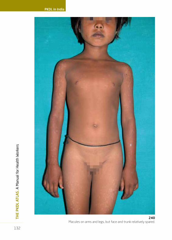
240 Macules on arms and legs, but face and trunk relatively spared.

133
3. P
KD
L in
Asi
a: c
linic
al p
rese
ntat
ion
and
diff
eren
tial
dia
gnos
is
Macules on limbs and face with some
small papules on the nose.
241
242
PKDL in India

ThE
PK
DL
ATLA
s. A
Man
ual f
or H
ealt
h W
orke
rs
134
PKDL in India
243 Nodules on the face; macules on the rest of the body with virtually
total hypopigmentation sparing axillae and inguinal areas.
135
3. P
KD
L in
Asi
a: c
linic
al p
rese
ntat
ion
and
diff
eren
tial
dia
gnos
is
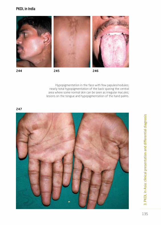
Hypopigmentation in the face with few papules/nodules; nearly total hypopigmentation of the back sparing the central
area where some normal skin can be seen as irregular macules; lesions on the tongue and hypopigmentation of the hand palms.
244 245 246
247
PKDL in India

ThE
PK
DL
ATLA
s. A
Man
ual f
or H
ealt
h W
orke
rs
136
PKDL in India
248Hypopigmented patches in the face, typically around the mouth.

137
3. P
KD
L in
Asi
a: c
linic
al p
rese
ntat
ion
and
diff
eren
tial
dia
gnos
is
Hypopigmented lesions involving the whole face sparing the neck; this is only appreciated in a lateral view. The lesions on the back spare the mid back and elbows (the arrow indicates the site of a biopsy).
249
250
PKDL in India
ThE
PK
DL
ATLA
s. A
Man
ual f
or H
ealt
h W
orke
rs
138
PKDL in India
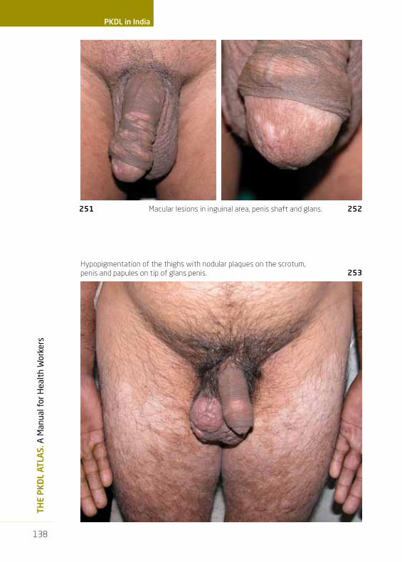
Macular lesions in inguinal area, penis shaft and glans.
Hypopigmentation of the thighs with nodular plaques on the scrotum, penis and papules on tip of glans penis.
251 252
253

139
3. P
KD
L in
Asi
a: c
linic
al p
rese
ntat
ion
and
diff
eren
tial
dia
gnos
is
PKDL: papules and nodules on inner thighs, scrotum and penis.254
PKDL in India

ThE
PK
DL
ATLA
s. A
Man
ual f
or H
ealt
h W
orke
rs
140
PKDL in India
Erythrodermic PKDL. This is uncommon; there is facial erythema and sparing of the axillae. The rest of the body is also faintly erythematous.
255
256
Papules and nodules on the tongue and
buccal mucosa.

141
3. P
KD
L in
Asi
a: c
linic
al p
rese
ntat
ion
and
diff
eren
tial
dia
gnos
is
Papules and nodules on the tongue.257
PKDL in India

ThE
PK
DL
ATLA
s. A
Man
ual f
or H
ealt
h W
orke
rs
142
PKDL in India
259
Discrete papules on chin (258, 259);
crops of nodules on chin and nose (260)
258
260

143
3. P
KD
L in
Asi
a: c
linic
al p
rese
ntat
ion
and
diff
eren
tial
dia
gnos
is
PKDL in India
Verrucous or hypertrophic form.261

ThE
PK
DL
ATLA
s. A
Man
ual f
or H
ealt
h W
orke
rs
144
PKDL in India
Tumor-like nodules on face (note sparing of eyebrows).
Tumor-like; there is a spontaneous furrow.263
262

145
3. P
KD
L in
Asi
a: c
linic
al p
rese
ntat
ion
and
diff
eren
tial
dia
gnos
is
Vitiligo. Note the total loss of pigment (depigmentation) and the involvement of the central part of the trunk.
Lepromatous leprosy.Lesions are prominent on the forehead and the cheeks while the central part of the face is spared. The patient on the right has madarosis. All these are not features of PKDL.
264
266
265
267
Differential diagnosis

ThE
PK
DL
ATLA
s. A
Man
ual f
or H
ealt
h W
orke
rs
146
Differential diagnosisPKDL in India
268Borderline leprosy.
Red, raised plaques on the face and limbs, note sparing areas around the nose and the mouth.

147
3. P
KD
L in
Asi
a: c
linic
al p
rese
ntat
ion
and
diff
eren
tial
dia
gnos
is
269Borderline tuberculoid leprosy; the nose and chin are free; note the madarosis.
Differential diagnosis

ThE
PK
DL
ATLA
s. A
Man
ual f
or H
ealt
h W
orke
rs
148
PKDL in Bangladesh
PKDL in Bangladesh
270 Macular lesions.

149
3. P
KD
L in
Asi
a: c
linic
al p
rese
ntat
ion
and
diff
eren
tial
dia
gnos
is
271
272
Macular lesions.
PKDL in Bangladesh

ThE
PK
DL
ATLA
s. A
Man
ual f
or H
ealt
h W
orke
rs
150
PKDL: macular lesions, some are confluent.
Large areas of confluent macules leaving some islands of normal skin
PKDL in Bangladesh
274
273

151
3. P
KD
L in
Asi
a: c
linic
al p
rese
ntat
ion
and
diff
eren
tial
dia
gnos
is
275
276
Combined confluent macular lesions
on the arms (275) and papulonodular
lesions on the face (276).
PKDL in Bangladesh

ThE
PK
DL
ATLA
s. A
Man
ual f
or H
ealt
h W
orke
rs
152
PKDL in Bangladesh
277
278
Extensive confluent macules on the back and face leaving little normal skin.

153
3. P
KD
L in
Asi
a: c
linic
al p
rese
ntat
ion
and
diff
eren
tial
dia
gnos
is
279 Extensive confluent macules on the back.
PKDL in Bangladesh

ThE
PK
DL
ATLA
s. A
Man
ual f
or H
ealt
h W
orke
rs
154
PKDL in Bangladesh
284283
280 281
282Mostly macular rash with papular rash on the shin.

155
3. P
KD
L in
Asi
a: c
linic
al p
rese
ntat
ion
and
diff
eren
tial
dia
gnos
is
PKDL in Bangladesh
285 Maculopapular rash.
ThE
PK
DL
ATLA
s. A
Man
ual f
or H
ealt
h W
orke
rs
156
PKDL in Bangladesh
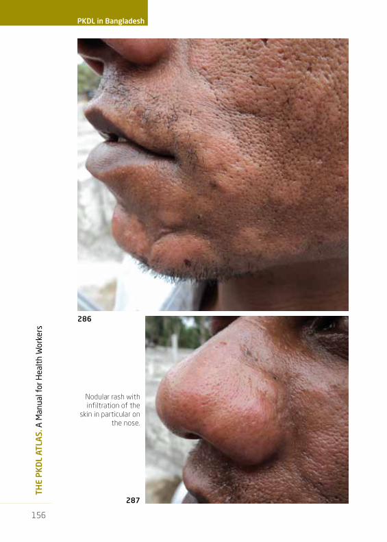
286
Nodular rash with infiltration of the
skin in particular on the nose.
287

157
3. P
KD
L in
Asi
a: c
linic
al p
rese
ntat
ion
and
diff
eren
tial
dia
gnos
is
Macular lesions on chest and nodular lesions on arms and fingers.
288
289
PKDL in Bangladesh

ThE
PK
DL
ATLA
s. A
Man
ual f
or H
ealt
h W
orke
rs
158
PKDL in Bangladesh Response to treatment
Macular rash before and after 6 weeks; no response can be noted.
Figures 290-318: In this section, a number of figures are shown of patients who were treated with liposomal amphotericine B 5 mg/kg twice weekly for three weeks. In some patients this leads to cure within 12 months; in others the rash is slow to disappear. It is difficult to assess if these lesions will further heal, leave residual hypopigmentation or indicate treatment failure; for the latter another diagnosis should also be considered.
290
292
291
293

159
3. P
KD
L in
Asi
a: c
linic
al p
rese
ntat
ion
and
diff
eren
tial
dia
gnos
is
Response to treatment
Macular rash before and after 6 weeks, 4 months and 7 months; note the slow recovery (e.g. around the mouth).
294
296
295
297

ThE
PK
DL
ATLA
s. A
Man
ual f
or H
ealt
h W
orke
rs
160
PKDL in Bangladesh Response to treatment
300 301 302
Residual lesions can be seen 10-11 months after treatment (301).
298 299
161
3. P
KD
L in
Asi
a: c
linic
al p
rese
ntat
ion
and
diff
eren
tial
dia
gnos
is
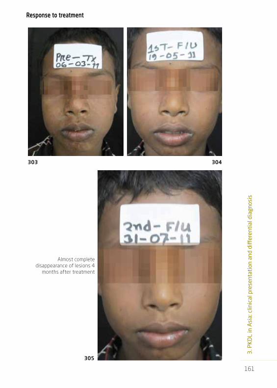
Response to treatment
305
Almost complete disappearance of lesions 4
months after treatment
303 304
ThE
PK
DL
ATLA
s. A
Man
ual f
or H
ealt
h W
orke
rs
162
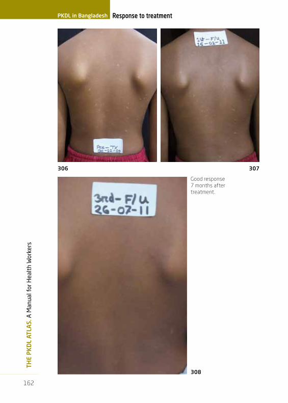
PKDL in Bangladesh Response to treatment
Good response 7 months after treatment.
308
306 307

163
3. P
KD
L in
Asi
a: c
linic
al p
rese
ntat
ion
and
diff
eren
tial
dia
gnos
is
Response to treatment
310
309
Good response of nodular lesions 6
weeks after start of treatment.

ThE
PK
DL
ATLA
s. A
Man
ual f
or H
ealt
h W
orke
rs
164
PKDL in Bangladesh Response to treatment
Clear residual lesions 5 months after start of treatment.
313
311 312

165
3. P
KD
L in
Asi
a: c
linic
al p
rese
ntat
ion
and
diff
eren
tial
dia
gnos
is
314
315
Response to treatment
Poor response to treatment; lesions are virtually unchanged.

ThE
PK
DL
ATLA
s. A
Man
ual f
or H
ealt
h W
orke
rs
166
PKDL in Bangladesh Response to treatment
Gradual disappearance of the macular lesions 9 months after start of treatment
318
316 317

167
3. P
KD
L in
Asi
a: c
linic
al p
rese
ntat
ion
and
diff
eren
tial
dia
gnos
is
Chronic arsenicosis. Hypopigmented and hyperpigmented lesions resembling PKDL. Common in Bangladesh; although epidemiologically the endemic areas do not overlap, this condition may be confused with PKDL. 319
Differential diagnosis
Table 6: Differential diagnosis of PKDL in Asia (most important)
Macular rash LeprosyChronic arsenic poisoningPityriasis versicolorVitiligoPityriasis alba
PapulonodularLeprosyNeurofibromatosisSecondary syphilis

ThE
PK
DL
ATLA
s. A
Man
ual f
or H
ealt
h W
orke
rs
168
PKDL in Bangladesh Differential diagnosis
Complications of chronic arsenicosis: solar elastosis
(320), squamous cell carcinoma (321,322) and
lentigo simplex (323). These complications are not seen in
chronic PKDL.
320
321
322
323

169
3. P
KD
L in
Asi
a: c
linic
al p
rese
ntat
ion
and
diff
eren
tial
dia
gnos
is
324
325
Chronic arsenicosisPapulonodular lesions on lower limbs and warty hyperkeratotic lesions on the soles (324); psoriatic plaques on the right shin and palmoplantar warty hyperkeratosis (325). The presence of these lesions are useful to differentiate from PKDL.
Differential diagnosis

a. PKDL in China
b. PKDL in Brazil
4. PKDL in other areas


173
4. P
KD
L in
oth
er a
reas
PKDL in China326
328327
Advanced nodular PKDL. The patients are from Taiwan but they contracted VL on mainland China.

ThE
PK
DL
ATLA
s. A
Man
ual f
or H
ealt
h W
orke
rs
174
PKDL in Brazil
PKDL in Brazil
PKDL from Brazil.Presented with skin lesions after
second episode of VL; L. infantum was isolated from the bone marrow.
Amastigotes were seen in a biopsy from the skin lesions.
There were no facial lesions; the patient was HIV negative.
329
330

5. PKDL in immunocompromised patients and other skin manifestations of Leishmania in HIV-positive patients


177
5. P
KD
L in
imm
unoc
ompr
omis
ed p
atie
nts
PKDL may occur in immunocompromised patients; most cases have been des-cribed among HIV infected patients. Other conditions include bone marrow transplant patients and patients with immunosuppressive therapy. In HIV infec-ted patients with VL, a variety of skin lesions has been described and these may occur before, during or after VL. They are sometimes referred to as atypical (dis-seminated) cutaneous lesions or diffuse cutaneous leishmaniasis in the cour-se of VL, but these may basically be the result of the same pathophysiological mechanism that underlies PKDL: an immune reaction to Leishmania parasites in the skin with subsequent clinical manifestations.
Although most evidence is anecdotal, one study found PKDL to be more common and more severe in patients who were HIV positive than in HIV negative patients.
The clinical presentation and characteristics may differ from those found in immunocompetent patients; the most important differences are the atypical distribution and evolution and the abundance of parasites in mainly nodular lesions (Table 7).
Table 7: Differences between PKDL in immunocompetent and immu-nocompromised patients
Immunocompetent ImmunocompromisedParasite L. donovani mainly Also L. chagasi/ L. infantum Frequency (reference) more frequent, more severeMain clinical presentation macular or maculopapular nodularOther post KA manifestations yes, uveitis yes, uveitisPost or para KDL post>>para para>>postParasites numbers scanty abundantParasites found in skin <60% 90%Ulcerating no genital ulcers describedFace affected the rule not alwaysAcra involved no often; symmetricalEvolution typical atypical
Disseminated cutaneous leishmaniasis without VL (history or present): L. tropica (India)L. chagasi / L. infantum (Nicaragua)Disseminated cutaneous lesions preceding VL:L. infantum (France) Disseminated cutaneous lesions with concomitant VLL. donovani (Ethiopia)L. infantum + L. donovani (Brazil)L. infantum (France)Disseminated cutaneous lesions after VL:L. donovani (Ethiopia)L. infantum (France, Italy, Greece)L. chagasi / L. infantum (Brazil)L. major (Burkina Faso)
Table 8: Clinical entities in hIV infection with disseminated cutaneous Leishmania lesions, parasites isolated and areas from where reported

ThE
PK
DL
ATLA
s. A
Man
ual f
or H
ealt
h W
orke
rs
178
PKDL in immunocompromised patients
331Mild papular rash in an HIV-positive patient from Spain, who had multiple relapses. L. infantum was isolated from the skin.
332Nodules and plaques in an HIV-positive patient.
179
5. P
KD
L in
imm
unoc
ompr
omis
ed p
atie
nts
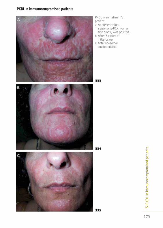
PKDL in an Italian HIV patienta. At presentation;
Leishmania PCR from a skin biopsy was positive.
b. After 3 cycles of miltefosine.
c. After liposomal amphotericine.
333
334
335
PKDL in immunocompromised patients

ThE
PK
DL
ATLA
s. A
Man
ual f
or H
ealt
h W
orke
rs
180
PKDL in immunocompromised patients
Cutaneous dissemination of visceral leishmaniasis in an HIV-positive patient from Brazil. Leishmania parasites were isolated in aspirates from bone marrow and skin.
There is a compact inflammatory infiltrate on deep dermis under a normal epidermis (338; arrows); numerous amastigotes can be seen (339, arrow).
336 337
338
339

181
5. P
KD
L in
imm
unoc
ompr
omis
ed p
atie
nts
PKDL in 2 HIV positive patients from Spain. Dermatomyositis-like presentation showing erythematous plaques with periungual erythema on dorsum of hands (340) and in the face particularly on the upper eyelids (341).
341
342 Diffuse infiltration around the nose.
PKDL in immunocompromised patients
340

ThE
PK
DL
ATLA
s. A
Man
ual f
or H
ealt
h W
orke
rs
182
PKDL in immunocompromised patients
Three patients from Ethiopia.HIV-positive with papules and nodules in the face, abdomen and extremities. Amastigotes were demonstrated in spleen aspirate and slit skin smear.
343
345 346
344
347
348HIV-positive and PKDL with Kaposi’s sarcoma-like lesions on the lower legs; amastigotes were found in a skin scraping.
349 HIV-positive with amastigotes
demonstrated from bone marrow and skin scrapings.

183
5. P
KD
L in
imm
unoc
ompr
omis
ed p
atie
nts
Cutaneous and mucosal lesions in an HIV- positive patient from Bolivia; L. (V) braziliensis was isolated from the skin. The ulceration shown (352, 353) is not a feature of PKDL.
350
352
353
351
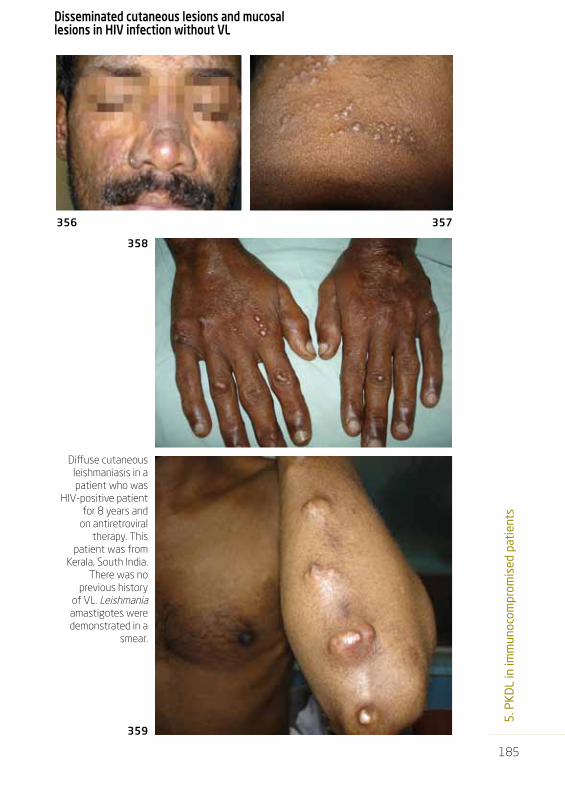
Disseminated cutaneous lesions and mucosal lesions in hIV infection without VL

ThE
PK
DL
ATLA
s. A
Man
ual f
or H
ealt
h W
orke
rs
184
PKDL in immunocompromised patients
Two HIV-positive patients from
India with low CD4 counts and no
previous history of VL. A smear
from the lesions in each patient
showed Leishmania amastigotes. There are nodular lesions on the hands (354)
and infiltrated plaques on the
nose, dorsum of the left wrist and
on the index finger of the right hand
(355).
354 355
Disseminated cutaneous lesions and mucosal lesions in hIV infection without VL
185
5. P
KD
L in
imm
unoc
ompr
omis
ed p
atie
nts
Diffuse cutaneous leishmaniasis in a patient who was
HIV-positive patient for 8 years and
on antiretroviral therapy. This
patient was from Kerala, South India.
There was no previous history
of VL. Leishmania amastigotes were demonstrated in a
smear.
356 357
359
358
Disseminated cutaneous lesions and mucosal lesions in hIV infection without VL

ThE
PK
DL
ATLA
s. A
Man
ual f
or H
ealt
h W
orke
rs
186
PKDL in immunocompromised patients
360
362
363
364
361
Disseminated cutaneous lesions and mucosal lesions in hIV infection without VL
Three HIV positive patients from India with multiple CL lesions due to L. tropica.
NoteThe conditions shown do not necessarily overlap in epidemiology with PKDL.
6. Other forms of leishmaniasis that resemble PKDL or that may be found in the same endemic area


189
6. O
ther
for
ms
of le
ishm
ania
sis
that
rese
mbl
e PK
DL
or t
hat
may
be
foun
d in
the
sam
e en
dem
ic a
rea
The presence of leishmania parasites was confirmed by PCR (L. donovani) (367). The lesion cured spontaneously and a scar was seen 6 months later (368).
Leishmanioma in sudanese patients
365
366

ThE
PK
DL
ATLA
s. A
Man
ual f
or H
ealt
h W
orke
rs
190
Other forms Leishmanioma in sudanese patients
Leishmanioma in a patient with PKDL (367); close-up (368). A smear from the lesion was positive in leishmanial PCR.
Healed leishmanioma .
367
368
369
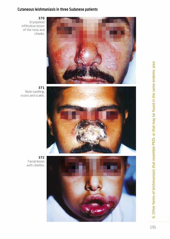
191
6. O
ther
for
ms
of le
ishm
ania
sis
that
rese
mbl
e PK
DL
or t
hat
may
be
foun
d in
the
sam
e en
dem
ic a
rea
370Erysipeloid
infiltrative lesion of the nose and
cheeks.
371Note swelling,
crusts and scales.
372Facial lesion
with cheilitis.
Cutaneous leishmaniasis in three sudanese patients
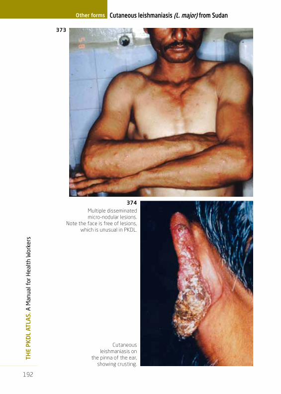
ThE
PK
DL
ATLA
s. A
Man
ual f
or H
ealt
h W
orke
rs
192
Cutaneous leishmaniasis (L. major) from sudanOther forms
Multiple disseminated micro-nodular lesions.
Note the face is free of lesions, which is unusual in PKDL.
Cutaneous leishmaniasis on
the pinna of the ear, showing crusting.
374
373
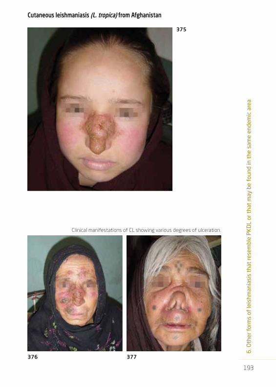
193
6. O
ther
for
ms
of le
ishm
ania
sis
that
rese
mbl
e PK
DL
or t
hat
may
be
foun
d in
the
sam
e en
dem
ic a
rea
Clinical manifestations of CL showing various degrees of ulceration.
Cutaneous leishmaniasis (L. tropica) from Afghanistan
376 377
375

ThE
PK
DL
ATLA
s. A
Man
ual f
or H
ealt
h W
orke
rs
194
Other forms Cutaneous leishmaniasis (L. tropica) from Afghanistan
381
378 379
380
Various degrees of ulceration in cutaneous leishmaniasis
(L. tropica) from Afghanistan.
Lupoid form.

195
6. O
ther
for
ms
of le
ishm
ania
sis
that
rese
mbl
e PK
DL
or t
hat
may
be
foun
d in
the
sam
e en
dem
ic a
rea
Cutaneous leishmaniasis (L. aethiopica) from Ethiopia
382
383 384
385
CL due to L. aethiopica: diffuse infiltration of the skin

ThE
PK
DL
ATLA
s. A
Man
ual f
or H
ealt
h W
orke
rs
196
Other forms
Leishmaniasis recidivans due to L. aethiopica in Ethiopian patients.
386
387
Leishmaniasis recidivans

197
6. O
ther
for
ms
of le
ishm
ania
sis
that
rese
mbl
e PK
DL
or t
hat
may
be
foun
d in
the
sam
e en
dem
ic a
rea
Leishmaniasis recidivans
388 Leishmaniasis recidivans due to (L. tropica) from Morocco. Note the healed scar from which new lesions develop.

ThE
PK
DL
ATLA
s. A
Man
ual f
or H
ealt
h W
orke
rs
198
Leishmaniasis recidivansOther forms
389 Leishmaniasis recidivans due to (L. tropica) from Afghanistan. Note the healed scars from which new lesions develop.

199
6. O
ther
for
ms
of le
ishm
ania
sis
that
rese
mbl
e PK
DL
or t
hat
may
be
foun
d in
the
sam
e en
dem
ic a
rea
Leishmaniasis recidivans
Leishmaniasis recidivans due to L. tropica from Morocco (390,) and Afghanistan
(391, 392).
390
391
392

ThE
PK
DL
ATLA
s. A
Man
ual f
or H
ealt
h W
orke
rs
200
Diffuse Cutaneous Leishmaniasis (DCL)
from Ethiopia. The patient originating from the
Highlands where CL and not VL is endemic; there is
no previous history of VL. Leishmania
parasites were found in a skin scraping.
Diffuse Cutaneous LeishmaniasisOther forms
393
395
394

201
6. O
ther
for
ms
of le
ishm
ania
sis
that
rese
mbl
e PK
DL
or t
hat
may
be
foun
d in
the
sam
e en
dem
ic a
rea
Diffuse Cutaneous Leishmaniasis
Diffuse Cutaneous Leishmaniasis (DCL); Venezuela.
Diffuse Cutaneous Leishmaniasis (DCL); Venezuela.
396
400
398
397
399

ThE
PK
DL
ATLA
s. A
Man
ual f
or H
ealt
h W
orke
rs
202
Diffuse cutaneous
leishmaniasis (DCL) in Venezuela
is normally by L. amazonensis.
Diffuse Cutaneous LeishmaniasisOther forms
401
403
402

203
6. O
ther
for
ms
of le
ishm
ania
sis
that
rese
mbl
e PK
DL
or t
hat
may
be
foun
d in
the
sam
e en
dem
ic a
rea
Diffuse Cutaneous Leishmaniasis
Diffuse cutaneous leishmaniasis; Venezuela (see also Figs 357, 358 an 361 for comparison)
404
405

ThE
PK
DL
ATLA
s. A
Man
ual f
or H
ealt
h W
orke
rs
204
Mucosal leishmaniasis Other forms
Mucosal leishmaniasis (sudan). Fungating or tumour-like form. May be primarily mucosal (L. major) or post-kala-azar mucosal leishmaniasis (L. donovani).Clinical clues: Chronic ulceration of the nasal mucosa; previous history of VL. Lesions on the palate and the gums should be looked for.
Mucosal leishmaniasis (sudan).
Before and after treatment with Pentostam®.
408
407
406

205
Leishmaniasis in tattoos
409
410
Cutaneous leishmaniasis in a tattoo due to L. infantum from Spain in an HIV –infected patient who had no previous history of CL or VL. The predilection for previous lesions or damaged skin is also a feature of PKDL (see Köbner’s phenomenon, Figs 186-189).


7. Literature


209
7. L
iter
atur
e
1. Zijlstra EE, Musa AM, Khalil EAG, El Hassan IM, El-Hassan AM. Post-kala-azar dermal leishmaniasis (Review). Lancet Infectious Diseases 2003;3:87-98.
2. Ritmeijer K, Dejenie A, Assefa Y, Hundie T, Mesure J, Boots G, den Boer M, Davidson RN. A Comparison of Miltefosine and Sodium Stibogluconate for Treatment of Visceral Leishmaniasis in an Ethiopian Population with High Prevalence of HIV Infection. Clinical Infectious Diseases 2006; 43:357–64.
3. Musa AM, Khalil EAG, Raheem MA, Zijlstra EE, Ibrahim ME, Elhassan IM, Mukhtar MM, El Hassan AM. The natural history of Sudanese post- kala-azar dermal leishmaniasis: clinical, immunological and prognostic features. Annals of Tropical Medicine and Parasitology 2002;96:765-72.
4. Zijlstra EE, El-Hassan AM. Leishmaniasis in Sudan.Trans¬actions of the Royal Society of Tropical Medi¬cine and Hygiene 2001; 95; supplement 1: S1-S76.
5. Zijlstra EE, Khalil EAG, Kager PA, El-Hassan AM. Post-kala-azar dermal leishmaniasis in the Sudan: clinical presentation and differential diagnosis. British Journal of Dermatology 2000;142:1-9.
6. El Hassan AM, Khalil EAG, El Sheikh EA, Zijlstra EE, Osman A, Ibrahim ME. Post kala-azar ocular leishmaniasis. Trans¬actions of the Royal Society of Tropical Medi¬cine and Hygiene, 1998;92:177-9.
7. El-Hassan AM, El-Sheikh EA, Eltoum IA, Ghalib HW, Ali MS, Zijlstra E, Sat¬ti M. Post-kala-azar anterior uveitis: demon¬strat¬ion of Leishmania paras¬ites in the lesion. Tran¬sac¬tions of the Royal Society of Tropical Medic¬ine and Hygiene 1991;85¬:471-3
8. Zijlstra EE, El-Hassan AM, Ismael A. Endemic kala-azar in eastern Sudan. Post-kala-azar dermal leishmaniasis. American Journal of Tropical Medicine and Hygiene 1995;52:299-305.
9. El-Hassan AM, Zijlstra EE, Ismael A, Ghalib HW. Recent observations on the epidemiology of kala-azar in the Eastern and Central States of the Sudan. Tropical and Geographical Medi¬cine 1995;47:151-156.
10. El-Hassan AM, Meredith SEO, Yagi HI, Khalil EAG, Ghalib HW, Abbas K, Zijlstra EE, Kroon CCM, Schoone GJ, Ismael A. Sudan mucosal leishmaniasis: Epidemiology, clinical features, diagnosis, immune responses and treatment. Trans¬actions of the Royal Society of Tropical Medi¬cine and Hygiene 1995;89:647-52.
11. El-Hassan AM, Ali MS, Zijlstra EE, Eltoum IA, Ghali¬b HW, Ahmed HMA. Peripheral neural involve¬ment in post kala-azar dermal leishma¬niasis: a clinical and path¬ological study. Interna¬tional Journal of Dermatology 1992;31:400-3.
12. El-Hassan AM, Ghalib HW, Zijlstra EE, El¬toum IA, Satti M, Ali MS, Ali HMA. Post Kala-azar Dermal Leishmaniasis in the Sudan: clinical features, path¬ology and treatment. Tran¬sac¬tions of the Royal Soci¬ety of Tropi¬cal Medicine and Hygiene 1992;86:245-8.
13. El-Hassan AM, Hashim FA, Abdallah M, Zijlstra EE, Ghali¬b HW. Distinguis¬hing post-kala-azar dermal leish¬mania¬sis from lepro¬sy: experience in the Sudan. Leprosy Revie¬w 1993;64:¬53-9.
14. Ramesh V, Singh N. A clinical and histopathological study of macular type of post-kala-azar dermal leishmaniasis. Tropical Doctor 1999;29:205-7.
15. Salotra P, Raina A, Ramesh V. Western blot analysis of humoral immune response to Leishmania donovani antigens in patients with post-kala-azar dermal leishmaniasis. Trans¬actions of the Royal Society of Tropical Medi¬cine and Hygiene 1999;93:98-101.
16. Singh N, Ramesh V, Arora VK, Bhatia A, Kubba A, Ramam M. Nodular post-kala-azar dermal leishmaniasis: a distinct histopathological entity. Journal of Cutaneous Pathology 1998;25:95-9.
17. Mukherjee A, Ramesh V, Misra RS. Post-kala-azar dermal leishmaniasis: a light and electron microscopic study of 18 cases. Journal of Cutaneous Pathology 1993;20:320-5.
18. Ramesh V, Ramam M, Singh R, Salotra P. Hypopigmented post-kala-azar dermal leishmaniasis. International Journal of Dermatology 2008;47:414-6.
19. Ramesh V. Post-kala-azar dermal leishmaniasis with visceral leishmaniasis, or a rare presentation of visceral leishmaniasis with extensive skin manifestations. International Journal of Dermatology 2007;46:1326.
20. Ramesh V, Kumar J, Salotra P. An unusual presentation of post-kala-azar dermal leishmaniasis.Tropical Doctor 2007;37:172-3.
21. Ramesh V, Singh R, Salotra P. Short communication: post-kala-azar dermal leishmaniasis—an appraisal. Tropical Medicine and International Health 2007;12:848-51.
22. Ramesh V, Singh N. A clinical and histopathological study of macular type of post-kala-azar dermal leishmaniasis. Tropical Doctor 1999;29:205-7.

ThE
PK
DL
ATLA
s. A
Man
ual f
or H
ealt
h W
orke
rs
210
Literature
23. Gelanew T, Hurissa Z, Diro E, Kassahun A, Kuhls K, Schönian G, Hailu A. Disseminated cutaneous leishmaniasis resembling post-kala-azar dermal leishmaniasis caused by Leishmania donovani in three patients co-infected with visceral leishmaniasis and human immunodeficiency virus/acquired immunodeficiency syndrome in Ethiopia. American Journal of Tropical Medicine and Hygiene 2011;84:906-12.
24. Gelanew T, Amogne W, Abebe T, Kuhls K, Hailu A, Schönian G. A clinical isolate of Leishmania donovani with ITS1 sequence polymorphism as a cause of para-kala-azar dermal leishmaniasis in an Ethiopian human immunodeficiency virus-positive patient on highly active antiretroviral therapy. British Journal of Dermatology 2010 Oct;163(4):870-4. doi: 10.1111/j.1365-2133.2010.09768.x.
25. Shah S, Shah A, Prajapati S, Bilimoria F. Post-kala-azar dermal leishmaniasis in HIV-positive patients: A study of two cases. Indian Journal of Sexually Transmitted Diseases 2010;31:42-4.
26. World Health Organization. Control of the leishmaniases. World Health Organization Technical Report Series 2010;(949):xii-xiii, 1-186.
27. Carnaúba D Jr, Konishi CT, Petri V, Martinez IC, Shimizu L, Pereira-Chioccola VL. Atypical disseminated leishmaniasis similar to post-kala-azar dermal leishmaniasis in a Brazilian AIDS patient infected with Leishmania (Leishmania) infantum chagasi: a case report. International Journal of Infectious Diseases 2009;e504-7. Epub 2009 May 17.
28. Guffanti M, Gaiera G, Bossolasco S, Ceserani N, Ratti D, Cinque P, Salmaso F, Gianotti N, Lazzarin A. Post-Kala-Azar dermal leishmaniasis in an HIV-1-infected woman: recovery after amphotericin B following failure of oral miltefosine. American Journal of Tropical Medicine and Hygiene 2008;79:715-8.
29. Roselino AM, Chociay MF, Costa RS, Machado AA, Figueiredo JF. L. (L.) chagasi in AIDS and visceral leishmaniasis (kala-azar) co-infection. Revistas Instituto Medico Tropical Sao Paulo. 2008;50:251-4.
30. Ramos A, Cruz I, Muñez E, Salas C, Fernández A, Alvarez-Espejo T. Post-kala-azar dermal Leishmaniasis and uveitis in an HIV-positive patient. Infection 2008;36:184-6. Epub 2008 Mar 10.
31. Antinori S, Longhi E, Bestetti G, Piolini R, Acquaviva V, Foschi A, Trovati S, Parravicini C, Corbellino M, Meroni L. Post-kala-azar dermal leishmaniasis as an immune reconstitution inflammatory syndrome in a patient with acquired immune deficiency syndrome. British Journal of Dermatology 2007;157:1032-6. Epub 2007 Sep 13. Review.
32. Daudén E, Peñas PF, Rios L, Jimenez M, Fraga J, Alvar J, García-Diez A. Leishmaniasis presenting as a dermatomyositis-like eruption in AIDS. Journal of the American Academy of Dermatology 1996;35:316-9.
33. Santos-Oliveira JR, Da-Cruz AM, Pires LH, Cupolillo E, Kuhls K, Giacoia-Gripp CB, Oliveira-Neto MP. Atypical lesions as a sign of cutaneous dissemination of visceral leishmaniasis in a human immunodeficiency virus-positive patient simultaneously infected by two viscerotropic Leishmania species. American Journal of Tropical Medicine and Hygiene 2011;85:55-9.
34. Chaudhary RG, Bilimoria FE, Katare SK. Diffuse cutaneous leishmaniasis: Co-infection with human immunodeficiency virus (HIV). Indian Journal of Dermatology, Venereology and Leprology 2008;74:641-3.
35. Torrico F, Parrado R, Castro R, Marquez CJ, Torrico MC, Solano M, Reithinger R, García AL. Co Infection of Leishmania (Viannia) braziliensis and HIV: report of a case of mucosal leishmaniasis in Cochabamba, Bolivia. American Journal of Tropical Medicine and Hygiene 2009;81:555-8.
36. Mehta V, Balachandran C, Rao R, Dil SK, Indusri L. Diffuse cutaneous leishmaniasis in HIV. Dermatology Online Journal 2009;15:9.
37. Soni P, Prasad N, Khandelwal K, Ghiya BC, Mehta RD, Bumb RA, Salotra P. Unresponsive cutaneous leishmaniasis and HIV co-infection: Report of three cases. Indian Journal of Dermatology, Venereology and Leprology 2011;77:251.
38. Morgan FM, Watten RH, Kuntz RE. Post-kala-azar dermal leishmaniasis. A case report from Taiwan (Formosa). Journal of the Formosan Medical Association 1962;61:282-291.
39. ICDDR,B Health and Science Bulletin Vol. 5 No. 4 December 2007

8. Acknowledgements/ List of contributors


213
8. A
ckno
wle
dgem
ents
/Li
st o
f co
ntrib
utor
s
LIsT Of CONTRIBuTORs
Aditya P DashVector-Borne and NeglectedDiseases, Tropical Diseases Control,WHO/SEARO, IndiaFigs 204, 205.
Academic Medical Centre Department of Dermatology, Amsterdam, The NetherlandsFig 149.
Philippe DesjeuxFormerly Senior program Officer,PATH / OWH, Divonne, FranceFigs 227-31, 238, 248, 375-80,386-9, 390-2.
Ermias DiroDepartment of Internal Medicine,University of Gondar, Gondar,EthiopiaFigs 220, 225-6, 235-6, 393-395.
Abul faizFormerly of SSMC, Dhaka &Chittagong Medical College, DhakaMedical College, Dhaka University,Ministry of Health. Dhaka,BangladeshFigs 273, 275, 276, 319, 324-5.
Tesfaye GelanewInstitut fur Mikrobiologieund Hygiene, CharitéUniversitätsmedizin, Berlin, Germany;CDC, Puerto RicoFigs 232, 343-349.
José Angelo Lauletta LindosoRosimeire silva
Laboratório de Soroepidemiologia eImunobiologia, Instituto de MedicinaTropical de São Paulo - USP. SãoPaulo, BrazilFigs 329-30.
Rogelio López-VélezTropical Medicine & ClinicalParasitology. Infectious DiseasesDepartment. Ramon y Cajal Hospital,Madrid, Spain.Fig 331.
Médecins sans frontières-holland
Amsterdam, the NetherlandsFigs 270-2, 277-85, 288-318.
Wim van der MeijdenDept of Dermatology, Erasmus MCRotterdam, the NetherlandsFigs 152, 168, 171.
Damas ObvalaMinistère de la Santé Publique,Brazzaville, Democratic Republic ofCongoFigs 169, 170.
Lluís PuigDept of Dermatology, Hospital dela Santa Creu i Sant Pau, Barcelona.SpainFig 332.
ACKNOWLEDGEMENTsWe are most grateful to all who generously contributed to this atlas. The Figures from Sudan were collected by the Leishmaniasis Research Group of the Institute of Endemic Diseases, Khartoum, under the leadership of Professor Ahmed Mohamed El-Hassan.We would like to express our sincere gratitude to all the patients who kindly consented to be photographed.

ThE
PK
DL
ATLA
s. A
Man
ual f
or H
ealt
h W
orke
rs
214
Acknowledgements/List of contributors
The following figures were reproduced with permission from:
– Transactions of the Royal Society of Tropical Medicine and Hygiene 2001; 95; supplement 1: S59-S76. Figs: 7, 11, 67, 73, 88, 113, 130, 153, 154, 163, 164, 193, 194, 207, 216, 217, 218, 366, 371-5, 406, 407.
– Lancet Infectious Diseases 2003;3:87-98: Figs 51, 63, 66, 190– British Journal of Dermatology 2000;142:1-9.: Fig 189.– Transactions of the Royal Society of Tropical Medicine and Hygiene,
1998;92:177-9. Fig 202.– Indian Journal of Sexually Transmitted Diseases 2010;31:42-4. Figs 233– Tropical Doctor 2007;37:172-3. Figs 250, 251. – Journal of the Formosan Medical Association 1962;61:282-291. Fig 328.– American Journal of Tropical Medicine and Hygiene 2008;79:715-8. Fig 334-6.– American Journal of Tropical Medicine and Hygiene 2011;85:55-9. Fig 337-40.– Journal of the American Academy of Dermatology 1996;35:316-9. Figs 341-3.– American Journal of Tropical Medicine and Hygiene 2011;84:906-12. Figs 344-50.– American Journal of Tropical Medicine and Hygiene 2009;81:555-8. Figs 351-4.– Indian Journal of Dermatology, Venereology and Leprology 2011;77:251. Figs 355-6.– Indian Journal of Dermatology, Venereology and Leprology 2011;77:251. Figs 361-5.– Dermatology Online Journal 2009;15:9. Figs 357-60.– Transactions of the Royal Society of Tropical Medicine and Hygiene 1995;89:647-
52. Fig 405.
Recognition Is given to the Fundación para la Cooperación Internacional, Salud y Política Social for support.
Iftekhar QuasemChild Health Research Foundation.Dhaka Shishu Hospital, Dhaka,BangladeshFigs 320-3.
V RameshDepartment of Dermatology,Safdarjung Hospital. New Delhi,IndiaFigs 240-7, 249-7, 261-9.
TALCSt Albans, UKFigs 203, 233-4.
Olga ZerpaInstituto de Biomedicina, UCV.MPPS. Caracas, VenezuelaFigs 174-5, 396-405.


![Immunogenicity Evaluation of a Rationally Designed ... · iasis and post Kala-Azar dermal leishmaniasis has been ascribed to CD8+ T cell exhaustion due to long lasting infection [29,30]](https://img.pdfslide.net/doc/110x75/5d273b4d88c9939b378b4d90/immunogenicity-evaluation-of-a-rationally-designed-iasis-and-post-kala-azar.jpg)

![The Post Kala-azar Dermal Leishmaniasis (PKDL) Atlas A ...Acknowledgements/List of contributors [pag. 211] Preface Preface Post Kala-azar dermal leishmaniasis (PKDL) is a well-recognized](https://img.pdfslide.net/doc/110x75/6050aaeeae32d42fa7003b62/the-post-kala-azar-dermal-leishmaniasis-pkdl-atlas-a-acknowledgementslist.jpg)