Embed Size (px)
Citation preview
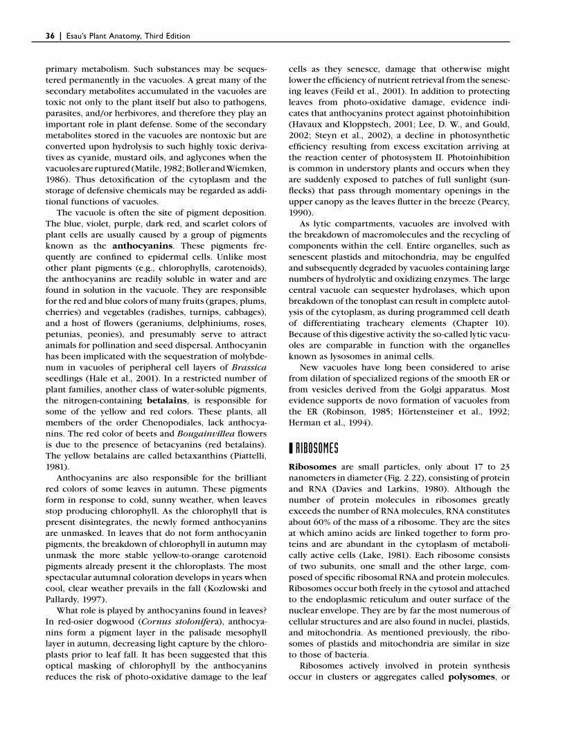
The Protoplast: Plasma Membrane, Nucleus, and Cytoplasmic Organelles
CHAPTER TWO
Cells represent the smallest structural and functional units of life (Sitte, 1992). Living organisms consist of single cells or of complexes of cells. Cells vary greatly in size, form, structure, and function. Some are mea-sured in micrometers, others in millimeters, and still others in centimeters (fi bers in certain plants). Some cells perform a number of functions; others are special-ized in their activities. Despite the extraordinary diver-sity among cells they are remarkably similar to one another, both in their physical organization and in their biochemical properties.
The concept that the cell is the basic unit of biologi-cal structure and function is based on the cell theory, which was formulated in the fi rst half of the nineteenth century by Mathias Schleiden and Theodor Schwann. In 1838, Schleiden concluded that all plant tissues are com-posed of cells. A year later, Schwann (1839) extended Schleiden’s observation to animal tissues and proposed a cellular basis for all life. In 1858, the idea that all living organisms are composed of one or more cells took on even broader signifi cance when Rudolf Virchow gener-alized that all cells arise only from preexisting cells. In its classical form, the cell theory proposed that the
bodies of all plants and animals are aggregates of indi-vidual, differentiated cells, and that the activities of the whole plant and animal might be considered the sum-mation of the activities of the individual constituent cells, with the individual cells of prime importance.
By the latter half of the nineteenth century, an alter-native to the cell theory was formulated. Known as the organismal theory, it maintains that the entire organ-ism is not merely a group of independent units but rather a living unit subdivided into cells, which are con-nected and coordinated into a harmonious whole. An often quoted statement is that of Anton de Bary (1879), “It is the plant that forms cells, and not the cell that forms plants” (translation by Sitte, 1992). Since then substantial evidence has accumulated in favor of an organismal concept for plants (see Kaplan and Hagemann, 1991; Cooke and Lu, 1992; and Kaplan, 1992; and literature cited therein).
The organismal theory is especially applicable to plants, whose cells do not pinch apart during cell divi-sion, as do animal cells, but are partitioned initially by insertion of a cell plate (Chapter 4). The separation of plant cells is rarely complete. Contiguous plant cells
15
Esau’s Plant Anatomy, Third Edition, By Ray F. Evert.Copyright © 2006 John Wiley & Sons, Inc.
16 | Esau’s Plant Anatomy, Third Edition
remain interconnected by cytoplasmic strands known as plasmodesmata, which traverse the walls and unite the entire plant body into an organic whole. Appropri-ately, plants have been characterized as supracellular organisms (Lucas et al., 1993).
In its modern form the cell theory states simply that: (1) all organisms are composed of one or more cells, (2) the chemical reactions of a living organism, including its energy-related processes and its biosynthetic pro-cesses, occur within cells, (3) cells arise from other cells, and (4) cells contain the hereditary information of the organisms of which they are a part, and this informa-tion is passed on from parent to daughter cell. The cell and organismal theories are not mutually exclusive. Together, they provide a meaningful view of the struc-ture and function at cellular and organismal levels (Sitte, 1992).
The word cell, meaning “little room,” was introduced by Robert Hooke in the seventeenth century to describe the small cavities separated by cell walls in cork tissue. Later Hooke recognized that living cells in other plant tissues were fi lled with “juices.” Eventually the contents of cells were interpreted as living matter and received the name protoplasm. An important step toward rec-ognition of the complexity of protoplasm was the dis-covery of the nucleus by Robert Brown in 1831. This discovery was soon followed by reports of cell division. In 1846, Hugo von Mohl called attention to the distinc-tion between protoplasmic material and cell sap, and in 1862, Albert von Kölliker used the term cytoplasm for the material surrounding the nucleus. The most con-spicuous inclusions in the cytoplasm, the plastids, were long considered to be merely condensations of proto-plasm. The concept of independent identity and conti-nuity of these organelles was established in the nineteenth century. In 1880, Johannes Hanstein intro-duced the term protoplast to designate the unit of protoplasm inside the cell wall.
Every living cell has a means of isolating its contents from the external environment. A membrane called the plasma membrane, or plasmalemma, brings about this isolation. Plant cells have, in addition, a more or less rigid cellulosic cell wall (Chapter 4) deposited outside the plasma membrane. The plasma membrane controls the passage of materials into and out of the protoplast and so makes it possible for the cell to differ structurally and biochemically from its surroundings. Processes within a cell can release and transfer the energy neces-sary for growth and for the maintenance of metabolic processes. A cell is organized to retain and transfer information so that its development and that of its progeny can occur in an orderly manner. This way the integrity of the organism, of which the cells are a part, is maintained.
In the three centuries since Hooke fi rst observed the structure of cork through his rudimentary microscope,
our capacity to see the cell and its contents has increased dramatically. With improvement of the light microscope, it became possible to observe objects with a diameter of 0.2 micrometer (about 200 nanometers), an improve-ment on the naked eye about 500 times. With the trans-mission electron microscope (TEM), the limit of resolution imposed by light was greatly reduced. Because of problems with specimen preparation, contrast, and radiation damage, however, the resolution of biological objects is more like 2 nanometers. Nonetheless, this is still 100 times better than the resolution of the light microscope. The TEM has distinct disadvantages, however: the specimen to be observed must be pre-served (dead) and cut into exceedingly thin, effectively two-dimensional slices. Optical microscopy using fl uo-rescent dyes and various methods of illumination have enabled biologists to overcome these problems and to observe subcellular components in live plant cells (Fricker and Oparka, 1999; Cutler and Ehrhardt, 2000). Notable is the use of green fl uorescent protein (GFP), from the jelly fi sh Aequorea victoria, as a fl uorescent protein tag and of confocal microscopy to visualize the fl uorescent probes in intact tissues (Hepler and Gunning, 1998; Fricker and Oparka, 1999; Hawes et al., 2001). The observation of subcellular components in live plant cells is providing new and often unexpected insights into subcellular organization and dynamics.
❙ PROKARYOTIC AND EUKARYOTIC CELLSBased on the degree of internal organization of their cells, two fundamentally distinct groups of organisms are now recognized: prokaryotes and eukaryotes. The prokaryotes (pro, before; karyon, nucleus) are repre-sented by the Archaea and Bacteria, including the cyano-bacteria, and the eukaryotes (eu, true; karyon, nucleus) by all other living organisms (Madigan et al., 2003).
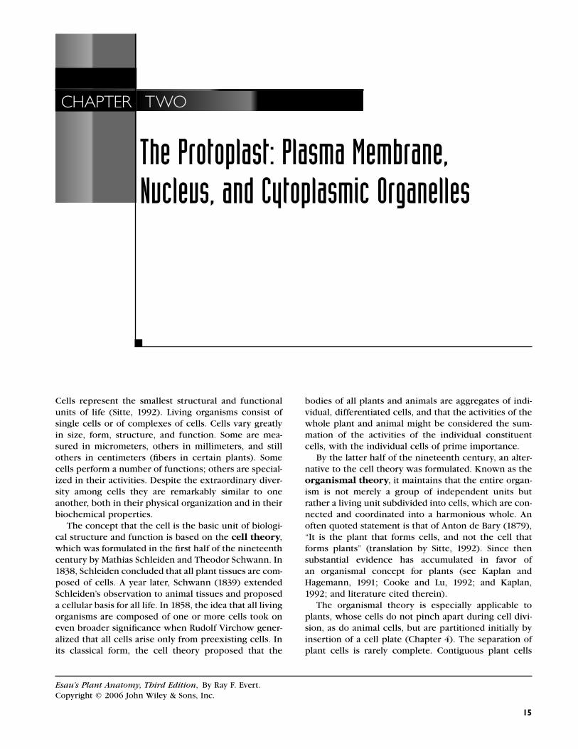
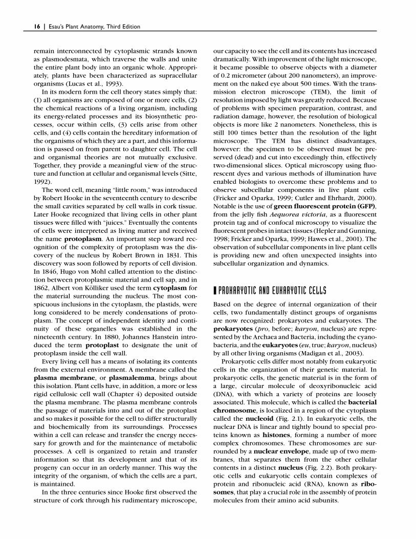
Prokaryotic cells differ most notably from eukaryotic cells in the organization of their genetic material. In prokaryotic cells, the genetic material is in the form of a large, circular molecule of deoxyribonucleic acid (DNA), with which a variety of proteins are loosely associated. This molecule, which is called the bacterial chromosome, is localized in a region of the cytoplasm called the nucleoid (Fig. 2.1). In eukaryotic cells, the nuclear DNA is linear and tightly bound to special pro-teins known as histones, forming a number of more complex chromosomes. These chromosomes are sur-rounded by a nuclear envelope, made up of two mem-branes, that separates them from the other cellular contents in a distinct nucleus (Fig. 2.2). Both prokary-otic cells and eukaryotic cells contain complexes of protein and ribonucleic acid (RNA), known as ribo-somes, that play a crucial role in the assembly of protein molecules from their amino acid subunits.
The Protoplast: Plasma Membrane, Nucleus, and Cytoplasmic Organelles | 17
Eukaryotic cells are subdivided by membranes into distinct compartments that perform different functions. The cytoplasm of prokaryotic cells, by contrast, typi-cally is not compartmentalized by membranes. Notable exceptions are the extensive system of photosynthetic membranes (thylakoids) of the cyanobacteria (Madigan et al., 2003) and the membrane-bounded entities called acidocalcisomes found in a variety of bacteria, including Agrobacterium tumefaciens, the plant pathogen that causes crown gall (Seufferheld et al., 2003).
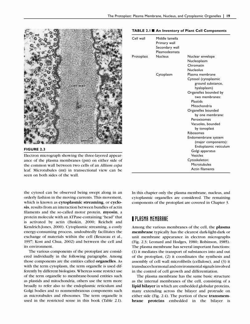
The appearance of membranes under the electron microscope is remarkably similar in various organisms. When suitably preserved and stained, these membranes have a three-layered appearance, consisting of two dark layers separated by a lighter layer (Fig. 2.3). This type of membrane was named unit membrane by
Robertson (1962) and interpreted as a bimolecular lipid layer covered on each side with a layer of protein. Although this model of membrane structure has been superseded by the fl uid mosaic model (see below), the term unit membrane remains a useful designation for a visually defi nable three-ply membrane.
Among the internal membranes of eukaryotic cells are those surrounding the nucleus, mitochondria, and plastids, which are characteristic components of plant cells. The cytoplasm of eukaryotic cells also contains systems of membranes (the endoplasmic reticulum and Golgi apparatus) and a complex network of nonmem-branous protein fi laments (actin fi laments and microtu-bules) called the cytoskeleton. A cytoskeleton is absent in prokaryotic cells. Plant cells also develop multifunc-tional organelles, called vacuoles, that are bound by a membrane called the tonoplast (Fig. 2.2).
In addition to the plasma membrane, which controls the passage of substances into and out of the protoplast, the internal membranes control the passage of sub-stances among compartments within the cell. This way the cell can maintain the specialized chemical environ-ments necessary for the processes occurring in the dif-ferent cytoplasmic compartments. Membranes also permit differences in electrical potential, or voltage, to become established between the cell and its environ-ment and between adjacent compartments of the cell. Differences in the chemical concentration of various ions and molecules and the electric potential across membranes provide potential energy used to power many cellular processes.
Compartmentation of cellular contents means divi-sion of labor at the subcellular level. In a multicellular organism a division of labor occurs also at the cellular level as the cells differentiate and become more or less specialized with reference to certain functions. Func-tional specialization fi nds its expression in morphologi-cal differences among cells, a feature that accounts for the complexity of structure in a multicellular organism.
❙ CYTOPLASMAs mentioned previously, the term cytoplasm was introduced to designate the protoplasmic material sur-rounding the nucleus. In time, discrete entities were discovered in this material, fi rst only those that were within the resolving power of the light microscope; later, smaller entities were discovered with the electron microscope. Thus the concept of cytoplasm has under-gone an evolution; with new technologies the concept undoubtedly will continue to evolve. Most biologists today use the term cytoplasm, as originally introduced by Kölliker (1862), to designate all of the material sur-rounding the nucleus, and they refer to the cytoplasmic
DNAregion
plasmamembrane
ribosomes
0.2 mm
FIGURE 2.1
Electron micrograph of the gram-negative bacterium, Azotobacter vinelandii. The granular appearance of the cytoplasm is largely due to the presence of numer-ous ribosomes. The clearer DNA-containing regions constitute the nucleoid. (Courtesy of Jack L. Pate.)
18 | Esau’s Plant Anatomy, Third Edition
w
p
v
er
v
pmm
nunununu
nene
n
m
nunu
ne
5 mm
n
o
o
FIGURE 2.2
Nicotiana tabacum (tobacco) root tip. Longitudinal section of young cells. Details: er, endoplasmic reticulum; m, mitochondrion; n, nucleus; ne, nuclear envelope; nu, nucleolus; o, oil body; p, plastid; v, vacuole; w, cell wall. (From Esau, 1977.)
matrix, in which the nucleus, organelles, membrane systems, and nonmembranous entities are suspended, as the cytosol. As originally defi ned, however, the term cytosol was used to refer specifi cally “to the cytoplasm minus mitochondria and endoplasmic reticulum compo-nents” in liver cells (Lardy, 1965). Cytoplasmic ground
substance and hyaloplasm are terms that commonly have been used by plant cytologists to refer to the cyto-plasmic matrix. Some biologists use the term cytoplasm in the sense of cytosol.
In the living plant cell the cytoplasm is always in motion; the organelles and other entities suspended in
The Protoplast: Plasma Membrane, Nucleus, and Cytoplasmic Organelles | 19
the cytosol can be observed being swept along in an orderly fashion in the moving currents. This movement, which is known as cytoplasmic streaming, or cyclo-sis, results from an interaction between bundles of actin fi laments and the so-called motor protein, myosin, a protein molecule with an ATPase-containing “head” that is activated by actin (Baskin, 2000; Reichelt and Kendrich-Jones, 2000). Cytoplasmic streaming, a costly energy-consuming process, undoubtedly facilitates the exchange of materials within the cell (Reuzeau et al., 1997; Kost and Chua, 2002) and between the cell and its environment.
The various components of the protoplast are consid-ered individually in the following paragraphs. Among those components are the entities called organelles. As with the term cytoplasm, the term organelle is used dif-ferently by different biologists. Whereas some restrict use of the term organelle to membrane-bound entities such as plastids and mitochondria, others use the term more broadly to refer also to the endoplasmic reticulum and Golgi bodies and to nonmembranous components such as microtubules and ribosomes. The term organelle is used in the restricted sense in this book (Table 2.1).
pmpm
mtmt
mtmt
mtmt
mtmt
pmpm
mtmt
pm
mt
mt
mt
mt
pm
mt
cell wallcell wallcell wall
0.12 0.12 mm0.12 mm
FIGURE 2.3
Electron micrograph showing the three-layered appear-ance of the plasma membranes (pm) on either side of the common wall between two cells of an Allium cepa leaf. Microtubules (mt) in transectional view can be seen on both sides of the wall.
TABLE 2.1 ■ An Inventory of Plant Cell Components
Cell wall Middle lamella Primary wall Secondary wall PlasmodesmataProtoplast Nucleus Nuclear envelope Nucleoplasm Chromatin Nucleolus Cytoplasm Plasma membrane Cytosol (cytoplasmic ground substance, hyaloplasm) Organelles bounded by two membranes: Plastids Mitochondria Organelles bounded by one membrane: Peroxisomes Vacuoles, bounded by tonoplast Ribosomes Endomembrane system (major components): Endoplasmic reticulum Golgi apparatus Vesicles Cytoskeleton: Microtubules Actin fi laments
In this chapter only the plasma membrane, nucleus, and cytoplasmic organelles are considered. The remaining components of the protoplast are covered in Chapter 3.
❙ PLASMA MEMBRANEAmong the various membranes of the cell, the plasma membrane typically has the clearest dark-light-dark or unit membrane appearance in electron micrographs (Fig. 2.3; Leonard and Hodges, 1980; Robinson, 1985). The plasma membrane has several important functions: (1) it mediates the transport of substances into and out of the protoplast, (2) it coordinates the synthesis and assembly of cell wall microfi brils (cellulose), and (3) it transduces hormonal and environmental signals involved in the control of cell growth and differentiation.
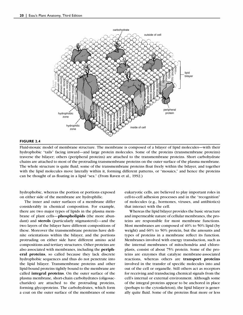
The plasma membrane has the same basic structure as the internal membranes of the cell, consisting of a lipid bilayer in which are embedded globular proteins, many extending across the bilayer and protrude on either side (Fig. 2.4). The portion of these transmem-brane proteins embedded in the bilayer is
20 | Esau’s Plant Anatomy, Third Edition
hydrophobic, whereas the portion or portions exposed on either side of the membrane are hydrophilic.
The inner and outer surfaces of a membrane differ considerably in chemical composition. For example, there are two major types of lipids in the plasma mem-brane of plant cells—phospholipids (the more abun-dant) and sterols (particularly stigmasterol)—and the two layers of the bilayer have different compositions of these. Moreover the transmembrane proteins have defi -nite orientations within the bilayer, and the portions protruding on either side have different amino acid compositions and tertiary structures. Other proteins are also associated with membranes, including the periph-eral proteins, so called because they lack discrete hydrophobic sequences and thus do not penetrate into the lipid bilayer. Transmembrane proteins and other lipid-bound proteins tightly bound to the membrane are called integral proteins. On the outer surface of the plasma membrane, short-chain carbohydrates (oligosac-charides) are attached to the protruding proteins, forming glycoproteins. The carbohydrates, which form a coat on the outer surface of the membranes of some
eukaryotic cells, are believed to play important roles in cell-to-cell adhesion processes and in the “recognition” of molecules (e.g., hormones, viruses, and antibiotics) that interact with the cell.
Whereas the lipid bilayer provides the basic structure and impermeable nature of cellular membranes, the pro-teins are responsible for most membrane functions. Most membranes are composed of 40% to 50% lipid (by weight) and 60% to 50% protein, but the amounts and types of proteins in a membrane refl ect its function. Membranes involved with energy transduction, such as the internal membranes of mitochondria and chloro-plasts, consist of about 75% protein. Some of the pro-teins are enzymes that catalyze membrane-associated reactions, whereas others are transport proteins involved in the transfer of specifi c molecules into and out of the cell or organelle. Still others act as receptors for receiving and transducing chemical signals from the cell’s internal or external environment. Although some of the integral proteins appear to be anchored in place (perhaps to the cytoskeleton), the lipid bilayer is gener-ally quite fl uid. Some of the proteins fl oat more or less
carbohydrate
outside of cell
lipidbilayer
peripheralprotein
inside of cellhydrophilic
zone
hydrophobiczone
FIGURE 2.4
Fluid-mosaic model of membrane structure. The membrane is composed of a bilayer of lipid molecules—with their hydrophobic “tails” facing inward—and large protein molecules. Some of the proteins (transmembrane proteins) traverse the bilayer; others (peripheral proteins) are attached to the transmembrane proteins. Short carbohydrate chains are attached to most of the protruding transmembrane proteins on the outer surface of the plasma membrane. The whole structure is quite fl uid; some of the transmembrane proteins fl oat freely within the bilayer, and together with the lipid molecules move laterally within it, forming different patterns, or “mosaics,” and hence the proteins can be thought of as fl oating in a lipid “sea.” (From Raven et al., 1992.)
The Protoplast: Plasma Membrane, Nucleus, and Cytoplasmic Organelles | 21
freely in the bilayer, and they and the lipid molecules can move laterally within it, forming different patterns, or mosaics, that vary from time to time and place to place—hence the name fl uid-mosaic for this model of membrane structure (Fig. 2.4; Singer and Nicolson, 1972; Jacobson et al., 1995).
Membranes contain different kinds of transport pro-teins (Logan et al., 1997; Chrispeels et al., 1999; Kjellbom et al., 1999; Delrot et al., 2001). Two of the types are carrier proteins and channel proteins, both of which permit the movement of a substance across a membrane only down the substance’s electrochemical gradient; that is, they are passive transporters. Carrier proteins bind the specifi c solute being transported and undergo a series of conformational changes in order to transport the solute across the membrane. Channel proteins form water-fi lled pores that extend across the membrane and, when open, allow specifi c solutes (usually inorganic ions, e.g., K+, Na+, Ca2+, Cl−) to pass through them. The channels are not open continuously; instead they have “gates” that open briefl y and then close again, a process referred to as gating.
The plasma membrane and tonoplast also contain water channel proteins called aquaporins that specifi -cally facilitate the passage of water through the membranes (Schäffner, 1998; Chrispeels et al., 1999; Maeshima, 2001; Javot and Maurel, 2002). Water passes relatively freely across the lipid bilayer of biological membranes, but the aquaporins allow water to diffuse more rapidly across the plasma membrane and tono-plast. Because the vacuole and cytosol must be in con-stant osmotic equilibrium, rapid movement of water is essential. It has been suggested that aquaporins facili-tate the rapid fl ow of water from the soil into root cells and to the xylem during periods of high transpiration. Aquaporins have been shown to block the infl ux of water into cells of the roots during periods of fl ooding (Tournaire-Roux et al., 2003) and to play a role in drought avoidance in rice (Lian et al., 2004). In addition evidence indicates that water movement through aqua-porins increases in response to certain environmental stimuli that induce cell expansion and growth; the cyclic expression of a plasma membrane aquaporin has been implicated in the leaf unfolding mechanism in tobacco (Siefritz et al., 2004).
Carriers can be classifi ed as uniporters and cotrans-porters according to how they function. Uniporters transport only one solute from one side of the mem-brane to another. With cotransporters, the transfer of one solute depends on the simultaneous or sequential transfer of a second solute. The second solute may be transported in the same direction, in which case the carrier protein is known as symporter, or in the oppo-site direction, as in the case of an antiporter.
The transport of a substance against its electrochemi-cal gradient requires the input of energy, and is called
active transport. In plants that energy is provided primarily by an ATP-powered proton pump, specifi -cally, a membrane-bound H+ -ATPase (Sze et al., 1999; Palmgren, 2001). The enzyme generates a large gradient of protons (H+ ions) across the membrane. This gradient provides the driving force for solute uptake by all proton-coupled cotransport systems. The tonoplast is unique among plant membranes in having two proton pumps, an H+ -ATPase and an H+ -pyrophosphatase (H+ -PPase) (Maeshima, 2001), although some data indicate that H+ -PPase may also be present in the plasma membrane of some tissues (Ratajczak et al., 1999; Maeshima, 2001).
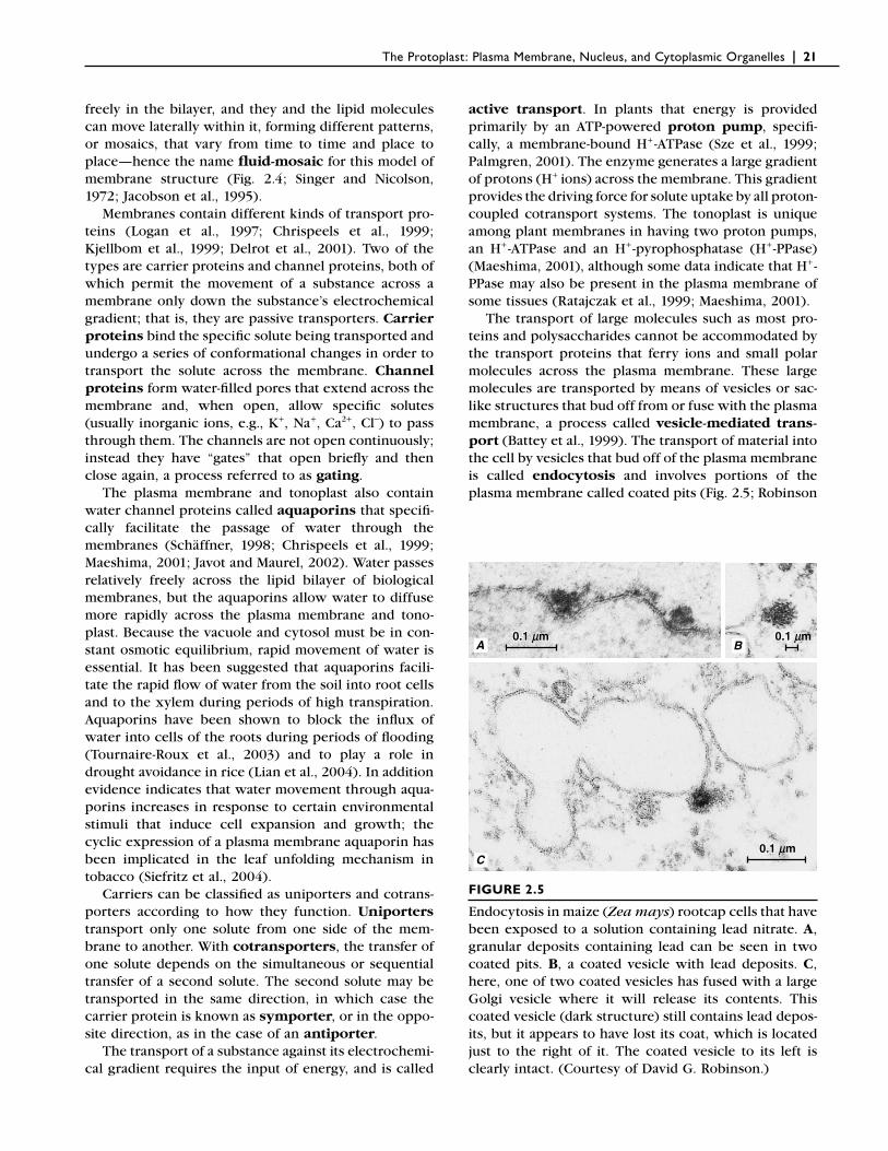
The transport of large molecules such as most pro-teins and polysaccharides cannot be accommodated by the transport proteins that ferry ions and small polar molecules across the plasma membrane. These large molecules are transported by means of vesicles or sac-like structures that bud off from or fuse with the plasma membrane, a process called vesicle-mediated trans-port (Battey et al., 1999). The transport of material into the cell by vesicles that bud off of the plasma membrane is called endocytosis and involves portions of the plasma membrane called coated pits (Fig. 2.5; Robinson
BA
C
0.1 0.1 mm0.1 mm
0.1 0.1 mm0.1 mm
0.1 0.1 mm0.1 mm
FIGURE 2.5
Endocytosis in maize (Zea mays) rootcap cells that have been exposed to a solution containing lead nitrate. A, granular deposits containing lead can be seen in two coated pits. B, a coated vesicle with lead deposits. C, here, one of two coated vesicles has fused with a large Golgi vesicle where it will release its contents. This coated vesicle (dark structure) still contains lead depos-its, but it appears to have lost its coat, which is located just to the right of it. The coated vesicle to its left is clearly intact. (Courtesy of David G. Robinson.)
22 | Esau’s Plant Anatomy, Third Edition
and Depta, 1988; Gaidarov et al., 1999). Coated pits are depressions in the plasma membrane containing spe-cifi c receptors (to which the molecules to be trans-ported into the cell must fi rst bind) and coated on their cytoplasmic surface with clathrin, a protein composed of three large and three smaller polypeptide chains that together form a three-pronged structure, called a triske-lion. Invaginations of the coated pits pinch off to form coated vesicles. Within the cell the coated vesicles shed their coats and then fuse with some other mem-brane-bound structures (e.g., Golgi bodies or small vacu-oles). Transport by means of vesicles in the opposite direction is called exocytosis (Battey et al., 1999). During exocytosis, vesicles originating from within the cell fuse with the plasma membrane, expelling their contents to the outside.
Relatively large invaginations, or infoldings, of the plasma membrane are frequently encountered in tissue prepared for electron microscopy. Some form pockets between the cell wall and protoplast, and may include tubules and vesicles. Some invaginations may push the tonoplast forward and intrude into the vacuole. Others, called multivesicular bodies, are often detached from the plasma membrane and embedded in the cytosol or appear suspended in the vacuole. Similar formations were fi rst observed in fungi and named lomasomes (Clowes and Juniper, 1968). Multivesicular bodies in Nicotiana tabacum BY-2 cells have been identifi ed as plant prevacuolar compartments that lie on the endo-cytic pathway to lytic vacuoles (see below; Tse et al., 2004).
❙ NUCLEUSOften the most prominent structure within the proto-plast of eukaryotic cells, the nucleus performs two important functions: (1) it controls the ongoing activi-ties of the cell by determining which RNA and protein molecules are produced by the cell and when they are produced, and (2) it is the repository of most of the cell’s genetic information, passing it on to the daughter cells in the course of cell division. The total genetic informa-tion stored in the nucleus is referred to as the nuclear genome.
The nucleus is bounded by a pair of membranes called the nuclear envelope, with a perinuclear space between them (Figs. 2.2 and 2.6; Dingwall and Laskey, 1992; Gerace and Foisner, 1994; Gant and Wilson, 1997; Rose et al., 2004). In various places the outer membrane of the envelope is continuous with the endo-plasmic reticulum, so that the perinuclear space is con-tinuous with the lumen of the endoplasmic reticulum. The nuclear envelope is considered a specialized, locally differentiated portion of the endoplasmic reticulum. The most distinctive feature of the nuclear envelope is
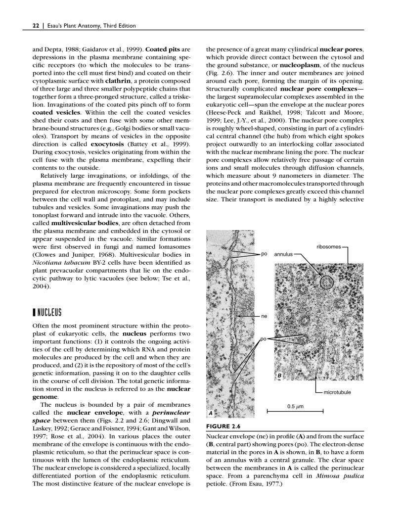
the presence of a great many cylindrical nuclear pores, which provide direct contact between the cytosol and the ground substance, or nucleoplasm, of the nucleus (Fig. 2.6). The inner and outer membranes are joined around each pore, forming the margin of its opening. Structurally complicated nuclear pore complexes—the largest supramolecular complexes assembled in the eukaryotic cell—span the envelope at the nuclear pores (Heese-Peck and Raikhel, 1998; Talcott and Moore, 1999; Lee, J.-Y., et al., 2000). The nuclear pore complex is roughly wheel-shaped, consisting in part of a cylindri-cal central channel (the hub) from which eight spokes project outwardly to an interlocking collar associated with the nuclear membrane lining the pore. The nuclear pore complexes allow relatively free passage of certain ions and small molecules through diffusion channels, which measure about 9 nanometers in diameter. The proteins and other macromolecules transported through the nuclear pore complexes greatly exceed this channel size. Their transport is mediated by a highly selective
po
po
0.5 mm
microtubule
annulusribosomes
ne
B
A
FIGURE 2.6
Nuclear envelope (ne) in profi le (A) and from the surface (B, central part) showing pores (po). The electron-dense material in the pores in A is shown, in B, to have a form of an annulus with a central granule. The clear space between the membranes in A is called the perinuclear space. From a parenchyma cell in Mimosa pudica petiole. (From Esau, 1977.)
The Protoplast: Plasma Membrane, Nucleus, and Cytoplasmic Organelles | 23
active (energy-dependent) transport mechanism that takes place through the central channel. The central channel has a functional diameter of up to 26 nanome-ters (Hicks and Raikhel, 1995; Görlich and Mattaj, 1996; Görlich, 1997).
In specially stained cells, thin threads and grains of chromatin can be distinguished from the nucleoplasm. Chromatin is made up of DNA combined with large amounts of proteins called histones. During the process of nuclear division, the chromatin becomes progres-sively more condensed until it takes the form of chro-mosomes. Chromosomes (chromatin) of nondividing, or interphase, nuclei are attached at one or more sites to the inner membrane of the nuclear envelope. Before DNA replication each chromosome is composed of a single, long DNA molecule, which carries the hereditary information. In most interphase nuclei the bulk of chro-matin is diffuse and lightly staining. This uncondensed chromatin, called euchromatin, is genetically active and associated with high rates of RNA synthesis. The remaining, condensed chromatin, called heterochro-matin, is genetically inactive; that is, it is not associated with RNA synthesis (Franklin and Cande, 1999). Overall, only a small percentage of the total chromosomal DNA codes for essential proteins or RNAs; apparently there is a substantial surplus of DNA in the genomes of higher organisms (Price, 1988). Nuclei may contain protein-aceous inclusions of unknown function in crystalline, fi brous, or amorphous form (Wergin et al., 1970), in addition to chromatin-containing “micropuffs” and coiled bodies composed of ribonucleoprotein (Martín et al., 1992).
Different organisms vary in the number of chromo-somes present in their somatic (vegetative, or body) cells. Haplopappus gracilis, a desert annual, has 4 chro-mosomes per cell; Arabidopsis thaliana, 10; Vicia faba, broad bean, 12; Brassica oleracea, cabbage, 18; Aspara-gus offi cinalis, 20; Triticum vulgare, bread wheat, 42; and Cucurbita maxima, squash, 48. The reproductive cells, or gametes, have only half the number of chromo-somes that is characteristic of the somatic cells in an organism. The number of chromosomes in the gametes is referred to as the haploid (single set) number and designated as n, and that in the somatic cells is called the diploid (double set) number, which is designated as 2n. Cells that have more than two sets of chromo-somes are said to be polyploid (3n, 4n, 5n, or more).
Often the only structures discernible within a nucleus with the light microscope are spherical structures known as nucleoli (singular: nucleolus) (Fig. 2.2; Scheer et al., 1993). The nucleolus contains high con-centrations of RNA and proteins, along with large loops of DNA emanating from several chromosomes. The loops of DNA, known as nucleolar organizer regions, contain clusters of ribosomal RNA (rRNA) genes. At these sites, newly formed rRNAs are packaged with
ribosomal proteins imported from the cytosol to form ribosomal subunits (large and small). The ribosomal subunits are then transferred, via the nuclear pores, to the cytosol where they are assembled to form ribo-somes. Although the nucleolus commonly is thought of as the site of ribosome manufacture, it is involved with only a part of the process. The very presence of a nucle-olus is due to the accumulation of the molecules being packaged to form ribosomal subunits.
In many diploid organisms, the nucleus contains one nucleolus to each haploid set of chromosomes. The nucleoli may fuse and then appear as one large struc-ture. The size of a nucleolus is a refl ection of the level of its activity. In addition to the DNA of the nucleolar organizer region, nucleoli contain a fi brillar component consisting of rRNA already associated with protein to form fi brils, and a granular component consisting of maturing ribosomal subunits. Active nucleoli also show lightly stained regions commonly referred to as vacu-oles. In living cultured cells these regions, which should not be confused with the membrane-bound vacuoles found in the cytosol, can be seen to be undergoing repeated contractions, a phenomenon that might be involved with RNA transport.
Nuclear divisions are of two kinds: mitosis, during which a nucleus gives rise to two daughter nuclei, each morphologically and genetically equivalent to the other and to the parent nucleus; meiosis, during which the parent nucleus undergoes two divisions, one of which is a reduction division. By a precise mechanism, meiosis produces four daughter nuclei, each with one-half the number of chromosomes as the parent nucleus. In plants, mitosis gives rise to somatic cells and to gametes (sperm and egg), and meiosis to meiospores. In both kinds of division (with some exceptions) the nuclear envelope breaks into fragments, which become indistin-guishable from ER cisternae, and the nuclear pore com-plexes are disassembled. When new nuclei are assembled during telophase, ER vesicles join to form two nuclear envelopes, and new nuclear pore complexes are formed (Gerace and Foisner, 1994). The nucleoli disperse during late prophase (with some exceptions) and are newly organized during telophase.
❙ CELL CYCLEActively dividing somatic cells pass through a regular sequence of events known as the cell cycle. The cell cycle commonly is divided into interphase and mitosis (Fig. 2.7; Strange, 1992). Interphase precedes mitosis, and in most cells, mitosis is followed by cytokinesis, the division of the cytoplasmic portion of a cell and the separation of daughter nuclei into separate cells (Chapter 4). Hence most plant cells are uninucleate. Certain spe-cialized cells may become multinucleate either only
24 | Esau’s Plant Anatomy, Third Edition
during their development (e.g., nuclear endosperm) or for life (e.g., nonarticulated laticifers). Mitosis and cyto-kinesis together are referred to as the M phase of the cell cycle.
Interphase can be divided into three phases, which are designated G1, S, and G2. The G1 phase (G stands for gap) occurs after mitosis. It is a period of intense bio-chemical activity, during which the cell increases in size, and the various organelles, internal membranes, and other cytoplasmic components increase in number. The S (synthesis) phase is the period of DNA replica-tion. At the onset of DNA replication, a diploid nucleus is said to have a 2C DNA value (C is the haploid DNA content); at completion of the S phase, the DNA value has doubled to 4C. During the S phase, many of the histones and other DNA-associated proteins are also syn-thesized. Following the S phase, the cell enters the G2
phase, which follows the S phase and precedes mitosis. The primary role of the S phase is to make sure chromo-some replication is complete and to allow for repair of damaged DNA. The microtubules of the preprophase band, a ring-like band of microtubules that borders the plasma membrane and encircles the nucleus in a plane corresponding to the plane of cell division, also develop during the G2 phase (Chapter 4; Gunning and Sammut, 1990). During mitosis the genetic material synthesized during the S phase is divided equally between two daughter nuclei, restoring the 2C DNA value.
The nature of the control or controls that regulate the cell cycle apparently is basically similar in all eukary-otic cells. In the typical cell cycle, progression is controlled at crucial transition points, called check-points—fi rst at the G1-S phase transition and then at the G2-M transition (Boniotti and Griffi th, 2002). The fi rst
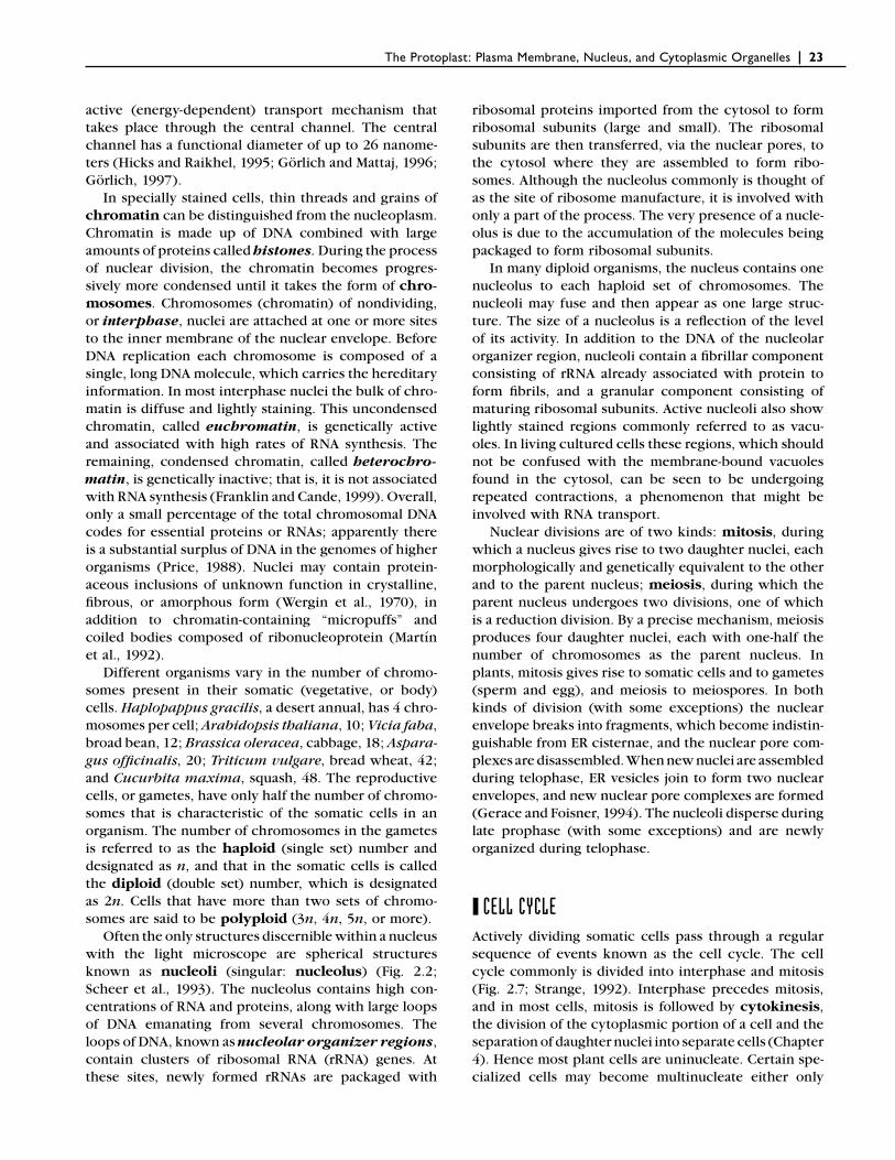
G2 phase: Structuresrequired for cell divisionbegin to assemble;chromosomes begin tocondense. G2 checkpoint
G2
M
Division
S Interphase
G1
S phase: DNA replicatedand associated proteinssynthesized; two copies ofcell’s genetic informationnow exist.
G1 checkpoint
G1 phase: Cell doublesin size; organelles,enzymes, and othermolecules increasein number.
M phase: The two setsof chromosomes areseparated (mitosis)and the cell divides(cytokinesis).
FIGURE 2.7
The cell cycle. Cell division, which consists of mitosis (the division of the nucleus) and cytokinesis (the division of the cytoplasm), takes place after the completion of the three preparatory phases (G1, S, and G2) of interphase. Pro-gression of the cell cycle is mainly controlled at two checkpoints, one at the end of G1 and the other at the end of G2. After the G2 phase comes mitosis, which is usually followed by cytokinesis. Together, mitosis and cytokinesis constitute the M phase of the cell cycle. In cells of different species or of different tissues within the same organism, the various phases occupy different proportions of the total cycle. (From Raven et al., 2005.)
The Protoplast: Plasma Membrane, Nucleus, and Cytoplasmic Organelles | 25
checkpoint determines whether or not the cell enters the S phase, the second whether or not mitosis is initi-ated. A third checkpoint, the metaphase checkpoint, delays anaphase if some chromosomes are not properly attached to the mitotic spindle. Progression through the cycle depends on the successful formation, activation, and subsequent inactivation of cyclin-dependent protein kinases (CDKs) at the checkpoints. These kinases consist of a catalytic CDK subunit and an activating cyclin subunit (Hemerly et al., 1999; Huntley and Murray, 1999; Mironov et al., 1999; Potuschak and Doerner, 2001; Stals and Inzé, 2001). Both auxins and cytokinins have been implicated in the control of the plant cell cycle (Jacqmard et al., 1994; Ivanova and Rost, 1998; den Boer and Murray, 2000).
Cells in the G1 phase have several options. In the presence of suffi cient stimuli they can commit to further cell division and progress into the S phase. They may pause in their progress through the cell cycle in response to environmental factors, as during winter dormancy, and resume dividing at a later time. This specialized resting, or dormant, state is often called the Go phase (G-zero phase). Other fates include differentiation and programmed cell death, a genetically determined program that can orchestrate death of the cell (Chapter 5; Lam et al., 1999).
Some cells feature only DNA replication and gap phases without subsequent nuclear division, a process known as endoreduplication (Chapter 5; D’Amato, 1998; Larkins et al., 2001). The single nucleus then becomes polyploid (endopolyploidy, or endoploidy). Endoploidy may be part of the differentiation of single cells, as it is in the Arabidopsis trichome (Chapter 9), or that of any tissue or organ. A positive correlation exists between cell volume and the degree of polyploidy in most plant cells, indicating that polyploid nuclei might be required for the formation of large plant cells (Kondorosi et al., 2000).
❙ PLASTIDSTogether with vacuoles and cell walls, plastids are char-acteristic components of plant cells (Bowsher and Tobin, 2001). Each plastid is surrounded by an envelope con-sisting of two membranes. Internally the plastid is dif-ferentiated into a more or less homogeneous matrix, the stroma, and a system of membranes called thylakoids. The principal permeability barrier between cytosol and plastid stroma is the inner membrane of the plastid envelope. The outer membrane, although a barrier to cytosolic proteins, has generally been assumed to be permeable to low molecular weight solutes (<600 Da), an assumption that may be in need of re-evaluation (Bölter and Soll, 2001). Stroma-fi lled tubules have been observed emanating from the surfaces of some plastids.
These so-called stromules can interconnect different plastids and have been shown to permit exchange of green fl uorescent protein between plastids (Köhler et al., 1997; Köhler and Hanson, 2000; Arimura et al., 2001; Gray et al., 2001; Pyke and Howells, 2002; Kwok and Hanson, 2004). In a study of stromule biogenesis, increases in stromule length and frequency correlated with chromoplast differentiation; it was proposed that stromules enhance the specifi c metabolic activities of plastids (Waters et al., 2004).
Plastids are semiautonomous organelles widely ac-cepted to have evolved from free-living cyanobacteria through the process of endosymbiosis (Palmer and Delwiche, 1998; Martin, 1999; McFadden, 1999; Reumann and Keegstra, 1999; Stoebe and Maier, 2002). Indeed, plastids resemble bacteria in several ways. For example, plastids, like bacteria, contain nucleoids, which are regions containing DNA. The DNA of the plastid, like that of the bacterium, exists in circular form (Sugiura, 1989); moreover it is not associated with his-tones. During the course of evolution most of the DNA of the endosymbiont (the cyanobacterium) was gradu-ally transferred to the host nucleus; hence the genome of the modern plastid is quite small compared to the nuclear genome (Bruce, 2000; Rujan and Martin, 2001). Both plastids and bacteria contain ribosomes (70S ribo-somes) that are about two-thirds as large as the ribo-somes (80S ribosomes) found in the cytosol and associated with endoplasmic reticulum. (The S stands for Svedbergs, the units of the sedimentation coeffi -cient.) In addition the process of plastid division—binary fi ssion—is morphologically similar to bacterial cell division.
Chloroplasts Contain Chlorophyll and Carotenoid Pigments
Mature plastids are commonly classifi ed on the basis of the kinds of pigments they contain. Chloroplasts (Figs. 2.8–2.10), the sites of photosynthesis, contain chloro-phyll and carotenoid pigments. The chlorophyll pig-ments are responsible for the green color of these plastids, which occur in green plant parts and are par-ticularly numerous and well differentiated in leaves. In seed plants, chloroplasts are usually disk-shaped and measure between 4 and 6 micrometers in diameter. The number of chloroplasts found in a single mesophyll (middle of the leaf) cell varies widely, depending on the species and the size of the cell (Gray, 1996). A single mesophyll cell of cocoa (Cacao theobroma) and Pep-eromia metallia leaves may contain as few as three chloroplasts, whereas as many as 300 chloroplasts occur in a single mesophyll cell of the radish (Raphanus sativus) leaf. The mesophyll cells of most leaves that have been examined for plastid development contain 50 to 150 chloroplasts each. The chloroplasts are usually
26 | Esau’s Plant Anatomy, Third Edition
found with their broad surfaces parallel to the cell wall, preferentially on cell surfaces bordering air spaces. They can reorient in the cell under the infl uence of light—for example, gathering along the walls parallel with the leaf surface under low or medium light inten-sity, thereby optimizing light utilization for photosyn-thesis (Trojan and Gabrys, 1996; Williams et al., 2003). Under potentially damaging high light intensity the chloroplasts can orient themselves along walls perpen-dicular to the leaf surface. The blue-UV region of the spectrum is the most effective stimulus for chloroplast movement (Trojan and Gabrys, 1996; Yatsuhashi, 1996; Kagawa and Wada, 2000, 2002). In the darkness the chloroplasts are distributed either randomly around all the cell walls or their arrangement depends on local factors inside the cells (Haupt and Scheuerlein, 1990). Presumably movement of the chloroplasts involves an actin-myosin-based system.
The internal structure of the chloroplast is complex. The stroma is traversed by an elaborate system of thyla-koids, consisting of grana (singular: granum)—stacks of disk-like thylakoids that resemble a stack of coins—and stroma thylakoids (or intergrana thylakoids) that traverse the stroma between grana and interconnect them (Figs. 2.8–2.10). The grana and stroma thylakoids and their internal compartments are believed to consti-tute a single, interconnected system. The thylakoids are not physically connected with the plastid envelope but are completely embedded in the stroma. Chlorophylls and carotenoid pigments—both of which are involved in light harvesting—are embedded, along with proteins, in the thylakoid membranes in discrete units of organi-zation called photosystems. The principal function of the carotenoid pigments is that of an antioxidant,
preventing photo-oxidative damage to the chlorophyll molecules (Cunningham and Gantt, 1998; Vishnevetsky et al., 1999; Niyogi, 2000).
Chloroplasts often contain starch, phytoferritin (an iron compound) and lipid in the form of globules called plastoglobuli (singular: plastoglobule). The starch grains are temporary storage products and accumulate only when the plant is actively photosynthesizing. They may be lacking in the chloroplasts of plants kept in the dark for as little as 24 hours but often reappear after the plant has been in the light for only 3 or 4 hours.
Mature chloroplasts contain numerous copies of a circular plastid DNA molecule and the machinery for the replication, transcription, and translation of that genetic material (Gray, J. C., 1996). With the limited coding capacity (approximately 100 proteins) of the chloroplast, however, the vast majority of proteins involved with chloroplast biogenesis and function are encoded by the nuclear genome (Fulgosi and Soll, 2001). These proteins, which are synthesized on ribosomes in the cytosol, are targeted into the chloroplast as precur-sor proteins with the aid of an amino-terminal exten-sion referred to as a transit peptide. Each protein imported into the chloroplast contains a specifi c transit peptide. The transit peptide both targets the protein to the chloroplast and mediates import into the stroma where it is cleaved off after import (Flügge, 1990; Smeekens et al., 1990; Theg and Scott, 1993). Transport across a thylakoid membrane is mediated by a second transit peptide unmasked when the fi rst one is cleaved off (Cline et al., 1993; Keegstra and Cline, 1999). Evi-dence indicates that part of the chloroplastic protein machinery is derived from the endosymbiotic cyanobac-terial ancestor of chloroplasts (Reumann and Keegstra, 1999; Bruce, 2000).
In addition to regulatory traffi c from the nucleus to the chloroplast, the chloroplasts transmit signals to the nucleus to coordinate nuclear and chloroplast gene expression. Moreover plastid signals also regulate the expression of nuclear genes for nonplastid proteins and for the expression of mitochondrial genes (see refer-ences in Rodermel, 2001). Chloroplasts are not only sites of photosynthesis; they are also involved in amino acid synthesis and fatty acid synthesis and provide space for the temporary storage of starch.
Chromoplasts Contain Only Carotenoid Pigments
Chromoplasts (chroma, color) are also pigmented plastids (Fig. 2.11). Of variable shape, they lack chloro-phyll but synthesize and retain carotenoid pigments, which are often responsible for the yellow, orange, or red colors of many fl owers, old leaves, some fruits, and some roots. Chromoplasts are the most heterogeneous category of plastids and are classifi ed entirely on the structure of the carotenoid-bearing components present
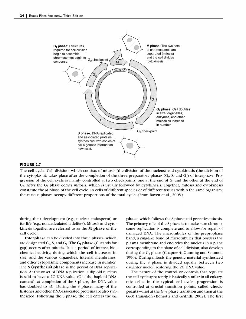
granum
granumthylakoid
stromathylakoids
intermembrane space
outer membrane
inner membranestroma
FIGURE 2.8
Three-dimensional structure of a chloroplast. Note that the internal membranes (thylakoids) are not connected with the plastid envelope. (From Raven et al., 1992.)
The Protoplast: Plasma Membrane, Nucleus, and Cytoplasmic Organelles | 27
tonoplast
vacuole
m
nncellwall
0.55 mm
plastoglobule
chloroplast envelope
plasmamembrane
tonoplast
stroma thylakoid
granum
peripheral reticulum
stroma
cell wall
granum
1 mmBA
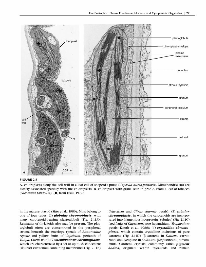
FIGURE 2.9
A, chloroplasts along the cell wall in a leaf cell of sheperd’s purse (Capsella bursa-pastoris). Mitochondria (m) are closely associated spatially with the chloroplasts. B, chloroplast with grana seen in profi le. From a leaf of tobacco (Nicotiana tabacum). (B, from Esau, 1977.)
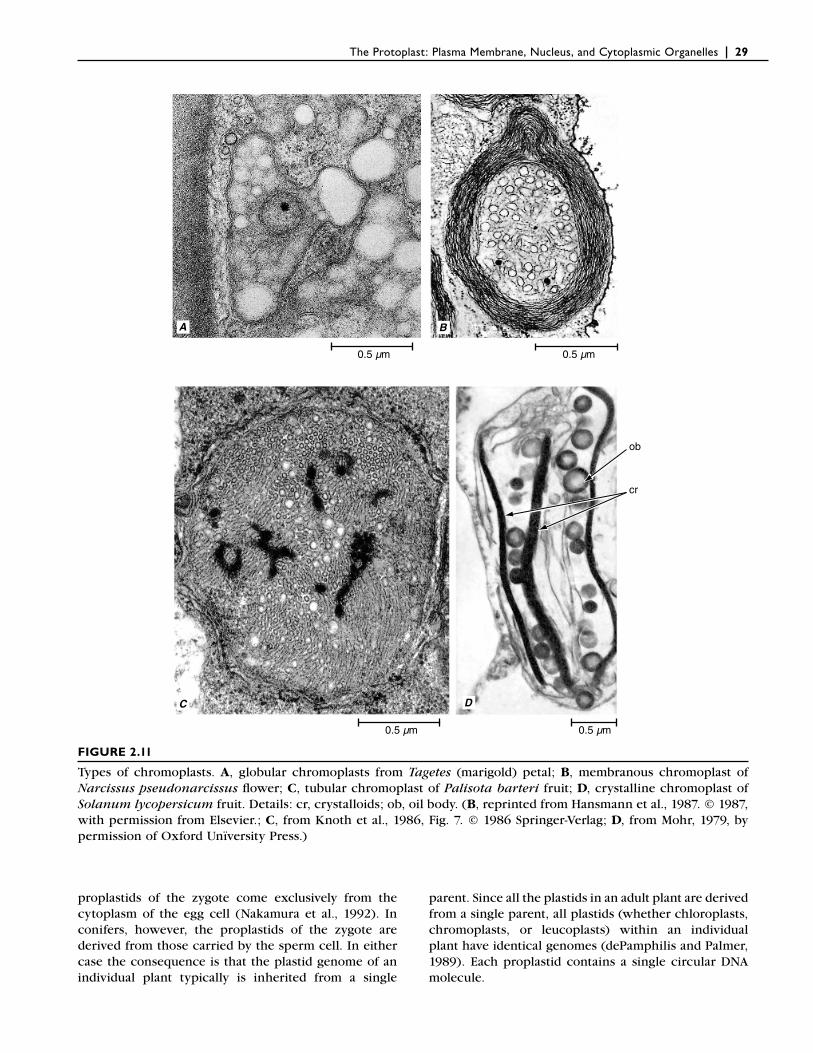
in the mature plastid (Sitte et al., 1980). Most belong to one of four types: (1) globular chromoplasts, with many carotenoid-bearing plastoglobuli (Fig. 2.11A). Remnants of thylakoids also may be present. The plas-toglobuli often are concentrated in the peripheral stroma beneath the envelope (petals of Ranunculus repens and yellow fruits of Capsicum, perianth of Tulipa, Citrus fruit); (2) membranous chromoplasts, which are characterized by a set of up to 20 concentric (double) carotenoid-containing membranes (Fig. 2.11B)
(Narcissus and Citrus sinensis petals); (3) tubular chromoplasts, in which the carotenoids are incorpo-rated into fi lamentous lipoprotein “tubules” (Fig. 2.11C) (red fruits of Capsicum, rose hypanthium; Tropaeolum petals; Knoth et al., 1986); (4) crystalline chromo-plasts, which contain crystalline inclusions of pure carotene (Fig. 2.11D) (β-carotene in Daucus, carrot, roots and lycopene in Solanum lycopersicum, tomato, fruit). Carotene crystals, commonly called pigment bodies, originate within thylakoids and remain
28 | Esau’s Plant Anatomy, Third Edition
surrounded by the plastid envelope during all stages of development. Globular chromoplasts are the most common type and are regarded as the oldest and most primitive in evolutionary terms (Camara et al., 1995).
Chromoplasts may develop from previously existing green chloroplasts by a transformation in which the chlorophyll and thylakoid membranes of the chloroplast disappear and masses of carotenoids accumulate, as occurs during the ripening of many fruits (Ziegler et al., 1983; Kuntz et al., 1989; Marano and Carrillo, 1991, 1992; Cheung et al., 1993; Ljubešic et al., 1996). Interest-ingly these changes apparently are accompanied by the disappearance of plastid ribosomes and rRNAs but not of the plastid DNA, which remains unchanged (Hansmann et al., 1987; Camara et al., 1989; Marano and Carrillo, 1991). With the loss of plastid ribosomes and rRNAs, protein synthesis can no longer occur in the chromoplast, indicating that it is necessary for chromoplast-specifi c proteins to be coded for in the nucleus and then imported into the developing chromo-plast. Chromoplast development is not an irreversible phenomenon, however. For example, the chromoplasts of citrus fruit (Goldschmidt, 1988) and of the carrot root (Grönegress, 1971) are capable of reverse differentiation into chloroplasts; they lose the carotene pigment and develop a thylakoid system, chlorophyll, and photosyn-thetic apparatus.
The precise functions of chromoplasts are not well understood, although at times they act as attractants to insects and other animals with which they coevolved, playing an essential role in the cross-pollination of fl ow-
ering plants and the dispersal of fruit and seeds (Raven et al., 2005).
Leucoplasts Are Nonpigmented Plastids
Structurally the least differentiated of mature plastids, leucoplasts (Fig. 2.12) generally have a uniform granu-lar stroma, several nucleoids, and, despite reports to the contrary, typical 70S ribosomes. They lack an elaborate system of inner membranes (Carde, 1984; Miernyk, 1989). Some store starch (amyloplasts; Fig. 2.13), others store proteins (proteinoplasts), fats (elaio-plasts), or combinations of these products. Amyloplasts are classifi ed as simple or compound (Shannon, 1989). Simple amyloplasts, such as those of the potato tuber, contain a single starch grain, whereas compound amy-loplasts contain several often tightly packed starch grains as in the endosperm of oats and rice. The starch grains of the potato tuber may become so large that the envelope is ruptured (Kirk and Tilney-Bassett, 1978). The compound amyloplasts in rootcaps play an essential role in gravity perception (Sack and Kiss, 1989; Sack, 1997).
All Plastids Are Derived Initially from Proplastids
Proplastids are small, colorless plastids found in undif-ferentiated regions of the plant body such as root and shoot apical meristems (Mullet, 1988). Zygotes contain proplastids that are the ultimate precursors of all plas-tids within an adult plant. In most angiosperms the
5 mmBA
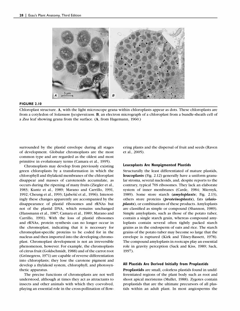
FIGURE 2.10
Chloroplast structure. A, with the light microscope grana within chloroplasts appear as dots. These chloroplasts are from a cotyledon of Solanum lycopersicum. B, an electron micrograph of a chloroplast from a bundle-sheath cell of a Zea leaf showing grana from the surface. (A, from Hagemann, 1960.)
The Protoplast: Plasma Membrane, Nucleus, and Cytoplasmic Organelles | 29
ob
cr
0.5 µm 0.5 µm
0.5 µm 0.5 µm
BA
DC
FIGURE 2.11
Types of chromoplasts. A, globular chromoplasts from Tagetes (marigold) petal; B, membranous chromoplast of Narcissus pseudonarcissus fl ower; C, tubular chromoplast of Palisota barteri fruit; D, crystalline chromoplast of Solanum lycopersicum fruit. Details: cr, crystalloids; ob, oil body. (B, reprinted from Hansmann et al., 1987. © 1987, with permission from Elsevier.; C, from Knoth et al., 1986, Fig. 7. © 1986 Springer-Verlag; D, from Mohr, 1979, by permission of Oxford Unïversity Press.)
proplastids of the zygote come exclusively from the cytoplasm of the egg cell (Nakamura et al., 1992). In conifers, however, the proplastids of the zygote are derived from those carried by the sperm cell. In either case the consequence is that the plastid genome of an individual plant typically is inherited from a single
parent. Since all the plastids in an adult plant are derived from a single parent, all plastids (whether chloroplasts, chromoplasts, or leucoplasts) within an individual plant have identical genomes (dePamphilis and Palmer, 1989). Each proplastid contains a single circular DNA molecule.
30 | Esau’s Plant Anatomy, Third Edition
proplastids must divide before the cells divide. The plastid population of mature cells typically exceeds that of the original proplastid population. The greater pro-portion of the fi nal plastid population may be derived from the division of mature plastids during the period of cell expansion. Although plastid division apparently is controlled by the nucleus (Possingham and Lawrence, 1983), a close interaction exists between plastid DNA replication and plastid division.
Plastid division is initiated by a constriction in the middle of the plastid (Fig. 2.14). With continued narrow-ing of the constriction, the two daughter plastids come to be joined by a narrow isthmus, which eventually breaks. The envelope membranes of the daughter plas-tids then reseal. The constriction process is caused by contractile rings referred to as plastid-dividing rings, which are discernible with the electron microscope as electron-dense bands. There are two concentric plastid-dividing rings, an outer ring on the cytosolic face of the plastid outer membrane and an inner ring on the stromal
1.3 1.3 mm1.3 mm
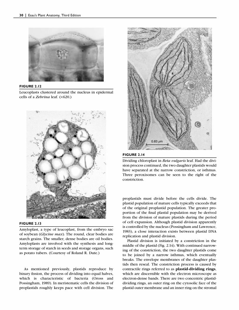
FIGURE 2.12
Leucoplasts clustered around the nucleus in epidermal cells of a Zebrina leaf. (×620.)
FIGURE 2.13
Amyloplast, a type of leucoplast, from the embryo sac of soybean (Glycine max . The round, clear bodies are starch grains. The smaller, dense bodies are oil bodies. Amyloplasts are involved with the synthesis and long-term storage of starch in seeds and storage organs, such as potato tubers. (Courtesy of Roland R. Dute.)
0.83 mm
FIGURE 2.14
Dividing chloroplast in Beta vulgaris leaf. Had the divi-sion process continued, the two daughter plastids would have separated at the narrow constriction, or isthmus. Three peroxisomes can be seen to the right of the constriction.
As mentioned previously, plastids reproduce by binary fi ssion, the process of dividing into equal halves, which is characteristic of bacteria (Oross and Possingham, 1989). In meristematic cells the division of proplastids roughly keeps pace with cell division. The
The Protoplast: Plasma Membrane, Nucleus, and Cytoplasmic Organelles | 31
face of the plastid inner membrane. Prior to the appear-ance of the plastid-dividing rings, two cytoskeletal-like proteins, FtsZ1 and FtsZ2—homologs of the bacterial cell division FtsZ protein—assemble into a ring at the future division site in the stroma within the plastid envelope. It has been suggested that the FtsZ ring determines the division region (Kuroiwa et al., 2002). Molecular analysis of chloroplast division indicates that the mechanism of plastid division has evolved from bacterial cell divi-sion (Osteryoung and Pyke, 1998; Osteryoung and McAndrew, 2001; Miyagishima et al., 2001).
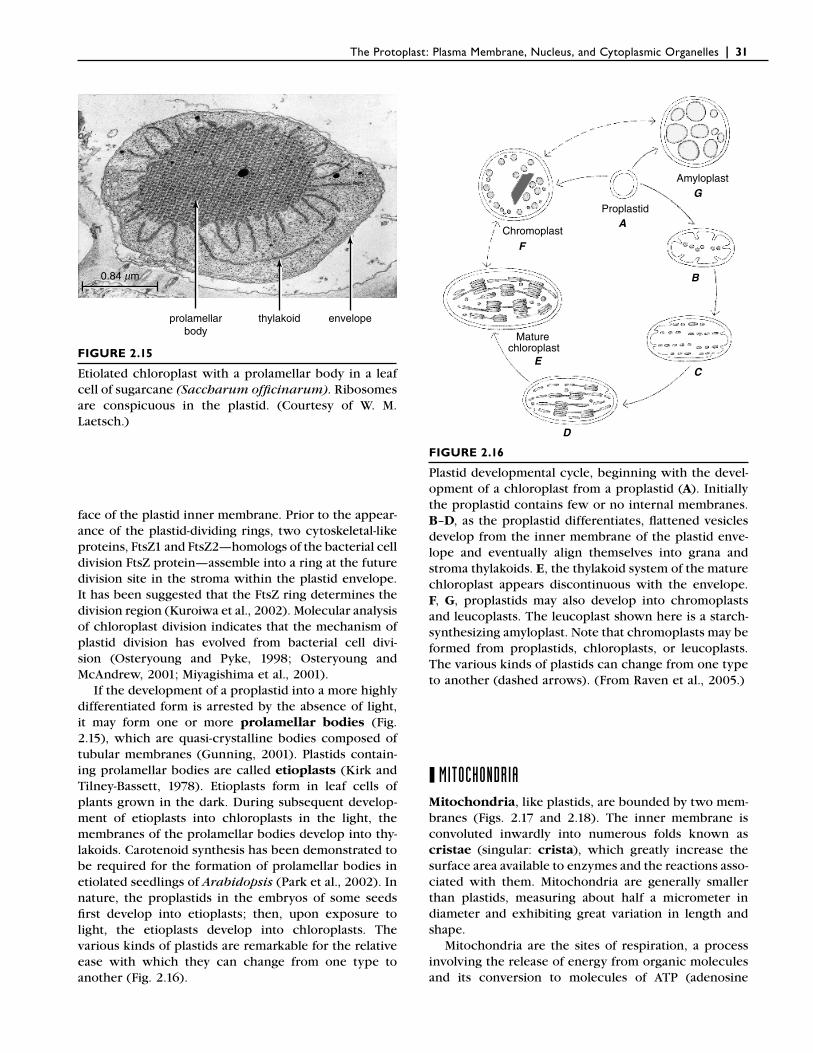
If the development of a proplastid into a more highly differentiated form is arrested by the absence of light, it may form one or more prolamellar bodies (Fig. 2.15), which are quasi-crystalline bodies composed of tubular membranes (Gunning, 2001). Plastids contain-ing prolamellar bodies are called etioplasts (Kirk and Tilney-Bassett, 1978). Etioplasts form in leaf cells of plants grown in the dark. During subsequent develop-ment of etioplasts into chloroplasts in the light, the membranes of the prolamellar bodies develop into thy-lakoids. Carotenoid synthesis has been demonstrated to be required for the formation of prolamellar bodies in etiolated seedlings of Arabidopsis (Park et al., 2002). In nature, the proplastids in the embryos of some seeds fi rst develop into etioplasts; then, upon exposure to light, the etioplasts develop into chloroplasts. The various kinds of plastids are remarkable for the relative ease with which they can change from one type to another (Fig. 2.16).
prolamellarbody
thylakoid envelope
0.84 mm
Proplastid
Chromoplast
Maturechloroplast
Amyloplast
B
C
D
E
F
G
A
FIGURE 2.15
Etiolated chloroplast with a prolamellar body in a leaf cell of sugarcane (Saccharum offi cinarum). Ribosomes are conspicuous in the plastid. (Courtesy of W. M. Laetsch.)
FIGURE 2.16
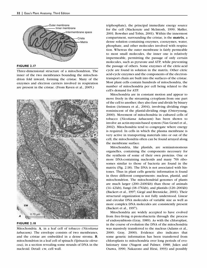
Plastid developmental cycle, beginning with the devel-opment of a chloroplast from a proplastid (A). Initially the proplastid contains few or no internal membranes. B–D, as the proplastid differentiates, fl attened vesicles develop from the inner membrane of the plastid enve-lope and eventually align themselves into grana and stroma thylakoids. E, the thylakoid system of the mature chloroplast appears discontinuous with the envelope. F, G, proplastids may also develop into chromoplasts and leucoplasts. The leucoplast shown here is a starch-synthesizing amyloplast. Note that chromoplasts may be formed from proplastids, chloroplasts, or leucoplasts. The various kinds of plastids can change from one type to another (dashed arrows). (From Raven et al., 2005.)
❙ MITOCHONDRIAMitochondria, like plastids, are bounded by two mem-branes (Figs. 2.17 and 2.18). The inner membrane is convoluted inwardly into numerous folds known as cristae (singular: crista), which greatly increase the surface area available to enzymes and the reactions asso-ciated with them. Mitochondria are generally smaller than plastids, measuring about half a micrometer in diameter and exhibiting great variation in length and shape.
Mitochondria are the sites of respiration, a process involving the release of energy from organic molecules and its conversion to molecules of ATP (adenosine
32 | Esau’s Plant Anatomy, Third Edition
triphosphate), the principal immediate energy source for the cell (Mackenzie and McIntosh, 1999; Møller, 2001; Bowsher and Tobin, 2001). Within the innermost compartment, surrounding the cristae, is the matrix, a dense solution containing enzymes, coenzymes, water, phosphate, and other molecules involved with respira-tion. Whereas the outer membrane is fairly permeable to most small molecules, the inner one is relatively impermeable, permitting the passage of only certain molecules, such as pyruvate and ATP, while preventing the passage of others. Some enzymes of the citric-acid cycle are found in solution in the matrix. Other citric acid-cycle enzymes and the components of the electron-transport chain are built into the surfaces of the cristae. Most plant cells contain hundreds of mitochondria, the number of mitochondria per cell being related to the cell’s demand for ATP.
Mitochondria are in constant motion and appear to move freely in the streaming cytoplasm from one part of the cell to another; they also fuse and divide by binary fi ssion (Arimura et al., 2004), involving dividing rings reminiscent of the plastid-dividing rings (Osteryoung, 2000). Movement of mitochondria in cultured cells of tobacco (Nicotiana tabacum) has been shown to involve an actin-myosin-based system (Van Gestel et al., 2002). Mitochondria tend to congregate where energy is required. In cells in which the plasma membrane is very active in transporting materials into or out of the cell, the mitochondria often can be found arrayed along the membrane surface.
Mitochondria, like plastids, are semiautonomous organelles, containing the components necessary for the synthesis of some of their own proteins. One or more DNA-containing nucleoids and many 70S ribo-somes similar to those of bacteria are found in the matrix (Fig. 2.18). The DNA is not associated with his-tones. Thus in plant cells genetic information is found in three different compartments: nucleus, plastid, and mitochondrion. The mitochondrial genomes of plants are much larger (200–2400 kb) than those of animals (14–42 kb), fungi (18–176 kb), and plastids (120–200 kb) (Backert et al., 1997; Giegé and Brennicke, 2001). Their structural organization is not fully understood. Linear and circular DNA molecules of variable size as well as more complex DNA molecules are consistently present (Backert et al., 1997).
Mitochondria are widely accepted to have evolved from free-living α-proteobacteria through the process of endosymbiosis (Gray, 1989). As with the chloroplast, in the course of evolution the DNA of the mitochondria was massively transferred to the nucleus (Adams et al., 2000; Gray, 2000). Evidence also indicates that some genetic information has been transferred from chloroplasts to mitochondria over long periods of evo-lutionary time (Nugent and Palmer, 1988; Jukes and Osawa, 1990; Nakazono and Hirai, 1993) and possibly
Outer membraneInner membrane
CristaMatrix
Intermembrane space
0.70 0.70 mm0.70 mm
cw
cristacrista
cristaecristae
envelopeenvelope
0.14 0.14 mm
crista
cristae
cw
cw
envelope
0.14 mm
A
B
FIGURE 2.17
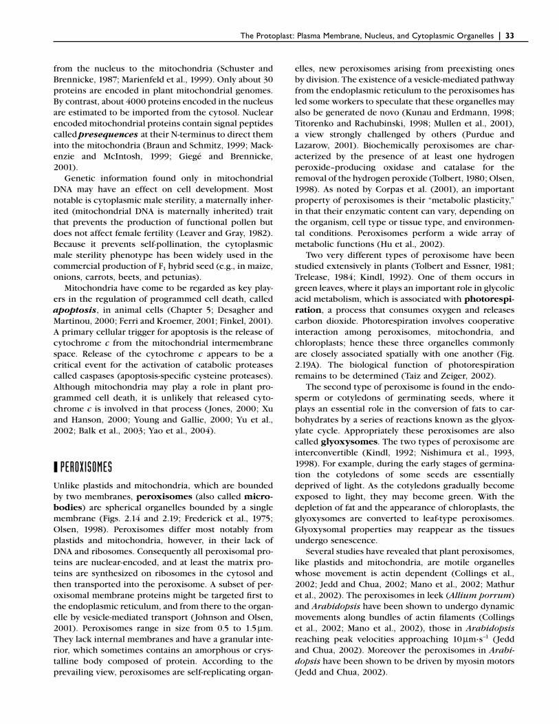
Three-dimensional structure of a mitochondrion. The inner of the two membranes bounding the mitochon-drion fold inward, forming the cristae. Many of the enzymes and electron carriers involved in respiration are present in the cristae. (From Raven et al., 2005.)
FIGURE 2.18
Mitochondria. A, in a leaf cell of tobacco (Nicotiana tabacum). The envelope consists of two membranes, and the cristae are embedded in a dense stroma. B, mitochondrion in a leaf cell of spinach (Spinacia olera-cea), in a section revealing some strands of DNA in the nucleoid. Detail: cw, cell wall.
The Protoplast: Plasma Membrane, Nucleus, and Cytoplasmic Organelles | 33
from the nucleus to the mitochondria (Schuster and Brennicke, 1987; Marienfeld et al., 1999). Only about 30 proteins are encoded in plant mitochondrial genomes. By contrast, about 4000 proteins encoded in the nucleus are estimated to be imported from the cytosol. Nuclear encoded mitochondrial proteins contain signal peptides called presequences at their N-terminus to direct them into the mitochondria (Braun and Schmitz, 1999; Mack-enzie and McIntosh, 1999; Giegé and Brennicke, 2001).
Genetic information found only in mitochondrial DNA may have an effect on cell development. Most notable is cytoplasmic male sterility, a maternally inher-ited (mitochondrial DNA is maternally inherited) trait that prevents the production of functional pollen but does not affect female fertility (Leaver and Gray, 1982). Because it prevents self-pollination, the cytoplasmic male sterility phenotype has been widely used in the commercial production of F1 hybrid seed (e.g., in maize, onions, carrots, beets, and petunias).
Mitochondria have come to be regarded as key play-ers in the regulation of programmed cell death, called apoptosis, in animal cells (Chapter 5; Desagher and Martinou, 2000; Ferri and Kroemer, 2001; Finkel, 2001). A primary cellular trigger for apoptosis is the release of cytochrome c from the mitochondrial intermembrane space. Release of the cytochrome c appears to be a critical event for the activation of catabolic proteases called caspases (apoptosis-specifi c cysteine proteases). Although mitochondria may play a role in plant pro-grammed cell death, it is unlikely that released cyto-chrome c is involved in that process (Jones, 2000; Xu and Hanson, 2000; Young and Gallie, 2000; Yu et al., 2002; Balk et al., 2003; Yao et al., 2004).
❙ PEROXISOMESUnlike plastids and mitochondria, which are bounded by two membranes, peroxisomes (also called micro-bodies) are spherical organelles bounded by a single membrane (Figs. 2.14 and 2.19; Frederick et al., 1975; Olsen, 1998). Peroxisomes differ most notably from plastids and mitochondria, however, in their lack of DNA and ribosomes. Consequently all peroxisomal pro-teins are nuclear-encoded, and at least the matrix pro-teins are synthesized on ribosomes in the cytosol and then transported into the peroxisome. A subset of per-oxisomal membrane proteins might be targeted fi rst to the endoplasmic reticulum, and from there to the organ-elle by vesicle-mediated transport (Johnson and Olsen, 2001). Peroxisomes range in size from 0.5 to 1.5 μm. They lack internal membranes and have a granular inte-rior, which sometimes contains an amorphous or crys-talline body composed of protein. According to the prevailing view, peroxisomes are self-replicating organ-
elles, new peroxisomes arising from preexisting ones by division. The existence of a vesicle-mediated pathway from the endoplasmic reticulum to the peroxisomes has led some workers to speculate that these organelles may also be generated de novo (Kunau and Erdmann, 1998; Titorenko and Rachubinski, 1998; Mullen et al., 2001), a view strongly challenged by others (Purdue and Lazarow, 2001). Biochemically peroxisomes are char-acterized by the presence of at least one hydrogen peroxide–producing oxidase and catalase for the removal of the hydrogen peroxide (Tolbert, 1980; Olsen, 1998). As noted by Corpas et al. (2001), an important property of peroxisomes is their “metabolic plasticity,” in that their enzymatic content can vary, depending on the organism, cell type or tissue type, and environmen-tal conditions. Peroxisomes perform a wide array of metabolic functions (Hu et al., 2002).
Two very different types of peroxisome have been studied extensively in plants (Tolbert and Essner, 1981; Trelease, 1984; Kindl, 1992). One of them occurs in green leaves, where it plays an important role in glycolic acid metabolism, which is associated with photorespi-ration, a process that consumes oxygen and releases carbon dioxide. Photorespiration involves cooperative interaction among peroxisomes, mitochondria, and chloroplasts; hence these three organelles commonly are closely associated spatially with one another (Fig. 2.19A). The biological function of photorespiration remains to be determined (Taiz and Zeiger, 2002).
The second type of peroxisome is found in the endo-sperm or cotyledons of germinating seeds, where it plays an essential role in the conversion of fats to car-bohydrates by a series of reactions known as the glyox-ylate cycle. Appropriately these peroxisomes are also called glyoxysomes. The two types of peroxisome are interconvertible (Kindl, 1992; Nishimura et al., 1993, 1998). For example, during the early stages of germina-tion the cotyledons of some seeds are essentially deprived of light. As the cotyledons gradually become exposed to light, they may become green. With the depletion of fat and the appearance of chloroplasts, the glyoxysomes are converted to leaf-type peroxisomes. Glyoxysomal properties may reappear as the tissues undergo senescence.
Several studies have revealed that plant peroxisomes, like plastids and mitochondria, are motile organelles whose movement is actin dependent (Collings et al., 2002; Jedd and Chua, 2002; Mano et al., 2002; Mathur et al., 2002). The peroxisomes in leek (Allium porrum) and Arabidopsis have been shown to undergo dynamic movements along bundles of actin fi laments (Collings et al., 2002; Mano et al., 2002), those in Arabidopsis reaching peak velocities approaching 10 μm·s−1 (Jedd and Chua, 2002). Moreover the peroxisomes in Arabi-dopsis have been shown to be driven by myosin motors (Jedd and Chua, 2002).

34 | Esau’s Plant Anatomy, Third Edition
peroxisome mitochondrion
chloroplast
starch
cristaribosome
1 mm
0.5 0.5 mm m 0.5 mm B
A
FIGURE 2.19
Organelles in leaf cells of sugar beet (Beta vulgaris, A) and tobacco (Nicotiana tabacum, B). The unit membranes enclosing the peroxisomes may be contrasted with the double-membraned envelopes of the other organelles. The peroxisome in B contains a crystal. Some ribosomes are perceptible in the chloroplast in A and in the mitochondrion in B. (From Esau, 1977.)
❙ VACUOLESTogether with the presence of plastids and a cell wall, the vacuole is one of the three characteristics that dis-tinguish plant cells from animal cells. As mentioned previously, vacuoles are organelles bounded by a single membrane, the tonoplast, or vacuolar membrane (Fig. 2.2). They are multifunctional organelles and are widely diverse in form, size, content, and functional dynamics (Wink, 1993; Marty, 1999). A single cell may contain more than one kind of vacuole. Some vacuoles function primarily as storage organelles, others as lytic compartments. The two types of vacuole can be char-acterized by the presence of specifi c tonoplast integral (intrinsic) proteins (TIPs): for example, whereas α-TIP is associated with the tonoplasts of protein-storage vacu-
oles, γ-TIP localizes to the tonoplasts of lytic vacuoles. Both types of TIP may colocalize to the same tonoplast of large vacuoles, apparently the result of merger of the two types of vacuole during cell enlargement (Paris et al., 1996; Miller and Anderson, 1999).
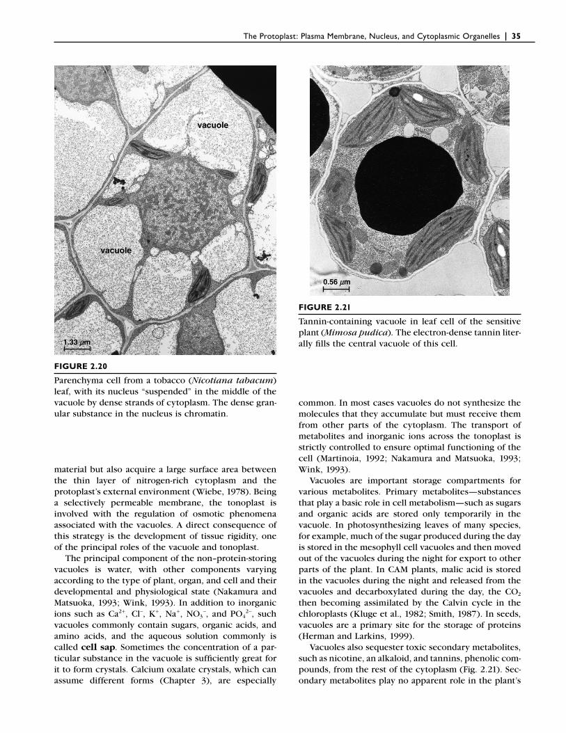
Many meristematic plant cells contain numerous small vacuoles. As the cell enlarges, the vacuoles increase in size and fuse into a single large vacuole (Fig. 2.20). Most of the increase in size of the cell in fact involves enlargement of the vacuoles. In the mature cell as much as 90% of the volume may be taken up by the vacuole, with the rest of the cytoplasm consisting of a thin peripheral layer closely pressed against the cell wall. By fi lling such a large proportion of the cell with “inex-pensive” (in terms of energy) vacuolar contents, plants not only save “expensive” nitrogen-rich cytoplasmic
The Protoplast: Plasma Membrane, Nucleus, and Cytoplasmic Organelles | 35
material but also acquire a large surface area between the thin layer of nitrogen-rich cytoplasm and the protoplast’s external environment (Wiebe, 1978). Being a selectively permeable membrane, the tonoplast is involved with the regulation of osmotic phenomena associated with the vacuoles. A direct consequence of this strategy is the development of tissue rigidity, one of the principal roles of the vacuole and tonoplast.
The principal component of the non–protein-storing vacuoles is water, with other components varying according to the type of plant, organ, and cell and their developmental and physiological state (Nakamura and Matsuoka, 1993; Wink, 1993). In addition to inorganic ions such as Ca2+, Cl−, K+, Na+, NO3
−, and PO42−, such
vacuoles commonly contain sugars, organic acids, and amino acids, and the aqueous solution commonly is called cell sap. Sometimes the concentration of a par-ticular substance in the vacuole is suffi ciently great for it to form crystals. Calcium oxalate crystals, which can assume different forms (Chapter 3), are especially
common. In most cases vacuoles do not synthesize the molecules that they accumulate but must receive them from other parts of the cytoplasm. The transport of metabolites and inorganic ions across the tonoplast is strictly controlled to ensure optimal functioning of the cell (Martinoia, 1992; Nakamura and Matsuoka, 1993; Wink, 1993).
Vacuoles are important storage compartments for various metabolites. Primary metabolites—substances that play a basic role in cell metabolism—such as sugars and organic acids are stored only temporarily in the vacuole. In photosynthesizing leaves of many species, for example, much of the sugar produced during the day is stored in the mesophyll cell vacuoles and then moved out of the vacuoles during the night for export to other parts of the plant. In CAM plants, malic acid is stored in the vacuoles during the night and released from the vacuoles and decarboxylated during the day, the CO2 then becoming assimilated by the Calvin cycle in the chloroplasts (Kluge et al., 1982; Smith, 1987). In seeds, vacuoles are a primary site for the storage of proteins (Herman and Larkins, 1999).
Vacuoles also sequester toxic secondary metabolites, such as nicotine, an alkaloid, and tannins, phenolic com-pounds, from the rest of the cytoplasm (Fig. 2.21). Sec-ondary metabolites play no apparent role in the plant’s
vacuole
vacuole
1.33 mm
FIGURE 2.20
Parenchyma cell from a tobacco (Nicotiana tabacum) leaf, with its nucleus “suspended” in the middle of the vacuole by dense strands of cytoplasm. The dense gran-ular substance in the nucleus is chromatin.
0.56 mm
FIGURE 2.21
Tannin-containing vacuole in leaf cell of the sensitive plant (Mimosa pudica). The electron-dense tannin liter-ally fi lls the central vacuole of this cell.
36 | Esau’s Plant Anatomy, Third Edition
primary metabolism. Such substances may be seques-tered permanently in the vacuoles. A great many of the secondary metabolites accumulated in the vacuoles are toxic not only to the plant itself but also to pathogens, parasites, and/or herbivores, and therefore they play an important role in plant defense. Some of the secondary metabolites stored in the vacuoles are nontoxic but are converted upon hydrolysis to such highly toxic deriva-tives as cyanide, mustard oils, and aglycones when the vacuoles are ruptured (Matile, 1982; Boller and Wiemken, 1986). Thus detoxifi cation of the cytoplasm and the storage of defensive chemicals may be regarded as addi-tional functions of vacuoles.
The vacuole is often the site of pigment deposition. The blue, violet, purple, dark red, and scarlet colors of plant cells are usually caused by a group of pigments known as the anthocyanins. These pigments fre-quently are confi ned to epidermal cells. Unlike most other plant pigments (e.g., chlorophylls, carotenoids), the anthocyanins are readily soluble in water and are found in solution in the vacuole. They are responsible for the red and blue colors of many fruits (grapes, plums, cherries) and vegetables (radishes, turnips, cabbages), and a host of fl owers (geraniums, delphiniums, roses, petunias, peonies), and presumably serve to attract animals for pollination and seed dispersal. Anthocyanin has been implicated with the sequestration of molybde-num in vacuoles of peripheral cell layers of Brassica seedlings (Hale et al., 2001). In a restricted number of plant families, another class of water-soluble pigments, the nitrogen-containing betalains, is responsible for some of the yellow and red colors. These plants, all members of the order Chenopodiales, lack anthocya-nins. The red color of beets and Bougainvillea fl owers is due to the presence of betacyanins (red betalains). The yellow betalains are called betaxanthins (Piattelli, 1981).
Anthocyanins are also responsible for the brilliant red colors of some leaves in autumn. These pigments form in response to cold, sunny weather, when leaves stop producing chlorophyll. As the chlorophyll that is present disintegrates, the newly formed anthocyanins are unmasked. In leaves that do not form anthocyanin pigments, the breakdown of chlorophyll in autumn may unmask the more stable yellow-to-orange carotenoid pigments already present it the chloroplasts. The most spectacular autumnal coloration develops in years when cool, clear weather prevails in the fall (Kozlowski and Pallardy, 1997).
What role is played by anthocyanins found in leaves? In red-osier dogwood (Cornus stolonifera), anthocya-nins form a pigment layer in the palisade mesophyll layer in autumn, decreasing light capture by the chloro-plasts prior to leaf fall. It has been suggested that this optical masking of chlorophyll by the anthocyanins reduces the risk of photo-oxidative damage to the leaf
cells as they senesce, damage that otherwise might lower the effi ciency of nutrient retrieval from the senesc-ing leaves (Feild et al., 2001). In addition to protecting leaves from photo-oxidative damage, evidence indi-cates that anthocyanins protect against photoinhibition (Havaux and Kloppstech, 2001; Lee, D. W., and Gould, 2002; Steyn et al., 2002), a decline in photosynthetic effi ciency resulting from excess excitation arriving at the reaction center of photosystem II. Photoinhibition is common in understory plants and occurs when they are suddenly exposed to patches of full sunlight (sun-fl ecks) that pass through momentary openings in the upper canopy as the leaves fl utter in the breeze (Pearcy, 1990).
As lytic compartments, vacuoles are involved with the breakdown of macromolecules and the recycling of components within the cell. Entire organelles, such as senescent plastids and mitochondria, may be engulfed and subsequently degraded by vacuoles containing large numbers of hydrolytic and oxidizing enzymes. The large central vacuole can sequester hydrolases, which upon breakdown of the tonoplast can result in complete autol-ysis of the cytoplasm, as during programmed cell death of differentiating tracheary elements (Chapter 10). Because of this digestive activity the so-called lytic vacu-oles are comparable in function with the organelles known as lysosomes in animal cells.
New vacuoles have long been considered to arise from dilation of specialized regions of the smooth ER or from vesicles derived from the Golgi apparatus. Most evidence supports de novo formation of vacuoles from the ER (Robinson, 1985; Hörtensteiner et al., 1992; Herman et al., 1994).
❙ RIBOSOMESRibosomes are small particles, only about 17 to 23 nanometers in diameter (Fig. 2.22), consisting of protein and RNA (Davies and Larkins, 1980). Although the number of protein molecules in ribosomes greatly exceeds the number of RNA molecules, RNA constitutes about 60% of the mass of a ribosome. They are the sites at which amino acids are linked together to form pro-teins and are abundant in the cytoplasm of metaboli-cally active cells (Lake, 1981). Each ribosome consists of two subunits, one small and the other large, com-posed of specifi c ribosomal RNA and protein molecules. Ribosomes occur both freely in the cytosol and attached to the endoplasmic reticulum and outer surface of the nuclear envelope. They are by far the most numerous of cellular structures and are also found in nuclei, plastids, and mitochondria. As mentioned previously, the ribo-somes of plastids and mitochondria are similar in size to those of bacteria.
Ribosomes actively involved in protein synthesis occur in clusters or aggregates called polysomes, or
The Protoplast: Plasma Membrane, Nucleus, and Cytoplasmic Organelles | 37
polyribosomes (Fig. 2.22), united by the messenger RNA molecules carrying the genetic information from the nucleus. The amino acids from which the proteins are synthesized are brought to the polysomes by transfer RNAs located in the cytosol. The synthesis of protein, known as translation, consumes more energy than any other biosynthetic process. That energy is provided by the hydrolysis of guanosine triphosphate (GTP).
The synthesis of polypeptides (proteins) encoded by nuclear genes is initiated on polysomes located in the cytosol and follows one of two divergent pathways. (1) Those polysomes involved in the synthesis of polypep-tides destined for the endoplasmic reticulum become associated with the endoplasmic reticulum early in the translational process. The polypeptides and their associ-ated polysomes are directed to the endoplasmic reticu-lum by a targeting signal, or signal peptide, located at the amino end of each polypeptide. The polypeptides are transferred across the membrane into the lumen of the ER (or are inserted into it, in the case of integral proteins) as polypeptide synthesis continues. (2) Those polysomes involved with the synthesis of polypeptides destined for the cytosol or for import into the nucleus,
mitochondria, plastids, or peroxisomes remain free in the cytosol. The polypeptides released from the free polysomes either remain in the cytosol or are targeted to the appropriate cellular component by a targeting sequence (Holtzman, 1992). Membrane-bound and free ribosomes are both structurally and functionally identi-cal, differing from one another only in the proteins they are making at any given time.
REFERENCES ADAMS, K. L., D. O. DALEY, Y.-L. QIU, J. WHELAN, and J. D.
PALMER. 2000. Repeated, recent and diverse transfers of a mitochondrial gene to the nucleus in fl owering plants. Nature 408, 354–357.
ARIMURA, S.-I., A. HIRAI, and N. TSUTSUMI. 2001. Numerous and highly developed tubular projections from plastids observed in tobacco epidermal cells. Plant Sci. 160, 449–454.
ARIMURA, S.-I., J. YAMAMOTO, G. P. AIDA, M. NAKAZONO, and N. TSUTSUMI. 2004. Frequent fusion and fi ssion of plant mito-chondria with unequal nucleoid distribution. Proc. Natl. Acad. Sci. USA 101, 7805–7808.
BACKERT, S., B. L. NIELSEN, and T. BÖRNER. 1997. The mystery of the rings: Structure and replication of mitochondrial genomes from higher plants. Trends Plant Sci. 2, 477–483.
BALK, J., S. K. CHEW, C. J. LEAVER, and P. F. MCCABE. 2003. The intermembrane space of plant mitochondria contains a DNase activity that may be involved in programmed cell death. Plant J. 34, 573–583.
BASKIN, T. I. 2000. The cytoskeleton. In: Biochemistry and Molecular Biology of Plants, pp. 202–258, B. B. Buchanan, W. Gruissem, and R. L. Jones, eds. American Society of Plant Physiologists, Rockville, MD.
BATTEY, N. H., N. C. JAMES, A. J. GREENLAND, and C. BROWNLEE. 1999. Exocytosis and endocytosis. Plant Cell 11, 643–659.
BOLLER, T., and A. WIEMKEN. 1986. Dynamics of vacuolar com-partmentation. Annu. Rev. Plant Physiol. 37, 137–164.
BÖLTER, B., and J. SOLL. 2001. Ion channels in the outer mem-branes of chloroplasts and mitochondria: Open doors or regulated gates? EMBO J. 20, 935–940.
BONIOTTI, M. B., and M. E. GRIFFITH. 2002. “Cross-talk” between cell division cycle and development in plants. Plant Cell 14, 11–16.
BOWSHER, C. G., and A. K. TOBIN. 2001. Compartmentation of metabolism within mitochondria and plastids. J. Exp. Bot. 52, 513–527.
BRAUN, H.-P., and U. K. SCHMITZ. 1999. The protein-import apparatus of plant mitochondria. Planta 209, 267–274.
BRUCE, B. D. 2000. Chloroplast transit peptides: Structure, func-tion and evolution. Trends Cell Biol. 10, 440–447.
CAMARA, B., J. BOUSQUET, C. CHENICLET, J.-P. CARDE, M. KUNTZ, J.-L. EVRARD, and J.-H. WEIL. 1989. Enzymology of isoprenoid
200 nm200 nm200 nm100 nm100 nm100 nm
BA
FIGURE 2.22
Ribosomes. A, in bundle-sheath cell of a maize (Zea mays) leaf. Arrow points to a bundle of actin fi laments. B, a polysome (polyribosome) attached to the surface of endoplasmic reticulum in a tobacco (Nicotiana tabacum) leaf cell. (B, from Esau, 1977.)
38 | Esau’s Plant Anatomy, Third Edition
biosynthesis and expression of plastid and nuclear genes during chromoplast differentiation in pepper fruits (Capsicum annuum). In: Physiology, Biochemistry, and Genetics of Nongreen Plastids, pp. 141–156, C. D. Boyer, J. C. Shannon, and R. C. Hardison, eds. American Society of Plant Physiologists, Rockville, MD.
CAMARA, B., P. HUGUENEY, F. BOUVIER, M. KUNTZ, and R. MONÉGER. 1995. Biochemistry and molecular biology of chro-moplast development. Int. Rev. Cytol. 163, 175–247.
CARDE, J.-P. 1984. Leucoplasts: A distinct kind of organelles lacking typical 70S ribosomes and free thylakoids. Eur. J. Cell Biol. 34, 18–26.
CHEUNG, A. Y., T. MCNELLIS, and B. PIEKOS. 1993. Maintenance of chloroplast components during chromoplast differentia-tion in the tomato mutant Green Flesh. Plant Physiol. 101, 1223–1229.
CHRISPEELS, M. J., N. M. CRAWFORD, and J. I. SCHROEDER. 1999. Proteins for transport of water and mineral nutrients across the membranes of plant cells. Plant Cell 11, 661–676.
CLINE, K., R. HENRY, C.-J. LI, and J.-G. YUAN. 1993. Multiple pathways for protein transport into or across the thylakoid membrane. EMBO J. 12, 4105–4114.
CLOWES, F. A. L., and B. E. JUNIPER. 1968. Plant Cells. Blackwell Scientifi c, Oxford.
COLLINGS, D. A., J. D. I. HARPER, J. MARC, R. L. OVERALL, and R. T. MULLEN. 2002. Life in the fast lane: Actin-based motility of plant peroxisomes. Can. J. Bot. 80, 430–441.
COOKE, T. J., and B. LU. 1992. The independence of cell shape and overall form in multicellular algae and land plants: Cells do not act as building blocks for constructing plant organs. Int. J. Plant Sci. 153, S7–S27.
CORPAS, F. J., J. B. BARROSO, and L. A. DEL RÍO. 2001. Peroxi-somes as a source of reactive oxygen species and nitric oxide signal molecules in plant cells. Trends Plant Sci. 6, 145–150.
CUNNINGHAM, F. X., JR., and E. GANTT. 1998. Genes and enzymes of carotenoid biosynthesis in plants. Annu. Rev. Plant Physiol. Plant Mol. Biol. 49, 557–583.
CUTLER, S., and D. EHRHARDT. 2000. Dead cells don’t dance: Insights from live-cell imaging in plants. Curr. Opin. Plant Biol. 3, 532–537.
D’AMATO, F. 1998. Chromosome endoreduplication in plant tissue development and function. In: Plant Cell Proliferation and Its Regulation in Growth and Development, pp. 153–166, J. A. Bryant and D. Chiatante, eds. Wiley, Chichester.
DAVIES, E., and B. A. LARKINS. 1980. Ribosomes. In: The Biochem-istry of Plants, vol. 1, The Plant Cell, pp. 413–435, N. E. Tolbert, ed. Academic Press, New York.
DE BARY, A. 1879. Besprechung. K. Prantl. Lehrbuch der Botanik für mittlere und höhere Lehranstalten. Bot. Ztg. 37, 221–223.
DELROT, S., R. ATANASSOVA, E. GOMÈS, and P. COUTOS-THÉVENOT. 2001. Plasma membrane transporters: A machinery for
uptake of organic solutes and stress resistance. Plant Sci. 161, 391–404.
DEN BOER, B. G. W., and J. A. H. MURRAY. 2000. Triggering the cell cycle in plants. Trends Cell Biol. 10, 245–250.
DEPAMPHILIS, C. W., and J. D. PALMER. 1989. Evolution and func-tion of plastid DNA: A review with special reference to nonphotosynthetic plants. In: Physiology, Biochemistry, and Genetics of Nongreen Plastids, pp. 182–202, C. D. Boyer, J. C. Shannon, and R. C. Hardison, eds. American Society of Plant Physiologists, Rockville, MD.
DESAGHER, S., and J.-C. MARTINOU. 2000. Mitochondria as the central control point of apoptosis. Trends Cell Biol. 10, 369–377.
DINGWALL, C., and R. LASKEY. 1992. The nuclear membrane. Science 258, 942–947.
ESAU, K. 1977. Anatomy of Seed Plants, 2nd ed. Wiley, New York.
FEILD, T. S., D. W. LEE, and N. M. HOLBROOK. 2001. Why leaves turn red in autumn. The role of anthocyanins in senescing leaves of red-osier dogwood. Plant Physiol. 127, 566–574.
FERRI, K. F., and G. KROEMER. 2001. Mitochondria—The suicide organelles. BioEssays 23, 111–115.
FINKEL, E. 2001. The mitochondrion: Is it central to apoptosis? Science 292, 624–626.
FLÜGGE, U.-I. 1990. Import of proteins into chloroplasts. J. Cell Sci. 96, 351–354.
FRANKLIN, A. E., and W. Z. CANDE. 1999. Nuclear organization and chromosome segregation. Plant Cell 11, 523–534.
FREDERICK, S. E., P. J. GRUBER, and E. H. NEWCOMB. 1975. Plant microbodies. Protoplasma 84, 1–29.
FRICKER, M. D., and K. J. OPARKA. 1999. Imaging techniques in plant transport: Meeting review. J. Exp. Bot. 50(suppl. 1), 1089–1100.
FULGOSI, H., and J. SOLL. 2001. A gateway to chloroplasts—Protein translocation and beyond. J. Plant Physiol. 158, 273–284.
GAIDAROV, I., F. SANTINI, R. A. WARREN, and J. H. KEEN. 1999. Spatial control of coated-pit dynamics in living cells. Nature Cell Biol. 1, 1–7.
GANT, T. M., and K. L. WILSON. 1997. Nuclear assembly. Annu. Rev. Cell Dev. Biol. 13, 669–695.
GERACE, L., and R. FOISNER. 1994. Integral membrane proteins and dynamic organization of the nuclear envelope. Trends Cell Biol. 4, 127–131.
GIEGÉ, P., and A. BRENNICKE. 2001. From gene to protein in higher plant mitochondria. C. R. Acad. Sci., Paris, Sci. de la Vie 324, 209–217.
GOLDSCHMIDT, E. E. 1988. Regulatory aspects of chloro-chromoplast interconversions in senescing Citrus fruit peel. Isr. J. Bot. 37, 123–130.
GÖRLICH, D. 1997. Nuclear protein import. Curr. Opin. Cell Biol. 9, 412–419.
The Protoplast: Plasma Membrane, Nucleus, and Cytoplasmic Organelles | 39
GÖRLICH, D., and I. W. MATTAJ. 1996. Nucleocytoplasmic trans-port. Science 271, 1513–1518.
GRAY, J. C. 1996. Biogenesis of chloroplasts in higher plants. In: Membranes: Specialized Functions in Plants, pp. 441–458, M. Smallwood, J. P. Knox, and D. J. Bowles, eds. BIOS Scientifi c, Oxford.
GRAY, J. C., J. A. SULLIVAN, J. M. HIBBERD, and M. R. HANSEN. 2001. Stromules: Mobile protrusions and interconnections between plastids. Plant Biol. 3, 223–233.
GRAY, M. W. 1989. Origin and evolution of mitochondrial DNA. Annu. Rev. Cell Biol. 5, 25–50.
GRAY, M. W. 2000. Mitochondrial genes on the move. Nature 408, 302–305.
GRÖNEGRESS, P. 1971. The greening of chromoplasts in Daucus carota L. Planta 98, 274–278.
GUNNING, B. E. S. 2001. Membrane geometry of “open” prola-mellar bodies. Protoplasma 215, 4–15.
GUNNING, B. E. S., and M. SAMMUT. 1990. Rearrangements of microtubules involved in establishing cell division planes start immediately after DNA synthesis and are completed just before mitosis. Plant Cell 2, 1273–1282.
HAGEMANN, R. 1960. Die Plastidenentwicklung in Tomaten-Kotyledonen. Biol. Zentralbl. 79, 393–411.
HALE, K. L., S. P. MCGRATH, E. LOMBI, S. M. STACK, N. TERRY, I. J. PICKERING, G. N. GEORGE, and E. A. H. PILON-SMITS. 2001. Molybdenum sequestration in Brassica species. A role for anthocyanins? Plant Physiol. 126, 1391–1402.
HANSMANN, P., R. JUNKER, H. SAUTER, and P. SITTE. 1987. Chro-moplast development in daffodil coronae during anthesis. J. Plant Physiol. 131, 133–143.
HANSTEIN, J. 1880. Einige Züge aus der Biologie des Protoplasmas. Botanische Abhandlungen aus dem Gebiet der Morphologie und Physiologie, Band 4, Heft 2. Marcus, Bonn.
HAUPT, W., and R. SCHEUERLEIN. 1990. Chloroplast movement. Plant Cell Environ. 13, 595–614.
HAVAUX, M., and K. KLOPPSTECH. 2001. The protective functions of carotenoid and fl avonoid pigments against excess visible radiation at chilling temperature investigated in Arabidopsis npq and tt mutants. Planta 213, 953–966.
HAWES, C., C. M. SAINT-JORE, F. BRANDIZZI, H. ZHENG, A. V. ANDREEVA, and P. BOEVINK. 2001. Cytoplasmic illuminations: in planta targeting of fl uorescent proteins to cellular organ-elles. Protoplasma 215, 77–88.
HEESE-PECK, A., and N. V. RAIKHEL. 1998. The nuclear pore complex. Plant Mol. Biol. 38, 145–162.
HEMERLY, A. S., P. C. G. FERREIRA, M. VAN MONTAGU, and D. INZÉ. 1999. Cell cycle control and plant morphogenesis: Is there an essential link? BioEssays 21, 29–37.
HEPLER, P. K., and B. E. S. GUNNING. 1998. Confocal fl uorescence microscopy of plant cells. Protoplasma 201, 121–157.
HERMAN, E. M., and B. A. LARKINS. 1999. Protein storage bodies and vacuoles. Plant Cell 11, 601–614.
HERMAN, E. M., X. LI, R. T. SU, P. LARSEN, H.-T. HSU, and H. SZE. 1994. Vacuolar-type H+ -ATPases are associated with the endoplasmic reticulum and provacuoles of root tip cells. Plant Physiol. 106, 1313–1324.
HICKS, G. R., and N. V. RAIKHEL. 1995. Protein import into the nucleus: An integrated view. Annu. Rev. Cell Dev. Biol. 11, 155–188.
HOLTZMAN, E. 1992. Intracellular targeting and sorting. Bio-Science 42, 608–620.
HÖRTENSTEINER, S., E. MARTINOIA, and N. AMRHEIN. 1992. Reap-pearance of hydrolytic activities and tonoplast proteins in the regenerated vacuole of evacuolated protoplasts. Planta 187, 113–121.
HU, J., M. AGUIRRE, C. PETO, J. ALONSO, J. ECKER, and J. CHORY. 2002. A role for peroxisomes in photomorphogenesis and development of Arabidopsis. Science 297, 405–409.
HUNTLEY, R. P., and J. A. H. MURRAY. 1999. The plant cell cycle. Curr. Opin. Plant Biol. 2, 440–446.
IVANOVA, M., and T. L. ROST. 1998. Cytokinins and the plant cell cycle: Problems and pitfalls of proving their function. In: Plant Cell Proliferation and Its Regulation in Growth and Development, pp. 45–57, J. A. Bryant and D. Chiatante, eds. Wiley, New York.
JACOBSON, K., E. D. SHEETS, and R. SIMSON. 1995. Revisiting the fl uid mosaic model of membranes. Science 268, 1441–1442.
JACQMARD, A., C. HOUSSA, and G. BERNIER. 1994. Regulation of the cell cycle by cytokinins. In: Cytokinins: Chemistry, Activity, and Function, pp. 197–215, D. W. S. Mok and M. C. Mok, eds. CRC Press, Boca Raton, FL.
JAVOT, H., and C. MAUREL. 2002. The role of aquaporins in root water uptake. Ann. Bot. 90, 301–313.
JEDD, G., and N.-H. CHUA. 2002. Visualization of peroxisomes in living plant cells reveals acto-myosin-dependent cytoplas-mic streaming and peroxisome budding. Plant Cell Physiol. 43, 384–392.
JOHNSON, T. L., and L. J. OLSEN. 2001. Building new models for peroxisome biogenesis. Plant Physiol. 127, 731–739.
JONES, A. 2000. Does the plant mitochondrion integrate cellular stress and regulate programmed cell death? Trends Plant Sci. 5, 225–230.
JUKES, T. H., and S. OSAWA. 1990. The genetic code in mitochon-dria and chloroplasts. Experientia 46, 1117–1126.
KAGAWA, T., and M. WADA. 2000. Blue light-induced chloroplast relocation in Arabidopsis thaliana as analyzed by microbeam irradiation. Plant Cell Physiol. 41, 84–93.
KAGAWA, T., and M. WADA. 2002. Blue light-induced chloroplast relocation. Plant Cell Physiol. 43, 367–371.
KAPLAN, D. R. 1992. The relationship of cells to organisms in plants: Problem and implications of an organismal perspec-tive. Int. J. Plant Sci. 153, S28–S37.
KAPLAN, D. R., and W. HAGEMANN. 1991. The relationship of cell and organism in vascular plants. BioScience 41, 693–703.
40 | Esau’s Plant Anatomy, Third Edition
KEEGSTRA, K., and K. CLINE. 1999. Protein import and routing systems of chloroplasts. Plant Cell 11, 557–570.
KINDL, H. 1992. Plant peroxisomes: Recent studies on function and biosynthesis. Cell Biochem. Funct. 10, 153–158.
KIRK, J. T. O., and R. A. E. TILNEY-BASSETT. 1978. The Plastids. Their Chemistry, Structure, Growth, and Inheritance, rev. 2nd ed. Elsevier/North-Holland Biomedical Press, Amsterdam.
KJELLBOM, P., C. LARSSON, I. JOHANSSON, M. KARLSSON, and U. JOHANSON. 1999. Aquaporins and water homeostasis in plants. Trends Plant Sci. 4, 308–314.
KLUGE, M., A. FISCHER, and I. C. BUCHANAN-BOLLIG. 1982. Meta-bolic control of CAM. In: Crassulacean Acid Metabolism, pp. 31–50, I. P. Ting and M. Gibbs, eds. American Society of Plant Physiologists, Rockville, MD.
KNOTH, R., P. HANSMANN, and P. SITTE. 1986. Chromoplasts of Palisota barteri, and the molecular structure of chromoplast tubules. Planta 168, 167–174.
KÖHLER, R. H., and M. R. HANSON. 2000. Plastid tubules of higher plants are tissue-specifi c and developmentally regu-lated. J. Cell Sci. 113, 81–89.
KÖHLER, R. H., J. CAO, W. R. ZIPFEL, W. W. WEBB, and M. R. HANSON. 1997. Exchange of protein molecules through con-nections between higher plant plastids. Science 276, 2039–2042.
KONDOROSI, E., F. ROUDIER, and E. GENDREAU. 2000. Plant cell-size control: Growing by ploidy? Curr. Opin. Plant Biol. 3, 488–492.
KOST, B., and N.-H. CHUA. 2002. The plant cytoskeleton: Vacu-oles and cell walls make the difference. Cell 108, 9–12.
KOZLOWSKI, T. T., and S. G. PALLARDY. 1997. Physiology of Woody Plants, 2nd ed. Academic Press, San Diego.
KUNAU, W.-H., and R. ERDMANN. 1998. Peroxisome biogenesis: Back to the endoplasmic reticulum? Curr. Biol. 8, R299–R302.
KUNTZ, M., J.-L. EVRARD, A. D’HARLINGUE, J.-H. WEIL, and B. CAMARA. 1989. Expression of plastid and nuclear genes during chromoplast differentiation in bell pepper (Capsicum annuum) and sunfl ower (Helianthus annuus). Mol. Gen. Genet. 216, 156–163.
KUROIWA, H., T. MORI, M. TAKAHARA, S.-Y. MIYAGISHIMA, and T. KUROIWA. 2002. Chloroplast division machinery as revealed by immunofl uorescence and electron microscopy. Planta 215, 185–190.
KWOK, E. Y., and M. R. HANSON. 2004. Stromules and the dynamic nature of plastid morphology. J. Microsc. 214, 124–137.
LAKE, J. A. 1981. The ribosome. Sci. Am. 245 (August), 84–97.
LAM, E., D. PONTIER, and O. DEL POZO. 1999. Die and let live—Programmed cell death in plants. Curr. Opin. Plant Biol. 2, 502–507.
LARDY, H. A. 1965. On the direction of pyridine nucleotide oxidation-reduction reactions in gluconeogenesis and lipo-
genesis. In: Control of Energy Metabolism, pp. 245–248, B. Chance, R. W. Estabrook, and J. R. Williamson, eds. Aca-demic Press, New York.
LARKINS, B. A., B. P. DILKES, R. A. DANTE, C. M. COELHO, Y.-M. WOO, and Y. LIU. 2001. Investigating the hows and whys of DNA endoreduplication. J. Exp. Bot. 52, 183–192.
LEAVER, C. J., and M. W. GRAY. 1982. Mitochondrial genome organization and expression in higher plants. Annu. Rev. Plant Physiol. 33, 373–402.
LEE, D. W., and K. S. GOULD. 2002. Why leaves turn red. Am. Sci. 90, 524–531.
LEE, J.-Y., B.-C. YOO, and W. J. LUCAS. 2000. Parallels between nuclear-pore and plasmodesmal traffi cking of information molecules. Planta 210, 177–187.
LEONARD, R. T., and T. K. HODGES. 1980. The plasma membrane. In: The Biochemistry of Plants, vol. 1, The Plant Cell, pp. 163–182, N. E. Tolbert, ed. Academic Press, New York.
LIAN, H.-L., X. YU, Q. YE, X.-S. DING, Y. KITAGAWA, S.-S. KWAK, W.-A. SU, and Z.-C. TANG. 2004. The role of aquaporin RWC3 in drought avoidance in rice. Plant Cell Physiol. 45, 481–489.
LJUBEŠIC, N., M. WRISCHER, and Z. DEVIDÉ. 1996. Chromoplast structures in Thunbergia fl owers. Protoplasma 193, 174–180.
LOGAN, H., M. BASSET, A.-A. VÉRY, and H. SENTENAC. 1997. Plasma membrane transport systems in higher plants: From black boxes to molecular physiology. Physiol. Plant. 100, 1–15.
LUCAS, W. J., B. DING, and C. VAN DER SCHOOT. 1993. Plasmo-desmata and the supracellular nature of plants. New Phytol. 125, 435–476.
MACKENZIE, S., and L. MCINTOSH. 1999. Higher plant mitochon-dria. Plant Cell 11, 571–586.
MADIGAN, M. T., J. M. MARTINKO, and J. PARKER. 2003. Brock Biology of Microorganisms, 10th ed. Pearson Education, Upper Saddle River, NJ.
MAESHIMA, M. 2001. Tonoplast transporters: organization and function. Annu. Rev. Plant Physiol. Plant Mol. Biol. 52, 469–497.
MANO, S., C. NAKAMORI, M. HAYASHI, A. KATO, M. KONDO, and M. NISHIMURA. 2002. Distribution and characterization of peroxisomes in Arabidopsis by visualization with GFP: Dynamic morphology and actin-dependent movement. Plant Cell Physiol. 43, 331–341.
MARANO, M. R., and N. CARRILLO. 1991. Chromoplast formation during tomato fruit ripening. No evidence for plastid DNA methylation. Plant Mol. Biol. 16, 11–19.
MARANO, M. R., and N. CARRILLO. 1992. Constitutive transcrip-tion and stable RNA accumulation in plastids during the con-version of chloroplasts to chromoplasts in ripening tomato fruits. Plant Physiol. 100, 1103–1113.
MARIENFELD, J., M. UNSELD, and A. BRENNICKE. 1999. The mitochondrial genome of Arabidopsis is composed of both native and immigrant information. Trends Plant Sci. 4, 495–502.
The Protoplast: Plasma Membrane, Nucleus, and Cytoplasmic Organelles | 41
MARTÍN, M., S. MORENO DÍAZ DE LA ESPINA, L. F. JIMÉNEZ-GARCÍA, M. E. FERNÁNDEZ-GÓMEZ, and F. J. MEDINA. 1992. Further investigations on the functional role of two nuclear bodies in onion cells. Protoplasma 167, 175–182.
MARTIN, W. 1999. A briefl y argued case that mitochondria and plastids are descendants of endosymbionts, but that the nuclear compartment is not. Proc. R. Soc. Lond. B 266, 1387–1395.
MARTINOIA, E. 1992. Transport processes in vacuoles of higher plants. Bot. Acta 105, 232–245.
MARTY, F. 1999. Plant vacuoles. Plant Cell 11, 587–599.
MATHUR, J., N. MATHUR, and M. HÜLSKAMP. 2002. Simultaneous visualization of peroxisomes and cytoskeletal elements reveals actin and not microtubule-based peroxisomal motility in plants. Plant Physiol. 128, 1031–1045.
MATILE, P. 1982. Vacuoles come of age. Physiol. Vég. 20, 303–310.
MCFADDEN, G. I. 1999. Endosymbiosis and evolution of the plant cell. Curr. Opin. Plant Biol. 2, 513–519.
MIERNYK, J. 1989. Leucoplast isolation. In: Physiology, Biochemistry, and Genetics of Nongreen Plastids, pp. 15–23, C. D. Boyer, J. C. Shannon, and R. C. Hardison, eds. American Society of Plant Physiologists, Rockville, MD.
MILLER, E. A., and M. A. ANDERSON. 1999. Uncoating the mecha-nisms of vacuolar protein transport. Trends Plant Sci. 4, 46–48.
MIRONOV, V., L. DE VEYLDER, M. VAN MONTAGU, and D. INZÉ. 1999. Cyclin-dependent kinases and cell division in plants—The nexus. Plant Cell 11, 509–521.
MIYAGISHIMA, S.-Y., M. TAKAHARA, T. MORI, H. KUROIWA, T. HIGASHIYAMA, and T. KUROIWA. 2001. Plastid division is driven by a complex mechanism that involves differential transition of the bacterial and eukaryotic division rings. Plant Cell 13, 2257–2268.
MOHR, W. P. 1979. Pigment bodies in fruits of crimson and high pigment lines of tomatoes. Ann. Bot. 44, 427–434.
MØLLER, I. M. 2001. Plant mitochondria and oxidative stress: Electron transport, NADPH turnover, and metabolism of reactive oxygen species. Annu. Rev. Plant Physiol. Plant Mol. Biol. 52, 561–591.
MULLEN, R. T., C. R. FLYNN, and R. N. TRELEASE. 2001. How are peroxisomes formed? The role of the endoplasmic reticulum and peroxins. Trends Plant Sci. 6, 256–261.
MULLET, J. E. 1988. Chloroplast development and gene expres-sion. Annu. Rev. Plant Physiol. Plant Mol. Biol. 39, 475–502.
NAKAMURA, K., and K. MATSUOKA. 1993. Protein targeting to the vacuole in plant cells. Plant Physiol. 101, 1–5.
NAKAMURA, S., T. IKEHARA, H. UCHIDA, T. SUZUKI, T. SODMERGEN. 1992. Fluorescence microscopy of plastid nucle-oids and a survey of nuclease C in higher plants with respect to mode of plastid inheritance. Protoplasma 169, 68–74.
NAKAZONO, M., and A. HIRAI. 1993. Identifi cation of the entire set of transferred chloroplast DNA sequences in the mito-chondrial genome of rice. Mol. Gen. Genet. 236, 341–346.
NISHIMURA, M., Y. TAKEUCHI, L. DE BELLIS, and I. HARA-NISHIMURA. 1993. Leaf peroxisomes are directly transformed to glyoxysomes during senescence of pumpkin cotyledons. Protoplasma 175, 131–137.
NISHIMURA, M., M. HAYASHI, K. TORIYAMA, A. KATO, S. MANO, K. YAMAGUCHI, M. KONDO, and H. HAYASHI. 1998. Microbody defective mutants of Arabidopsis. J. Plant Res. 111, 329–332.
NIYOGI, K. K. 2000. Safety valves for photosynthesis. Curr. Opin. Plant Biol. 3, 455–460.
NUGENT, J. M., and J. D. PALMER. 1988. Location, identity, amount and serial entry of chloroplast DNA sequences in crucifer mitochondrial DNAs. Curr. Genet. 14, 501–509.
OLSEN, L. J. 1998. The surprising complexity of peroxisome bio-genesis. Plant Mol. Biol. 38, 163–189.
OROSS, J. W., and J. V. POSSINGHAM. 1989. Ultrastructural fea-tures of the constricted region of dividing plastids. Proto-plasma 150, 131–138.
OSTERYOUNG, K. W. 2000. Organelle fi ssion. Crossing the evo-lutionary divide. Plant Physiol. 123, 1213–1216.
OSTERYOUNG, K. W., and R. S. MCANDREW. 2001. The plastid division machine. Annu. Rev. Plant Physiol. Plant Mol. Biol. 52, 315–333.
OSTERYOUNG, K. W., and K. A. PYKE. 1998. Plastid division: Evi-dence for a prokaryotically derived mechanism. Curr. Opin. Plant Biol. 1, 475–479.
PALMER, J. D., and C. F. DELWICHE. 1998. The origin and evolution of plastids and their genomes. In: Molecular Systematics of Plants. II. DNA Sequencing, pp. 375–409, D. E. Soltis, P. S. Soltis, and J. J. Doyle, eds. Kluwer Academic, Norwell, MA.
PALMGREN, M. G. 2001. Plant plasma membrane H+ -ATPases: Powerhouses for nutrient uptake. Annu. Rev. Plant Physiol. Plant Mol. Biol. 52, 817–845.
PARIS, N., C. M. STANLEY, R. L. JONES, and J. C. ROGERS. 1996. Plant cells contain two functionally distinct vacuolar compart-ments. Cell 85, 563–572.
PARK, H., S. S. KREUNEN, A. J. CUTTRISS, D. DELLAPENNA, and B. J. POGSON. 2002. Identifi cation of the carotenoid isomer-ase provides insight into carotenoid biosynthesis, prolamellar body formation, and photomorphogenesis. Plant Cell 14, 321–332.
PEARCY, R. W. 1990. Sunfl ecks and photosynthesis in plant cano-pies. Annu. Rev. Plant Physiol. Plant Mol. Biol. 41, 421–453.
PIATTELLI, M. 1981. The betalains: Structure, biosynthesis, and chemical taxonomy. In: The Biochemistry of Plants, vol. 7, Sec-ondary Plant Products, pp. 557–575, E. E. Conn, ed. Academic Press, New York.
POSSINGHAM, J. V., and M. E. LAWRENCE. 1983. Controls to plastid division. Int. Rev. Cytol. 84, 1–56.
42 | Esau’s Plant Anatomy, Third Edition
POTUSCHAK, T., and P. DOERNER. 2001. Cell cycle controls: Genome-wide analysis in Arabidopsis. Curr. Opin. Plant Biol. 4, 501–506.
PRICE, H. J. 1988. DNA content variation among higher plants. Ann. Mo. Bot. Gard. 75, 1248–1257.
PURDUE, P. E., and P. B. LAZAROW. 2001. Peroxisome biogenesis. Annu. Rev. Cell Dev. Biol. 17, 701–752.
PYKE, K. A., and C. A. HOWELLS. 2002. Plastid and stromule morphogenesis in tomato. Ann. Bot. 90, 559–566.
RATAJCZAK, R., G. HINZ, and D. G. ROBINSON. 1999. Localization of pyrophosphatase in membranes of caulifl ower infl ores-cence cells. Planta 208, 205–211.
RAVEN, P. R., R. F. EVERT, and S. E. EICHHORN. 1992. Biology of Plants, 5th ed. Worth, New York.
RAVEN, P. R., R. F. EVERT, and S. E. EICHHORN. 2005. Biology of Plants, 7th ed. Freeman, New York.
REICHELT, S., and J. KENDRICK-JONES. 2000. Myosins. In: Actin: A Dynamic Framework for Multiple Plant Cell Functions, pp. 29–44, C. J. Staiger, F. Baluška, D. Volkmann, and P. W. Barlow, eds. Kluwer Academic, Dordrecht.
REUMANN, S., and K. KEEGSTRA. 1999. The endosymbiotic origin of the protein import machinery of chloroplastic envelope membranes. Trends Plant Sci. 4, 302–307.
REUZEAU, C., J. G. MCNALLY, and B. G. PICKARD. 1997. The endomembrane sheath: A key structure for understanding the plant cell? Protoplasma 200, 1–9.
ROBERTSON, J. D. 1962. The membrane of the living cell. Sci. Am. 206 (April), 64–72.
ROBINSON, D. G. 1985. Plant membranes. Endo- and plasma membranes of plant cells. Wiley, New York.
ROBINSON, D. G., and H. DEPTA. 1988. Coated vesicles. Annu. Rev. Plant Physiol. Plant Mol. Biol. 39, 53–99.
RODERMEL, S. 2001. Pathways of plastid-to-nucleus signaling. Trends Plant Sci. 6, 471–478.
ROSE, A., S. PATEL, and I. MEIER. 2004. The plant nuclear enve-lope. Planta 218, 327–336.
RUJAN, T., and W. MARTIN. 2001. How many genes in Arabidopsis come from cyanobacteria? An estimate from 386 protein phylogenies. Trends Genet. 17, 113–120.
SACK, F. D. 1997. Plastids and gravitropic sensing. Planta 203 (suppl. 1), S63–S68.
SACK, F. D., and J. Z. KISS. 1989. Plastids and gravity perception. In: Physiology, Biochemistry, and Genetics of Nongreen Plastids, pp. 171–181, C. D. Boyer, J. C. Shannon, and R. C. Hardison, eds. American Society of Plant Physiologists, Rockville, MD.
SCHÄFFNER, A. R. 1998. Aquaporin function, structure, and expression: Are there more surprises to surface in water relations? Planta 204, 131–139.
SCHEER, U., M. THIRY, and G. GOESSENS. 1993. Structure, function and assembly of the nucleolus. Trends Cell Biol. 3, 236–241.
SCHLEIDEN, M. J. 1838. Beiträge zur Phytogenesis. Arch. Anat. Physiol. Wiss. Med. (Müller’s Arch.) 5, 137–176.
SCHUSTER, W., and A. BRENNICKE. 1987. Plastid, nuclear and reverse transcriptase sequences in the mitochondrial genome of Oenothera: Is genetic information transferred between organelles via RNA? EMBO J. 6, 2857–2863.
SCHWANN, TH. 1839. Mikroskopische Untersuchungen über die Übereinstimmung in der Struktur und dem Wachstum der Thiere und Pfl anzen. Wilhelm Engelmann, Leipzig.
SEUFFERHELD, M., M. C. F. VIEIRA, F. A. RUIZ, C. O. RODRIGUES, S. N. J. MORENO, and R. DOCAMPO. 2003. Identifi cation of organelles in bacteria similar to acidocalcisomes of unicellular eukaryotes. J. Biol. Chem. 278, 29971–29978.
SHANNON, J. C. 1989. Aqueous and nonaqueous methods for amyloplast isolation. In: Physiology, Biochemistry, and Genetics of Nongreen Plastids, pp. 37–48, C. D. Boyer, J. C. Shannon, and R. C. Hardison, eds. American Society of Plant Physiolo-gists, Rockville, MD.
SIEFRITZ, F., B. OTTO, G. P. BIENERT, A. VAN DER KROL, and R. KALDENHOFF. 2004. The plasma membrane aquaporin NtAQP1 is a key component of the leaf unfolding mechanism in tobacco. Plant J. 37, 147–155.
SINGER, S. J., and G. L. NICOLSON. 1972. The fl uid mosaic model of the structure of cell membranes. Science 175, 720–731.
SITTE, P. 1992. A modern concept of the “cell theory.” A perspec-tive on competing hypotheses of structure. Int. J. Plant Sci. 153, S1–S6.
SITTE, P., H. FALK, and B. LIEDVOGEL. 1980. Chromoplasts. In: Pigments in Plants, 2nd ed., pp. 117–148, F.-C. Czygan, ed. Gustav Fischer Verlag, Stuttgart.
SMEEKENS, S., P. WEISBEEK, and C. ROBINSON. 1990. Protein trans-port into and within chloroplasts. Trends Biochem. Sci. 15, 73–76.
SMITH, J. A. 1987. Vacuolar accumulation of organic acids and their anions in CAM plants. In: Plant Vacuoles: Their Importance in Solute Compartmentation in Cells and Their Applications in Plant Biotechnology, pp. 79–87, B. Marin, ed. Plenum Press, New York.
STALS, H., and D. INZÉ. 2001. When plant cells decide to divide. Trends Plant Sci. 6, 359–364.
STEYN, W. J., S. J. E. WAND, D. M. HOLCROFT, and G. JACOBS. 2002. Anthocyanins in vegetative tissues: A proposed unifi ed function in photoprotection. New Phytol. 155, 349–361.
STOEBE, B., and U.-G. MAIER. 2002. One, two, three: Nature’s tool box for building plastids. Protoplasma 219, 123–130.
STRANGE, C. 1992. Cell cycle advances. BioScience 42, 252–256.
SUGIURA, M. 1989. The chloroplast chromosomes in land plants. Annu. Rev. Cell Biol. 5, 51–70.
SZE, H., X. LI, and M. G. PALMGREN. 1999. Energization of plant cell membranes by H+ -pumping ATPases: Regulation and bio-synthesis. Plant Cell 11, 677–690.
The Protoplast: Plasma Membrane, Nucleus, and Cytoplasmic Organelles | 43
TAIZ, L., and E. ZEIGER. 2002. Plant Physiology, 3rd ed. Sinauer Associates, Sunderland, MA.
TALCOTT, B., and M. S. MOORE. 1999. Getting across the nuclear pore complex. Trends Cell Biol. 9, 312–318.
THEG, S. M., and S. V. SCOTT. 1993. Protein import into chloro-plasts. Trends Cell Biol. 3, 186–190.
TITORENKO, V. I., and R. A. RACHUBINSKI. 1998. The endoplasmic reticulum plays an essential role in peroxisome biogenesis. Trends Biochem. Sci. 23, 231–233.
TOLBERT, N. E. 1980. Microbodies—Peroxisomes and glyoxy-somes. In: The Biochemistry of Plants, vol. 1, The Plant Cell, pp. 359–388, N. E. Tolbert, ed. Academic Press, New York.
TOLBERT, N. E., and E. ESSNER. 1981. Microbodies: Peroxisomes and glyoxysomes. J. Cell Biol. 91 (suppl. 3), 271s–283s.
TOURNAIRE-ROUX, C., M. SUTKA, H. JAVOT, E. GOUT, P. GERBEAU, D.-T. LUU, R. BLIGNY, and C. MAUREL. 2003. Cytosolic pH regulates root water transport during anoxic stress through gating of aquaporins. Nature 425, 393–397.
TRELEASE, R. N. 1984. Biogenesis of glyoxysomes. Annu. Rev. Plant Physiol. 35, 321–347.
TROJAN, A., and H. GABRYS. 1996. Chloroplast distribution in Arabidopsis thaliana (L.) depends on light conditions during growth. Plant Physiol. 111, 419–425.
TSE, Y. C., B. MO, S. HILLMER, M. ZHAO, S. W. LO, D. G. ROBINSON, and L. JIANG. 2004. Identifi cation of multivesicular bodies as prevacuolar compartments in Nicotiana tabacum BY-2 cells. Plant Cell 16, 672–693.
VAN GESTEL, K., R. H. KÖHLER, and J.-P. VERBELEN. 2002. Plant mitochondria move on F-actin, but their positioning in the cortical cytoplasm depends on both F-actin and microtubules. J. Exp. Bot. 53, 659–667.
VIRCHOW, R. 1858. Die Cellularpathologie in ihrer Begründung auf physiologische und pathologische Gewebelehre. A. Hirschwald, Berlin.
VISHNEVETSKY, M., M. OVADIS, and A. VAINSTEIN. 1999. Carot-enoid sequestration in plants: The role of carotenoid-associ-ated proteins. Trends Plant Sci. 4, 232–235.
WATERS, M. T., R. G. FRAY, and K. A. PYKE. 2004. Stromule for-mation is dependent upon plastid size, plastid differentiation status and the density of plastids within the cell. Plant J. 39, 655–667.
WERGIN, W. P., P. J. GRUBER, and E. H. NEWCOMB. 1970. Fine structural investigation of nuclear inclusions in plants. J. Ultrastruct. Res. 30, 533–557.
WIEBE, H. H. 1978. The signifi cance of plant vacuoles. BioScience 28, 327–331.
WILLIAMS, W. E., H. L. GORTON, and S. M. WITIAK. 2003. Chlo-roplast movements in the fi eld. Plant Cell Environ. 26, 2005–2014.
WINK, M. 1993. The plant vacuole: A multifunctional compart-ment. J. Exp. Bot. 44 (suppl.), 231–246.
XU, Y., and M. R. HANSON. 2000. Programmed cell death during pollination-induced petal senescence in Petunia. Plant Physiol. 122, 1323–1334.
YAO, N., B. J. EISFELDER, J. MARVIN, and J. T. GREENBERG. 2004. The mitochondrion—An organelle commonly involved in programmed cell death in Arabidopsis thaliana. Plant J. 40, 596–610.
YATSUHASHI, H. 1996. Photoregulation systems for light-oriented chloroplast movement J. Plant Res. 109, 139–146.
YOUNG, T. E., and D. R. GALLIE. 2000. Regulation of programmed cell death in maize endosperm by abscisic acid. Plant Mol. Biol. 42, 397–414.
YU, X.-H., T. D. PERDUE, Y. M. HEIMER, and A. M. JONES. 2002. Mitochondrial involvement in tracheary element programmed cell death. Cell Death Differ. 9, 189–198.
ZIEGLER, H., E. SCHÄFER, and M. M. SCHNEIDER. 1983. Some metabolic changes during chloroplast-chromoplast transition in Capsicum annuum. Physiol. Vég. 21, 485–494.