Embed Size (px)
Citation preview
“The Red Face”and More Clinical Pearls
Courtney R. Schadt, MD, FAADAssistant Professor
Residency Program DirectorUniversity of Louisville
Associates in Dermatology

I have no disclosures or conflicts of interest
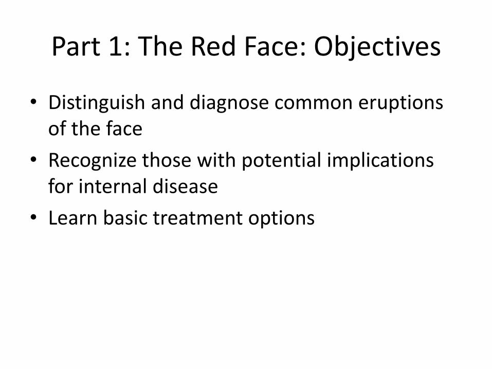
Part 1: The Red Face: Objectives
• Distinguish and diagnose common eruptions of the face
• Recognize those with potential implications for internal disease
• Learn basic treatment options
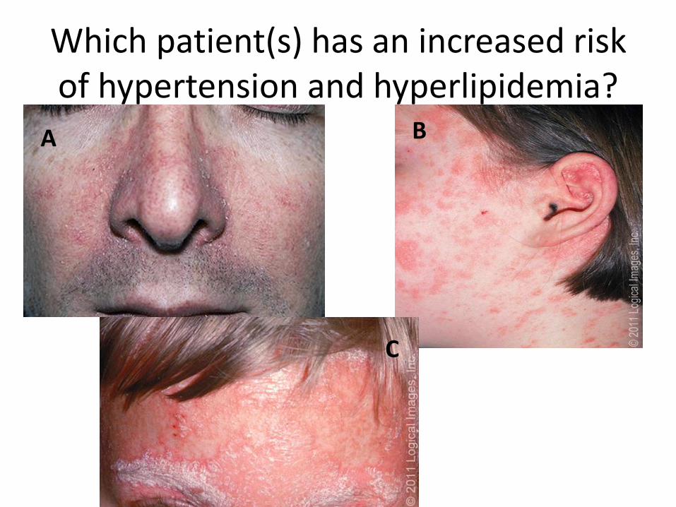
Which patient(s) has an increased risk of hypertension and hyperlipidemia?
A B
C

Which patient(s) has an increased risk of hypertension and hyperlipidemia?
A B
C
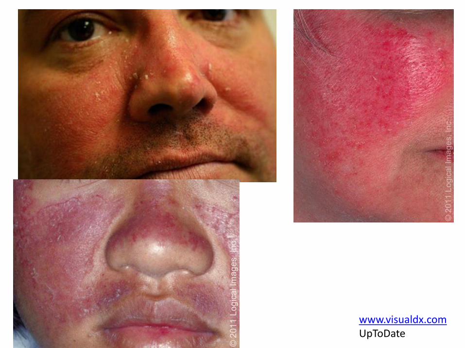
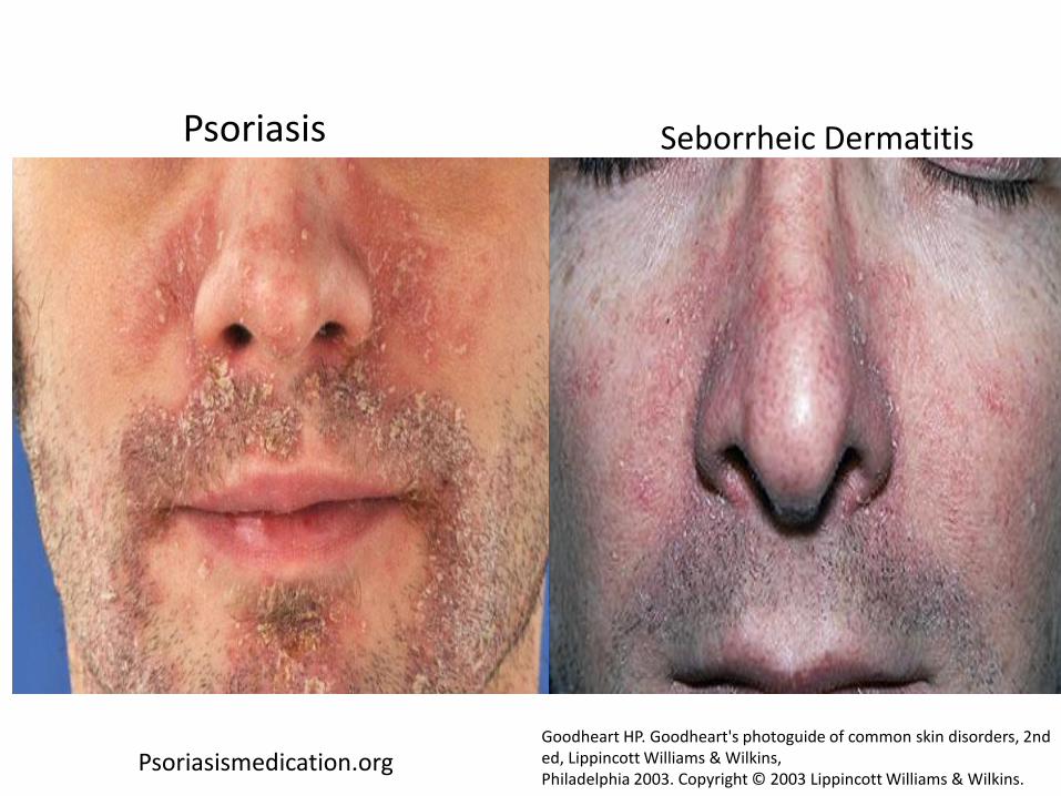
Psoriasis
Seborrheic Dermatitis
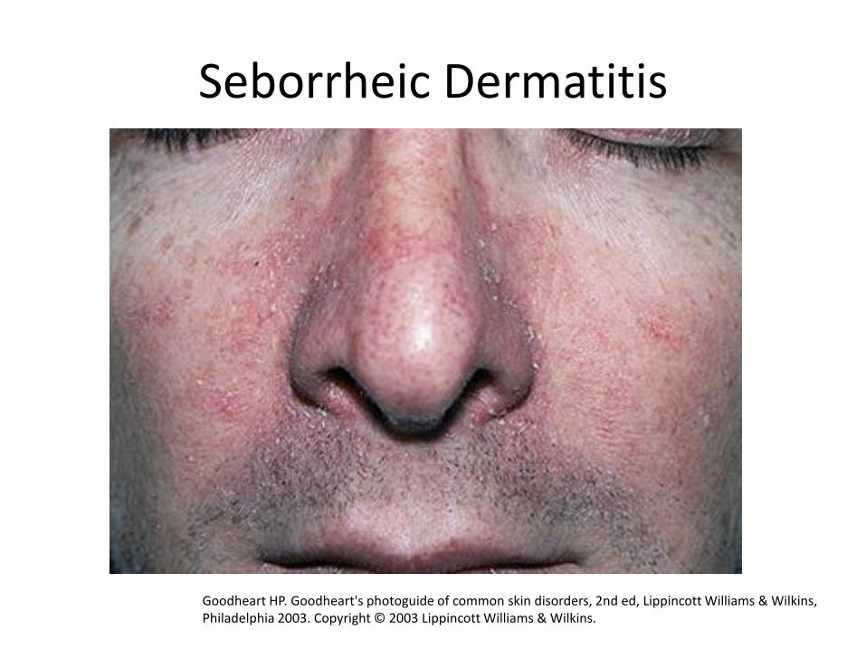
Seborrheic Dermatitis
Goodheart HP. Goodheart's photoguide of common skin disorders, 2nd ed, Lippincott Williams & Wilkins, Philadelphia 2003. Copyright © 2003 Lippincott Williams & Wilkins.
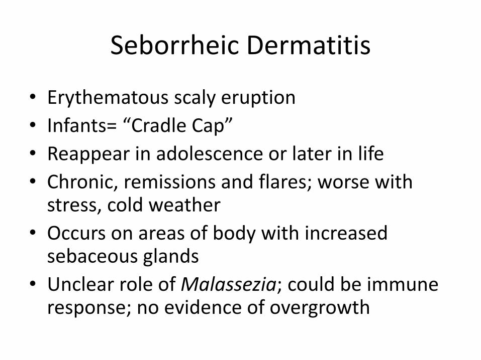
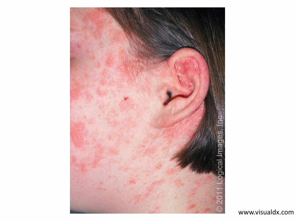
Seborrheic Dermatitis
• Erythematous scaly eruption
• Infants= “Cradle Cap”
• Reappear in adolescence or later in life
• Chronic, remissions and flares; worse with stress, cold weather
• Occurs on areas of body with increased sebaceous glands
• Unclear role of Malassezia; could be immune response; no evidence of overgrowth

Seborrheic DermatitisSevere Seb Derm: THINK:
• HIV (can also be more diffuse on trunk)
• Parkinson’s (seb derm improves with L-dopa therapy)
• Other neurologic
disorders
• Neuroleptic agents
• Unclear etiology
5MinuteClinicalConsult
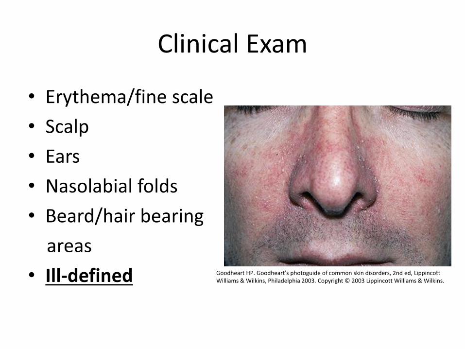
Clinical Exam
• Erythema/fine scale
• Scalp
• Ears
• Nasolabial folds
• Beard/hair bearing
areas
• Ill-defined Goodheart HP. Goodheart's photoguide of common skin disorders, 2nd ed, LippincottWilliams & Wilkins, Philadelphia 2003. Copyright © 2003 Lippincott Williams & Wilkins.

Treatment
• Topical steroids: hydrocortisone 2.5% cream or triamcinolone 0.025% cream if severe
– **Desonide now $200!!!! so I never prescribe**
• Topical antifungals: ketoconazole shampoo (to face and scalp), ciclopirox shampoo, ketoconazole cream
• Severe: itraconazole or fluconazole 200mg a day x 7 days

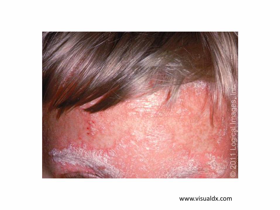
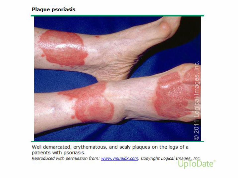
Facial Psoriasis
• Well-defined, more erythematous patches/plaques, +/- silvery scale
• Usually on scalp if also on face; ears
• Check elbows and knees as well
• Symmetric
• Other variants: inverse (folds), guttate(raindrop-like), nails
www.visualdx.com
www.visualdx.com
Psoriasismedication.orgGoodheart HP. Goodheart's photoguide of common skin disorders, 2nd ed, Lippincott Williams & Wilkins, Philadelphia 2003. Copyright © 2003 Lippincott Williams & Wilkins.
Psoriasis Seborrheic Dermatitis


Psoriasis
• 2 peaks in onset:– Between ages 30-39, and 50-69
• Complex pathogenesis: genetic + environment– PSORS1 locus within MHC on chromosome 6p21
– HLA-Cw6: higher susceptibility to early onset
– HLA-B17: more severe phenotype
– Other susceptibility loci: genes that encode for IL-12 and IL-23
– Multiple other gene products, including those affecting TNFα
Parisi, R, et al. J Invest Dermol 2013;133:337.

Psoriasis: environmental
Risk and exacerbating factors
• Smoking: intensity and duration
• Obesity: may contribute to more severe psoriasis, pro-inflammatory cytokines– Limited studies on weight loss and impact on
psoriasis
• Drugs: beta-blockers, lithium, anti-malarials
• Infections: Strep guttate; HIV
• Alcohol abuse
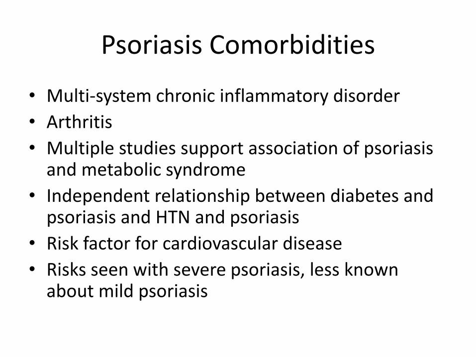
Psoriasis Comorbidities
• Multi-system chronic inflammatory disorder
• Arthritis
• Multiple studies support association of psoriasis and metabolic syndrome
• Independent relationship between diabetes and psoriasis and HTN and psoriasis
• Risk factor for cardiovascular disease
• Risks seen with severe psoriasis, less known about mild psoriasis
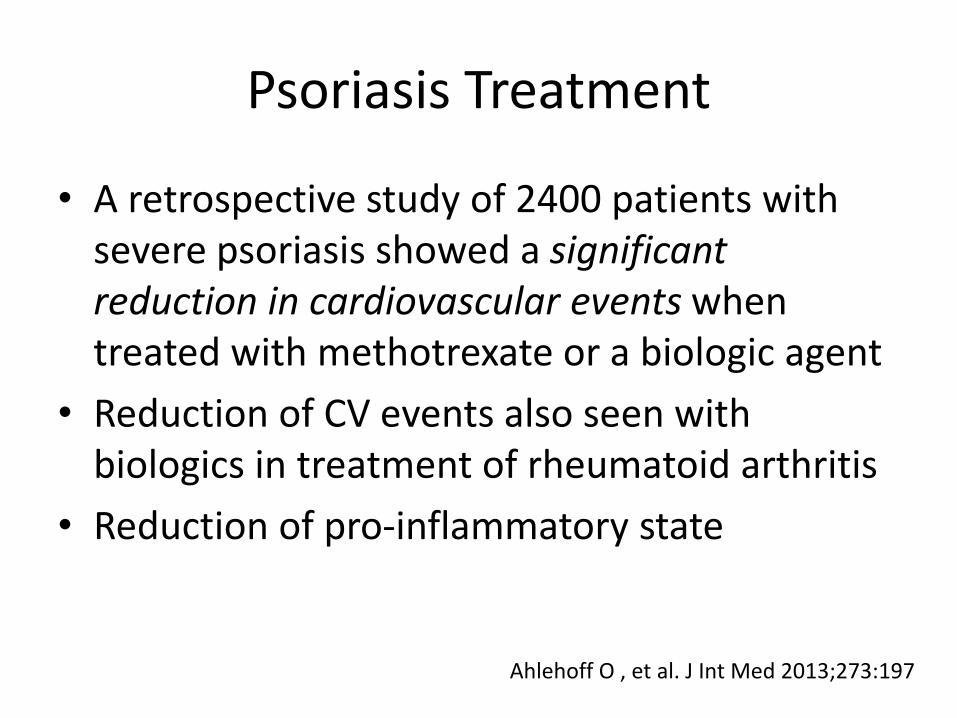
Psoriasis Treatment
• A retrospective study of 2400 patients with severe psoriasis showed a significant reduction in cardiovascular events when treated with methotrexate or a biologic agent
• Reduction of CV events also seen with biologics in treatment of rheumatoid arthritis
• Reduction of pro-inflammatory state
Ahlehoff O , et al. J Int Med 2013;273:197
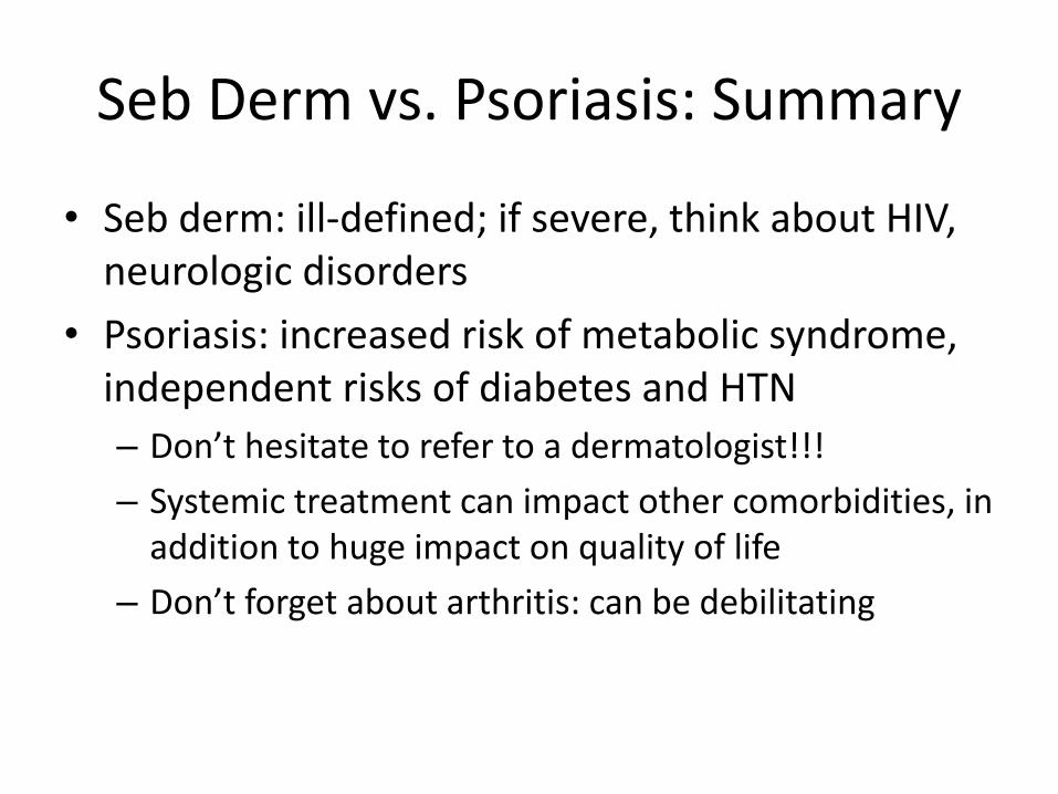
Seb Derm vs. Psoriasis: Summary
• Seb derm: ill-defined; if severe, think about HIV, neurologic disorders
• Psoriasis: increased risk of metabolic syndrome, independent risks of diabetes and HTN
– Don’t hesitate to refer to a dermatologist!!!
– Systemic treatment can impact other comorbidities, in addition to huge impact on quality of life
– Don’t forget about arthritis: can be debilitating

More Red Faces
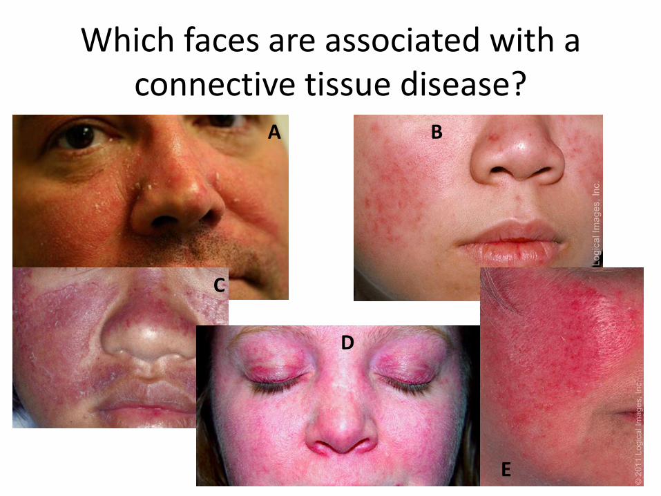
Which faces are associated with a connective tissue disease?
A B
C
D
E

Which faces are associated with a connective tissue disease?
A B
C
D
E
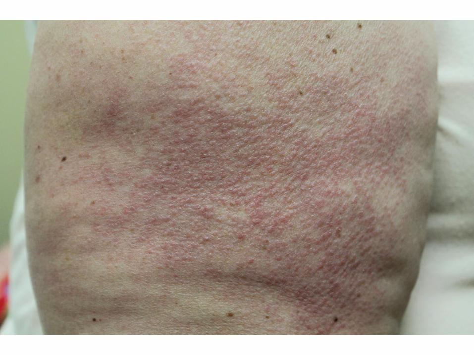
SLE
SLE
Seborrheic dermatitis
Dermatomyositis Rosacea
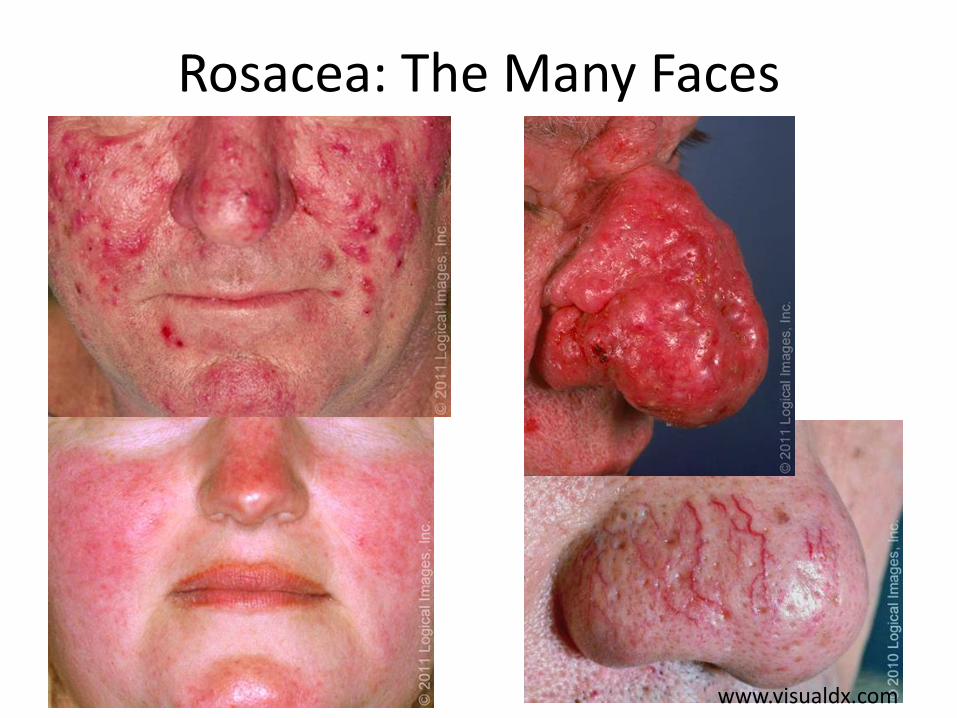
Rosacea: The Many Faces
www.visualdx.com

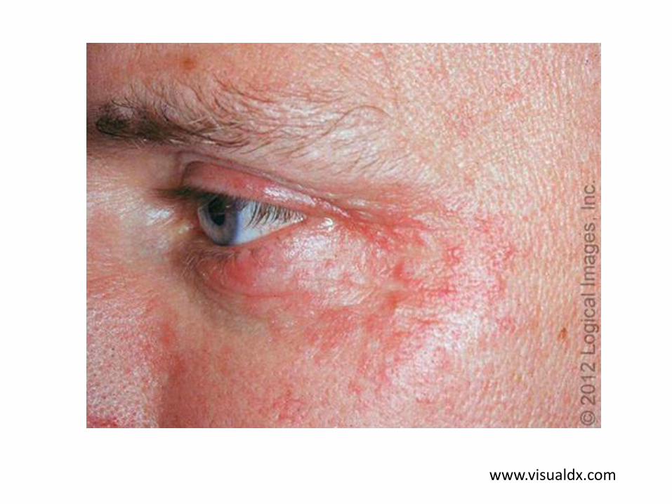
Rosacea
• Most frequently in fair-skinned individuals
• Women > men
• Dysfunction of innate immune system
Chronic inflammation, vascular hyperreactivity
• Debatable: demodex mites, bacteria
• UV radiation reactive oxidative species
• **Lacks comedones (distinguish from acne)**
What should you NOT prescribe topically for rosacea?
1. Clindamycin
2. Hydrocortisone
3. Metronidazole
4. Ivermectin
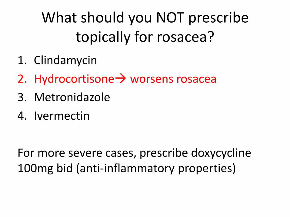
What should you NOT prescribe topically for rosacea?
1. Clindamycin
2. Hydrocortisone worsens rosacea
3. Metronidazole
4. Ivermectin
For more severe cases, prescribe doxycycline 100mg bid (anti-inflammatory properties)
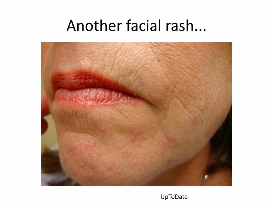
Another facial rash...
UpToDate
www.visualdx.com
www.visualdx.com
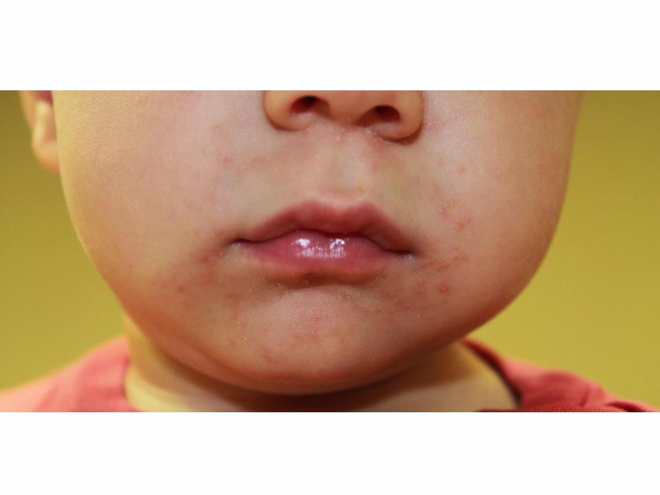
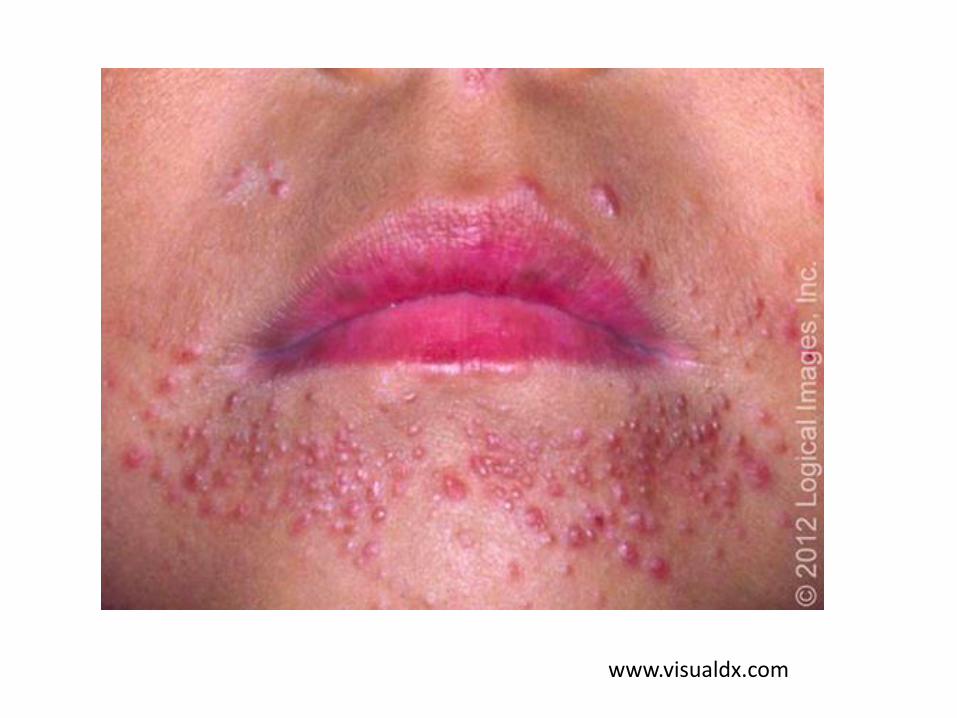
Perioral (orificial) Dermatitis
• Multiple small erythematous papules/pustules around the mouth (spares vermillion border), nose, and/or eyes
• + scaly patches
• NO comedones
• Most common: women, between 16 and 45
• Children common (average age 6)
• Cause?: possible irritant + skin barrier dysfunction

Perioral dermatitis
• Common: previous topical steroid use
• Can occur with oral, inhaled, or topical steroids
• Initially improves, then flares with continued use
• MUST stop topical steroid
• May have to taper to a less potent topical steroid if severe flare initially
Perioral dermatitis: Treatment
• Bland, nonocclusive lotions
• Pimecrolimus cream
• Metronidazole cream, erythromycin gel
If severe and/or fails topicals x 1 month:
• Doxycycline
• Erythromycin (under age 9)

Back to connective tissue disease...
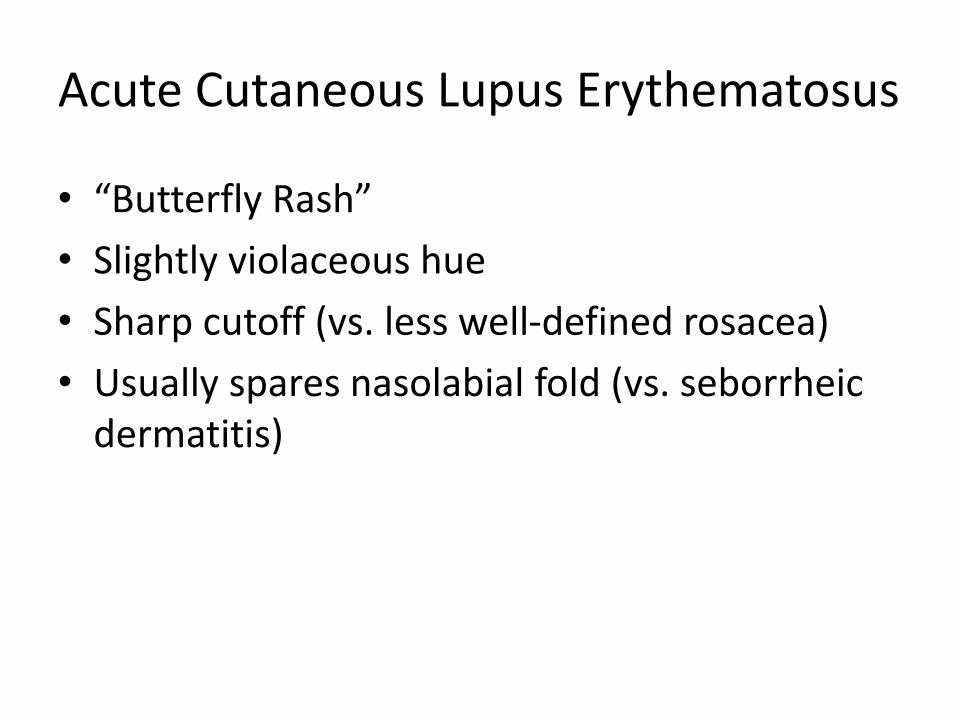
Acute Cutaneous Lupus Erythematosus
• “Butterfly Rash”
• Slightly violaceous hue
• Sharp cutoff (vs. less well-defined rosacea)
• Usually spares nasolabial fold (vs. seborrheicdermatitis)
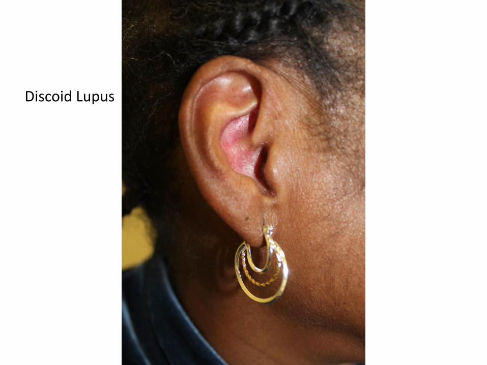
Discoid Lupus

Courtesy of Jeff Callen, MD
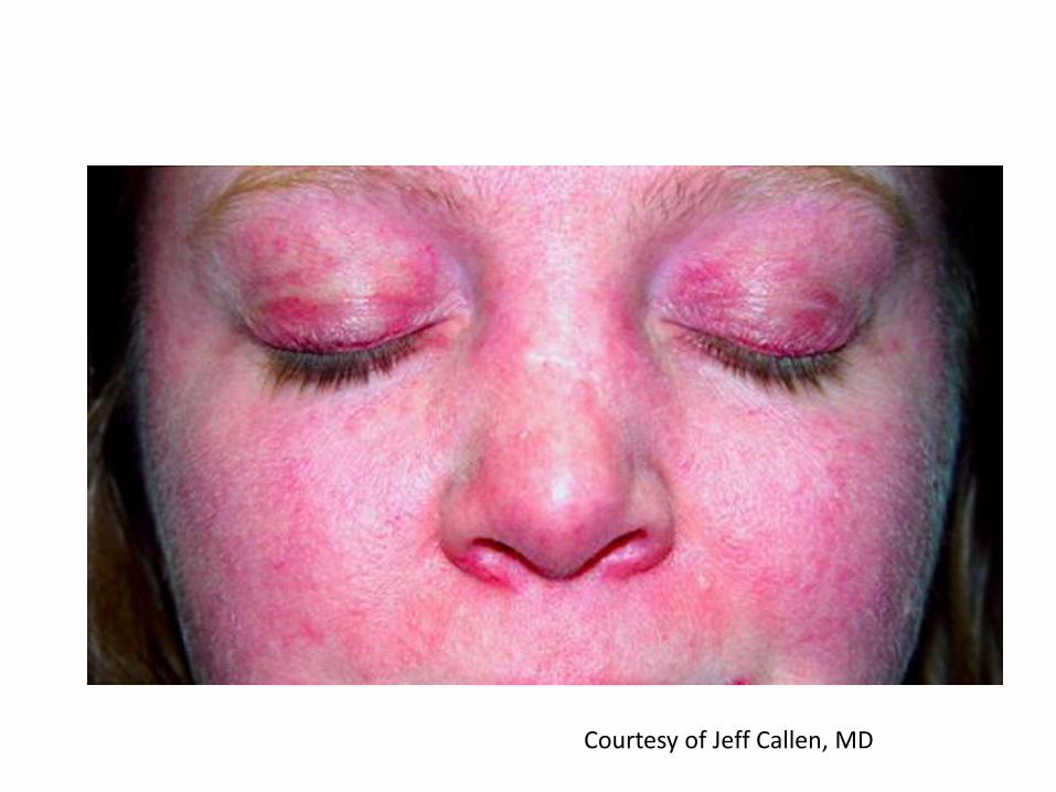
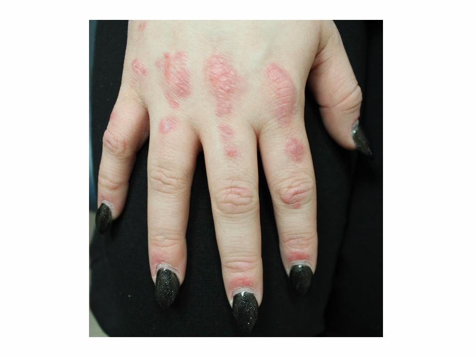
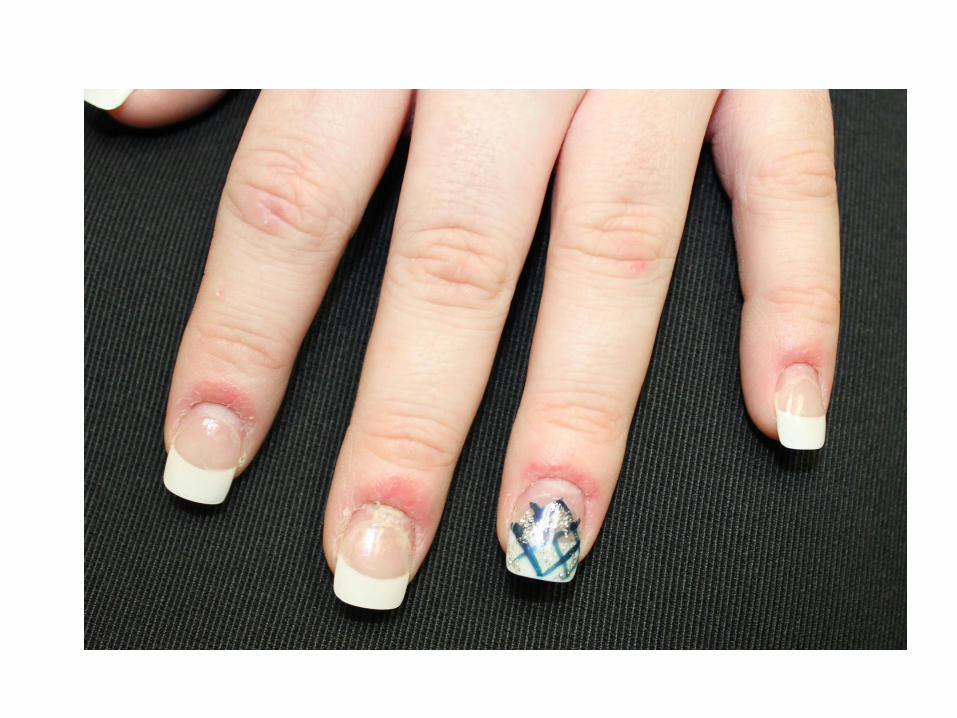
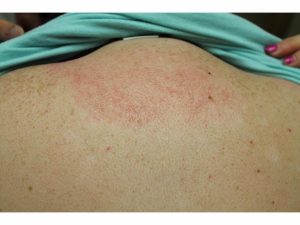
Dermatomyositis
• Idiopathic inflammatory myopathy and skin eruption
• Proximal muscle weakness
• Approximately 20% have no myositis
• Erythematous eruption on the face, joints, periungual, upper back and chest, scalp
• Can be associated with interstitial lung disease, cardiomyopathy, and internal malignancy


Dermatomyositis
If suspicious:
• Refer to dermatology; biopsy can rule out other conditions
• Ensure that patient is up to date on age appropriate malignancy screening
• Sun protection
• Derm/Rheum: can prescribe anti-malarials, methotrexate, etc.
Summary: Face Rashes
• Rosacea, perioral dermatitis: NO TOPICAL STEROIDS
• Lupus: spares nasolabial fold, well-demarcated
• Dermatomyositis: heliotrope rash (eyelids), violaceous
• Psoriasis: well-defined, thick scale
• Seborrheic dermatitis: ill-defined, fine scale,
Part 2: More Pearls:
1. Not all that is red is cellulitis.
2. The perils of oral steroids for rashes.
3. The perils of topical steroids for rashes.

Pearl #1
• Not all that is red is cellulitis.
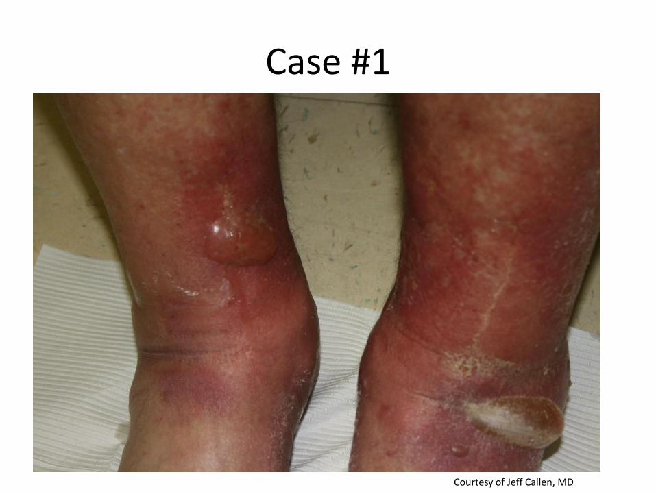
Case #1
Courtesy of Jeff Callen, MD

Treatment?
1. Give oral antibiotics
2. Do a wound culture
3. Admit patient for iv antibiotics
4. Elevation and compression
5. Topical steroids
6. Topical steroid + topical antifungal
7. Topical antibiotic ointment (triple antibiotic or bacitracin)

Treatment?
1. Give oral antibiotics
2. Do a wound culture
3. Admit patient for iv antibiotics
4. Elevation and compression
5. Topical steroids
6. Topical steroid + topical antifungal
7. Topical antibiotic ointment (neosporin or bacitracin)
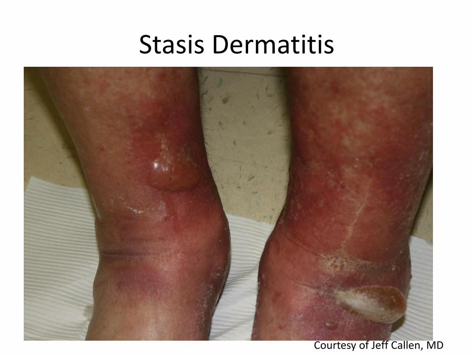
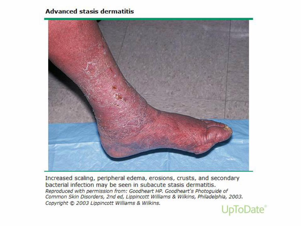
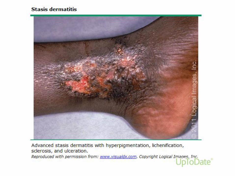
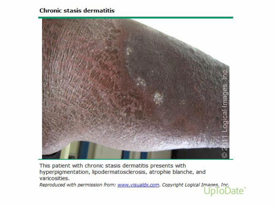
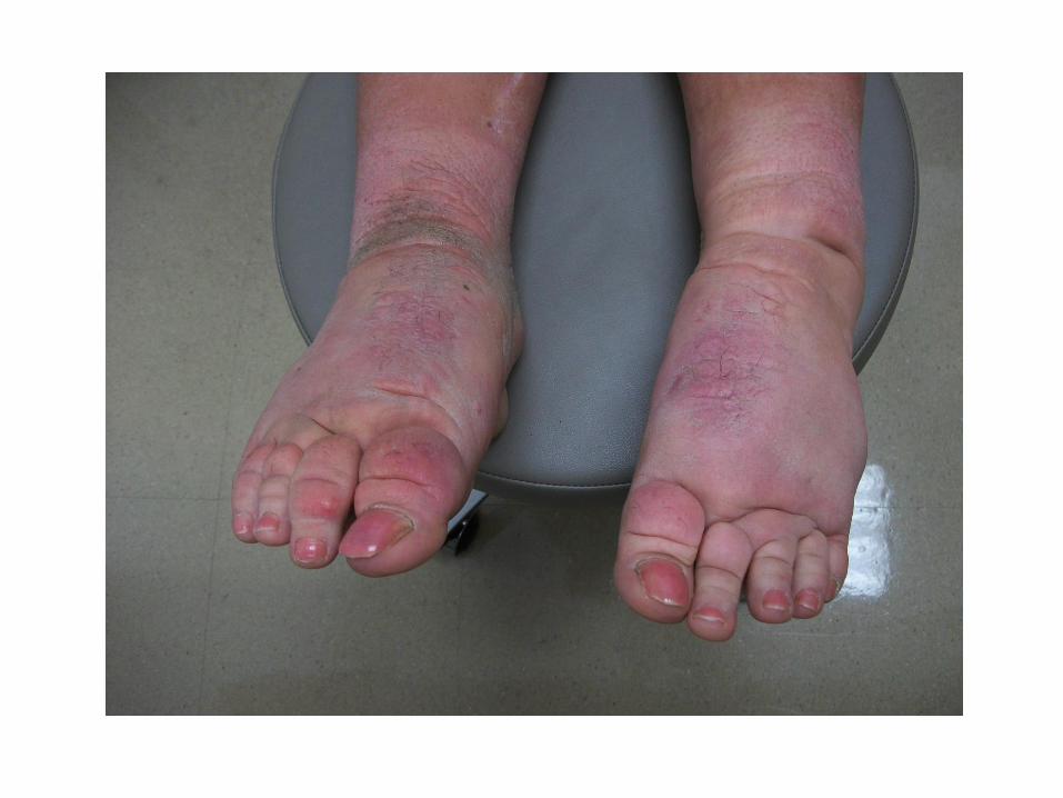
Stasis Dermatitis
Courtesy of Jeff Callen, MD
Stasis Dermatitis
• 2/2 chronic venous insufficiency
• Venous HTN from dysfunctional venous pump or valves chronic edema
• Proliferation of small blood vessels in the dermis, extravasation of RBCs into the dermis, inflammation in the skin
• Acute: Erythema, warmth, eczema-like rash, acute vesiculation
• Chronic: Sclerosis and ulceration, brawny look


Risk factors
• Age
• Female gender
• Obesity
• Family history
• Standing occupation
• History of DVT
• Aggravating factors: HTN, CHF

Differential diagnosis


Complications of Stasis Dermatitis
• Contact dermatitis
• Autosensitization
• Superinfection

Contact Dermatitis
• Much higher risk!!• Repeated use, increased blood flow
to area, chronic inflammation• Fragrances• Neomycin (Triple antibiotic oint*),
bacitracin• Lanolin• Adhesives• Anti-itch creams• Topical steroids (OTC hydrocortisone)
*Neosporin is the only available brand name triple antibiotic ointment*

Autosensitization or “Id” reaction
• Nonspecific rash on arms, thighs, trunk after flare of stasis dermatitis
• Pathogenesis unknown: triggering the immune system elsewhere
• Stasis dermatitis “all over”
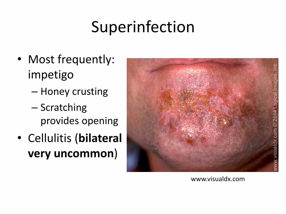
Superinfection
• Most frequently: impetigo
– Honey crusting
– Scratching provides opening
• Cellulitis (bilateral very uncommon)
www.visualdx.com

Management
• Compression: start light, be realistic
• Mild bland soaps: Dove, Cetaphil
– NOT Dial, Zest, or fragranced products
• Vaseline
• Topical steroids: if erythema, pruritus, vesiculation, oozing
– Ointments preferred over creams
– Triamcinolone 0.1% ointment briefly
Underdiagnosis of Stasis Dermatitis
Misdiagnosis of Cellulitis
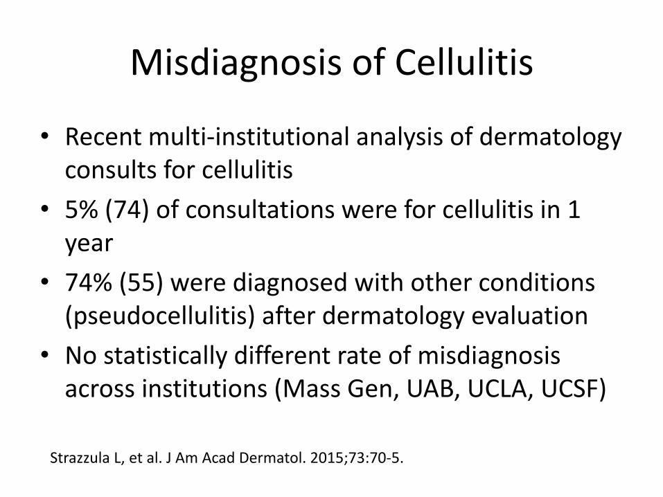
• Recent multi-institutional analysis of dermatology consults for cellulitis
• 5% (74) of consultations were for cellulitis in 1 year
• 74% (55) were diagnosed with other conditions (pseudocellulitis) after dermatology evaluation
• No statistically different rate of misdiagnosis across institutions (Mass Gen, UAB, UCLA, UCSF)
Strazzula L, et al. J Am Acad Dermatol. 2015;73:70-5.

What did the patients have?
• 31% stasis dermatitis
• 14.5% contact dermatitis
• 9% tinea
• Other conditions included: psoriasis, vasculitis, lymphedema
• 38% had more than 1 cutaneous condition
Predictors
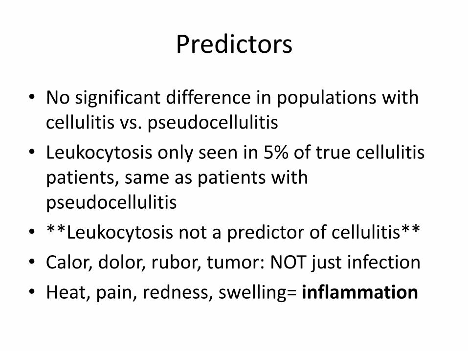
• No significant difference in populations with cellulitis vs. pseudocellulitis
• Leukocytosis only seen in 5% of true cellulitis patients, same as patients with pseudocellulitis
• **Leukocytosis not a predictor of cellulitis**
• Calor, dolor, rubor, tumor: NOT just infection
• Heat, pain, redness, swelling= inflammation

Cellulitis is Expensive
• More $3.7 billion spent on approximately 24,000 adult patient admissions for cellulitis in 2004
• 74% in previous multi-center studied were incorrectly diagnosed
• Use of a dermatologist more frequently upon admission may decrease costs, hospital duration, and use of unnecessary antibiotics
The DRG Handbook Comparative Clinical and Financial Benchmarks. 2006, Evanston,IL: Solucient.

Summary
Stasis dermatitis:
• Usually bilateral
• Can be red, hot, painful, and swollen
• Underlying chronic venous insufficiency
• Compression, vaseline, topical steroids if acute
• Avoid triple antibiotic ointment and other topical OTC creams

Pearl #2
• The perils of oral steroids (or certain ones) for rashes.
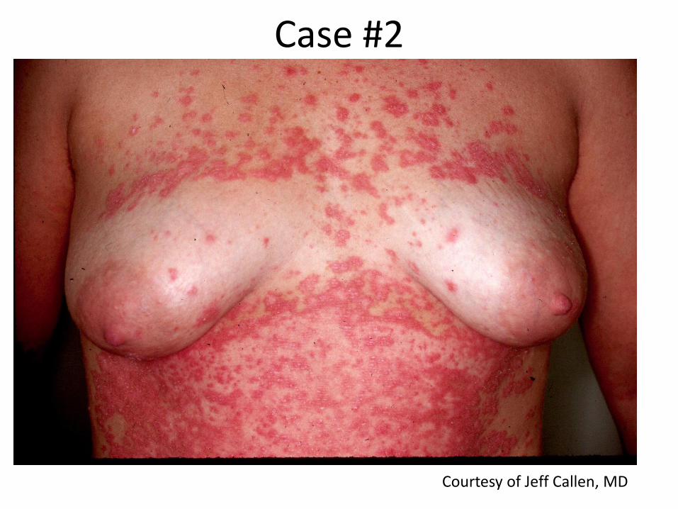
Case #2
Courtesy of Jeff Callen, MD

Courtesy of Jeff Callen, MD
Flare of Pustular Psoriasis 2/2 Oral Steroids

Triggers of generalized pustular psoriasis
• Withdrawal from **systemic corticosteroids** (44%)
• Other meds withdrawal (cyclosporine, biologics)
• Infections (16%)
• Pregnancy (17%)
Choon SE, et al. Int J Dermatol 2014;53:676
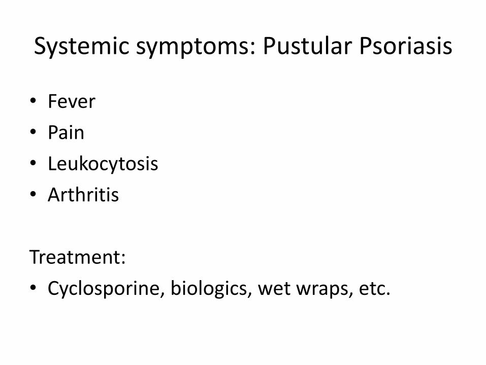
Systemic symptoms: Pustular Psoriasis
• Fever
• Pain
• Leukocytosis
• Arthritis
Treatment:
• Cyclosporine, biologics, wet wraps, etc.

Rhus (Poison Ivy)
Courtesy of Jeff Callen, MD
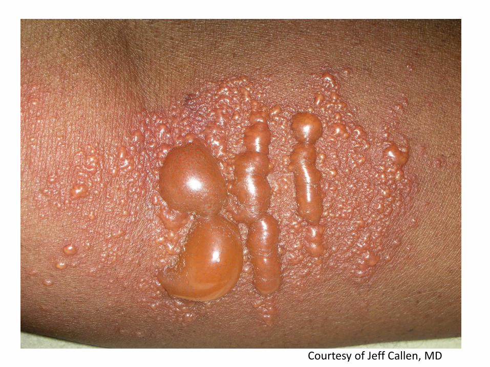
Courtesy of Jeff Callen, MD

Allergic Contact Dermatitis
• Avoid short courses of oral steroids or IM triamcinolone
• Methylprednisolone dose pack: too brief
• Patients will flare when they complete pack or shot wears off
• Instead: prescribe a Prednisone 20 day taper
• 60mg qam x 5days, 40mg qam x 5days, 20mg qam x 5days, 10mg qam x 5days
• Generalized psoriasis: do not give oral steroids
• Severe rashes (that aren’t psoriasis): do not use steroid dose packs or IM triamcinolone
• Prednisone 20 day taper
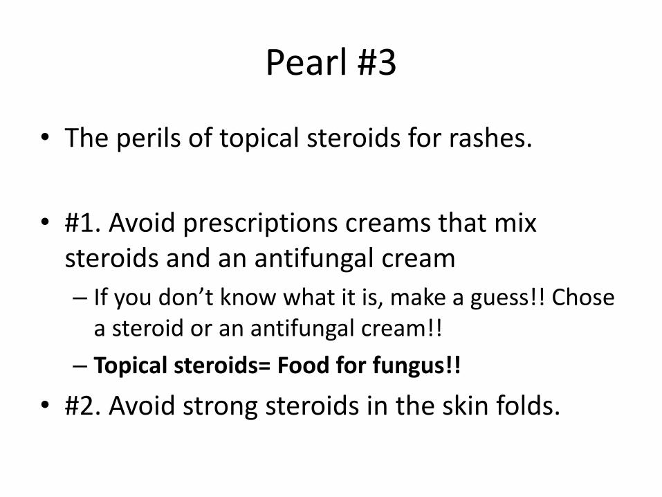
Pearl #3
• The perils of topical steroids for rashes.
• #1. Avoid prescriptions creams that mix steroids and an antifungal cream
– If you don’t know what it is, make a guess!! Chose a steroid or an antifungal cream!!
– Topical steroids= Food for fungus!!
• #2. Avoid strong steroids in the skin folds.
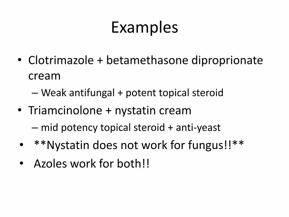
Examples
• Clotrimazole + betamethasone diproprionatecream
– Weak antifungal + potent topical steroid
• Triamcinolone + nystatin cream
– mid potency topical steroid + anti-yeast
• **Nystatin does not work for fungus!!**
• Azoles work for both!!
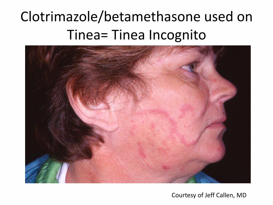
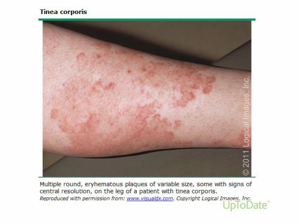
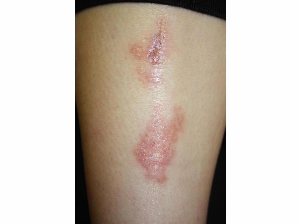
Clotrimazole/betamethasone used on Tinea= Tinea Incognito
Courtesy of Jeff Callen, MD

More Tinea Incognito
Courtesy of Jeff Callen, MD

Clotrimazole/betamethasone striae
Courtesy of Jeff Callen, MD
Appropriate steroids
• For the folds (groin, axilla, etc): hydrocortisone, triamcinolone 0.025% sparingly;
• Only time I ever mix a steroid with an antifungal cream:
– Intertrigo: hydrocortisone 2.5% + ketoconazole cream

Summary
• Bilateral red lower legs: most likely stasis dermatitis
• Compression, mild soaps, steroid ointments
• Steroid dose packs DO NOT work for rashes
• Do not treat psoriasis with oral steroids
• When in doubt, pick either a steroid or an antifungal cream, never both

Thank you!!


![Welcome [louisville.edu]louisville.edu/medicine/departments/familymedicine/... · Eating better can help you feel better. A variety of healthy foods can help fight infections, prevent](https://img.pdfslide.net/doc/110x75/5f58211d31f7a838b910d5f9/welcome-eating-better-can-help-you-feel-better-a-variety-of-healthy-foods-can.jpg)