Embed Size (px)
Citation preview

The Respiratory System
There is the Upper respiratory tract and
Lower respiratory tract
What organs do you think are in What organs do you think are in each tract? each tract?
Come up and list them. Come up and list them.

LUNGS
ORAL CAVITY
PHARYNX
PARANASAL CAVITIES
NASAL CAVITY
NOSE
UPPER RESPIRATORY SYSTEM
LOWER RESPIRATORY SYSTEM
LARYNX
TRACHEA
BRONCHI

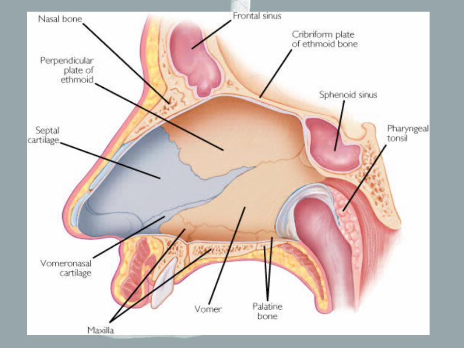
THE NOSE / THE NOSE / Nasal CavityNasal Cavity
• Air enters through two openings, THE EXTERNAL NARES or NOSTRILS.
• A midsagittal NASAL SEPTUM divides the NASAL CAVITY.
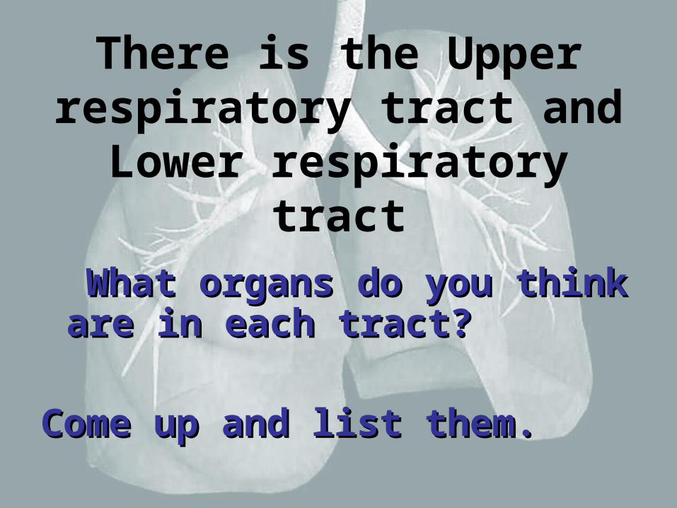
• The maxillary, nasal, frontal, ethmoid and sphenoid bones form the lateral and superior walls of the nasal cavity.
• The HARD and SOFT PALATES form the floor of the cavity. ( the posterior part of the soft palate is the UVULA)
• The external portion of the nose is composed of cartilage that forms the BRIDGE and the TIP of the nose.
• Posteriorly the INTERNAL NARES open into the nasopharynx.
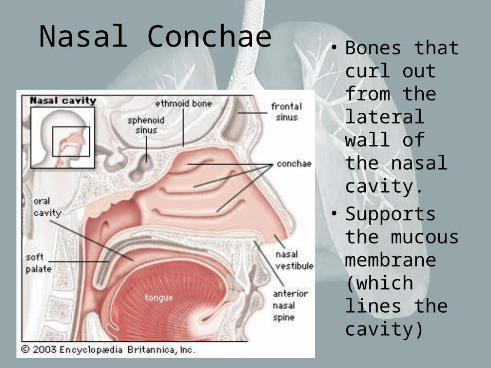
Nasal Conchae • Bones that curl out from the lateral wall of the nasal cavity.
• Supports the mucous membrane (which lines the cavity)

What tissues are in the nasal cavity and what role does it play? • Pseudostratified ciliated epithelium.
• Has mucous secreting goblets that combined with the cilia help trap dust and move them out of the nasal passageway or through the nasal conchae out the nose or into the pharynx which gets swallowed.

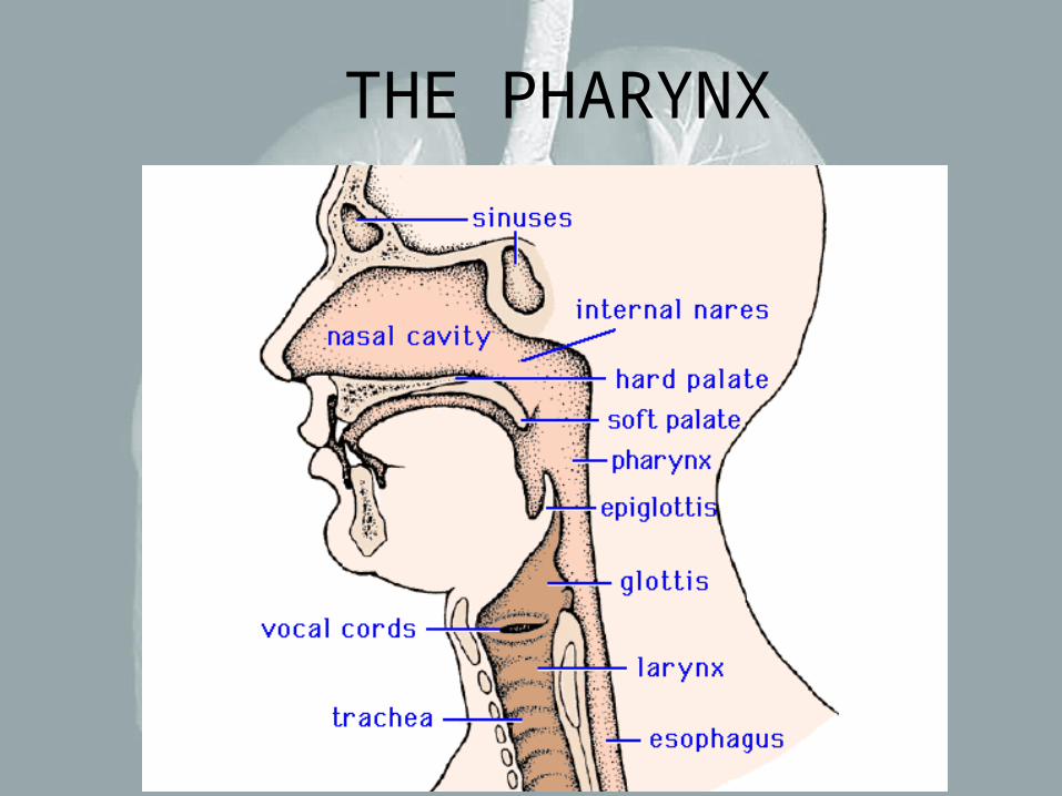
THE PHARYNX (Throat)
• Is a chamber shared by the digestive and respiratory systems.
• It extends between the internal nares and the entrances to the larynx and esophagus.
• A stratified squamous epithelium lines the pharynx.
THE PHARYNX
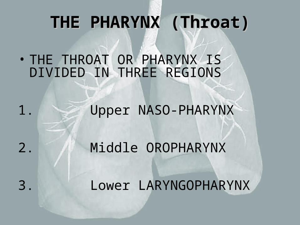
• THE THROAT OR PHARYNX IS DIVIDED IN THREE REGIONS
1. Upper NASO-PHARYNX
2. Middle OROPHARYNX
3. Lower LARYNGOPHARYNX
THE PHARYNX (Throat)THE PHARYNX (Throat)

TTHE NASOPHARYNXHE NASOPHARYNX
LIES SUPERIOR TO THE SOFT PALATE
• SERVES AS A PASSAGEWAY FOR AIRFLOW FROM NASAL CAVITY
• IT CONTAINS THE PHARYNGEAL TONSILS (adenoids ) IN POSTERIOR WALL, AND THE OPENNINGS OF THE AUDITORY TUBES
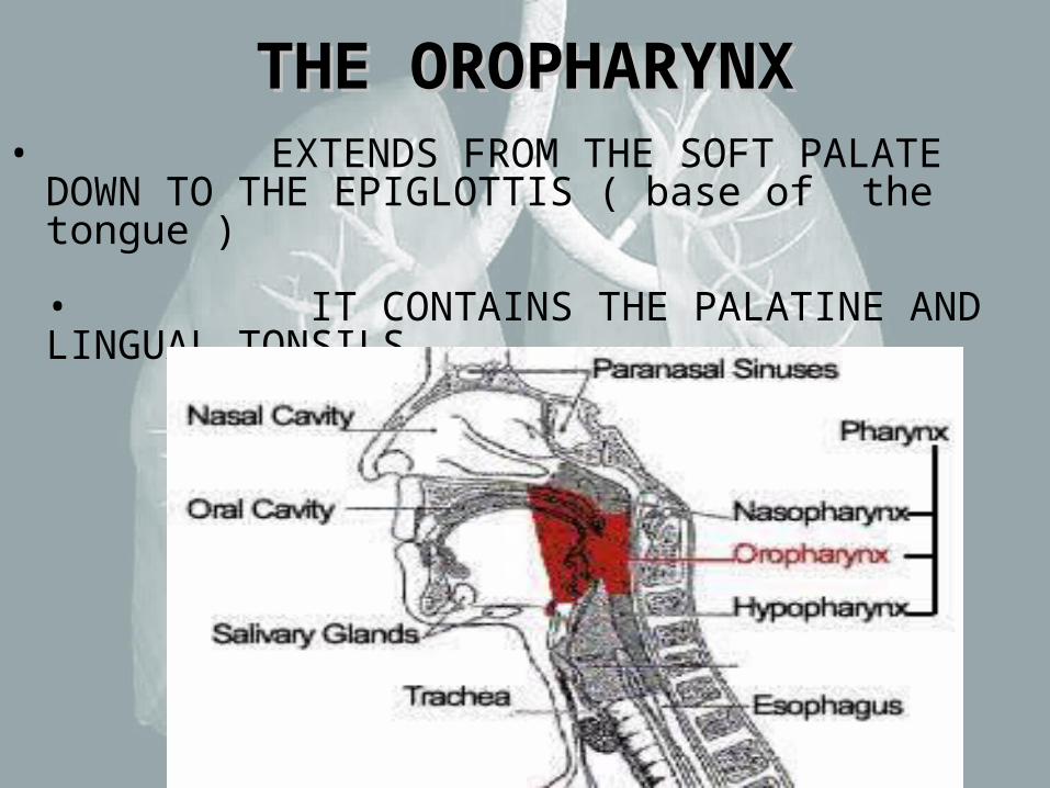
THE OROPHARYNXTHE OROPHARYNX• EXTENDS FROM THE SOFT PALATE DOWN
TO THE EPIGLOTTIS ( base of the tongue )
• IT CONTAINS THE PALATINE AND LINGUAL TONSILS.
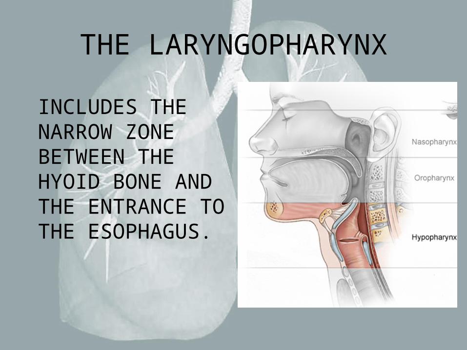
THE LARYNGOPHARYNX
INCLUDES THE NARROW ZONE BETWEEN THE HYOID BONE AND THE ENTRANCE TO THE ESOPHAGUS.
Functions of the Pharynx:
1. A passageway for food traveling from the oral cavity to the esophagus.
2. A passageway for air passing between the nasal cavity and the larynx.
3. Helps produce the sounds of speech.

• If a function of the pharynx is to both let in air and food, how does this process work?
• What is happening when “Food goes down the wrong tube?”
WHEN FINISHED:
1. What is the entrance point for air?
2. Explain where or what air does in the respiratory system.
3. What is happening when you sneeze?
4. Explain what happens when “food goes down the wrong tube”
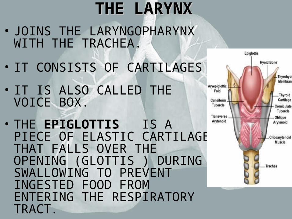
THE LARYNXTHE LARYNX• JOINS THE LARYNGOPHARYNX
WITH THE TRACHEA.
• IT CONSISTS OF CARTILAGES
• IT IS ALSO CALLED THE VOICE BOX.
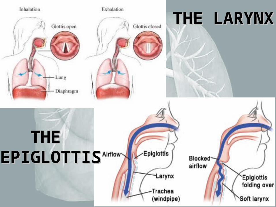
• THE EPIGLOTTISEPIGLOTTIS IS A PIECE OF ELASTIC CARTILAGE THAT FALLS OVER THE OPENING (GLOTTIS ) DURING SWALLOWING TO PREVENT INGESTED FOOD FROM ENTERING THE RESPIRATORY TRACT.
THE LARYNXTHE LARYNX• TWO PAIRS OF FOLDS SPAN THE GLOTTAL
OPENING. FALSE VOCAL CORDS AND TRUE VOCAL CORDS.
• DURING EXPIRATION AIR FLOWING THROUGH THE LARYNX VIBRATES THE VOCAL CORDS (TRUE VOCAL CORDS) AND PRODUCES SOUND WAVES.
• COUGHING AND LARYNGEAL SPASMS ARE PROTECTIVE REFLEXES THAT PROTECT THE GLOTTIS AND TRACHEA FROM FOREIGN OBJECTS AND IRRITANTS.
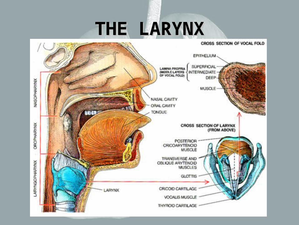
THE LARYNXTHE LARYNX
THE LARYNXTHE LARYNX
THE THE EPIGLOTTISEPIGLOTTIS
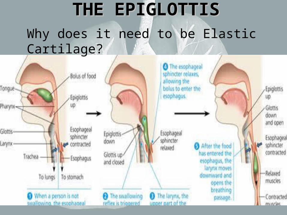
THE EPIGLOTTISTHE EPIGLOTTISWhy does it need to be Elastic Cartilage?
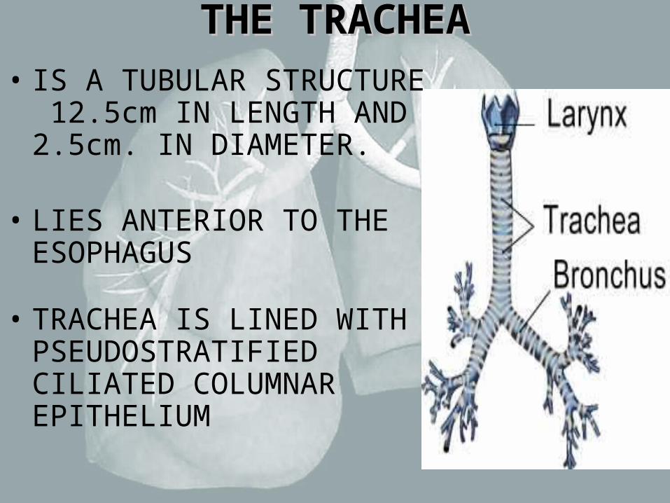
THE TRACHEATHE TRACHEA• IS A TUBULAR STRUCTURE
12.5cm IN LENGTH AND 2.5cm. IN DIAMETER.
• LIES ANTERIOR TO THE ESOPHAGUS
• TRACHEA IS LINED WITH PSEUDOSTRATIFIED CILIATED COLUMNAR EPITHELIUM

THE TRACHEATHE TRACHEA
ALONG THE LENGTH OF THE TRACHEA ARE 15-20 C-SHAPED PIECES OF HYALINE CARTILAGE (TRACHEAL CARTILAGES)
THE TRACHEAL MUSCLE HOLDS THE TWO SIDES OF THE C-SHAPED CARTILAGE TOGETHER POSTERIORLY
THE TRACHEA BRANCHES WITH-IN THE MEDIASTINUM, FORMING THE LEFT AND RIGHT PRIMARY BRONCHI

THE TRACHEATHE TRACHEA
LEFT AND RIGHT PRIMARY LEFT AND RIGHT PRIMARY BRONCHIBRONCHI
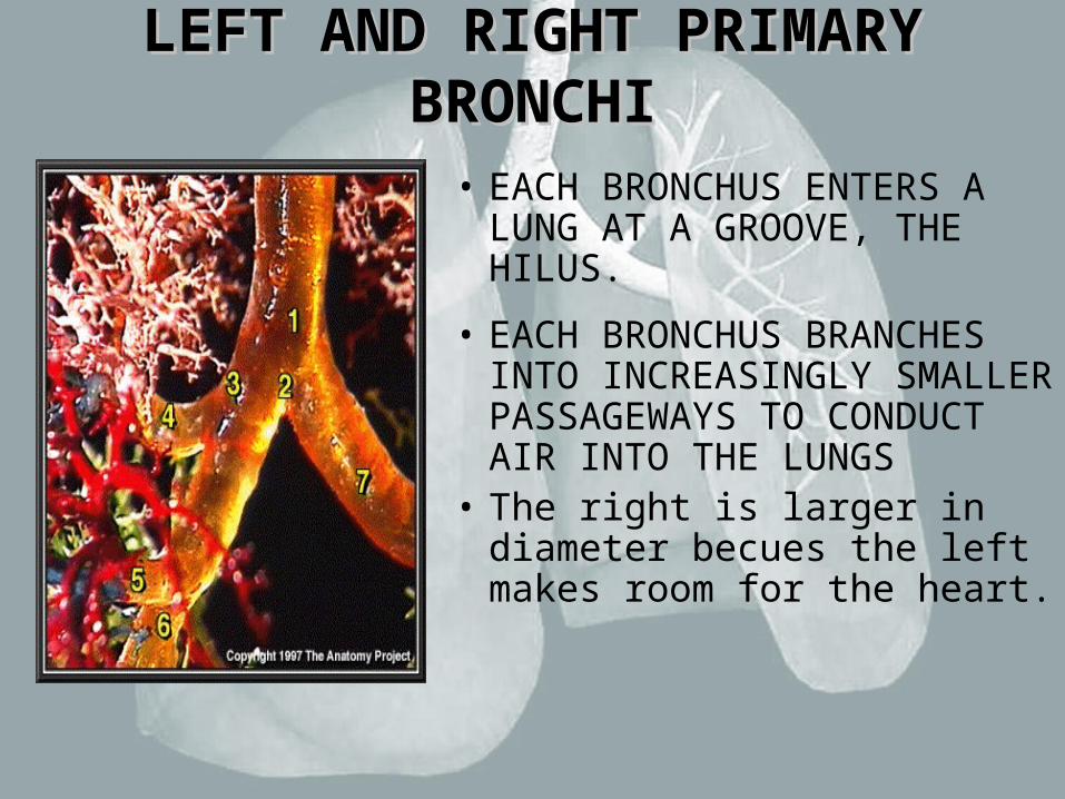
• EACH BRONCHUS ENTERS A LUNG AT A GROOVE, THE HILUS.
• EACH BRONCHUS BRANCHES INTO INCREASINGLY SMALLER PASSAGEWAYS TO CONDUCT AIR INTO THE LUNGS
• The right is larger in diameter becues the left makes room for the heart.
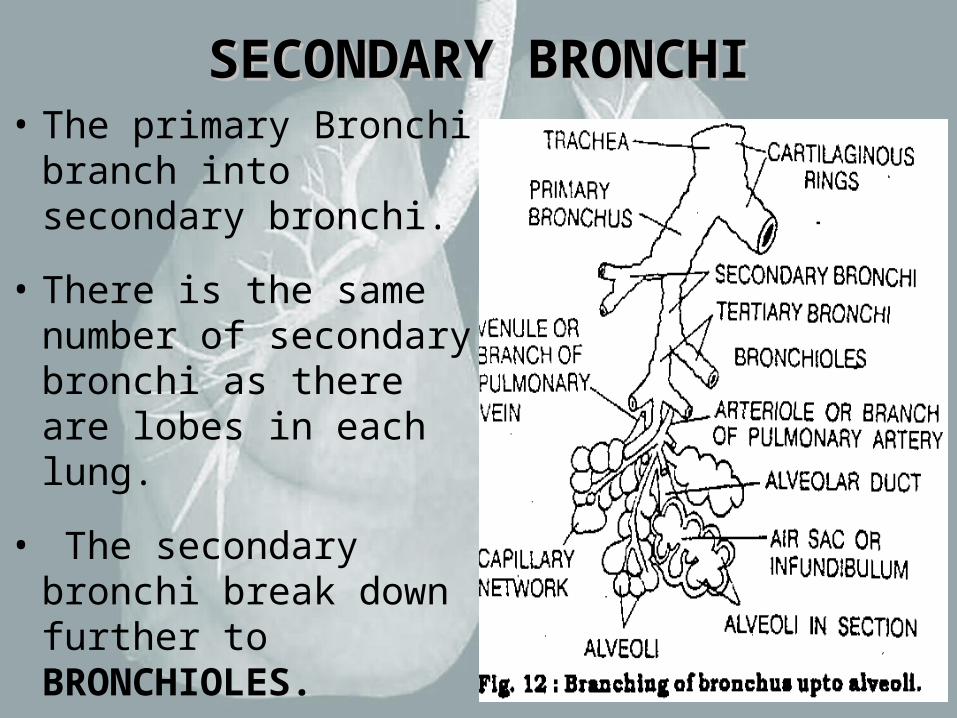
SECONDARY BRONCHISECONDARY BRONCHI• The primary Bronchi
branch into secondary bronchi.
• There is the same number of secondary bronchi as there are lobes in each lung.
• The secondary bronchi break down further to BRONCHIOLES.
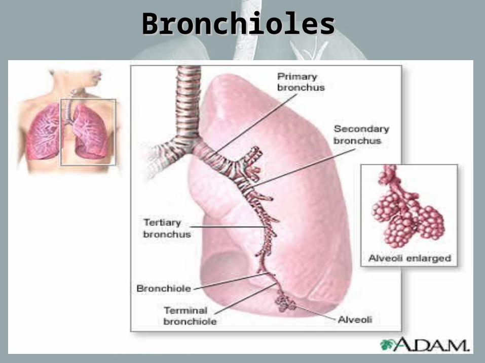
BronchiolesBronchioles
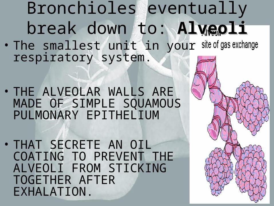
Bronchioles eventually break down to: AlveoliAlveoli
• The smallest unit in your respiratory system.
• THE ALVEOLAR WALLS ARE MADE OF SIMPLE SQUAMOUS PULMONARY EPITHELIUM
• THAT SECRETE AN OIL COATING TO PREVENT THE ALVEOLI FROM STICKING TOGETHER AFTER EXHALATION.
AlveoliAlveoli
• ALSO IN THE ALVEOLAR WALL ARE MACROPHAGES THAT PHAGOCYTIZE DEBRIS OR POTENTIAL PATHOGENS
• PULMONARY CAPILLARIES COVER THE EXTERIOR OF THE ALVEOLI
• THIS IS THE SITE WHERE GASES (Oxygen and Carbon dioxide are exchanged)
Alveoli Exchange of Gases: Alveoli Exchange of Gases:
• Oxygen diffuses from aveolar walls and enters the blood in capillaries.(where it can now go to other cells in the body)
• Carbon Dioxide diffuses from the blood through the walls and enters the aveoli. (where it can be exhaled and released)

Alveoli Exchange of Gases: Alveoli Exchange of Gases: Using the picture below EXPLAIN the gas Using the picture below EXPLAIN the gas exchange process: exchange process:
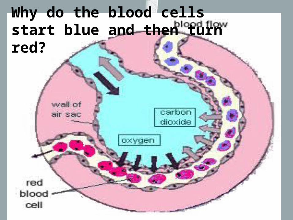
Why do the blood cells start blue and then turn red?

THE LUNGSTHE LUNGS
• ARE A PAIR OF CONE SHAPED ORGANS LYNING IN THE PLEURAL CAVITIES.
• EACH LUNG HAS A HILUS, A MEDIAL SLIT WHERE THE BRONCHIAL TUBES, VASCULARIZATION, LYMPHATICS, AND NERVES REACH THE LUNG.
• EACH LUNG IS DIVIDED INTO LOBES BY DEEP FISURES
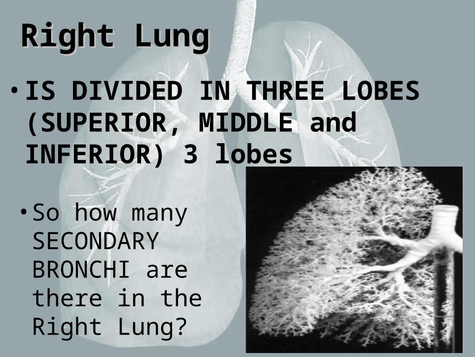
Right LungRight Lung
• IS DIVIDED IN THREE LOBES (SUPERIOR, MIDDLE and INFERIOR) 3 lobes
• So how many SECONDARY BRONCHI are there in the Right Lung?
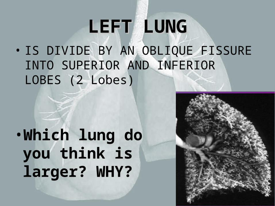
LEFT LUNGLEFT LUNG• IS DIVIDE BY AN OBLIQUE FISSURE
INTO SUPERIOR AND INFERIOR LOBES (2 Lobes)
• Which lung do you think is larger? WHY?

Right Lung / Left LungRight Lung / Left Lung
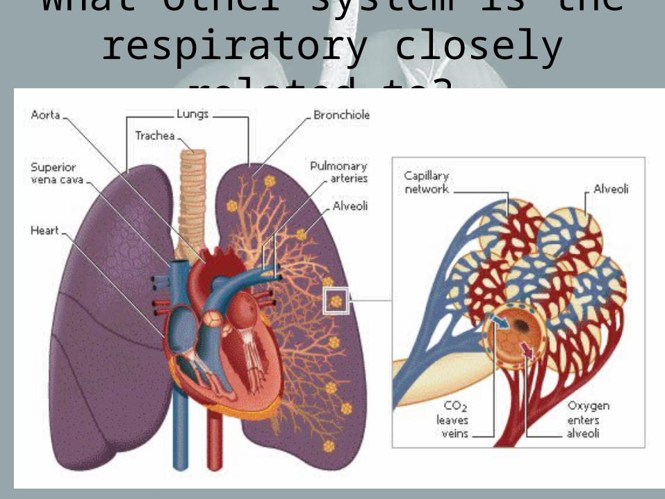
What other system is the respiratory closely related to?
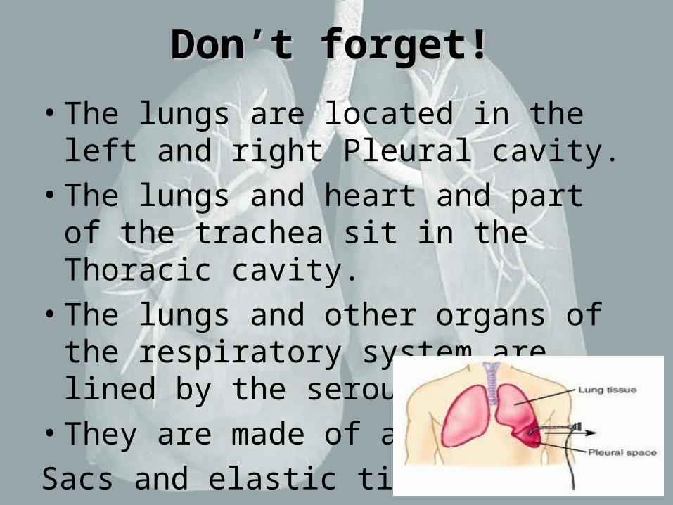
Don’t forget!Don’t forget!
• The lungs are located in the left and right Pleural cavity.
• The lungs and heart and part of the trachea sit in the Thoracic cavity.
• The lungs and other organs of the respiratory system are lined by the serous membrane.
• They are made of air
Sacs and elastic tissue.

Checking for Understanding: Answer the following Questions.
1. What organs are in the upper respiratory tract? What about the lower?
2. Name the bones that make up the nose.
3. Explain what tissues are involved in the nasal cavity and what they help do.
4. What organ also refers to your throat?
5. How do vocal cords produce sound?
6. What is the function of the epiglottis?
7. Explain starting with your trachea how it “branches” out.
8. Explain how gases are exchanged in the alveoli.
9. Where does oxygen go? Where does carbon dioxide go?
10. What else is in the lungs beside the bronchial tubes?