Embed Size (px)
Citation preview
THE RH FACTOR
A CLINICAL AND FUNDAMENTAL STUDY OF ITS
SIGNIFICANCE IN ISO- AND AUTO-HAEMOLYTIC
ANAEMIAS
By
Gerardus Hubertus Vas O.B.E., M.R.C. Path.
Durban
Submitted in partial fulfilment
of the
requirements for the degree of
Doctor of Philosophy
in the
Institute of Immunology
Faculty of Science
University of Natal
November, 1973.

For Dell
ACKNOWLEDGEMENTS
I wish to express my appreciation to the following
persons for their contributions towards the presentation
of this thesis:
Professor B.G. Grobbelaar, Director of the Natal
Institute of Immunology, for his constant enthusiasm,
friendship and support. Without his personal encour
agement this thesis would never have been submitted.
Professor T.A. Villiers, Department of Biological
Sciences, University of Natal, and Dr P. Brain, Deputy
Director of the Natal Institute of Immunology, for
their unstinted assistance in the preparation of the
manuscript.
In particular I would like to thank Drs G.A. Kelsall
R.L. Kirk, H.H. Fudenberg and L.D. Petz who, at the
time this material was published, stimulated me with
new ideas and searching ques t ions. I am the richer
for their help and guidance.
,
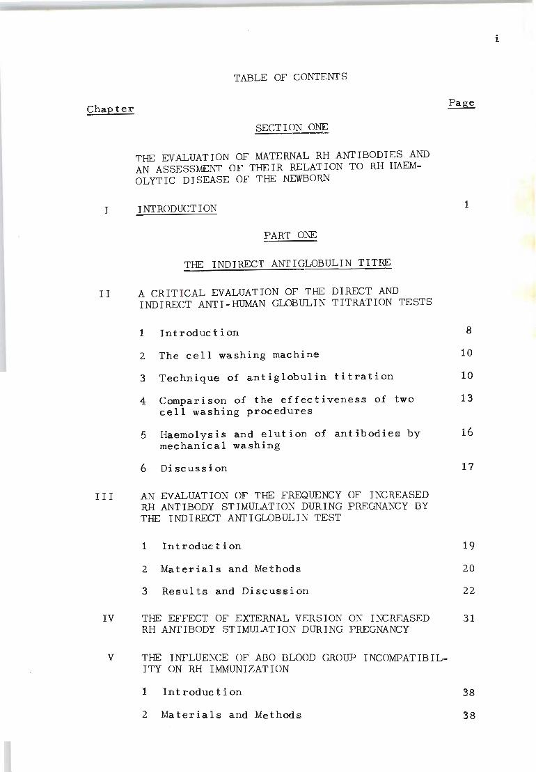
Chap ter
I
TABLE OF CONTENTS
SECTION ONE
THE EVALUATION OF MATERNAL RH ANTIBODIES AND AN ASSESSMENT OF THEIR RELATION TO RH HAEMOLYTIC DISEASE OF THE NEWBORN
INTRODUCTION
PART ONE
THE INDIRECT ANTIGLOBULIN TITRE
II A CRIT I CAL EVALUATION OF THE DIRECT AND INDIRECT ANTI-HUMAN GLOBULIN TITRATION TESTS
1
1 Introduction 8
2 The cell washing machine 10
3 Technique of antiglobulin titration 10
4 Comparison of the effectiveness of two 13 cel l washing procedures
5 Haemolysis and elution of antibodies by 16 mechanical washing
6 Discussion 17
III AN EVALUATION OF THE FREQUENCY OF INCREASED RH ANTIBODY STIMULATION DURING PREGNANCY BY THE INDIRECT ANTIGLOBULIN TEST
IV
1 In trod uc t i on
2 Materials and Methods
3 Results and Discussion
THE EFFECT OF EXTERNAL VERSION ON INCREASED RH ANT I BODY STIMULATION DURING PREGNANCY
V THE INFLUENCE OF ABO BLOOD GROUP INCOMPATIBILITY ON RH IMMUNIZATION
lInt roduc t i on
2 Materials and Methods
19
20
22
31
38
38
i
Chapter
V (contd.)
3 Results
4 Discussion
VI THE EVALUATION OF THE INDIRECT ANTI-HUMAN GLOBULIN TITRE AS A PROGNOSTIC INDEX
40
54
1 Rh antibody levels in the maternal serum 57
2 The relationship of maternal indirect 63 antiglobulin titre, cord-blood haemoglobin and reticulocyte perc entage in the prognosis of Rh haemolytic disease
PART TWO
THE PARTIAL ABSORPTION TEST
VII THE PARTIAL ABSORPTION TEST (PA) FOR TITRATING RH ANTIBODIES
1 Introduction
2 Materials and Method s
3 Results
4 Discussion
VIII THE EVALUATION OF THE PARTIAL ABSORPTION TEST IN THE PROGNOSIS OF RH HAEMOLYTIC DISEASE
1 In trod uc t i on
2 Materials and Methods
3 Results
4 Discussion
PART THREE
THE RH ANTIBODY INHIBITION TEST
IX THE SIGNIFICANCE OF THE RH ANTIBODY INHIBITION TEST IN DETERMINING THE SEVERITY OF RH HAEMOLYTIC DISEASE OF THE NEWBORN
69
70
72
73
81
85
85
86
97
99
ii
Chapter Page
IX (contd. )
1 In trod uc t i on 100
2 Materials and Methods 101
3 Results and Discussion 104
X THE VALUE OF THE RH ANTIBODY INHIBITION TEST IN RELATION TO LIQUOR AMNII STUDIES
XI
1 Introduction 116
2 Materials and Methods 117
3 Results and Discussion 118
SUMMARY 132
SECTION TWO
THE SPECIFICITY AND IMMUNOGLOBULIN CHARACTER· 137 ISTICS OF AUTOANTIBODIES IN ACQUIRED HAEMOLY-TIC ANAEMIA OF THE 'WARM' TYPE
INTRODUCTION 138
X I I THE RH SYSTEM OF BLOOD GROUPS
1 Introduction 144
2 The normal Rh gene complex 144
3 The missing Rh antigen types 148
4 The LW antigen and the Rhnull phenotype 150
5 The association of mUltiple phenotypic abnormalities wi th Rhnull 152
XIII THE SPECIFICITY OF ACQUIRED HAEMOLYTIC ANAEMIA AUTOANTIBODIES
1 In trod uc t i on
2 Materials and Methods
3 Results
4 Di scuss ion
158
159
160
168
iii

Chapter
SUMMARY
REFERENCES
216
222
v
LIST OF TABLES
Table
1 Comparison of indirect antiglobulin titrations 14 using two methods of washing sensitized cells
2 Comparison of direct antiglobulin titration 15 using two methods of washing sensitized cells
3 Accuracy in evaluation of Rh-antibody titres 21 by standardized indirect antiglobulin method when determined by 2 investigators {blind test}
4 Analysis of frequency of antenatal episodes of 23 Rh-immunization observed among 818 Rh-immunized mothers examined at various stages of pregnancy
5 Comparative analysis of frequency of antenatal episodes of Rh immunization observed among mothers not immunized and those already immunized before present pregnancy
24
6 Evaluation of incidence of fetal wastage 29 {stillbirths and neonatal deaths} in relation to antenatal episodes of increased Rh-antibody production observed for 208 mothers who were not immunized before present pregnancy
7 Evaluation of incidence of fetal wastage 30 {stillbirths and neonatal deaths} in relation to antenatal episodes of increased Rh-antibody production observed for 610 mothers wh o were already immunized before present pregnancy
8 Comparison of the frequency of antenatal epis- 33 odes of Rh immunization in Rh negative mothers on whom external version was carried out after 32 weeks gestation, and Rh negative mothers on whom such manipulations were not done
9 The frequency of external version and antenatal 34 immunization by the Rh factor observed in 6 380 consecu t ive admissions for pregnancy
10 The frequency of ABO compatible and incompat- 41 ible combinations in Rh-immunized father-mother and mother-child combinations
11 Distribution of ABO compatible and incompatible 43 combina t ions in Rh negative mothers with var-ious titres of Rh antibodies
12 The relationship of maternal Rh antibody values to ABO incompatible combinations ity of the father's Rh genotype and mea~ of pregnancies
titre zygosnumber
45
vi
Table
13 Incidence of Rh immunized mothers with history 48 of abortion in relation to ABO compatible and incompatible matings
14 Rh antibody titre values for ABO incompatible 50 combinations of Rh immunized families
15 Maternal Rh antibody titre values during the 52 first and third trimesters of pregnanc y in relation to the frequency of mother- c hild ABO compatible and incompatible combinations
16 Distribution of 373 c ases of incompatible 59 infants born to Rh immunized mothers by maternal indirect antiglobulin titre at term
17 Relationship between maternal indirect antiglob- 62 ulin index and outcome of pregnancies of Rhsensitized mothers
18 Comparison of maternal partial absorption titre 74 at the time of birth with titre of free antibody in the cord serum
19 Comparative tests on maternal Rh sera 76
20 Partial absorption titre, blocking titre and 78 indirect antiglobulin titre for various maternal sera
21 Distribution and out come of cases of Rh-incompat- 87 ihle infant s born to immunized mothers by part ial absorption titre of the maternal serum at term
22 Relationship between c ord-blood haemoglobin 92 values in liveborn Rh-inc ompatible infants and the difference between the maternal indirect antiglobulin titre and PA titre at term
23 Outcome of cases in which the maternal PA titre 94 was determined before the end of the second trimester
24 Distribution of Rh-negative infants born to 96 immunized mothers by indirect antiglobulin titre and PA titre of the maternal serum at birth
25 Results of Rh-antibody inhibition test 105
26 Calculating inhibition index 106
27 Relationship between maternal indirect antiglob- 107 ulin titre and outcome of pregnancy
vi i
Table
28 Distribution of Rh positive infants born alive 109 to immunized mothers with indirect antiglobulin titres of 1:1024 or more by values for cord-blood haemoglobin and neonatal deaths
29 Comparative analysis between relationship of Rh 111 antibody titres and inhibition test
30 Rh antibody inhibition index as a measure of 112 severity of Rh haemolytic disease
31 Frequency of other immune red cell antibodies 115 among mothers who were initially sensitized by the Rh factor
32 A statistical evaluation of the accuracy in pre- 120 dieting the severity of Rh haemolytic disease before birth of Rh-incompatible infants by the Rh antibody inhibition test and amniotic fluid studies
33 Rh antibody and liquor amnii follow-up studies 123 on Mrs. H. L.
34 Rh antibody and · liquor amnii follow-up studies 125 on Mrs p. K.
35 Rh antibody and liquor amnii follow-up studies 126 on Mrs. G. M.
36 Rh antibody and liquor amnii follow-up studies 128 on Mrs D. B.
37 Rh antibody "and liquor amnii follow-up studies 130 on Mrs R. K.
38 Comparison of the Fisher-Race linked gene theory 147 and the Wiener multiple allele theory
39 Rh gene complexes with their corresponding 149 antigens
40 Absorption and elution studies of anti-LW using 153 Rh-negative and RhnuII red cells
41 Red cell agglutination test of known U-positive 155 and U-negative bloods for various antisera
42 Classification of 60 acquired haemolytic anaemia 162 red cell eluates
43 Distribution of the various classes of eluates 163 between male and female, patients and donors
vi i i
Table Page
44 Absorption experiments on eluate B.A.B.
45 Absorption experiments on eluate L.A.
46 Agglutination scores of eluates against enzymated cells
47 Suggested antigenic make-up of "normal", "partially" deleted and "fully" deleted cells
48 Ob served frequency of "ant i-nl" and "ant i-dl" autoantibodies in the serum and the red cell eluates of 12 patients with acquired haemolytic anaemia.
165
166
167
169
176
49 Frequency of "anti-nl" and "anti~dl" autoanti- 177 bodies observed in 12 serums and red cell eluates by the ficin test and combined ficin and antiglobulin test
50 Number of ficin and antiglobulin-reactive 179 autoantibodies observed by examining the red cell eluates against three "normal" and three Rhnull samples of blood.
51 An evaluation of the simultaneous occurrence 180 of f ic in- reac t i ve "ant i-nl" and f ic in- reac t ive "anti-dl" or antiglobulin-reactive "anti-dl".
52 An evaluation of the simultaneous occurrence of 181 antiglobulin-reactive " ant i-nl" and antiglob-ulin reactive "anti-dl" or ficin-reactive " an t i - n I "
53 Relationship of indirect antiglobulin pattern 183 to presence of "anti-nl" and " anti ~ dl " autoantibodies in 24 red cell eluates
54 Distribution of "anti-nl", " anti-pdl " and 191 "anti-dl" in 55 red cell eluates
55 Absorption experiments with red cell eluate 193 S.C.
56 Indirect antiglobulin reactivity of various 194 AHA autoantibodies for specific preparations of anti-IgG, anti-IgM, anti-IgA and anti-complement
57 Frequency of various immunoglobulin classes 196 observed in 55 acquired haemolytic anaemia eluates ..
58 Indirect antiglobulin reactions of various AHA 202 red cell eluates for known red cell types
ix
Table
59 Specificities of autoantibod'ies recognized by 203 selective absorption and elution studies using different panels of red cells
60 Absorpt i on and elution experiments with red 205 cell eluate FR
61 Absorption experiments with red cell eluate PM 207
62 Absorption experiments using guinea pig anti-sera to Rh positive LW-(Mrs Bigelow} cells 209
63 Absorpt i on experiments with red cell eluate KP 211
64 Absorption experiments using a selected red 213 cell eluate which did not react with Rhnull or partial l y deleted red cells
Appendix:
Serology, biochemistry and haematology of 66 131 Rh-immunized mothers who delivered Rh-positive infants
x
LIST OF FIGURES
Figure
1 Photograph of cell-washing machine, with the 11 rack for tubes mounted on the rack holder
2 The distribution of 147 cases of 'incompat- 65 ible' infants born alive to inununized mothers, by values for cord-~lood haemoglobin and the rna t erna I an t i glob u lin tit rea t term
3 The distribution of 145 cases of 'incompatible' 67 infants born alive to inununized mothers, by
4
5
6
7
8
values for cord-blood haemoglobin and reticulocyte percentage
The distribution of 179 live-born Rh-incompatible infants by cord-blood haemoglobin value and maternal P.A. titre at the time of birth
The Wiener and Fisher-Race concept of the Rh system
An illustration of two types of condition that can lead to Rhnull expression
A model of the Rh chromosomes based on the evaluation of acquired haemolytic anaemia aut oan t i bod i e s
Rh chromosomes of man and apes and their relationship to determinants 'nl', 'pdl '" and , d I' .
89
146
157
217
219
xi
SECTION ONE
THE EVALUATION OF MATERNAL RH ANTIBODIES AND AN
ASSESSMENT OF THEIR RELATION TO RH HAEMOLYTIC
DISEASE OF THE NEWBORN
Chapter I
INTRODUCTION
The association of haemolytic disease of the newborn
with blood group differences between the mother and her
unborn child was first recognized by Levine and Stetson
(1939) in a patient who gave birth to a stillborn macer-
at ed fetus. They established that the mother had been
immunized with an antigen which the infant had inherited
from the father. It was subsequently confirmed by Land-
steiner and Wiener (1940) that the antibody described by
Levine and Stetson was identical to the antibody they had
obtained when rabbits were injected with Rhesus monkey
red cells. The blood group factor associated with this
haemolytic condition was thereafter called the Rhesus
( Rh) f ac t or.
It is now well established that Rh-immunization is the
result of transplacental passage of fetal Rh-positive
red cells into Rh-negative mothers. The effect of this
process of immunization on the clinical condition of the
infant starts when the maternal Rh-antibody enters the
infant's circulation through the placenta. The an t i body
becomes attached to the fetal Rh-positive red cells and
shortens their life span. The degree of severity of this
haemolytic process depends on the rate of fetal red cell
destruction induced by the antibody and the ability of
1
the fetus to compensate by increased red cell production.
If the rate of red cell destruction during gestation is
greater than its production through extramedullary erythro
poietic activity then anaemia results and may be followed
by intrauterine death. Should the affected fetus surv ive
the hostile in utero environment complications associa ted
with hyperbilirubinaemia may develop which can lead to
severe brain damage (kernicterus).
In white American, British and South African populat-
ions haemolytic disease of the newborn due to Rhesus in-
compatibility occurs in one out of 150 full-term pregnan-
cies (Levine, Vogel and Rosenfield, 1953; Vos, 1967).
Approximately 70 per cent of infants born with Rh haem-
olytic disease will be severely enough affected to require
treatment (Kelsall, Vos and Kirk, 1958). Universal accept -
ance of the necessity for routine Rh-typing of all preg-
nant women, coupled with detailed antenatal serological
investigations has now greatly simplified the problem of
anticipating Rh haemolytic disease. However, for the
obstetrician and paediatrician there remains the issue of
individual prognosis and case management and it is with
this respect that a multitude of intriguing and often un-
answerable questions can be provided by experienced immuno-
haematologists.
During the past decade significant advances have been
made towards the prevention of Rh haemolytic disease.
Based on the fundamental concept that passive immunity
can prevent active immunity (Smith, 1909), investigators
in England and the United States were able to show that
the simple injection of Rh antibodies into Rh-negative
mothers can also suppress Rh immunization (Finn, Clark~,
Conohoe, McConnell, Sheppard, Lehane and Kulke, 1961;
Freda, Gorman and Pollack, 1963). The efficacy of this
2
approach has now been firmly established (Clarke, 1967;
Pollack, Gorman, Hager, Freda and Tripodi, 1968).
In spite of these advances in the prevention of Rh
immunization, the risk of Rh-negative mothers becoming
immunized to the Rhesus antigen is still high. Condition:>
influencing the continuous occurrence of Rh immunization
are related to several factors among which the failure of
the mother to receive passive therapy during and after
pregnancy IS probably the main cause. There is al so a
good deal of evidence to suggest that the suppression of
Rh immunization produced by anti-Rh is antigen-specific
(Mollison, 1972). This means that the administration of
specific anti-D prevents the formation of anti-D but not
the formation of other types of antibodies which are
likely to cause haemolytic disease of the newborn, e.g.
anti-C, anti-E, anti-Kell and Fya and anti-Jka . Therefore,
haemolytic disease of the newborn remains a significant
c 1 in i c a I pro b I em.
In the antenatal diagnosis of Rh haemolytic disease it
is important that the presence of placenta-permeable anti ..
bodies is first recognized in the maternal serum. From
then on it can generally be expected that the severity of
the disease process in utero will be related to the inten.o
sity of antibody formation in the mother. Under this
stimulus destruction of the unborn ~hild's Rh-positive
red cells leads to the formation of various end products
including the toxic unconjugated bilirubin which has an
affinity for certain nerve cells in the brain.
3
Quantitative tests for the determination of unconjug
ated bilirubin in liquor amnii will therefore directly
reflect the intensity of fetal red cell destruction tak
ing place in utero. The ability to predict the severity
of Rh haemolytic disease by examining the amniotic fluid
has been confirmed by Bevis (1956), Mackay (1961), Liley,
(1961) and Freda, (1964).
Although prognosis by amniotic fluid studies appears
to be simple , a considerable number of complications can
occur following the practise of inserting a needle into
the uterus (amniocentesis). The most common hazards are:
(1) damage to fetal blood vessels on the surface of the
placenta, which has been reported to occur in nearly 15
per cent of patients (Zilliacus and Ericksson, 1958);
this complication has induced early separation of the
placenta causing fetal death in a number of instances
(Mayer, 1963); (2) if the placenta is traversed at the
puncture, fetal Rh-incompatible cells invariably enter
the maternal circulation to intensify Rh-antibody product
ion. Walker and Jennison (1962) reported a post-amnio-
centesis antibody rise in 41 per cent of mothers. Thus
in those cases where fetal blood has been aspirated by
amniocentesis the chances of increasing Rh-immunization
and intens~fying the haemolytic process in utero are
considerably greater. It has therefore been accepted
that amniocentesis should be carried out only when the
maternal antibody value is raised above a certain crit
ical level (Queenan, 1966 ; Kubli, 1966; Vos, 1969).
4
A major problem in the application of Rh antibody
determinations is the selection of suitable serological
methods. In a study concerning the in vitro reactivities
of Rh antibodies Hill and Soules (1957) showed that three
orders of antibodies are clearly recognizable by differ
ent testing procedures: (1) the "agglutinin"; this is an
Rh antibody best demonstrated by its ability to agglutin
ate cells in a medium of physiological saline; (2) the
"agglutinoid", which can be demonstrated by the blocking
test and ind i rect anti-human globulin test but not by any
enzyme test, and (3) the " cryptagglutinoid", which is
detectable by the indirect anti-human globulin test,
albumin and other colloids and by enzyme-treated cells.
A fourth order of Rh antibody, the "papain type" which
was described by Dodd and Eeles (1961) is detectable
solely by enzyme techniques.
The relat i onship between the various orders of Rh anti
bodies to the general immunization process has established
that Rh antibodies measured by the indirect anti-human
globulin test are of far greater prognostic significance
than those determined by the albumin or enzyme tests
(Vos, 1958; Tovey and Valaes, 1959; Zeitlin and Boorman,
1963 ; Jacobs, 1962; Hubinot, 1961).
A disturbing complication of the indirect anti-human
globulin test is that considerable variation in results
may be obtained when the same method is used by other
workers in different laboratories (Goldsmith, Mourant
5
and Bangham, 1967). Thus it is difficult to compare the res -
ul ts obtained by one laboratory wi th those of another.
Although this lack of agreement may reflect variations In
individual standards of measuring Rh antibody activity,
several other causes also cannot be ignored. For ins tanc e,
it has now become clear that Rh antigens may occupy a
greater, or lesser, area of the red cell membrane, de
pending upon the zygosity of the Rh antigen and its quan
titative distribution in family members (Rochine and
Hughes-Jones, 1965; Masouredis, Dupuy and Elliott, 1967).
In view of the difficulties in assessing the clinical
conditions of this disease by serological means, extens
ive investigations were carried out to examine the relat
ionship of several methods of Rh antibody determination.
(1) the standardized indirect anti-human globulin titrat
ion test (Vos and Kirk, 1958); (2) the partial absorption
test (Vos, 1 958) and (3) the Rh antibody inhibition test
(Vos, 1969). It was hoped that these studies would pro
vide valuab l e information for the evaluation of differ
ent methods in the diagnosis of the severity of Rh haem-
olytic disease of the newborn. The development of satis-
factory testing procedures was of fundamental importance
to the progress of the entire study.
A notable achievement resulting from these investigat
ions has been the acknowledgement that the partial absorpt
ion test, described in Chapter VII, has been recommended
as a testing procedure by the Council on Immunohaematology
of the American Society of Clinical Pathologists.
6
7
PART ONE
THE INDIRECT ANTIGLOBULIN TITRE
Chapter II
A CRITICAL EVALUATION OF TI-IE DIRECT AND INDIRECT ANTI
HUMAN GLOBULIN TITRATION TESTS
1. INTRODUCT ION
Since the discovery of Rh haemolytic disease of the
newborn by Levine and Stetson (1939) and Landsteiner and
Wiener (1940), investigators have encountered many diff
iculties in predicting the severity of the disease before
birth. To evaluate the condition of the infant by clin
ical signs only can be most misleading and clinicians are
obliged to look to the laboratory for information to clar
ify an otherwise obscure clinical picture.
After much trial and error we adopted the antiglobulin
test of Coombs, Mourant and Race (1945) as a routine pro
c edure for the determination of the severity of Rh haem
olytic disease. Both the direct and indirect methods
were employed and standardized in every detail (Vos and
Kirk, 1958). In Coombs et aI's description of the use
of rabbit anti-human globulin serum for detecting the
sensitization of red blood cells by Rh antibodies, they
emphasized the necessity for several washings of the sen
sitized cells prior to adding the antiglobulin reagent.
In many laboratories investigators wash the sensitized
cells by adding an excess (10 to 20 vol.) of 0.85 per
cent saline solution, inverting the tube to resuspend
the cells, centrifuging and removing the supernatant sal.
ine solution and then repeating the procedure until three
washings have been completed.
8
In
Serologists are aware that simple resuspension of cells
a large volume of physiological saline solution IS in-
adequ~te for reproducible end points in titrations with
antiglobulin reagents. We found that other laboratories
using the same test serum and the same antiglobulin serum
often obtained titration values that were less than our
own; in some instances the discrepancies were startling.
A technique was therefore devised to eliminate these diff
erences and to make titration values obtained in various
laboratories more comparable. This is particularly im-
portant when maternal Rh antibody titres are used as a
guide for the induction of labour and the treatment of the
newborn infant (Kelsall and Vos, 1955; Kelsall, Vos, Kirk
and Shield, 1957).
Initially our improved washing procedure consisted of
using a Pasteur pipette to withdraw a suspension of sensit
ized cells out of the test tube and then vigorously expell-
ing the cells back into the tube . This was repeated 10 to
15 times to ensure thorough washing. Although this proced-
ure yielded consistent end-points in all of the titrations
it was tedious and time-consuming. A simple machine was
therefore designed to provide a means of shaking a rack
of tubes at a constant speed. The results obtained show
that sensitized cells that are washed by means of mechan
ical agitation yield consistently higher titration values
than do cells that are washed by means of simple resuspen
sion In a large volume of physiological saline.
9
2. THE CELL-WASHING MACHINE
A rack-ho l der is mounted on a rocker arm that is rigid
ly coupled through an eccentric to a 1/20-h.p. electric
motor than can run at 1 000 rpm. The reciprocal motion
moves the rack-holder horizontally with a maximal displace
ment of 1.3cm. The light aluminium rack carries 20 tubes
(0.5 by 5,0 cm). The tubes are closed with small rubber
stoppers and are seated on sponge-rubber cushions; they
are held rigidly in position by a hinged plate that folds
forward against the rubber stoppers when the rack is mount
ed on the rack-holder. In order to facilitate rapid place
ment and removal the rack is held on the rack-holder by
means of a keyed slide. When removed it may be fitted
into a stand on the operator's bench. Fig.1 IS an illus
tration of the cell-washing machine with a rack mounted on
the rack-holder.
3. TECHNIQUE OF ANTIGLOBULIN TITRATION
Indirect antiglobulin titration - The specifically react
ive test cells are washed mechanically three times in 10
vol of 0.85 per cent saline solution and then resuspended
in a sufficient amount of physiological saline solution
t o yield a 10 per cent suspension of cells. 0.4 ml of the
undiluted se r um to be tested is pipetted into the first
and second serial test tubes. To the second and to each
succeeding tube, 0.4 ml of 0.85 per cent saline is added.
Serial doubling dilutions are then prepared In the usual
manner. To each of the tubes 0.4 ml of the 10 per cent
suspension is added and the tubes incubated at 37° C for
1 hr. (We have adopted the convention of calling the dil-
10
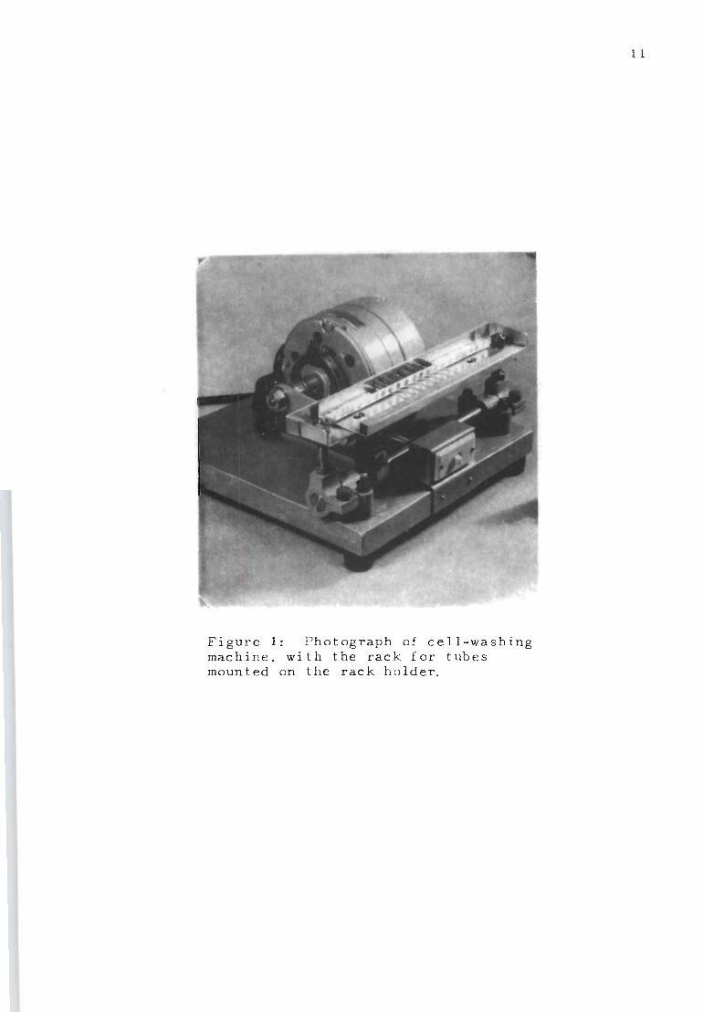
Figure 1: Photograph of cell-washing "machine, with the rack for tubes mounted on the rack holder.
1 1
ution in the first tube 1:2).
After incubation the sensitized cells are washed three
times by mechanical shaking for 30 sec. each time with 20
vol. of 0.85 per cent physiological saline. After removal
of the supernatant fluid from the final washing two drops
of anti-human globulin reagent (known to yield optimal re
actions with Rh-sensitized cells) are added to the washed
sensitized cells in the test tube. This suspension is
then pipetted onto a glass slide so that a pool of cells
approximately one inch in diameter is formed. The slide
is then placed in a moist chamber at room temperature.
After being left undisturbed for 10 minutes the slide is
gently tilted forward and the agglutination read against
a well-illuminated ground-glass screen using a 5 x magnif
ication head fitting eyepiece. As a control, unsensitized
group 0, Rh-positive cells are tested in parallel with the
sensitized cells.
Direct antiglobulin titration ~ Adult or infant cells
sensitized in vivo are washed mechanically three times in
20 vol. of O. 85 per cen t sa 1 ine sol uti on. Af ter remov ing
the supernatant saline from the final washing, two drops
of anti-human globulin reagent are added to the washed
cells in the test tube. This suspension is then pipet ted
onto a glass slide. The results are read as described
for the indirect antiglobulin ti tration test . Group 0,
Rh-positive nonsensitized red blood cells are used as con
t rol s.
12
4. COMPARISON OF THE EFFECTIVENESS OF TWO CELL WASHING PROCEDURES
Indirect ant i globulin titrations - Seven Rh antisera
were titrated by the indirect antiglobulin technique and
two procedures were used for washing the sensitized cells.
In the first series the cells were washed three times by
adding 10 vol. of 0.85 per cent saline solution and then
inverting the tube in order to resuspend the cells. In
the other series the cells were washed three times by mech-
anical agitation on the cell-washing machine for 30 sec.
The results in Table 1 indicate that in all instances the
end point of the titration was higher for the mechanically
washed cells . In cases 2, 3, 5 and 6 the prozone observed
in the lower dilutions of the manually washed cells was
not present when the cells were washed mechanically.
Direct antiglobulin titrations - A similar set of results
for the direc t antiglobulin test was obtained using the
same two methods of washing cells (Table 2) . Cases 1 to
4 were red cells obtained from the umbilical cords of in-
fants born to Rh-immunized mothers. Cases 5 and 6 were
adult cells that were sensitized with known Rh antisera.
In case 5 the Rh antiserum had an indirect antiglobulin
titre of 1:128; case 6 a titre of 1:4096. The mechanically
washed cells yielded higher end points in four out of six
cases. In five cases a conspicuous prozone was observed
in the lower dilutions for the manually washed cells; this
was absent wh en the cells were washed mechanically.
13
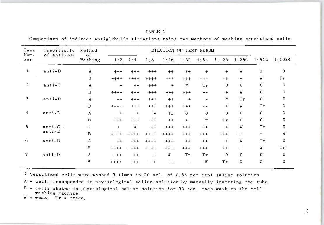
TABLE 1
Compari son of indirect antiglobulin titrations using two methods of washing sensitized cells
Case Specificity Method DILUTION OF TEST SERUM Num- of an t ibody of ber Washing 1:2 1: 4 1:8 1:16 1:32 1: 64 1: 128 1:256 1: 512 1:1024
1 an t i-D A +++ +++ +++ ++ ++ + + W 0 0
B ++++ ++++ ++++ +++ +++ +++ ++ + W Tr
2 an t i-C A + ++ +++ + W Tr 0 0 0 0
B ++++ +++ +++ +++ +++ ++ + W 0 0
3 anti-D A ++ +++ +++ ++ + + W Tr 0 0
B ++++ +++ +++ +++ +++ ++ + W Tr 0
4 an t i-D A + + W Tr 0 0 0 0 0 0
B +++ +++ ++ ++ + Vi Tr 0 0 0
5 anti-C + A 0 W ++ +++ +++ ++ + W Tr 0 an t i-D
B ++++ ++++ ++++ ++++ +++ +++ +++ ++ + W
6 an t i-D A ++ +++ ++++ +++ ++ ++ + W Tr 0
B ++++ ++++ ++++ +++ +++ +++ ++ + W Tr
7 an t i-D A +++ ++ + W Tr Tr 0 0 0 0
B ++++ +++ +++ ++ + W Tr 0 0 0
* Sensitized cells were washed 3 times in 20 vol. of 0.85 per cent sa line solution
A - cells resuspended in physiological sa line solution by manually inverting the tube
B - cells shaken in physiological sa line solution for 30 sec. each wash on the cell-washing machine.
W = weak; Tr = trace.
~
~
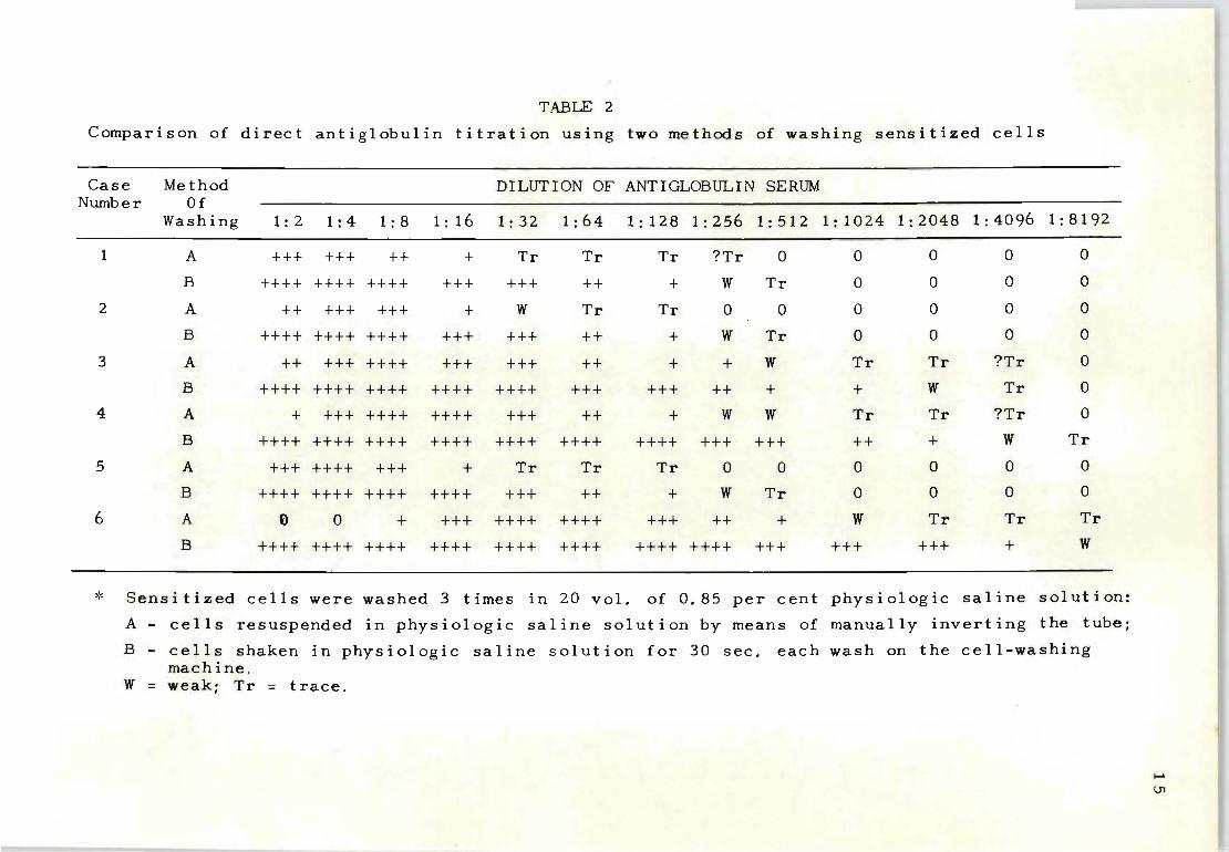
TABLE 2
Comparison of direct an t i glob u 1 in titration using two methods of washing sensitized cells
Case Method DILUTION OF ANTIGLOBULIN SERUM Number Of
Washing 1: 2 1: 4 1: 8 1: 16 1:32 1: 64 1: 128 1:256 1:512 1:1024 1:2048 1: 4096 1:8192
1 A +++ +++ ++ + Tr Tr Tr ?Tr 0 0 0 0 0
B ++++ ++++ ++++ +++ +++ ++ + W Tr 0 0 0 0
2 A ++ +++ +++ + W Tr Tr 0 0 0 0 0 0
B ++++ ++++ ++++ +++ +++ ++ + W Tr 0 0 0 0
3 A ++ +++ ++++ +++ +++ ++ + + W Tr Tr ?Tr 0
B ++++ ++++ ++++ ++++ ++++ +++ +++ ++ + + W Tr 0
4 A + +++ ++++ ++++ +++ ++ + W W Tr Tr ?Tr 0
B ++++ ++++ ++++ ++++ ++++ ++++ ++++ +++ +++ ++ + W Tr
5 A +++ ++++ +++ + Tr Tr Tr 0 0 0 0 0 0
B ++++ ++++ ++++ ++++ +++ ++ + W Tr 0 0 0 0
6 A () 0 + +++ ++++ ++++ +++ ++ + W Tr Tr Tr
B ++++ ++++ ++++ ++++ ++++ ++++ ++++ ++++ +++ +++ +++ + W
~< Sensitized cells were washed 3 times in 20 vol. of 0.85 per cent physiologic sa 1 i ne sol ut ion:
A - cells resuspended in physiologic sed ine solution by means of manually inverting the tube;
B - cells shaken in physiologic saline solution for 30 sec. each wash on the cell-washing machine .
W = weak; Tr = trace.
..... \Jl
5. HAEMOLYSIS AND ELUTION OF ANTIBODIES BY MECHANICAL WASHING
The amoun t of cellular damage caused by the mechanical
washing procedure was studied by measuring the percentage
of red cell haemolysis. Adult and cord-blood cells that
were stored as heparinized blood at 4 0 C and then washed
mechanically for 60 sec. with 0.85 per cent saline solut-
ion showed no measurable haemolysis when used within 24
hours of collection. The amount of haemolysis was O. 1
per cent for cells stored between 24 to 48 hours and it
increased to 1.0 per cent for cells that were stored at
4 0 C for more than 72 hours .
The possibility that antibodies may be released from
sensitized cells during the washing procedure was also
studied. Adult cells that were sensitized with various
Rh antisera were washed mechanically three times and the
supernatant f luid from the washings was pooled. The wash
liquor was then dialysed against gum acacia and the con-
tents of the bag reconstituted to the original volume of
serum by add i tion of AlB serum. No unabsorbed antibody
was detected in the wash-liquor when the ratio of cells
to serum (10% suspension of cells) removed all the Rh
antibodies from the original serum. When an excess of
antibody was present in the original serum it was invar-
iably noted that similar excesses could also be found in
the wash-liquor, although at a lower concentration than
in the original serum. It is in such instances that a
prozone is frequently obse r ved in direct or indirect anti-
globulin titrations when the cells are washed manually.
16
It is important to note however that even though antibod
ies are detectable in the wash-liquor from these cells when
they are mechanically washed, the end point in the titrat
ion when using the anti-human globulin test was always
considerably higher than by the manual method.
6. DISCUSSION
Although investigators have emphasized the importance
of cell washing to free Rh-sensitized cells from contamin
ation with human serum prior to adding the anti-human
globulin reagent, little direction has been given as to
the precise method to be used. Our studies have revealed
that great differences in end-point can occur in the dir
ect and indirect antiglobulin test when two different
methods are used for washing the sensitized cells. The
mechanical procedure for washing red blood cells for the
anti-human globulin test has the advantage of (1) yielding
higher titration values and (2) enabling persons to obtain
constant and reliable results, even when they are using
the technique for the first time. If the directions for
performing anti-human globulin titrations are followed as
outlined, wor kers in various laboratories should obtain
identical values. This is particularly important if the
anti-human g l obulin test for the quantitation of Rh anti
bodies in the maternal serum is to be used as a prognostic
aid in the management of such patients.
An observation of special interest with regard to anti
human globulin titrations is that the prozone phenomenon
which can be observed when using manual procedures is
17
absent when the sensitized cells have been vigorously
washed mechanically. A prozone is obtained with Rh anti
bodies only when the sensitized cells do not absorb all
of the antibody from the serum. If the ratio of cells to
serum is changed in such a manner that no excess antibody
remains, a prozone is not formed when the antiglobulin
reagent is added.
18
The results of experiments in which the wash-liquor
from mechanically washed, sensitized cells was reconstit
uted to the original volume of serum, by means of dialysis,
demonstrated that in instances where excess antibody was
present in the original serum, antibody was also rem0ved
from the cells by the washing procedure. This suggests
that additional antibody is coated 100sely around the cells
in these instances; this loosely bound antibody, if not re
moved by vigorous washing, interferes with the antiglobulin
reaction. The interference occurs (1) when this loosely
bound Rh-antibody is present in excess, as in the lower
dilutions of the indirect antiglobulin reaction, or (2)
when the antiglobulin antibody is in excess, as in the
lower dilutions of the direct titration with antiglobulin.
The fact that the end point of the anti-human globulin
titre is higher when the cells are washed mechaniGally
than when they are washed manually (even in instances
where free Rh-antibody is detected in the wash liquor),
indicates that loosely bound antibody may enhance a pro-
zone phenomenon. The warning given by Coombs and his co
workevs (1945) against too much washing because of the theor
etical possibility that antibodies may be washed from the
cells does not appear to apply when dealing with Rh-antibodies.
Chapter III
AN EVALUATION OF THE FREQUENCY OF INCREASED RH-ANTIBODY
STIMULATION DURING PREGNANCY BY THE INDIRECT ANTIGLOBULIN
TEST.
1. INTRODUCT I ON
19
Since the discovery that Rh-negative mothers can be
protected against Rh immunization by the passive adminis
tration of anti-D gammaglobulin (Finn, Clarke, Donohoe,
McConnell, Sheppard, Lehane and Kulke, 1961; Freda, Gorman
and Pollack, 1964), considerable attention has been focused
on the incidence and effect of feto-maternal bleeds occurr-
ing at delivery. Evidence that fetal red cells can find
their way into the maternal circulation during pregnancy
indicates that antigenic stimuli for Rh-antibody formation
can be expected before, as well as after, the delivery of
an Rh-positive infant (Taylor and Kullman, 1961; Robinson,
'Villiams, Jakobowicz, Moore and Silberman, 1966).
Even though investigators employ different methods for
the antenatal detection of fetal red cells there is close
agreement that the frequency of positive findings increases
during the final months of pregnancy. While not every
feto-maternal bleed necessarily constitutes an active pro
cess of iso-immunization (because individual differences
in the mechanism of antibody formation can be expected),
it nevertheless raises the problem that antenatal haemorr
hage from the fetus to the mother can initiate or intensify
antibody stimulation. Antenatal variations in antibody
specificity, nature of immunoglobulin involved, or simple
antibody titre differences can therefore be considered as
evidence that renewed fetal bleeds have taken place .
Results of this investigation show that Rh-antibody
follow-up studies performed throughout pregnancy can often
clearly reveal the frequency of transplacental haemorrhage
and its effect on fetal wastage. Although past observat
ions have indicated that the initial development of Rh
immunization occurs as a result of the trauma of parturit
ion, we have found that it can also occur as a consequence
of fetal bleeds during pregnancy.
2. MATERIALS AND METHODS
Rh-immunized mothers in this analysis were examined
from the first trimester of pregnancy. In no ins tance
were mothers included in whom the production of Rh anti
bodies might have been the result of previous Rh-incompat
ible blood transfusions and only Rh-incompatible mother
infant combinations were examined .
For the evaluation of Rh-antibody titres the standard
ized method of indirect antiglobulin titration was used
(Chapter 11) . Since the frequency of antenatal episodes
of Rh immunization is known to differ in Rh-negative
mothers who become immunized by fetal Rh-positive red
cells, the question of when and how often these mothers
do show increased episodes of antibody production was im
portant . To determine ~hese variations the investigator
must first resolve the accuracy of his indirect antiglob
ul in t i tra t ion procedure. Tab Ie 3 shows tha t wi thin the
range of one dilution tube nearly 20% of inaccurate res-
20
TABLE 3
Accuracy in evaluation of Rh-antibody titres by standardized indirect
antiglobulin method when determined by 2 investigators (blind test)
134 Samples of Rh Antibodies Examined
Ti tre First Second Average Accuracy in predicting differences invest- invest- variat- increased ep i sodes of Rh
igator igator ion immunization when variat-% error % error % error ions exceed
3 d i 1 ut ion 0.0 0.0 0.0 100% tubes
2 d i 1 ut i on 6. 7 5. 9 6.3 93. "70/0 tubes
1 dilution 15.6 17. 9 16. 7 83.3% tube
N t-
ults can be recorded for the same samples of blood when
tested by two investigators. For a 2-dilution-tube diff
erence the percentage of error is narrowed to 5%. The
absence of titration inaccuracies exceeding three dilution
tubes is significant in that an observed increase of this
magnitude can confidently be regarded as a renewed episode
of immunization. Assuming that titre difference's (!If three
dilution tubes or more can also imply that transplacental
passage of Rh-incompatible fetal red cells has taken place,
then it should also be possible to assess how often such
occurrences generally take place during pregnaney.
3. RESULTS AND DISCUSSION
Table 4 records an analysis of 818 Rh-negative mothers
who were either already immunized before the present preg
nancy or became immunized at some stage during the present
pregnancy. The findings for different gestation periods
clearly show that a significantly greater frequency of ep
isodes of increased Rh-antibody production occurs after
22
32 weeks gestation. Considering that this indicates in
creased fetal red cell leakage across the placental barrier,
the results appear to be in agreement with the findings of
Betke (1966) who also found a greater percentage of fetal
red cells in t he maternal circulation towards the end of
pregnancy.
Table 5 records a similar investigation of the 818 Rh
immunized mothers, comparing those who were immunized be-
fore the present pregnancy with those who were not. Of
importance here was the observation that the frequency of
TABLE 4
Analysis of frequency of antenatal episodes of Rh-immunization
observed among 818 Rh-immunized mothers examined at various
stages of pregnancy
Gestation range Total number Epi sodes of Rh immunization of mothers observed >!' examined No. %
25 weeks or less 818 0 0.0%
26-28 weeks 818 53 6.4
29-31 weeks 805 106 13. 1
32-34 weeks 773 166 21. 4
35-37 weeks 713 254 35.6
38-40 weeks 289 167 57. 7
* Represents Rh-antibody titre increase of 3 dilution tubes or more
tv W
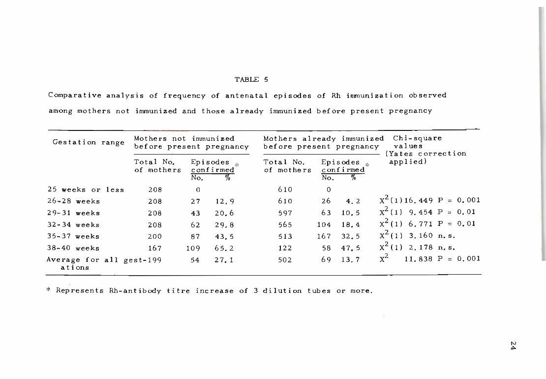
TABLE 5
Comparative analysis of frequency of antenatal episodes of Rh immunization observed
among mothers not immunized and those already immunized before present pregnancy
Gestation range
25 weeks or less
26-28 weeks
29-31 weeks
32-34 weeks
35-37 weeks
38-40 weeks
Mothers not immunized before present pregnancy
Total No. of mothers
208
208
208
208
200
167
Ep i sod e s _'_ confirmed "-No. %
0
27 12. 9
43 20.6
62 29.8
87 43.5
109 65.2
Average for all gest-199 54 27. 1 at ions
Mothers already immunized before present pregnancy
Chi-square val ues
Total No. of mothe rs
610
610
597
565
513
122
502
Ep is odes _" c onf i rmed ''-No. %
0
26 4.2
63 10. 5
104 18.4
167 32. 5
58 47. 5
69 13.7
(Yates correction applied)
X2 (1)16.449 P = 0.001
X2 ( 1) 9.454 P = 0.01
X2(1) 6.771 P = 0.01
X2(1) 3. 160 n. s.
X2(1) 2.178n.s. X2 11. 838 P = 0.001
>:< Represents Rh-antibody titre increase of 3 dilution tubes or more.
N ~
raised episodes of immunization was significantly greater
among mothers in whom Rh antibodies were detected for the
first time. This increase appears to be closely associated
with the duration of pregnancy. Mothers not immunized be-
fore the present pregnancy recorded a significantly greater
frequency of episodes of immunization before 35 weeks of
pregnancy than the Rh-negative mothers who were already
immunized.
It would be unrealistic to assume that such differences
are directly influenced by significant variations in the
incidence of transplacental bleeds occurring between moth
ers immunized and not immunized before the present preg
nancy, and the most likely explanation for the decreased
occurrence of episodes of immunization is that the mother
who is already immunized has a remarkable ability to con
ceal the recognition of the fetal Rh-positive antigen as a
potential stimulus. Such a hypothesis seems quite accept-
25
able when we consider that these mothers can only be the
recipients of fetal Rh-positive red cells which were al
ready sensitized in utero by placenta-permeable antibodies,
whereas mothers who were not immunized before the present ·
pregnancy were more often exposed to fetal red cells poss
essing the full expression of the Rh antigen. As such, the
mechanism of Rh-antibody stimulation is comparable with the
studies of Stern, Goodman and Berger (1961). They showed
that sensitized Rh-positive red cells are far less antig
enic than unsensitized red cells and these findings led to
the evaluation of the prevention of Rh immunization by the
injection of anti-D immunoglobulin.
If the indication of apparent suppression of increased
antibody production is an accurate deduction of the exper
i mental findings of Stern et al. (1961) it can be seen that
this protection is not constant throughout pregnancy_ In
fac t, it appears to decrease among the pre-immunized moth
ers as pregnancy progresses towards term. Since only small
transplacental bleeds are expected before 30 weeks of preg
nancy, with more profuse bleeds generally occurring after
this time (Betke, 1966), it is possible that a breakdown
26
in protection is induced by the introduction of signific
ant variations in the amount of Rh-positive red cells cross-
ing the placental barrier after 30 weeks' gestation. This
situation bears a striking resemblance to the inability of
anti-D gammaglobulin to secure complete protection against
rhesus immunization when massive feto-maternal bleeds take
plac e (Dudok de Wit and Borst-Eilers, 1968; Woodrow, Bow
ley, Gilliver and Strong, 1968).
These deve l opments suggest that there are at least two
way s in which the intensity of increased Rh- antibody pro-
duction can be stimulated during pregnancy. One is the re-
suI t of imperfectly understood defects of the placenta,
which allow greater quantities of fetal red cells into the
maternal circulation, while the other is undoubtedly close
I y assoc ia ted wi th external vers ion, (Vos, 1967; Rob inson,
Williams, Jakobowicz, Moore and Silberman, 1966), the re
peated application of abdominal amniocentesis, (Zipursky,
Pollock, Chown and Israels, 1963), or certain modes of
obstetric management during the third stage of labour
(Morrison, 1967).
27
The results so far presented show that fetal red cells
often cross the placental barrier to stimulate increased
Rh-antibody pr,oduction in the mother. Not yet firmly
established, however, is the effect of these episodes of
immunization on the clinical manifestation of Rh-haemolytic
disease. Table 6 details an analysis of 208 mothers' who
for the first time showed Rh antibodies during ap Rh-incom-
patible pregnancy. It is interesting to note that fetal
wastage {combined stillb~rths and neonatal deaths} can be
anticipated in the first immunizing pregnancy. Thus, the
tendency to consider all primary immunization cases as
mildly affected infants, who generally do not require
further treatment, will involve a certain amount of risk
unless constant follow-up studies are carried out to meas
ure the intensity of the haemolytic process in utero.
Compared with the values of fetal wastage observed
among the already immunized series of mothers {Table 7},
it is significant that fetal losses among the primary
immunization ca'ses consistently took place after 35 weeks
of pregnancy when Rh-antibody stimulation appeared to be
most intense. This type of pattern suggests that the time
during which an Rh-positive fetus is exposed to Rh-antibod
ies is of some importance in determining the severity of
the h~emolytic process in utero.
The findings presented in Tables 6 and 7 show that a
significant percentage of fetal wastage can be attributed
to the occurrence of renewed episodes of Rh-antibody stim ..
ula t ion. This implies that the protective factor against
increased Rh-antibody formation in the already immunized

mother can be reversed through the reactivities of pro
gressively large fetal bleeds, which in turn intensify
the haemolytic process.
28
TABLE 6
Evaluation of incidence of fetal wastage (stillbirths and neonatal deaths) in relation
to antenatal episodes of increased Rh-antibody production observed for 208 mothers who
were not immunized before present pregnancy
Total No. Gestation range of mothers
observed
ANTENATAL EPI SODES OF IMMUNIZATION
Observed* Not observed
Fetal wastage Fetal wastage
No. % No. % No. 0/0 No. % 25 weeks or less 208 0 0 0 0 208 100.0 0 0
26-28 weeks 208 27 12. 9 0 0 181 87. 1 0 0
29-31 weeks 208 43 20.6 0 0 165 73.4 0 0
32-34 weeks 208 62 29. 8 0 0 146 70.2 0 0
35-37 weeks 200 87 43.5 2 2.3 113 56.5 0 0
38-40 weeks 167 109 65.2 5 4.6 58 34.8 1 1.7
':< Represents Rh-antibody titre increase of 3 dilution tubes or more
N -.D
TABLE 7
Evaluation of incidence of fetal wastage (stillbirths and neonatal deaths) in relation
to antenatal episodes of increased Rh-antibody production observed for 610 mothers who
were already immunized before present pregnancy
Total No. ANTENATAL EPISODES OF IMMUNIZATION of mothers
Gestation range observed Ob served ':' Not ob served Fetal wastage Fetal wastage
No. % No. % No. % No. "0 25 weeks or less 610 0 0 0 0 610 100.0 0 0
26-28 weeks 610 26 4. 2 10 38.4 584 95.8 3 O. 5
29-31 weeks 597 63 10. 5 27 42.7 534 89.5 5 1. 0
32. 34 weeks 565 104 18.4 26 25.0 461 81. 6 8 1.7
35-37 weeks 513 167 32. 5 49 29. 3 346 67.5 21 6. 0
38-40 weeks 122 58 47.5 8 13. 7 64 52. 5 3 4.6
':< Represents Rh-antibody titre increase of 3 dilution tubes or more
w o
Chapter IV
THE EFFECT OF EXTERNAL VERSION ON INCREASED RH-ANTIBODY
STIMULATION DURING PREGNANCY
External version is known to be associated with in
creased transplacental haemorrhage (Robinson et al. 1966).
It was therefore important to investigate the potential
hazards of this procedure in Rh haemolytic disease.
To determine the frequency and intensity of antenatal
Rh-immunization as a result of external manipulation we
examined two separate hospital recording systems. They
comprised laboratory estimations and clinical follow-up
reports which when combined revealed those patients who
were subjected to external version and those who were not.
The fact that this information was not routinely requested
by the investigator before this study commenced, or during
the time that the Rh-negative mothers were examined for Rh
antibodies, shows that bias was avoided in evaluating the
effect of external version on antenatal immunization.
Only Rh-negative mothers examined from the first tri
mester of pregnancy were included so that the frequency of
the first occurrence of Rh antibodies or subsequent in
creases in Rh antibody production could be measured.
Of the 227 cases selected 119 formed the external ver-
sion series and 108 the control series. Only Rh-incompat-
ible mother-child combinations were used. The indirect
antiglobulin titre values of maternal Rh antibodie's were
divided into three groups: (i) those with values of 1:64
31
3 2
or less; (ii) 1:128,1:256 and 1:512 and (iii) 1:1024 or
more. The evaluation of Rh antibody titre differences re-
corded in the first trimester of pregnancy and before del
ivery would then indicate the intensity of Rh-immunization
for that pregnancy.
Table 8 details the occurrence of episodes of Rh-immun
ization among the external version and control series of
mothers. It shows a higher percentage of cases with init-
ial production of Rh antibodies in the external version
series (43.7% compared with 26.8%). Although the number
of cases is small the difference is significant (X2 (1) = 7.046; p = <.0.01).
When the overall frequency of antenatal Rh-immunization
is examined i t is evident that the version cases not only
show more frequent involvement in the secondary production
of Rh-antibodies (among those already immunizeq) but also
appear to produce higher titres than the control series
(X2 (1) = 17.025; P = <: 0.001).
To assess whether the results in Table 8 may have been
obtained by chance selection it was necessary to evaluate
the frequency of primary evidence of episodes of Rh-immun
ization in a random series of hospital clinic admissions.
The phrase 'primary evidence of Rh-immunization' refers to
the observation that Rh-antibodies were detected for the
first time at some stage during the second half of preg
nancy among mothers who were confirmed not to have had Rh
antibodies during the first half of the present pregnancy.
Table 9 details this analysis. Of an unselected series of
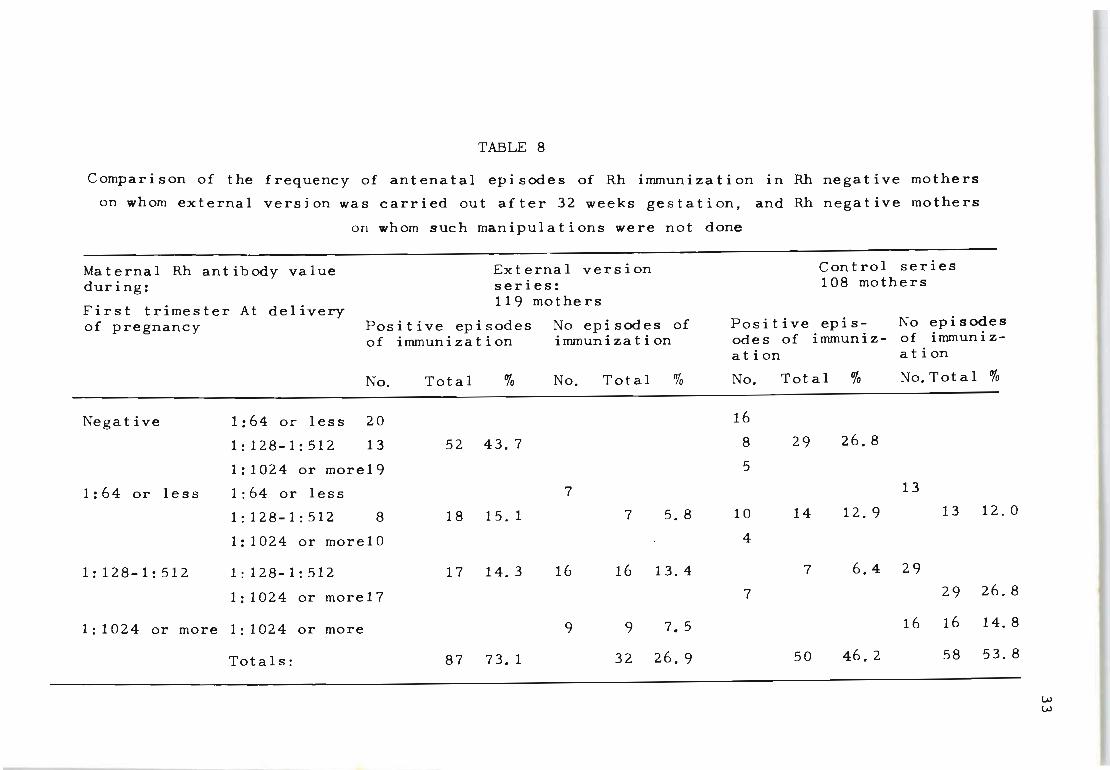
TABLE 8
Comparison of the frequency of antenatal episodes of Rh immunization in Rh negative mothers
on whom external version was carried out after 32 weeks gestation, and Rh negative mothers
on whom such manipulations were not done
Maternal Rh antibody value External version Con t rol series during: series: 108 mothers
First trimester At delivery 119 mothers
of pregnancy Positive episodes No epi sodes of Positive epis- No episodes of immunization immunization odes of immuniz- of immuniz-
ation ation
No. Total % No. Total % No. Total % No. Total %
Negative 1: 64 or less 20 16
1:128-1:512 13 52 43.7 8 29 26.8
1:1024 or more19 5
1:64 or less 1:64 or less 7 13
1:128-1:512 8 18 15. 1 7 5. 8 10 14 12. 9 13 12.0
1:1024 or more10 4
1:128-1:512 1:128-1:512 17 14. 3 16 16 13.4 7 6.4 29
1:1024 or more17 7 29 26.8
1: 1024 or more 1:1024 or more 9 9 7.5 16 16 14.8
Totals: 87 73. 1 32 26.9 50 46.2 58 53. 8
w w
TABLE 9
The frequency of external version and antenatal immunization by the Rh factor observed
6380 consecutive pregnancy admissions
in 6380 consecutive admissions for pregnancy
5260 Rh positive (82. 5%)
1120 Rh negative (17.5%)
External version performed 492
(9. 5% ) No external version performed4768
External version 113
Number and percentage of Rh negative mothers immunized by Rh factor
performed (100.0%) 9
(7.9%) 33
(3. 2%) No external 1007 version performed
Rate of Rh immunization in total number of pregnancies
1 in 151 pregnancies
w ~
6 380 consecutive admissions, 5 260 mothers were classi-
fied as Rh positive (82.5%) and 1 120 as Rh negative
(17.5%). The frequency of external version carried out
after 32 weeks gestation did not differ significantly be
tween Rh positive (9.5%) and Rh-negative (10.0%) mothers.
Of the 1 120 Rh-negative mothers tested routinely on an
35
average of 2.8 occasions during the first half of pregnancy
and on an average of 4.3 occasions during the second half
of pregnancy, 42 (3.7%) produced Rh antibodies before the
birth of their infants. This represents a rate of one case
of Rh immunization out of every 151 random pregnancies. It
was found that 7.9% of the external version series produced
Rh antibodies compared with only 3.2% of the controls. For
the total number of Rh-negative mothers examined the diff-
2 erence is significant (X (1) = 6.186; P = .::: 0.02). This
observation indicates that the results presented in Table
8 are not due to chance selection and that the higher freq-
uency of antibody production in patients subjected to ex-
ternal version is real.
The antibody-producing response in Rh negative volunteers
deliberately transfused with Rh positive blood is known to
vary significantly (Wiener, 1949; Waller 1949). Mollison
(1956) suggests that 'some volunteers are readily sensit-
ized, others are difficult to sensitize and some, perhaps,
cannot be sensitized at all'. We have found further evid-
ence to indicate that this situation also exists with res-
pect to antenatal Rh immunization. Thus, whilst it has
been shown that Rh antibody production is increased among
those mothers who received external manipulation, a far
greater percentage (92.2%) (Table 9) of the Rh-negative
female population at risk consistently fail to become
immunized.
That Rh immunization cannot always be induced even
among 'high risk' cases as a result of the delivery of an
Rh-positive infant was also established by Woodrow et al.
(1965). Even among their selected Rh-negative mothers
who consistently had Rh incompatible fetal red cells in
36
the maternal circulation, a larger percentage always failed
to become immunized. It therefore seems evident that Rh
immunization involves more than the single event of receiv
ing Rh-incompatible blood, either by deliberate transfus
ions or by disruption of the placental circulation. The
distinction between those who can be readily immunized and
those who can not is a matter for future study. The sus-
ceptible class of Rh-negative mothers who have the ability
to develop Rh-antibodies could be considered as a separate
group capable of ready sensitization to primary exposure
and also to subsequent episodes of immunization. This
appears evident from the results we have obtained. In
these mothers the intensity of Rh antiQody production was
significantly increased by the possible disruption of th~
placental circulation as a direct result of external vers
ion. A similar effect was also reported by Wood~ow et al.
(1965) with respect to transplacental haemorrhage as a
result of delivery. In their studies a significantly
greater percentage of mothers produced Rh antibodies dur
ing the post-natal period when transplacental haemorrhage
had been evident.
While there may be differences of opinion with regard
to the risk of increasing antenatal immunization by exter
nal version the possibility of intensifying the haemolytic
process in the child as a consequence of this procedure
cannot be ignored. Therefore, the policy of limiting all
antenatal manipulations which might disrupt the placental
circulation should be strictly maintained.
37
Chap ter V
THE INFLUENCE OF ABO BLOOD GROUP INCOMPAT IB ILITY ON RH
I MMUNI ZAT ION
1. INTRODUCTION
Since Levine (1943) first observed the influence of
the ABO blood group system and its relationship to Rh
immunization during pregnancy, other investigators have
confirmed that Rh-immunization in pregnancy is signific
antly less in ABO incompatible matings than in ABO com
patible matings. This deficiency had been interpreted
as being due to ABO compatible pregnancies providing a
more favourable condition for the survival of Rh positive
fetal red cells in the Rh negative mother (Levine, 1958).
In ABO incompatible conceptions it is assumed that the
presence of anti-A and anti-B in the maternal serum has
an inhibiting effect on the production of Rh antibodies
by the transplacental passage of fetal Rh positive red
cells of blood groups A or B.
Because the prognosis of Rh-haemolytic disease is
closely related to the maternal Rh antibody titre during
pregnancy, the frequency of ABO incompatibility in relat
ion to the maternal Rh antibody titre was examined using
a standardized indirect antiglobulin technique, as des
cribed in Chapter II.
2. MATERIALS AND METHODS
The obstetric records of 836 Rh-immunized mothers were
available for examination. Of these, 25 were rejected
where previous transfusion of Rh-positive blood might
38
have caused Rh-sensitization. Another 80 Rh-immunized
mothers were rejected since the paternal blood groups and
Rhesus genotype were unknown. 85 unaffected Rh-negative
infants compa t ible with their mother's Rh type were ex
cluded. Forty-three Rh-immunized mothers found during
the postnatal period were also rejected for two important
reasons: (1) t he maternal Rh-antibody titre could differ
considerably f rom the antenatal value due to episodes of
immunization i nduced by the trauma of parturition, and
(2) the postnatal cases were initially diagnosed by the
clinical manifestation of Rh-haemolytic disease and not
by routine an t enatal testing. This would increase the
number of moderately and severely affected Rh cases in
the random sample, while mildly affected Rh-positive
infants without clinical signs of Rh haemolytic disease
are not referred to the laboratory. A total of 603 mother,
father, and infant combinations among Rh-immunized famil
ies remained as suitable for study.
In some in s tances when blood from stillborn infants
could not be examined for their ABO and Rh status by
the convention al method of testing, the ability of the
red cell stroma to inhibit the appropriate antiserum was
used to establish these factors.
For the evaluation of ABO compatible and incompatible
matings in the control series 214 families with histor-
ies of normal deliveries were examined. Mothers with
obstetric histories of spontaneous abortion were not
included. As these cases were drawn from the same hospital
they represented a population similar to that under study.
39
The second group of control cases used for the evaluat
ion of ABO incompatible combinations was composed of Rh
negative mothers and their Rh-positive infants in whom
the absence of Rh-sensitization was confirmed during ante
natal testing and by the direct Coombs test on the infant's
red cells at birth . . 548 mother-infant combinations were
examined in this category. The mean number of pregnancies
experienced by these mothers was not sign.ificantly diff
erent from that in the mothers producing Rh antibodies
(3.2 pregnancies per mother compared with 3.8 pregnancies
per mother in the Rh-immunized series).
3. RESULTS
Table 10 compares the frequency of ABO compatible and
incompatible combinations in Rh-immunized father-mother
and mother-child combinations with a similar combination
of non-immunized cases. As expected, the overall occurr
ence of ABO incompatible matings in the Rh-immunized fam
ilies was lower than the values observed in the control
series.
The distribution of ABO incompatible combinations in
the control series (31.6%) compared with the value (32.8%)
reported by Reepmaker, Nijenhuis and van Loghem (1962) who
examined 1 210 Rh-negative non-immunized mothers and their
husbands. The 24.0 per cent incidence of ABO incompatible
mating s recorded in the current series of 603 Rh-immunized
families also compares favourably with the reported incid
ence of 24.7 per cent by Levine (1943). However, it
differs from the value of 18.5 per cent reported by Reep-
40
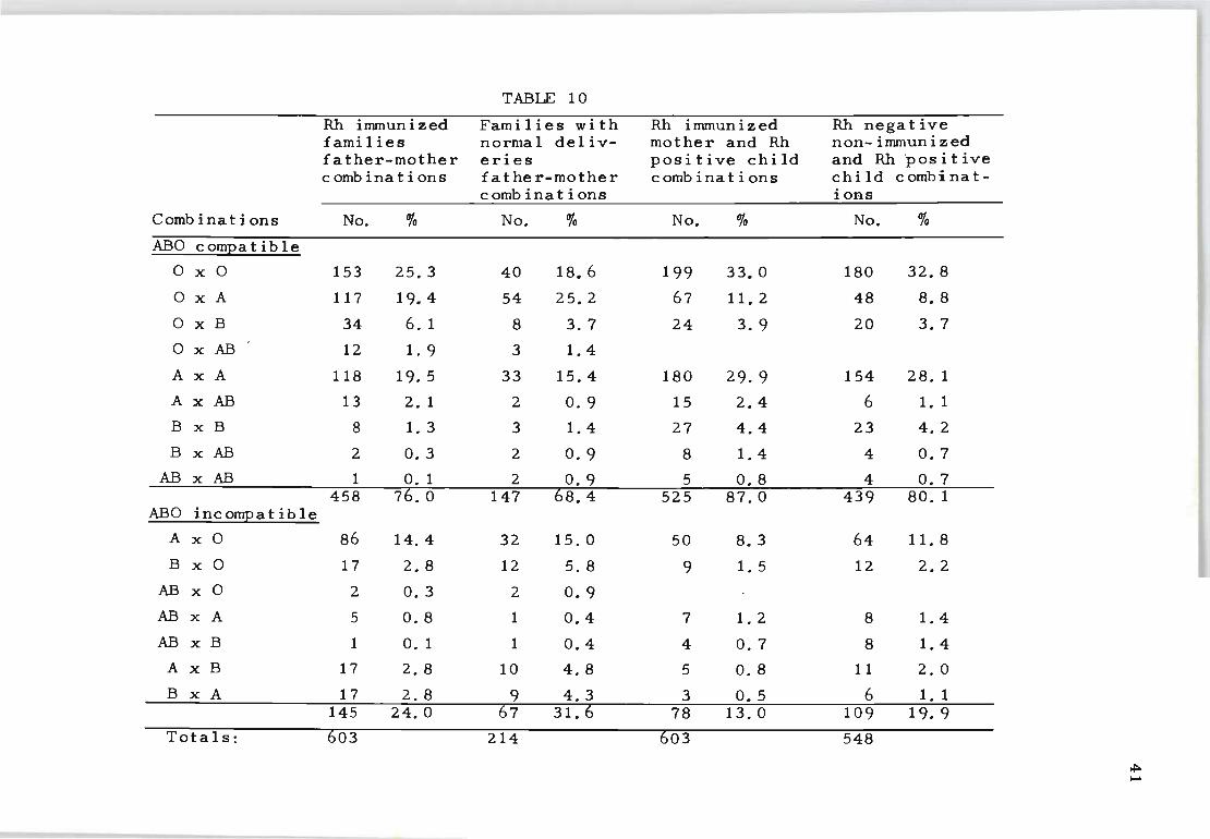
TABLE 10
Rh inununized Fami I ies wi th Rh immunized Rh nega t ive families normal deliv- mother and Rh non-inununized father-mother eries positive child and Rh 'pas it ive comb ina t ions father-mother combinations child combinat-
comb ina t ions ions
Combinations No. % No. % No. % No. % ABO cornEa t ible
o x 0 153 25.3 40 18.6 199 33.0 180 32.8
o x A 117 19.4 54 25.2 67 11. 2 48 8. 8
o x B 34 6. 1 8 3. 7 24 3.9 20 3.7
o x AB 12 1.9 3 1.4
A x A 118 19. 5 33 15.4 180 29.9 154 28. 1
A x AB 13 2. 1 2 o. 9 15 2.4 6 1.1
B x B 8 1.3 3 1.4 27 4.4 23 4.2
B x AB 2 O. 3 2 O. 9 8 1.4 4 O. 7
AB x AB 1 O. 1 2 O. 9 5 0.8 4 O. 7 458 76.0 147 68.4 525 87.0 439 80. 1
ABO incomEa t ib Ie
A x 0 86 14.4 32 15.0 50 8.3 64 11. 8
B x 0 17 2.8 12 5. 8 9 1.5 12 2. 2
AB x 0 2 0.3 2 0.9
AB x A 5 0.8 1 0.4 7 1.2 8 1.4
AB x B 1 O. 1 1 0.4 4 0.7 8 1.4
AxB 17 2. 8 10 4.8 5 0.8 11 2.0
B x A 17 2.8 9 4.3 3 0.5 6 1. 1 145 24.0 67 31.6 78 13.0 109 19. 9
Totals: 603 214 603 548
~ .....
maker, Nijenhuis and van Loghem (1962) on 1 742 families
immunized by the Rh factor.
The differences in the distribution of ABO incompat
ible combinations between mother and infant among the Rh
immunized series are smaller than the corresponding values
observed for mother-infant combinations in the non-immun
ized Rh-negative mothers. The 13.0 per cent frequency of
mother-child ABO incompatibility noted in the present Rh
immunized series is practically the same as the value of
12.6 per cent reported by Levine in 1958 but it differs
considerably from the 7.3 'per cent frequency reported by
Reepmaker (1955) on 1 608 Rh-immunized mothers.
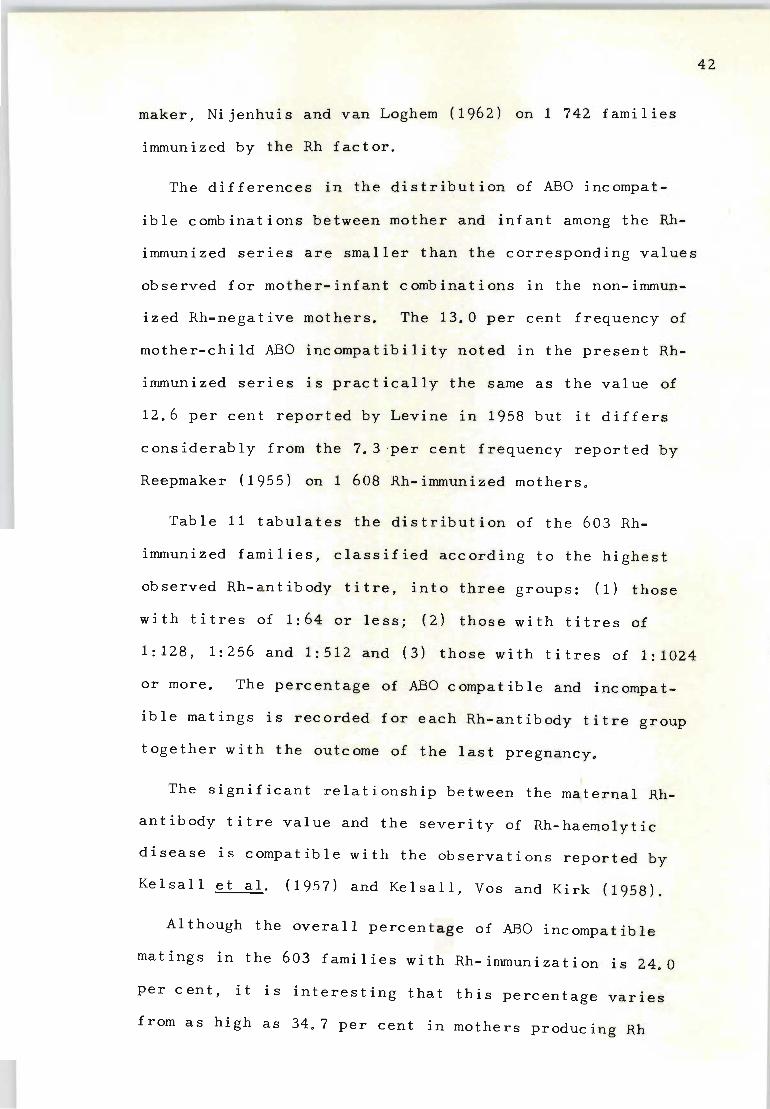
Table 11 tabulates the distribution of the 603 Rh
immunized families, classified according to the highest
observed Rh-antibody titre, into three groups: (1) those
with titres of 1:64 or less; (2) those with titres of
1:128, 1:256 and 1:512 and (3) those with titres of 1:1024
or more. The percentage of ABO compatible and incompat-
ible matings is recorded for each Rh-antibody titre group
together with the outcome of the last pregnancy.
The significant relationship between the maternal Rh
antibody titre value and the severity of Rh-haemolytic
disease is compatible with the observations reported by
Kelsall et al. (1957) and Kelsall, Vos and Kirk (1958).
Although the overall percentage of ABO incompatible
matings in the 603 families with Rh-immunization is 24.0
per cent, it IS interesting that this percentage varies
from as high as 34.7 per cent in mothers producing Rh
42
TABLE 11
Distribution of ABO compatible and incompatible combinations in Rh negative
mothers with various titres of Rh antibodies
Maternal Rh Classification ABO compa t ib Ie ABO incompatible an t i body tit re of infant** matings matings at term>:' No. 0/0 No. % Total
GrouE 1 Livebirths 63 64.8 34 35. 2 97
1:64 or less Died Nil Nil Ni I Stillbirths 1 Nil 1 All cases 64 65.3 34 34.7 98
GrouE 2 Livebirths 199 73.7 71 26.3 270
1:128-1:512 Died 7 70.0 3 30.3 10 Stillbirths 12 80.0 3 20.0 15 All cases 218 73.9 77 26. 1 295
GrouE 3 Livebirths 92 84.4 17 15.6 109
1:1024 or more Died 27 84.4 5 15. 6 32 Stillbirths 57 82.6 12 17.4 69 All cases 176 83.8 34 16. 2 210
All cases com- Livebirths 354 74.3 122 25.7 476 bined Died 34 80.9 8 19. 1 42
Stillbirths 70 82.3 15 17.7 85 All cases 458 75.9 145 24. 1 603
* Tested by the standardized 1958).
indirect antiglobulin technique (Vos and Kirk
** Last born infants.
~ w
antibodies with titres of 1:64 or less, down to 16.2 per
cent in those producing Rh-antibodies with titres of
1:1024 or more.
44
When the distribution of ABO compatible and incompat
ible matings among Rh-immunized mothers with low Rh-anti
body titres (1:64 or less) was compared with those pro
ducing higher titres (1:1024 or more) it was confirmed
that the distribution of ABO incompatibility in these
two groups differed significantly (X2 = 12.24; P = 0.001).
Table 12 compares the same three groups of Rh-antibody
titre values with the frequency of ABO incompatible com-
binations of the last born Rh-positive child. For ease
of comparison this table also shows the ABO incompatibil
ity of father-mother combinations as recorded in Table 11.
The results of these two sets of combinations detail the
mangitude of selection in Rh-antibody titre values, and,
whilst ABO incompatibility between fathers and mothers
appears twice as great (34.7% as opposed to 16.2%) in the
lower Rh-antibody titre group (1:64 or less) when compared
with the results observed In the higher titre group (1:1024
or more), this difference is at least five times greater
(26.5% as opposed to 3.7%) when the actual ABO incompatib
ility status of the mother and child is taken into account.
The 26.5 per cent value of mother-child ABO incompatible
combination observed in the lower titre group of Rh-immun
ized mothers is also greater than the values recorded for
the control series of non-immunized mother-child combinat
ions, which was 19.9 per cent.
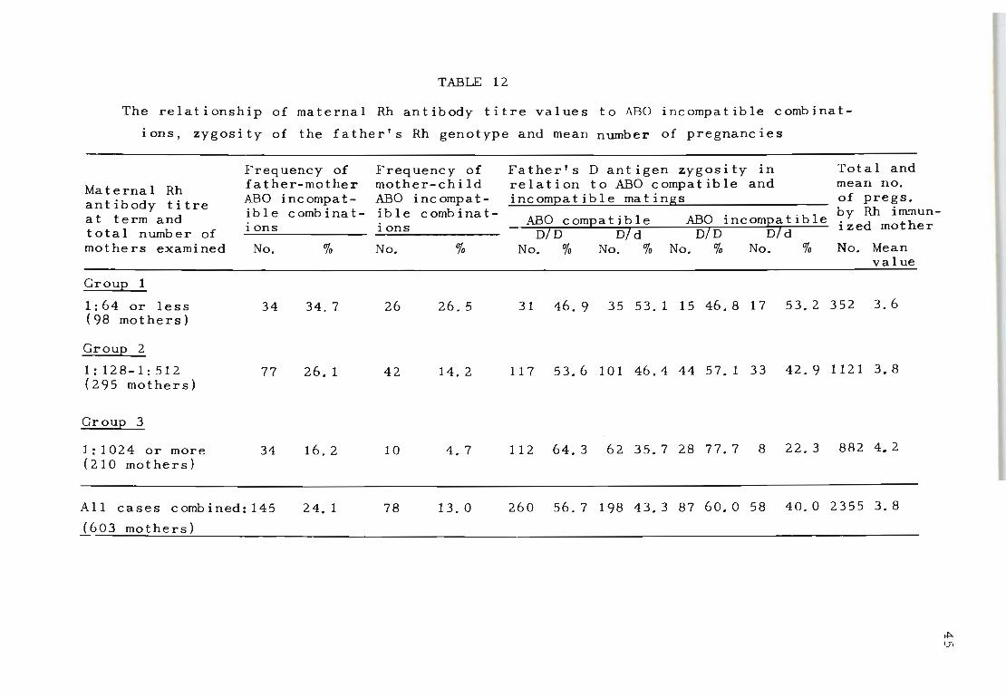
TABLE 12
The relationship of maternal Rh antibody titre values to ABO incompatible combinat
ions, zygosity of the father's Rh genotype and mean number of pregnancies
Freq uency of
Maternal Rh fa ther-mothe r
ant ibody titre ABO incompat-i b I e comb ina t-at term and ions total number of
mothers examined No. %
GrouE 1
1:64 or less 34 34. 7 (98 mothers)
GrouE 2
1 : 128-1 : 512 77 26 . 1 (295 mothers)
Groul?..-.l.
1: 1 024 or more 34 16. 2 ( 2 10 mothers)
All cases combined: 145
(?03 mothers)
24. 1
Frequency of mother-chi ld ABO incompat-i b I e comb ina t -ions
No. %
26 26 . 5
42 14. 2
10 4.7
78 13.0
Father's D antigen zygosity in Total and relation to ABO compatible and mean no. incomEatible matings of pregs.
. . by Rh im..'llun-ABO cornEa t ib Ie ABO IncomEatlble. d th
DID DId DID DId lze mo er
No. % No. % No. % No. % No. Mean value
31 46.9 35 53. 1 15 46.8 17 53. 2 352 3.6
117 53.6 101 46.4 44 57. 1 33 42 . 9 1121 3. 8
112 64.3 62 35.7 28 77. 7 8 22.3 882 4. 2
260 56.7 198 43.3 87 60.0 58 40.0 2355 3.8
~ (J'I
When the distribution of mother-child ABO incompatibil-
ity was compared between mothers with low Rh antib~dy
titre values (1:64 or less) and those with higher titre
values (1:1024 or more) it was confirmed that mother-child
ABO incompatibility differed significantly in the two cat-
(X2
1 ) egories of Rh-immunized mothers = 20.31; P = 0.00 .
The same table also compares the relationShip of mater-
nal Rh-antibody titre values with the father's D antigen
zygosity status. These results' were obtained by the con-
ventional use of specific D,C,E,c and e antisera, while
the genotypes were assigned on the basis of reactions
against these sera, using the table of most frequent
occurrence (Race and Sanger, 1962). The zygosity distrib-
ution among ABO compatible and incompatible matings does
not appear to differ in mothers with Rh antibody titres
of 1:64 or less (ABO compatible 46.9% DID and 53.1% Did;
ABO incompatible 46.8% DID and 53.2% Did), but marked
variations were seen in ' those with Rh antibody titres of
1:1024 or more (ABO compatible 64.3% DID and 35.7% Did,
ABO incompatible 77.7% DID and 22.3% Did) to implicate
homozygosity (DID) alone as a most suitable condition for
the production of high titre Rh antibodies even among ABO
incompatible matings.
46
The overall distribution of homozygous DID fathers also
differed significantly in the low titre group of Rh-immun
ized mothers (1:64 or less) when compared with the frequency
of their occurrence in the high-titred group of mothers 2
(1: 1024 or mor e) (X = 10.06; P = 0.001).
When the influence of the mean number of pregnancies on
the variations in Rh-antibody titre was studied (Table 12)
it was noted that there was a slight increase in pregnan
cies where the maternal Rh- antibody titre was 1:1024 or
more. These higher values are most probably associated
with the mother's increased desire to give birth to a
live-born ch i ld, particularly in a category where the
fetal wastage due to stillbirth and neonatal death are
significantly greater than among the lower titre group
of mothers (Table 11). The distribution of parity does
not otherwise appear to be associated with the intensity
of Rh-antibody titre values in the mother.
When the total number of pregnancies among the low
titre group of Rh-immunized mothers was compared with
the numbers of pregnancies observed in the high t~tre
group of mothers, no sign i ficant deviations were observed
(X2 = 2.77; P = 0 . 09).
Rh-immunized mothers with previous histories of spon
taneous abortion were also investigated in relation to
their maternal Rh-antibody titre value (Table 13). Of
the 603 mothers in this series, a total of 60 (9.9%) had
a history of early abortion. It was interesting to find
47
that the greater percentage of mothers with previous hist
ory of abortion (22 . 4%) fell into the lowest Rh-antibody
titre group (1:64 or less) . This was different from the
Rh-immunized mothers with antibody titre values of 1:1024
or more, where only 6.1% of mothers had experienced one
or more abortion.
A differential study of the frequency of abortion in
relation to ABO compatible and incompatible matings among
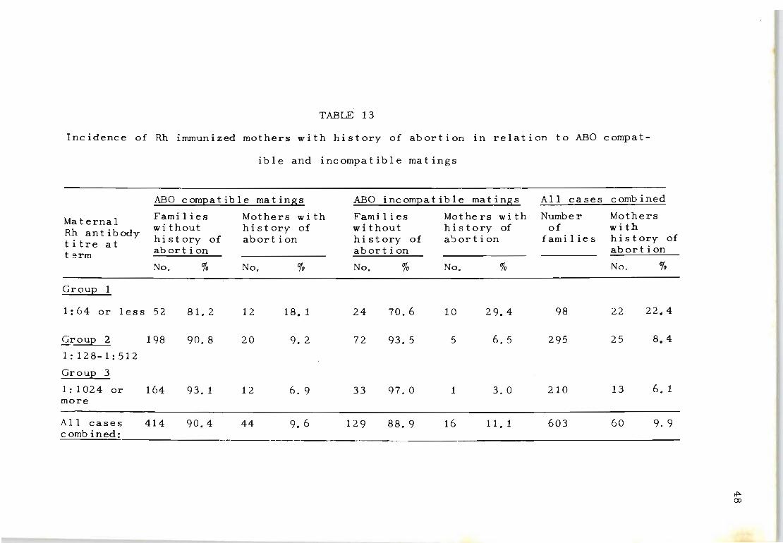
TABLE 13
Incidence of Rh immunized mothers with history of abortion in relation to ABO compat-
ible and incompatible matings
ABO compatible matings
Maternal Rh ant ibody titre at term
Group 1
Families wi thout history of abortion
No. %
1:64 or less 52 81. 2
Group 2
1:128-1:512
Group 3
1:1024 or more
All cases
198
164
414
90.8
93. 1
90.4
Mothers wi th history of abort ion
No. %
12 18. 1
20 9. 2
12 6. 9
44 9.6
ABO incompatible matings
Families wi thout hi st ory of abort ion
No. %
24 70.6
72 93.5
33 97.0
129 88.9
Mo the r s wit h hi s tory of a~ortion
No. %
10 29.4
5 6. 5
1 3.0
16 11. 1
All cases combined
Number of
f ami I ie s
98
295
210
603
Mothers wi th hi story of abort ion
No. %
22 22.4
25 8.4
13 6. 1
60 9. 9 combined: _________________________________ _____________________________________________________________________ _
~ CD
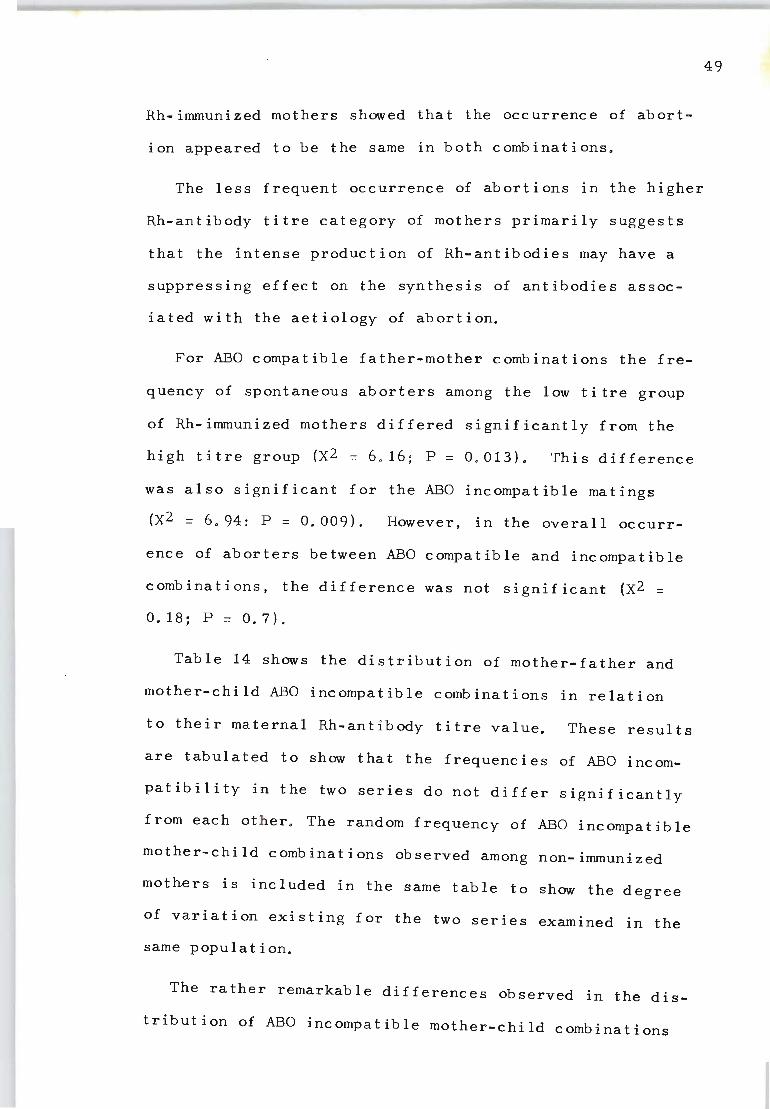
Rh-immunized mothers showed that the occurrence of abort
ion appeared to be the same in both combinations.
The less frequent occurrence of abortions in the higher
Rh-antibody titre category of mothers primarily suggests
that the intense production of Rh-antibodies may have a
suppressing effect on the synthesis of antibodies assoc
iated with the aetiology of abortion.
For ABO compatible father-mother combinations the fre
quency of spontaneous aborters among the low titre group
of Rh-immunized mothers differed significantly from the
high titre group (X2 = 6 . 16; P = 0.013). This difference
was also significant for the ABO incompatible matings
(X2 = 6.94: P = 0.009), However, in the overall occurr
ence of aborters between ABO compatible and incompatible
combinations, the difference was not signjficant (X2 =
0.18;P=0.7).
Table 14 shows the distribution of mother-father and
mother-child ABO incompatible combinations in relation
to their maternal Rh-antibody titre value. These results
are tabulated to show that the frequencies of ABO incom
patibility in the two series do not differ significantly
from each other. The random frequency of ABO incompatible
mother-child combinations observed among non-immunized
mothers is included in the same table to show the degree
of variation existing for the two series examined in the
same populat ion.
The rathe r remarkable differences observed in the dis
tribution of ABO incompatible mother-child combinations
49
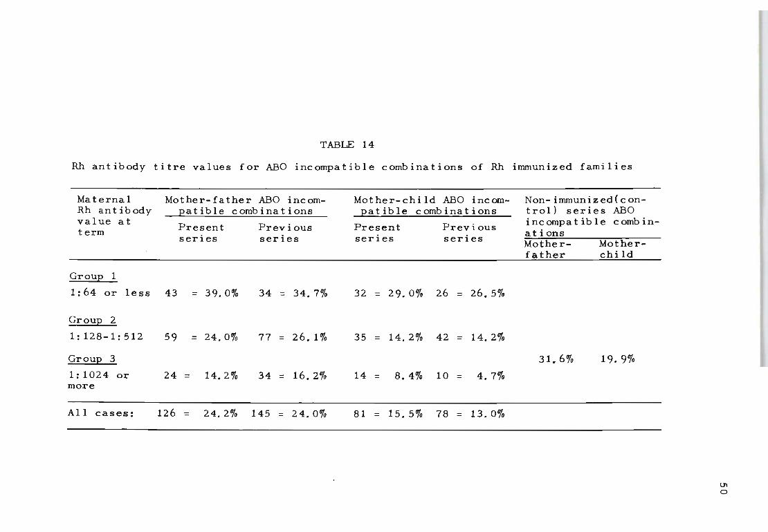
TABLE 14
Rh antibody titre values for ABO incompatible combinations of Rh immunized families
Maternal Rh antibody value at term
Group 1
Mother-father ABO incompatible combinations
Present series
Previous series
1:64 or less 43 = 39.0% 34 = 34.7%
GrouE 2
1: 128-1: 512 59 = 24.0% 77 = 26.1%
GrouE 3
1:1024 or 24 = 14.2% 34 = 16.2% more
All cases: 126 = 24.2% 145 = 24.0%
Mother-child ABO incompatible combinations
Present series
Previous series
32 = 29.0% 26 = 26.5%
35 = 14.2% 42 = 14.20/0
14 = 8.4% 10 = 4.7%
81 = 15. 5% 78 = 13.0%
Non-immunizedCcontroll series ABO incompatible combinations Motherfather
31.6%
Motherchi ld
19.9%
Ul o
between the group (i) series of Rh-immunized mothers
(29%) and the group (iii) series (8.4%) demonstrates
clearly the effect on maternal Rh antibody production
when immunization is induced by both ABO and Rh-incompat
ible fetal red cells.
Table 15 evaluates the number and percentage of ABO
compatible and ABO incompatible mother-child combinations
among Rh incompatible conceptions followed throughout the
antenatal period for the behaviour pattern of Rh immunizat
ion. In 195 cases the first evidence for the production
of Rh antibodies was observed during the second or third
trimester of pregnancy in pat i ents known not to be sensit
ized in the first trimeste r. A total of 30, or 15.3 per
cent of the pregnancies resu l ted in the delivery of ABO
incompatible infants.
These findings confirm t hat ABO incompatibility does
not prevent Rhesus negative mothers from producing Rh
antibodies. Considering that the random frequency of ABO
incompatibility in non-immunized mothers observed in the
same population area is on l y 19.9 per cent (Table 14), the
much higher percentage recorded among Rh-immunized mothers
with Rh antibody titre values of 1:64 or less , either dur
ing the initial stage of Rh antibody production (26.5%)
or in mothers who were already immunized prior to the
present pregnancy (32.6%) suggests that combined ABO and
Rh incompatibility may in f act enhance the mother's potent
ial i ty to become immunized . If th i s is so, then the pre
sent explanation for the much lower frequency of ABO in
compatibility observed among the high titred (1:1024 or
51
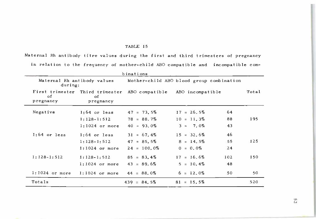
TABLE 15
Maternal Rh antibody titre values during the first and third trimesters of pregnancy
in relation to the frequency of mother-child ABO compatible and incompatible com-
Maternal Rh antibody values during:
First trimester Third trimester of of
pregnancy pregnancy
Negative 1:64 or less
1:128-1:512
1:1024 or more
1:64 or less 1:64 or less
1:128-1:512
1:1024 or more
1:128-1:512 1:128-1:512
1:1024 or more
1:1024 or more 1:1024 or more
Totals
binations
Mother-child ABO blood group combination
ABO compa t ib Ie ABO incompa t ib Ie Total
47 = 73.5% 17 = 26.5% 64
78 = 88.7% 10 = 11. 3% 88 195
40 = 93.0% 3 = 7.0% 43
31 = 67.4% 15 = 32.6% 46
47 = 85.5% 8 = 14.5% 55 125
24 = 100.0% 0=0.0% 24
85 = 83.4% 17 = 16.6% 102 150
43 = 89.6% 5 = 10.4% 48
44 = 88.0% 6 = 12.0% 50 50
439 = 84.5% 81 = 15.50/0 520
11l N
more) Rh-immunized mothers cannot be accepted as the re
sult of direct interaction of maternal anti-A or anti-B
antibodies on ABO and Rh-incompatible fetal red cells.
One possible explanation is that when the maternal
antibody-forming mechanism is called on to produce two
different types of antibodies (anti-A or anti-B which
are generally associated with haemolysin production and
anti-Rh which is not), it may do so but with reduced
ability to form high titred Rh-antibodies.
The possibility that Rh-immunized mothers producing
high titred Rh-antibodies possess the ability to select
at the cervical level conceptions which are ABO compat
ible rather than ABO incompatible, as postulated among
classical infertility conditions by Behrnman, Buettner
Janusch, Heglar, Gershowitz and Tew (1960) and Vas (1965),
must remain in doubt until the incidence of immune anti-A
or anti-B haemolysins can be assessed among Rh-immunized
mothers.
53
Levine's hypothesis (1943) that ABO incompatible Rh
positive fetal red cells are more likely to be eliminated
from the maternal circulation before mothers can respond
to the production of Rh-antibodies is not compatible with
the findings in this analysis . However, our observations
are in agreement with others (Nevahlinna and Vainio (1956),
Murray, Knox and Walker (1965) that fetal ABO incompatib
ility among Rh-immunized mothers affords a significant
degree of protection against the occurrence of severe
manifestations of Rh-haemolytic disease in the newborn.

This protection appears to be based upon the mother's
inability to produce higher titres of Rh-antibodies in
approximately 80 per cent of the ABO incompatible combin
ations examined.
4. DISCUSSION
This investigation has shown that differences in the
distribution of ABO incompatible matings not only occur
between Rh-immunized families and the control series,
but also can be expected when the maternal response to
the production of Rh antibodies is taken into account .
These differences range from the normal random distribut
ion of ABO incompatible matings where the maternal Rh
antibody titre value is 1:64 or less (26.5%) down to val
ues frequently reported by others when the Rh antibody
titre is 1:1024 or more (12 . 0%),
ABO incompatibility among Rh immunized families has
been reported to range between 24.7% (Levine, 1953) and
13.0% (Stern, Davidsohn and Masaitis, 1956), There has
been no information to indicate that such wide variations
may in some instances be influenced by the inclusion of
a greater proportion of severe cases of Rh incompatibility
with mothers possessing higher Rh-antibody titre's than
would normally be expected in a random selection of Rh-
immunized mothers . This situation can occur when the Rh-
immunized families used for this type of investigation
are composed of a large number of Rh cases which are
brought to the attention of the investigator because of
the obvious clinical manifestation of Rh-haemolytic dis-
54
ease, while mild forms of Rh-disease in infants not re
quiring treatment are not included. The use of Rh
immunized families detected by routine antenatal testing
alone would therefore appear to be the only approach
that would ensure that the normal distribution of mild
and severe forms of Rh-haemolytic conditions are included
In an analysis of this nature. By observing this rule
of selection we found that the frequency of father-mother
and mother-child ABO incompatible combinations did not
differ significantly in mothers producing low-titred Rh
antibodies (1:64 or less) when compared with the distrib
ution of these combinations in non-immunized mothers.
Where the Rh-immunized mothers produced high titred
(1:1024 or more) Rh antibodies the frequency of mother
child ABO incompatibility differed significantly from
the random results (4.7% as opposed to 19.9%). The wide
variations of mother-child ABO incompatible combinations
reported by Levine (1958) of 12.6% and Lucia and Hunt
(1950) of 4.4% can most likely be attributed to the marked
differences in the maternal Rh antibody titres of the
mothers investigated in the two series.
The influence of the father's D antigen zygosity on
maternal Rh antibody titres showed the expected higher
frequency of DID matings in mothers with Rh antibody
titres of 1:1024 or more, particularly among the ABO in
compatible matings (ABO compatible: DID, 64.3% and Did,
35.7%; ABO incompatible: DID, 77.7% and Did, 22.3%),
suggesting that group A or B homozygous DID fathers among
55
the incompatible combinations possess a greater potential
ity to induce the formation of high titre Rh-antibodies
than heterozygous DId fathers of the same blood groups.
The effect of parity on the maternal Rh-antibody titre
response does not appear to be significant and confirms
the general observation that mothers with only two pre
vious pregnancies can sometimes possess extremely high Rh
antibody titres while those with at least six pregnancies
may possess lower Rh-antibody values.
56
The belief that ABO incompatible matings in Rh-haem
olytic disease are a sound protection against Rh-immuniz
ation was only confirmed in mothers with high titre Rh-anti
bodies. The normal frequency of ABO incompatible matings
found in either father-mother or mother-child combinations
among those producing lower values (1:64 or less) of Rh
antibodies provided evidence to suggest that the trans
placental passage of fetal ABO incompatible Rh-positive
red cells may not necessarily prevent the mother from
being immunized. The protective mechanism assumed by ABO
incompatibility does not appear to be of any significance
in this category of Rh-immunized mothers.
Chapter VI
THE EVALUATION OF THE INDIRECT ANTI-HUMAN GLOBULIN TITRE
AS A PROGNOSTIC INDEX
1. RH ANTIBODY LEVELS IN THE MATERNAL SERUM
A most important factor observed from comparing Rh
antibody titres with the clinical severity of the disease
has been the wide variations of results that different
investigators obtain. This is probably caused from
lack of standardization in technique and methods adopted.
Of the several methods known to determine the presence of
Rh antibodies (saline test, enzyme test, albumin replace
ment test and anti-human globulin test), we have found
the indirect antiglobulin test to be the most dependable
(Vos,1969).
Using the standardized indirect anti-human globulin
test (described in Chapter II), the relationship between
the Rh antibody titre of the mother at delivery and fetal
wastage was examined. 429 infants ' were studied where Rh
antibodies were detected in the mother1s serum. All cases
were included in which an antiglobulin titration of the
mother's serum was carried out either on the day of birth,
or within two or three days prior to birth, and where the
pregnancy continued for at least 20 weeks. 56 (13.0%) of
the 429 infants were Rh-negative. Of the remaining 373
Rh-incompatible infants, 65 (17.4%) were stillborn and
308 (82.6%) were liveborn. Thirty-four of the liveborn
infants (9.1% of the incompatible infants) subsequently
died, 20 of them despite prompt and adequate exchange-
t ransfus ion. The other 14 were untreated, having died
57
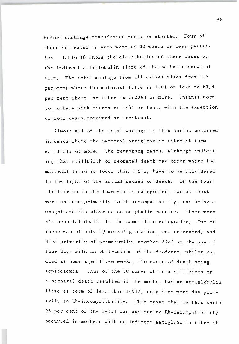
before exchange-transfusion could be started. Four of
these untreated infants were of 30 weeks or less gestat
ion. Table 16 shows the distribution of these cases by
the indirect antiglobul i n ti t re of the mother's serum at
term. The fetal wastage from all causes rises from 1.7
per cent where the maternal titre is 1:64 or less to 63.4
per cent where the titre i s 1:2048 or more. Infants born
58
to mothers with titres of 1:64 or less, with the exception
of four cases,received no treatment.
Almost all of the fe t al wastage in this series occurred
in cases where the maternal antiglobulin titre at term
was 1:512 or more. The remaining cases, although indicat-
ing that stillbirth or neonatal death may occur where the
maternal titre is lower than 1:512, have to be considered
in the light of the actual causes of death. Of the four
stillbirths in the lower-tit r e categories, two at least
were not due primarily to Rh-incomp~tibility, one being a
mongol and the other an anencephalic monster. There were
six neonatal deaths in the same titre categories. One of
these was of only 29 weeks' gestation, was untreated, and
died primarily of prema t u ri ty; another died at the age of
four days with an obstruct ion of the duodenum, whilst one
died at home aged three weeks , the cause of death being
septicaemia. Thus of the 10 cases where a stillbirth or
a neonatal death resulted if the mother had an antiglobulin
titre at term of less than 1:512, only five were due prim~
arily to Rh-incompatibility. This means that in this series
95 per cent of the fetal wastage due to Rh-incompatibility
occurred in mothers with an indirect antiglobulin titre at
TABLE 16
Distribution of 373 cases of incompatible infants born to Rh-immunized
mothers by maternal indirect antiglobulin titre at term
Maternal No. of Li veborn Rh+ infants Stillbirths % Foetal titre at cases Survived Died wastage term
1:64 or less 60 59 0 1 ':' 1.7
1: 128 30 28 1 1*':' 6.7
1:256 60 53 5 2 11. 7
1:512 70 62 4 4 11.4
1: 1024 82 46 9 27 43.9
1:2048 or more 71 26 15 30 63.4
All titres: 373 274 34 (9.1%) 65 ( 17. 4%) (73. 5%)
>!' Mongol ** Anencephalic monster
\J1 -..D
term of 1:512 or more.
Although there is a close relationship between the
maternal indirect antiglobulin titre at term and the
chance of having a successful outcome of pregnancy, this
by itself does not mean that early induction of labour
should be carried out if the maternal titre at 36 weeks
is 1:512, because it could remain at this level during
the next four weeks . The relationship between maternal
60
indirect antiglobulin titre at delivery and outcome, as
shown in Table 16, indica t es the chance of successful
outcome in such cases to be greater than 90 per cent.
However, the picture obtained from considering the relat
ionship of a single maternal indirect antiglobulin titrat
ion at term and the chance of survival can be most mislead
ing. It does not indicate whether the titre determined
at the time of delivery has just arrived at that level,
perhaps quite suddenly from a low or zero value, or
whether the fetus has been exposed to the same quantity
of antibody for a cons i derably longer length of time.
In 222 Rh-immunized mothers with Rh-incompatible preg
nancies, antibody titrations were carried out weekly or
fortnightly during the last 10 weeks of pregnancy. This
made possible the calculation of a 'titre index' to give
some measure of the effect of the titre level and the
duration of the response on the severity of the disease
in the affected child. The titre index is calculated by
mUltiplying the titre code number by the number of weeks
and summing for the total 10- weeks period. The code num-
bers used range from 1 , f or a titre of 1:16 or less, 2,
f 1 32 3 f 1 64 e tc to 9, for a titre of 1 : 4096 or : , , or : , . ,
or more. Thus a mother who has maintained a titre of
1:1024 over the whole 10-weeks period will have a titre
index of 7xl0 = 70. On the other hand, a mother who has
a titre of 1:1024 at term but started the 10-weeks period
with a titre of 1:64 and rose progressively each two weeks
up to this titre will have an index of 2x3 + 2x4 + 2x5 +
2x6 + 2x7 = 50.
The distribution of the 222 cases in four categories
of titre index where this information is available IS
given in Table 17. Where the titre index is 40 or less,
as it was in 87 of the cases (39%), the chance of death
is only 3.4 per cent. The one stillbirth was the mongol
61
referred to previously. If the titre index is between 41
and 60 the chance of death is 23.9 per cent and from 61-70
it is 59 per cent. If the titre index is above 70, as it
was in 25 of the cases, the chance of death is 80 per cent.
On the basis of these results it is possible to estim-
ate the increase in severity which will occur if a preg-
nancy is allowed to continue for any particular length of
time. For example, if a mother has reached 36 weeks and
has a titre of 1:1024 and during the previous six weeks
her titre score has reached 40, then even if her titre
remains unchanged she will accumulate 4x7 = 28 points to
give a final titre index of 68 if the pregnancy continues
to full term. This means that there is almost a 50 per
cent chance that the infant will be stillborn, or, if
liveborn, will be very seriously affected, with a 30 per

TABLE 17
Relationship between maternal indirect antiglobulin index and outcome of
pregnancies of Rh-sensitized mothers
See text for method of calculating the index
Indirect No. of Liveborn Rh+ infants Stillbirths antiglobulin cases Survived Died index
40 or less 87 84 2 1 ;'<
41-60 71 54 8 9 61-70 39 16 3 20
71 and over 25 5 4 16
All indexes 222 159 17 46
>!< Mongol
0/0 Foe ta 1 wastage
3.4
23.9
59.0
80.0
28.4
0"N
cent chance of death. In some of the liveborn cases
death will occur before exchange-transfusion can be
started. It is clear, therefore, that accurate knowledge
of the maternal antibody level and its behaviour from the
26th week of pregnancy onward enables one to predict be-
fore birth the degree of severity of the disease in the
affected child with a high degree of accuracy.
2. THE RELATIONSHIP OF MATERNAL INDIRECT ANTIGLOBULIN TITRE, CORD-BLOOD HAEMOGLOBIN AND RETICULOCYTE PERCE~AGE IN THE PROGNOSIS OF RH HAEMOLYTIC DISEASE
This series comprises 218 consecutive pregnant women
In whom anti-Rh antibodies were detected by the indirect
antiglobulin technique. 24 women gave birth to Rh-negative
infant s. In 35 cases the pregnancy ended in a stillbirth
and in the remaining 159 cases an Rh-positive infant was
born al ive. The cells of these infants when examined
immediately after birth gave a direct Coombs positive
result in each case.
Values for cord-blood haemoglobin were determined using
an MRC grey wedge photometer. Statistical analysis re-
vealed no significant difference between different observ-
ers determining haemoglobin by this method. Reticulocyte
percentages were determined according to the technique of
Steensma (1955). Of the total of 159 Rh-positive infants
of immunized mothers born alive in the present series,
estimations of cord-blood haemoglobin were made in 147
and reticulocyte percentage in 145.
In an attempt to increase the prognostic value of the
indirect antiglobulin titre, this titre was correlated
63
with the value for cord-blood haemoglobin of liveborn Rh
positive infants. The results for the 147 cases for
which both sets of information were available and for the
various treatment categories are shown in Figure 2. When
the value for cord-blood haemogl~in results are plotted
against the logarithm of the indirect antiglobulin titre
a significant correlation is evident (r = -0.612).
Figure 2 shows that all but three of the 19 deaths
occurred among babies born to mothers whose indirect
antiglobulin titre was 1:1024 or higher. These 16 deaths
represent 30.5 per cent of all the cases in this category.
Similar observations were also made for babies born with
cord-blood haemoglobin values of 10 gm/100 ml or less.
There were 17 deaths in this category, representing 40.5
per cent of the cases. It is clear, then, that although
the maternal indirect antiglobulin titre at delivery is
highly correlated with values for cord-blood haemoglobin,
it is not an ideal indicator for forecasting the severity
of the disease. However, because the antiglobulin titre
of the maternal serum is determined before birth it is a
valuable guide for the management of the case. Under
these circumstances a slight loss in efficiency may be
more than compensated by the practical value of inducing
labour at the earliest time consistent with safety factors
involved in premature birth if the mother's titre is ris
ing toward the level of 1:1024.
Pickles (1949) considers that the number of immature
cell types present during the first day of life is of
considerable prognostic significance in cases of haemoly-
64

20. • + NO TREATMENT DIED.
• • NO TREATMENT. SURVIVED • 8 o EXCHANGE TRANSFUSED. I • I SURVIVED.
18. 0 0 • EXCHANGE TRANSFUSED • • I DIED. 0 8 • 0
16. • I 0 0 , 8 0 j +. 8 0 • .A c 0 &, 00
14 • I 0 0 0 0 • <w 8 0 O~ c3> &, {P
~ ~ 12 , 0
c • 0 0
C"::: • ~ 8 0
iii 8 a: .c 10 -0,. 0
OJ: I 8 0 0
U qp 0 If) 0 • •
8 • ~ E 0 • 0 00 - 0 Z~ • ~
0 ~
LL 6 z
41 • • " 0
• 21 • 0
16 32 6.4 128 256 512 1024 2048 4096 8192 16384
MATERNAL INDIRECT COOMBS TITER. NO OF CASES (147)
Figure 2: The distribution of 147 cases of 'incompatible' infants born alive to immunized mothers, by values for cord-blood haemoglobin and the maternal antiglobulin titre at term.
'" \.1l

tic disease of the newborn. Figure 3 reveals, as expected,
an inverse correlation which is highly significant between
haemoglobin values and reticulocyte percentage in the cord
bloods of 145 Rh-positive infants born alive to Rh-immun
ized mothers. The data in Figure 3 is best fitted by a
non-linear curve, the equation for which is Hb = 16.429 -
0.299 X + 0.002X2 where X is the reticulocyte percentage.
In this series, 17 of the 19 deaths had values for
cord-blood haemoglobin of 10 gm/100 ml or less. But of
these deaths 15 occurred among the 25 cases with a retic-
ulocyte percentage of 28 or higher. It is clear that re-
ticulocyte counts can be used in conjunction with values
for cord-blood haemoglobin to give a good index of the
chance of survival. Used alone, however, the reticulocyte
percentage is not quite such a good prognostic index as
cord blood haemoglobin. If a discrimination is based on
a reticulocyte percentage of 20 or higher, which includes
16 of the 19 deaths in the series, and is therefore com
parable with a discrimination based on an indirect anti
globulin titre of 1:1024 or higher, the number of deaths
represents 37 per cent of the cases in the category. How
ever, reticulocyte percentages, like values for cord-blood
haemoglobin, suffer from the disadvantage that their use
for prognostic purposes must wait upon the birth of the
ch i ld.
Mollison and Cutbush (1951) correlated values for cord
blood haemoglobin in their series with concentration of
bilirubin. They concluded that predictions based on val
ues for cord-blood haemoglobin and concentration of bili-
66
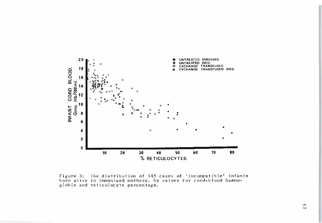
20 ••
e 18 0 0 16 ...J cc"":
EU cg ~~ 12 U~
.10 I- en z E oCt (!) 8 LL z
6
: I 0
Figure 3: born alive glob in and
0 •
. • UNTREATED. SURVIVED. 0 0 • UNTREATED. DIED.
0 EXCHANGE TRANSFUSED 0 • EXCHANGE TRANSFUSED. DIED. . °0 0 0 .
0.0 0 0 0
.0Jlo· 4h oOS. 0
*::o>t o 0 0
0 0 g 3> 00 0
0° 00 0 0 0 co 0
8 ~ o 0 0
• 0 0 0 o 00 0
o 0 ... 0 0 • • 0 0 q, 0 0 0_ • 0
8 0 • • III 0 0 0 • • • 0 • 0
• •
0 • • •
•
10 20 30 40 50 60 10 10 % RE T ICUlOCY T ES.
The distribution of 145 cases of I incompatible' infants to immunized mothers, by values for cord-blood haemoreticulocyte percentage.
0' -..J
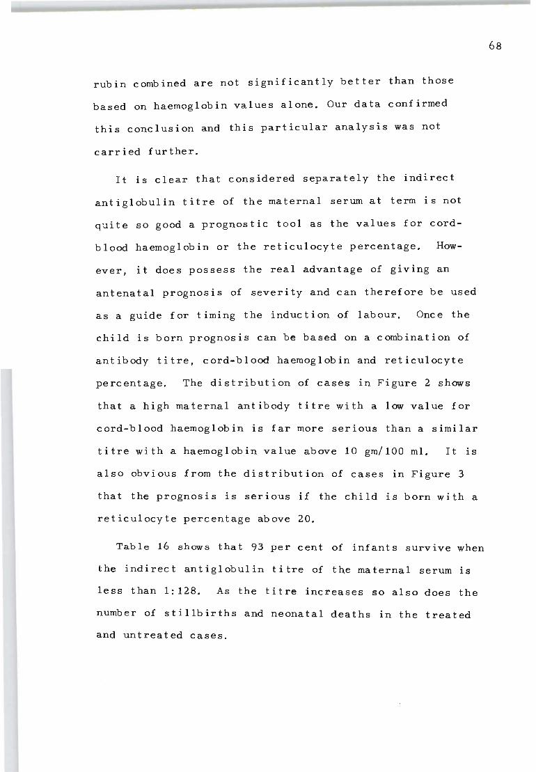
rubin combined are not significantly better than those
based on haemoglobin values alone. Our data confirmed
this conclusion and this particular analysis was not
carr i ed fur ther.
It is clear that considered separately the indirect
antiglobulin titre of the maternal serum at term is not
quite so good a prognostic tool as the values for cord
blood haemoglobin or the reticulocyte percentage. How
ever, it does possess the real advantage of giving an
antenatal prognosis of severity and can therefore be used
as a guide for timing the induction of labour. Once the
child is born prognosis can be based on a combination of
antibody titre, cord-blood haemoglobin and reticulocyte
percentage. The distribution of cases in Figure 2 shows
that a high maternal antibody titre with a low value for
cord-blood haemoglobin is far more serious than a similar
titre with a haemoglobin value above 10 gm/100 ml. It is
also obvious from the distribution of cases in Figure 3
that the prognosis is serious if the child is born with a
reticulocyte percentage above 20.
Table 16 shows that 93 per cent of infants survive when
the indirect antiglobulin titre of t~e maternal serum is
less than 1:128. As the titre increases so also does the
number of stillbirths and neonatal deaths in the treated
and untreated cases.
68
69
PART TWO
THE PARTIAL ABSORPTION TEST
Chap ter VI I
THE PARTIAL ABSORPTION (PA) TEST FOR TITRATING RH
ANTIBODIES
1. INTRODUCTION
The main reason for carrying out serological tests on
the Rh-immunized mother during pregnancy is to provide
the obstetrician with a reliable indication of the sever.
ity of the haemolytic process in the fetus. Ideally he
would like to know before the infant is born whethe+ it
w·ill be mildly or severely affected; whether if the preg
nancy is allowed to continue to its natural term the fetus
will be liveborn or stillborn, and whether, if the father
has been shown to be a heterozygote the fetus is Rh-posit
ive or Rh-negative.
Previous studies (Part I, Chapte~ VI), have shown that
the titre of Rh-antibodies in the maternal serum, when
de t ermined by the ind i rec t anti g 1 obul in method, is high 1 Y
correlated with the severity of the haemolytic process in
the child. An indirect antiglobulin titre at term of 1;64
or less is of no pathological significance and in such
cases almost all infants survive without treatment.
70
As the indirect antiglobulin titre increases, so does
the severity of the disease, and even though the infant
may be born alive and exchange-transfused, the chance of
survival is less. The majority of stillbirths due to Rh
incompatibility are associated with maternal indirect anti
globulin titres of 1:1024 or more. The indir,ect antiglob
ulin titre has therefore been found to be a valuaple guide
for determining whether early induction of labour is nec
essary. However, the indirect antiglobulin titre alone
can sometimes give rise to misleading predictions. In
some cases an antiglobulin titre of 1: 1024 may be assoc
iated with cord haemoglobin values well within the normal
range in the newborn infant. At the other extreme, the
same titre may be associated with a moribund infant having
a haemoglobin of 4 grammes/100 millilitres and a retic
ulocyte count of 50%; or, even more misleading, a mother
may have an indirect antiglobulin titre of 1:1024 through
out pregnancy, only to be induced and delivered of an Rh-
negative infant. A similar serological picture in another
case may result in a macerated stillborn fetus at 35 weeks.
Detailed studies carried out to investigate these discrep
ancies revealed that if the newborn infant has no, or
little free Rh antibody in circulation, the case is always
mild; whereas the most severe cases are associated with
high indirect antiglobulin titres of free Rh antibody in
the infant's serum. Attention was focussed, therefore,on
the absorbing capacity of adult non-sensitized Rh-positive
cells toward maternal sera from mild and severe cases. It
was found that when one volume of maternal serum, obtained
on the day of delivery , was incubated with one volume of
adult Rh-positive c ells, the amount of free antibody lef t
behind in the serum was almost always identical with the
amount found in one volume of the cord-serum of the infant.
Further observations showed that it was not necessary to
use intact Rh-pos i tive cells for carrying out this test,
as identical resu lts were obt a i ned if one volume of a sus-
71
pension of haemolysed adult Rh- positive cells was substit
uted "for the intact cells.
On the basis of these findings we developed a new anti
body test which, when used in conjunction with the indir
ect antiglobulin test, enables a much more accurate prog
nosis of the outcome of pregnancy to be made. The test
is referred to as the part i al absorption (PA) test. The
relationship of this test to other tests of Rh antibody
determinations currently in use, and its behaviour during
pregnancy was investigated.
2. MATERIALS AND METHODS
Antibody titrations - Indirect antiglobulin titrations
were carried out as described in Chapter II. The blocking
and conglutination tests were performed using the method
of Wiener and Hurst (1947) and Wiener, Hurst and Sonn
(1947). Albumin titrations followed the procedure of
Diamond and Denton (1945).
Preparation of standard haemolysed cell suspensions
72
Heparinized or oxalated blood was collected from the arm
veins of healthy Group 0, CDe/CDe (RIR1) or Group 0, CDe/
cDE (RIR2) donors . The cells were packed and washed three
times in large volumes of physiological saline, the samples
pooled and the cells finally packed by centrifuging at
3 000 rpm for 30 minutes. An accurate 25 per cent saline
suspension of the packed cells was made and this was di
vided into two-millilitre samples in screw-cap McCartney
bottles ready for daily use. This standard suspension of
cells was haemolysed by freezing to -20 0 C for 48 hours.
These haemolysed cell suspensions may be stored for at
least twelve months at this temperature without detectable
loss of absorbing capacity.
Procedure for the partial absorption (PA) test - The
standard haemolysed cell suspension was thawed to room
temperature and shaken thoroughly before use. 0.4 milli-
litre of this suspension was added to 0.4 millilitre of a
1:4 saline dilution of the maternal serum in a small test
tube. The two solutions were mixed by shaking and left
at room temperature for 15 minutes. The free Rh antibody
remaining in the mixture was titrated in doubling dilut-
ions using the indirect antiglobulin technique. The first
tube in this titration was read as 1: 16, SInce this is the
dilution at this stage of the original maternal serum.
Partial absorption titres lower than 1:16 have been found
to be of no significance and were scored as negative.
3. RESULTS
The partial absorption titre of the maternal serum and
the free Rh antibody in the infant's serum.
In Table 18 the PA titre of the maternal serum at the
time of delivery is compared with the titre of free anti-
body in the infant's cord blood as determined by the in-
direct antiglobulin method. A total of 187 cases was
available where the infant was liveborn, Rh-positive, and
in which both titrations had been carried out. In all
these cases the infant's cells gave a positive direct re-
sponse to the Coombs test at the time of birth.
of the cases (89%) the PA titre of the maternal
In 166
serum was
either identical with, or within one tube dilution of the
73
TABLE 18
Comparison of maternal partial absorption titre at
the time of birth with titre of free antibody in the
Tube dilutions of maternal titre compared wi th 1 cord serum tit re
-3
-2
-1
o +1
+2
+3
+4
All cases
cord serum
Number
1
6
29
109
28
4
7
3
187
Percentage
O. 5
3.2
15. 5
58.3
15. 0
2. 1
3. 7
1.6
99.9
1 "0" = maternal PA titre identical with indirect antiglobulin titre of cord serum.
-1,-2,-3 = maternal PA titre one, two or three tube dilutions below indirect antiglobulin titre of cord serum.
+1,+2,+3,+4 = maternal PA titre one, two, three or four tube dilutions above indirect antiglobulin titre of cord serum.
74

75
titre of free Rh antibody in the cord serum. In a further
10 cases the two titration values did not differ by more
than two dilution tubes.
Of the 11 cases in Table 18 where the maternal PA titre
differs from the free antibody titre in the cord-serum by
more than two dilution tubes, one mother was grossly oed-
ematous, and five infants were oedematous at birth; simple
dilution may account for part at least of the differences .
In the five cases in which oedema was not present in the
infant no satisfactory explanation can be offered for the
discrepant results.
Despite the wide range of clinical conditions in the
infants in this series, the PA titre of the maternal serUI>1
at the time of delivery in 90 per cent of the cases was
within the limits of one tube dilution, the same as the
free antibody titre of the infant's cord sercum. This fact
suggests that in the majority of cases the PA titre may be
used during the course of pregnancy as a reliable indicator
of the amount of free antibody in the fetal circulation.
Comparison of the partial absorption test with conglutin
ation, albumin, blocking and indirect antiglobulin tests
Cases were chosen to compare the PA titre with the
blocking, conglutination, albumin and indirect antiglob
ulin titres as determined by standard techniques. The
results of ten cases are given in Table 19. No correlat-
ion was observed between the PA titre and the plasma con-
glutination or albumin titres. There is a correlation,
however, between the PA titre and either the blocking
Case Number
302
213
273
261
342
267
231
278
274
340
TABLE 19
Comparative tests on maternal Rh sera
Partial Blocking Indirect Plasma Absorpt- titre antiglob- c ongl-ion titre ul in utinat-
titre ion titre
1:2048 1:64 1: 4096 1: 16 tr
1:1024 1: 64 1: 4096 1 : 64 tr
1: 512 1: 64 1:2048 1:256tr
1:256 1: 4 1: 4096 1: 1024
1:256 1: 8 1:2048 1: 128
1:256 1:4 1:1024 1: 512
Negative Negative 1: 512 1 : 1024
Negative Negative 1: 512 1:256
Negative Negative 1:256 1:64
1: 8 Negative 1: 128 1: 1024
Albumin titre
1:1024
1:256
1: 512
1:512
1: 512
1: 4096
1:2048
1:4096
1:2048
1: 512
-J
'"
or the indirect antiglobul i n titre. These relationships
were then examined more thoroughly on a larger series of
cases. Table 20 shows the results for 42 samples of mat
ernal sera examined . The r e is a significant positive cor
relation between both FA titre and blocking titre, and FA
titre and indirect antiglobulin titre, but the correlation
coefficient (r = 0 . 90) fo r the former is slightly higher
than for the latter (r = 0 . 83) .
Another interesting observation was that although the
FA titre was correlated with the blocking titre, the
method was more sensitive than the blocking test. In some
cases indirect FA titres as high as 1:256 were recorded
even though the results of the blocking tests were still
negative. In this series of cases the highest blocking
titre observed was 1:128.
77
Studies carried out to compare the FA titre with methods
of determining Rh-antibody by various enzyme tests (ficin ,
papain and bromelin), failed to show a positive correlation
between these two procedures.
Behaviour of the partial absorption titre during pregnancy
Examination of detailed case histories in which the
maternal FA titre was determined at regular intervals
throughout pregnancy revealed certain basic patterns in
the FA titre response associated with clinical outcome ,
ranging from mildly affected infants to grossly hydropic
stillborn infants. Since the pattern of response is some-
times more informative than a single titration value ob
tained near term, typical examples are described.
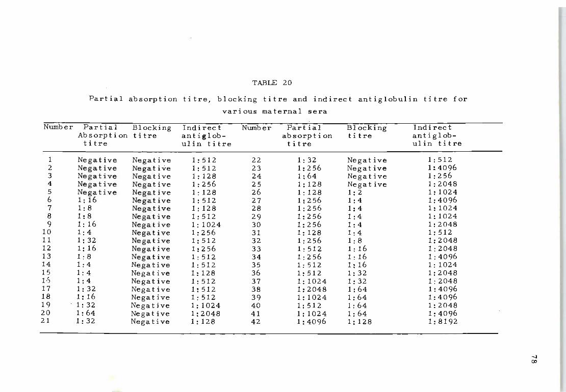
TABLE 20
Partial absorption titre, blocking titre and indirect antiglobulin titre for
various maternal sera
Number Partial Blocking Absorption titre titre
1 2 3 4 5 6 7 8 9
10 11 12 13 14 15 16 17 18 19 20 21
Negative Negative Negative Negative Negative 1: 16 1: 8 1: 8 1: 16 1: 4 1:32 1: 16 1: 8 1: 4 1: 4 1: 4 1:32 1: 16 1:32 1:64 1:32
Negative Negative Negative Negative Negative Negative Negative Negative Negative Negative Negative Negative Negative Negative Nega t i ve Negative Negative Negative Negative Negative Negative
Indirect antiglobulin titre
1:512 1: 512 1: 128 1:256 1 : 128 1:512 1: 128 1:512 1: 1024 1:256 1: 512 1:256 1: 512 1:512 1: 128 1: 512 1: 512 1:512 1:1024 1:2048 1: 128
Number
22 23 24 25 26 27 28 29 30 31 32 33 34 35 36 37 38 39 40 41 42
Partial absorp t i on titre
1:32 1:256 1:64 1:128 1 : 128 1:256 1:256 1:256 1:256 1: 128 1:256 1:512 1:256 1: 512 1:512 1:1024 1:2048 1:1024 1:512 1:1024 1: 4096
Blocking titre
Negat ive Negative Negative Negative 1: 2 1: 4 1: 4 1: 4 1: 4 1: 4 1:8 1: 16 1:16 1: 16 1:32 1:32 1:64 1:64 1:64 1:64 1: 128
Indirect antiglobulin tit re
1:512 1: 4096 1:256 1:2048 1:1024 1:4096 1: 1024 1:1024 1:2048 1: 512 1:2048 1:2048 1: 4096 1:1024 1:2048 1:2048 1: 4096 1: 4096 1:2048 1: 4096 1:8192
-..J 00
(a) Stillbirths - In nearly all ca~es of stillbirth due
to Rh-incompatibility the indirect antiglobulin titre at
the time of delivery was 1:1024 or more and was maintained
at a high level for at least 10 weeks before delivery
(Part I, Chapter VI). The PA titre in these cases can re
main at a relatively low level throughout the greater part
of pregnancy but may then rise rapidly. If death occurs
In utero the PA t itre may start to fall again, though the
indirect antiglobulin titre generally remains steady (vide
infra). A high indirect antiglobulin titre maintained
throughout pregnancy does not necessarily mean that the
fetus will be seriously affected. But if a high indirect
antiglobulin titre is associated with a high PA titre
throughout pregnancy and the fetus is Rh-positive, the
outcome is invariably fatal.
(b) Severely affected liveborn infants - These are nearly
always associated with a maternal indirect antiglobulin
titre of 1:1024 or higher, but the PA titre is often low
early in pregnancy and shows a sudden rise, often to a
high level shortly before birth.
(c) Moderately affected infants - One of the puzzling
features of previous work on the relationship between mat
ernal antibody levels and severity of the disease in the
infant has been the occurrence of relatively high antibody
levels in the maternal serum throughout pregnancies which
have terminated with the birth of a moderately affected
infant. In typical cases the indirect antiglobulin titre
of the maternal serum may persist at a moderately high
level of 1:512 throughout the greater part of pregnancy,
79
but the FA titre remains low, at 1:16 or less, and does
not rise appreciably near term. Another category of mod
erately affected infants can be distinguished in those
cases in which the indi r ect antiglobulin titre is low or
zero at the commencement of pregnancy, but rises to a
high value during the third trimester. The FA titre may
show a parallel rise . In these cases, however, the rise
in antibody level occurs too late to seriously affect the
infant before birth.
80
(d) Rh-negative infants - In families In which the father
is heterozygous Did it is important to be able to distin
guish an Rh-negative fetus from an Rh-positive fetus if
a correct prognosis is to be made before birth. At pre
sent there is no certain way by which this may be done.
The behaviour of the FA titre during pregnancy, however,
is a somewhat better guide than the behaviour of the in
direct antiglobulin titre alone . If the indirect antiglob
ulin titre remains high through the last four months of
pregnancy, as a result of antibodies from a previous preg
nancy, but the FA titre remains negative or very low, it
is an indication that no fresh immunization has taken
place. In general the FA titre responds more rapidly
than the indirect antiglobulin titre to a new episode of
immunization and it tends to fall again to low levels be
fore the next pregnancy commences. Fersis'tently low FA
titres throughout pregnancy are therefore more frequent
when the fetus is Rh-negative and is failing to stimulate
the production of more Rh- antibody.
(e) The effect of death in utero and maternal oedema on
the FA t i tre - In some cases a fall in the FA titre
may occur as pregnancy progresses. This may be due to
three causes : (a) an Rh-negative fetus, (b) death in
utero and (c) maternal oedema. Both (b) and (c) indicate
the remarkable sensitivity of the FA titre as an index of
what is occurring in the mother and the fetus. Somet imes,
in severe cases of Rh-immunization, the fetal heart stops
several weeks before parturition. If the FA titre is
high when death occurs it shows a fall in about one-half
of the cases . In a series of 20 stillbirths in which the
FA titre was determined at regular intervals through the
last ten weeks of pregnancy, eight cases showed a marked
decline of two or more dilution tubes in the FA titre
before the end of pregnancy. In each of these cases a
badly macerated fetus was born. In six of the remaining
12 cases the fetus was again macerated but there was no
detectable fall in the FA titre before delivery. It IS
important to note that although the FA titre fell in
nearly half of the 20 stillbirth cases, in no si n gle case
did we observe the indirect antiglobulin titre to show a
similar fall.
4. DISCUSSION
A number of attempts have been made to analyse the
complexity of the maternal antibody response to Rh anti
gens by both physico-chemical and serological means (Cann
et al. 1952; Krieger and Williams, 1955; Wiener and Gordon
1953; Greenwalt and Wagner, 1955). So far no one has
found a relatively simple test which, when applied to
81
,
the maternal serum antenatally, can predict with high
probability the severity of the haemolytic process in the
child and its chances of being stillborn, or, if born
alive, of living with or without exchange-transfusions.
82
It has been reported (Davidsohn and Stern, 1948; Wiener
eta 1. 1 952 ) t ha t bloc king an t i bod i e s are ass oc i at ed wit h
more severe manifestations of erythroblastosis in the baby,
and de Kromme and Vervaat (1953) attempted to show that
variations in conglutination-positive and conglutination
negative reactions can be used to explain differences in
the severity of the disease.
Titration values obtained by the FA test have there
fore been of importance in the understanding of the com
plexity of Rh-antibody production in response to the stim
ulus of an Rh-incompatible pregnancy. Diamond (1947)
noted that severe cases of erythroblastosis were assoc
iated with the presence of free-Rh antibody In the cord
serum of an affected infant. Our results show that if
one volume of packed adult cells or the equivalent volume
of haemolysed cells is used to absorb one volume of mat
ernal serum the antibody left free in the serum, as meas
ured by the indirect antiglobulin technique, can vary
over a wide range of values. For instance, in cases
where the indirect antiglobulin titre is 1:1024 the anti
body left free in the serum can vary from zero up to, but
never exceeding 1: 1024. The titre of free ant ibody is the
FA titre, and our results confirm and extend the observat-
ions of Diamond. The first conclusion to be drawn from
these results is that in vivo the absorbing power of
the fetal red cells is roughly the same as that of adult
red cells. Secondly, since all infants in whom the FA
titre is zero are mildly affected, it would suggest that
the haemolytic process in the fetus does not commence
until all the antigenic sites on the red cells have been
saturated with antibody, or blocked from accepting more .
Work in progress indicates that at least two kinds of
antigen-antibody reaction are involved in the process of
absorbing Rh-antibodies on Rh-positive cells. In the
first of these the antibody is bound firmly to the cell
and can be released only by extracting with ether which
completely alters the structure of the Rh antigen on the
cell surface. In the second the antibody bound to the
Rh antigen can be released simply by freezing and thawing
83
the cell. In this way the cell is haemolysed but the anti-
genic sites are relatively undamaged. When cells are sen
sitized with an Rh serum with a zero FA titre and then
thoroughly washed, no Rh-antibody is released after haem-
olysis caused by freezing and thawing. If, however, the
cells are sensitized with an Rh serum which has a positive
FA titre, the antibodies released from the cells after
freezing and thawing are found to be identical with the FA
titre of the original serum.
These results indicate that under conditions where the
sensitizing serum has a positive FA titre, a portion of
the Rh antibodies is in equilibrium with its corresponding
antigen. A tentative hypothesis suggests that it is only
when sensitized cells carry antibody in this manner that
they are readily haemolysed in the fetal circulation. If
this interpretation is correct, then the amount of free
antibody in the cord serum, as estimated by the PA tech
nique, would be related directly to the rate of cell de-
struction in vivo. The persistence of such a process. at
a high rate over several weeks is almost certain to result
in severe or fatal manifestations of erythroblastosis in
the inf an t.
84
Chapter VIII
THE EVALUATION OF THE PARTIAL ABSORPTION TEST IN
THE PROGNOSIS OF RH HAEMOLYTIC DISEASE
1. INTRODUCTION
It was shown in Chapter VI that the prognostic value of
the maternal antiglobulin titre is considerably improved
if titrations are carried out regularly during the last
ten weeks of pregnancy. The titration values are given a
code number from 1 to 9 and these are summed for each
week to give a titre index. This is a measure not only
of the height of the titre, but of the length of time that
the titre has been operating. Although the titre index
is a much better guide to the likely outcome of any part
icular pregnancy than is a single titre value for antibod
ies in the maternal serum at term, there is still a small
number of cases which do not fit into the general pattern.
It is believed that the explanation for some, at least, of
these exceptional cases is to be found in the differing
amounts of antibody which are left in the infant's serum.
In Chapter VII a simple partial absorption (PA) test was
described which enables an estimate of the amount of anti-
body in the fetal serum to be made from titrations carried
out on the maternal serum. The present chapter provides a
more rigorous evaluation of this test.
2. MATERIALS AND WETHODS
Venous blood samples were collected from Rh-negative
women as part of the routine antenatal tests. When Rh
antibodies were present in the maternal serum every effort
was made to obtain samples at regular intervals. During
85

the last three months of pregnancy samples were taken at
fortnightly, or even weekly intervals, whenever possible.
Samples of infant blood were obtained from the fetal end
of the umb i I ical cord at birth. The indirect antiglobulin
procedure used was described in Chapter II. The me thod of
carrying out the partial absorption test on the maternal
serum was described in Chapter VII.
The determination of cord-blood haemoglobin values and
reticulocyte percentages, and the methods of carrying out
exchange-transfusion have been described in detail by
Kelsall, Vos, Kirk and Shield (1957).
3. RESULTS
290 Rh~immunized mothers were examined for whom the in-
direct antiglobulin titre and the PA titre of the maternal
serum were available at the time of delivery. There were
41 liveborn Rh-negative infants. Of the remaining 249 Rh-
86
incompatible pregnancies, 36 resulted in stillborn infants:
of the 213 liveborn Rh - positive infants 21 subsequently
died despite prompt and adequate exchange-transfusion.
Maternal PA titre at delivery and survival of Rh-incompat -
ible infants The single death among the 57 cases in
which the PA titre at the time of delivery was zero was an
anencephalic monster (see Table 21). There were no pathol-
ogical signs that death in this case was In any way due to
Rh incompatibility. In addition, one of the neonatal
deaths, where the mother had a PA titre of 1:16, was due
primarily to obstruction of the duodenum. Of the other
deaths in the low-titre categories of 1'. 16 d 1 32 an : , shown
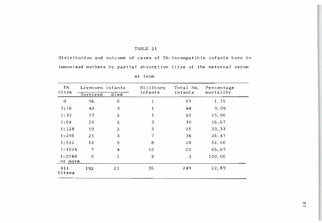
TABLE 21
Distribution and outcome of cases of Rh-incompatible infants born to
immunized mothers by partial absorption titre of the maternal serum
at term
PA Liveborn infants St i llborn Total No. Percentage titre Survived Died infants infants mortality
0 56 0 1 57 1. 75
1: 16 40 3 1 44 9.09
1:32 17 2 1 20 15. 00
1: 64 25 2 3 30 16.67
1: 128 10 2 3 15 33.33
1:256 25 3 7 34 26.47
1:512 12 5 8 25 52. 00
1 : 1024 7 4 10 21 66.67
1:2048 0 1 2 3 100.00 or more
All 192 21 36 249 22.89 titres
(Xl
-..J
in Table 21, at least one of the neonatal deaths and both
of the stillbirths were to mothers who were suffering from
toxaemia of pregnancy with marked oedema. In many cases
oedema associated with a stillbirth results in a fall in
the PA titre before birth (Vos, 1958) and this may well
have happened in the other case where the infant died very
soon after birth. The fall in the PA titre reduces its
usefulness as a prognostic guide in such cases, particul
arly if the only titration carried out is near the end of
pregnancy.
It can therefore be concluded that the risk of still
birth due primarily to Rh-incompatibility (when the mater
nal PA titre at birth is lower than 1:64), In the absence
8 8
of marked oedema in the mother, is no greater than in normal
pregnanc ies . For PA titres of 1:64 or more the risk of
neonatal death or stillbi r th is greater. In two- th i rds of
the cases with a maternal PA titre at birth of 1:1024 the
infants were either stillborn or died soon after birth,
whilst no infant has survived yet when the maternal PA
titre was 1:2048.
Correlation of maternal FA titre with cord-blood
haemoglobin values It was shown in Chapter VI that
the maternal indirect antiglobulin titre at term is highly
correlated with the cord-blood haemoglobin values (r = -0.62). For 178 of the l i veborn Rh-incompatible infants
in the present series both cord-blood haemoglobin values
and the PA titre of the maternal serum at term are avail-
able. The distribution of these values, together with
the method of treatment and outcome is shown in Figure 4.

89
20 .. NO TREATMENT DIED
10 • NO TIlEAn.lENT IURVIVED
o EXCHANGE TAANII'UIED
• IIUIVIVED
Ie • E.CHANGE TAANlfUlED 0 1 OlEO
t 0 CO Ob 16 0 0
11 0
0 § 0 Q)
~ 0 0
14 ~o 00 0 00 ~
§ 0 0
8 00
e 12 8 0 CO ~ [() 0 • 00 0
~ 0 8 ., CD 0 • .. 0 a: ~ 10 • 0 8 0 0 0 0
U I 0 CD 0 0 0
t- ~ 0 • 0
8 0 0 ~ z
it C> 0 0 • 0
z • 6 • ..
00 0 • .. .. 0
• .. 4 8
2 •
0 0 16 32 64 128 256 512 1024 2048
Or leu
MATERNAL PARTIAL ABSORPTION TITRE No OF CAlES '79
Figure 4: The dis t rib uti on of 179 live-born Rh-incompatible infants by cord-blood haem-oglob in value and maternal P. A. titre at the time of birth.
When this distribution is compared with the evaluation of
maternal indirect antiglobulin titre at term and cord
blood haemoglobin value, as shown in Chapter VI, it is
clear that the PA titre of the maternal titre gives a
much better indication of the severity of the haemolytic
process in the child than does the maternal indirect anti-
globulin titre. This is in fact borne out by the statis-
tical analysis which gives a correlation coefficient be
tween the PA titre and cord-blood haemoglobin value of
- O. 72.
For infants compatible with their mothers for the Rh
factor, the range of cord-blood haemoglobin values is from
11.0 to over 20.0 g/100ml, with a mean value of 16.24 and
a standard deviation of 1.7. It might be expected there-
fore that for any particular level of the maternal PA
titre there would be a corresponding spread of cord-blood
haemoglobin values. Figure 4 shows this to be true.
Relationship between maternal indirect antiglobulin titre
and partial absorption titre - If all cases are consid-
90
ered in which the maternal indirect antiglobulin titre at
the time of birth is 1:1024, the cord-blood haemoglobin
values can range from 3.0 to 17.0 g/100m1 with approximately
half of the cases possessing haemoglobin values above 10.0
g/100ml. (Figure 2, Chapter VI). It has been found that
infants with haemoglobin values above 10.0 g/100ml. are
not bad risks for exchange-transfusion. However, in cases
in which the maternal indirect antiglobulin titre at birth
is 1:1024 but the cord-blood haemoglobin value is below
10.0 g/100ml. the infants have a much poorer prognosis for
survival even when exchange-transfused.
For any given maternal indirect antiglobulin titre the
PA titres of the maternal serum can vary over a wide range
in different cases. In Table 22 the distribution of cord
blood haemoglobin values for 95 Rh-positive liveborn in
fants born to mothers with a maternal indirect antiglob
ulin titre at term of 1:512 to 1:4096 is given in relat
ion to the difference between the indirect antiglobulin
titre and the PA titre of the maternal serum. The differ
ence in titre is expressed in dilution tube numbers. This
means that for a particular indirect antiglobulin titre
(1:1024 for example), the corresponding PA titre can range
from the same value (difference = 0) down to 1:16 (differ
ence = 6 dilution tubes).
From the distribution of cases In Table 22 it is clear
that there is a high correlation between the difference
in titre and the cord-blood haemoglobin value (r = 0.61).
This means that the PA titre enables us to improve consid
erably on the prognosis based on the maternal indirect
antiglobulin titre at term. Of the 95 liveborn infants
recorded in Table 22, 15 subsequently died. None of these
deaths occurred, however, among the 24 infants in whom the
PA titre was more than three dilution tubes lower than the
maternal indirect antiglobulin titre at term. Thus, a
high indirect antiglobulin titre In the mother need not be
considered a serious sign if the PA titre at the same time
is low.
Stillbirths - There were 36 stillbirths among the 249
incompatible infants. One of these was an anencephalic
91
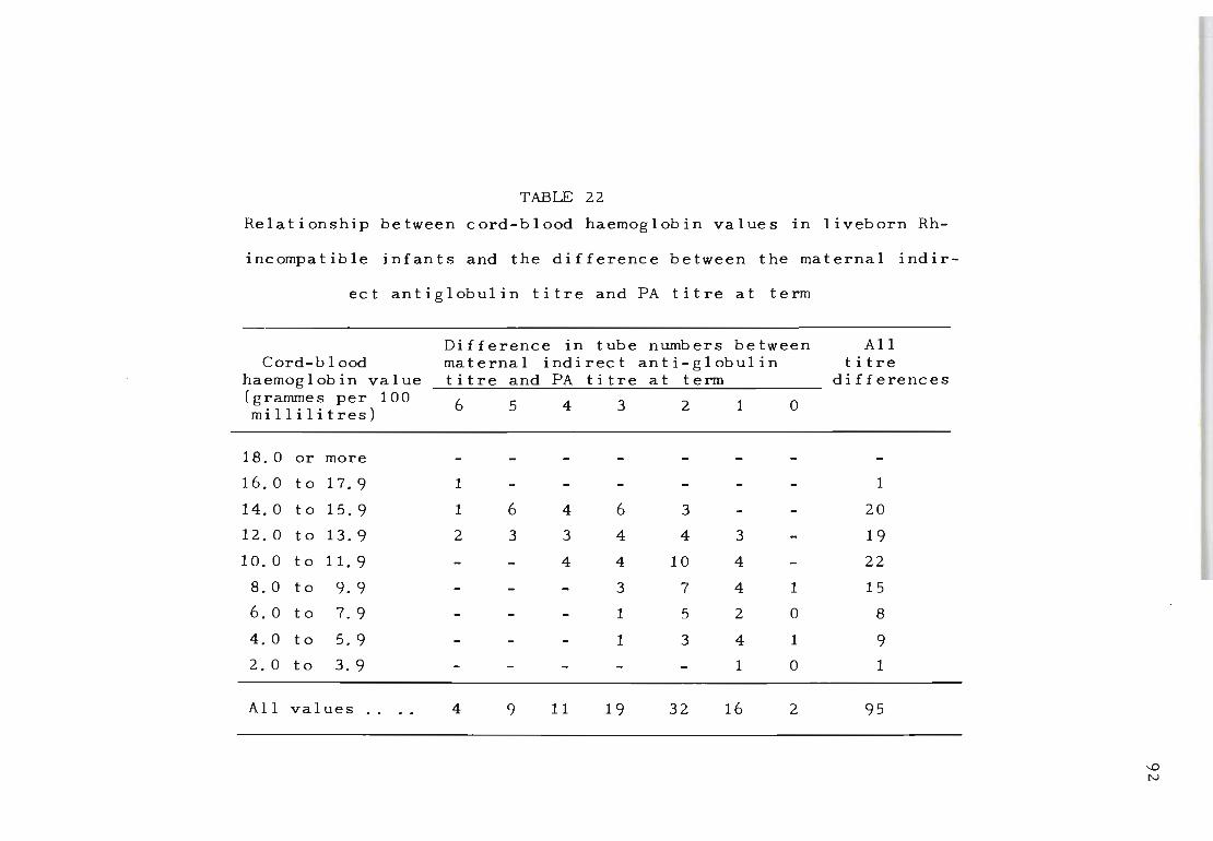
TABLE 22
Relationship between cord-blood haemoglobin values in Ii veb orn Rh-
inc ompa t i b Ie infants and the difference between the maternal indir-
ec t antiglobulin titre and FA titre at term
Difference in tube numbers between All Cord-b lood maternal indirect ant i-globul in titre
haemoglobin value titre and FA titre at term differences (grammes per 100 6 5 4 3 2 1 0 mi 11 i Ii t res)
18. 0 or more
16. 0 to 17.9 1 1
14.0 to 15.9 1 6 4 6 3 20
12.0 to 13.9 2 3 3 4 4 3 19
10. 0 to 11.9 4 4 10 4 22
8.0 to 9.9 3 7 4 1 15
6.0 to 7.9 1 5 2 0 8
4. 0 to 5.9 1 3 4 1 9
2.0 to 3.9 1 0 1
All values 4 9 11 19 32 16 2 95
...0 N
monster: the maternal indirect antiglobulin titre was only
1:128 and the PA titre was zero at term, so that it seems
reasonable to consider that Rh incompatibility was not of
aetiological significance. Of the remaining 35, 27 were
associated with peak PA titres of 1:256 or more (see Table
21). It is necessary to use the peak PA titre in these
cases since in many cases of stillbirth the PA titre drops
rapidly after death has occurred in utero. A terminal
value of the PA titre in such cases therefore does not
represent the true value to which the living fetus has
been exposed.
The rapid drop in the maternal PA titre which often
occurs after the fetus has died means that prognosis
based on a single titration value obtained near term is
likely to be misleading. This probably explains why eight
of the stillbirths in Table 21 were associated with PA
titres of 1:128 or less. In a number of these it was
possible to obtain blood samples from the mother only
near term. If these cases had been titrated at regular
intervals over at least the last ten weeks of pregnancy
the peak values obtained would almost certainly have been
significantly higher.
For 73 of the incompatible pregnancies in the present
series antibody titrations have been carried out on the
maternal serum before the end of the second trimester.
Table 23 shows that in these cases, when the PA titre was
already 1:64 or higher at the twenty-fourth week of gest
ation, only one out of 17 infants survived; 12 were still-
born and four died after birth. Indeed, of the children
93
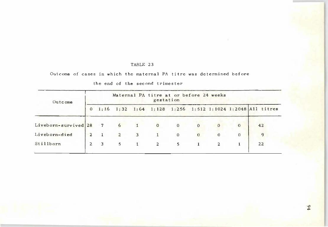
TABLE 23
Outcome of cases in which the maternal FA titre was determined before
Outcome
the end of the second trimester
Maternal FA titre at or before 24 weeks gestation
o 1:16 1 : 32 1:64 1:128 1:256 1:5121 : 1024 1:2048iAll titres
Liveborn-survivedl28 7 6 1 o o o o o 42
Li veb orn-d i ed 2 1 2 3 1 o o o o 9
St i llborn 2 3 5 1 2 5 1 2 1 22
....0 ~
of mothers who had a FA titre at all at the end of the
second trimester, only 14 out of 43 incompatible infants
survived. Clearly, the occurrence of a FA titre in the
maternal serum at any level early during pregnancy is an
unfavourable sign. If it is 1:64 or more the fetus almost
always dies.
The Rh-negative infant - There were 41 Rh-negative infants
born to mothers who had been immunized by a previous preg
nancy. In the majority of these cases the indirect anti
globulin titre was of little value in helping to predict
before birth whether the infant would be Rh-positive or
Rh. negative. In fact, 34 of these Rh-negative infants
were born to mothers with an indirect antiglobulin titre
of 1:128 or more - that is, in the range which would make
an exchange-transfusion necessary if the infant was Rh
positive (Table 24). Actually 25 of the pregnancies were
terminated artifically, since a correct determination of
the infant's genotype could not be made before birth.
The FA titres of the same cases show an entirely diff.
erent distribution; 18 of the mothers had a zero FA titre
at the time of birth and a further 13 had a FA titre only
in the lowest dilution. In these latter cases the FA
titre value of 1:16 had been maintained unchanged during
the greater part of pregnancy. The constant antibody
level was true also of all but two of the 10 cases with
a maternal FA titre higher than 1:16. In one of the ex-
ceptional cases the FA titre fell from an early value of
1:512 to 1:128 at term. In the other case the FA titre
rose from 1:64 to 1:128. This is unique in our experience
9 $
96
TABLE 24
Distribution of Rh-negative infants born to immunized
mothers by indirect antiglobulin titre and PA titre of
the maternal serum at birth
Ti tre Dis t rib uti on of infants
PA titre Indirect anti-globul in titre
Zero 18
1: 16 13 3
1:32 4 2
1:64 3 2
1: 128 3 1 1
1:256 9 1: 512 1 1
1: 1024 3
Total 41 41
and represents the only example in which an Rh-negative
infant waS born to a mothe r who had a rise in either in
direct antiglobulin or FA titre during the course of preg
nancy.
The distribution of cases In Table 24 shows that the
FA titre is of greater value than the maternal indirect
antiglobulin titre in deciding whether a pregnancy should
be allowed to terminate spontaneously. If the father is
known to be heterozygous Did, then a zero or constantly
low FA titre maintained throughout pregnancy is a reason
able indication that the infant is Rh-negative. In such
cases, even i f the indirect antiglobulin titre is high,
the pregnancy can be allowed to continue. It must be
emphasized, however, that if this course is adopted,weekly
checks of the FA titre are essential during the last two
months. In some cases In which an incompatible infant is
involved the FA titre may remain at a low level until the
last few weeks, only to rise rapidly with serious conseq
uences for the infant if labour is not induced immediately.
4. DISCUSSION
A technique for estimating indirectly the amount of Rh
antibody free in the infant's serum by a partial absorpt
ion test on the maternal serum gave early indications that
it would improve the reliability of prognosis based on the
maternal indirect antiglobulin titre alone. The present
evaluation of this test, based on a series of 290 cases
has confirmed this.
The correlation between cord~blood haemoglobin value
97
and maternal FA titre is higher than between cord-blood
haemoglobin value and the ordinary indirect antiglobulin
titre. Indeed, the variability in cord-blood haemoglobin
value for any particular maternal indirect antiglobulin
titre is due almost entirely to the variability in the FA
titre.
severe
The value of the discrimination between mild and
cases which the use of the FA test makes possible
is indicated by the fact that no deaths have occurred
among cases with a maternal indirect antiglobulin titre
of 1:512 or more where the maternal FA titre was more
than three dilution tubes lower (Table 22).
The use of the FA test enables also a much safer pre
diction of an Rh-negative infant in cases in which the
father is known to be heterozygous Did. This is an im-
98
portant use of the test if it means that some pregnancies
which would otherwise merit early induction will be allowed
to continue to full term when the FA titre is zero or
steady at 1:16. However, the FA test is not so valuable
in predicting stillbirths unless the titrations have been
carried out at regular intervals during pregnancy. This
appears to be due mainly to the very marked drop in the
FA titre which may take place when death occurs in utero.
Terminal titration values obtained shortly before birth
therefore mislead, since they may suggest a milder degree
of involvement than is true when the pregnancy terminates
with the birth of a grossly hydropic fetus. However, the
records show that if the maternal FA titre at the end of
the second trimester is 1:64 or more the fetus almost al
ways dies.
99
PART THREE
THE RH ANTIBODY INHIBITION TEST
Chapter IX
THE SIGNIFICANCE OF THE RH ANTIBODY INHIBITION TEST
IN DETERMINING THE SEVERITY OF RH HAEMOLYTIC
DISEASE OF THE NEWBORN
1. INTRODUCTION
Evidence was presented in Chapter III to show that
increased Rh-antibody stimulation can be expected more
often towards the last few weeks of pregnancy than in
early pregnancy. Since only Rh-incompatible fetal red
100
cells entering the maternal circulation can be responsible
for this increase, it is likely that stronger fetal move-
ments during the latter part of pregnancy may also be the
cause of some placental bleeds which lead to increased
Rh-antibody stimulation. While it is accepted that epis-
odes of immunization are associated with renewed antigenic
stimuli, it is also known that the over-all effect of such
stimuli often leads to increased fetal wastage in the form
of stillbirth or neonatal death. This was confirmed by
investigating a large series of Rh-immunized mothers who,
during pregnancy, showed Rh-antibody titre increases of 3
dilution tubes or more, but since an analysis of this nat
ure only showed the general effect of antibody behaviour
over a large series of cases, it does not necessari ly
imply that the severity of Rh-haemolytic disease can be
accurately predicted in each individual case by this pro-
cedure. This is obvious when it is considered that a 3-
fold increase in Rh-antibody titre from a value of 1~8 to
1:64 is of a smaller magnitude than an increase which be-
gins at 1:256 and rises to 1:4096. Also, it has been
established that Rh-antibody titrations alone do not
clearly prognosticate the severity of Rh-haemolytic dis
ease in all instances.
These findings led to the introduction of a 'titre
index' system and a partial absorption test. With the
former procedure some quantification can be obtained by
computing the product of the maternal titre and the time
during which the Rh-ant {bodies were present, while with
the latter procedure partial antibody quantification is
obtained by recording the amount of Rh-antibody not re
moved after one absorption by Rh-positive red cells.
101
A procedure of Rh-antibody quantification is now intro
duced which considers both the time factor and the amount
of antibody to which the infant has been exposed during
pregnancy. With this method it is not necessary to meas
ure the amount of antibody by the conventionai titration
method, which, as shown previously, was found to lack re
producibility (Chapter II). The striking variations found
by Race and Sanger (1954) in their attempts to remove Rh
antibodies by repeated absorption with packed Rh-positive
red cells were taken into consideration. The effective-
ness of Rh-antibody absorption values was compared with
conventional titration procedures with the aim of estimat
ing the extent of accuracy for predicting the severity of
Rh-haemolytic disease in individual cases.
2. MATERIALS AND METHODS
Rh-immunized mothers were examined from the first tri
mester of pregnancy onwards . Mothers were excluded where
the production of Rh-antibodies may have been the result
of previous Rh-incompatible blood transfusions, and only
Rh-incompatible mother-infant combinations were used. For
the evaluation of Rh-antibody titres the standardized In
direct antiglobulin titration method was used.
Rh antibody inhibition test
Preparation of standard haemolysed Rh-positive cell
suspensions Blood collected in ACD solution from a
102
pooled source (2 or 3 donors) of group 0, R1 r (CDe/cde)
donors was washed three times in large volumes of physiol
ogical saline and finally packed by centrifugation at
3 000 rpm for 30 minutes. An accurate 50% saline suspens-
ion of the packed cells was made and divided into 10 mI.
aliquots for daily use. This standard suspension of
cells was haemolysed by freezing at -20 0 C for 48 hours.
It may be stored for many months at this temperature with
out detectable loss of capacity to inhibit Rh-antibodies.
Since significant differences in the inhibition of Rh
antibody can be expected when CDe/cde or cDE/cde red cells
are used it is important that only CDe/cde donors are
selected for the preparation of standard haemolysed test
cells. The heterozygous genotype is always preferred .
because it not only resembles more directly the phenotype
of all Rh-immunized infants' red cells but also ensures
that consistent inhibition results are obtained. DU_ pos
itive red cells should not be included in the donor pool
as the antibody inhibition capacity of these red cells is
quite different from that of D+ DU bloods.
Method of testing - The standardized suspension of haem
olysed Rh-positive cells was thawed at room temperature
103
and shaken thoroughly before use. A constant volume
(0.4 mI.) of haemolysed cells was placed into six test
tubes, numbered 1 to 6. To tube 1 was added one volume
(0 4 I) of a 1.·2 saline dilution of Rh antiserum (1 part • m.
undiluted serum and 1 part saline in this instance was re-
cognized as a 1:2 dilution of serum and not 1: 1); to tube
2, 1 volume (0.4 ml.) of a 1:4 saline dilution of Rh anti-
serum and to tubes 3,4,5 and 6 similarly increased dilut-
ions of Rh antiserum were added (1:8, 1:16, 1:32 and 1:64).
The diluted serum and haemolysed cells in each test tube
were mixed and left undisturbed at room temperature for
10-15 minutes. The free Rh-antibody not inhibited by the
haemolysed suspension of Rh-positive red cells was meas-
ured by adding one volume (0.4 ml.) of unhaemolysed Rh-
positive red cells and left for a further 20 minutes at
room temperature. The presence or absence of free Rh-
antibodies in each test tube was then determined by the
indirect antiglobulin test. The need to use avid broad-
spectrum anti-human gammaglobulin serum must be emphasized
because antiglobulin reagents of lesser quality cannot al -
ways detect free antibody on the red cells by this proced -
ure.
Interpretation of results - Since an increasing amount
of Rh antigen (in the form of haemolysed Rh-positive red
cells) is actually combined with the Rh antiserum, a meas-
urement can be made of the amount of Rh antigen that is
required to inhibit the Rh antibody. From tubes 1 to 6
the ratio of serum to Rh antigen varies from 1·.1 t 1 2 o : ,
1 : 4, 1: 8 , 1: 16 and 1: 32. The absence of Rh-antibodies in
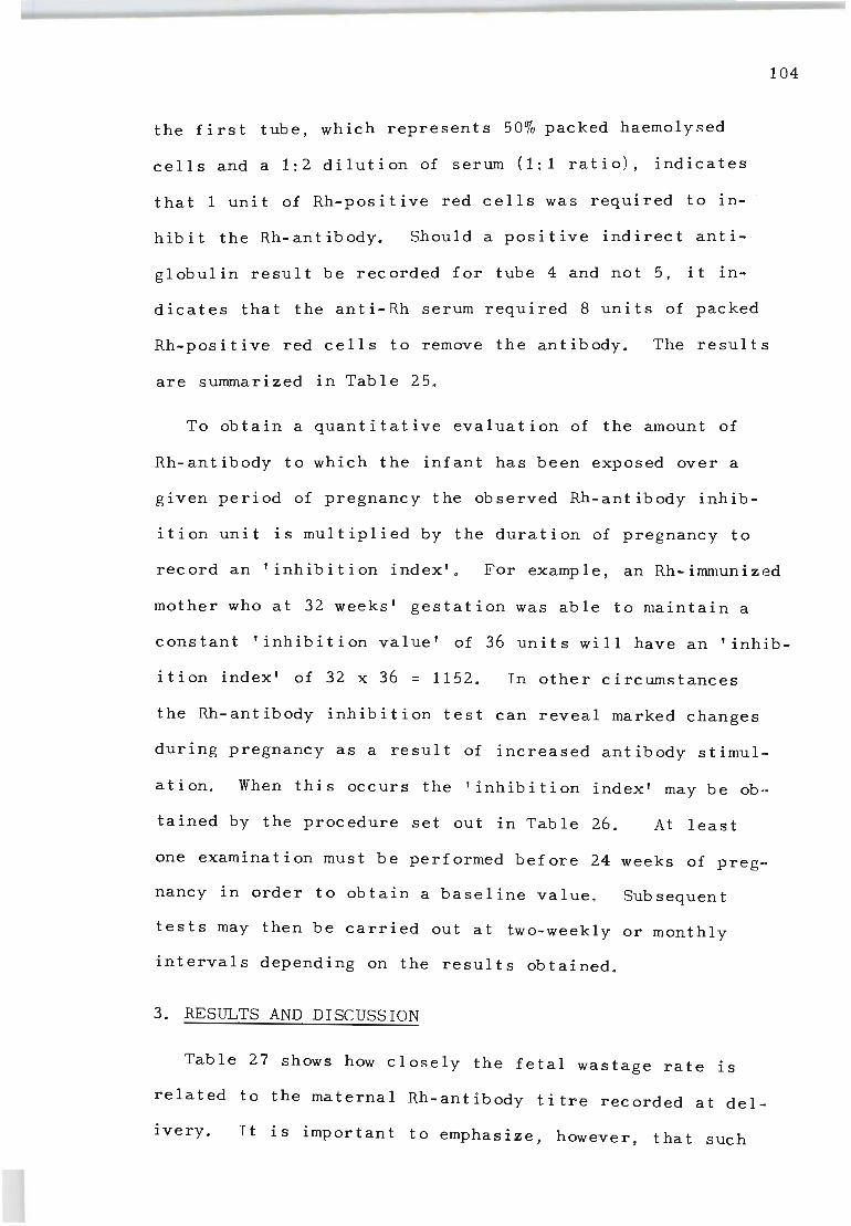
104
the first tube, which represents 50% packed haemolysed
cells and a 1:2 dilution of serum (1:1 ratio), indicates
that 1 unit of Rh-positive red cells was required to in
hibit the Rh-antibody. Should a positive indirect anti-
g I obul in re suI t be rec orded f or tube 4 and not 5, it in
dicates that the anti-Rh serum required 8 units of packed
Rh-positive red cells to remove the antibody. The results
are summarized in Table 25 .
To obtain a quantitative evaluation of the amount of
Rh-antibody to which the infant has been exposed over a
given period of pregnancy the observed Rh-antibody inhib
ition unit is multiplied by the duration of pregnancy to
record an 'inhibition index' . For example, an Rh-immunized
mother who at 32 weeks' gestation was able to maintain a
constant 'inhibition value' of 36 units will have an 'inhib-
ition index' of 32 x 36 = 1152. In other circumstances
the Rh-antibody inhibition test can reveal marked changes
during pregnancy as a result of increased antibody stimul
ation. When this occurs the 'inhibition index' may be ob-
tained by the procedure set out in Table 26. At least
one examination must be performed before 24 weeks of preg-
nancy in order to obtain a baseline value. Sub sequent
tests may then be carried out at two-weekly or monthly
intervals depending on the results obtained.
3. RESULTS AND DISCUSSION
Table 27 shows how closely the fetal wastage rate is
related to the maternal Rh-antibody titre recorded at del
i very. It is impor tan t to emphas i ze, however, t ha t such
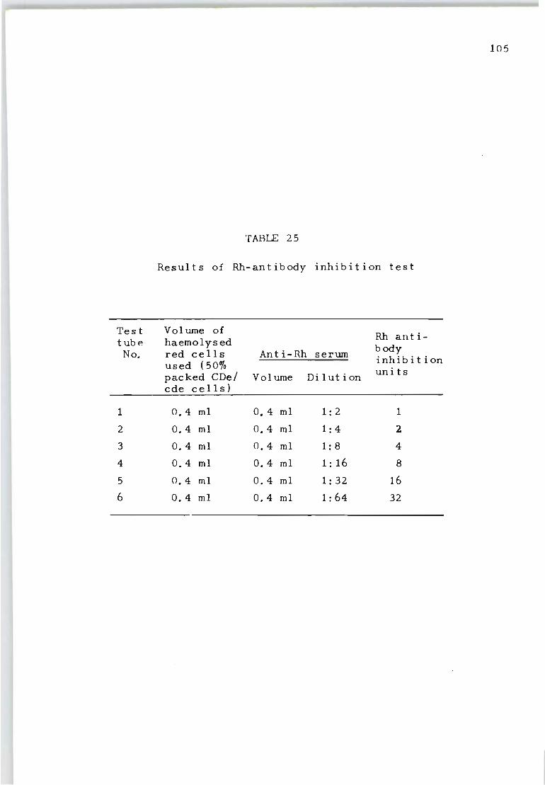
105
TABLE 25
Results of Rh-antibody inhibition test
Test Volume of Rh anti-tube haemolysed body No. red cells Ant i-Rh serum
used ( 50% inhibition
packed CDe/ Vol ume Dilution units
cde cells)
1 0.4 ml 0.4 ml 1: 2 1
2 0.4 ml 0.4 ml 1: 4 2
3 0.4 ml 0.4 ml 1: 8 4
4 0.4 ml 0.4 ml 1: 16 8
5 0.4 ml 0.4 ml 1:32 16
6 0.4 ml 0.4 ml 1:64 32
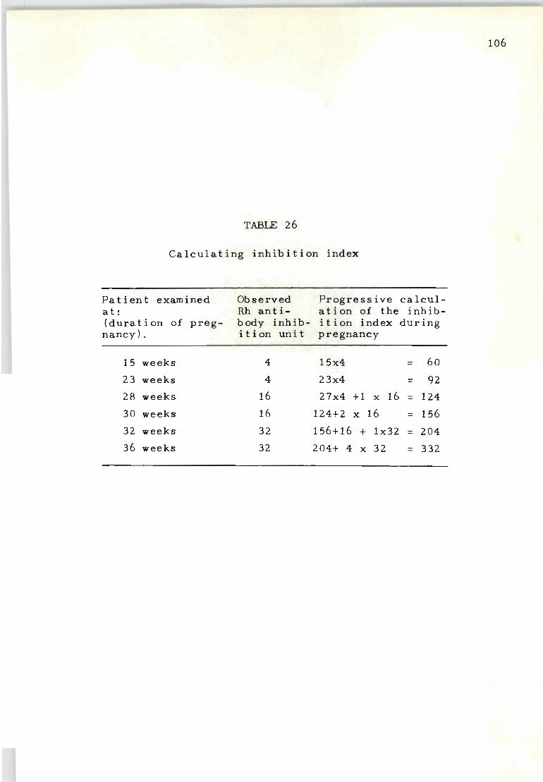
106
TABLE 26
Calculating inhibition index
Patient examined Observed Progressive calcul-at: Rh ant i- at ion of the inhib-(duration of preg- body inhib- ition index during nancy) . ition uni t pregnancy
15 weeks 4 15x4 = 60
23 weeks 4 23x4 = 92
28 weeks 16 27x4 +1 x 16 = 124
30 weeks 16 124+2 x 16 = 156
32 weeks 32 156+16 + 1x32 = 204
36 weeks 32 204+ 4 x 32 = 332
TABLE 27
Relationship between maternal indirect antiglobulin titre and outcome of pregnancy
Maternal Rh ant i body Rh nega t i ve Rh positive infants Overa 11 fetal titre at delivery mothers wastage
No. Survived Died S t i llb orn No. %
1 : 64 or less 176 176 0 0 0 0.0
1:128-1 : 512 326 310 10 6 16 4.9
1 : 1024 or more 316 164 53 99 152 48. 1
Total: 818 650 63 105 168 20.5
~
o --J
108
a correlation can be seen only in a combined analysis of
many cases and that misleading information can easily be
deduced from it if it is assumed that all Rh-immunized
mothers with antibody titres of 1:1024 or more will have
severely affected Rh-positive infants. The findings do,
however, confirm that no fetal wastage due to Rh-immuniz
ation need be expected if a mother has an antibody titre
of 1:64 or less. For practical purposes a number of inves
tigators (Crawford, Cameron and Walker, 1966; Farre ll and
Davis, 1967) have therefore accepted that amniocentesis
need not be performed on Rh-immunized mothers with low
Rh-antibody titres.
It has often been reported that Rh-antibody tit rations
cannot be regarded as a reliable criterion for the pre
diction of the severity of Rh-haemolytic disease in indiv-
idual cases. We also found this to be so by correla ting
the range of cord-blood haemoglobin values against the
maternal Rh-antibody titres observed at birth. Since the
greatest percentage of false predictions can be expected
among mothers with titres of 1:1024 or more a selective
analysis was carried out (Table 28) . At least 40 per cent
of the mothers had Rh-positive infants with cord-blood
haemoglobin values greater than 10 GI100 mI. These evid
ently tolerated the intensity of a 1:1024 titre much better
than those with cord-blood haemoglobins less than 10 GI100
mI. The last-mentioned group also experienced a high per
centage of neonatal deaths.
The variations observed between the apparent clinical
significance of low Rh-antibody titres (1:64 or less) a nd

TABLE 28
Distribution of Rh positive infants born alive to immunized
mothers with indirect antiglobulin titres of 1:1024 or more
by values for cord-blood haemoglobin and neonatal deaths
Infant cord-blood haemoglobin G/100 ml
14. 0
10.0-13.9
6.0- 9.9
5. 9
Total:
Rh negative mothers
No. %
46) 42) 40.5
) 98) 31) 59. 5
)
217
Rh positive infants Survived Died
No. No. %
46 0 O. 0
42 0 O. 0
71 27 27. 5
5 26 83.8
164 53 24.4
..... o -.D
lack of such correlation when the Rh-antibody titres are
high (1:1024 or more) draws a tt ention to the fact that
the conventional procedure of antibody titration does not
adequately measure the true quantity of Rh-antibody, par
ticularly in the higher dilutions .
110
In an attempt to discover other recognizable variations
between low and high-titred Rh-antibodies it was found that
significant differences often existed in the capacity of
Rh-positive red cells to absorb Rh-antibodies. By modify
ing the procedure in order to obtain a greater degree of
standardization and reproducibility the Rh-antibody inhib
ition test was compared with the antibody titration pro-
cedure. It was found (Table 29) that serum samples with
Rh-antibody titres of 1:64 or less are not generally diff
icult to inhibjt by one unit of haemolysed Rh-positive
red cells, although a few cases may require four units of
haemolysed cells. By comparison, Rh-antibody titres of
1:1024 or more showed a significantly greater range of
inhibition results, some requiring 2-4 units of haemolysed
red cells while others required 16 units or more. Although
some Rh-antibodies possessed titres of 1:1024 or more they
appeared to have the same inhibition value as those with
titre values of 1:64 or less. This presupposes that the
haemolytic effect of some 1:1024 Rh-antibody titres is
the same as some observed within the 1:64 category.
With such marked variations observed in the ability to
inhibit Rh-antibodies it was important to determine the
value of this test In prognosticating the severity of Rh
haemolytic disease before birth. Table 30 details an
TABLE 29
Comparative analysis between relationship of Rh antibody
titres and inhibition test
Maternal indir- No. of Units of haemolyzed cells
ec t anti g I obul in mothers required for the i nh i bit i on of
titre value examined Rh antibodies 124 8 16 32 64
1:64 or less 56 37 13 6 1: 128 ) 1:256 ) 74 17 21 15 10 7 4 1: 512 ) 1: 1024 or more 39 2 6 13 8 6 4
...-
TABLE 30
Rh antibody inhibition index as a measure of severity of Rh haemolytic disease
Maternal Rh ant ibody inhibition index
80 uni t s or less
80 - 500 units
500 units or more
No. of mothe rs examined
82
54
33
Al ive and well
82
48
15
Rh positive infants
Died
4
6
St i llborn
2
12
Exchange transfused
7
46 21
Live-births Not
exchange transfused
75
6
Foe tal wastage
%
o. 0
11. 1
54.5
......
...... N
analysis of 169 Rh-immunized mothers examined throughou t
pregnancy. The inhibition test is expressed as a measure
of 'inhibition index' which estimates the period that a n
Rh-incompatible infant has been exposed to maternal Rh-
antibodies. By such calculations a wide range of inhib -
ition index values were recorded which, for the purpose
113
of this analysis, have been divided into three categories .
The first category represents Rh-immunized mothers who
throughout pregnancy have accumulated an Rh-antibody in
hibition index of only 80 units or less . Not one of the
Rh-incompatible pregnancies resulted in a stillbirth or
neonatal death and more than 90 per cent of the liveb orn
infants did not require exchange-transfusion. From this
information it is apparent that a good correlation exists
between a low Rh-antibody inhibition index and a less
severe haemolytic manifestation of the disease neonatall y.
For those mothers who had higher inhibition index value s
during pregnancy the effec t of in utero haemolysis was
most obvious when judged by the increased frequency of
fetal death and the significantly raised incidence of
infants who required exchange-transfusions.
The findings in Table 30 show the close correlat i on
between the Rh-antibody inhibition index and the final
outcome of pregnancy. It was not intended to show the
antenatal behaviour pattern of the inhibition test, nor
the manner in which the findings should be interpreted
to make an accurate prediction of the haemoly~ic process
in each individual case. This will be more fully detail ed
in the next chapter. However, the observations do emphas -
ize that an inhibition index of 500 units or more is un
questionably associated with an intense process of immune
Rh-antibody producti on wh ich, as shown in Table 31, can
be responsible for the introduction of other complicating
immunological responses o
The presence of other antibodie s apart from anti-Rh is
not unusual and their occurrence would seem to follow the
same course as documented by Nilsson (1965) with regard
114
to the influence of multiple Rh- antibodies. When multiple
Rh-antibodies are present the severity of the disease is
usually greater; t his is probably because the total amount
of Rh-antibodies bound to the infant's red cells is, for
example, greater in CDe/cde infants born to mothers with
anti-C plus anti-D, than if the same mothers had produced
only anti-D. It is quite possible that there is a connect
ion between increased antibody loading on the red cells
and the severity of t he disease . However, the primary
cause of the developmen t of mUltiple antibodies appears
to be directly associ ated with the intensity of the stim
ulation of the antibodies as a result of repeated episodes
of placental haemorrhage.
It has been shown that antibodies other than anti-Rh
are produced in response to frequent episodes of re-immun
ization. This complication is most often found among
mothers with significantly raised Rh-antibody values
(Table 31), who as a rule have more severely affected
infants (Table 30), and possibly a greater incidence of
placental haemorrhage. Future studies directed towards
the prevention of plac en t al haemorrhage must therefore
lead to an even grea t e r reduction in fetal wastage.
TABLE 31
Frequency of other immune red cell antibodies among mothers who were
initially sensitized by the Rh factor
Maternal Rh ant ibody inhibition index
No. of mothers examined
8 a un its 0 r Ie s s
80 - 500 units
500 units or more
82
54
33
Immunizing antibody Anti-Rh ' alone Anti-Rh plus
others*
82
49 27
a 5
6
Percentage of two ant ibody systems observed
'0 O. a 9.2
18. 1
* Rh immunization found in association with immune anti-A, B, Kell, Fya
or Jka .
.... .... U1
Chapter X
THE VALUE OF THE RH-ANTIBODY INHIBITION TEST
IN RELATION TO LIQUOR AMNII STUDIES
1. INTRODUCTION
116
Considerable progress was made in the management of
pregnancies complicated by Rh-immunization with the intro
duction of amniotic fluid studies (Bevis, 1950, 1952).
Investigators have now confirmed the relationship of bile
pigment concentration in the amniotic fluid with the
intensity of Rh-haemolytic disease in utero. However,
the spectrophotometric (Walker, 1957), chemical (Mackay
and Watson, 1962) and pigment-protein ratio procedures
(Morris, Murray and Ruthven, 1967) cannot always be
accepted as being infallible, their shortcomings being
too seldom emphasized while their advantages are simply
taken for granted (Vos, 1969).
Some investigators (Pirofsky, 1965; Robertson, 1961)
believe that with the introduction of liquor amnii stud-
ies nothing useful can be gained by the inclusion of mat-
ernal Rh-antibody investigations. Others (Liley, 1963;
Hyslop and Whiley, 1960; Walker, 1967) emphasize that
the prediction of the severity of Rh-haemolytic disease
by amniotic fluid analysis can at times be most mislead-
ing. Fully endorsed cautions about relying on the pre
dictions obtained from amniotic fluid studies alone have
led investigators to believe that bile pigment levels
should be used only in conjunction with maternal Rh-anti
body estimations (Nilsson, 1965; Crawford, Cameron and
Walker, 1966).
117
Since repeated abdominal paracentesis on every Rh
immunized mother can result in errors in the antenatal
prediction of the severity of the disease (Zipursky,
Pollock, Chown and Israels, 1963; Misenhimer, 1966),
selective amniocentesis has become an accepted practice.
The recording of low maternal Rh-antibody values is
really not indicative of severe haemolytic manifestat
ions of the disease.
We found that by using Rh-antibody inhibition studies
we could predict far more reliably the severity of Rh
haemolytic disease, particularly when the significance
of liquor amnii values appears to be in some doubt.
Routine maternal antibody studies form an effective
supplement to the results obtained by spectrophotometric
analysis. Rh-antibody studies, can, in fact, be regarded
as a test to show whether a clinically favourable fore
cast of severity obtained by liquor examinations at 26-30
weeks gestation continues to be the same at 34 weeks'
gestation, especially when it is taken into account that
more frequent episodes of re-immunization may be expected
after 30 weeks of pregnancy (see Chapter III).
2. MATERIALS AND METHODS
Rh-immunized mothers were examined from the first
trimester of pregnancy onwards . Those who might have
produced Rh-antibodies as a result of previous Rh-incom
patible blood transfusions were excluded and only Rh
incompatible mother-child combinations were studied.
To evaluate the Rh-antibody inhibition index, the
procedure described in Chapter IX was used. Rh-ant ibody
titre values were determined by the standardized method
of indirect antiglobulin titration (Chapter II).
Amniotic fluid studies - Amniotic fluid obtained by
abdominal paracentesis was centrifuged immediately after
collection at 4 000 rpm for 30 minutes to remove cellular
elements and was then placed in a light-proof container
if an immediate analysis was not carried out. Bile pig
ment concentration in the specimen was analysed using
the spectrophotometric procedure of Fleming and Woolf
(1965) and total protein value by the biuret reaction
de-scribed by Morris, Murray and Ruthven (1967). Pigment
protein ratio was calculated as recommended by the same
authors. Serum bilirubin estimations on the infants'
118
cord bloods were determined by the method of Powell (1954).
3. RESULTS AND DISCUSSION
In order to compare objectively the efficacy of amnio
tic fluid measurements with that of Rh-antibody inhibition
studies, an investigation was carried out on 66 Rh-immun
ized mothers who had given birth to Rh-incompatible infants.
In the assessment of how closely these two procedures can
forecast the severity of fetal red cell destruction in
utero it is important to remember that the Rh-antibody
inhibition test determines the measured intensity of anti
body effect up to the time of delivery.
The amniotic fluid studies (particularly when performed
before 30 weeks of pregnancy) can only measure the inten
sity of fetal red cell haemolysis present at the time
119
the sample was obtained by amniocentesis.
To reduce such variations to a minimum, only the high-
est projected spectrophotometric measurement obtained
from a series of amniotic studies on the same patient
was used. An average of 2 . 4 amniocentesis samples were
tested from the 66 mothers examined. Their time of coll-
ection ranged from 28 to 33 weeks of gestation and in no
instance were single amniocentesis studies included if
they were performed after the 33rd week of pregnancy. A
detailed analysis of the serological, biochemical and
haematological findings of the 66 Rh-immunized mothers
and their infants is shown in the Appendix of this chapter.
To determine how closely the Rh-antibody 'inhibition
index' and bilirubin protein values are related to the
severity of the infant's anaemia at birth, a biserial
correlation coefficient analysis was carried out for the
two testing procedures against (a) the haemoglobin con-
centration of the infant's cord blood at birth; (b) total
serum bilirubin value at birth, and (c) the severity of
Rh-haemolytic disease as determined by the infant's
clinical condition. Table 32 shows the results of this ,
analysis and it can be seen that both procedures (amnio-
tic fluid values and antibody inhibition studies) accur-
ately predicted that an infant's cord blood haemoglobin
level will be low when the bilirubin/protein ratio or
Rh-antibody inhibition index values are high. A corres-
ponding correlation is also confirmed for the ability of
these two procedures to measure the overall severity of
the disease. However, no real correlation was established
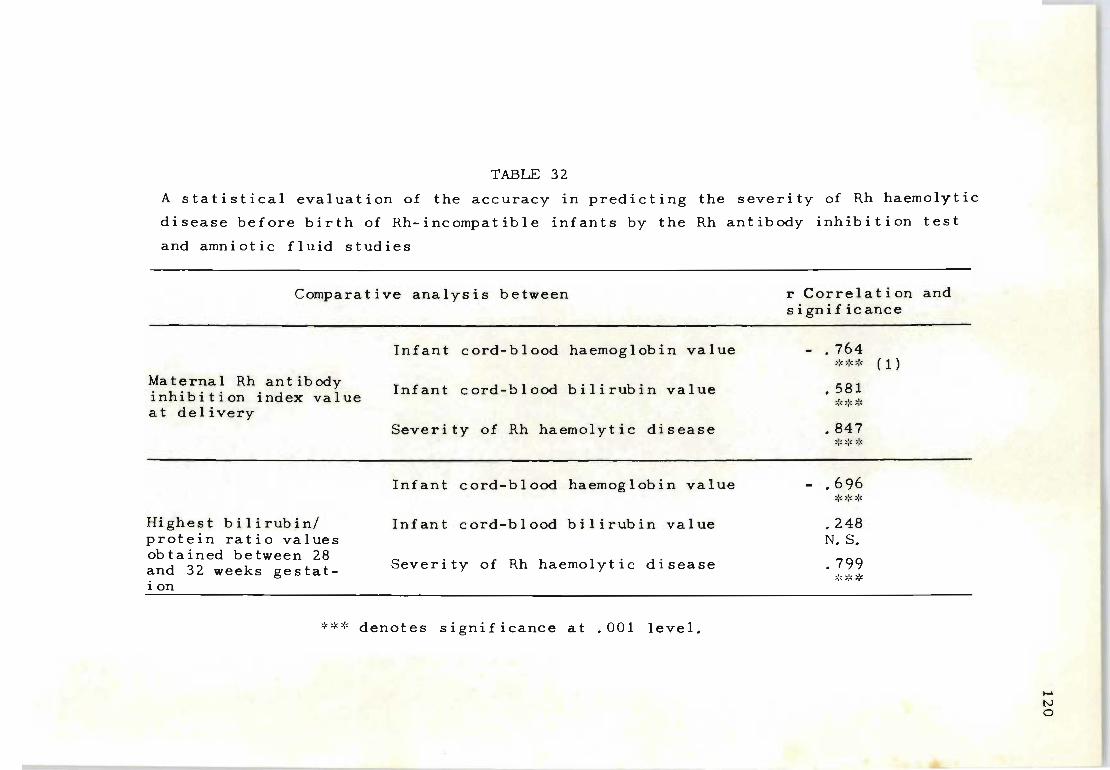
TABLE 32
A statistical evaluation of the accuracy in predicting the severity of Rh haemolytic
disease before birth of Rh-incompatible infants by the Rh antibody inhibition test
and amniotic fluid studies
Comparative analysis between
Maternal Rh ant ibody inhibition i ndex value a t delivery
Highest bilirubin/ protein ratio values obtained between 28 and 32 weeks gestation
Infant cord-blood haemoglobin value
I nfant cor d-blood b ili rubin value
Severity of Rh haemolytic disease
Infant cord-blood haemoglobin value
Infant cord-blood bilirubin value
Severity of Rh haemolytic disease
*** denotes significance at .001 level.
r Correlation and sign if ic ance
- . 764 >!<>.'<* (1)
• 581 ~::: ~~ :::<
• 847 :::~~, *
- .696 ~,~:::*
.248 N. S.
• 799 ::::::: ':' ~(
...... IV o
between the bilirubin/protein ratio and the cord-blood
bilirubin values, whereas a significant association be
tween Rh-antibody inhibition results and cord-blood bili
rubin values is evident.
121
Such variations lend support to the argument that the
bilirubin/protein ratio test (done at least four weeks
before the birth of the infant) IS able to determine only
the intensity of in utero haemolysis noted at the time of
collection and does not necessarily indicate the true
intensity of red cell haemolysis to be expected at birth.
As more frequent episodes of antibody stimulation can
be expected as pregnancy progresses towards term, the
lack of positive correlation between bilirubin/protein
ratio values and cord-blood bilirubin may be influenced
by the increased rate of red cell destruction seen after
the amniocentesis samples were taken. The possibility
that raised Rh-antibody production (induced by the dis
ruption of fetal-placental circulation at amniocentesis),
is a contributing factor cannot be ignored, and like the
practice of external version (Vos, 1967) can be considered
as furthering the intensity of the disease (Zipursky, Poll
ock, Chown and Israels, 1963).
A real measure of confidence in the prognosis of Rh
haemolytic disease can be attained when two different
testing procedures are employed. However, it was found
that even if a statistical correlation between the test
ing procedures and the severity of the disease is evident
in a combined analysis of many cases, the same may not
necessarily be true for some individual cases. Marked
differences can occasionally be observed due to the con
dition of an infant being influenced by factors other
than Rh-immunization (Mollison , 197 2) .
122
With the knowledge that such variations can be expected
in individual cases it appeared that the prognosis of the
disease should ultimately be based on the assessment of a
combination of parameters (previous history, father's Rh
genotype, Rh-antibody value and amniotic fluid results)
concurrent with any additional experienc e gained from
other statistical studies . For this r eason a number of
individual cases are included to show how the Rh-antibody
inhibition test can be of particular significance in pro
viding extra information about the haemolytic condition
In utero.
Table 33 details the study of an Rh-immunized mother
with a previous history of two normal pregnancies which
were not complicated by Rh- immunization. During her third
pregnancy Rh-immunization was confirmed at 12 weeks' ges
tation, the preliminary observations strongly suggesting
that this was the direct result of postnatal immunization.
A high Rh-antibody value scored at 12 weeks' gestation
also served as an indication that the present pregnancy
could result in a very severely affe~ted infant. This
prediction was reached by noting that the Rh-antibody
inhibition index value at 26 weeks would have attained a
level of at least 832 units which, as shown in Chapter IX,
is dangerously high. Subsequent amniotic fluid studies
clearly confirmed that the infant was indeed likely to be
badly affected, indicating that only intra-uterine trans-
I \
\
Pe r i od of
gestation
12/52
24/52
26/52
27/52
29/52
30/52
32/52
TABLE 33
Mrs H.L.: Age 25. Previous obstetric history: Two infants alive and well. No stillbirths or
spontaneous abortions. Father's Rh genotype: CDe/CDe
Rh antibody studies by:
Indirect Inhibition ant i glob u lin titre Value Index
1: 1024 32 384
1: 1024 32 768
1:1024 32 832
1:1024 32 864
1: 1024 32 928
1: 1024 32 960
1: 1024 32 1024
Opt ical dens i ty
at . 450 mu
O. 170
O. 140
Liguor amnii studies
B iIi rub in pgm/ml
2. 72
2.40
Protein mgm/ml
4,8
3.9
Ra t i 0 b ilirubin
figm/mgm of protein
o. 56
0.61
Outc orne: Spontaneous labour at 32 weeks. Macerated stillborn fetus,
..... [\J
w
fusion may have saved the infant from being stillborn at
32 weeks.
The case presented in Table 34 is very similar to the
one described in Table 33, the only d:ifference being that
the comparative intensity of in utero haemolysis, as
judged by the amniocentesis value, appeared to be more
pronounced. The bilirubin/protein ratio values between
this and the previous case were at least three times
greater, whereas the relative Rh-antibody index value
scored at delivery differed only slightly.
124
These variations do not necessarily imply that the two
testing procedures differed in their prognostic reliabil
ity, but it does show that individual variations can be
expected within certain limits. There would be reason
for real concern, of course, if the two values had been
significantly different. Table 35 describes a case where
it was very difficult for the clinicians to decide whether
intra-uterine transfusion or veTY early induction of lab-
our was the better mode of treatment. This mother had a
moderately low Rh-antibody inhibition index value when
examined at 30 weeks' gestation and at this stage a very
severely affected infant would not have been suspected.
On the other hand, liquor amnii studies clearly showed
that the infant was severely affected, the bilirubin/
protein ratio values obtained being greater than the re
sults recorded for the stillborn infant in Table 33.
Since no other immune antibodies were detected in the
maternal serum to aggravate the haemolytic process in
utero, no measures were taken to treat the infant premat-

Period of
TABLE 34
Mrs P.K.: Age 32 Previous obstetric history: First infant died 12 days after birth. Second infant alive and well. Third infant stillborn at 33 weeks. Fourth infant Rh-affected) delivered by Caesarean section; exchange-transfused, alive and well.
Rh antibody studies by: Liguor amni i studies
Indirect Inhibition Opt ical Bilirubin Protein Ra t i 0 b i I i-gestation antiglobulin density /Ugm/ ml mgm/ml rubin
22/52
26/52
31/52
33/52
34/52
35/52
titre Value Index at ).lgm/ mgm of 450 mu protein
1: 512 16 352
1: 512 16 416
1: 1024 32 512 1. 001 10. 7 6.7 1.5
1:2048 32 576 O. 710 10. 2 5 . 3 1.9
1:4096 32 608
1: 4096 32 640
Outcome: Spontaneous labour at 35 weeks. Macerated stillborn fetus: intra-uterine death was established at 34 weeks.
IV Ln
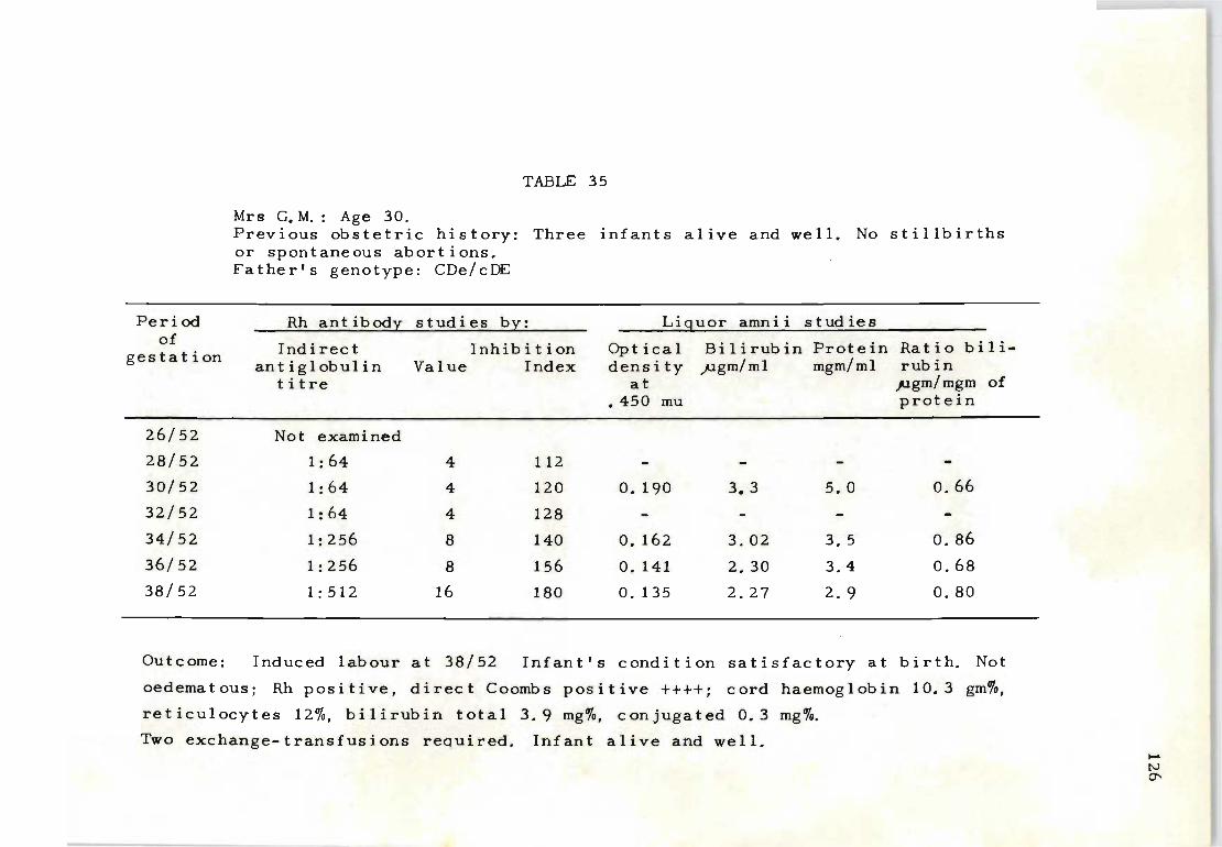
Peri od of
gestation
26/52
28/52
30/52
32/52
34/52
36/52
38/52
TABLE 35
Mrs C.M.: Age 30. Previous obstetric history: Three infants alive and well. No stillbirths or spontaneous abortions. Father's genotype: CDe/cDE
Rh antibody studies by: Liguor amnii stud ies
Indirect Inhibition Optical Bilirubin Protein Ratio bili-ant ig I obul in Value Index dens i ty flgm/ml mgm/ml rubin
titre at pgm/ mgm of .450 mu protein
Not examined
1:64 4 112
1:64 4 120 o. 190 3.3 5. 0 0.66
1:64 4 128
1: 256 8 140 O. 162 3.02 3. 5 0.86
1:256 8 156 O. 141 2.30 3.4 0.68
1: 512 16 180 O. 135 2.27 2.9 0.80
Outcome: Induced labour at 38/52 Infant's condition satisfactory at birth. Not
oedematous; Rh positive, direct Coombs positive ++++; cord haemoglobin 10.3 gm%,
reticulocytes 12%, bilirubin total 3.9 mg%, conjugated 0.3 mg%.
Two exchange-transfusions required. Infant alive and well. ..... tv 0'

127
urely. Follow-up studies gradually showed raised Rh-anti-
body formation while the amniocentesis values continued
to indicate an apparently intense haemolysis in utero . At
birth the clinical condit i on and haematological findings
were in c'omplete accord wi th the serological findings but
disagreed with the amniocentesis values scored since 30
weeks of pregnancy.
Whilst it can be accepted that a high percentage of
predictions scored by amniocentesis investigations are
r~liable indicators of the intensity of in utero haemoly
sis, the recognition of some shortcomings may help future
reasoning regarding the mechanism of bilirubin entry into
the amniotic sac.
Table 36 describes a case where it is again evident
that the initial process of Rh-immunization occurred as
a possible consequence of the trauma of parturition. In
this instance Rh-antibody production was not very intense
during the early stages of her second pregnancy ; however,
from 32 weeks gestation onwards a marked increase in Rh
antibody titre was noted, whilst the inhibition index
value remained low. Amniocentesis values confirmed that
the intensity of in utero haemolysis was not of clinical
concern and that the postnatal course of the disease would
be commensurate with such findings. It is important to
remember however that should this mother conceive another
Rh-positive infant soon after the present pregnancy the
clinical outcome would most likely follow a course very
similar to the case described in Table 34 where an already
immunized mother commenced pregnancy with lethal Rh-anti-
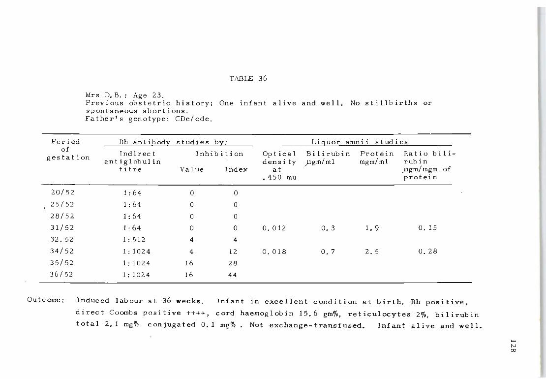
Peri od of
TABLE 36
Mrs D.B.: Age 23. Previous obstetric history: One infant alive and well. No stillbirths or spontaneous abortions. Father's genotype: CDe/cde.
Rh ant ibody studies by: Liguor amni i studies
Indirect Inhibition Opt ica I Bi I i rub i n Protein Ra t i 0 b i I i-gestation antiglobulin density Fgm/ml mgm/ml rubin
titre Value Index at ).lgm/ mgm of • 450 mu protein
20/52 1:64 0 0
25/52 1:64 0 0
28/52 1:64 0 0
31/52 1:64 0 0 0.012 O. 3 1. 9 O. 15
32.52 1: 512 4 4
34/52 1:1024 4 12 0.018 O. 7 2. 5 0.28
35/52 1: 1024 16 28
36/52 1: 1024 16 44
Outcome: Induced labour at 36 weeks. Infant in excellent condition at birth. Rh positive,
direct Coombs positive ++++, cord haemoglobin 15.6 gm%, reticulocytes 2%, bilirubin
total 2.1 mg% conjugated 0.1 mg% Not exchange-transfused. Infant alive and well.
tN ex>
body values which resulted in stillbirth.
In an effort to help women in these circumstances to
avoid pregnancies with a high risk of stillbirth, pre
natal counselling and examination is advocated until the
mother can be assured that the Rh-antibody value indic
ates that there is a good possibility of her delivering
a live born child.
129
Finally, it must also be acknowledged that the use of
liquor examinations is of real practical value in deter
mining the presence of Rh-negative infants. Table 37 de
tails a case where it was obvious from a combination of
maternal Rh-antibody and liquor amnii values that the
baby was Rh-negative. The prediction by amniocentesis of
a mildly affected infant did not agree with the forecast
of a very severely affected infant by Rh-antibody inhibit-
ion values. If we accept that Rh-antibodies destroy fetal
red cells then the lack of haemolysis by amniocentesis
studies almost certainly indicated that the infant's Rh
type had to be compatible with the mother's.
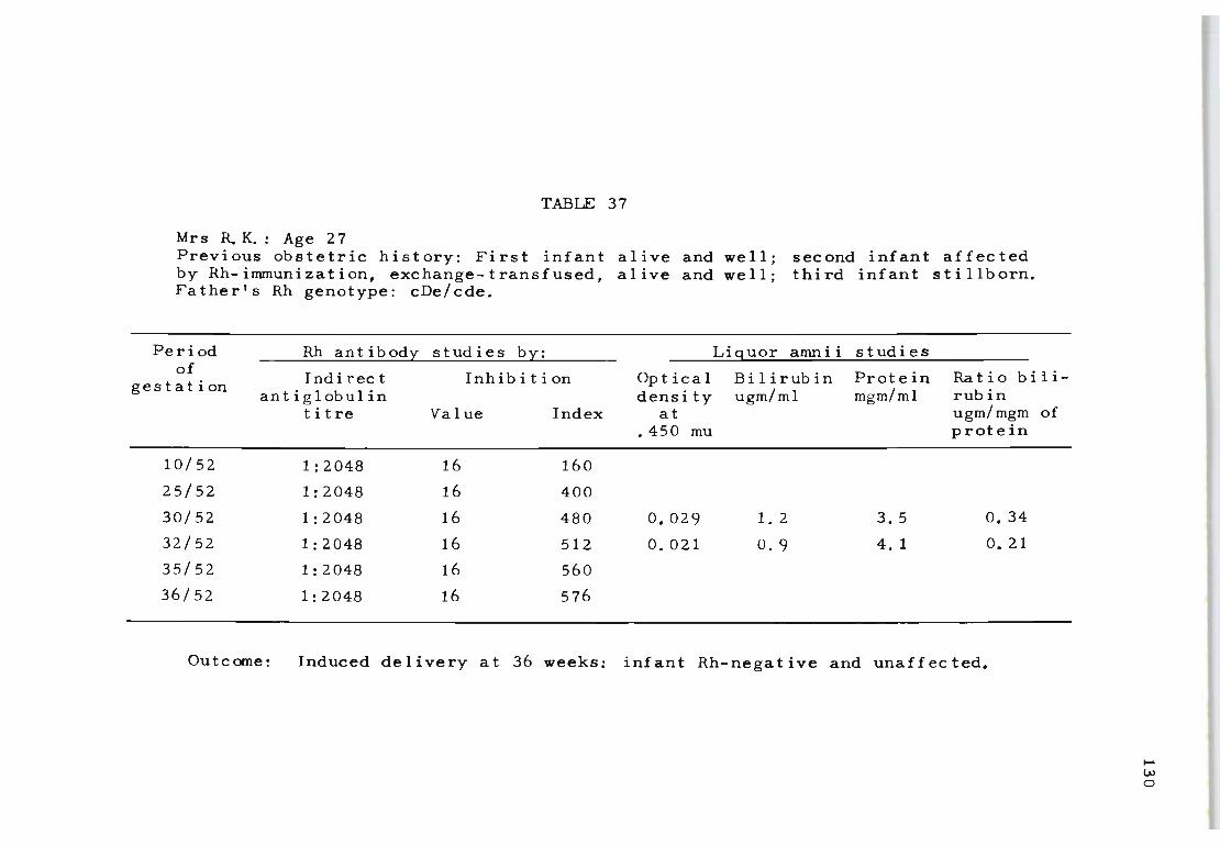
TABlE 37
Mrs R.K.: Age 27 Previous obstetric history: First infant alive and well; second infant affected by Rh-imrnunization, exchange-transfused, alive and well; third infant stillborn. Father's Rh genotype: cDe/cde.
Period Rh antibody studies by: Liguor amnii studies of
Indirect Inhib i t ion Opt ical Bi I i rub in Protein Ratio bili- -gestation antiglobulin density ugm/ml mgm/ml rubin
titre Value Index at ugm/mgm of .450 mu protein
10/52 1:2048 16 160
25/52 1:2048 16 400
30/52 1:2048 16 480 0.029 1.2 3. 5 0.34
32/52 1:2048 16 512 0.021 0.9 4. 1 O. 21
35/52 1:2048 16 560
36/52 1:2048 16 576
Outcome: Induced delivery at 36 weeks: infant Rh-negative and unaffected.
...... w o

131
APPENDIX
SEROLOGY, BIOCHEMISTRY AND HAEMATOLOGY OF 66 RH-IMMUNIZED MOTHERS WHO DELIVERED RH-POSITIVE INFANTS
Liquor amnii studies Maternal Cord-blood estimations
Case Rh-antibody Ratio Time of delivery , Clinical evaluation No. inhibition Bilirubin Protein bilirubin Haemoglobin Bilirubin treatment, and of the diseaset
index at ",g. /ml. mg./ml. p.g. /mg. of G/IOO mi. total mg./IOO mi. outcome· delivery protein
1 450 2 · 5 6·4 0·39 11 · 8 5 ·4 35/52, Ex. Tr. A & W 2 2 560 3·2 3·6 0·89 Stillborn 28/52 4 3 640 12·4 6 ·8 1·82 Stillborn 30/52 4 4 204 1·4 5· 2 0·26 16·2 2·2 36/52, A & W 1 5 620 10·7 6·7 1· 56 Stillborn 35/52 4 6 570 1·9 6·3 0·31 6·4 8 ·6 36/52, Ex. Tr. A & W 3 7 312 2·3 5 · 7 0 ·40 11 ·6 3·7 35/52, Ex. Tr. A & W 3 8 36 1·1 5·4 0·20 12 · 3 3·3 36/52, A & W I 9 144 1·2 3 ·8 0·31 12 ·0 3·0 36/52, Ex. Tr. A & W 2
10 896 6·2 4·9 1·26 Stillborn 31 /52 4 11 206 2 ·9 7·1 0 ·40 10 ·8 4 ·8 35/52, Ex. Tr. A & W 2 12 428 1·4 3·4 0 ·44 9 ·4 3 ' 1 36/52, Ex. Tr. A & W 3 13 68 1·2 5·8 0 ·20 13'6 3·4 36/52, Ex. Tr. A & W I 14 560 1·9 3 · 1 0 · 61 10·2 2 ' 5 35/52, Ex. Tr. A & W 2 15 488 2·4 3·8 0·63 6·8 5·2 35/52, Ex. Tr. A & W 3 16 1,120 3·9 6·2 0·63 8 ·0 5 ' 7 35/52, Ex. Tr. A & W 3 17 10 1'1 3·8 0·28 17 ·8 2 ' 1 40/52, A & W I 18 72 0·8 2·8 0 ·28 13 ·3 3 · 1 38/52, A & W I 19 24 0·6 2·9 0 ·20 15·0 3·3 40/52, A & W I 20 180 3·3 5·0 0·66 10 ·3 3 ·9 38/52, Ex. Tr. A & W 2 21 280 1·0 2·7 0 · 37 9 · 3 2·6 36/52, Ex. Tr. A & W 3 22 320 2 ·0 2·8 0 · 71 10 · 7 3 ·8 35/52, Ex. Tr. A & W 2 23 700 12 ·0 7·0 1·70 Stillborn 31 /52 4 24 220 1·6 5'4 0·29 15 · 1 2·4 37/52, Ex. Tr. A & W I 25 800 2·0 5·8 0·34 5 ' 1 7'2 36/52. Ex. Tr. A & W 3 26 350 2 ·6 6·1 0·42 10 ·8 2·9 36/52, Ex. Tr. A & W 3 27 54 1·3 6·3 0·20 13 ·8 2 ' 7 36/52, A & W I 28 400 2·2 5·8 0·37 \1 ·0 5·0 36/52, Ex. Tr. A & W 2 29 600 3·0 3·4 0 · 88 Stillborn 30/52 4 30 820 8·4 7·3 1· 15 Stillborn 29/52 4 31 840 9·7 7·2 1·34 Stillborn 34/52 4 32 150 1·5 4·8 0·31 13 ·4 2 ·6 37/52, Ex. Tr. A & W 2 33 230 2 ·4 5 ·4 0·44 11 · 7 4·3 36/52, Ex. Tr. A & W 2 34 454 1·6 3 ·8 0 ·42 9 ' 1 3·7 36/52, Ex. Tr. A & W 3 35 80 0·9 4·2 0·21 14·0 2 ' 7 37/52, Ex. Tr. A & W I 36 540 2·0 3 · 1 0 ·64 \1 ·9 2'9 36/52, Ex. Tr. A & W 3 37 522 2·8 4·2 0·66 7 ·0 5 ·6 35/52, Ex. Tr. A & W 3 38 980 4 ·2 5 ·8 0 ' 72 6 ' 7 5·0 35/52, Ex. Tr. A & W 3 39 26 1·7 7·3 0·23 15·4 2 ' 1 38/52, A & W 1 40 92 0·5 2 ·4 0·20 14 ·3 2 ·4 39/52, A & W 1 41 78 0 ·9 4 ·3 0·21 13 ·9 3 ·1 40/52, A & W I 42 200 4 · 1 6 ·8 0·60 12·0 4·8 37/52, Ex. Tr. A & W 2 43 310 1·5 4·4 0·34 10'1 3· 7 36/52, Ex. Tr. A & W 2 44 420 1·9 2'4 0 ' 79 9 ' 4 3 ' 9 35/52, Ex. Tr. A & W 3 45 640 3'4 3 ·8 0·89 Stillborn, 29/52 4 46 500 2'8 6 ' 6 0 ' 42 12'0 6'1 36/52, Ex. Tr. A & W 2 47 180 1·2 5·8 0·20 14·3 2 ' 7 36/52, Ex. Tr. A & W I 48 620 2 ·3 5·2 0·44 8·7 4'8 35/52, Ex. Tr. A & W 3 49 300 1·8 4·1 0·43 12·1 3·9 36/52, Ex. Tr. A & W 2 50 68 1·4 6·1 0·26 14 ·2 2'4 36/52, A & W I 51 96 1·0 3 · 3 0·30 15 · 1 2 · 7 37/52, A & W 1 52 190 2·0 5·2 0·38 \1·7 3'9 36/52, Ex. Tr. A & W 2 53 720 11·4 6'5 1· 75 Stillborn, 30/52 4 54 400 1·3 4 · 1 0 · 31 10 · 7 4'2 36/52, Ex. Tr. A & W 3 55 72 0 ·8 3·8 0 ·21 13'7 2 ' 1 38/52, A & W 1 56 510 1·8 3 ·0 0·60 12 · 1 3·8 36/52, Ex. Tr. A & W 2 57 480 2·3 4 ·3 0·53 9·7 4 ' 9 36/52, Ex. Tr. A & W 3 58 846 4 · 7 7·4 0·63 8·4 5'5 35/52, Ex. Tr. A & W 3 59 42 2'1 9 ' 4 0·22 16·3 2·0 39/52, A & W 1 60 960 5·2 4·8 1·08 Stillborn, 33/52 4 61 864 9·1 6'4 1' 57 Stillborn, 30/52 4 62 78 1'3 5 ·4 0·24 14 · 7 2 ·4 39/52, A & W 1 63 88 0·7 3'1 0·22 16·4 2'0 40/52, A & W 1 64 210 3·9 6·3 0·61 11·8 3 ' 9 38/52, Ex. Tr. A & W 2 65 248 1' 9 4·6 0 ' 41 12·6 3'7 37/52, Ex. Tr. A & W 2 66 310 1·7 3·2 0 ·53 10 · 7 4'3 36/52, Ex. Tr. A & W 3
• Ex. Tr. = exchange transfusion; A & W = alive and well t I = mild; 2 = moderate; 3 = severe; 4 = very severe. . . For comparative statistical analysis of the Rh·antibody inhibition index allainst other values scored results were divided by a factor of I 000 ' ~ I I Index of 450 ~ 0 ·45. '. • or examp e. case
132
SUMMARY
Using a standardized antiglobulin test for the deter
mination of Rh-antibodies it was found that Rh- immunizat
i on occurs in one or more episodes during pregnancy and
that the resulting antibody titre can either be high or
low. The frequency of increased antibody stimulation, as
determined by a three-fold rise in the indirect antiglob
ulin titre, appears to be directly related to a large per-
centage of fetal wastage. The findings also show that a
significantly greater number of episodes of increased Rh
antibody production occur after 32 weeks gestation, indic
ating more frequent fetal red cell leakage across the
placental barrier. The practice of external manipulation
for breech presentation or transverse lie, which is known
to cause fetal-maternal haemorrhage, should therefore be
avoided among Rh-immunized mothers. The results presented
confirm that these procedures can disrupt the placental
circulation and the ultimate risk involved in intensifying
the haemolytic process in utero cannot be ignored.
Antenatal follow-up studies also established that the
incidence and intensity of Rh-immunization can differ as
a consequence of fetal-maternal ABO blood group variations.
In mothers who produced lower values of Rh-antibody titres
(1 : 64 or less) the incidence of mother-child ABO blood
group incompatible combinations was generally within the
normal range (20%), whilst the reverse (4.7%) was true
for mothers producing high-titre Rh-antibodies (1:1024 or
more). This disparity between the production of high and
low Rh-antibody titres by ABO and Rh-incompatible fetal
133
cells suggests that significant suppression of Rh-antibody
formation can result from 'double incompatibilities'.
The antibody titre of the maternal serum at term, as
measured by the indirect antiglobulin technique, is not
quite such a good prognostic index for forecasting the
chance of survival of individual Rh-positive infants as
are the values for cord-blood haemoglobin or reticulocyte
percentage. However, titration values of the maternal
serum have the distinct advantage of being determined be
fore birth and can indicate the best management of the
case.
Another aspect of this investigation was the assess
ment of a partial absorption (FA) test which enables the
quantity of Rh-antibodies free in the fetal serum to be
estimated by titrations carried out on the maternal serum.
With this test a more accurate prognosis can be made of
the outcome for the infant than if the indirect antiglob-
ulin titre IS used alone. In a study of 290 cases of in-
fants born to Rh-immunized women it was found that the FA
test could determine mildly and severely affected cases
of haemolytic disease more accurately than the indirect
antiglobulin titre. The correlation coefficient between
the FA titre of the maternal serum at delivery and cord
blood haemoglobin for liveborn Rh-positive infants was
0.72. However, the FA titre was found to be unreliable
for predicting intra-uterine death unless regular titrat
ions were carried out from at least the twenty-fourth
week of pregnancy. Follow-up studies showed tfiat this IS
due to the rapid fall in FA titre observed in many cases
134
when the fetus has died in utero.
In the final study a new procedure of Rh-antibody quan
tification was introduced. Of importance here was the
determination of the number of absorptions an Rh-antibody
requires by Rh-positive red cells before it is completely
removed from the serum. The Rh-antibody inhibition test
was designed to consider both the time factor and the
amount of antibody to which the infant is exposed during
pregnancy. Extensive investigations confirmed that the
application of this test, in conjunction with spectrophoto
metric analysis of liquor amnii, can introduce a real meas
ure of prognostic reliability in predicting the severity
of Rh-haemolytic disease before the birth of the infant.
The Rh-antibody inhibition test can in fact be considered
as a testing procedure for revealing whether a clinically
favourable forecast by liquor amnii studies between 26-30
weeks gestation continues to be the same after 33 weeks
gestation. This is particularly important when it is con
sidered that (a) the spectrophotometric test can be com
pletely misleading when performed after 33 weeks gestat
ion and (b) that more frequent episodes of re-immunization
can be expected from 30 weeks of pregnancy onwards. This
was clearly established by finding a positive correlation
between Rh-antibody inhibition index values and cord-blood
bilirubin results. The absence of a similar positive co
rrelation between amniotic fluid bilirubin values and
cord-blood bilirubin indicates that liquor amnii studies
cannot always predict whether increased fetal red cell
destruction can be expected after the amniotic fluid
135
samples have been taken.
The findings presented indicate that the effect of Rh
antibodies on the fetus differs in almost every Rh-immun
ized mother. In some immunized pregnancies the antibody
value remains the same throughout pregnancy whilst sig
nificant changes may increase fetal mortality in others .
From the numerous tests performed it is also evident that
the relationship between Rh-antibody titres and the sever
ity of Rh-haemolytic disease is more complicated than has
been thought up until now . In many laboratories antibod -
ies are titrated by the simple method of diluting the
serum containing the antibody. In this way it is safe to
predict that Rh-immunized mothers with antibody values
below a certain critical dilution (titre of less than
1:32) will generally give birth to Rh-positive infants
who are only mildly affec t ed . Howev er , similar predict -
ions are not possible when the antibody values are above
a certain cr i tical level. In an effort to find a much
closer association between maternal Rh- antibodies and the
severity of the diseas e the 'partial absorption test' was
introduced. In terms of p r edi c ting the condition of the
fetus in utero, the part i al absorption test is signific
antly better than any other method of Rh-antibody deter
mination based on serial dilutions of serum.
A more refined innovation of the partial absorption
test is the 'Rh antibody inhibi t ion test' . This test is
based on the observation that some Rh- antibodies require
many absorptions with Rh- positive red cells to remove the
antibody, whilst others require only few absorptions. As
a method for predicting the severity of Rh-haemolytic
disease it is more sens itive than the partial absorption
test .
136
These studies show that Rh-antibody quantification by
serological procedures may be more accurately performed
by measuring the amount of Rh-antigen utilized in absorpt
ion studies than by expre s s i ng Rh- antibody reactivity in
dilutions.
SECTION TWO
THE SPECIFICITY AND IMMUNOGLOBULIN
CHARACTERISTICS OF AUTOANTIBODIES
IN ACQUIRED HAEMOLYTIC ANAEMIA
OF THE 'WARM' TYPE
13 7
Chapter XI
1. INTRODUCTION
Early in this century Widal, Abrami and Brule (1908)
and Chauffard and Troisie r (1908) described factors re
sembling autoantibodies in the serum of patients with
acquired haemolytic anaemia. They were the first invest
igators to observe autoagglutination of patients' red
cells in vitro and recognized it as a useful diagnostic
indicator for this haematological disorder. Significant
advances in the immunology of acquired haemolytic anaem
ias were hindered for many years because of the lack of
appropriate methods for detecting red cell antibodies,
especially those that could not be detected by direct
in vitro autoagglutination. It was not until Coombs,
Mourant and Race (1945) introduced the anti-human glob
ulin test (Chapter II) for detecting Rh-isoimmunization
that Loutit and Mollison (1945) discovered the signific
ance of this test to recognize the incomplete form of
autoantibodies in acquired haemolytic anaemias.
138
The subsequent development of other serological meth
ods, such as the procedure for recovering antibodies from
sensitized red cells (Kidd, 1949; Vos and Kelsall, 1956;
Weiner, 1957) enabled investigators to describe these
autoantibodies in detail . The important demonstration
followed that autoantibodies can be classified into two
main groups: (1) the 'warm' type of autoantibodies and
(2) the 'cold' type of autoantibodies. Patients in whom
these two types of autoantibodies are found fall corres
pondingly into two broadly distinguishable clinical types.
The 'warm' type affects all ages and the patients may
suffer from chronic mild anaemia to severe acute haemoly
tic episodes. Jaundice due to excess unconjugated bili
rubin in their blood and moderate enlargement of their
spleen is common. The 'cold' type of autoantibody mainly
affects elderly people and is generally a very chronic
139
disorder in which the anaemia is seldom severe. For much
of the clinical information available concerning auto
immune haemolytic anaemias we are indebted to Dacie's
authoritative monograph published in 1962.
In nearly one-third of the patients with autoimmune
haemolytic anaemia of the 'warm' type the disease is des
cribed as being 'secondary' because it is associated with
other diseases, particularly chronic lymphocytic leukaemia,
reticulosarcoma and also virus pneumonia and disseminated
lupus erythematosus. Some of these 'secondary' conditions
are considered to be autoimmune themselves. In the other
two-thirds of patients there is no other disease specific
ally associated with the haemolytic condition and these
are described as being 'idiopathic', meaning a disease for
which no cause is so far known.
The 'warm' type of autoantibodies characteristically
show maximal sensitizing activity for red cells at 37 0 C
without lysing them, whereas the 'cold' types are only
slightly or completely inactive at 37 0 C. They differ
from the 'warm' type in their ability to lyse enzyme
treated red cells. It is significant that both the 'warm'
and 'cold' autoantibodies react not only with the patient's
own red cells but also with almost all other human red
cell s. For this reason it was originally thought that
the autoantibodies were 'non-specific' in their combining
activity for red cells. The term 'non-specific' used for
these autoantibodies is however not immunologically corr-
t S ince all antibodies must have a specificity of reec ,
actions.
It has since been shown that nearly all examples of
140
autoantibodies of the 'cold' type have anti-I specificity
(Wiener, Unger, Cohen and Feldman, 1956). For the 'warm'
type of autoantibodies to be discussed from now on the
recognition of antibody specificity is not as simple.
Using the antibodies recovered from the red cells of pat-
ients with autohaemolytic anaemia of the 'warm' type,
Sturgeon (1947), Kidd (1949) and Wiener, Gordon and
Gallop (1953) found that these autoantibodies reacted
with the red cells of man, chimpanzees and Rhesus monkeys
but not with red cells of the rabbit, fowl, sheep, guinea
pig or horse. In this respect the reactions seemed to
parallel the behaviour of Rhesus isoantibodies. However,
these observations did not help to establish the specific-
ity of the autoantibodies. The first reported recognit-
ion of the role of Rh specificity in acquired haemolytic
anaemia of the 'warm' type came from Weiner, Battey, Cleg
horn, Marson and Meynell (1953) who demonstrated the pres-
ence of anti-e. Many investigators have since confirmed
that other varieties of Rh-antibodies, e.g. anti-D, anti-
c, anti-c+e could also be recognized as autoantibodies.
Using red cells possessing the normal complement of Rh
antigen determinants, Mevli (1957) and Hollander and
Batschelet (19~8) were able to show that the relative
incidence of the specific Rh-antibodies found in auto
immune haemolytic anaemia of the 'warm' type corresponds
well with the incidence of the frequency of the D,C,E,e
and e antigens in the population.
14 1
By combining the findings of other investigators Dacie
(1962) established that only 34 per cent of the 152 pat
ients examined actually had autoantibodies possessing
true 'Rh specificity'. The nature of the autoantibodies
which lacked Rh specificity could apparently not be re
solved by testing the antibodies against red cells with
a wide range of Rh-phenotypes. The first indication that
the unidentified autoantibodies do possess detectable 'Rh
related specificities' was established by Weiner (1961).
He confirmed that the autoantibodies which always reacted
with any combination of red cells possessing a normal
complement of Rh antigens (DCe/DcE, DcE/ce, ce/ce, etc.),
sometimes failed to sensitize red cells lacking the normal
complement of Rh-antigens (Dc-/Dc- or D--/D--). This ob
servation showed that specific autoantibodies directed
against a high incidence antigen can appear to lack spec
ificity unless tested against rare samples of blood from
which some Rh-antigens have been lost by chromosomal de
letion or suppression. With the aid of red cells lacking
all recognizable Rh-antigens (Vos et al. 1961) and those
lacking the Cc and Ee series of Rh-antigens (Race, Sanger
and Selwyn, 1950), it was established that the so-called
unidentified autoantibodies were in fact related to
the Rh system (Weiner and Vos, 1963).
142
The discovery that a high percentage of autoantibodies
do possess 'Rh-related specificities' has raised two quest
ions: What is the significance of the source of the antig
en involved in this autoimmune system, and to what extent
can it provide additional information about the composit
ion of the Rh genome?
The investigations described from here on were designed
to evaluate both the specificity and the nature of the
immunoglobulin involved in acquired haemolytic anaemia of
the 'warm' type.
Another important fact to be considered in this disease
is how the reactions of autoantibodies for Rh-antigens or
Rh-related antigens could have developed. There is reason
to suspect that under pathological conditions the red cell
surface may be modified by some exogenous factor which in
turn alters the physicochemical structure of the Rh-antigen
to elicit the formation of autoantibodies. Proof of this
concept has not yet been established because studies along
these lines require a profound knowledge of the chemistry
of Rh-antigens. Alternatively, one could explain the ab
normality by postulating some defect of the patient's
immunological apparatus and the emergence of 'forbidden'
clones of lymphocytes which are unable to distinguish the
body's own Rh-antigen from foreign Rh-antigen. This trend
of thought would be in line with the immunological ideas
of 'self and not self' of Burnet and Fenner (1949).
Us ing a number of c las s ical serol ogica I ob serva t ions
as a guide, Milgrom (1969) suggested that autoantibodies
to Rhesus antigens may result from the inability of these
antigens to elicit self-recognition when they are altered
by certain viral or chemical agents. In this situation
it was postulated that antibodies are not only produced
against the affected Rh-antigen sites but also against
143
the autoantigens. The mechanism of autoantibody format
ion to Rh-antigens is obviously of profound interest to
the whole problem of autoimmune haemolytic anaemia. So
far there is evidence available to indicate that auto
antibodies with Rh-related specificities may be the result
and not the cause of the disease.
144
Chapter XII
THE RH SYSTEM OF BLOOD GROUPS
1. INTRODUCTION
Since the blood group specificities of autoantibodies
found among patients suffering from acquired haemolytic
anaemia of the 'warm' type have been identified as Rh or
Rh-related antibodies it is important that a definition
of the Rh system from 'normal' to the 'missing' Rh antigen
types is included in this study. An appreciation of the
complex factors surrounding the conventionaJ Rh system is
of particular value in helping to classify the nature of
Rh-related autoantibodies. These antibodies do not recog
nize the well-defined DCEce antigens of the Rh system and
are therefore difficult to incorporate into t~e accepted
theories of today. Indeed , very little information about
the relationship of these autoantibodies to the Rh system
would have been available if rare bloods lacking some or
all Rh antigens had not been discovered. Thus the inclus
ion of the so-called 'unidentified' antibodies into the
Rh system of antibodies begins with a basic perception of
the present day structure of the Rh genome.
2. THE NORMAL RH GENE COMPLEX
Rh antigens are molecular configurations on the surface
of red cells of which the chemical composition has not
been clearly defined to date. Recent studies by Green
(1968a, 1968b) do however suggest that Rh antigens are
lipoproteins capable of being inactivated by certain sulf
hydryl reagents by heating at 56 0 C and by enzymatic dig-

145
est ion using proteases and urea. Their presence or absence
on the red cells is determined by genes.
Two theories have been proposed to explain the heredity
of the Rh system (Figure 5). According to Wiener and Wex
ler (1958) one gene at a single locus on a chromosome con
trols the entire Rh system, whereas the Fisher-Race theory
(1944) suggests that the Rh system is determined by three
closely linked genes on each chromosome. A comparison of
the Wiener and Fisher-Race notations for the eight major
Rh alleles is shown in Table 38. Regardless of which con
cept is considered to be correct, the pattern of inherit-
ance has been shown to fit both theories. The two con-
cepts have unfortunately led to the introduction of two
nomenclatures of the Rh system. For the study presented
here the Fisher Race notation has been used because most
investigators regard it as a versatile representation of
the genetical situation.
The expression of Rh blood groups in man is demonstrated
by specific antisera which determine the presence or absence
of a particular Rh antigen on the red cells. The five
main antisera concerned with this expression are anti-D,
anti-e, anti-E, anti-c and anti-e and they define only the
presence of the corresponding Rh antigen and not the geno
type. As can be seen from Fig.5, the gene complex Dee
determines the presence of antigens D,e and e on the red
cells. The combinations of genes which the single chromo
some carries are transmitted as a unit from mother to in-
fant. The Rh genotype of the infant is determined by pairs
of these chromosomes, one de r i ved from the father and one
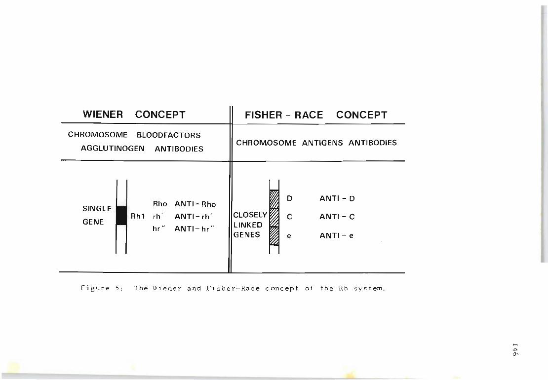
WIENER CONCEPT
CHROMOSOME BLOODFACTORS
AGGLUTINOGEN ANTIBODIES
SINGLE _ Rho ANTI-Rho
GENE _ Rh1 rh' ANTI-rh'
hr" ANTI- hr"
FISHER - RACE CONCEPT
CHROMOSOME ANTIGENS ANTIBODIES
D ANTI- D
"CLOSELY t:'2l C ANTI- C LINKED GENES ~ e ANTI- e
Figure 5: The Wiener and Fisher-Race concept of the Rh system.
...... t+>O'
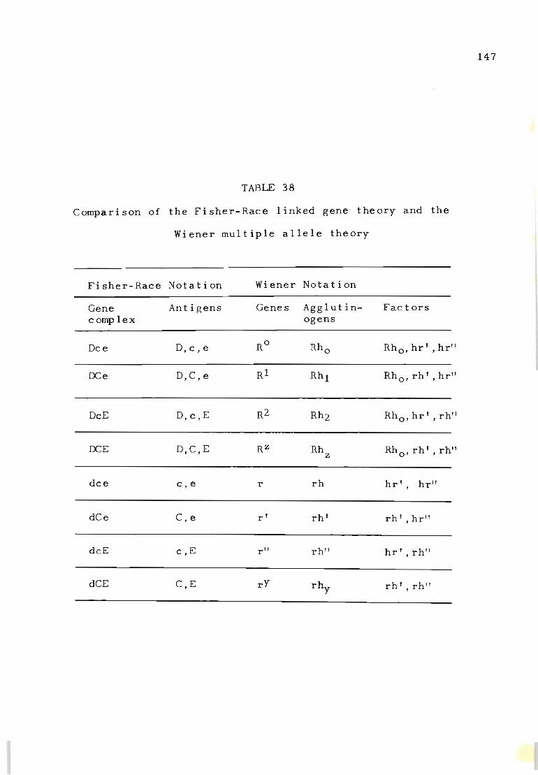
147
TABLE 38
Comparison of the Fisher-Race linked gene theory and the
Wiener multiple allele theory
Fisher-Race Notation Wiener Notation
Gene Antigens Genes Agglutin- Fac tors complex ogens
Dce D,c,e RO Rho Rho, hr' , hr"
OCe D,C,e Rl Rhl Rho, rh' , hr"
DcE D,c,E R2 Rh2 Rho, hr' , rh"
OCE D,C,E RZ Rh Z Rh rh' rh" 0' ,
dce c,e r rh hr' , hr"
dCe C , e r' rh' rh' , hr"
dcE c,E r" rh" hr' ,rh"
dCE C,E rY rhy rh' ,rh"
from the mother during fertilization. For example, all
infants of a CDe/CDe x cde/cde parental combination will
be of the genotype CDe/cde because one CDe chromosome
would have been received from one parent and one cde
chromosome from the other parent. Of the eight basic
types of Rh chromosomes recognized through extensive
family studies (Table 38) each individual is known to
carry a pair of these chromosomes which represents his
genotype. In numerous clinical and anthropological stud-
148
ies carried out throughout the world the Rh gene complex
has always been observed to follow a simple Mendelian pat
tern of inheritance. Table 39 lists some of the Rh gene
complexes known to exist today with the resulting Rh ant
igens they possess.
3. THE MISSING RH ANTIGEN TYPES
In 1950 Race, Sanger and Selwyn described a blood which
failed to react with any Rh antisera except those contain-
ing anti-D. The Rh genotype of the person concerned was
described as D--/D-- and it was considered possible that
the Ce and Ec portions of the Rh gene complex had been
lost by deletion from the Rh chromosome. Since then sev
eral other examples of D--/D-- have been described and
people whose Rh genotype appears to be DCw_/DCw_ have been
reported. In describing this particular Rh genotype Race
and Sanger (1968), for the lack of absolute evidence ,
suggested that it could be the result of (1) a short de-
letion of a small piece of chromosome, or (2) a built in
inhibitor which fails to express the missing antigens.
During routine testing of blood collected for anthrop-
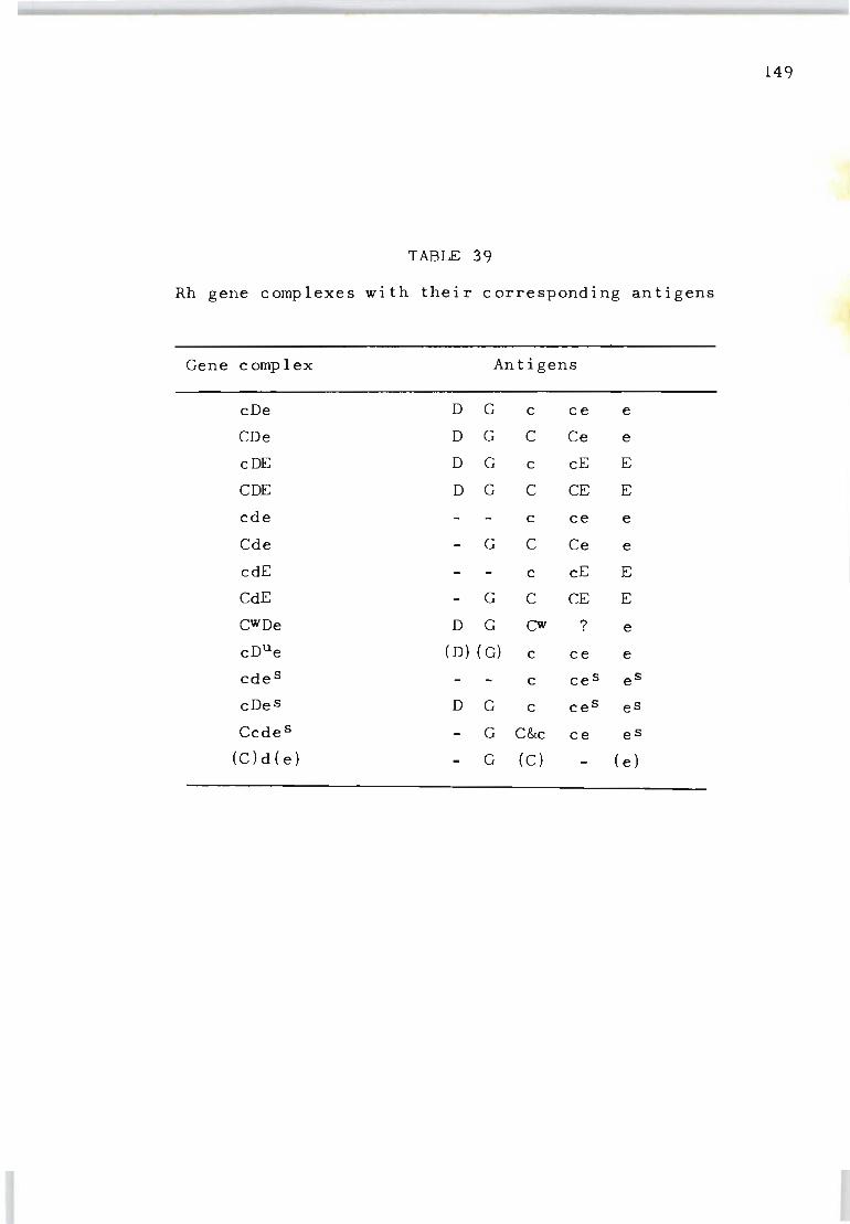
149
TABLE 39
Rh gene complexes with their corresponding antigens
Gene complex Antigens
cDe D G c ce e
CDe D G C Ce e
cDE D G c cE E
CDE D G C CE E
cde c ce e
Cde G C Ce e
cdE c cE E
CdE G C CE E
CWDe D G Cw ? e cDue (D) (G) c ce e
cde s c ce s e S
cDe s D G c ce s e S
Cedes G C&c ce e S
(C)d(e) G (C) (e)
ological purposes from Australian aborigines from the
Western Desert of Western Australia, a sample of blood
was found (Vos, Vos, Kirk and Sanger, 1961) which failed
to react with the following sera: 25 anti-D, 13 anti-C+D
(+G), 13 anti-C (including some of the specificity anti-
Ce and anti-CE), 2 anti-Cw, 10 anti-E, 1 anti-Ew, 3 anti-
150
c +E, 14 an t i - c , 14 an t i - e , 2 an t i - f (an t i - c e) 2 an t i-V
(anti-ces), 2 anti-VS (anti-e s ), 5 sera from immunized D--/
D-- persons, 1 from an immunized DCw_/DCw_ person, 2 from
immunized Dc-Dc- persons and with 7 sera from immunized
Negroes who have made antibody not unlike anti-e (anti
HrS) •
The cells of individuals with such deletions have since
been named 'Rhnull' by Ceppellini (1964) . The view that
the Rhnull genotype is the result of suppression or delet
ion of that part of the chromosome bearing the Rh locus
has been considered (Vos, 1961). Another possibility may
be that Rhnull (---/---) represents basic material remain
ing after loss of products of the Rh genes by one or another
genetic mechanism, but on the other hand, it could repres
ent basic material on which the Rh genes deposit their
produc t s.
4. THE LW ANT I GEN AND THE RHn u 11 PHENOTYPE
When Landsteiner and Wiener (1940) injected the red
cells of Rhesus monkeys into rabbits they observed that
the resulting antibody could, after partial absorption
with human Rh negative cells, react strongly with Rh
positive red cells and weakly with Rh-negative red cells.

The strong positive reactions against Rh-positive red
cells suggested that the specificity was the same as that
of human anti -D. It is now accepted that the reaction of
151
the rabbit antibody to Rhesus monkey red cells is directed
against a different specificity referred to as 'LW' in
honour of Landsteiner and Wiener (Levine, Celano, Wallace
and Sanger, 1963). Levine has also proposed that although
the genes for Rh and LW are inherited independently they
may act on the same basic Rh substance to produce their
respective red cell antigens .
With the discovery of the Rhnull (---/---) blood an
excellent opportunity was provided to determine whether
these red cells also carried the LWantigen. If not, the
question arose of the relationship of the LW antigen to
basic Rh substance.
Parallel studies were undertaken, based on three crit
erla: (a) absorption of guinea pig anti-LW with Rhnull
cells, (b) elution of the antibody and (c) antigenicity
of the Rhnull factor in guinea pigs. Immunization experi-
ments were carried out with Rh-negative and Rhnull red
cells. At three-day intervals each of two series of
guinea pigs received two injections of 2.0 ml of the
bloods. When tested ten days later five guinea pigs in
jected with Rhnull cells failed to produce anti-LW, two
of these five produced an antibody identified as anti-No
Since the Rhnull red cells used for this study were of the
genotype NN the source of this antibody stimulation is ob-
vi ous. Of the four guinea pigs injected with Rh-negative
red cells, three showed very weak anti-LW specificity
when absorbed with Rh-negative red cells. After absorbing
these sera with the Rhnull cells all four gave anti-LW
specificity of stronger activity than that obtained on
absorption with Rh- negative red cells . In contrast to Rh
negative bloods, the Rhnull cells failed to yield eluates
with anti-LW specificity, as shown in Table 40.
The results of our study indicate that when a gene pro-
duces an Rh antigen determinant it almost always produces
LW antigen and that the LW material constitutes unaltered
basic Rh substance which, by a series of mutations in man ,
has evolved into the complex of Rh factors as it exists
today. This theory was mentioned as early as 1952 by
Murray and Clark. In any event it can be concluded that
the LW antigen is associated with all human blood bearing
152
anyone or more of the antigenic determinant groups of the
Rh system (see Chapter XVI) .
5. THE ASSOCIATION OF MULTIPLE PHENOTYPIC ABNORMALITIES
WITH RHnull.
A most important observation with the discovery of the
original Rhnull blood and some 15 or 20 other Rhnull pro-
positi which have now been found, is the failure of some
Rhnull bloods to react with most anti-U or anti-s reagents.
Although there is no evidence for linkage between the gene
loci of Rh determinants and SsU blood group systems, there
may, nevertheless, be a structural relationship between
the antigenic determinants in these two systems .
Examples of blood which fail to react with anti-U re
agents have always been found in Negroes and have so far
not been reported i n Caucas i ans , Mongeloids, Australoids
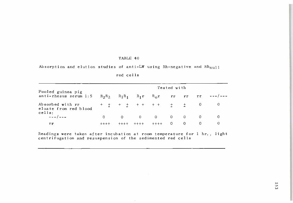
TABLE 40
Absorption and elution studies of anti-LW using Rh-negative and Rhnull
red cells
Tested wi th Pooled guinea pig anti-rhesus serum 1:5 R2R2 R1R1 R1 r Ror rr rr rr ---/---
Absorbed with rr + + + + + + + + + + 0 0 - -eluate from red blood cells:
---/--- 0 0 0 0 0 0 0 0
rr ++++ ++++ ++++ ++++ 0 0 0 0
Readings were taken after incubation at room temperature for 1 hr., light centrifugation and resuspension of the sedimented red cells
..... \.Jl W
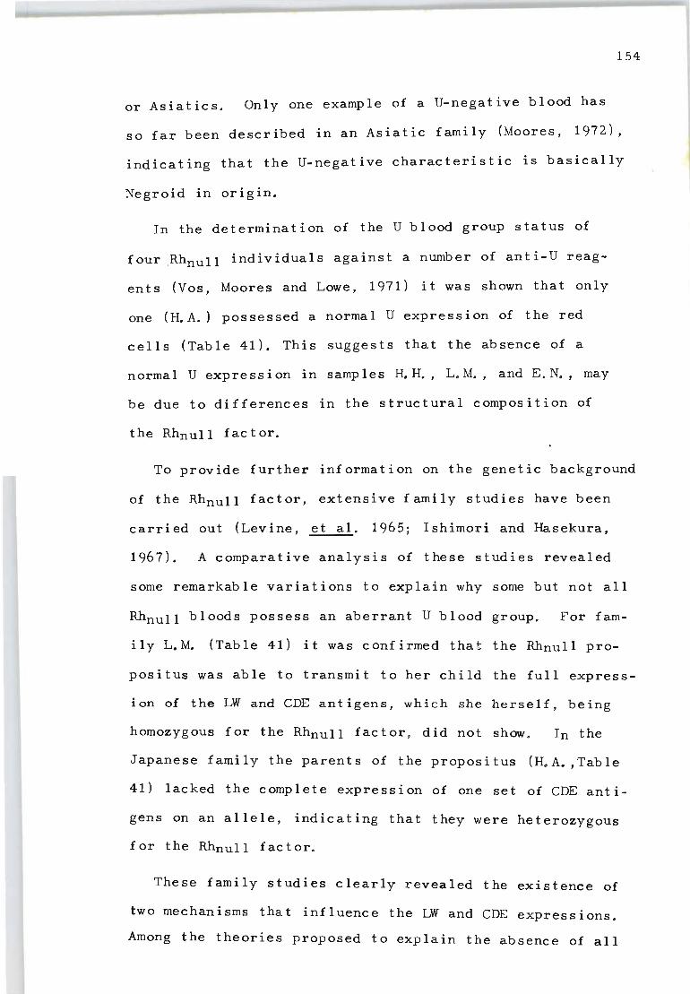
154
or Asiatics. Only one example of a U-negative blood has
so far been described in an Asiatic family (Moores, 1972),
indicating that the U-negative characteristic is basically
Negroid in origin.
In the determination of the U blood group status of
four .Rhnull individuals against a number of anti-U reag
ents (Vos, Moores and Lowe, 1971) it was shown that only
one (H. A.) possessed a normal U expression of the red
cells (Table 41). This suggests that the absence of a
normal U expression in samples H.H., L.M., and E.N., may
be due to differences in the structural composition of
the Rhnull factor.
To provide further information on the genetic background
of the Rhnull factor, extensive family studies have been
carried out (Levine, et al. 1965; Ishimori and Hasekura,
1967 ). A comparative analysis of these studies revealed
some remarkable variations to explain why some but not all
Rhnull bloods possess an aberrant U blood group. For fam-
i I y L. M. (Tab Ie 41) it was c onf i rmed that the Rhnull pro
positus was able to transmit to her child the full express
ion of the LW and CDE antigens, which she herself, being
homozygous for the Rhnull factor, did not show. In the
Japanese family the parents of the propositus (H.A. ,Table
41) lacked the complete expression of one set of CDE anti
gens on an allele, indicating that they were heterozygous
for the RhnuII factor.
These family studies clearly revealed the existence of
two mechanisms that influence the LW and CDE expressions.
Among the theories proposed to explain the absence of all
TABLE 41
Red cell agglutination test of known U-positive and U-negative bloods for various antisera
SsU phenotYT.>e Reactivity of red cells for antisera
Ant i-LW Ant i-S Anti-s An t i-U AHA G.Pig 1172 159 60 385 7 Dub 1. Perth 176 959 31 Mah 128
Control S+s+U+ 4 4 4 4 4 4 4 4 2 2 4 4 3 4 Con t rol S- s+U+ 4 4 0 0 0 4 4 4 2 2 4 4 4 4 Control S+s-U+ 4 4 4 4 3 0 0 0 0 0 4 4 4 4
2671 S- s- U- 4 4 0 0 0 0 0 0 0 0 0 0 0 0 2538 S- s- U- 4 4 0 0 0 0 0 0 0 0 0 0 0 0 1784 S- s- U- 4 4 0 0 0 0 0 0 0 0 0 0 0 0 9484 S- s- U- 4 4 0 0 0 0 0 0 0 0 0 0 0 0 5816 S- s- U- 4 4 0 0 0 0 0 0 0 0 0 0 0 0 7799 S- s- U- 4 4 0 0 0 0 0 0 0 0 0 0 0 0 4166 ~- s- U- 4 4 0 0 0 0 0 0 0 0 0 0 0 0 3836 S- s- U- 4 4 0 0 0 0 0 0 0 0 0 0 0 0 3357 S- s- U- 4 4 0 0 0 0 0 0 0 0 0 0 0 0 4661 S- s- U- 4 4 0 0 0 0 0 0 0 0 0 0 0 0 1458 S- s- U- 4 4 0 0 0 0 0 0 0 0 0 0 0 0 2439 S- s- U- 4 4 0 0 0 0 0 0 0 0 0 0 0 0 Krum S- s- U- 4 4 0 0 0 0 0 0 0 0 0 0 0 0 Amich S- s- U- 4 4 0 0 0 0 0 0 0 0 0 0 0 0 Kamla S- s- U- 4 4 0 · 0 0 0 0 0 0 0 0 0 0 0 Sara S- s- u- 4 4 0 0 0 0 0 0 0 0 0 0 0 0
H. A. S- s+ U+ 0 0 0 0 0 4 4 4 2 2 4 3 3 4 H. H. S- s+U- 0 0 0 0 0 4 0 4 2 0 0 0 0 0 L. M. S- s+U- 0 0 0 0 0 4 0 4 2 0 0 0 0 0 E. N. S- s+U- 0 0 0 0 0 4 0 4 2 0 0 0 0 0
..... LTI LTI
156
Rh antigens on the red cells are (1) chromosomal deletions
(Ishimori and Hasekura, 1967) and (2) the existence of an
independent amorphic gene XOr which blocks the expression
of the Rh genes (Levine et al., 1965). Fig.6 illustrates
a tentative model for the two types of conditions that
can lead to Rhnull expression.
It is difficult to explain why some Rhnull red cells
(samples H.H., L.M., and E.N.) do not react with anti-U
reagents. The fact that the expression of the U antigen
can be altered by the same mechanism that is involved in
the blocking phenomenon of the Rh precursor substance
suggests that the two independent systems Rh and Ssu are
influenced by a single aberration. Schmidt et al., (1967)
proposed that this aberration may be due to the sequential
action of genes controlling shared terminal sugar(s) which
in turn has the capacity to alter the various specificities
depending on the basic precursor substance involved (Wat
kins, 1966).
Race and Sanger (1968) made the important observation
that discrepant anti-S, anti-s and anti-U results against
unrelated Rhnull bloods were not given when saline or al
bumin tests could be used in place of the indirect anti
globulin test. The question thus arises whether the ab
sence of U reactivity in some Rhnull bloods may be assoc
iated with a spatial membrane arrangement of the U antigen
whereby the receptors are not fully exposed above the sur
face.
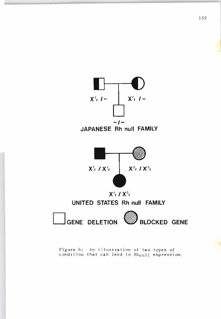
X'r 1- X'r /-
-/-JAPANESE Rh null FAMILY
X'r I X'r
X'r / XOr
UNITED STATES Rh null FAMILY
D GENE DELETION _ BLOCKED GENE
Figure 6: · An illustration of two types of condition that can lead to Rhnull expression.
157
Chapter XIII
THE SPECIFICITY OF ACQUIRED HAEMOLYTIC
ANAEMIA AUTOANTIBODIES
1. INTRODUCTION
If a serum contains a gammaglobulin which will react
with (agglutinate, haemolyse or sensitize) certain genet
ically well-defined red blood cells but not others, it is
158
said to possess an antibody, and the antibody is said to
be specific for a certain property of the red cells. Thus
the specificity of an antibody depends on this selective
reactivity. This simple approach for determining anti
body specificity is often difficult to demonstrate if the
range of activity is widespread and if there are 9nly a
few cells available which lack the property against which
the antibody reacts. A good many blood grouping laborat
ories have in their freezers sera containing antibodies
against 'Public Antigens'. These sera have up to now re
mained unclassified because they react with all available
r ed cells except those of the patient or donor. Nobody
will dispute the antibody nature of these reagents or call
them 'non-specific'. If these 'non-specific' agents
could be shown to be 'specific', that is not to react with
c ertain genetically well-defined red cells, more workers
migh t be persuaded to regard them as true antibodies.
An important feature of the antibodies found in ac
quired haemolytic anaemia of the 'warm' type is their
ability to react with the patient's own red cells in
vivo. Until 1953 when Weiner, Battey, Cleghorn, Marson
and Meynell discovered that such autoantibodie~ possess
definite 'specificity' it was generally thought that the
antibodies reacted with an undetermined antigen.
Using red cells with a wide variety of well~defined
Rhesus phenotypes it was established that about 30 per
cent of patients with acquired haemolytic anaemia of the
'warm' type possess autoantibodies with specificities
directed against several of the known Rhesus antigens on
159
the surface of the red cells. In the other 70 per cent of
patients the specificity of the autoantibodies could not
be identified because they did not share the true charact
eristics of known Rhesus antibodies.
With an added knqwledge of the genetics of the Rh
system from the discovery of Rhnull cell~ (Vos et al . 1961)
and red cells from which the Cc and Ee portions of the Rh
gene complex had been lost (Race, Sanger and Selwyn, 1950)
the nature of non-specific autoantibodies was re-examined.
The autoantibodies from 60 patients suffering from ac
quired haemolytic anaemia were tested against a variety of
cells with common and very rare Rhesus genotypes.
2. MATERIALS AND METHODS
Eluates obtained from the red cells were used rather
than sera because sera often do not contain all the anti
bodies that can be detected in eluates.
The eluates were prepared by the ether elution method
of Vos and Kelsall (1956). They were tested against Cde/
Cde, cdE/cdE, CdE/cde, CDE/CDE, cde/cde KK cells, one
D--/D-- blood, one Dc~/Dc- sample and the original Rhnull
160
blood. The cell panel included the following Rhesus
antigens: 'normal' cells (not deleted: nI), 'partially'
deleted cells (pdl) and 'fully' d~leted cells: (dI). The
tests were performed with positive and negative controls.
Indirect Coombs tests we~e done to confirm doubtful results
but generally the enzyme method proved to be a valuable
qualitative test. None of the eluates produced agglutin-
ation of saline-suspended cells. Most eluates agglutinated
cells suspended in bovine aloumin but the reactions were
much better with enzyme~treated cells and the latter method
was generally used.
Absorptions were done in the following way: 1 volume
of packed red cells (usually unmodified) was incubated
with 1 volume of eluate at 37 0 C for 2 hours. The super-
natant was removed and kept. rhe red cells were then
(without washing) incubated for another 2 hours with 3- 4
volumes of the same fresh eluate and were then eluted.
Titrations were done using doubling dilutions of the
eluates in saline and enzyme-treated cells. 66 eluates
were available in q\lantities large enough for more de
tailed investigation. Six showed 'blood group specificity'
and were excluded. Of the remaining 60, 10 were from
'normal' donors who were found to have 'coated' red cells.
The remaining 50 eluates were from patients SUffering from
typical autoimmune haemolytic anaemia (w<;l.rm type) of vary
ing sever i ty.
3. RESULTS
Weiner (1961a) established that when 'partially' del-
eted cells (D--/D--) were used together with other 'nor-
mal'
were
red cells, the eluates fell into three groups which
called Class 1, Class 2 and Class 3. This classi~
fication depended on whether they reacted as strongly
wit h D- - / D- - cell s as with 'norma l' cell s (C 1 ass 1), more
weakly with the former (Class 2) or not at all with them
(Class 3). This classification was confirmed by us, but
161
a further subdivision was necessary to include the obser
ved reactions obtained with completely deleted (---/--~)
cells. Some Class 1 eluates reacted equally strongly with
---/--- cells (la), others reacted more weakly (lb) and
the rest showed no reactivity with them (lc). Class 2
eluates could be subdivided in the same manner (2a, 2b,2c)
but Class 3 eluates gave only one pattern of reactivity,
i.e. they did not react with the deleted (---/---) cells
at all. Table 42 shows the reactivity of the 60 eluates.
There was no correlation between the class and subdiv
ision of the eluates with the ABO groups or Rhesus geno
types of the patients or donors. Both ABO groups and Rh
genotypes showed the expected distribution. Amongst the
patients there were 28 men and 24 women. Of the I normal'
donors all 10 were men (amongst the donor population from
which these donors were drawn, the ratio of men to women
is almost exactly 1: I).
It would appear that more women than men develop an
antibody which will react with the ---/--- cell, but the
differences shown in Table 43 are statistically not sig
nificant. This table also shows that of the 60 eluates
examined, 32 failed to react against the ---/--- cell and
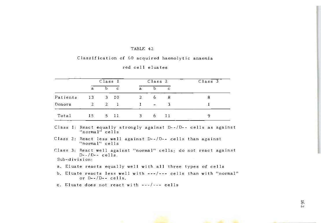
TABLE 42
Classification of 60 acquired haemolytic anaemia
red cell eluates
Class 1 Class 2 Class 3
a b c a b c
Patients 13 3 10 2 6 8 8
Donors 2 2 1 1 3 1
Total 15 5 11 3 6 11 9
Class 1: React equally strongly against D--/D-- cells as against " norma 1" cell s .
Class 2: React less well against D--/D-- cells than against "normal" cells
Class 3: React well against "normal" cells; do not react against D- - / D- - cell s .
Sub - d i vis i on:
a. Eluate reacts equally well with all three types of cells
b. Eluate reacts less well with ---/--- cells than with "normal" or D--/D-- cells.
c. Eluate does not react with ---/--- cells
I-' erN

Total
Fa t i en t s
Donors
TABLE 43
Distribution of the various classes of eluates
between male and female, patients and donors
Class 1 Class 2 Class 3
a b c a b c
(Male 6 3 7 2 1 10 7
~Female 8 2 5 1 5 1 2
(Male 4 1 6 1 1 7 6
~Female 8 2 5 1 5 1 2
2 2 1 1 0 3 1
...... '" \..oJ
11 eluates showed less activity against these cells than
'normal' cells. Thus 43 (71 per cent) eluates did not
react, or reacted weakly against ---1--- red cells when
compared with the reactivity against 'normal' red cells.
This pattern of reactivity suggested a mixture of anti
bodies in the 'reacting' eluates which was confirmed by
absorption and re-elution experiments.
A few typical examples are given. Eluate B.A. B. (Table
44) gave the following reactions: ++++ against normal
cells, ++++ against the D--/D-- cell and +++ against the
---1--- cell. When this eluate was absorbed with ---1--cells the antibody against ---1--- was taken out but the
reactivity against normal cells and D--/D-- was almost
identical with the pre-absorption activity. A further
absorption with D--/D-- left an antibody in the supernat
ant which reacted only with 'normal' cells. Re-elution
of the antibodies from the absorbing red cells produced
reagents which had the expected reactivity. Table 45
shows a similar absorption experiment on eluate L.A.
Non- reactivity of certain eluates against D--/D-- and/
or ---/--- red cells might have been thought to be the
result of quantitative rather than qualitative factors.
However, the results demonstrate that the classes of the
eluates obtained were independent of the titres of the
eluates against 'normal' cells. Scores of different el-
uates against different red cells are shown in Table 46.
It can be seen that the scores (or titres) against normal
cells have no influence on the class or subdivision of
the eluate. In Class 3, for instance, scores range from
164
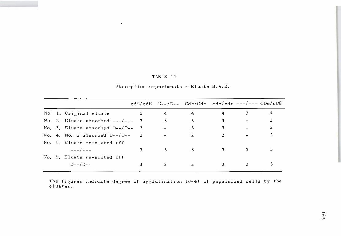
TABLE 44
Absorption experiments - Eluate B.A.B.
cdE/cdE D--/D-- Cde/Cde cde/cde ---/--- CDe/cDE
No. 1. Original eluate 3 4 4 4 3 4
No. 2. Eluate absorbed ---/--- 3 3 3 3 3
No. 3. Eluate absorbed D--/D-- 3 3 3 3
No. 4. No. 2 absorb~d D--/D-- 2 2 2 2
No. 5. Eluate re-eluted off
---/--- 3 3 3 3 3 3
No. 6. Eluate re-eluted off
D--/D-- 3 3 3 3 3 3
The figures indicate degree of agglutination (0-4) of papainized cells by the eluates.
.... '" IJl
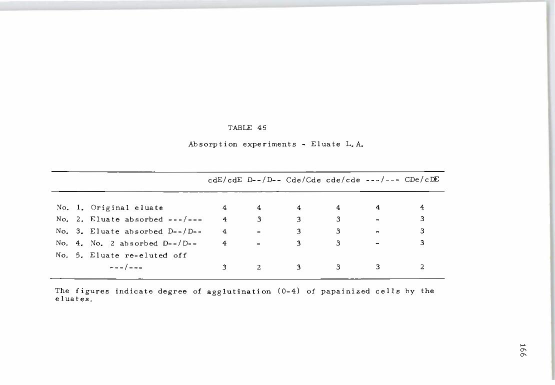
TABLE 45
Absorption experiments - Eluate L.A.
cdE/cdE D--/D-- Cde/Cde cde/cde ---/--- CDe/cDE
No. 1. Original eluate 4 4 4 4 4 4
No. 2. Eluate absorbed ---/--- 4 3 3 3 3
No. 3. Eluate absorbed D--/D-- 4 3 3 3
No. 4. No. 2 absorbed D--/D-- 4 3 3 3
No. 5. Eluate re-eluted off
---/--- 3 2 3 3 3 2
The figures indicate degree of agglutination ( 0-4) of papainized cells by the el uates.
..... 0' 0'
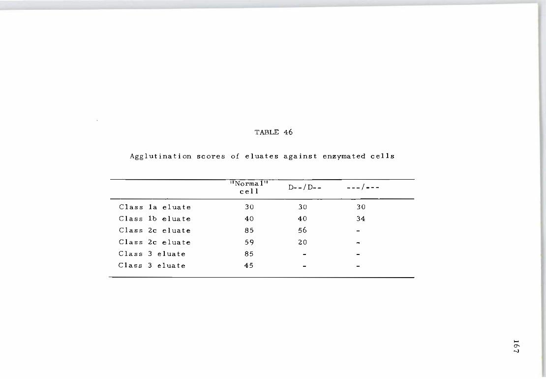
TABLE 46
Agglutination scores of eluates against enzymated cells
"Normal" D- -/D-- ---/---cell
Class Ia eluate 30 30 30
Class Ib eluate 40 40 34
Class 2c eluate 85 56
Class 2c eluate 59 20
Class 3 eluate 85
Class 3 eluate 45
..... C1' -.J
45 to 85 and in other classes conditions were similar.
4. DISCUSSION
It was established that the eluates of cells from pat ..
ients with AHA and from 'normal' donors contained three
different types of antibodies. The red cell antigens re-
active with these antibodies are listed in Table 47. The
results suggest that the eluates may contain anti-nl,
anti-pdl, and anti-dl, singly or in combination.
168
It was found that one person may form one antibody
whereas another may form two or three against related
antigens. This is common in people developing antibodies
within the Rhesus system (e.g. anti-D plus am i-C and anti
G). It appears that all 'normal' cells contain the antigen
designated as 'nl'. This may be produced by a gene on
the Ee locus as it is · absent from the D--/D-- and the
Dc-/Dc- cells. Or it may be a 'composite' antigen whose
production depends on the presence of a full complement
of 'normal' genes. Its absence from the partially del
eted cells could be explained by either hypothesis. The
anti-pdl antibody may react with an antigen produced by
a gene near the D locus, nearer in fact than the C gene,
as both D--/D-- and Dc-/Dc- carry it. It is not a part
of th~ D antige~ as it can be found on D negative cells
in the same strength as on D positive cells. As far as
we know the 'dl' antigen has a universal distribution.
The factors determining the type of antibody which is
formed in different patients are not known. The severi ty
of the illness has apparently no bearing on this question
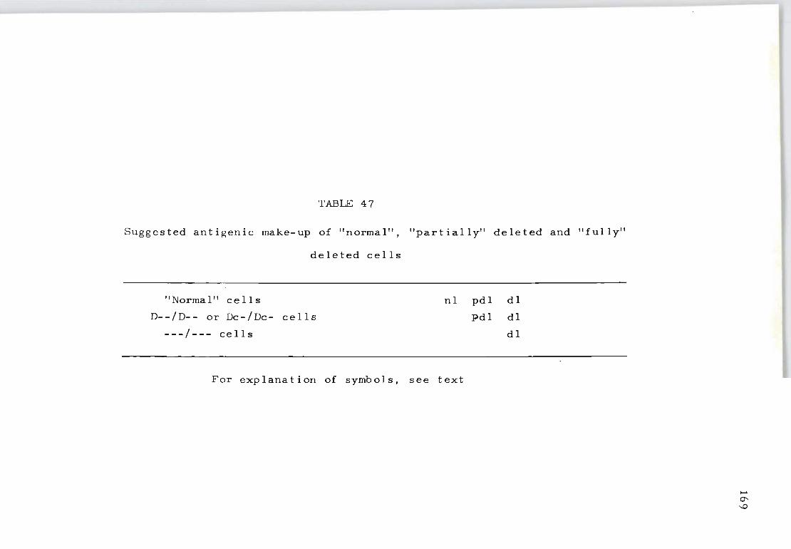
TABLE 47
Suggested antigenic make-up of "normal", "partially" deleted and "fully"
deleted cells
"Norma I" cell s
D--/D-- or Dc-/Dc- cells
nl pdl dl
Pdl dl
---/--- cells dl
For explanation of symbols, see text
I-'
0' -.D
as all types of eluates were found in all types of pat
ients, even 'normal' donors. Similarly, the genotype of
the patient or donor has no bearing on the development
of a particular class of antibody. At present it is un-
170
explained why some people form an antibody, eog. against
c or e antigens and others against the antigens described
in this study, though all patients or donors who were in
vestigated carried the latter antigens on their cells.
Investigation of antibodies contained in the eluates
of patients and 'normal' donors has demonstrated three
antigens within the Rh system, antigens which seem to be
additional to those recognized by the commonly used Rh
antisera. They have pemonstrated that the 'fully deleted
cell' still carries an antigen. It is not known whether
this antigen is the 'ground substance' of the Rh system
or part of the complex Rh system necessary for the ex
pression of other Rh antigens.
Chapter XIV
THE SEROLOGICAL CHARACTERISTICS OF ACQUIRED
HAEMOLYTIC ANAEMIA AUTOANTIBODIES
1. INTRODUCTION
171
The present study was designed to determine the serol
ogical characteristics of the various autoantibodies
found in patients with AHA. Using normal erythrocytes
and Rhnull erythrocytes the autoantibody specificity was
studied and the reactivity defined both with the antiglob
ulin test and erythrocytes treated with a proteolytic
enzyme (f ic in) . A further subject for study was the eval-
uation of complement components on the erythrocytes of
patients with warm AHA autoantibodies. The presence of
both IgG and complement components in erythrocyte eluates
was defined with the antiglobulin test using monospecific
antiglobulin sera (anti-IgG and anti-complement). From
these results a hypothesis for the apparent discrepancy
in the complement fixing properties of Rh isoantibodies
and the autoantibodies found in AHA of the 'warm' type
is presented.
2. MATERIALS AND METHODS
Selection of patients depended upon a positive direct
antiglobulin test of the red cells and the haematological
association of an autohaemolytic anaemia of the 'warm'
type. Of the 24 patients on whom red cell eluate studies
could be performed only 12 were available for a detailed
investigation of the serum as well as the red cells.
To avoid in vitro sensitization of the red cells due
172
to cold antibodies on storage at 50 C, separation of the
red cells from the defibrinated blood was performed on
the same day as collection. For the preparation of el
uates red cells were washeo four times with 20-40-fold
volumes of isotonic sodium chloride solution (0.85%) and
stored at -20 0 C for varying periods as a 50% concentrat
ion of packed red cells in saline.
Antibody elution - To recover autoantibodies from the
patient's in vivo sensitized red cells the ether elution
procedure of Vos and Kelsall (1956) was used. The only
modification to the published method was that ether was
added directly to the 50% suspension of packed washed
red cells. At no stage before or after the elution pro-
cedure was fresh human serum added to the suspension of
haemolyzed red cells.
Antiglobulin test ~ The antiglobulin test was performed
on a translucent white rectangular tile as described by
Dacie and Lewis (1963). A 'broad spectrum' commercial
anti-human serum capable of detecting IgG and complement
components was used. A specific anti-IgG was made by ab-
sorbing the broad spectrum anti-human globulin serum with
cells sensitized with incomplete Lea (IgM) antibody and
complement components. An 'anti-non-gamma' reagent was
made by adding purified human IgG globulin to the broad
spectrum serum as described by Dacie (1962). In the
more recent experiments an antiserum to comp.lement com-
ponents C3 and C4 was used. Parallel experiments using
the anti-complement and 'anti-non-gamma' serums showed
similar reactivity to that reported previously by Harboe
173
et al. (1963) and Leddy and Bakemeier (1967).
Ficin test - For the enzyme test a solution of 1% comm
ercial ficin in isotonic sodium ch19ride solution and
buffered to pH 7.2 with Sorensen buffer was prepared. To
detect antibodies one drop of this enzyme solution was
added to one drop of serum or red cell eluate, followed
by one drop of a 5% suspension of known red cells. At
no stage were the red cells pre-treated with this enzyme
for the recognition of ficip-reactive antibodies. The
enzyme agglutination tests were performed on translucent
tiles and left in a damp chamber at room temperature
(22 0 C) for one hour. Agglutinations were read macros
copically with a 5 x magnification head fitting eyepiece.
Red cells useq - To detect the presence of known anti
bodies a commercial panel of red cells was used. Anti
body absorption and elution studies were performed with
DCe/DcE and Rhnull red cells, each obtained from a single
donor.
Definitions Rf antibody specificity - In acquired haem~
olytic anaemia it is not upusual to find more than one
specific autoantibody. Antibodies characterized in this
study were defined as detailed in Chapter XIII.
(a) Antibodies for well~defined red cell antigens: ~
These antibodies show distinct specificity against
any of the well-defined blaod graup antigens such as C,c,
E,e,Kell, Fya, Lea etc. Since undiluted serum .or eluate
same times reac ted wi th all narmal ce 11 s (i. e. thase wi th
aut ~h gene deletians), absorptian studies were necessary
ta demanstrate such specificity.
(b) Antibodies reactive with 'normal' red cells (anti
nl): Anti-nl antibodies are defined as those that
react with normal human red cells possessing the common
Rh phenotypes ce/ce-, OCe/ce, DcE/DcE etc. but fail to
react with the fully deleted Rhnull red cells (---/---).
Since partially deleted red cells of the phenotype D--/
174
D-- were not available for this study, it is highly prob-
able that some antibodies for normal red cells could
also contain anti-pdl (Weiner and Vos, 1963).
(c) Antibodies reactive with Rhnull red cells (anti-dl):
Anti-dl antibodies are defined as those reacting with
Rhnull erythrocytes. The Rhnull bloods used were obtain
ed from three sources: (1) Mrs.E.N. (the original Rhnull
Australian Aborigine, Vos et al. 1961); (2) the Japanese
Rhnull (Ishimori et al. 1966); (3) a white Californian
Rhnull (P. Sturgeon, unpublished). Serum or red cell el-
uates reactive with normal cells and with Rhnull red cells
were absorbed using equal volumes of serum or eluate
against washed packed RhnuII red cells. If the ant ibody
was completely removed by these red cells the specificity
was considered to be anti-dl. If the RhnuII cells re-
moved the antibody against Rhnull cells but left an anti-
body against normal cells the antibody in the serum or
red cell eluate was considered to be a combination of
anti-nl and anti-pdl.
Differentiation of ficin and antiglobulin-reactive auto~
antibodies - AHA autoantibodies can be detected by sero-
logical techniques such as the antiglobulin and ficin
tests (Dacie, 1962). These techniques can also be used

to further characterize AHA autoantibodies of multiple
specificity. For example, should an AHA autoantibody
sensitize normal red cells by the antiglobulin as well
as the ficin test, and Rhnull red cells by the ficin
test only, then the first procedure is to absorb this
serum or eluate with ficin ... treated Rhnull red cells.
Subsequent tests may then show that the absorbed serum
175
or eluate still contains the antiglobulin-reactive anti
body for normal red cells but not the ficin-reactive anti
body for Rhnull red cells. Using this mode of testing a
clear differentiation of the type of serological reactiv
ity involved can be established.
3. RESULTS
The specificity of antibodies found in the serum and
red cell eluates of 12 patients with AHA is shown in
Tab Ie 48. Antibodies reactive for well-defined red cell
antigens (anti-e, anti-E etc.) are at times observed in
the serum. In this series such antibodies were not found
in the e I ua t e s • Since antibody activity in an erythro-
cyte eluate more strongly implicates it in the pathogen
esis of the disease than antibody activity which is pres.
ent only in the serum, emphasis was placed on those anti
bodies present both in serum and eluates.
Investigations were carried out to record how often
anti-nl and anti-dl autoantibodies could he detected as
ficin or antiglobulin-reactive varieties or a combination
of both.
Table 49 shows that autoantibodies detectable only by
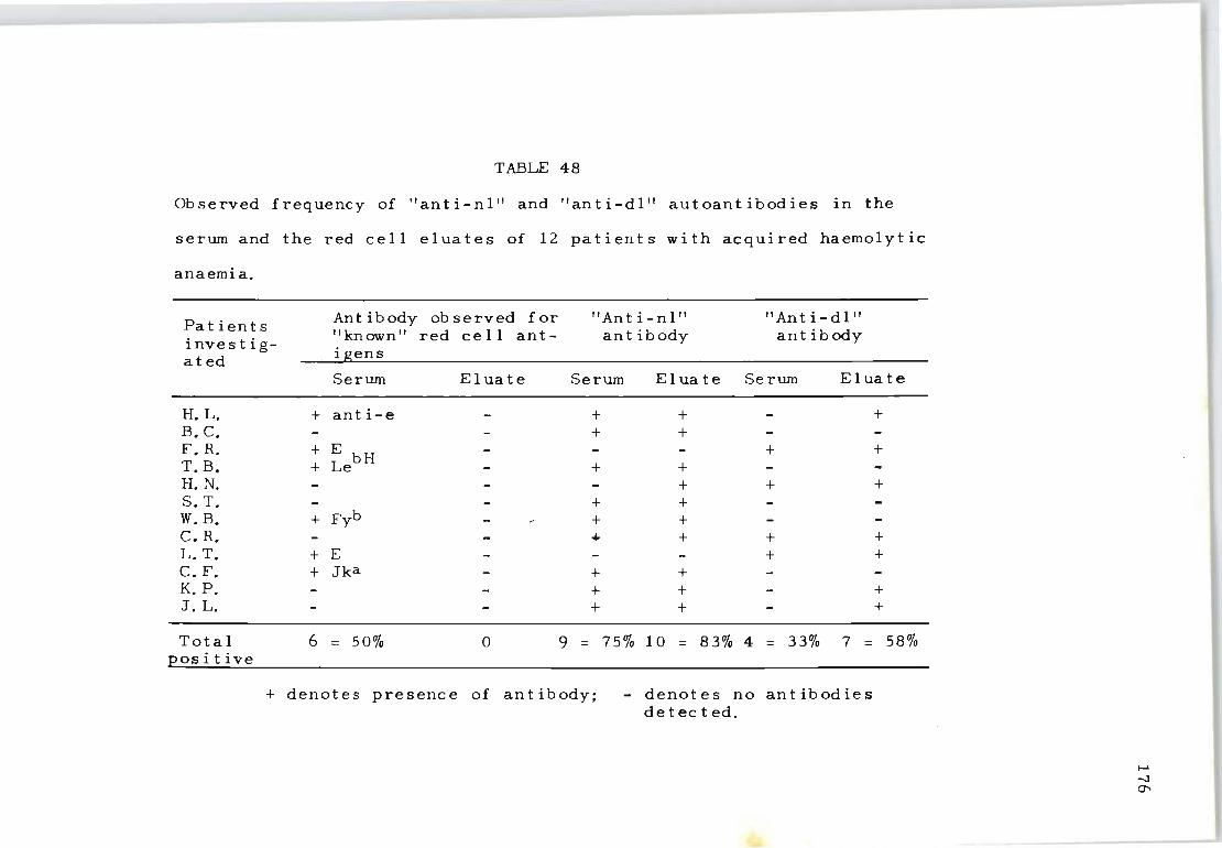
TABLE 48
Observed frequency of "anti-nl" and "anti-dl" autoantibodies in the
serum and the red cell eluates of 12 patients with acquired haemol yt ic
anaemia.
Patients Ant ibody ob served for " An t i - n I " "Anti-dl"
investig- "known" red cell ant- ant ibody ant ibody
ated igens
Serum Eluate Serum Eluate Serum Eluate
H. L. + anti-e + + + B. c. + + F. R. + E bH + + T. B. + Le + + H. N. + + + S. T. + + w. B. + Fyb + + c. R. + + + + L. T. + E + + c. F. + Jka + + K. p. + + + J. L. + + +
Total 6 = 50% 0 9 = 75% 10 = 83% 4 = 33% 7 = 58% positive
+ denotes presence of antibody; - denotes no antibodies detected.
..... -.J 0"
!
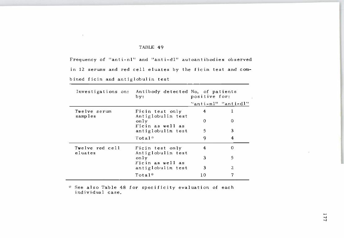
TABLE 49
Frequency of "anti-nl" and "anti-dl" autoantibodies observed
in 12 serums and red cell eluates by the ficin test and com-
bined ficin and antiglobulin test
Investigations on:
Twelve serum samples
Twelve red cell eluates
Antibody detected No. of patients by: positive for:
"anti-nl" "an t i-dl"
Fic in test only 4 1 An t i glob u lin test only 0 0 Ficin as well as an t i glob u lin test 5 3
Total ':' 9 4
Ficin test only 4 0 An t i glob u lin test only 3 5 Ficin as well as ant i glob u lin test 3 2
Total':' 10 7
* See also Table 48 for specificity evaluation of each individual case.
--..] -..]
the antiglobulin te~t were not found in the serum but
were frequeptly present in the red ~ell eluates.
178
Table 50 shows the frequency of anti-nl and anti-dl
autoantibodies detected by the ficin and indirect anti
globulin tests on the same 24 red cell eluates. The
Rhnull red cells from three donors gave similar results
and were mo~e often reactive by the antiglobulin test
than by the ficin test. No marked differences were ob
served between the ability of the ficin or the antiglob
ulin test to recognize the autoimmune anti.nl antibodies.
To determine whether this is because patients cannot pro
duce at one time two different antibodies reactive by
the same serological technique, the data were analyzed
to determine how often ficin-reactive anti-nl could be
found in the red cell eluates together with ficin-react
ive anti-dl. Tables 51 and 52 detail this analysis for
combinations possible considering the two different auto-
antibodies and the two testing procedures. Table 51 in-
dicate·s that the red cell eluates possessing strong ficin
reactive anti-nl very seldom appeared to have strong
ficin-reactive anti-dl. Table 52 shows that antiglobulin.
reactive anti-nl, together with antiglobulin-reactive
anti .. dl was also uncommon. In contrast, Tables 51 and
52 show that strong £icin-reactive anti-nl and strong
antiglobulin-reactive anti .. dl, or strong antiglobulin
reactive anti-nl and strong ficin-reactive anti-nl were
combinations of autoantibodies frequently recovered in
eluates. The simultaneous recovery of anti-nl and anti
dl antibodies which were stropgly reactive by the anti-
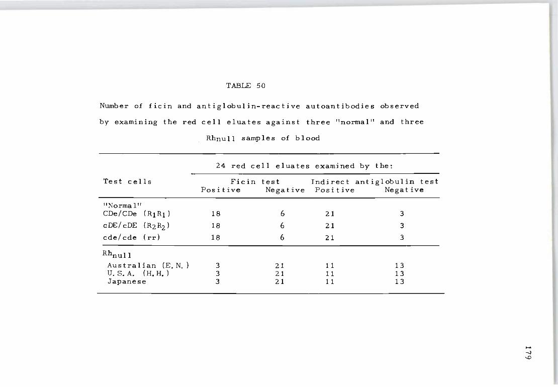
TABLE 50
Number of ficin and antiglobulin-reactive autoantibodies observed
by examining the red cell eluates against three "normal" and three
Rhnull samples of blood
24 red cell eluates examined by the:
Test cells Ficin test Indirect antiglobulin test Pos it ive Negative Positive Negative
"Normal" CDe/CDe (Rl Rl ) 18 6 21 3
cDE/ cDE (R2 R2 ) 18 6 21 3
cde/cde ( rr) 18 6 21 3
Rhnull
Australian (E. N. ) 3 21 11 13 U. S. A. (H. H. ) 3 21 11 13 Japanese 3 21 11 13
..... -J ..0
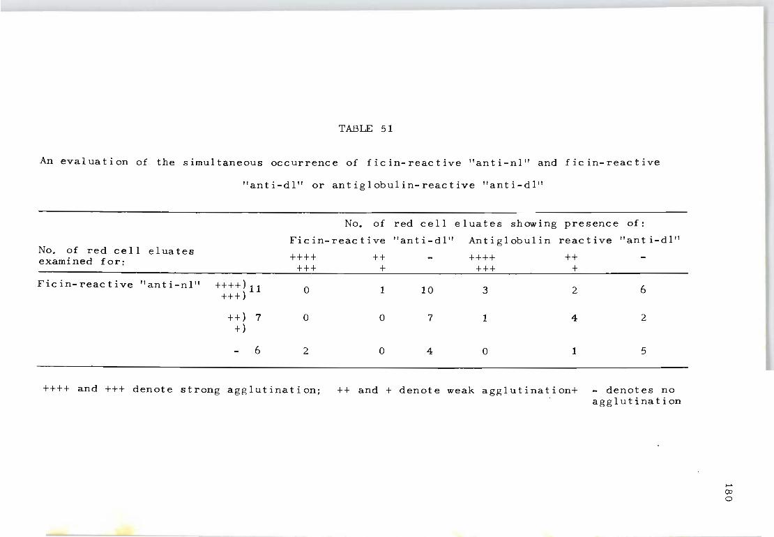
TABLE 51
An evaluation of the simultaneous occurrence of ficin-reactive "anti-nl" and ficin-reactive
"anti-dl" or antiglobulin-reactive "anti-dl"
No. of red cell eluates showing presence of:
Ficin-reactive "anti-dl" An t i glob u 1 in reactive "anti-dl" No. of red cell eluates examined for: ++++ ++ ++++ ++
+++ + +++ +
Ficin-reactive "anti-nl" ++++)11 0 1 10 3 2 6 +++)
++ ) 7 0 0 7 1 4 2 +)
6 2 0 4 0 1 5
++++ and +++ denote strong agglutination; ++ and + denote weak agglutination+ - denotes no agglutination
..... (Xl
o
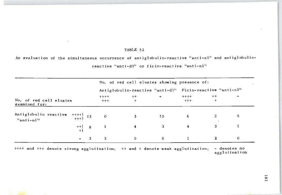
TABLE 52
An evaluation of the simultaneous occurrence of antiglobulin-reactive "anti-nl" and antiglobulin-
No. of red cell eluates examined for:
Antiglobulin reactive
"an t i - n I"
++++) +++)
++) +)
13
8
3
reactive 1'anti-dl" or ficin-reactive "anti-nl"
No. of red cell eluates showing presence of:
Ant ig 1 obul in- reac t i ve "ant i-dl" Fic in- reac t i ve "ant i -nl"
++++ +++
o
1
3
++ +
3
4
o
10
3
o
++++ +++
6
4
1
++ +
2
3
2
5
1
o
++++ and +++ denote strong agglutination; ++ and + denote weak agglutination; - denotes no agglutination
.... (XI
.....
globul in test was uncommon. Thus it was distinctly
unusual to demonstrate both autoantibodies by a single
serological technique.
In an experiment designed to dete~mine the frequency
182
of complement activity in relation to a number of other
parameters so far mentioned it could be shown that com
plement components appeared only in those red cell eluates
which also revealed strong ficin-reactive autoantibodies.
In addition, strong ficin~reactive autoantibodies were
often strongly reactive in the indirect antiglobulin
test using anti-IgG and generally possessed a high incid
ence of both anti-nl and anti-dl autoantibodies, the
latter being uncommon among the weak ficin-reactive auto-
an t i b od i e s . It was also observed that the occurrence of
complement components in the red cell eluates is not nec
essarily associated with the presence of antibodies
strongly reactive in the indirect antiglobulin test using
ant i- IgG. Thus, using anti-complement sera nearly the
same percentage of eluates reactive for the indirect
antiglobulin test was found whether the eluates gave a
strong or weak positive reaction with anti-IgG.
A much better correlation was therefore evident be
tween the occurrence of complement and a strongly posit
ive ficin test. Table 53 demonstrates that six out of
eight red cell eluates containing a combination of both
anti-nl and anti-dl activity possessed complement compon
ents. In contrast, none of the 16 eluates with either
specific anti-nl or specific anti-dl activity (but not
both) contained complement components. Thus all the red
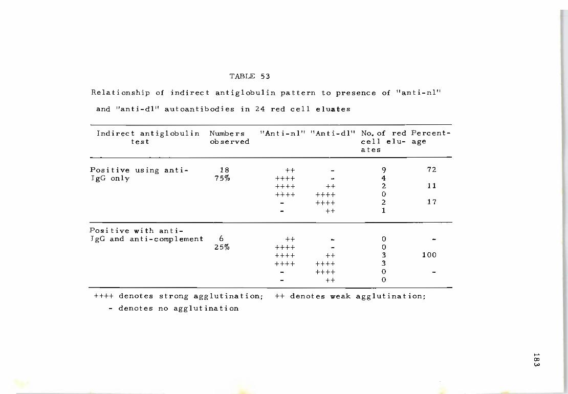
TABLE 53
Relationship of indirect antiglobulin pattern to presence of "anti-nl"
and "anti-dl" autoantibodies in 24 red cell eluates
Indirect antiglobulin test
Nurnbe rs ob served
"Anti-nl" "Anti-dl" No. of red Percentcell elu- age
Positive using anti- 18 IgG only 75%
Positive with anti-IgG and anti-complement 6
25%
++++ denotes strong agglutination;
- denotes no agglutination
++ ++++ ++++ ++ ++++ ++++
++++ ++
++ ++++ ++++ ++ ++++ ++++
++++ ++
ates
9 4 2 o 2 1
o o 3 3 o o
72
11
17
100
++ denotes weak agglutination;
~
co w
cell eluates containing complement components had both
anti-nl and anti-dl activity.
4. DISCUSSION
This investigation was carried out to characterize
autoantibodies found in the serum and on the red cells of
patients with acquired haemolytic anaemia of the 'warm'
type. Antibodies of varying specificity were found in
cluding those specifically reactive against well-.defined
red cell antigens (anti-e, anti-E), against all normal
red cells (anti~nl) and against Rhnull red cells (anti
dl). Antibodies against well-defined red cell antigens
were found in the serum only and not in the red cell
eluates, suggesting that in thls series such antibodies
were isoantibodies resulting from blood transfusion.
In contrast, anti-nl and anti-dl autoantibodies were
often detected in the red cell eluates as well as in the
184
serum; they were observed either singly or in combination
(anti-nl 54%, anti-nl plus anti-dl 33%, anti-dl 13%). Such
antibody specificity was best delineated using both the
an t i g I obul in t est and the f ic in t es t. Some autoantibodies
were detected only by one or the other of these techniques.
This is analogous to some Rh isoantibodies since on rare
occasions Rh isoantibodies can be identified only by serol
ogical techniques utilizing erythrocytes treated with pro
teolytic . enzymes but not by the antiglobulin test (Vas et
al. 1962; Kornstad et al. 1960) or conversely, only by
the latter procedure but not the former (Lucia et al. 1960:
Viessen et al. 1964). This confirms Race and Sanger's
(1954) observation that Rh antibodies can exist which are
185
sharply separated from each other both by the nature of
their antigenic specificity as well as by the technique
needed for their demonstration. The latter distinctions
are presumably explained by subtle differences in anti
body molecular structure even within a given major immuno-
globul in class. Hence, antibody formation in AHA could
be the result of sequential immunization which closely
resembles the manner in which different combinations of
specific Rh-isoantibodies are produced. For example, re-
peated episodes of immunization by DCe cells can result
in the production of Rh-antibodies commencing from anti-D
alone to anti-D and separable anti-C and then to a combin
ation of anti-D and anti-CD (anti-G) (Vos, 1960). This
might be analogous to the formation of anti-nl, anti-dl
and then combined anti-nl and anti-dl.
Another aspect of this study relates to the finding
that AHA autoantibodies frequently fix complement to ery
throcytes in vivo (Dacie, 1962). Red cell eluates may
contain not only antibody but also components of comple
ment which can be detected by the indirect antiglobulin
test using an anticomplement reagent (Leddy, et al. 1965;
Eyster et al. 1967; Petz, et al. 1968). It appears that
such components of complement are fixed to the antibody
and are carried with it in the eluate. Since in this
study a source of fresh complement was not added during
the incubation period of the indirect antiglobulin test,
the antibody in the elua t e did not fix complement to the
red cells at that time .
Leddy and Bakemeier (1967) considered the possibility
186
that non-complement-fixing autoantibodies were 'Rh-related'
and that complement-fixing autoantibodies were 'non-Rh-
related'. In support of this suggestion they found that
the eluates from AHA red cells with a positive direct
antiglobuljn reaction for both IgG and complement usually
contained autoantibodies reactive with Rhnull cells whereas
eluates from red cells that had a direct antiglobulin test
positive for IgG only usually lacked reactivity with Rhnull
cells. Results obtained with 24 eluates indicate, however,
that anti-nl autoantibodies may be present with anti-dl ,
autoantibodies and that when both are present complement
components will also be detectable in the eluate, whereas
with anti-dl autoantibodies alone complement components
need not necessarily be found.
Since there is ample precedent for the fixation of
complement components by multiple Rh isoantibodies (Harboe
et al. 1963; Rosse, 1968) and since the absorption studies
revealed multiple autoantibodies in those eluates contain-
ing complement components, there would seem to be no need
to propose a 'non-Rh-related' autoantibody when complement
fixation is present (Leddy et al. 1967). Hence it is pre-
ferred to hypothesize that the presence of complement com-
ponents in association with a combination of anti-nl and
anti-dl autoantibodies is analogous to complement fixation
produced by mUltiple Rh isoantibodies. In considering
these autoantibodies of mUltiple specificity the more fre-
quent occurrence of anti-nl (Table 53) suggests that this
autoantibody might indicate an early state of autoantibody
production. The progressive formation of anti-nl with
anti-dl which then tends to result i~ the fixation of
complement on the eryth~ocytes may well be a natural pro
gression to a more advanced ~tate of autoimmunization
comparable with the development of Rh iso~ntibodies where
as a rule anti-D is more often noted in the early stages
of isoimmu~ization than ~nti-CD.
187
Chapter XV
THE IMMUNOGLOBULIN CHARACTERISTICS OF
ACQUIRED HAEMOLYTIC ANAEMIA AUTOANTIBODIES
1. INTRODUCTION
188
The red cells of patients with acquired haemolytic
anaemia of the 'warm' type are known to be coated with
immunoglobulins which not only represent various specific
ities of autoantibodies but also structural differences
in the physicochemical composition of the autoantibody
(Weiner and Vos, 1963; Vos, Petz and Fudenberg, 1970;
Dacie and Worlledge, 1969; Leddy, Peterson, Yeaw and Bak
emeier, 1970). Therefore the activity of the antibody can
sometimes only be determined by using antisera specific
against a variety of immunoglobulins.
It has also been shown that some patients may form a
mixture of specific autoantibodies, some components of
which may react with all, or almost all, human red cells.
Although such antibodies appear to lack specificity, in
dividual specificities can often be demonstrated by sel
ective absorptio~ and elution studies using rare red cell
antigens. In this way it was shown that the autoantibod-
ies recovered from the red cells may possess different
specificities against one or another well-defined red
cell antigen closely associated with the Rh system (see
Chap t er XI II).
This study was designed to determine the incidence of
IgG, IgM or IgA coating the red cells of 55 patients with
acquired haemolytic anaemia of the 'warm' antibody type.
It was important to establish whether one or more aut o-
189
antibodies specific for each red cell antigen determinant
found in erythrocyte eluates was correlated with the pres
ence of more than one immunoglobulin class of antibody and
wi th the presence of complement components.
2. MATER IALS AND METHODS .
Fifty-five consecutive patients with autoimmune haem
olytic anaemia of the 'warm' type, as defined by haematol
ogical and serological criteria (Dacie, 1962) and with
strongly positive dire~t Coombs' tests, were studied.
Red cells were s,eparated from defibrinated blood immed
iately after collection. Tpe cells were then washed four
times' with 40-fold volumes of isotonic sodium chloride
solution (0.85%) and stored at _20 0 C for varying periods
as a 50% concentration of packed red cells in saline.
Reagents - Monospecific anti-IgG, ~nti-IgM and IgA were
used. Specificity of these reagents was determined by
haemagglutination techniques and not by immunoelectrophor
esis. The reaction patterns of these antisera conformed
with the findings of Engelfriet et aI, (1968). The anti
complement serum used was prepared as described by Harboe
et al. (1963). Previous experiments revealed that the
antiserum was specific for complement components C3 and
C4.
Red cells used - To detect known antibody systems a sel
ected panel of red cells was used. Antibody absorption
and elut~on studies were performed with CDe/cDE red cells
partially deleted red cells of the phenotype -D-/-D- and
,
fully deleted Rhnull ---/--- red cells. The Rhnull bloods
used were obtained from the original Rhnull Australian
Aborigine and the Japanese Rhnullo
190
Absorptions - Red cell eluates reactive with normal cells,
partially deleted cells and fully deleted Rhnull red cells
were absorbed using equal volumes of eluate against washed,
packed Rhnull red cells. If all the antibody was completely
removed by these red cells the specificity was considered
to be 'anti-dl'. If the Rhnull cells removed the antibody
against Rhnull cells but left an antibody that still re
acted with normal and partially deleted red cells a second
absorption was carried out using partially deleted cells.
Removal of all antibody activity for partially deleted
cells indicated that the eluate also contained 'anti-pdl'.
If the eluate continued to show antibody activity for
normal red cells the specificity was considered to be
'anti-nl'. Using this method of abscrption a clear diff
erentiation of the type of specific antibody or antibodies
present could be established.
3. RESULTS
Three specific varieties of autoantibodies were recov
ered from the red cells of patients with acquired haemoly
tic anaemia. These antibodies were classified in accord
ance with the antigenic make-up of a number of selected
red cells as shown in Table 47. (page 169).
55 red cell eluates were examined for the presence of
one or more varieties of autoantibodies. As shown in
Table 54, antibodies with a single specificity (anti-nI,
or anti-pdl or anti-dl) were more often present (63%) in
the eluates than antibodies with mUltiple specificities
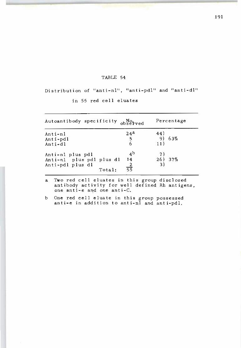
TABLE 54
Distribution of "anti-nl", "anti-pdl ll and "anti-dIll
in 55 red cell eluates
Au toant ibody spec i f ic i ty No observed Percentage
An t i-nl 24a 44) Ant i-pdl 5 9) 63% Anti-dl 6 11 )
Anti-nl plus pdl 4b 7) Anti-nl plus pdl plus dl 14 26) 37% Anti-pdl plus dl 2 3)
Total: 55
a Two red cell eluates in this group disclosed antibody activity for well defined Rh antigens, one anti-e and one anti-C.
b One red cell eluate in this group possessed anti-e in addition to anti-nl and anti-pdl.
191
(37%). Table 5~ also shows that antibodies with 'nl'
specificity were more frequent (77%) than antibodies with
specificities 'anti-pdl' (46%) or'anti-dl' (40%). Three
red cell eluates showed the presence of antibodies for
192
well-defined Rh antigen in addition to 'anti-nl' or 'anti
pdt' . Tab Ie 55 descr ibes thi s ob serva t i on f or one examp Ie
(eluate S.C.). This eluate failed to react with Rhnull
cells but not with partially deleted (-0-/-0-) cells,
indicating the absence of 'anti-dl' and the presence of
'anti-pdl' (experiment 1). After absorption with -D-/-D-
red cells (experiment 2) the eluate retained antihody
activity for six examples of normal red cells, suggesting
the presence of 'anti-nl'. Subsequent absorption of this
eluate by cDE/cDE red cells (experiment 3) disclosed the
presence of anti-e. The 'anti-nl' activity was absorbed
onto cDE/cDE cells and could be recovered by re~elution
from the sensitized cells (experiment 4).
Table 56 relates the autoantibody specificity to the
results of indirect antiglobulin tests using antiglobulin
reagents specific for IgG, IgM, IgA and complement. As
shown, antibodies with single-blood-group specificity
usually consist of a single class of immunoglobulin, but
this is not true of red cell eluates containing two or
three specific autoantibodies. Thus eluates of mUltiple
specificities always contained more than one class of
immunoglobulin. As further indicated in Table 56, comple
ment compone;nts were detected in 18 of 55 red cell eluates
examined (33%). All these pa t ients had positive direct
an t i g 1 obul in tes t s with ant i-c omp 1 ement reagen t s. The

TABLE 55
Absorption experiments with red cell eluate S.C.
Expt. Procedure Test Cells
No. 1 2 3 4 5 6 7 8
cDE/cDE CDe/CDe CDe/cDE cdE/cdE Cde/Cde cde/cde -D-/-D- ---/---
1 Original eluate 4 a 4 4 4 4 4 2 0
2 Original eluate after ab-s orp't i on wit h test cell 4 4 4 4 4 4 0 0 No. 7
3 Absorbed eluate from ex-periment 2 reabsorbed 0 2 1 0 2 2 0 0 wi th test cell No.1.
4 Re-elution of test cell No.1. after sensitizat- 4 4 4 4 4 4 0 0 ion with absorbed eluate from Experiment 2
a "4", "3" and "1" denote intensity of agglutination reaction, with 0 indicating no reaction.
..... ...0 W
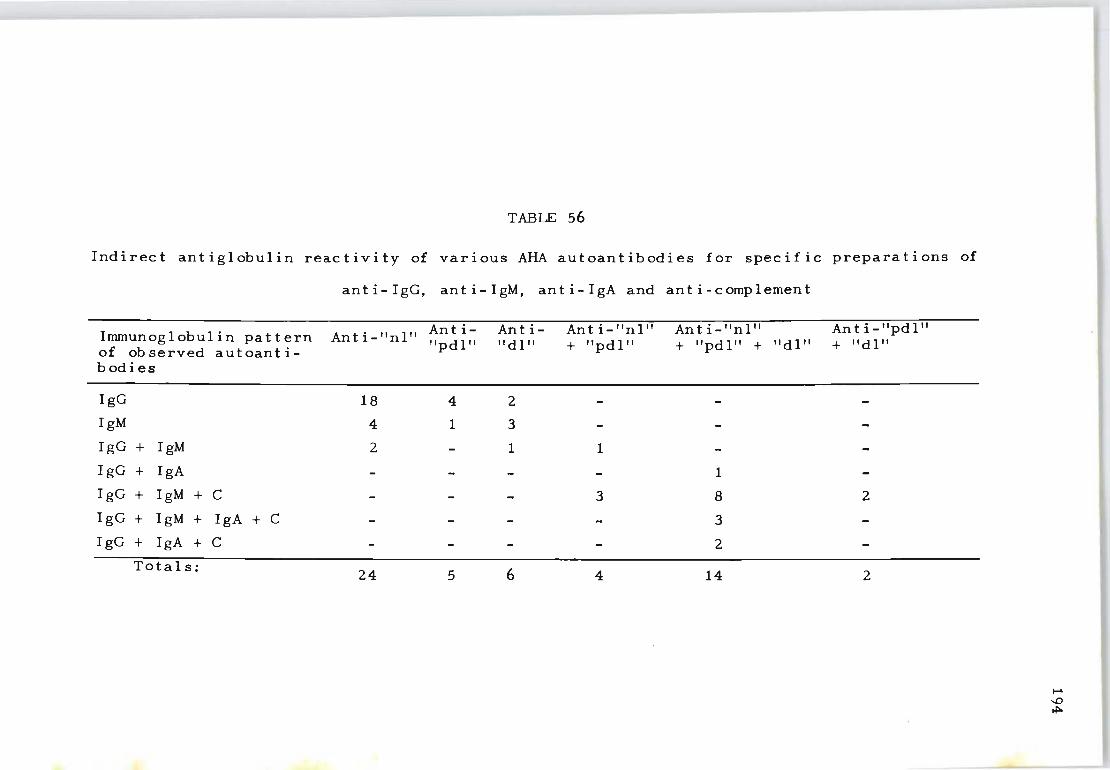
Indirect antiglobulin reactivity of
anti-IgG,
Immunoglobulin pattern Ant i-"nl" of observed autoanti-bodies
IgG 18
IgM 4
IgG + IgM 2
IgG + IgA
IgG + IgM + C
IgG + IgM + IgA + C
IgG + IgA + C
Totals: 24
TABLE 56
various AHA autoantibodies for specific preparations
anti-IgM, anti-IgA and anti - c omp lemen t
Ant i- Ant i- Anti-"nl" Anti-"nl" Anti-"pdl" "pd I " "dl" + "pdl" + "pdl" + "dl" + "dl"
4 2
1 3
1 1
1
3 8 2
3
2
5 6 4 14 2
of
..... ...0 ~
195
demonstration of complement components was associated
only with antibodies of multiple immunoglobulin clas ses
or multiple antibody specificities. In contrast, c omple-
ment was not demonstrable in red cell eluates poss e ssing
only one immunoglobulin type and a single specif i c auto~
ant ibody. Table 57 shows that autoantibodies of a single
specificity were more often of the IgG than IgM o r I gA
class. IgG autoantibody (and to a lesser extent IgM auto-
antibody) was most often observed when the autoantibody
had specificity for a single red cell antigen. Multiple
specificities were frequently associated with a wider
range of immunoglobulin types, particularly IgM and IgA.
The observed variation in the distribution of different
types of immunoglobulin might perhaps be attributed to
renewed episodes of antibody stimulation by reactive red
cell antigens that are not coated by a specific antibody.
4. DISCUSSION
This investigation has shown that eluates of red ce lls
f r om patients with acquired haemolytic anae~ia conta in
autoantibodies of differing Rh specificities.
specifically reactive against well-defined red cell a nt-
igens of the Rh system (anti~e, anti-C and the like) , 1
others against all normal red cells (anti-nl), partiall y
deleted Rh red cells (anti-pdl) and fully deleted Rh red
cells (anti-dI). Autoantibodies directed against the
patient's own well-defined CDEce antigens are seldom
found in the red cell eluates. In contrast, anti-nl ,
anti-pdl and anti-dl were the predominating autoantibod-
ies, either singly or in combination. Such ant ibodie s
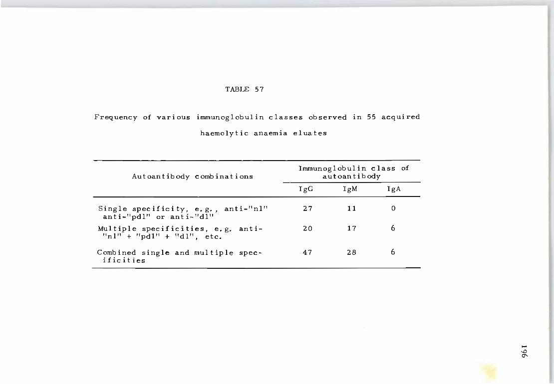
TABLE 57
Frequency of various immunoglobulin classes observed in 55 acquired
haemolytic anaemia eluates
Autoantibody combinations
Single specificity, e. g., anti-"nl" anti-"pdl" or anti-"dl"
Multiple specificities, e.g. anti"nl" + "pdl" + "dl", etc.
Combined single and multiple specificities
Immunoglobulin class of autoan t i body
IgG IgM IgA
27 11 0
20 17 6
47 28 6
.....
"" C1'
could be formed by a sequential process of in vivo
immunization.
197
Serum complement components on the red cells seem to
be associated exclusively with autoantibodies of multiple
immunoglobulin classes or mUltiple red cell specificities.
The fixation of complement on the red cells of patients
with acquired haemolytic anaemia together with the pro
gressive formation of multiple red cell antibodies may
represent the natural evolution of the disease.
The variability in multiple antibodies may reflect
continuous differences in the inciting stimulus resulting
from altered configuration of red cell antigenic determin-
ants. Assuming ' that trapping and processing of antigen
are prerequisites for the induction of antibody response
it may be postulated that an adequate concentration of
anti-nl on the red cell antigen site inhibits fu~ther
synthesis of this antibody but not necessarily the enhance
ment ' of antibody synthesis for other unbound antigens on
the immunizing red cells (anti-pdl and anti-dl). The
suggestion that multiple antibody and immunoglobulin form
ation follows a predetermined course of development as a
consequence of variability in antigen presentation is not
in full agreement with the idea that the disease primar
ily results from an aberration of the immune mechanism.
The findings suggest that the initial site of action of
autoantibody development to Rhesus-antibody-implicated
haemolytic anaemia (anti-nl, anti-pdl and anti-dl) res
ults from a defect of the structural composition of the
Rhesus genome which is thereafter rejected by the normal
immune mechanism. The subsequent development of addit-
ional specificities to other red blood cell antigens
involving multiple immunoglobulin classes does not nec
essarily indicate an aberrant immune apparatus.
198
The observed pattern of serological and immunological
findings does not support the concept that red cell auto
antibodies are a homogeneous population of immunoglobulins,
at least not in the same sense that myeloma globulins are
products of monoclonal proliferation.
The concept that modification of an individual's red
cell antigen can lead to the production of autoantibodies
has been reported by others (Worlledge, Carstairs and
Dacie, 1966; Mohn, Lambert, Bowman and Brason, 1965; van
Loghem, 1965). It is postulated that the initial defect
in most cases of autoimmune haemolytic anaemia does not
involve an aberration of the antibody-forming mechanism
but a modification of the Rh antigen determinant.
Chapter XVI
THE VARIOUS SPECIFICITIES OF AUTOANTIBODIES
FOUND IN ACQUIRED HAEMOLYTIC ANAEMIA WITH
SPECIAL REFERENCE TO THE LW FACTOR
1. INTRODOCTION
It has been established that patients with autoimmune
haemolytic anaemia of the 'warm' type may form mixtures
199
of specific autoantibodies which react with all, or almost
all, human red cells (Table 54). It has also been found
that acquired haemolytic anaemia of the 'warm' type can
be associated with autoantibodies to blood group factors
LW and U (Celano and Levine, 1967, Nugent, Coledge and
Marsh, 1971), suggesting that not all autoantibodies
possess specificities against 'Rh-associated antigens'
( an t i - n I, an t i - pd I, an t i - d 1) •
The LW antigen is almost always found in human bloods
bearing anyone or more of the known Rh antigen determin-
ants (DCEce), and is always lacking from the fully del
eted RhnuII (---/---) red cells, indicating that the LW
factor is closely linked to the Rh system (see Chapter
XI1) • It was particularly important in the present in-
vestigation to establish whether autoantibodies to the LW
factor may be regarded as a possible variant of one of the
'Rh-associated antibodies' so far described. Confirmat-
ion of an association between LW and the various types of
autoantibodies detailed in the previous chapters would
not only further our knowledge of the LW system but would
also help to place the 'nl', 'pdl' and 'dl' varieties of
antigens (Table 47) more closely within the framework of
the Rh system.
Reports that autoantibodies are directed against other
blood group antigens such as Kidd (van Loghem, J.J. and
Hart, v.d.M., 1954), Kell (Da14sset and Colombani, 1959)
and Xga are rare.
2. MATERIALS AND METHODS
200
Selected AHA patients were studied because their red
cell eluates had previously been shown to contain multiple
antibodies. The panel of red cells used (Table 59) had
been stored in a buffered glycerol citrate solution at
-20 0 C. Frozen cells were recovered by Weiner's method
(1961) using Visking dialysis tubing. In no instance did
the frozen and thawed red cells react differently from
fresh cells before storage.
Antiglobulin tests were performed using standard tech
niques and antiglobulin serum that had previously been
evaluated in detail (Chapter XV).
Absorption studies on AHA eluates were carried out
using equal v~lumes of eluate and washed packed red cells.
After incubation at 37 0 C for one hour the red cells were
washed four times in saline. Antibody elution was then
performed on a 50 per cent saline suspension of the red
cells. The ether elution procedure of Vos and Kelsall
(1956) was used, modified only in that ether was added
directly to the 50 per cent suspension of packed washed
red cells. The sequence of absorption and re-elution
used for the differentiation of the type of specific anti
body to be recovered is detailed in the Results.
For the formation of anti-LW guinea pigs were given
subcutaneous injections, at two-weekly intervals, of a
5 per cent suspension of washed LW positive cells mixed
in equal volumes of Freund's complete adjuvant. This
produced intense antibody responses within 8 to 10 weeks.
For absorption studies, the highest dilution of guinea
pig serum giving the strongest reaction with the immuniz
ing red cells was accepted as the optimal starting dilut
ion. Using this procedure repeated absorption of the
antibody with valuable LW negative red cells was avoided.
The range of antibody titre values scored was between
201
1:512 and 1:4096. It was found that the method of Levine
and Celano (1967) in which guinea pigs were given six
intraperitoneal injections of a 25 per cent suspension
of LW positive cells at intervals of three to four days
resulted in considerably lower responses.
3. RESULTS
Table 58 records the indirect antiglobulin titre val
ues of eight selected AHA eluates tested against a panel
of red cells capable of differentiating a variety of spec
ific antibodies. Six of the eight eluates revealed signif
icant differences in reactivity when they were tested
against red cells of varying genotypes, suggesting that
mixtures of specific autoantibodies may be involved.
Table 59 summarizes the results of antibody determinat
ions performed on the same eluates using two different
sets of panel cells. The findings show that cell panel 1
was limited in its scope for detecting anti-U and anti-LW
because it lacked the required cell types to differentiate

TABLE 58
Indirect antiglobulin reactions of various AHA red cell eluates for known red
cell types
Red Cell Eluates from Acquired Haemolytic Anaemia Patients
Red Cell Type BN BL SF CT FR MT VP BS
cDE/cDE, LW+, u+ 512 >:< 16 256 4 32 128 64 256
cde/cde, LW+, u+ 256 16 256 4 32 128 64 256
CDe/CDe, LW+, u- 512 16 256 4 32 128 64 256
cDE/cde, LW+, u- 256 16 256 4 32 128 64 256
CDe/ cde, LW-, u+ 256 16 256 4 8 128 64 256
-D- / -D-, LW+, u+ 256 16 256 4 32 128 64 32
---/---, LW- , u+ 8 4 256 4 0 0 64 0
---/---, LW- , u- 0 4 256 0 0 0 64 0
* Titres are expressed as reciprocals of highest dilution giving definite positive results
f5 tv

Patients BN BL SP CT FR MI' VP BS
Observed
TABlE 59
Specificities of autoantibodies recognized by selective absorption and
elution studies using different panels of red cells.
Antibodies in eluates from patients' red cells recognized by:
Panel 1
CDe/CDe, LW+, U+ crE/ cDE, LW+, U+ cde/cde, LW+, U+ -D- / -D-, LW+, U+ ---/---, LW- , u-
anti-pdl anti-pdl, anti-dl anti-pdl, anti-dl anti-pdl anti-pdl anti-nl, anti-pdl an t i-dl anti-nl, anti-e, anti-pdl
13 specific autoantibodies
Panel 2
CDe/CDe, LW+, UcDE/ cDE, LW+, U+ cde/cde, LW+, U+ cDE/ cde, LW+, UCDe/cde, LW-, U+
anti-pdl, anti-U
-D-/-D- LW+ U+ , , ---/--- LW- U+ , , - - - / - - - , LW - , U-
anti-pdl, anti-dl, anti-LW anti-pdl, anti-dl, anti-LW an t i - LW , an t i - U anti-pdl, anti-LW anti-nl, anti-pdl, anti-LW an t i - d I, anti - U anti-nl, anti-e, anti-LW
20 specific autoantibodies
N o l.tJ
anti-U from anti-nl and anti-pdl, and also anti-LW from
ant i-pdl.
Restrictions on the availability of rare cell types,
particularly the CDe/cde, LW-negative, U-positive cells
204
of Mrs Bigelow (De Veber, Clarke, Hunking and Stroup, 1971)
and the Japanese Rhnull, LW-negative, U-positive blood
(Ishimori and Hasekura, 1967) reduced the opportunity of
isolating the full spectrum of autoantibodies.
Table 60 shows the results of specificity testing using
red cell eluate FR. This autoantibody had previously been
classified as anti-pdl (Table 59) when a more limited
panel of test cells was used. The demonstration of anti
LW specificity and the close association of this antibody
with anti-pdl was recognized when the rare CDe/cde, LW
negative red cells of Mrs Bigelow were included. As shown
in Table 60, step 1, the antibody in eluate FR reacted
more strongly with LW positive cells than the LW negative
cells of Mrs Bigelow. The eluate did not react with the
two Rhnull LW negative red cells indicating the presence
of anti-pdl, or anti-pdl + nl and possibly anti-LW as
well. Step 2: Absorption of the antibody from the orig-
inal eluate with test cell number four (-D-, LW+,U+) which
has pdl, LW and U antigens but not nl, completely removed
all antibody activity indicating pdl, or pdl plus LW spec
ificity. Step 3: The original eluate after one absorpt
ion with test cell number 3 (CDe/cde, LW-, U+) retained
antibody activity for LW positive test cells only. Step 4:
Re-elution from test cells number 4 (-D~, LW+) following
step 2, established the presence of an antibody that sen-
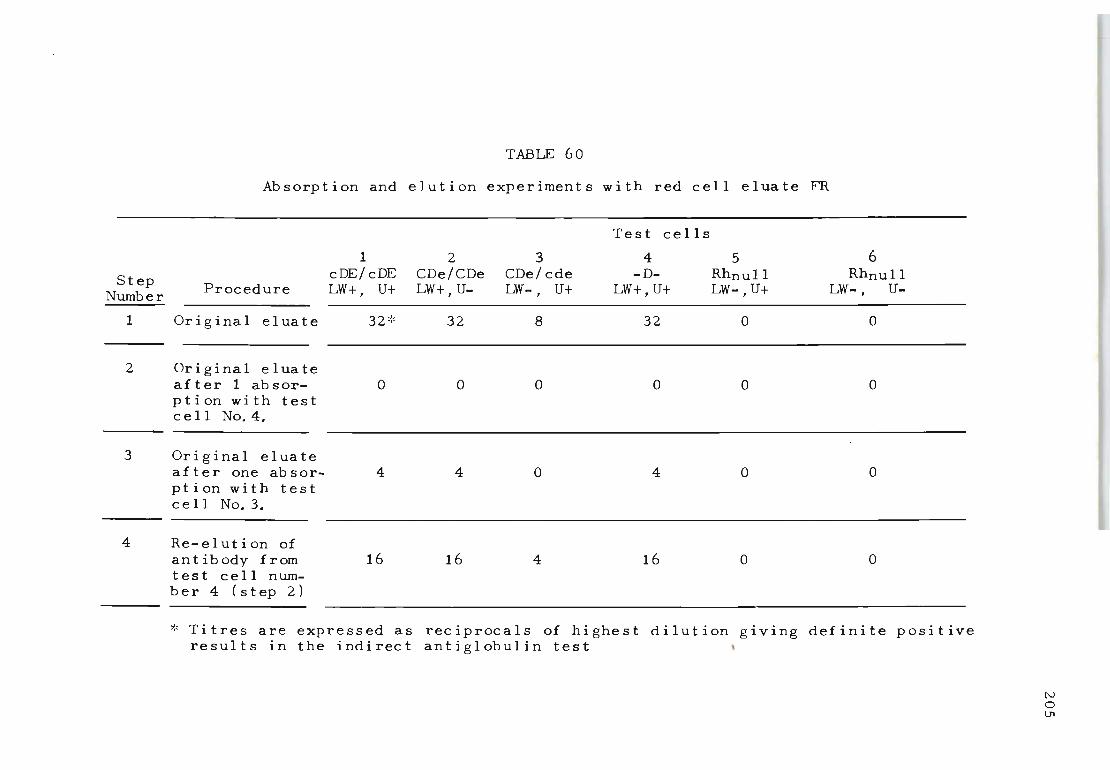
TABLE 60
Absorption and elution experiments with red cell eluate FR
Step Number
1
2
3
Procedure
Original eluate
Original eluate after 1 absorp t i on wit h t est cell No.4.
Original eluate after one absorption with test cell No.3.
4 Re-elution of ant ibody from test cell numb e r 4 (s t ep 2)
1 2 cDE/ cDE CDe/CDe LW+, U+ LW+, U-
32 ':' 32
o o
4 4
16 16
Test cells
3 4 5 CDe/cde -D- Rhnull LW- , U+ LW+,U+ LW-,U+
8 32 0
o o o
o 4 o
4 16 o
6 Rhnull
LW-, U-
o
o
o
o
* Titres are expressed as reciprocals of highest dilution giving definite positive results in the indirect antiglobulin test
N o l.J'l
206
sitized LW positive red cells more strongly than the LW
negative red cells of Mrs Bigelow (test cell 3). This
eluate was then tested against over 200 random samples of
blood to determine whether other examples of weak positive
reactions similar to the weak reactions observed with Mrs
Bigelow's red cells could be recognized but none were
found.
Table 61 illustrates the results obtained when eluate
PM, which reacted with all cells tested except Rhnull,
was absorbed with various cells including animal cells.
One absorption of the eluate with Mrs. Bigelow's CDe/cde
LW negative cells removed activity for her cells completely
and reduced the reaction against cde/cde LW positive and
CDe/cde LW positive cells. Absorption with cde/cde LW
positive cells removed the activity against Mrs Bigelow's
cells and CDe/cde LW positive cells and reduced the activ-
ity against CDe/cDE LW positive cells. Ab sorpt i on wi th
CDe/cDE LW positive cells removed all antibody activity.
Baboon and monkey cells also removed all antibody activ
ity but absorption with sheep, rabbit or horse did not.
These results expand the observations made in Table 60.
The data may be interpreted as follows: If Mrs Bigel-
ow's cells represent a weak variant of LW the antibody
recovered from the -D-/-D-, LW positive sensitized red
cells (Table 60), step 4) can be designated as anti-LW
ra ther than an t i-pdl. This implies that Rhnull red cells
would remain as the true LW negative bloods, as originally
described by Levine, Celano, Vas and Morrison (1962), The
ability of anti-pdl to react more strongly with LW posit-
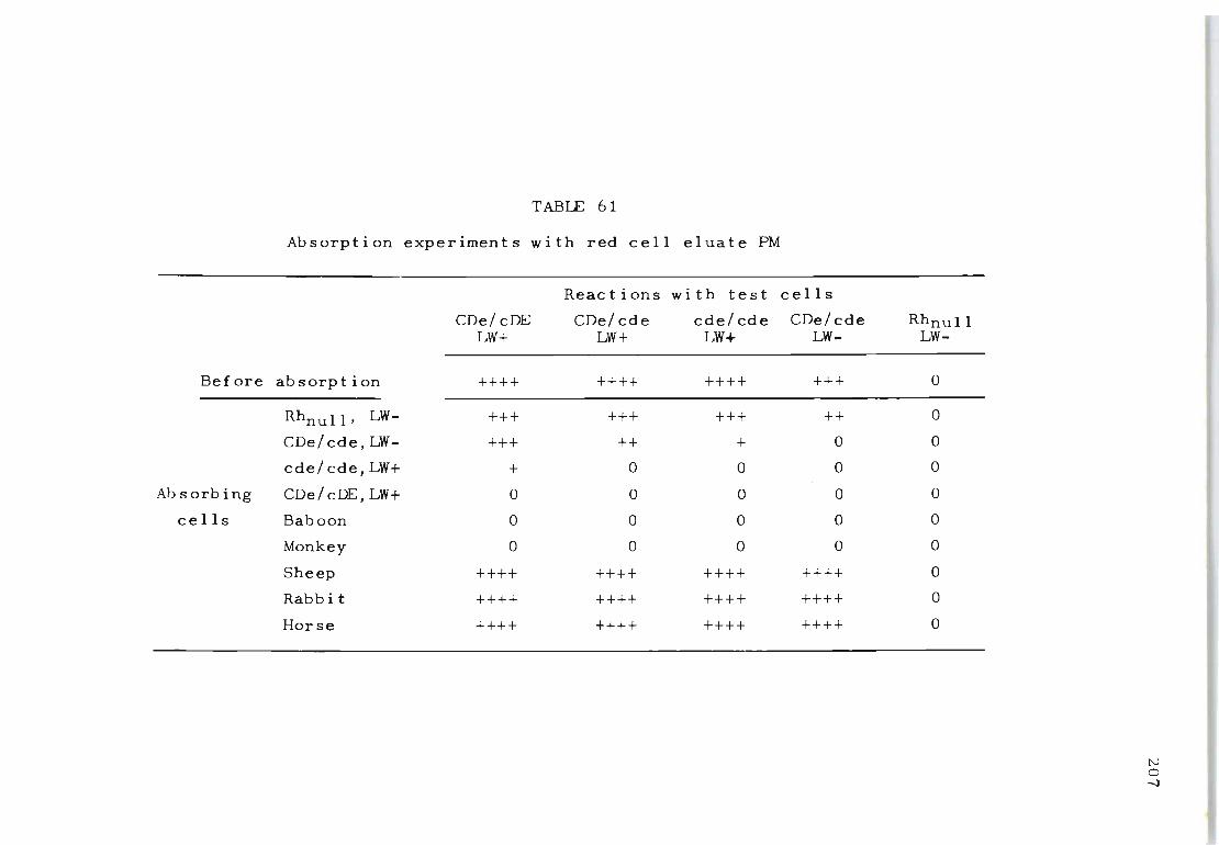
TABLE 61
Absorption experiments with red cell eluate PM
Reactions with test
CDe/cDE CDe/cde cde/cde LW+ LW+ LW+
Before absorpt ion ++++ ++++ ++++
Rhnull, LW- +++ +++ +++
CDe/cde,LW- +++ ++ +
cde/cde,LW+ + 0 0
Ab sorbing CDe/cDE,LW+ 0 0 0
cells Baboon 0 0 0
Monkey 0 0 0
Sheep ++++ ++++ ++++
Rabbi t ++++ ++++ ++++
Horse ++++ ++++ ++++
cells
CDe/ cde LW-
+++
++
0
0
0
0
0
++++
++++
++++
Rhnull LW-
0
0
0
0
0
0
0
0
0
0
tv o -J
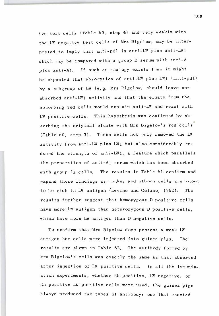
ive test cells (Table 60, step 4) and very weakly with
the LW negative test cells of Mrs Bigelow, may be inter
preted to imply that anti-pdl is anti-LW plus anti-LW1
which may be compared with a group B serum with anti-A
plus anti-A1. If such an analogy exists then it might
208
be expected that absorption of anti-LW plus LW1 (anti-pdl)
by a subgroup of LW (e.g. Mrs Bigelow) should leave un
absorbed anti-LW1 activity and that the eluate from the
absorbing red cells would contain anti-LW and react with
LW positive cells. This hypothesis was confirmed by ab
sorbing the original eluate with Mrs Bigelow's red cells
(Table 60, step 3). These cells not only removed the LW
activity from anti-LW plus LW1 but also considerably re
duced the strength of anti-LW1, a feature which parallels
the preparation of anti-A1 serum which has been absorbed
with group A2 cells. The results in Table 61 confirm and
expand these findings as monkey and baboon cells are known
to be rich in LW antigen (Levine and Celano, 1962). The
results further suggest that homozygous D positive cells
have more LW antigen than heterozygous D positive cells,
which have more LW antigen than D negative cells .
To confirm that Mrs Bigelow does possess a weak LW
antigen her cells were injected into guinea pigs. The
results are shown in Table 62. The antibody formed by
Mrs Bigelow's cells was exactly the same as that observed
after injection of LW positive cells. In all the immuniz-
ation experiments, whether Rh positive, LW negative, or
Rh positive LW positive cells were used, the guinea pigs
always produced two types of antibody; one that reacted
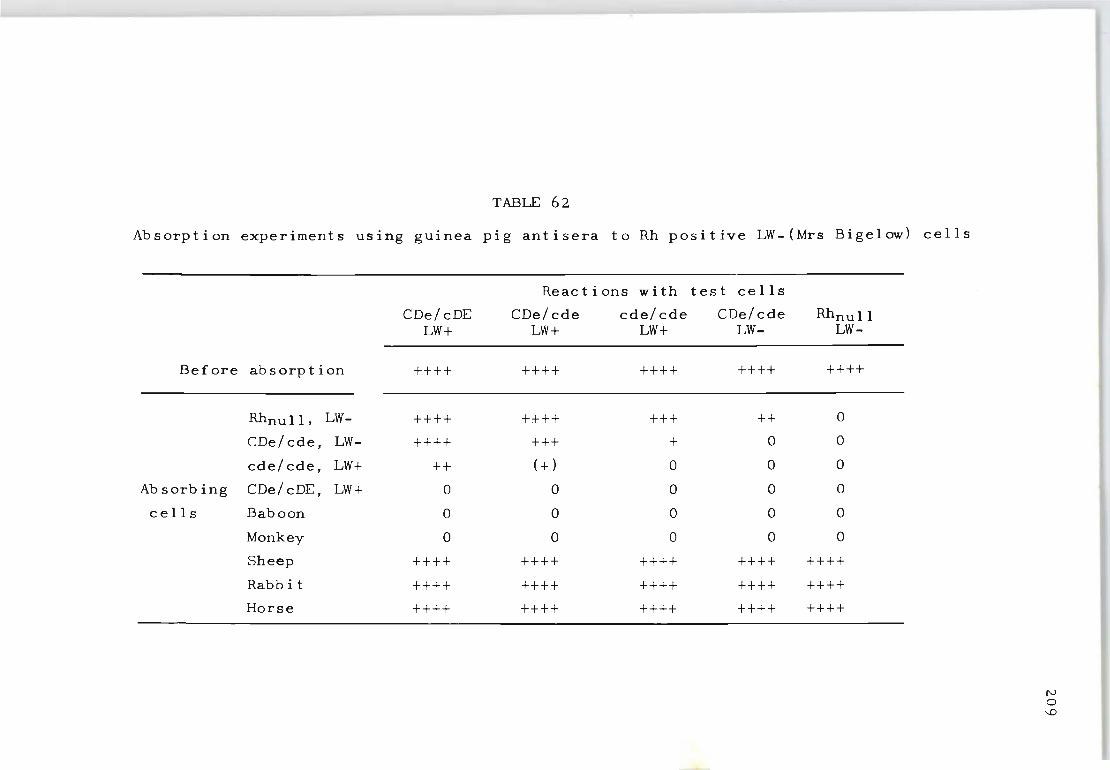
TABLE 62
Absorption experiments using guinea pig antisera to Rh positive LW-(Mrs Bigelow) cells
Reactions with test cells
CDe/cDE CDe/cde cde/cde CDe/cde Rhnull LW+ LW+ LW+ LW- LW-
Before absorption ++++ ++++ ++++ ++++ ++++
RhnuII, LW- ++++ +:+- ++ +++ ++ 0
CDe/cde, LW- ++++ +++ + 0 0
cde/cde, LW+ ++ (+) 0 0 0
Ab sorbing CDe/cDE, LW+ 0 0 0 0 0
cells Baboon 0 0 0 0 0
Monkey 0 0 0 0 0
Sheep ++++ ++++ ++++ ++++ ++++
Rabb it ++++ ++++ ++++ ++++ ++++
Horse ++++ ++++ ++++ ++++ ++++
N o -.D
210
with all human cells and could be removed with one absorpt
ion with Rhnull cells {probably anti-dl)j and an antibody
that reacted exactly the same as the eluate shown in Table
61. The reactions of this antibody were apparent only
after the guinea pig serum had been absorbed with Rhnull
cells. The guinea pig antibody for Rh positive, LW posit-
ive red cells could always be completely removed by one
absorption with Rh positive, LW positive cells and red
cells from baboons and monkeys, suggesting that these
red cells possess an abundance of LW sites.
Autoantibodies reactive for Rhnull cells were observed
in nearly 40 per cent of 51 red cell eluates from AHA
patients (Chapter XIII). They were found either singly
or in combination with other Rh-related autoantibodies.
The activity of autoantibodies for Rhnull red cells can
be removed by absorption with any human or non-human
primate red cells but not sheep, rabbit or horse red cells.
This is illustrated in the results obtained with eluate
KP. (Table 63) where it was found that the specificity
of the antibody may be directed against the 'nucleus of
the Rh substance', as postulated by Wiener, Gordon and
Gallop (1953). The ability of Rhnull cells to separate
this autoantibody from other varieties of Rh-related anti
bodies influenced us to classify this autoantibody as
an t i - d I (i. e. ' del e t i on' ) •
Guinea pigs injected with the original RhnuII red cells
produced antibodies which were identical to those recovered
in the eluates from the red cells of some patients with
AHA (Tab Ie 63) . In no ins tance was the produc t i on of other

Before absorption
Rhnull, LW-
CDe/ cde, LW-
cde/cde, LW+
Absorbing CDe/cDE, LW+
cells Baboon
Monkey
Sheep
Rabbit
Horse
TABLE 63
Absorption experiments with red cell eluate KP
Reac t ions wi th test cells
CDe/cDE CDe/cde cde/cde CDe/cde LW+ LW+ LW+ Uf-
++++ ++++ ++++ ++++
0 0 0 0
0 0 0 0
0 0 0 0
0 0 0 0
0 0 0 0
0 0 0 0
++++ ++++ ++++ ++++
++++ ++++ ++++ ++++
++++ ++++ ++++ ++++
Rhnull LW-
++++
0
0
0
0
0
0
++++
++++
++++
tv ..... .....
known varieties of Rh-related antibodies observed, e.g.
anti-LW in guinea pigs injected with Rhnull red cells.
212
Table 64 shows the results obtained with a third var
iety of Rh-related autoantibody often recovered from the
red cells of AHA patients. These antibodies do not agglut
inate partially deleted -D-/-D-, LW positive red cells
and Rhnull cells but react with all other red cells (i.e.
anti-nIL Their specific activity seems to be directed
towards a region of the Rh antigen representing the CcEe
sequences. The antibody can be completely absorbed by Rh
positive, LW negative, and Rh positive, LW positive cells
but not with red cells from baboons and monkeys. In our
immunization studies we were never successful in produc
ing this type of antibody in guinea pigs. Experiments
are now in progress to establish whether antibodies to
the CcE:e reg i on may be more read i I y produced in non-human
primates. Like the donor of the partially deleted -D-/-D
red cells they are known to lack these particular determin
ant s.
4. DISCUSSION
Nugent, Coledge and Marsh (1971) and Marsh, Reid and
Scott (1972) recently reported anti-U as a specific auto-
an t ibody in AHA. They also postulated that the 'Rh related'
autoantibodies which often fail to be identified as spec
ific Rh antibodies may in fact be anti-U. In our study
the reactions of red cell eluates from eight AHA patients
were examined against a selected combination of extremely
rare types of red cells. Absorption and elution experi
ments showed autoantibodies with U specificity to be found
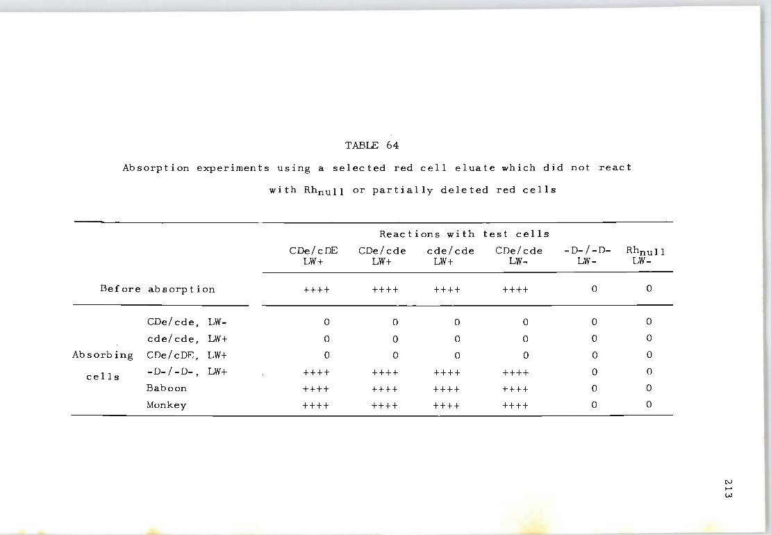
TABLE 64
Absorption experiments using a selected red cell eluate which did not react
with Rhnull or partially deleted red cells
Reac t ions wit h test cells
CDe/cDE CDe/cde cde/cde CDe/ cde -D- / -D- Rhnull LW+ LW+ LW+ LW- LW- LW-
Before absorption ++++ ++++ ++++ ++++ 0 0
CDe/ cde, LW- 0 0 0 0 0 0
cde/ cde, LW+ 0 0 0 0 0 0
Absorbing CDe/cDE, LW+ 0 0 0 0 0 0
cells -D-/ -D-, LW+ ++++ ++++ ++++ ++++ 0 0
Baboon ++++ ++++ ++++ ++++ 0 0
Monkey ++++ ++++ ++++ ++++ 0 0
N .... W
in some, but not all, preparations of red cell eluates.
Thus, no association between anti-U activity and other
214
'Rh related' autoantibodies was found and the suggestion
by Nugent et al. (1971) that the reported 'Rh related'
antibodies may be anti-U in specificity was not confirmed.
In eight selected AHA eluates two examples of anti-nl,
five anti-pdl, three anti-dl, six anti-LW, one anti-e and
three anti-U autoantibodies were observed. This repres-
ents an 85 per cent incidence of antibodies with specific
ities related to the Rh system and a 15 per cent incidence
of anti-U. Considering that almost all autoantibodies so
far described are related to the Rh system, the finding
of a 15 per cent incidence of anti-U lends support to the
suggestion that some association may exist between Rh and
U antigens (Schmidt, Lostumbo, English and Hunter, 1967).
The 'pdl' autoantibody is often found in AHA eluates
and is known to react with all human red cells except
Rhnull. Previously this antibody could not be classified
as anti-LW because it sensitized Mrs Bigelow's 'LW negat-
ive' red cells. In the present study it was particularly
interesting to find that Mrs Bigelow's LW negative red
cells could separate anti-pdl into anti-pdl plus anti-LW
(Table 60), It therefore appears that although Mrs Big
elow has been accepted to be LW negative there is a weak
form of LW antigen present. The presence of LW antigen
on Mrs Bigelow's red cells was also established by inject
ing her red cells into guinea pigs (Table 62),
It is tempting to suggest an analogy to the ABO system
and postulate that Rh positive cells represent LWl; Rh
negative cells, LW2 (as suggested by Levine and Celano,
1967) and Mrs Bigelow, LW3. Rhnull appears to be the
true LW negative (? lw). It will be interesting to see
whether known Rh negative, LW negative samples represent
215
a weaker form of LW than Mrs Bigelow, who is Rh positive.
A contradiction of this analogy would be that if Mrs. Big
elow represents LW3, comparable to A3 in the ABO system,
then the anti-LW in her serum should react with only LWl
(i.e. Rh positive) red cells, comparable to the anti-Al
found in the serum of an A3 individual. However, the
anti-LW reacts strongly with Rh positive (LW1) cells and
weakly with LW2 (Rh negative) cells. It is possible that
the results are simply reflecting a quantitative rather
than qualitative difference. Even though anti-Al usually
does not agglutinafe A2 cells, some workers have reported
that anti-Al found in Ax individuals reacts with both Al
and A2 cell s (I s sit t, 1 970 ) •
We know there is a quantitative effect, as anti-Al can
be removed by several absorptions with A2 cells. This dem
onstrates that anti-Al does react with A2 cells. By anal
ogy ant i-LWl reacts wi th LWl strongly and LW2 weakly. The
only difference between the reactions of LWl and those of
anti-Al with Al and A2 cells is that the LW reactions are
demonstrated by strong agglutination versus weak agglutin
ation, whereas the Al, A2 reactions are demonstrated by
agglutination versus absorption. The results of these
studies indicate that LW represents a spectrum of variat
ions from LW1, LW2, LW3 and maybe LW4. LW vari es even be ..
tween homozygous D positive and heterozygous D positive
individuals which adds to the evidence of the very close
assoc ia t i on between D and LW.
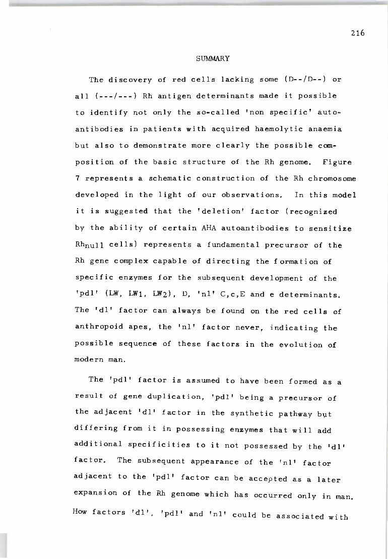
SUMMARY
The discovery of red cells lacking some (D--/D--) or
all (---/---) Rh antigen determinants made it possible
216
to identify not only the so-called 'non specific' auto
antibodies in patients with acquired haemolytic anaemia
but also to demonstrate more clearly the possible com
position of the basic structure of the Rh genome. Figure
7 represents a schematic construction of the Rh chromosome
developed in the light of our observations. In this model
it is suggested that the 'deletion' factor (recognized
by the ability of certain AHA autoantibodies to sensitize
Rhnull cells) represents a fundamental precursor of the
Rh gene complex capable of directing the formation of
specific enzymes for the subsequent development of the
'pdl' (LW, LW1, LW2), D, 'nl' C,c,E and e determinants.
The 'dl' factor can always be found on the red cells of
anthropoid apes, the 'nl' factor never, indicating the
possible sequence of these factors in the evolution of
modern man.
The 'pdl' factor is assumed to have been formed as a
r,esult of gene duplication, 'pdl' being a precursor of
the adjacent 'dl' factor in the synthetic pathway b~t
differing from it in possessing enzymes that will add
additional specificities to it not possessed by the 'dl'
fac tor. The subsequent appearance of the 'nl' factor
adjacent to the 'pdl' factor can be accepted as a later
expansion of the Rh genome which has occurred only in man.
How factors 'dl', 'pdl' and 'nl' could be associated with
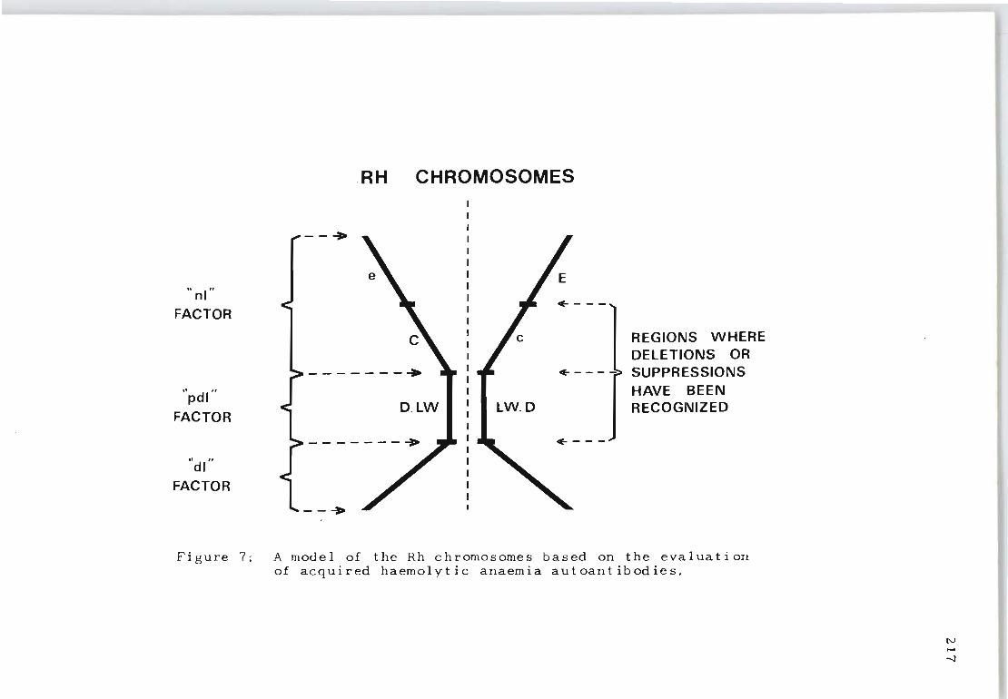
"nl"
FACTOR
"pdl
FACTOR
"d!"
FACTOR
.RH CHROMOSOMES
--+
-------+
D. LW LW.D
of----
REGIONS WHERE DELETIONS OR
of- - - - -> SUPPRESSIONS HAVE BEEN RECOGNIZED
Figure 7 : A model of the Rh chromosomes based on the evaluation of acquired haemolytic anaemia autoantibodies.
N ~
-.J

the D,C,E,c and e determinants is not clear. The i dl'
factor, being present on Rhnull red cells (---/---) as
well as on those possessing the D,C,E,c,e determinants
218
(Figure 8) is the least complicated part of the Rh syst~m
since it has no substance of the LW, D,C,E,c,e variety
in its structure. It could therefore be considered
as the primary precursor with enzymatic potential to de
velop secondary precursors on which the genes giving rise
to the development of antigens LW, D,C,E,c and e can ex
ert their effects.
Evidence that 'non specific' autoantibodies possess
distinct blood group specificities within the Rh system
also contributed to a better understanding of the serol
ogical and immunological characteristics of these auto
an t i bod i e s.
In a study of many red cell eluates from patients with
acquired haemolytic anaemia of the 'warm' type it was
established that antibodies of varying Rh specificities
can be involved in the disease. Some are specifically
reactive against well-defined red cell antigens of the
Rh system (anti-e, anti-C and the like), some against
all normal Rh red cells (anti-nl), some against partially
deleted Rh red cells (anti-pdl) and some against fully
deleted Rh red cells (anti-dl). Antibodies against well
defined Rh antigens (CDEce) were not often detected in
the red cell eluates. In contrast, anti-nl, anti-pdl and
anti-dl autoantibodies were frequently found.
The demonstration of serum complement on patients'
red cells seems to be associated only with autoantibodies
\\ nl"
.. pdl"
"dl"
NORMAL "nl"
MAN II APES
DLWIILWD
----7---"'---- - - - - - -~-
PARTIALLY DELETED
"pdl"
DELETED "d!"
GIBBON
LW
BABOON
Figure 8: Rh chromosomes of man and apes and their relationship to determinants 'nl', 'pdl' and 'dl'.
N ~
-..0

220
of multiple immunoglobulin classes or multiple red cell
specificities. The structural heterogeneity of immuno-
globulin types (IgG, IgM and IgA) for the various variet
ies of autoantibodies (anti-nl, anti-pdl and anti-dl) can
be pictured as arising from multiple antibody-forming
cell lines.
The findings suggest that the initial site of auto-
antibody development in AHA results from a defect in the
structural composition of the Rhesus genome, which is
thereafter rejected by a normal immune mechanism. The
subsequent development of a more generalized auto-immune
abnormality involving the formation of multiple autoanti-
body-forming cell lines does not necessarily indicate
that an aberrant immune apparatus has been established.
Additional studies using red cell eluates revealed that
no direct correlation could be established between the
presence of complement components and the intensity of
the indirect antiglobulin test using anti-IgG serum. How-
ever, the simultaneous presence of 'anti-nl' and 'anti-dl'
autoantibodies was often associated with the presence of
complement components in the red cell eluates. Fixation
of complement by Rh isoantibodies of mUltiple specificity
has been reported and thus the presence of complement
components in association with anti-nl and anti-dl may be
analogous to complement fixation by mUltiple Rh isoanti
bodies. The formation of anti-nl with anti-dl which ,
tends to result in complement components being fixed by
the antibody, may be considered as a natural progression
of antibody synthesis associated with an advanced state
of autoimmunization.
Absorption and elution studies using rare Rh positive
LW negative cells (Mrs Bigelow) showed that anti-pdl may
in fact represent anti-LW plus LW1 and that Mrs Bigelow's
cells may possess a weak variant of LW. Injection of her
red cells into guinea pigs produced an anti-LW that re
acted similarly to the antibody produced by injecting Rh
positive LW positive cells . It is suggested that normal
Rh positive LW positive cells represent LW1; Rh negative
LW positive cells represent LW2, Mrs Bigelow's cells LW3
and that Rhnull cells are the only true LW negative (lw)
cells.
221
REFERENCES
BEHRMAN, S.J., BDETTNER-JANUSCH, J., HEGLAR, R. , GERSHOW
ITZ, H. and TEW, W.L.: J . Obstet . Gynec. 79:847 (1960) .
BETKE, K. : XIth Congress of the International Soc i e t y of
Blood Transfusion. Sydney, Australia (1966).
BEVIS, D. C. A. : Lancet, 2:443 (1950).
BEVIS, D.C.A. : Lancet, 1:395 (1952).
222
BEVIS, D. C. A. : J. Ob stet . Gynaec. Bri t. Cwl tho 63:68 (1956 ).
BURNET, F.M. and FENNER , F.: The Production of Antibodies.
Macmillan, Melbourne (1949) .
CANN, J.R., BROWN, R.A., KIRKWOOD , J.G., STURGEON, p. and
CLARKE, D.W.: J. Immunol. 68:243 (1952).
CELANO, M.J. and LEVINE, p.: Transfusion 7:265 (1967).
CEPPELLINE, R. as cited by LEVINE, P., CHAMBERS, J.W.,
CELANO, M.J., FALKOWSKI, F., HUNTER, O.B. and ENGLISH,
C.T.: Proc. 10th Congr. Int. Soc . Blood Transf. Stock
holm, 350 (1964).
CHAUFFARD, A. and TROISIER, J.: Sem. Med . 28:345 (1908).
CLARKE, C.A.: Brit. Med. J. 4:7 (1967).
COOMBS, R.R.A., MOURANT, A.E . and RACE, R.R. : Lanc e t,
2:15 (1945).
CRAWFORD, J.W., CAMERON, C. and WALKER, C.H.M.: Lancet,
2:887 (1966).
DACIE, J.V.: Lancet ii:954 (1951).
DACIE, J.V.: The Haemolytic Anaemias, Congenital and
Acquired. Part II. The Autoimmune Haemolytic Anaemias,
2nd. Ed. Grune & Stratton , New York (1962).
DACIE, J.V. and LEWIS, S.M.: Practical Haematology, 3rd.
Ed. p. 166, Grune & Stratton, New York (1963).
223
DACIE, J.V. and WORLLEDGE, S.M. Progr. Haemat. 6:82 (1969)
DAUSSET, J., and COLDMBANI, J.: Blood 14: 1280 (1959).
DE VEBER, L.L., CLARKE, G.W., HUNKING, M. and STROUP,M:
Transfusion 11:33 (1971).
DAVIDSOHN, I. and STERN, K.: Am.J. Clin. Path. 18:690
(1948 ).
DIAMOND, L.K. and DENTON, R.L.: J. Lab. & Clin. Med. 31:
821 (1945).
DIAMOND, L.K.: Proc. Roy. Soc. Med. 40:546 (1947).
DODD, B.E. and EELES, D~A.: Immunology IV, 337 (1961).
DUDOK de WIT, C. and BORST-EILERS, E.: Brit. Med. J. 1:152
(1968).
ENGELFRIET, C.P., BORNE, A.E., v.d. GIESSEN, M. v.d.,
BECKERS, D. and LOGHEM, J.J. van. Clin. Exp. Immunol.
3:605 (1968).
EYSTER, M.E., JENKINS, D.E. and MOORE, W.H.: (Abstract)
Clinical Research 15:276 (1967).
FARRELL, A.G.W. and DAVI~, G.S.D.: Sth. Afr. J. Obstet.
Gynaec. 5: 1 (1967).
FINN, R., CLARKE, C.A., DONOHOE, W.T.A., MCCONNELL, R.B.,
SHEPPARD, P. M., LEHANE, D. and KULKE, W.: Bri t. Med. J. , 1:1486 (1961).
FISHER, R.A.: Cited by Race, R.R., Nature, 153:771 (1944).
FLEMING, A.F. and WOOLF, A. J.: Clin. Chim. Acta. 12:67 . (1965).
FREDA, V.J., GORMAN, J.G. and POLLACK, W.: Fed. Proc. 22: 374 (1963).
FREDA, V.J.: Bull. Sloane Hosp. 10:194 (1964).
FREDA" V.J., GORMAN, J.G. and POLLACK, W.: Transfusion
4: 1 (1964).
FUDENBERG, H. H., MANDY, W. J. and NISONOFF, A.: J. CI in.
Invest. 41:2123 (1962).
GIESSEN, M. van der, HART, M. van der and WEERDT, C.M.
van der. Vox Sanguinis, 9:25 (1964).
GOLDSMITH, K.L.G., MOURANT, A.E. and BANGHAM, D.R.:
Bull. WId. Health. Organ. 36:435 (1967).
224
GOODMAN, H.S., MASAITIS, L. and MIZERA, G.: J. of Immunol.
98:425 (1967).
GREEN, F.A.: Nature, 219 : 86 (1968a).
GRE~N, F.A.: J. BioI. Chern. 243:5519 (1968b).
GREENWALT, J. and WAGNER, A.J. Blood 10:29 (1955).
HARBOE, M., MULLER-EBERHARD, H. J., FUDENBERG, H. H. ,
POLLY, M.J. and MOLLISON, P.L.: Immunology 6:412
( 1963).
HILL, J.M. and SOULES, D.E.: Papers in dedication of
P.H. Andresen's Sixtieth Birthday. Munksgaard,
Copenhagen. p. 82 (1957).
HOLLANDER, L. and BATSCHELET, E.: Proc. 6th Congr. Europ.
Soc. Haernatol. Copenhagen. p.680 (1957).
HUBINOT, P.: Bruxelles Med. 41:864 (1961).
HYSLOP, R.S. and WHILEY, W.: Med. J. Aust. 1:622 (1960).
ISHIMORI, T. and HASEKURA, H.: Proc. Japanese Acad. 6:658 (1966).
ISHIMORI, T. and HASEKURA, H.: Transfusion 7:84 (1967).
ISSITT, P.D.: Applied Blood Group Serology (ed.l).
Oxnard, Spectra Biologicals (1970).
JACOBS, W.M.: Obstet. Gynec. (N. Y.) 19:806 (1962).
KELSALL, G.A. and VOS, G.H.: Lancet, 2:161 (1955).
KELSALL, G.A., VOS, G.H., KIRK, R.L. and SHIELD, J.W.:
Pediatrics 20;221 (1957).
KELSALL, G.A., VOS, G.H. and KIRK, R.L.: Brit. Med.J.
2:468 (1958).
KIDD, p.: J. Clin. Path. 2:103 (1949).
KORNSTAD, L., RYTTINGER, L. and HOGMAN, C.: Vox Sang.
5:330 (1960).
KRIEGER, V. I. and WILLIAMS, E.J.: J.Lab.& Clin.Med.,
46:199 (1955).
KROMME, L. de and VERVAAT, M.: Vox Sang. 3:9 (1953).
KUBLI, F.: Bibl. Gynaec. 38:30 (1966).
LANDSTEINER, K. and WIENER, A.S.: Proc. Soc. Exp.Biol.
(N.Y.) 43:223 (1940).
LEDDY, J.p., BAKEMEIER, R.F. and VAUGHAN, J.H. : J.of
Clin. Investig. 44:1066 (1965).
LEDDY, J.p. and BAKEMEIER, R.F.: Proceedings of the
Soc. for Experimental BioI. and Med. 125:808 (1967).
225
LEDDY, J.P., PETERSON, p., YEAW, M.A. and BAKEMEIER,R.F.:
J. Immunol. 105:677 (1970).
LEVINE, p. and STETSON, R.E.: J. Amer. Med. Ass. 113:126
(1939) .
LEVINE, P.: H.Hered. 37:71 (1943).
LEVINE, p.: Hum. BioI. 30:14 (1958).
LEVINE, P., VOGEL, p. and ROSENFIELD, R.E.: Advances
Pediat. VI:97 (1953).
LEVINE, p. and CELANO, M. J.: Nature 193:184 (1962).
LEVINE, P., CELANO, M.J., VOS, G. H., and MORRISON, J.:
Nature, 194:304 (1962).
LEVINE, P., CELANO, M.J. WALLACE, J o and SANGER, R.:
Nature 198:596 (1963)0
LEVINE, P., CHAMBERS, J.W., CELANO, M. J., FALKOWSKI,F.,
HUNTER, O.B. and ENGLISH, C.T.: Proc. 10th Congr.
Int. Soc. Blood Transf. Stockholm, 1964, p. 350
Karger, Basel (1965).
LEVINE, P., CHAMBERS, J.Wo, CELANO, M.J., FALKOWSKI,F.,
HUNTER, O.B. and ENGLISH, C.T.: Transfusion 5:492
(1965).
LEVINE, p. and CELANO, M.J.: Science 156:1744 (1967).
LILEY, A.W.: Amer. J. Obstet. Gynec. 82:1359 (1961).
LILEY, A.W.: Am. J. Obst. & Gynec. 86:485 (1963).
LOGHEM, J.J. van.: Series Haemat., Fasc.9, Munksgaard,
Copenhagen (1965).
LOUTIT, . J.F. and MOLLISON, P.L.: J. Path. and Bact.
58:711 (1946).
LUCIA, S.P, and HUNT, M.L.: J, Hematol. 5:762 (1950).
LUCIA, S.P., WILD, G.M. and HUNT, M.L.: Vox Sang. 5:377
(1960).
MACKAY, E.V.: Austr. N.Z. J o Obstet. Gynaec.1:78 (1961).
MACKAY, E.V. and WATSON, D.: M.J. Aust. 2:942 (1962).
MARSH, W.L., REID, M.E. and SCOTT, E.P.: Brit. J o Haemat.
22:625 (1972).
MASOUREDIS, So, DUPUY, M.E. and ELLIOT, M.: J. Clin.
Invest. 46:681 (1967).
226
MEULI, H.C.: Blut. 3: 270 (1957).
MILGROM, F.: Vox Sang. 16:286 (1969).
MISENHIMER, H.R.: Am.J. Obst. & Gynec. 94:1133 (1966).
MOHN, J.F., LAMBERT, R.M., BOWMAN, H.S. and BRASON, F.W.:
Ann. New York Acad. Sci: 124:477 (1965).
MOLLISON, P.L.: Blood Transfusion in Clinical Medicine,
2nd Ed. p. 210 (Blackwell Scientific Publications,
Oxf ord, 1956 ) .
MOLLISON, P.L.: Blood Transfusion in Clinic~l Medicine.
5th Ed. Blackwell Scientific Publications, Oxford.
p. 323 (1972).
MOLLISON, P.L. and CUTBUSH, M.: Blood 6:777 (1951).
MOORES, P.: Vox Sang. 23:452 (1972).
MORRISON, J.: J. Obstet. Gynec. Brit. Cwlth. 74:419
(1967).
MORRIS, E.D., MURRAY, J., and RUTHVEN, C.R.J.: Brit.
M.J. 2:352 (1967).
MORTON, J.A. and PICKLES, M.M.: Nature (Lond.) 159:779
( 1947) .
MURRAY, J. and CLARK, E.C.: Nature, 169:886 (1952~.
MURRAY, Sheilagh, KNOX, E.G. and WALKER, W.: Vox Sang.
10:6 (1965).
NEVAHLINNA, H.R. and VAINIO, T.: Vox Sang. 1:26 (1956)
NILSSON, B.A.: Acta Obstet. Gynec. Scand. Suppl.2:44
(1965).
NUGENT, M.E., COLEDGE, J. I. and MARSH, W.L.: Vox Sang.
20:519 (1971).
227
PETZ, L.D., FINK, D.J., LETSKY, E.A., FUDENBERG, H.H.,
and MULLER-EBERHARD, H.J.: J. Clin. Invest.
47:2469 (1968).
PICKLES, M.M.: Haemolytic Disease of the Newborn.
Springfield, Thomas; Oxford, Blackwell (1949).
PIROFSKY, B.: Am.J. Obstet & Gynec. 92:720 (1965).
POLLACK, W., GORMAN, J.G., HAGER, H.J., FREDA, V.J. and
TRIPODI, D.: Transfusion, 8;134 (1968).
POWELL, W. N.: Am. J. Cl in. Pa tho 14: 55 (1954).
QUEENAN, J.T.: Clin. Obstet. Gynec. 9:491 (1966).
RACE, R.R., SANGER, R., and SELWYN, J.G.: Nature, Lond.
166:520 (1950).
RACE, R.R. and SANGER, R.: Blood Groups in Man, 2nd Ed.
p. 202, Blackwell Scientific Publications, Oxford
(1954).
RACE, R.R. and SANGER, R.: Blood Groups in Man. 2nd Ed.
p. 280. OxfQrd Blackwell Scientific Publications
(1954).
RACE, R.R. and SANGER, R.: Blood Groups in Man, 4th Ed.
Oxford Blackwell (1962).
RACE, R.R. and SANGER, R.: Blood Groups in Man, 5th Ed.
Blackwell Scientific Publications, Oxford (1968).
REEPMAKER, J., NIJENHUIS, L.E. and van LOGHEM, J.J.:
Amer. J. Hum. Genet. 14:185 (1963).
REEPMAKER, J.: ABO Antagonisme en Morbus Haemolyticus
Neonatorum. Thes-is. Leyden (1955).
ROBERTSON, S.E.J.: M.J. Aust. 1:398 (1961).
ROBINSON, M., WILLIAMS, L., JAKOBOWICZ, R., MOORE, F. and
SILBERMAN, F.: XIth Congress of the International
Society of Blood Transfusion, Sydney, Aust. (1966).
228
229
ROCHNA, E. and HUGHES-JONES, N.C.: Vox Sang. 10:675 (1965~
ROSSE, W.F.: J. Clin. Investig. 47:2430 (1968).
SCHMIDT, P.J. and VOS, G.H.: Vox Sang. 13:18 (1967)
SCHMIDT, P.J., LOSTUMBO, ~M., ENGLISH, L.T. and
HUNTER, O.B.: Transfusion 7:33 (1967).
SMITH, T.: J. Exp. Med. 11:241 (1909).
STEENSMA, F.A.: Klinisch Laboratoriumboek. Amsterdam,
Scheltema and Holkemas (1954).
STERN, K., DAVIDSOHN, I., and MASAITIS, L.: Amer. J.
CI in. Pa the 26: 833 (1956).
STERN, K., GOODMAN, H.S. and BERGER, M.: J. Immunol.
87: 189 (1 96 1 ) .
STURGEON, P.: Science, 106:293 (1947).
TAYLOR, W.C. and KULLMAN, A.K.: J. Obstet. Gynaec.Brit.
Emp. 68:261 (1961).
TOVEY, G.H. and VALAES, T.: Lancet, 2:521 (1959).
VAN LOGHEM, J.J. and v.d. HART, M.: Vox Sang. 4:2 (1954)
VOS, G.H. and KELSALL, G.A.: Brit. J. of Haematol.2:342
(1956).
VOS, G.H.: Australasian Ann. Med . 7:340 (1958).
VOS, G.H. and KIRK, R.L.: Amer. J. Clin. Path. 29:284 (1958).
VOS, G.H.: Vox Sang. 5:472 (1960).
VOS, G.H., VOS, D., KIRK, R.L. and SANGER, R., Lancet, 1:14 (1961).
VOS, G.a and KIRK, R.L.: Vox Sang. 7:22 (1962).
VOS, G.H.: Transfusion 5:327 ( 1965).
VOS, G.H. : Vox Sang. 12:390 (1967)
VOS, G. H. : Sth. Afr. Med. J. 43: 130 (1969) .
VOS, G. H. : Sth. Afr. Med. J. 43 : 241 (1969).
VOS, G.H. : Sth. Afr. Med. J. 43 : 364 ( 1969).
VOS, G. H. , PETZ, L. D. and FUDENBERG, H. H. : Bri t. J.
Haema t. 19:57 (1970),
VOS, G.H., PETZ, L.D. and FUDENBERG, H.H.: Immunology,
106:1172 (1971).
VOS, G.H., MOORES, P. and LOWE, R.F. : Sth. Afr. J. Med.
Sc i. 36: 1 (1971).
WALKER, A.H.C. : Brit.M. J. 2:376 (1957).
WALKER, A.H.C. and JENNISON, R.F.: Brit. Med. J. 2:1152
(1962).
WALKER, W.: B r it. M. J, 1 : 1 75 (1 96 7 )
WALLER, R.K. : J. Lab. Clin. Med. 34:270 (1949),
WATKINS, W.M. : Science, 152 : 172 (1966).
WEINER, W., BATTEY, D.A., CLEGHORN, T.E., MARSON, F.G.W.
and MEYNELL, M.J. : Brit. Med. J. 2:125 (1953).
WEINER, W.: Brit. J. Haematol. 3:276 (1957).
WEINER, W.: Proc. 8th Congr. Europe. Soc. Haematol.
Vie nna (1 96 1 ) a.
WEINER, W.: Lancet 1:1264 (1961)b.
WEINER, W. and VOS, G.H. : Blood, 22:606 (1963).
WIDAL, F., ABRAM I , p. and BRULE, M.: Arch. Mal. Coeur.
1:193 (1908).
WIENER, A.S. and HURST, Jane G. and SONN, E.B.: J. Exper. Med. 86:267 (1947).
230
WIENER, A.S. and HURST, J.G.: Exper. Med. & Surg. 5:285
(1947) .
WIENER, A.S.: Proc. Soc. Exp. BioI. 70:576 (1949).
WIENER, A.S., NAPPI, R. and GORDON, E.B.: Am.J. Obst. & Gynec. 63:6 (1952).
WIENER, A.S., GORDON, E.B., and GALLOP, C.: J. Immunol.
71:58 (1953).
WIENER, A.S. and GORDON, E.B.: Am. J. Clin. Path. 23:429
(1953).
WIENER, A.S., UNGER, L.J., COHEN, L. and FELDMAN, J.:
Ann. Intern. Med. 44:221 (1956).
WIENER, A.S. and WEXLER, I.B.: Heredity of the Blood
Groups. Grune & Stratton, New York (1958).
\
WOODROW., J. C., CLARKE, C. A., DONOHOE, W. T. A., FINN, R.,
McCONNELL, R.B" SHEPPARD, P,M., LEHANE, D.,
RUSSELL, S.H., KULKE, W. and DURKIN, C.M.:
Brit. Med. J. 1:279 (1965).
WORLLEDGE, S., CARSTAIRS, K,C. and DACIE, J.V.: Lancet,
1:135 (1966).
231
YOKOYAMA, M., EITH, D.T. and BOWMAN, M.: Vox Sang. 12:138
(1967) .
ZEITLIN, R.A. and BOORMAN, K.E.: J. Obstet. Gynec. Brit. Cwlth. 70:798 (1963).
ZILLIACUS, H. and ERIKSSON, A.: Acta. Obstet. Gynec, Sc and. 37: 1 (1 958 ) .
ZIPURSKY, A., POLLOCK, J., CHOWN, B. and ISRAELS L.G. , Lancet, 2:493 (1963).