Embed Size (px)
Citation preview

The role of end-of-treatment PET
in lymphoma management
Ulrich Duehrsen
Department of Hematology
West German Cancer Center
University Hospital Essen
Germany

Disclosures
I have no conflicts of interest to declare.

1. Performance of EoT PET
2. Prognostic information
3. Therapeutic implications
End-of-treatment PET in lymphoma Key objectives
1. Performance of EoT PET
2. Prognostic information
3. Therapeutic implications
End-of-treatment PET in lymphoma Key objectives

Positron emission tomography Increased glucose utilization in lymphomas
Entity % positive cases Maximum SUV
Aggressive lymphomas
HL, DLBCL, PMBCL, BL, PTCL 85 - 100 % 19,6 ± 9,3
Indolent lymphomas
Mantle cell lymphoma 100 % 8,7 ± 1,3
Follicular lymphoma 95 % 7,7 ± 4,6
Nodal marginal zone lymphoma 100 %
Splenic MZL 67 % 3,8 ± 1,3
Extranodal MZL (MALT) 55 %
Waldenstrom’s macroglobulinemia 83 % n.r.
Lymphocytic lymphoma / CLL 83 % 2,5 ± 0,7
Schöder et al, J Clin Oncol 23: 4643, 2005; Weiler-Sagie et al, J Nucl Med 51: 25, 2010;
Karam et al, Nucl Med Comm 30: 770, 2009;
Karam et al, Cancer 107: 175, 2006;
Banwait et al, Am J Hematol 86: 567, 2011

Juweid et al, J Clin Oncol 25: 571, 2007
CT
PET
PET/CT
Hodgkin lymphoma
Residual bulk
of 5 x 4 cm
after 6 x ABVD
Viable
lymphoma
≠
necrosis or
fibrosis
End-of-treatment PET Comparison with computed tomography

Juweid et al, J Clin Oncol 23: 4652, 2005
End-of-treatment PET Superior outcome prediction
EoT PET negative: always CR (independent of residual mass)
EoT PET positive: never CR
IWC IWC+PET
CR CR
CRu
PR PR
SD SD
PD PD
(n=24) (n=35)
(n=19) (n=12)
IWC+PET: CR
IWC+PET: PR
IWC: CR/CRu
IWC: PR

End-of-treatment PET Timing
Lugano recommendations:
6 - 8 weeks after chemotherapy (minimum of 3 weeks)
2 weeks after cessation of G-CSF treatment
3 months after radiotherapy
(avoidance of inflammatory reactions)
Barrington et al, J Clin Oncol 32: 3048, 2014

End-of-treatment PET Response criteria
Locally developed response criteria
International Harmonization Project (IHP) criteria – 2007
Small lesions no uptake above background
Lesions ≥ 2 cm uptake ≤ mediastinal blood pool
Deauville criteria – 2009, 2014
1 no uptake
2 uptake ≤ mediastinal blood pool
3 uptake > mediastinal blood pool, but ≤ liver
4 uptake moderately > liver
5 uptake markedly > liver or new lesions
Semiquantitative criteria (SUVmax, MTV, …)
Juweid et al, J Clin Oncol 25: 571, 2007;
Meignan et al, Leuk Lymphoma 50: 1257, 2009;
Barrington et al, J Clin Oncol 32: 3048, 2014
1. Performance of EoT PET
2. Prognostic information
3. Therapeutic implications
End-of-treatment PET in lymphoma Key objectives

End-of-treatment PET Prognostic information
Indolent lymphomas
Follicular lymphoma
Mantle cell lymphoma
Aggressive lymphomas
Diffuse large B-cell lymphoma
Primary mediastinal B-cell lymphoma
Burkitt lymphoma
Hodgkin lymphoma
Peripheral T-cell lymphoma
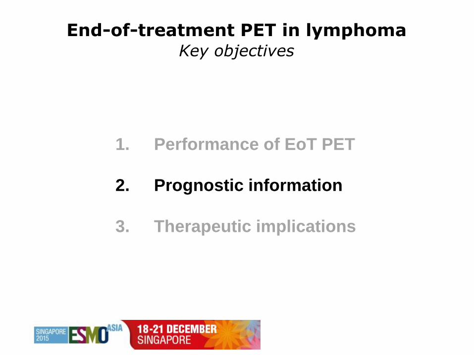
Indolent lymphomas Studies reporting the prognostic value of EoT PET
Entity
No. of
studies
No. of
patients
PET
criteria
PFS
OS
Follicular lymphoma
8 6 retrospective
2 prospective
16 - 202
local
IHP
D5S
8 / 8
3 / 3
Mantle cell lymphoma
4 retrospective
28 - 55
IHP
3 / 4
3 / 4
Prognostic value
Follicular lymphoma Zinzani et al, Clin Lymphoma Myeloma 7: 291, 2007; Bishu et al, Leuk Lymphoma 48: 1548, 2007;
Le Dortz et al, Eur J Nucl Med Mol Imaging 37: 2307, 2010; Trotman et al, J Clin Oncol 29: 3194, 2011;
Dupuis et al, J Clin Oncol 30: 4317, 2012; Zinzani et al, Am J Hematol 88: E273, 2013;
Luminari et al, Ann Oncol 25: 442, 2014; Lu et al, Ann Nucl Med 28: 805, 2014
Mantle cell lymphoma Bodet-Milin et al, Eur J Nucl Med Mol Imaging 37: 1633, 2010;
Hosein et al, Am J Hematol 86: 841, 2011;
Mato et al, Cancer 118: 3565, 2012;
Kedmi et al, Leuk Lymphoma 55 : 2484, 2014

Follicular lymphoma PFS and OS according to EoT PET
Dupuis et al, J Clin Oncol 30: 4317, 2012
Overall Survival
6 x R-CHOP + 2 x R → EoT PET → Deauville 1-3 vs. 4+5
2-yr-PFS 2-yr-OS
PET neg. 87 % 100 %
PET pos. 51 % 88 %
(n=119) Log-rank P < 0.001 Log-rank P = 0.01
EoT PET negative: 78 %
EoT PET positive: 22 %
Progression-free Survival

Mantle cell lymphoma PFS and OS according to EoT PET
Mato et al, Cancer 118: 3565, 2012;
Kedmi et al, Leuk Lymphoma 55: 2484, 2014
R-Hyper-CVAD/MA → IHP
Follow-up: 32 months
R-CHOP ± AutoSCT → IHP
Follow-up: 36 months
EoT PET negative: 84 %
Log-rank P = 0.0002
EoT PET positive: 16 %
(n=51)
Pro
gre
ssio
n-f
ree S
urv
ival
(n=55)
EoT PET negative: 78 %
EoT PET positive: 22 %
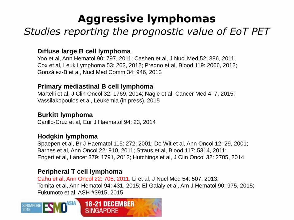
Aggressive lymphomas Studies reporting the prognostic value of EoT PET
Diffuse large B cell lymphoma Yoo et al, Ann Hematol 90: 797, 2011; Cashen et al, J Nucl Med 52: 386, 2011;
Cox et al, Leuk Lymphoma 53: 263, 2012; Pregno et al, Blood 119: 2066, 2012;
González-B et al, Nucl Med Comm 34: 946, 2013
Primary mediastinal B cell lymphoma Martelli et al, J Clin Oncol 32: 1769, 2014; Nagle et al, Cancer Med 4: 7, 2015;
Vassilakopoulos et al, Leukemia (in press), 2015
Burkitt lymphoma Carillo-Cruz et al, Eur J Haematol 94: 23, 2014
Hodgkin lymphoma Spaepen et al, Br J Haematol 115: 272; 2001; De Wit et al, Ann Oncol 12: 29, 2001;
Barnes et al, Ann Oncol 22: 910, 2011; Straus et al, Blood 117: 5314, 2011;
Engert et al, Lancet 379: 1791, 2012; Hutchings et al, J Clin Oncol 32: 2705, 2014
Peripheral T cell lymphoma Cahu et al, Ann Oncol 22: 705, 2011; Li et al, J Nucl Med 54: 507, 2013;
Tomita et al, Ann Hematol 94: 431, 2015; El-Galaly et al, Am J Hematol 90: 975, 2015;
Fukumoto et al, ASH #3915, 2015

Aggressive lymphomas Studies reporting the prognostic value of EoT PET
Entity
No. of
studies
No. of
patients
PET
criteria
PFS
OS
Diffuse large B cell
lymphoma
5 2 retrospective
3 prospective
42 - 155 IHP
D5S 5 / 5 3 / 3
Primary mediastinal
B cell lymphoma
3 2 retrospective
1 prospective
27 - 115
IHP
D5S
SUV
3 / 3
2 / 3
Burkitt lymphoma 1 retrospective
27 SUV 1 / 1 - - -
Hodgkin lymphoma 6
3 retrospective
3 prospective
37 - 739
Local
IHP
D5S
6 / 6
1 / 2
Peripheral T cell
lymphoma 5
retrospective 31 - 80
IHP
D5S 4 / 5 3 / 4
Prognostic value

Primary mediastinal B-cell lymphoma PFS and OS according to EoT PET
Martelli et al, J Clin Oncol 32: 1769, 2014
Progression-free Survival Overall Survival
R-M/VACOB-B / R-CHOP → EoT PET → Deauville
Deauville
1+2 vs. 3-5
Deauville
1-3 vs. 4+5
(n=115)
EoT PET negative: 47 %
EoT PET positive: 53 %
EoT PET negative: 70 %
EoT PET positive: 30 %

Primary mediastinal B-cell lymphoma PFS and OS according to EoT PET
Vassilakopoulos et al, Leukemia (in press), 2015
R-CHOP → EoT PET → IHP / Deauville / SUVmax
Deauville
1-3 vs. 4+5
SUVmax
< 5 vs. ≥ 5 IHP
IHP negative (59%)
IHP positive (41%)
Deauville 1-3 (71%) IHP negative (59%)
Deauville 4+5 (29%)
SUVmax < 5 (20%)
SUVmax ≥ 5 (21%)

End-of-treatment PET in lymphoma Negative and positive predictive values (PFS)
Entity NPV PPV
Follicular lymphoma 91 – 100 % 75 – 92 %
Mantle cell lymphoma 100 % 63 %
Diffuse large B-cell lymphoma 90 – 100 % 50 – 82 %
Peripheral T-cell lymphoma 59 – 64 % 33 – 89 %
Hodgkin lymphoma 94 – 100 % 46 – 91 %
Primary mediastinal B-cell lymphoma 98 – 100 % 32 – 63 %
Bishu et al, Leuk Lymphoma 48: 1548, 2007; Le Dortz et al, Eur J Nucl Med Mol Imaging 37: 2307, 2010;
Bodet-Milin, Eur J Nucl Med Mol Imaging 37: 1633, 2010; Cahu et al, Ann Oncol 22: 705, 2011;
Li et al, J Nucl Med 54: 507, 2013; Tomita et al, Ann Hematol 94: 431, 2015;
Martelli et al, J Clin Oncol 32: 1769, 2014;
Nagle et al, Cancer Med 4: 7, 2015;
Barrington et al, J Clin Oncol 32: 3048, 2014
High Low
Dupuis et al, J Clin Oncol 30: 4317, 2012
End-of-treatment PET in lymphoma Independence of IPI, FLIPI, MIPI, IPS, PIT, …
Follicular Lymphoma Progression-free Survival according to FLIPI and EoT PET
(n=116)

1. Performance of EoT PET
2. Prognostic information
3. Therapeutic implications
End-of-treatment PET in lymphoma Key objectives

End-of-treatment PET in lymphoma Possible therapeutic implications
EoT PET negative Omission of maintenance therapy
Omission of radiotherapy
EoT PET positive Biopsy
Radiotherapy
Radioimmunotherapy
Alternative chemotherapy
High-dose therapy
Allogeneic transplantation
Experimental treatment

End-of-treatment PET Therapeutic implications investigated
Omission of radiotherapy
Hodgkin lymphoma
Primary mediastinal B-cell lymphoma
Addition of radiotherapy
Diffuse large B cell lymphoma
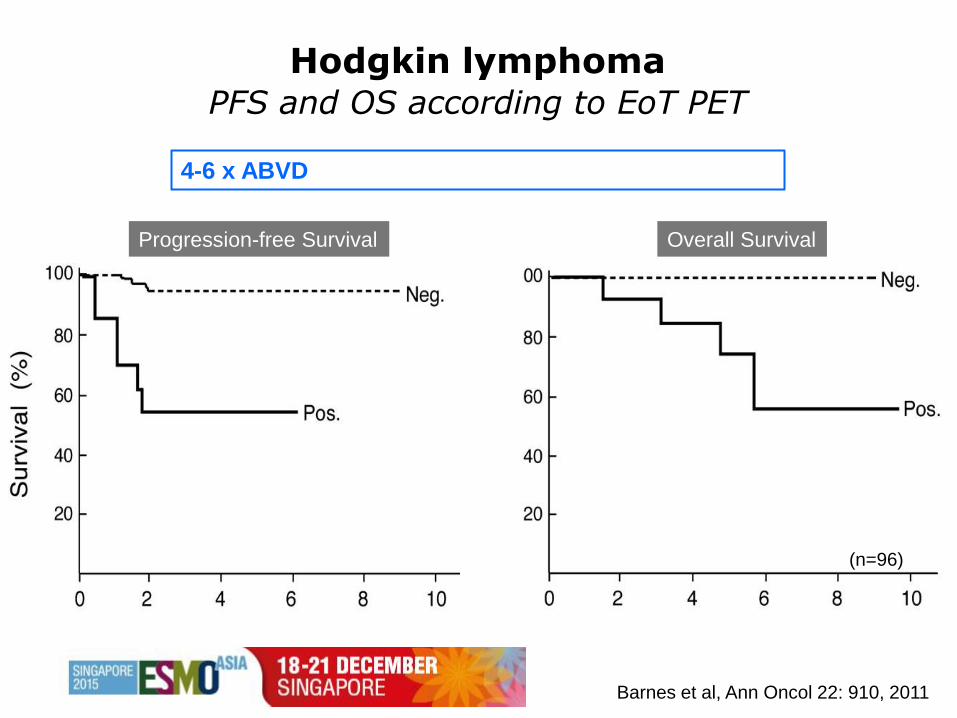
Hodgkin lymphoma PFS and OS according to EoT PET
Barnes et al, Ann Oncol 22: 910, 2011
Progression-free Survival Overall Survival
(n=96)
4-6 x ABVD ± IFRT → EoT PET → local 4-point scale
86 %
14 %
Log-rank P = 0.0001 Log-rank P = 0.0001

Non-inferiority no further treatment vs. standard IFRT :
Difference in 3-year PFS ≤ 7 %
3 x ABVD EoT PET
PET
negative
PET
positive 1 x ABVD + IFRT 30 Gy
IFRT 30 Gy
No further treatment
Stage
IA / IIA
(n = 602)
75 %
25 %
R
Radford et al, N Engl J Med 372: 1598, 2015
Hodgkin lymphoma, early stages British standard: 3 x ABVD + IFRT
Can radiotherapy be omitted in EoT PET-negative patients ?

Overall Survival Progression-free Survival
D PFS - 3,9 %
95 % CI -8,8 - 1,3
(n=420)
Radford et al, N Engl J Med 372: 1598, 2015
Hodgkin lymphoma, early stages No radiotherapy ?
Superiority standard IFRT vs. no further treatment :
Difference in EFS ≥ 10 %
Picardi et al, Leuk Lymphoma 48: 1721, 2007
Hodgkin lymphoma, bulky disease Italian standard: 6 x VEPED + IFRT
Can radiotherapy be omitted in EoT PET-negative patients ?
6 x VEPED EoT PET
PET negative
residual mass
PET positive High-dose therapy
IFRT 32 Gy
No further treatment
Stage
IA - IVB
(n = 260)
62 %
12 %
R
PET negative
no mass No further treatment
26 %
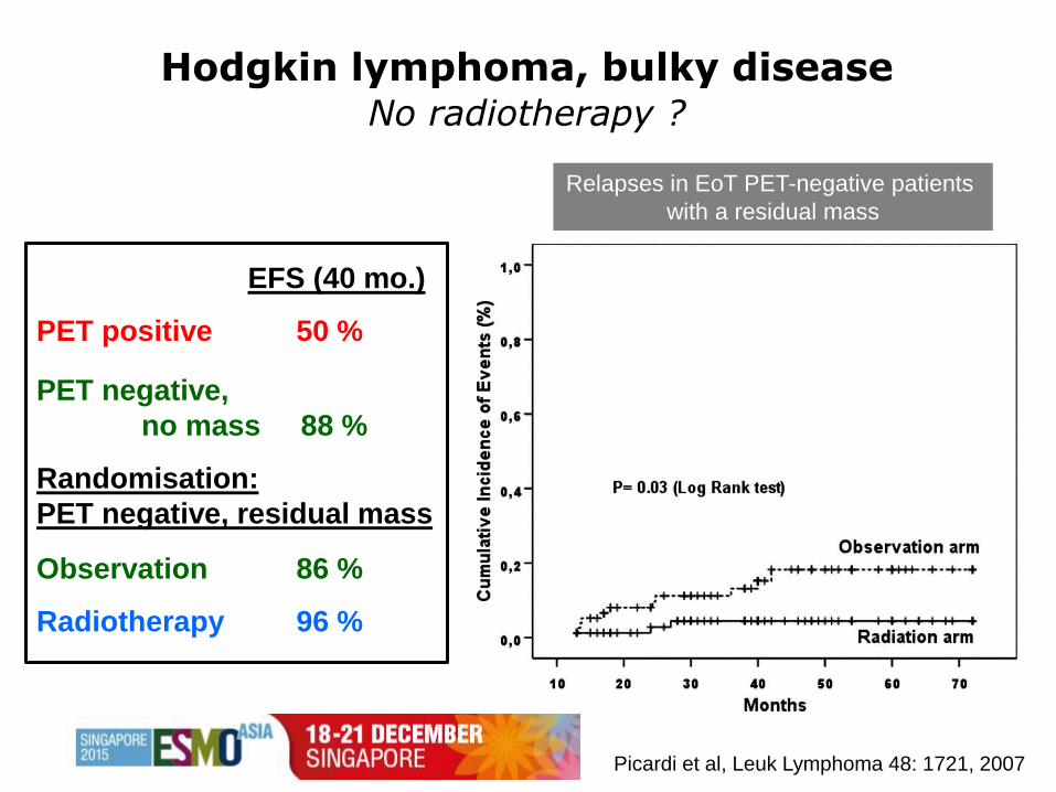
Picardi et al, Leuk Lymphoma 48: 1721, 2007
Relapses in EoT PET-negative patients
with a residual mass
EFS (40 mo.)
PET positive 50 %
PET negative,
no mass 88 %
Randomisation:
PET negative, residual mass
Observation 86 %
Radiotherapy 96 %
Hodgkin lymphoma, bulky disease No radiotherapy ?
D 10 %
(n=160)

Hodgkin lymphoma, advanced stages German standard: 6 x BEACOPPesc + IFRT (residual m.)
Engert et al, Lancet 379: 1791-99, 2012
Progression-free Survival
(n=1620)
11 % of patients
EoT CT: residual mass > 2,5 cm
→ PET: negative → observation
positive → radiotherapy

Primary mediastinal B-cell lymphoma Is radiotherapy required in EoT PET-negative patients ?
Vassilakopoulos et al, Leukemia (in press), 2015
Radiotherapy
No radiotherapy
Freedom from Progression
in EoT PET-negative patients
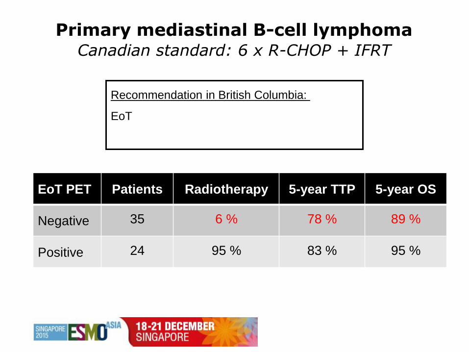
Primary mediastinal B-cell lymphoma Canadian standard: 6 x R-CHOP + IFRT
EoT PET Patients Radiotherapy 5-year TTP 5-year OS
Negative 35 6 % 78 % 89 %
Positive 24 95 % 83 % 95 %
Recommendation in British Columbia:
EoT → PET → negative → observation
→ positive → radiotherapy
Savage et al, ASH #303, 2012

Primary mediastinal B-cell lymphoma Can radiotherapy be omitted in PET-negative patients ?
ClinicalTrials.gov: NCT01599559

Diffuse large B-cell lymphoma PFS and OS according to EoT PET
Cashen et al, J Nucl Med 52: 386, 2011
Progression-free Survival Overall Survival
6 x R-CHOP → EoT PET → IHP
(n=42)
EoT PET negative: 83 %
EoT PET positive: 17 %

Diffuse large B-cell lymphoma Should radiotherapy be given to EoT PET-positive sites ?
Sehn et al,
Hematol Oncol 31 (suppl 1): abstr 123, 2013
EoT PET Patients Radiotherapy 4-year TTP 4-year OS
Negative 167 1 % 74 % 83 %
Positive 82 Yes: 73 %
No: 27 %
81 %
33 %
85 %
30 %
Recommendation in British Columbia:
EoT CT: residual mass > 2,0 cm → PET → negative → observation
→ positive → radiotherapy

Diffuse large B-cell lymphoma Should radiotherapy be given to EoT PET-positive sites ?
Dabaja et al,
Int J Radiation Oncol Biol Phys 89: 384, 2014
Chemotherapy only Chemotherapy + radiotherapy
(n=206) (n=88)

1. Performance of EoT PET
2. Prognostic information
3. Therapeutic implications
must be based on prospective trials !
End-of-treatment PET in lymphoma Key objectives