Embed Size (px)
Citation preview
THE ROLE OF PLAIN FILM RADIOGRAPHY IN THE DIAGNOSIS
AND MANAGEMENT OF KNEE PAIN
By
Chantelle Ann Damon
Dissertation submitted in partial compliance with the requirements for the Master’s Degree in Technology: Chiropractic
Durban University of Technology
I, Chantelle Ann Damon, do hereby declare that this dissertation is representative of my own work in both conception and execution (except where acknowledgements indicate to the
contrary)
……………………………………........... Date: …………………………… Chantelle Ann Damon
Approved for Final Submission
…………………………………………….. Date: …………………………… Dr. J. Shaik M. Tech. Chiro., M. Med. Sci. (SM), MCASA

i
DEDICATION
I dedicate this dissertation to:
My parents, Norma and Raymond Damon. Thank you for all the love and support you have
given me. Thank you for providing me with all I ever wanted or needed. I love you and
appreciate everything you have done.
My brother, Darren and sister, Theresa, even though you have been so far away, you are
always close to my heart. Thank you for your support and love.
My boyfriend, Leroy Ryan, for being there all these years. For lifting me up in my lowest
moments and loving me when I know I was impossible to love. I love you.
My friends, Marencia and Laken, you guys always believed in me. Thank you for always being
around when I needed you to make me laugh and thank you for your support, love and care. I
couldn’t ask for better people to walk with me on my journey of life.

ii
ACKNOWLEDGEMENTS
It is with sincere gratitude and appreciation that I would like to thank the following individuals: 1. My supervisor, Dr. J. Shaik, of the Department of Chiropractic, Faculty of Health
Sciences, Durban University of Technology, for his guidance and advice during the entire process of my research project. This time and knowledge was greatly appreciated.
2. Mrs. Tonya Esterhuizen for her assistance with the statistical analyses used in this study.
3. Funding for this study was provided by the Durban University of Technology’s Research
Fund.
iii
ABSTRACT
Background: Attempts to determine the association between the radiographic and clinical findings of knee
pathology have produced conflicting results. It is also not yet known how knee radiographs
influence the conservative management of patients with knee pain.
Objectives: 1. To determine the association between the clinical and radiographic diagnoses of knee pain.
2. To record the consultation at which a radiograph of the knee was requested by the student
or clinician and the reasons thereof.
3. To record the suspected clinical diagnoses and management of the patients prior to referral
for radiographs of the knee.
4. To determine the number of incidental radiographic findings in the selected radiographs.
5. To determine any change in the clinical diagnoses and management following radiographic
reporting of the selected radiographs.
Method: Radiographic and clinical data from 1 January 1997 to 31 December 2010 were retrospectively
collected from knee radiographs and corresponding patient files from the archives of the
Chiropractic Day Clinic (CDC). Statistical analysis included the use of percentages, mean,
standard deviation, range and frequency counts for the descriptive objectives. Diagnoses were
categorized into specific groups and to construct two-by-two tables of absence or presence of
radiographic vs. clinical diagnosis for each specific diagnosis to determine the association
indicator variables were used.
Results: The overall agreement between the clinical and radiographic diagnoses was 85.5%. For
degenerative joint disease there was a 97.8% agreement while in Osgood Schlatter’s disease
the agreement was 100%, and in chondromalacia patella the agreement was 50%. However,
there was no agreement between the clinical and radiographic diagnoses for each of the other
specific conditions. Degenerative changes were the most common radiographic findings. The
iv
majority of the knee radiographs were requested at the initial consultation and as the length of
treatment increased, the frequency of radiograph requests decreased. The most common
reasons for referral for radiographs were to identify degenerative changes (47.5%) and to
assess for unspecified pathology (37.4%). Of the 146 patients in this study, 125 patients did not
have a change in diagnosis after radiographs were obtained which means that 85.6% of the
diagnoses remained the same after radiographic examination. There was a wide range of
treatment modalities utilized in the management of patients with knee pain, including soft tissue
therapy, electrotherapeutic modalities and manual therapy (manipulation and mobilization). The
use of manual therapy increased from 67.8% prior to radiographs being taken to 82.9% after
radiographs were obtained.
Conclusion: Knee radiographs were over-utilized at the CDC and the findings on radiography did not have
much influence on the diagnosis and the management of the patient presenting with knee pain.
The majority of the clinical diagnoses were degenerative causes of knee pain.
v
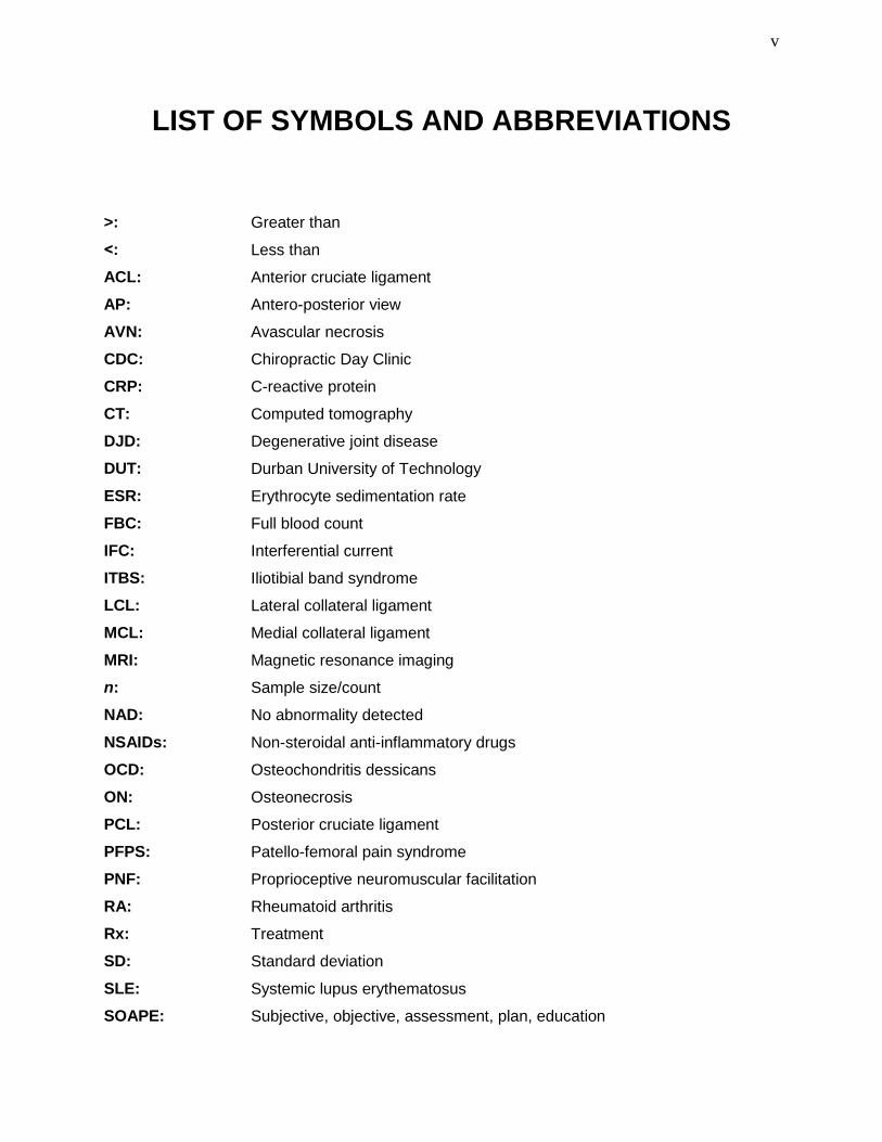
LIST OF SYMBOLS AND ABBREVIATIONS
>: Greater than
˂: Less than
ACL: Anterior cruciate ligament
AP: Antero-posterior view
AVN: Avascular necrosis
CDC: Chiropractic Day Clinic
CRP: C-reactive protein
CT: Computed tomography
DJD: Degenerative joint disease
DUT: Durban University of Technology
ESR: Erythrocyte sedimentation rate
FBC: Full blood count
IFC: Interferential current
ITBS: Iliotibial band syndrome
LCL: Lateral collateral ligament
MCL: Medial collateral ligament
MRI: Magnetic resonance imaging
n: Sample size/count
NAD: No abnormality detected
NSAIDs: Non-steroidal anti-inflammatory drugs
OCD: Osteochondritis dessicans
ON: Osteonecrosis
PCL: Posterior cruciate ligament
PFPS: Patello-femoral pain syndrome
PNF: Proprioceptive neuromuscular facilitation
RA: Rheumatoid arthritis
Rx: Treatment
SD: Standard deviation
SLE: Systemic lupus erythematosus
SOAPE: Subjective, objective, assessment, plan, education

vi
TENS: Transcutaneous electric nerve stimulation
Yrs: years
vii
LIST OF TABLES
CHAPTER TWO Table 2.1 Classification of the causes of knee pain 6 Table 2.2 Red flags associated with knee pain 10 Table 2.3 Indications for radiographing the knee 15 Table 2.4 Ottawa knee rules for requesting knee radiographs 15
CHAPTER THREE Table 3.1 Data collected and the source of the data 25 CHAPTER FOUR Table 4.1 The mean, standard deviation and the range of patients whose clinical files
and radiographs were examined 27
Table 4.2 The frequencies of each radiographic diagnosis by clinical diagnosis 29 Table 4.3 The agreement between clinical and radiographic diagnoses 30 Table 4.4 A summary of the consultation at which knee radiographs were requested
and the reasons thereof 32
Table 4.5 Suspected clinical diagnosis and management prior to radiographic referral 33 Table 4.6 Details of change of diagnosis 36 Table 4.7 Incidental findings and related suspected clinical diagnoses 37
viii
LIST OF FIGURES CHAPTER TWO Figure 2.1 Suggested algorithm for the management of knee pain 19 CHAPTER FOUR Figure 4.1 Gender distribution 28
Figure 4.2 Management prior to requests for radiographs 35
Figure 4.3 Management of the patients after knee radiographs were obtained 36

ix
LIST OF APPENDICES
Appendix 1: Clinical and Radiographic Data Collection sheet
Appendix 2: Patient Information sheet
Appendix 3: Ethical Clearance Certificate
x
TABLE OF CONTENTS
DEDICATION i ACKNOWLEDGEMENTS ii ABSTRACT iii LIST OF ABBREVIATIONS v LIST OF TABLES vii LIST OF FIGURES viii LIST OF APPENDICES ix
CHAPTER ONE…………………………………………………………….…………………….….1 1.1 INTRODUCTION TO THE STUDY 1.2 AIMS AND OBJECTIVES
1.1.1 The aims of the study 1.1.2 The objectives of the study
1.3 HYPOTHESES 1.4 SCOPE OF THE STUDY
1.5 LIMITATIONS OF THE STUDY CHAPTER TWO……………………………………………………………………………….…….5 2.1 INTRODUCTION TO KNEE PAIN 2.2 THE AETIOLOGY AND DIAGNOSIS OF KNEE PAIN
2.2.1 Factors associated with the diagnosis of knee pain 2.2.2 The association between the history and examination findings and the diagnosis
of knee pain
2.3 THE ROLE OF PLAIN FILM RADIOGRAPHY IN THE DIAGNOSIS OF KNEE PAIN 2.3.1 Utilization of radiographs in clinical practice 2.3.2 Advantages of radiographs in diagnosing knee pain
xi
2.3.3 Limitations of radiographs in diagnosing knee pain
2.4 THE CORRELATION BETWEEN THE CLINICAL AND RADIOGRAPHIC FINDINGS 2.5 AN OVERVIEW OF THE MANANGEMENT OF KNEE PAIN
2.5.1 Chiropractic management of knee pain
2.6 CONCLUSION CHAPTER THREE……………………………………………………………………….……......23 3.1 STUDY DESIGN 3.2 PATIENT CONFIDENTIALITY 3.3 SAMPLING METHOD AND SAMPLE SIZE 3.4 INCLUSION AND EXCLUSION CRITERIA
3.4.1 Inclusion criteria 3.4.2 Exclusion criteria
3.5 RESEARCH PROCEDURES 3.6 STATISTICAL ANALYSIS CHAPTER FOUR…………………………………………………………………..…….………..27 4.1 AGE AND GENDER 4.2 THE ASSOCIATION BETWEEN THE CLINICAL AND THE RADIOGRAPHIC
DIAGNOSES OF PATIENTS WITH KNEE PAIN 4.3 THE CONSULTATION WHEN A RADIOGRAPH WAS REQUESTED AND THE
REASONS THEREOF 4.4 SUSPECTED CLINICAL DIAGNOSES AND MANAGEMENT PRIOR TO REFERRAL
FOR KNEE RADIOGRAPHS 4.5 CHANGES IN CLINICAL DIAGNOSIS AND MANAGEMENT AFTER RADIOGRAPHS
4.6 INCIDENTAL RADIOGRAPHIC FINDINGS
xii
CHAPTER FIVE…………………………………………………………………………….……..38 5.1 AGE AND GENDER 5.2 THE ASSOCIATION BETWEEM THE CLINICAL AND THE RADIOGRAPHIC
DIAGNOSES OF PATIENTS WITH KNEE PAIN
5.3 THE CONSULTATION WHEN A KNEE RADIOGRAPH WAS REQUESTED AND THE REASONS THEREOF
5.4 THE CLINICAL DIAGNOSIS AND MANAGEMENT OF PATIENTS PRESENTING WITH KNEE PAIN BEFORE AND AFTER RADIOGRAPHY
5.5 INCIDENTAL RADIOGRAPHIC FINDINGS
5.6 PROPOSED RECOMMENDATIONS FOR THE CHIROPRACTIC DAY CLINIC
CHAPTER SIX……………………………………………………………………………….…….46 6.1 CONCLUSION 6.2 RECOMMENDATIONS REFERENCES……………………………………………………………………………….…….48 APPENDICES
1
CHAPTER ONE
INTRODUCTION
1.1. INTRODUCTION TO THE STUDY
Low back pain and knee pain are the two most common musculoskeletal complaints seen in
private practice (Antonopoulou et al., 2009). Knee pain occurs in approximately 20% of the
general population (Levy and Dickey-White, 2009) and this incidence is thought to be
increasing due to the rise in the activity levels of individuals (Calmbach and Hutchens,
2003). Since the differential diagnoses of knee pain are considerable, the exact cause of the
pain may be difficult to isolate. A clinical history and physical examination are required to
identify possible causes of knee pain, and to determine whether the use of diagnostic testing
is warranted (Calmbach and Hutchens, 2003). Following the clinical examination, a
treatment program may be initiated depending on the suspected diagnosis (Levy and
Dickey-White, 2009). A thorough clinical examination may lead to the detection of certain
features termed “red flags” which indicate serious underlying pathologies. These “red flags”
which include a history of direct trauma, inability to move the joint, and no response to
appropriate treatment may warrant secondary investigations such as radiographs and
magnetic resonance imaging (MRI) (Fagan and Davies, 2000; Levy and Dickey-White,
2009).
Chiropractors’ utilization of radiographs began soon after the discovery of ionizing radiation
in 1895 (Cooperstein and Gleberzon, 2004). Besides being important to the chiropractor in
aiding in the clinical diagnosis, radiographs also provide information regarding
biomechanical anomalies, recognizing contra-indications to manipulative therapy, and to
follow the course of degenerative processes. The evidence provided by radiographs is then
utilized for formulating or changing a treatment plan and identifying the need for referral for
further management (Cooperstein and Gleberzon, 2004).
Chiropractors have been criticized for overuse of radiographs (Ammendolia et al., 2008), but
this is not unique to this profession as general practitioners are also known for radiograph
overuse (Phillips, 1992; Morgan et al., 1997). The overuse of knee radiographs is particularly
related to medico-legal considerations, and in identifying degenerative changes that were
2
previously expected (Morgan et al., 1997), despite exposing patients to unnecessary ionizing
radiation which may have adverse effects such as the risk of developing cancer, or genetic
abnormalities (Shapiro, 2002).
In conclusion, the reported overuse of radiographs by chiropractors and observations of
Morgan et al. (1997) emphasise the need to determine the correlation between the
radiographic diagnoses and clinical diagnoses of knee pain. It is also important to not only
investigate the role of knee radiographs in determining the treatment protocol, but also how
the radiographic findings may result in alterations to the treatment or management protocol.
Therefore, the aim of this research was to determine the correlation between clinical and
radiographic diagnosis and to determine if or how the radiographic diagnosis influenced a
change in the management of the patient with knee pain.
1.2. AIMS AND OBJECTIVES
1.2.1. THE AIMS OF THE STUDY
The aims of this study were to:
1. Determine whether there was an association between the clinical and the
radiographic diagnoses of patients who presented with knee pain at a chiropractic
teaching clinic and;
2. Whether radiographs of the knee influenced a change in the diagnosis or
management of these patients.
1.2.2. THE OBJECTIVES OF THE STUDY
Specific objectives were identified and these included:
1. To determine the association between the clinical and the radiographic diagnoses of
knee pain.
2. To record the consultation at which a radiograph of the knee was requested by the
student or clinician and the reasons thereof.
3. To record the suspected clinical diagnoses# and management of the patients prior to
referral for radiographs of the knee.
3
4. To determine the number of incidental radiographic findings* in the selected
radiographs.
5. To determine any change in the clinical diagnoses# and management following
radiographic reporting of the selected radiographs.
* Definition of an incidental finding: “any abnormality not related to the illness or causes that prompted the diagnostic imaging test” (Lumbreras et al., 2010).
# This refers to the clinical suspicion as it appeared either on the SOAPE note or the radiographic request form at the point of referral for radiographs.
1.3. HYPOTHESES
The Alternate Hypothesis (Ha) was set for the first objective which stated that there would be
a significant association between the clinical and the radiographic diagnoses of patients with
knee pain.
The Alternate Hypotheses (Ha) was set for the fifth objective which stated that the
radiographic diagnosis would significantly influence a change in the clinical diagnosis and
management of the patient.
1.4. SCOPE OF THE STUDY
The results of 146 radiographs of the knee and the corresponding patient files that met the
inclusion and exclusion criteria are discussed in this dissertation. All radiographs and patient
files were obtained from the Chiropractic Day Clinic (CDC) archives at the Durban University
of Technology (DUT). Informed consent for the use of clinical and radiographic information
was obtained from patients at their initial consultation. Codes were assigned to each
patient’s name to maintain patient confidentiality and these were used instead of patients’
names in the data sheets. Access to the patient files and knee radiographs were restricted to
the researcher and supervisor.
4
1.5. LIMITATIONS OF THE STUDY
The study was limited to including only the plain film radiographs of the knee and the
corresponding patients’ files within the CDC archives. Knee radiographs in this study were
required to be taken during the patient’s management at the CDC. Therefore, this study
excluded patients with knee pain who presented at the initial consultation with knee
radiographs. This was due to the fact that it could not be determined if there would be an
influence in the diagnosis or management of the patient by the radiographic findings if
patient presented at the initial consultation at the CDC with the knee radiographs.
Since this study was retrospective, there was no way to verify the clinical findings as the
details were already recorded in the patient’s files. This study may not be representative of
all knee pain patients at the CDC, as there was probability of some radiographs being
removed from the archives for teaching purposes and others being taken home by patients.
5
CHAPTER TWO
LITERATURE REVIEW
2.1. INTRODUCTION TO KNEE PAIN
The knee joint is the largest and one of the most complex joints in the body (Calmbach and
Hutchens, 2003). The structure of the knee joint and its location subject it to many stresses,
diseases and trauma. This causes the knee joint to be the most injured joint in the body
(Smillie, 1978; Calliet, 1992; Calmbach and Hutchens, 2003). Knee pain is second to low
back pain as the most common musculoskeletal conditions seen in private practice
(Antonopoulou et al., 2009; Levy and Dickey-White, 2009).
The incidence of knee pain is increasing rapidly as there is a rise in the activity levels of
individuals in today’s society (Calmbach and Hutchens, 2003). The majority of patients with
knee pain present to medical doctors for their assessment and treatment. However, with the
growth of the alternate health care sector, more patients are presenting to chiropractors for
the management of their knee pain and other musculoskeletal complaints (Goldstein, 1999).
2.2. THE AETIOLOGY AND DIAGNOSIS OF KNEE PAIN
Musculoskeletal conditions are generally difficult to diagnose. This is true for the causes of
knee pain due to the extensive differential diagnosis (Calmbach and Hutchens, 2003). The
knee is one of the most complex joints of the body and is subjected to multiple stresses and
pathological factors making it difficult to reach a definitive diagnosis (Smillie, 1978; Calliet,
1992; Calmbach and Hutchens, 2003), although a thorough clinical history and physical
examination can narrow the list of differential diagnoses (Calmbach and Hutchens, 2003).
A useful classification of the causes of knee pain proposed by Wallace and Staats (2004) is
presented in Table 2.1.
6
Table 2.1 Classification of the causes of knee pain
Extra-articular causes Intra-articular causes
• Iliotibial band syndrome
• Patellar tendonitis
• Pes anserine bursitis
• Ligament injuries (MCL and LCL)
• Pain referred from the lumbar spine
or hip joints
• Meniscal lesions
• Ligament injuries (ACL and PCL)
• Plica syndrome
• Patello-femoral pain syndrome
• Degeneration
• Inflammatory arthritis
• Infection
• Fractures
• Osteonecrosis
• Osteochondritis dessicans
• Tumours
*Table adapted from Wallace and Staats (2004)
ACL= anterior cruciate ligament; LCL= lateral collateral ligament; MCL= medial collateral ligament; PCL= posterior cruciate ligament
Several extra-articular or intra-articular causes may be responsible for knee pain (Table 2.1).
The intra-articular causes include lesions affecting the structures directly related to the knee
joint such as the bones (femur, tibia, fibula and patella), cartilage, ligaments and soft tissue
surrounding and within the knee joint (Wallace and Staats, 2004).
The extra-articular causes include lesions involving joints proximal to the knee joint (hip joint
and lumbar spine pathologies referring to the knee joint), tendons and soft tissues that cross
or surround the knee joint and refer pain to the joint (pes anserine bursa, Iliotibial band,
patella tendon, medial collateral ligament (MCL) and lateral collateral ligament (LCL).
The common extra-articular causes can be divided into three categories: referred pain,
overuse syndromes and ligament injuries (Wallace and Staats, 2004). The most common of
these are the overuse syndromes which affect the tendinous insertions around the knee and
are caused by repetitive movements of the knee which result in inflammation of the tendons
e.g. repetitive flexion and extension of the knee results in the iliotibial band friction syndrome
(ITBS). The region of knee which is affected is specific to the tendon structure involved.
(Anterior knee pain is caused by patellar tendonitis and lateral knee pain is caused by ITBS).
Although the diagnosis of overuse syndromes are not difficult to reach, the clinical history
must be thorough to identify the mechanism of injury and the physical examination must
include movements of the knee which will reproduce the pain and palpation to identify which
structures are affected. Further investigations are not usually necessary although they may
be used to confirm the diagnosis. These investigations may include radiographs, ultrasound,
7
computed tomography (CT) scanning or magnetic resonance imaging (MRI) (Calmbach and
Hutchens, 2003; Martinez, 2009). Pes anserine bursitis is usually diagnosed on clinical
grounds and further investigations are usually not necessary (Glencross, 2009). However, in
certain cases, investigations such as ultrasonography may aid the clinician in the diagnosis
(Glencross, 2009).
Two extra-articular knee ligaments commonly injured are the MCL and LCL (DeBerardino,
2010). The MCL is the most commonly injured ligament of the knee due to either direct or
indirect trauma to the lateral knee or overuse injury which may occur as a result of repetitive
valgus loading at the knee joint (DeBerardino, 2010). The LCL is less commonly injured as
the opposite leg acts as a guard to the medial aspect of the knee. However, direct trauma to
the medial aspect of the knee may take place while the knee is in extension and placed in
front of the body. These ligament injuries can be diagnosed on clinical history and
examination as they are often sports-related and will present with a specific mechanism of
injury (DeBerardino, 2010; Ho, 2010).
Referred pain to the knee from the hip or the lumbar spine is difficult to identify as all other
possible causes of knee pain need to be excluded (diagnosis of exclusion) (Hollis, 2010).
Pain referred from the lumbar spine is most commonly due to nerve root entrapment
especially at L4. Pain referred to the knee from the hip may result from specific conditions
(e.g. slipped capital femoral epiphysis) or may be due to active myofascial trigger points of
muscles of the hip region (e.g. hamstrings and adductors). Nerves exiting the hip (e.g.
femoral nerve) may become entrapped and refer pain to the knee and cause other
symptoms in the knee (e.g. weakness). Once the region is identified as the primary cause,
investigations may be required to evaluate the region to reach a definitive diagnosis
(Calmbach and Hutchens, 2003; Hollis, 2010).
Intra-articular structures, including the menisci (medial and lateral) and anterior and posterior
cruciate ligaments (ACL and PCL) (Table 2.1) may also be injured (Aiello, 2008; Allen,
2010). Meniscal tears may be due to either traumatic injury or degenerative processes.
Traumatic injuries are more commonly observed in athletes whereas degeneration is more
likely in the elderly. Traumatic meniscal injuries are often associated with ACL and MCL
injuries resulting in the “unhappy triad” syndrome (Freitas, 2011). The PCL is injured most
commonly in the hyperextended position whereas the ACL in the partially flexed position
(Aiello, 2008; Allen, 2010). The ACL is the most commonly injured ligament of the knee and
injury occurs in both athletes and non-athletes, either with or without contact to the knee.
Ligament injuries are graded depending on the severity, from partial to complete tears (Allen,
2010).
8
The plica syndrome results from a remnant of foetal tissue in the knee which usually
decreases in size in the second trimester of pregnancy (Dupont, 1997). However, in some
individuals this process does not happen and the tissue continues into adult life. It is then
referred to as plica and can be injured as a result of direct trauma or an overuse injury when
the knee is flexed (Dupont, 1997).
Although patello-femoral pain syndrome (PFPS) is one of the most common of all musculo-
skeletal complaints, the evaluation, diagnosis and treatment of this condition are often
difficult due to the multifactorial aetiology (Servi, 2009). Aggravating factors include joint
malalignment, unbalanced muscle pull, excessive knee valgus deformity and excessive
loading, all of which need to be evaluated in order to reach the definitive diagnosis of PFPS.
These factors also lead to the development of degenerative joint disease (DJD) (Potter,
2009). A common cause of knee pain in the middle-aged and elderly individuals is DJD
which is a non-inflammatory arthritide as opposed to the majority of the other arthritic causes
of knee pain. The inflammatory causes (which also may be referred to as systemic causes)
of knee pain include rheumatoid arthritis (RA), septic arthritis and gout. These inflammatory
causes always have symptoms that warrant further investigation as the clinical features will
suggest the underlying pathology. These features include severe knee pain, swelling, fever,
exquisite tenderness and redness of the joint (Brusch, 2010; Patel, 2011). It should be noted
that several of these features may also be observed in infections or tumours around the
knee and even in post-traumatic states.
Infections of the knee (including bone infections) may be seen in individuals of any age, but
are most common in immuno-compromised individuals as in cases of alcoholism, prolonged
corticosteroid therapy and acquired immunodeficiency syndrome (AIDS) (Calmbach and
Hutchens, 2003). The onset of pain and swelling is sudden with no history of trauma.
Physical examination reveals a warm, swollen and tender joint with intense pain on any
movement (Calmbach and Hutchens, 2003).
There are many different fractures of the knee such as stress fractures, pathological
fractures, open and closed fractures. Closed fractures and stress fractures can be treated
conservatively with rest and immobilisation (Handoll and Parker, 2008). More severe
fractures that are open and displaced require surgical reduction. These, therefore, require
immediate referral for surgical intervention (Handoll and Parker, 2008; Steele, 2011).
Osteonecrosis (ON) or avascular necrosis (AVN) is a condition characterised by necrosis of
osseous tissue as a result of derangement of circulation (Rajadhyaksha, 2008). Often the
cause is not found as the condition occurs spontaneously, but in some cases the clinician is
able to identify a cause, such as trauma. Imaging studies such as plain film radiography and
9
MRI may be used to diagnose ON (Rajadhyaksha, 2008). Osteochondritis dessicans (OCD)
is a disorder of the calcification centres characterised by avasular necrosis and
recalcification. The aetiology is multifactorial and includes trauma, ischaemia and genetic
predisposition. This disorder may be diagnosed on plain film radiography, radioisotope bone
scan or MRI (Jacobs, 2011).
The knee joint is the most common location for primary bone tumours which occur as either
benign or malignant. Osteochondromas (exostoses), the most common of the benign
tumours, are composed of cortical and medullary bone with an overlying hyaline
cartilaginous cap. They may be solitary or multiple resulting in deformity of the knee joint,
and malignant change may occur in less than 5% of cases (Murphey et al., 2000; Yochum
and Rowe, 2005; Breitenseher and Dominkus, 2006). Malignant tumours are one of the most
important of all pathological conditions affecting the knee (Dickinson et al., 1997). The most
common of all tumours are metastatic bone tumours. Malignant tumours account for 70% of
all metastatic tumours and those affecting bone have a primary extra-skeletal site (Yochum
and Rowe, 2005). Primary malignant bone tumours include multiple myeloma and
osteosarcoma. Osteosarcomas are often located in the long bones of the extremities and
most commonly affect the knee and shoulder joints. A clinician should be aware of the
appearance of this tumour on radiographs as it is similar to that of myositis ossificans
(Yochum and Rowe, 2005). Even though a clinician may suspect a tumour on clinical
grounds, radiographic confirmation may not be possible due to the latent period. This is the
time interval between the onset of clinical symptoms and the appearance of the radiographic
features. Further diagnostic testing and imaging may be required e.g. erythrocyte
sedimentation rate (ESR) and alkaline phosphatase enzyme, ultrasound MRI and bone
biopsy to identify the specific tumour (Longmore et al., 2007; Cameron and Howard, 2010).
As there are several causes of knee pain, a definitive diagnosis may be difficult to reach.
However, with a thorough clinical history and a complete physical examination, clinical
findings may suggest a specific diagnosis or a list of differential diagnoses (Calmbach and
Hutchens, 2003). Depending on the suggested list of differential diagnoses there may be the
need to request for special investigations such as radiographs to guide the clinician to the
diagnosis or to exclude other diagnoses.
10
2.2.1. FACTORS ASSOCIATED WITH THE DIAGNOSIS OF KNEE PAIN
The differential diagnosis of knee pain is considerable which makes it difficult to reach an
exact diagnosis (Calmbach and Hutchens, 2003). Clinicians should focus on the clinical
history as this leads to the identification of symptoms which are characteristic to a specific
condition. Once these symptoms are identified, the physical examination is easier and is
used as a confirmation in the diagnosis of the condition suggested by the symptoms
(Calmbach and Hutchens, 2003). If the history is not thorough and specific, the physical
examination may be confusing making it difficult to reach a clear diagnosis. Once the clinical
history and physical examination is complete, a suspected diagnosis is reached and a
management plan is initiated (Levy and Dickey-White, 2009). The suspected diagnosis is a
working diagnosis which allows the clinician to assess the response of the condition to the
management. If there is no response to treatment or the response is insignificant, further
investigations may be necessary to identify any underlying causes (Fagan and Davies, 2000;
Levy and Dickey-White, 2009).
During the clinical assessment (history and physical examination) there may be certain
features which indicate serious underlying pathologies (Table 2.2). These features are
termed ‘red flags’ and require a thorough evaluation to determine the exact pathology
(Fagan and Davies, 2000; Levy and Dickey-White, 2009). Red flags may present at any time
during the natural development of a condition. Therefore, a careful assessment of the
patient’s status is important to identify the development of any red flags during patient
management. Serious pathologies indicated by red flags include infections, neoplasms,
inflammatory arthritides and fractures (Steele, 2011).
Table 2.2 Red flags associated with knee pain
Category History findings Physical findings
General Innocuous symptoms
Nocturnal pain¹
Knee pain with or without referral²
Failure to improve
No response to treatment
Inability to bear weight
Locking of knee
Neurological deficit
Joint deformity*
Malignancy Constitutional symptoms¹
Night pain¹
Severe knee pain
Neurological deficits¹
11
Age ˂18 years¹ Immobility
Deformity*
Infection
Constitutional symptoms¹
History of infection¹
Constitutional symptoms¹
Pain, warmth and swelling¹
Nerve root entrapment in the lumbar spine; nerve entrapment in hip region
History of neurological symptoms² Generalised knee pain²
Neurological deficits²
Fracture History of trauma¹
Age > 60 years*
Knee pain¹
Swelling and ecchymosis¹
Point tenderness¹
Inflammatory arthritides Constitutional symptoms
Age > 40 years*
Pain and swelling¹
Constitutional symptoms*
¹Adapted from Cameron and Howard (2010); ²adapted from Wallace and Staats et al. (2004); *adapted from Yochum and Rowe (2005)
There are symptoms that are not considered red flags when they present alone (innocuous
symptoms). These include localised knee pain, swelling, morning stiffness, pain worse on
walking and on weight-bearing. However, if these symptoms present with other red flag
symptoms, they may indicate serious underlying pathologies. For example, localised knee
pain and swelling may not be red flags if they present alone, but if they present with a history
of trauma or with constitutional symptoms (e.g. fever, night sweats or weight loss of
unknown cause), these may indicate fractures or malignancy (Cameron and Howard, 2010).
Red flags may also be related to the duration of symptoms which may occur in several ways;
symptoms may occur acutely (e.g. with a fracture, severe pain and swelling after trauma), or
they may be present over a longer period of time (e.g. neurological deficit) (Levy and Dickey-
White, 2009).
The patients’ age may provide a clue to the potential diagnosis. Knee pain caused by
malignancy usually occurs in individuals younger than 18 years and Osgood Schlatter’s
disease (tibial apophysitis) is most commonly observed in boys aged 13 or 14 years and
girls aged 10 or 11 years. Patients over the age of 60 years are more at risk of fractures
which may be as a result of repetitive stress on the area or due to osteoporosis. Rheumatoid
arthritis is most commonly observed in young to middle-aged adults whereas crystal induced
12
inflammatory arthropathies such as gout most commonly present in the elderly. Infection of
the knee is suspected by clues such as recurrent respiratory or urinary infections, drug
abuse or in individuals who are immuno-compromised (Calmbach and Hutchens, 2003;
Doherty et al., 2010).
The duration of a patient’s symptoms is an important clue because a condition lasting for
more than six weeks may be due to a serious underlying pathology, but a clinician should be
aware that benign conditions such as DJD may also be symptomatic for several weeks or
months. Patients whose symptoms persist for more than six weeks should undergo a
thorough re-evaluation to determine the nature of the condition and clinicians should request
further investigations to rule out serious pathologies. An example of a condition that may be
misdiagnosed resulting in chronicity of symptoms is malignancy. Symptoms such as pain
and swelling may be mistaken for degeneration, but if the symptoms persist and increase in
severity over time, malignancy should be considered (Cameron and Howard, 2010). With a
thorough re-evaluation and further investigation the correct diagnosis may be reached. A red
flag which is also very important to consider is non-responsiveness to treatment. This is
important because the clinician may have diagnosed the patient incorrectly, and therefore,
instituted the incorrect management protocol or, there may have been an underlying cause
which was missed during the evaluation of the patient. However, not all conditions that don’t
respond to treatment are red flags. Degenerative conditions are notorious for not responding
to treatment (Stauffer et al., 2011). Another factor which affects the response to treatment is
patient (non-) compliance. Patients are educated with regards to their management and
taught home-exercises and stretches to supplement the management program. If patients do
not perform these activities, the management program may be partially effective or only be
effective over a longer period of time. However, with the help of the patient in being involved
with his or her treatment, the response to treatment would be much quicker (Schenk, 2005).
Clinical features such as temperature above 38°C, night sweats, unexplained weight loss of
and generalised lymphadenopathy are suggestive of malignancy or infection (Cameron and
Howard, 2010; Doherty et al., 2010). A previous or current medical history of cancer and
nocturnal pain may indicate metastatic spread to the knee. Deformity of any nature should
be considered as red flags as these require further investigation to determine the cause
(Stevens, 2010). The deformities may be either valgus (knock-knees) or varus (bow-leg) and
causes range from physiological, genetic disorders or metabolic bone disease such as
Charcot’s (neuropathic) arthropathy (Stevens, 2007; Shah, 2011). If, at any point during the
clinical history the main complaint is not determined for any reason, the clinician should
suspect a more serious cause of knee pain as a matter of precaution. Examples of cases in
13
which this occurs are patients with mental illness or unconscious patients and the inability to
communicate (e.g. language barriers).
Neurological symptoms may present as dermatome, myotome or reflex deficits (Bradley,
2004). Dermatomes affected present as diminished sensation including, light touch, crude
touch, vibration, temperature and pain (e.g. as seen in diabetes mellitus). Myotome deficits
present as weakness or atrophy in the musculature surrounding the knee joint (e.g.
quadriceps weakness seen in L4 lumbar nerve root entrapment). Reflexes affected may
include the knee jerk or the ankle reflexes which may be either decreased or absent. It is
important for a clinician such as a chiropractor to be aware that non-musculoskeletal
conditions such as hypothyroidism may also cause neurological deficits (Longmore et al.,
2007). Sometimes, even with a history that provides clues to a condition, the physical
examination may not provide any useful information to the clinician. This may be a result of
the timing of the natural history of the condition i.e. the condition may not have reached the
clinical phase in which features are present on physical examination. A Baker’s cyst may
present with constitutional symptoms whereas with internal knee derangement there may be
some degree of deformity e.g. a mild varus deformity. Vascular symptoms may be produced
by popliteal artery calcification or aneurysm and cause the leg, distal to the knee to lose
warmth and colour (Bradley, 2004; Longmore et al., 2007).
2.2.2. THE ASSOCIATION BETWEEN THE HISTORY AND EXAMINATION FINDINGS AND THE DIAGNOSIS OF KNEE PAIN
Several clinicians have attempted to link findings from the clinical history and physical
examination (Oberholder et al., 1993; O’Shea et al., 1996; Wagemakers et al., 2008) but
have been unsuccessful. It has been reported that the history has more of a diagnostic value
than the physical examination (Wagemakers et al., 2008). The relationship between knee
pain and knee joint abnormality is controversial as there are cases in which there is knee
abnormality (e.g. valgus deformity seen in DJD), but the patient presents with no pain or
other symptoms besides pain (Yochum and Rowe, 2005). Furthermore, many patients
present with knee pain and other symptoms characteristic of a condition, but the patient
does not have a confirmatory diagnosis. It is, therefore, difficult to reach a diagnosis in these
cases as the association between the presenting symptoms and the pathology present is
poor (Claessens et al., 1990; Bedson and Croft, 2008).
Conflicting information has been reported by authors highlighting the controversy around the
diagnosis of knee pain. Bedson and Croft (2008) reported that knee pain is not a precise
marker for knee osteoarthritis and that radiographic evidence of osteoarthritis is not a
14
precise guide to the likelihood that knee disability or pain will be present. Neogie et al.
(2008), on the other hand, reported that radiographic evidence of osteoarthritis is strongly
associated with knee pain.
Chronic knee pain in the elderly and children or adolescents is commonly diagnosed as
degenerative joint disease or patella-femoral pain syndrome respectively (McAlindon, 1999;
Calmbach and Hutchens, 2003). The severity of the knee pain and the degree of disability in
any age has been reported to correlate poorly to the pathological findings of these conditions
(McAlindon, 1999).
2.3. THE ROLE OF PLAIN FILM RADIOGRAPHY IN THE DIAGNOSIS OF KNEE PAIN
2.3.1. UTILIZATION OF RADIOGRAPHS IN CLINICAL PRACTICE
Plain film radiography is utilised in clinical practice to identify pathological changes and to aid
the clinical diagnosis of patients (Table 2.2). Occasionally, radiographs are utilised in
circumstances where the clinical diagnosis is not known (Cooperstein and Gleberzon, 2004).
Radiography is a relatively inexpensive and readily available as a diagnostic tool compared
to other diagnostic imaging tests (Koplas and Schils, 2008). Radiographic imaging is
relatively quick and the films produced are easy to evaluate (Yochum and Rowe, 2005).
Soon after the discovery of ionizing radiation in 1895, chiropractors began utilising
radiographs to aid in the diagnosis of their patients (Cooperstein and Gleberzon, 2004).
Radiographic imaging is an important diagnostic tool for the chiropractor as it aids in
formulating a clinical diagnosis, provides information regarding biomechanical anomalies
and the course of degenerative processes, identifies red flags and contra-indications to
manipulative therapy and for protection against medico-legal challenges.
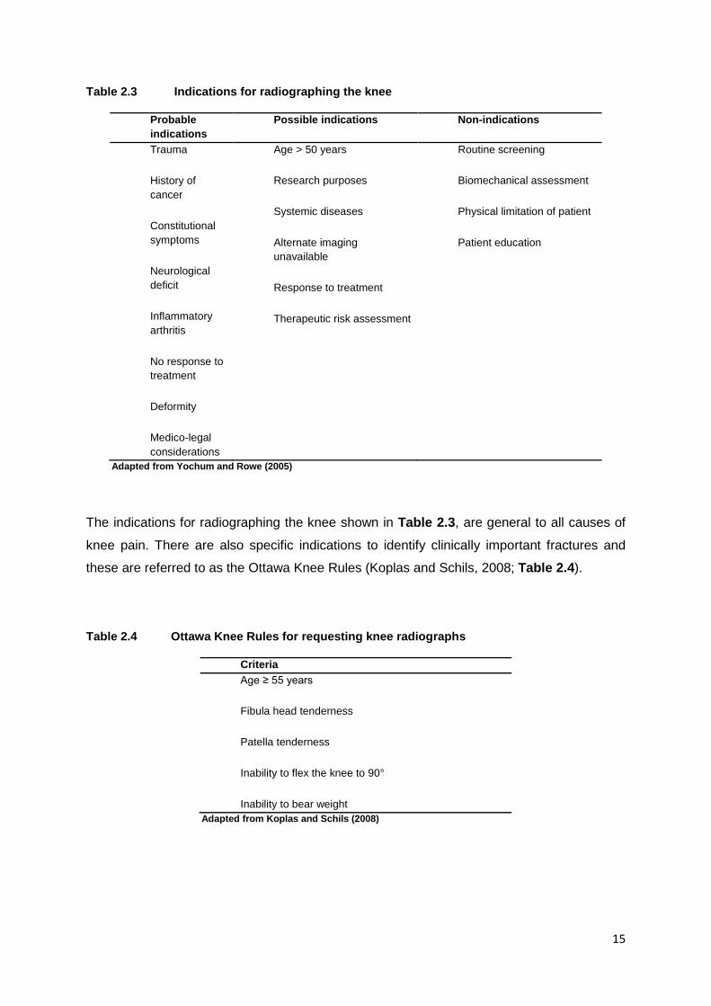
Yochum and Rowe (2005) provide a list of general indications for skeletal diagnostic
imaging, but some of these indications are applicable to patients presenting with knee pain.
These are shown in Table 2.3.
15
Table 2.3 Indications for radiographing the knee
Probable indications
Possible indications Non-indications
Trauma
History of cancer
Constitutional symptoms
Neurological deficit
Inflammatory arthritis
No response to treatment
Deformity
Medico-legal considerations
Age > 50 years
Research purposes
Systemic diseases
Alternate imaging unavailable
Response to treatment
Therapeutic risk assessment
Routine screening
Biomechanical assessment
Physical limitation of patient
Patient education
Adapted from Yochum and Rowe (2005)
The indications for radiographing the knee shown in Table 2.3, are general to all causes of
knee pain. There are also specific indications to identify clinically important fractures and
these are referred to as the Ottawa Knee Rules (Koplas and Schils, 2008; Table 2.4).
Table 2.4 Ottawa Knee Rules for requesting knee radiographs
Criteria Age ≥ 55 years
Fibula head tenderness
Patella tenderness
Inability to flex the knee to 90°
Inability to bear weight Adapted from Koplas and Schils (2008)
16
The guidelines of the Royal College of Radiologists for the request of radiographs of the
knee include indications such as locking and signs of restricted movement of the knee joint
(Morgan et al., 1997). Plain film imaging in the acute phase may not provide information that
is useful to the clinician unless there are red flags present (Morgan et al., 1997). The most
important red flags in the acute phase are those represented by the Ottawa Knee Rules
(Koplas and Schils, 2008; Table 2.4). Other important indications for radiographic imaging
when managing knee pain include no response to conservative treatment over a suggested
period, or aggravation of pain or other symptoms (Morgan et al., 1997).
Lumbreras et al. (2010) state that incidental findings on radiographs include any
abnormalities that are not related to the condition for which the imaging test was requested
i.e. they are not expected to be seen on the radiograph. The difficulty arises when the
clinician needs to determine whether the incidental finding is significant or not. An incidental
finding may be clinically important as it may lead to a diagnosis that was not suspected and
may warrant further investigations or changes in management. The majority of incidental
findings are found in the elderly, specifically in those who have co-morbid pathologies such
as diabetes mellitus where there may be radiographic suggestions of atheroma in the
arteries (Lumbreras et al., 2010). The most common incidental findings observed on
radiographs of the knee are those of degenerative joint disease, congenital patella
abnormalities (e.g. bipartite or tripartite patella) and soft tissue abnormalities (e.g.
calcification of the popliteal artery) (Yochum and Rowe, 2005; Frier and Fisher, 2010).
Lumbreras et al. (2010) conducted a study to determine the frequency of incidental findings
and observed that incidental findings are present in 15% of all diagnostic imaging.
A minimum of two perpendicular views are required to evaluate plain radiographs. Therefore,
to investigate knee conditions, a minimum of an antero-posterior (AP) and a lateral view are
required (Yochum and Rowe, 2005). These views show the alignment of the knee joint
including the tibio-femoral joints and the patella-femoral joints, bony anatomy and joint
spaces (Yochum and Rowe, 2005). The AP and lateral views are taken for every patient who
is referred for radiographic imaging, but there are other views (e.g. skyline view) which may
be ordered depending on the cause of the knee pain and the structure that needs to be
evaluated (Yochum and Rowe, 2005).
17
2.3.2. ADVANTAGES OF RADIOGRAPHS IN DIAGNOSING KNEE PAIN
Plain film radiography is the most readily available diagnostic imaging test and it is non-
invasive, relatively quick and inexpensive and is, therefore, often the first diagnostic imaging
investigation to be requested for skeletal disorders or abnormalities (Yochum and Rowe,
2005; Koplas and Schils, 2008). Furthermore, plain film radiographs are easy to evaluate,
and the films show all the necessary bony landmarks and to a much lesser extent, the
surrounding soft tissue structures that need to be evaluated (Yochum and Rowe, 2005). The
information provided may lead to a list of differential diagnoses or lead to an exact diagnosis
being made.
2.3.3. LIMITATIONS OF RADIOGRAPHS IN DIAGNOSING KNEE PAIN
Despite its benefits, plain film imaging does have a few limitations. The most important of
these is the exposure of patients to ionizing radiation which may have adverse effects such
as the development of cancer or genetic abnormalities (Shapiro, 2002; Yochum and Rowe,
2005). Other limitations of plain film radiography include (Yochum and Rowe, 2005):
• Lack of soft tissue discrimination
• Decreased sensitivity in identifying bone density changes
• Difficulty in identifying small lesions
• Radiographic latent periods
• Exposure differences effect
The soft tissue seen is determined mainly by fat and this is limited on radiographs.
Radiographs have diminished sensitivity in identifying bone density changes. For a lesion to
be visible on a radiograph, 30-50% bone density loss needs to have taken place and the
size of the lesion should be between 1cm and 5cm. There is a time interval between the
onset of clinical symptoms and the appearance of radiographic features which is referred to
as the radiographic latent period. The patient may have a radiograph taken during this period
and there would be no evidence of the condition on the radiograph. An example of this is
osteomyelitis in the peripheral bone which has a latent period of 10-14 days before it is
visible on radiography (Yochum and Rowe, 2005). To the clinician who is unaware of the
radiographic latent period, it would appear that there is no pathology. If exposure factors are
not optimal there may be an overexposed or an underexposed film resulting in the clinician
missing a radiographic diagnosis. It may appear that there is no disease process as there is
no evidence on radiography, but there may be a histologic disease present. For these
18
reasons tumours and infection may be missed in the early phases (Yochum and Rowe,
2005).
2.4. THE CORRELATION BETWEEN THE CLINICAL AND RADIOGRAPHIC FINDINGS
Radiographic findings may be non-specific and correlate poorly to the clinical findings. Plain
film imaging may not be sensitive in identifying soft tissue causes of knee pain which may,
therefore, be missed by the clinician (Morgan et al., 1997).
Many asymptomatic individuals may also have abnormal radiographic findings (Morgan et
al., 1997). This leads to doubt whether an abnormal structure appearing on radiography is
the actual cause of the presenting symptoms in symptomatic individuals. Asymptomatic
individuals have been observed to have abnormalities such as degenerative changes of the
knee joint visible on plain film radiographs (Morgan et al., 1997). Researchers at a college of
radiologists evaluated the use of knee radiographs by general practitioners (Morgan et al.,
1997). They correlated the patients’ history and physical examination findings with the
radiographic findings. It was observed that 90% of the knee radiographs were diagnosed as
normal or had features of degenerative changes that were previously clinically suspected.
The majority of the knee radiographs taken at the college, therefore, had no evidence of
pathology or incidental findings supporting the view that many radiographs of the knee are
unnecessarily taken (Morgan et al., 1997).
2.5. AN OVERVIEW OF THE MANAGEMENT OF KNEE PAIN The management of a patient with knee pain depends on the presenting condition and the
health care professional in charge. Chiropractors will utilise manual therapy most often,
whereas medical doctors will utilise pharmacological interventions and orthopaedic surgeons
will proceed with surgical intervention. A general algorithm for the management of knee pain
adapted from Palmer and Toombs (2004) and Hertling and Kessler (2006) is presented in
Figure 2.1.
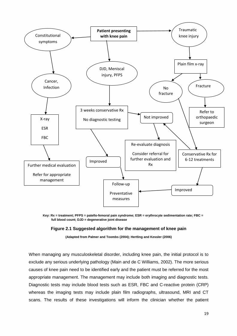
19
Key: Rx = treatment; PFPS = patello-femoral pain syndrome; ESR = erythrocyte sedimentation rate; FBC = full blood count; DJD = degenerative joint disease
Figure 2.1 Suggested algorithm for the management of knee pain
(Adapted from Palmer and Toombs (2004); Hertling and Kessler (2006)
When managing any musculoskeletal disorder, including knee pain, the initial protocol is to
exclude any serious underlying pathology (Main and de C Williams, 2002). The more serious
causes of knee pain need to be identified early and the patient must be referred for the most
appropriate management. The management may include both imaging and diagnostic tests.
Diagnostic tests may include blood tests such as ESR, FBC and C-reactive protein (CRP)
whereas the imaging tests may include plain film radiographs, ultrasound, MRI and CT
scans. The results of these investigations will inform the clinician whether the patient
Patient presenting with knee pain Constitutional
symptoms
Traumatic knee injury
Cancer, Infection
DJD, Meniscal injury, PFPS
Plain film x-ray
X-ray
ESR
FBC
Further medical evaluation
Refer for appropriate management
3 weeks conservative Rx
No diagnostic testing
Improved
Follow-up
Preventative measures
No fracture
Fracture
Refer to orthopaedic
surgeon
Conservative Rx for 6-12 treatments
Not improved
Re-evaluate diagnosis
Consider referral for further evaluation and
Rx
Improved
20
requires referral for conservative, medical or surgical management. The main goal of
treatment when managing knee pain is to reduce pain and inflammation and to maintain or
restore range of motion and function.
After the initial evaluation of a patient and there is no evidence of underlying serious
pathology, the initial treatment should be conservative management for six weeks (Hertling
and Kessler, 2006). Conservative management of knee pain may include exercise,
stretching and strengthening, heat and cryotherapy, other soft tissue techniques (e.g.
massage, dry needling, acupuncture, soft tissue manipulation), patient education and
modification of activity. If it is necessary, the patient may be advised to take medication such
as non-steroidal anti-inflammatory drugs (NSAIDs) and, if required manual therapy may be
utilized to maintain or increase range of motion (Palmer and Toombs, 2004).
An important aspect of patient management is patient education. It is important that the
patient understands the cause of the knee pain, the role of the investigations performed and
how they relate to the patient’s condition and the recommended management protocol.
Although a patient may be advised to limit bed rest and increase mobility, the clinician
should advise activities that are not strenuous or exacerbate pain. Activities such as
stretching in the pain free range of motion and strengthening of specific muscle groups are
suggested to the patient and the patient is instructed to return to normal activity when there
has been a significant response to treatment (Palmer and Toombs, 2004; Hertling and
Kessler, 2006).
After the initial period of conservative management of approximately six weeks, the patient
should be re-evaluated to determine if there has been any improvement. If there has been
no improvement, the patient should be sent for radiographic imaging of the knee in
anticipation of identifying anatomical changes, biomechanical anomalies, degenerative
changes and the development of possible red flags. These radiographic findings will guide
the clinician on the next step of patient management. Fractures, infections or malignancies,
are classified as medical emergencies and require immediate referral for medical or surgical
management (Cooperstein and Gleberzon, 2004; Palmer and Toombs, 2004).
21
2.5.1. CHIROPRACTIC MANAGEMENT OF KNEE PAIN
The initial approach of a chiropractor to the management of knee pain is to evaluate the
patient and determine if the patient has any red flags. If these are present the patients
should be referred for medical evaluation and management. If there is no evidence of
serious pathology, the condition may be managed conservatively for at least six weeks.
However, if there is no response to the management after this period, the patient should be
re-evaluated, referred for further investigations or to another clinician for a second opinion
(Palmer and Toombs, 2004).
During the assessment of the patient, the clinician should assess for the presence of
indications and contra-indications to manipulative therapy. Indications for manipulative
therapy include knee joint (patella-femoral, tibio-femoral, tibio-fibular joints) dysfunction and
myofascial pain syndromes (Wieting, 2008). However, there are relative and absolute
contra-indications to manipulation. Relative contra-indications refer to the conditions such as
osteoporosis and inflammatory arthritis which require modification of technique during
manipulation. Absolute contra-indications to manipulation include malignancy, infection and
joint instability or ligament laxity (Wieting, 2008).
There are many management options available to the chiropractor. These include manual
therapies (including manipulation or mobilisation), soft tissue techniques (including massage,
dry needling, ischaemic compression and soft tissue manipulation), electrotherapy (IFC,
ultrasound, TENS) and patient education (Palmer and Toombs, 2004). Patient education
involves advice on lifestyle modification as well as the teaching of stretches and specific
exercises (Palmer and Toombs, 2004).
There are several treatment modalities available to the chiropractic students at the
Chiropractic Day Clinic (CDC) at the Durban University of Technology (DUT) for the
treatment of patients (Chiropractic Clinic Manual, 2010). Manual therapy is the primary
modality utilised by the student at the CDC. The manual therapy techniques are often
utilised in combination with soft tissue techniques, electrotherapy, stretching and
strengthening exercises and other techniques such as heat and cryotherapy. Soft tissue
techniques include dry needling, massage, ischaemic compression and soft tissue
manipulation techniques and electrotherapeutic modalities available include ultrasound, IFC
and TENS. Proprioceptive neuromuscular facilitation (PNF), static stretches and
strengthening exercises are included when appropriate.
It is stated in the CDC Radiographic Guidelines and Procedures that radiographs should not
be used as a tool for general screening and should only be requested if they will contribute
22
significantly to patient diagnosis or management. The radiographs should be used to confirm
or refute a clinical diagnosis that was suggested by the clinical history or physical
examination. The intention of these guidelines is to decrease the amount of ionizing radiation
the patients are exposed to when undergoing radiographic imaging and avoiding the taking
of unnecessary radiographs (Chiropractic Clinic Manual, 2010).
2.6. CONCLUSION Knee pain is the second most common presenting musculoskeletal complaint. There are
many different causes of knee pain ranging from benign to serious causes. There have been
attempts by several researchers to link specific features in the clinical history and physical
examination, but the results are conflicting. Radiographs have been utilised to identify
serious pathologies and have been utilised by chiropractors to aid in the diagnosis of knee
pain. However, it has been observed that clinical findings poorly correlate with radiographic
findings (Morgan et al., 1997). Although guidelines for the use of radiographs have been
provided to eliminate the overuse of radiographs, researchers have observed that often
there is overuse of radiographs in clinical practice (Morgan et al., 1997).
Radiographs of the knee are used to provide information when formulating a diagnosis,
identifying contra-indications to manipulative therapy, following degenerative processes and
identifying red flag conditions that require referral for the necessary assessment and
management. Therefore, the aim of this study is to determine whether there is an
association between the clinical and the radiographic diagnoses of patients who present with
knee pain at a chiropractic teaching clinic and whether radiographs of the knee influence a
change in the diagnosis or management of these patients.
23
CHAPTER THREE
MATERIALS AND METHODS
3.1. STUDY DESIGN
This research was designed as a retrospective, non-experimental study. Data was obtained
from knee radiographs and the corresponding patient files of patients who presented to the
Chiropractic Day Clinic (CDC) at the Durban University of Technology (DUT) with knee pain
from 1 January 1997 to 31 December 2010. Ethical clearance for this study was obtained
from the Faculty of Health Sciences Research Committee at DUT (Ethical clearance certificate number: 006/11).
3.2. PATIENT CONFIDENTIALITY
Throughout the process of this study, steps were taken to maintain the confidentiality of the
patients. Before any examination or treatment at the CDC all patients are required to sign an
informed consent form. By signing the informed consent form, they provided written consent
to the use of their clinical and radiographic records for the purpose of research and strict
guidelines (according to DUT policy) were followed to maintain the confidentiality of the
patients. Codes were assigned to all patient names and recorded on a data sheet
(Appendix 2). These codes were then used instead of patient names in all documentation
that followed. Patient names were not recorded in this dissertation or any publication likely to
follow from this research. Access to patient files and radiographs was restricted to the
researcher and the supervisor.
24
3.3. SAMPLING METHOD AND SAMPLE SIZE
In this study, purposive sampling was used. Data sheets (Appendix 1) were used to record
all information. Knee radiographs of patients who presented to the CDC with knee pain taken
during the management at the CDC from 1 January 1997 to 31 December 2010 were
included in this sample. A total of 176 knee radiographs were found in the archives at the
CDC at the end of December 2010. Patient files and radiographs which did not meet the
inclusion and exclusion criteria were excluded. Therefore, the final sample size in this study
was 146.
3.4. INCLUSION AND EXCLUSION CRITERIA
3.4.1. INCLUSION CRITERIA
1. Clinical files of patients who presented to the CDC for treatment of knee pain.
2. Radiographs of the knee must have been taken during or prior to the selected
patient’s treatment at the CDC.
3. A minimum of an antero-posterior (AP) and lateral knee views were required.
3.4.2. EXCLUSION CRITERIA
1. Radiographs of the knee taken prior to the first consultation at the CDC.
2. Any patient files with no patient history, physical examination, knee orthopaedic
examination and a SOAPE (Subjective, Objective, Assessment, Plan and Education)
note corresponding to the date of the radiographic examination.
3. Any radiographs which were not reported by a radiologist or if the radiology report
was missing.
25
3.5. RESEARCH PROCEDURE
This study took place in three phases:
Phase 1
Radiographs from the CDC archives were sorted and all knee radiographs were placed
aside. The patients’ names and date of birth were recorded on a data sheet (Appendix 2).
This was done to locate the corresponding patient files using the CDC computer archive
system. A code was then assigned to each patient’s name. The data sheets with patients’
names were destroyed using a shredder once a code had been assigned to each patient.
These codes were used on all other data sheets.
Phase 2
All radiographs and patient files were briefly inspected and evaluated in order to determine if
they met the inclusion criteria.
Phase 3
Knee radiographs and the corresponding patient files were evaluated and the following data
was recorded (Appendix 1) as shown in Table 1:
Table 3.1 Data collected and the source of the data
Data collected Source Code and basic demographic information: code, age, gender, ethnicity
Patient confidentiality coding sheet (Appendix 2) and case history form
Date of the initial consultation Case history form and SOAPE note Main complaint (knee pain with/without radiation)
Case history form
Outline of treatment before radiographs* SOAPE note Reason for radiograph referral/ suspected clinical diagnosis
SOAPE note and/or radiology request form
Date of radiographs Radiology report and/or identification marker on radiographic films
Radiographic diagnosis Radiology report Radiographic incidental findings Radiology report and/or findings of the
researcher (and confirmed by supervisor/radiologist)
Clinical diagnosis after radiographs SOAPE note Any change (or no change) in treatment outlines after radiographs
SOAPE note
* Radiographs refer to the radiographs of the knee Table adapted from the approved PG4a proposal of McPhail (2010)
26
3.6. STATISTICAL ANALYSIS Version 15.0 of the Statistical Package for the Social Sciences (SPSS Inc, Chicago, Illinois,
USA) was used in the analyses of the data in this study.
Method of data analysis:
The association between the radiographic diagnosis and the clinical diagnosis was
determined. Diagnoses were categorised into specific groups and indicator variables were
used to construct two-by-two tables of absence or presence of radiographic vs. clinical
diagnosis for each specific diagnosis. This way, the associations between radiographic and
clinical diagnoses were assessed for each condition separately. The other objectives were
purely descriptive and were analysed and the outcomes reported using frequency tables and
bar charts in the case of categorical variables, or summary statistics such as mean, standard
deviation and range in the case of quantitative variables (Esterhuizen, 2010).
27
CHAPTER FOUR
RESULTS
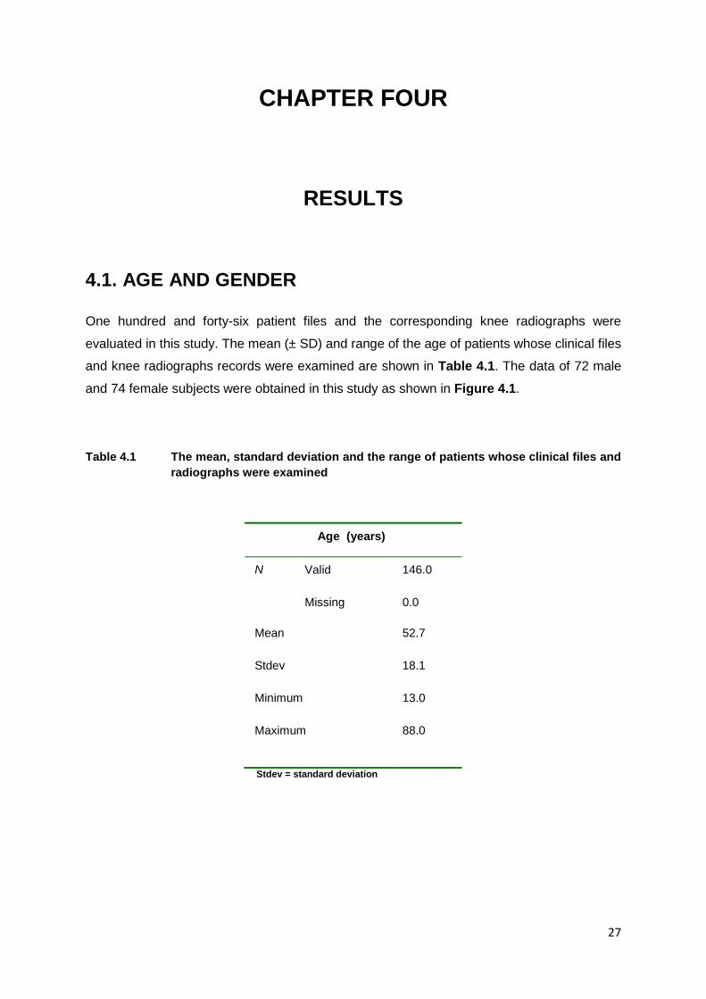
4.1. AGE AND GENDER
One hundred and forty-six patient files and the corresponding knee radiographs were
evaluated in this study. The mean (± SD) and range of the age of patients whose clinical files
and knee radiographs records were examined are shown in Table 4.1. The data of 72 male
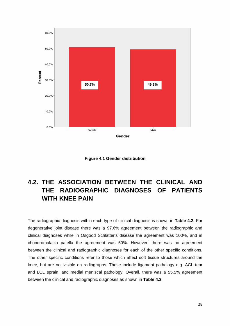
and 74 female subjects were obtained in this study as shown in Figure 4.1.
Table 4.1 The mean, standard deviation and the range of patients whose clinical files and radiographs were examined
Age (years)
N Valid 146.0
Missing 0.0
Mean 52.7
Stdev 18.1
Minimum 13.0
Maximum 88.0
Stdev = standard deviation
28
Figure 4.1 Gender distribution
4.2. THE ASSOCIATION BETWEEN THE CLINICAL AND THE RADIOGRAPHIC DIAGNOSES OF PATIENTS WITH KNEE PAIN
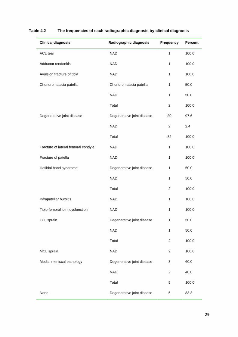
The radiographic diagnosis within each type of clinical diagnosis is shown in Table 4.2. For
degenerative joint disease there was a 97.6% agreement between the radiographic and
clinical diagnoses while in Osgood Schlatter’s disease the agreement was 100%, and in
chondromalacia patella the agreement was 50%. However, there was no agreement
between the clinical and radiographic diagnoses for each of the other specific conditions.
The other specific conditions refer to those which affect soft tissue structures around the
knee, but are not visible on radiographs. These include ligament pathology e.g. ACL tear
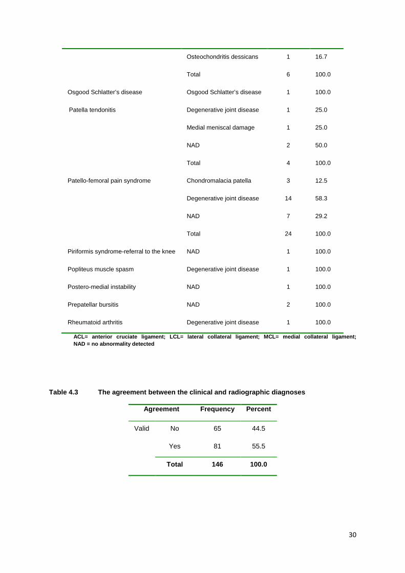
and LCL sprain, and medial meniscal pathology. Overall, there was a 55.5% agreement
between the clinical and radiographic diagnoses as shown in Table 4.3.
49.3% 50.7%
29
Table 4.2 The frequencies of each radiographic diagnosis by clinical diagnosis
Clinical diagnosis Radiographic diagnosis Frequency Percent
ACL tear NAD 1 100.0
Adductor tendonitis NAD 1 100.0
Avulsion fracture of tibia NAD 1 100.0
Chondromalacia patella Chondromalacia patella 1 50.0
NAD 1 50.0
Total 2 100.0
Degenerative joint disease Degenerative joint disease 80 97.6
NAD 2 2.4
Total 82 100.0
Fracture of lateral femoral condyle NAD 1 100.0
Fracture of patella NAD 1 100.0
Iliotibial band syndrome Degenerative joint disease 1 50.0
NAD 1 50.0
Total 2 100.0
Infrapatellar bursitis NAD 1 100.0
Tibio-femoral joint dysfunction NAD 1 100.0
LCL sprain Degenerative joint disease 1 50.0
NAD 1 50.0
Total 2 100.0
MCL sprain NAD 2 100.0
Medial meniscal pathology Degenerative joint disease 3 60.0
NAD 2 40.0
Total 5 100.0
None Degenerative joint disease 5 83.3
30
Osteochondritis dessicans 1 16.7
Total 6 100.0
Osgood Schlatter’s disease Osgood Schlatter’s disease 1 100.0
Patella tendonitis Degenerative joint disease 1 25.0
Medial meniscal damage 1 25.0
NAD 2 50.0
Total 4 100.0
Patello-femoral pain syndrome Chondromalacia patella 3 12.5
Degenerative joint disease 14 58.3
NAD 7 29.2
Total 24 100.0
Piriformis syndrome-referral to the knee NAD 1 100.0
Popliteus muscle spasm Degenerative joint disease 1 100.0
Postero-medial instability NAD 1 100.0
Prepatellar bursitis NAD 2 100.0
Rheumatoid arthritis Degenerative joint disease 1 100.0
ACL= anterior cruciate ligament; LCL= lateral collateral ligament; MCL= medial collateral ligament; NAD = no abnormality detected
Table 4.3 The agreement between the clinical and radiographic diagnoses
Agreement Frequency Percent
Valid No 65 44.5
Yes 81 55.5
Total 146 100.0
31
4.3. THE CONSULTATION WHEN A RADIOGRAPH WAS REQUESTED AND THE REASONS THEREOF
The consultation at which knee radiographs were requested by the student or clinician and
the reason(s) for referral are shown in Table 4.4. The majority of the knee radiographs were
requested at the initial consultation and as the length of treatment increased, the frequency
of radiograph requests decreased. It is interesting to note that no knee radiographs were
requested at treatment 9 and after treatment 10, only one radiograph was requested at
treatments 16 and 19.
The most common reasons for the referral for radiographs were to identify degenerative
changes and to evaluate unspecified pathology (Table 4.4). The term ‘unspecified pathology’
refers to any pathology that the student or clinician suspected on consultation, but did not
state on the referral form which pathology was suspected. This was a common observation
in this study group, with 33.6% (n = 49) of knee radiographs being requested without
providing a specific pathology that was suspected. It was also noted that eight radiographs
were requested to evaluate the reason why the patient was not responding to treatment
(Table 4.4). Surprisingly, this was the reason provided for requesting a few radiographs at
treatments 1 and 2.
There were circumstances where the student or clinician referred patients for radiographs as
there were red flags present. The conditions suspected included avascular necrosis (AVN) of
the knee, tumours and fractures. These radiographs were requested at treatment 1.
Interestingly, a radiograph was ordered at treatment 16 to determine calcification of the
patella tendon.
32
Table 4.4 A summary of the consultation at which knee radiographs were requested and the reasons thereof
Treatment number
Frequency Percent Reason for radiographic referral
1 99 67.8 • Degeneration (47.5%) • Unspecified pathology (37.4%) • Fracture (5.1%) • No response to treatment, instability or pathology,
degeneration or pain progression, AVN or fracture, rule out OCD, rule out OCD or degeneration, OCD or tumour, degeneration or tumour, degeneration or fracture, degeneration or pathology (1.0% each)
2 16 11.0 • Degeneration (62.5%) • Unspecified pathology (25.0%) • No response to treatment, fracture (6.2% each)
3 6 4.1 • Degeneration (50.0%) • Unspecified pathology (33.3%) • Suspected arthropathy (16.7%)
4 4 2.7 • Degeneration (50.0%) • Unspecified pathology, trauma (25.0% each)
5 4 2.7 • Unspecified pathology (50.0%) • Degeneration, meniscal pathology (25.0% each)
6 4 2.7 • Degeneration (75.0%) • No response to treatment (25.0%)
7 4 2.7 • Degeneration (75.0%) • Unspecified pathology (25.0%)
8 4 2.7 • No response to treatment (50.0%) • Degeneration, unspecified pathology (25.0% each)
10 3 2.1 • No response to treatment (66.7%) • Unspecified pathology (33.3%)
16 1 0.7 • Calcification of patella tendon
19 1 0.7 • No response to treatment
Total 146 100.0
OCD = osteochondritis dessicans; AVN = avascular necrosis
33
4.4. SUSPECTED CLINICAL DIAGNOSES AND MANAGEMENT PRIOR TO REFERRAL FOR KNEE RADIOGRAPHS
The management options for each of the clinical diagnoses are summarized in Table 4.5
and show that many treatment approaches were utilized for these conditions. Manual
therapy included knee joint manipulation, knee joint mobilisation or patella mobilisation.
Modalities such as ischaemic compression, dry needling and massage of myofascial trigger
points are referred to as soft tissue techniques. Electrotherapy modalities refer to treatments
such as interferential current therapy (IFC), ultrasound and transcutaneous electrical nerve
stimulation (TENS) which were used in the treatment of patients presenting with knee pain.
Stretching and strengthening included static stretches, proprioceptive neuromuscular
facilitative (PNF) stretches or strengthening exercises. The category ‘other’ included
treatments such as heat, ice or strapping (Table 4.5; Figure 4.2).
The most frequently utilized modalities in the management of patients with knee pain prior to
the request of radiographs included manual therapy, electrotherapy and soft tissue
techniques (Figure 4.2).
Table 4.5 Suspected clinical diagnosis and management prior to radiographic referral
Manual therapy
Soft tissue techniques
Electrotherapy Stretching or strengthening
No treatment, pending x-ray
results
Other
ACL tear
Adductor tendonitis
Avulsion fracture of tibia
Chondromalacia patella
Degenerative joint disease
Diagnosis pending x-ray results
Fracture of lateral femoral
34
condyle
Fracture patella
Iliotibial band syndrome
Infrapatellar bursitis
Tibio-femoral joint dysfunction
LCL sprain
MCL sprain
Medial meniscal pathology
Osgood Schlatter’s disease
Patella tendonitis
Patello-femoral pain syndrome
Pes anserine bursitis
Piriformis syndrome- referral to the knee
Popliteal muscle spasm
Postero-medial instability
Prepatellar bursitis
Rheumatoid arthritis
= yes; = no; ACL= anterior cruciate ligament; LCL= lateral collateral ligament; MCL= medial collateral ligament
35
1.4
4.1
12.3
13
36.3
37
67.8
0 20 40 60 80
Other
None
Referral
Stretch and strengthening exercises
Soft tissue techniques
Electrotherapy
Manual therapy
%
Figure 4.2 Management prior to requests for radiographs
4.5. CHANGES IN CLINICAL DIAGNOSIS AND MANAGEMENT AFTER RADIOGRAPHS
Of the 146 patients whose data is included in the study, 125 did not have a change in
diagnosis. This means that 85.6% of the diagnoses remained the same after radiographic
examination. There were 21 patients (14.4%) who had a change in the clinical diagnosis
following radiographic examination (Table 4.6). There were 33 patients (22.6%) who had a
change in management after radiographic examination. After radiographic examination, the
most common suspected clinical diagnoses changed to degenerative joint disease,
chondromalacia patella and osteochondritis dessicans.
The change in patient management after the student or clinician had received the
radiographic report is shown in Figure 4.3. Manual therapy was utilised more often after
radiographs were obtained (82.9%), than before the radiographs were taken (67.8%). Across
the spectrum of all treatment modalities, all of the modalities were used more often after
radiographs were obtained. The percentage of patients being referred after the radiographs
were obtained was 6.8% which was a significant decrease from 12.3% of patients being
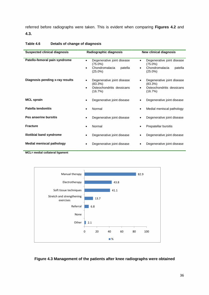
36
referred before radiographs were taken. This is evident when comparing Figures 4.2 and
4.3.
Table 4.6 Details of change of diagnosis
Suspected clinical diagnosis Radiographic diagnosis New clinical diagnosis
Patello-femoral pain syndrome • Degenerative joint disease (75.0%)
• Chondromalacia patella (25.0%)
• Degenerative joint disease (75.0%)
• Chondromalacia patella (25.0%)
Diagnosis pending x-ray results • Degenerative joint disease (83.3%)
• Osteochondritis dessicans (16.7%)
• Degenerative joint disease (83.3%)
• Osteochondritis dessicans (16.7%)
MCL sprain • Degenerative joint disease • Degenerative joint disease
Patella tendonitis • Normal • Medial meniscal pathology
Pes anserine bursitis • Degenerative joint disease • Degenerative joint disease
Fracture • Normal • Prepatellar bursitis
Iliotibial band syndrome • Degenerative joint disease • Degenerative joint disease
Medial meniscal pathology • Degenerative joint disease • Degenerative joint disease
MCL= medial collateral ligament
2.1
6.8
13.7
41.1
43.8
82.9
0 20 40 60 80 100
Other
None
Referral
Stretch and strengthening exercises
Soft tissue techniques
Electrotherapy
Manual therapy
%
Figure 4.3 Management of the patients after knee radiographs were obtained
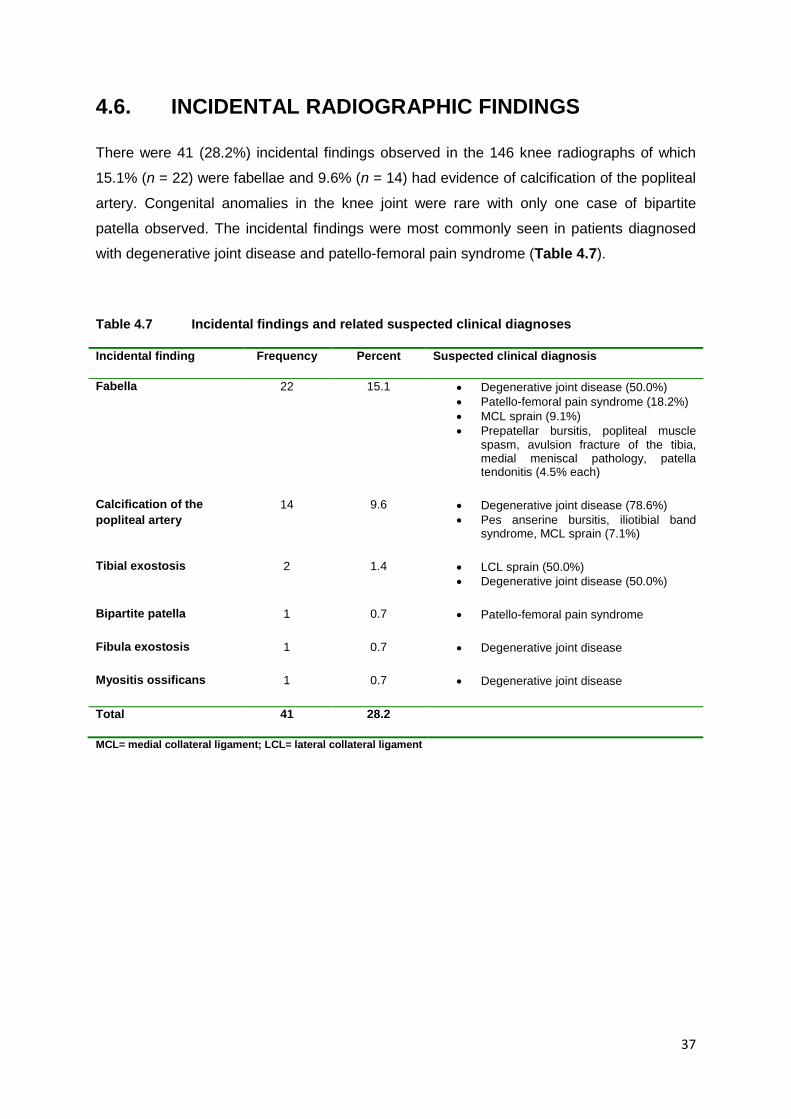
37
4.6. INCIDENTAL RADIOGRAPHIC FINDINGS
There were 41 (28.2%) incidental findings observed in the 146 knee radiographs of which
15.1% (n = 22) were fabellae and 9.6% (n = 14) had evidence of calcification of the popliteal
artery. Congenital anomalies in the knee joint were rare with only one case of bipartite
patella observed. The incidental findings were most commonly seen in patients diagnosed
with degenerative joint disease and patello-femoral pain syndrome (Table 4.7).
Table 4.7 Incidental findings and related suspected clinical diagnoses
Incidental finding Frequency Percent Suspected clinical diagnosis
Fabella 22 15.1 • Degenerative joint disease (50.0%) • Patello-femoral pain syndrome (18.2%) • MCL sprain (9.1%) • Prepatellar bursitis, popliteal muscle
spasm, avulsion fracture of the tibia, medial meniscal pathology, patella tendonitis (4.5% each)
Calcification of the popliteal artery
14 9.6 • Degenerative joint disease (78.6%) • Pes anserine bursitis, iliotibial band
syndrome, MCL sprain (7.1%)
Tibial exostosis 2 1.4 • LCL sprain (50.0%) • Degenerative joint disease (50.0%)
Bipartite patella 1 0.7 • Patello-femoral pain syndrome
Fibula exostosis 1 0.7 • Degenerative joint disease
Myositis ossificans 1 0.7 • Degenerative joint disease
Total 41 28.2
MCL= medial collateral ligament; LCL= lateral collateral ligament
38
CHAPTER FIVE
DISCUSSION OF RESULTS
5.1. AGE AND GENDER
The mean age and range of the patients whose clinical and radiographic records were
examined (Table 4.1) were comparable to those of the participants of a study at the Royal
College of Radiologists in which the mean age was 53 years and the range was 16-90 years
(Morgan et al., 1997). The age range observed in this study suggests that patients younger
than 13 years and older than 88 years do not visit the Chiropractic Day Clinic (CDC) for
treatment, but this conclusion would be erroneous as this study only included data of those
patients who presented to the CDC with knee pain and had radiographs taken at some point
during their management. There could have been patients who were treated for knee pain at
the CDC, but their data was not included due to the exclusion criteria of this study. The
number of males and female participants (Figure 4.1) were similar to the reports of Morgan
et al. (1997).
5.2. THE ASSOCIATION BETWEEN THE CLINICAL AND THE RADIOGRAPHIC DIAGNOSES OF PATIENTS WITH KNEE PAIN
The overall agreement between the clinical and radiographic diagnoses is shown in Table 4.3. Degenerative joint disease was the most suspected clinical diagnosis and in 97.6% of
these cases the radiographic diagnosis confirmed this. This percentage is more than twice
the figure reported by Morgan et al. (1997) in which 42% of knee radiographs were
requested to confirm degenerative changes that had been previously suspected. This
indicates the overuse of radiographs ordered to diagnose degenerative joint disease as the
majority of the cases were clinically correctly diagnosed since it is relatively easy to do so
especially in the late stages due to cardinal features such as joint swelling and crepitus.
39
Six patients had no clinical diagnosis prior to radiographs being taken (Table 4.2). In the
patient files and the radiograph request forms the students had recorded ‘no diagnosis’ or
‘diagnosis pending x-ray results’. There was no clinical suspicion of what radiographic
condition was suspected and the patient probably should not have been referred for
radiographs as it is unethical to do so (Shapiro, 2002; Chiropractic Clinic Manual, 2010).
Prior to radiographic examination there was no clinical diagnosis given to these patients
which meant that no management protocol could be initiated. Once the radiographs were
obtained, the patients were found to have degenerative changes and were diagnosed with
degenerative joint disease and osteochondritis dessicans. Degenerative joint disease has
specific clinical features and the student or clinician should, therefore, have provided a
suspected clinical diagnosis when referring for the radiograph. Osteochondritis dessicans,
on the other hand, is difficult to diagnose clinically due to the paucity of clinical features
(Jacobs, 2011). One patient in this study was suspected of having Osgood Schlatter’s
disease which was confirmed radiographically. Osgood Schlatter’s disease can be
diagnosed clinically as the symptoms are typical and radiographs are ordered to identify the
extent of the calcification or to confirm the clinical diagnosis (Chang, 2010).
For all the other specific clinical conditions (Table 4.2), there was no agreement between the
clinical and radiographic diagnoses. Of all the other specific conditions in 27 cases the
radiographs did not show any anatomical changes and, therefore, these were reported to
have no abnormality detected (NAD). Another 20 cases were diagnosed with degenerative
joint disease after the radiographs were obtained. Degenerative joint disease was the most
common radiographic diagnosis in this study and this may be attributed to the higher
average age of the patients. Three patients were referred for radiographs as a fracture was
suspected in each case (Table 4.2). The referral was based on clinical suspicion of a
fracture, but there was no evidence of fractures upon radiographic evaluation. It is, however,
possible that hairline fractures which are often not visible or are difficult to visualise on
radiographs, were missed in these cases (Drabicki et al., 2006). Nonetheless, the students
in these cases identified red flags in the history and physical examinations and adhered to
the Ottawa Knee Rules (Table 2.4) by referring the patients for radiographs.
It is evident that at the CDC there is overuse and overreliance on radiographs especially for
diagnosing degenerative joint disease. In these cases, students requested radiographs
unnecessarily, especially in cases where a clinical diagnosis could have been made. This
probably highlights the lack of confidence in making clinical decisions which is further
reflected in cases where the students did not provide a suspected clinical diagnosis when
referring for radiographs, but only diagnosed their patients once radiographs were obtained.
It is unethical to refer patients if no clinical condition is suspected as the patients are
40
exposed to unnecessary ionizing radiation which may have adverse effects such as the risk
of developing cancer or genetic abnormalities (Shapiro, 2002). In the cases of OCD and
fracture, the patients were referred for radiographs to confirm the clinical diagnosis which
was the correct protocol for requesting radiographic imaging (Chiropractic Clinic Manual,
2010).
5.3. THE CONSULTATION WHEN A KNEE RADIOGRAPH WAS REQUESTED AND THE REASONS THEREOF
It was observed that the majority of radiographs were obtained at the initial consultation
(Table 4.4) and more than a third were requested without a valid reason being provided as
the request forms stated ‘unspecified pathology’. Radiographs should not be requested
unless there are red flags present or if the patient does not respond to conservative
management over a period of six weeks (Palmer and Toombs, 2004; Hertling and Kessler,
2006). In keeping with the evidence-based guidelines for the requests of radiographs
(Morgan et al., 1997; Koplas and Schils, 2008), merely stating ‘unspecified pathology’ or not
providing a reason on the radiograph request form is not a proper indication for radiographic
evaluation (Table 2.3). Therefore, these requests for radiographic imaging were not in
keeping with the evidence-based guidelines (Morgan et al., 1997; Koplas and Schils, 2008).
At the CDC, the supervising clinicians are responsible for guiding the students, especially
with respect to making accurate clinical diagnoses and decisions, and advising students on
the appropriate diagnostic tests and reasons for referral. This also includes guidance on the
request for plain film radiographs. After performing a thorough history and physical
examination, the students provide a clinical diagnosis based on their findings. As stated in
the Chiropractic Clinic Manual under the CDC Radiographic Guidelines and Procedures
(Chiropractic Clinic Manual, 2010), the radiographs taken should be used to confirm or reject
the clinical diagnosis made by the student. It is observed that a number of referrals for
radiographic examination did not abide by these guidelines. This is an interesting
observation as all radiographic request forms are signed-off by the clinician on duty before
radiographs can be taken. It is recommended that the results of this study be presented to
the clinicians at the CDC so that appropriate measures are taken to ensure that patients are
not referred for unnecessary radiographs. Furthermore, students should be encouraged to
have confidence in their suspected clinical diagnosis and only refer for radiographs in the
presence of red flags or non-responsiveness to appropriate treatment.
41
Some radiographs were utilised to identify soft tissue structures including calcification of the
patella tendon (Table 4.4). It is not recorded in the patient file why this was the suspected
diagnosis. The patient did not respond to treatment and was eventually referred for
radiographs at treatment 16. One of the limitations of plain film radiography is that soft tissue
structures are poorly visualised except when there is calcification (Yochum and Rowe,
2005), but in this case, it was not evident on the radiograph nor was there evidence of any
anatomical abnormalities. Other investigations such as MRI or diagnostic ultrasound or even
referral to a medical practitioner are probably more appropriate in the management of
patients with suspected soft tissue lesions.
Red flag conditions clinically suspected included fractures, tumours, osteochondritis
dessicans, pain progression and no response to treatment (Table 4.4). These patients were
appropriately sent for plain film radiographs as the initial investigation. No treatment was
given to these patients at the CDC until these red flag conditions were excluded as their
presence could alter the management protocol of patients (Palmer and Toombs, 2004).
At treatment six, a radiograph was requested to determine pathology as the patient had not
responded to treatment. The patient was diagnosed with degenerative joint disease and
treated with manual therapy prior to radiographic examination which was appropriate in
keeping with recommendations of Palmer and Toombs (2004). ‘No response to treatment’
(as in this case) is an accepted reason for the referral for radiographic imaging (Hertling and
Kessler, 2006). However, it must be noted that patient response to treatment of degenerative
joint disease varies considerably (Allen, 2007; Stauffer et al., 2011) and non-responsiveness
is not necessarily a red flag. In this case, the argument is supported by the radiographic
examination findings of degenerative joint disease which did not have an influence on the
management as this did not change. Perhaps the student could have explored alternative
conservative treatment approaches and continued treatment for a little longer. Besides the
possible lack of confidence by students in making clinical diagnoses, the overuse and
overreliance may also be due to the free radiographic facilities available at the Radiographic
Clinic to patients treated at the CDC. It is also possible that students requested radiographs
in order for their patients to feel that “something important” was being done to identify their
problem.
42
5.4. THE CLINICAL DIAGNOSIS AND MANAGEMENT OF PATIENTS PRESENTING WITH KNEE PAIN BEFORE AND AFTER RADIOGRAPHY
Of all the patients in this study the majority were diagnosed with degenerative joint disease.
This is in agreement with the reported findings of Morgan et al. (1997) that 42% of knee
patients will have degenerative causes of knee pain. There were six patients who were not
diagnosed clinically. It was reported in these cases that the diagnosis was pending the
radiographic results (Table 4.1). These patients were eventually diagnosed with
degenerative joint disease and osteochondritis dessicans once the radiographs were
obtained.
A number of treatment modalities were used to manage patients prior to requesting
radiographs. Manual therapy which included manipulation and mobilisation was utilised more
often than all the other modalities (Table 4.5; Figure 4.2). Chiropractors utilise manual
therapy to decrease pain and increase range of motion and function which may explain why
manual therapy was utilised the most. The majority of diagnoses did not influence a change
in treatment protocols. Treatments were similar irrespective of the diagnosis. This raises an
interesting question as to why a specific diagnosis is required for non-pathological knee pain
if all the diagnoses are treated similarly. Another observation was that when the diagnoses
were pending radiographic results, no treatment modalities were employed (Table 4.5) as
these patients presented with red flags that were contra-indications to all modalities
(Cooperstein and Gleberzon, 2004; Palmer and Toombs, 2004).
In the case of fractures, different management protocols were observed (Table 4.5). The
patients suspected of having an avulsion fracture of the tibia or patella fracture were not
treated prior to the request for radiographs. However, in the case of a suspected fracture of
the lateral femoral condyle, the patient was initially managed with manual therapy and soft
tissue therapy and then referred for radiographs. Although the pain was recorded as severe,
the patient was ambulant and this could have influenced the student to proceed with
conservative treatment, although the correct procedure would have been to rule out the
suspected fracture and then initiate conservative treatment. In any case, the patient had no
response to treatment and was referred for further medical assessment and management.
One hundred and twenty five patients (out of 146) did not have a change in diagnosis. This
means that the radiographic examination did not offer any new diagnostic information in
85.6% of the cases which is comparable to the findings of Morgan et al. (1997) who
43
observed that 90% of knee radiographs yielded information that was insignificant or non-
diagnostic. They believed that this high percentage indicates the unnecessary referral and
the overuse of radiographs at the CDC.
Of the diagnoses that had changed after radiographic examination, 19 patients were
diagnosed with degenerative joint disease, osteochondritis dessicans or chondromalacia
patella. This observation was also in agreement with those of Morgan et al. (1997) who
reported that the majority of patients with knee pain have degenerative changes. One patient
was diagnosed with medial meniscal pathology and one was diagnosed with a prepatellar
bursitis. It was observed that there were no red flag conditions in this group.
In the cases where a fracture was suspected, the radiographic examination showed no
changes to the anatomical structures of the knee and was considered normal. The eventual
clinical diagnosis was prepatellar bursitis. Prepatellar bursitis is easy to diagnose as it has
specific features such as localised swelling over the patella, but there may be similarities
with suspected fracture such as severe knee pain, swelling, redness, difficulty with
ambulation and even a history of trauma (Drabicki et al., 2006; Cameron and Howard, 2010;
Steele, 2011). It is, therefore, likely that there may have been one or more of these features
present that made the student suspect a fracture. The diagnosis of prepatellar bursitis also
indicated that there was exclusion of the red flag condition (i.e. the fracture). In cases when
the student or clinician were unsure of the diagnosis and stated ‘diagnosis pending x-ray
results’ in the patient’s clinical file, the patients were eventually diagnosed with degenerative
joint disease and osteochondritis dessicans and there was no evidence of serious pathology.
When the diagnosis was changed, it was not always to a totally new diagnosis. For example,
anterior knee pain was initially diagnosed as PFPS, but after the radiographic examination,
the diagnosis changed to degenerative joint disease or chondromalacia patella. The results
of this study show that radiographs were not influential in the diagnosis of patients as the
diagnosis did not change in 85.6% of cases. This lends credence to the view that many
patients are referred for radiographic examination unnecessarily. When assessing the
management of the patients, there was a change in management after radiographs were
obtained. All the modalities were used more frequently after radiographic examination
(Figure 4.3). The most commonly used modality before and after radiographic imaging was
manual therapy, an unsurprising finding as the primary treatment modality utilised by
chiropractors is manual therapy (Dagenais and Haldeman, 2002). It is also possible that the
students were more confident utilising manual therapies once the radiographic examination
ruled out pathological conditions.
44
5.5. INCIDENTAL RADIOGRAPHIC FINDINGS
Incidental findings were observed in 28.2% of the knee radiographs which is greater than the
figure of 16.2% reported by Lumbreras et al. (2010) for all musculoskeletal conditions. The
most commonly observed incidental findings were fabellae followed by calcification of the
popliteal artery (Table 4.7). The fabellae were evident in individuals of any age and gender
and were not significant as they do not have any influence on the knee pain (Lumbreras et
al., 2010). Calcification of the popliteal artery was observed in patients above the age of 65
years. The majority of incidental findings are seen in the elderly, especially in those who
present with comorbid pathologies (Frier and Fisher, 2010; Lumbreras et al., 2010). This was
evident in this study as in the cases of calcification of the popliteal artery which was present
in ten patients who had comorbid pathologies including hypertension, diabetes mellitus and
hypercholesterolaemia, suggestive of peripheral vascular involvement associated with these
conditions (Frier and Fisher, 2010).
Tibial and fibular exostoses (also called osteochondroma) were observed in three patients.
They are usually asymptomatic, but may cause discomfort if close to the surface or when
impinging on underlying structures such as a nerve (Yochum and Rowe, 2005). Sometimes
they are covered with a cartilaginous cap which is invisible on a radiograph unless there is
calcification (Yochum and Rowe, 2005). Although they are benign, there is a small likelihood
of malignant change either in the bony exostosis (osteosarcoma) or in the cartilaginous cap
(chondrosarcoma) (Murphey et al., 2000; Yochum and Rowe, 2005; Breitenseher and
Dominkus, 2006). Patients with bony exostosis should, therefore, be monitored for any
changes suggestive of malignancy.
In the patient with myositis ossificans a history of trauma to the quadriceps muscle was
noted. Myositis ossificans is a clinically important incidental finding as it may be a reason
why the patient has pain and reduced range of motion in the affected knee and hip (Yochum
and Rowe, 2005). The main radiographic differential diagnosis of myositis ossificans is
osteosarcoma, but there are differences in the radiographic appearance. Myositis ossificans
presents with a calcified peripheral shell and a radiolucent centre whereas the opposite is
true for osteosarcoma (Yochum and Rowe, 2005). Nonetheless, it is possible for
misdiagnosis. Therefore, it is important to note the history of the patient to identify if there
was previous trauma, to examine for red flags and to be familiar with the radiographic
appearances of bone diseases such as osteosarcoma.
45
5.6. PROPOSED RECOMMENDATIONS FOR THE CHIROPRACTIC DAY CLINIC
The following recommendations are proposed for the Chiropractic Day Clinic based on the
results of this study:
• The supervising clinicians should encourage all students to base their clinical
diagnosis on history and physical examination findings and to have confidence in
their clinical approach.
• The guidelines for the referral for radiographs stated in the Chiropractic Clinic Manual
should be adhered to. An improvement in the confidence in the clinical diagnosis
together with adhering to the guidelines for radiograph referral will ensure that
patients are not sent for unnecessary radiographs. The radiographic request form
should never include ‘unspecified pathology’ or ‘no diagnosis’ as reasons for referral
as it is unethical to do so. If radiographs are requested at the initial consultation, this
should only be done if there are red flag conditions present.
• Patients presenting with suspected red flag conditions should not be treated
conservatively at the CDC until these have been excluded.
• It should be noted by clinicians and students that not all red flag conditions (e.g.
tumours) are visible on radiography. This may be due to early stages of a disease or
the radiographic latent periods or the poor visualisation of soft tissue structures on
plain film radiographs (Yochum and Rowe, 2005). Patients suspected of malignancy
should be referred for another opinion and further assessments.
• All changes, to the clinical and radiographic diagnoses or management, should be
recorded correctly in detail in the SOAPE note. This is important for new students or
locum students who take over and in medico-legal cases.
46
CHAPTER SIX
CONCLUSION AND RECOMMENDATIONS
6.1 CONCLUSION
The association between the clinical and the radiographic diagnoses of knee pain were
determined in this study. Diseases with strong agreements between clinical and radiographic
diagnoses included DJD, Osgood Schlatter’s disease and chondromalacia patella. However,
there was no agreement for each of the other specific knee conditions. Of the knee
radiographs taken at the CDC the majority were requested at the initial consultation and as
the length of treatment increased, the frequency of radiograph requests decreased. The
most common reasons for referral for radiographs were to identify degenerative changes
(47.5%) and to evaluate unspecified pathology (37.4%). Of the 146 patients in this study,
125 patients did not have a change in diagnosis after radiographs were obtained which
means that 85.6% of diagnoses remained the same after radiographic examination. There
was a wide range of treatment modalities utilized in the management of patients with knee
pain, including soft tissue therapy, electrotherapeutic modalities and manual therapy. The
use of manual therapy increased from 67.8% prior to radiographs being taken to 82.9% after
radiographs were obtained. This indicates that findings on radiography did influence the
conservative management of patients presenting with knee pain at the CDC.
It was observed that radiographs are not requested following the evidence-based guidelines
as in the case of ‘unspecified pathology’. Knee radiographs were over-utilized at the CDC
and the findings on radiography did not influence the diagnosis of the patients presenting
with knee pain. This indicates the large amount of patients at the CDC who presented with
knee pain who were sent for radiographs unnecessarily.

47
6.2 RECOMMENDATIONS
Recommendations for future studies include the following:
• To determine if these findings are similar to other teaching clinics in South Africa or if
they are unique to the CDC at DUT. A similar study should be carried out at the
University of Johannesburg Chiropractic Clinic or at another teaching clinic.
• A similar study could be conducted with a sample of private chiropractic practices to
determine a comparison between an academic environment of a teaching clinic and
the environment of private practice.
The findings of this study should be presented to the clinicians and the students at the CDC
to provide information to improve clinical practices at the CDC.
48
REFERENCES
1. Aiello, M.R. 2008. Knee, Posterior Cruciate Ligament Injuries (MRI). (online). Available at: http://emedicine.medscape.com/article/400845-overview (Accessed 11 April 2011).
2. Allen, A.M. 2010. Knee, Anterior Cruciate Ligament Injuries (MRI). (online). Available at: http://emedicine.medscape.com/article/400547-overview (Accessed 11 April 2011).
3. Allen, K.D., 2007. The value of measuring variability in osteoarthritis pain. The Journal of Rheumatology; 34: 2132-2133.
4. Ammendolia, C., Taylor, J.A.M., Pennick, V., Cote, P., Hogg-Johnson, S and Bombardirer, C. 2008. Adherence to radiography guidelines for low back pain: A survey of chiropractic schools worldwide. Journal of Manipulative and Physiological Therapeutics; 31: 412-418.
5. Antonopoulou, M.D., Alegakis, A.K., Hadjipaulou, A.G., Lionis, C.D. 2009. Studying the association between musculoskeletal disorders, quality of life and mental health. A primary care pilot study in rural Crete, Greece. BMC Musculoskeletal Disorders; 10: 143.
6. Bedson, J., Croft, P.R. 2008. The discordance between clinical and radiographic knee osteoarthritis: A systemic search and summary of literature. BMC Musculoskeletal Disorders; 9: 116.
7. Bradley, W. G. 2004. Neurology in Clinical Practice: Principles of Diagnosis and Management. Philadelphia: Butterworth-Heinemann.
8. Breitenseher, M., Dominkus, M. 2006. Tumours and tumour like lesions of the knee joint. In Maeuer, J. Imaging strategies of the knee. New York: Thieme.
9. Brusch, J.L. 2010. Septic Arthritis (online). Available at: http://emedicine.medscape.com/article/236299-overview (Accessed 15 April 2011).
10. Calliet, R. 1992. Knee pain and disability. Philadelphia: F.A. Davis.
11. Calmbach, W.L., Hutchens, M. 2003. Evaluation of patients presenting with knee pain. Part II differential diagnosis. American Family Physician; 68: 917-922.
12. Cameron, D.A., Howard, G.C.W. 2010. Oncology. In Boon, N. A., Colledge, N.R., Walker, B.R., Ralston, S.H. Davidsons’s Principles and Practices of Medicine. 21th ed. Edinburgh: Churchill Livingstone.
13. Chang, A.K. 2010. Osgood-Schlatter’s Disease in Emergency Medicine (online). Available at: http://emedicine.medscape.com/article/827380-overview (Accessed 31 August 2011).
14. Chiropractic Clinic Manual. 2010. Department of Chiropractic and Somatology, Durban University of Technology.
49
15. Claessens, A.A.M.C., Schouten, J.S.A.G., van den Ouweland, F.A., Valkenburg, H.A. 1990. Do clinical findings associate with radiographic osteoarthritis of the knee? Annals of the Rheumatic Diseases; 49: 771-774.
16. Cooperstein. R., Gleberzon. B.J., 2004. Technique Systems in Chiropractic. Britain: Churchill Livingstone.
17. Dagenais, S., Haldeman, S. 2002. Chiropractic. Primary Care; 29: 419-437.
18. DeBerardino, T.M. 2010. Medial Collateral Knee Ligament Injury (online). Available at: http://emedicine.medscape.com/article/89890-overview (Accessed 11 April 2011).
19. Dickinson, F.L., Harper, W.M., Finlay, D.B. 1997. Knee tumours- duration and nature of symptoms prior to investigation. The British Journal of Radiology; 70: 635-637.
20. Doherty, M., Lanyon, P., Ralston, S.H. 2010. Musculoskeletal disorders. In Boon, N. A., Colledge, N.R., Walker, B.R., Ralston, S.H. Davidsons’s Principles and Practices of Medicine. 21th ed. Edinburgh: Churchill Livingstone.
21. Drabicki, R.R., Greer, W.J., DeMeo, P.J. 2006. Stress fractures around the knee. Clinical Sports Medicine; 25: 105-115.
22. Dupont, J.Y. 1997. Synovial plicae of the knee. Controversies and review. Clinical Sports Medicine; 16: 87-122.
23. Esterhuizen, T. ([email protected]), 12 July 2010. Chiro research. E-mail to S. McPhail ([email protected]) [Accessed 12 July 2010].
24. Esterhuizen, T. ([email protected]), 26 April 2011. Re: Chantelle damon data collection. E-mail to C.A. Damon ([email protected]) [Accessed 26 April 2011].
25. Fagan, D.J.T., Davies, S. 2000. The clinical indications for plain radiography in acute knee trauma. Injury, International Journal of the Care of the Injured; 31: 723-727.
26. Freitas, A, 2011. Collateral Ligament Injury MRI (online). Available at: http://emedicine.medscape.com/article/401472-overview (Accessed 31 August 2011).
27. Frier, B.M., Fisher, M. 2010. Diabetes mellitus. In Boon, N. A., Colledge, N.R., Walker, B.R., Ralston, S.H. Davidsons’s Principles and Practices of Medicine. 21th ed. Edinburgh: Churchill Livingstone.
28. Glencross, P.M. 2009. Pes Anserine Bursitis (online). Available at: http://emedicine.medscape.com/article/308694-overview (Accessed 20 March 2011).
29. Goldstein, M.S. 1999. Alterantive health care: medicine, miracle or mirage? Philadelphia: Temple University Press.
30. Handoll, H.G.G., Parker, M.J. 2008. Conservative versus operative treatment for hip fractures in adults (Review). Edinburgh: John Wiley & Sons, Ltd.
31. Hertling, D., Kessler, R.M. 2006. Management of common musculoskeletal disorders: physical therapy principles and methods. Philadelphia: Lippincott Williams & Wilkins.
50
32. Ho, S.S.W. 2010. Lateral Collateral Knee Ligament Injury (online). Available at: http://emedicine.medscape.com/article/89819-overview (Accessed 11 April 2011).
33. Hollis, M.H. 2010. Nerve entrapment syndromes of the lower extremity (online). Available
at: http://emedicine.medscape.com/article/1234809-overview#aw2aab6b2 (Accessed 18
August 2011).
34. Jacobs, B.A. 2011. Knee Osteochondritis Dessicans (online). Available at: http://emedicine.medscape.com/article/89718-workup (Accessed 15 June 2011).
35. Koplas, M., Schils, J. 2008. The painful knee: Choosing the right imaging test. Cleveland Clinic Journal of Medicine; 75: 377-384.
36. Levy, D.B. Dickey-White, H. 2009. Knee Injury. Soft Tissue (online). Available at: http://emedicine.medscape.com/article/826792-overview (Accessed 19 October 2010).
37. Longmore, M., Wilkinson, I.B., Turmezei, T., Cheung, C.K. 2007. Oxford Handbook of Clinical Medicine. 7th ed. New York: Oxford University Press.
38. Lumbreras, B., Gonzalez-Alvarez, I., Lorente, M., Calbo, J., Aranaz, J., Hernandez-Aguado, I. 2010. Unexpected findings at imaging: predicted frequency in various types of studies. European Journal of Radiology; 74: 269-274.
39. Main, C.J., de C Williams, A.C. 2002. Musculoskeletal pain. British Medical Journal; 325: 534-537.
40. Martinez, J.M. 2009. Physical Medicine and Rehabilitation for Iliotibial Band Syndrome (online). Available at: http//emedicine.medscape.com/article/307850-overview (Accessed 20 March 2011).
41. McAlindon, T.E. 1999. The Knee. Bauliere’s Clinical Rheumatology; 13: 329-344.
42. McPhail, S. 2010. The role of lumbar spine x-rays in the diagnosis and management of patients who present with low back pain. M.Tech: Chiropractic Dissertation, Durban University of Technology.
43. Morgan, B. Mullick, S., W.M. Finlay, D.B. 1997. An audit of knee radiographs performed for general practitioners. The British Journal of Radiology; 70: 256-260.
44. Murphey, M.D., Choi, J.J., Kransdorf, M.J., Flemming, D.J., Gannon, F.H. 2000. Imaging of osteochondroma: Variants and complications with radiologic pathologic correlations. Radiographics; 20: 1407-1434.
45. Neogie, T., Felson, D.., Niu, J., Nevitt, M., Lewis, C.E., Aliabadi, P., Sack, B., Torner, J., Bradley, L., Zhang, Y. 2008. Radiographic features of osteoarthritis are strongly associated with knee pain in two cohorts: Most and Kiningham. Osteoarthritis and Cartilage; 16: S144-S145.
46. Oberholder, M.A., Shalvoy, R.M., Hughston, J.C. 1993. The accuracy of the clinical knee examination documented by arthroscopy: A prospective study. The American Journal of Sports Medicine; 21: 773-778.
51
47. O’Shea, K.J., Murphy, K.P., Heekin, R.D., Herzwurm, P.J. 1996. The diagnostic accuracy of history, physical examination, and radiographs in the evaluation of traumatic knee disorders. The American Journal of Sports Medicine; 24: 164-167.
48. Palmer, T., Toombs, J.D. 2004. Managing joint pain in primary care. The Journal of the American Board of Family Practice; 17: 532-542.
49. Patel, P. 2011. Patellofemoral Arthritis (online). Available at: http://emedicine.medscape.com/article/1933589-overview (Accessed 20 April 2011).
50. Phillips, R.B. 1992. Plain film radiology in chiropractic. Journal of Manipulative and Physiological Therapeutics; 15: 47-50.
51. Potter, P.J. 2009. Patellofemoral syndrome (online). Available at: http://emedicine.medscape.com/article/308471-overview (Accessed 12 April 2011).
52. Rajadhyaksha, A. 2008. Osteonecrosis, knee (online). Available at: http://emedicine.medscape.com/article/1252556-overview (Accessed 20 May 2011).
53. Schenk, K.W. 2005. Chiropractic management of chronic low back pain: A report of positive outcomes with patient compliance. Journal of Chiropractic Medicine; 4: 39-42.
54. Servi, J.T. 2009. Patellofemoral Joint Syndrome (online). Available at: http://emedicine.medscape.com/article/90286-overview (Accessed 12 April 2011).
55. Shah, M. 2011. Charcot Arthropathy (online). Available at: http://emedicine.medscape.com/article/1234293-overview (Accessed 15 June 2011).
56. Shapiro, J. 2002. Radiation protection: a guide for scientists, regulators and physicians. United States of America: La Editorial.
57. Smillie, I.S. 1978. Injuries of the knee joint. New York: Churchill Livingstone.
58. Stauffer, M.E., Taylor, S.D., Watson, D.J, Peloso, P.M., Morrison, A. 2011. Definition of nonresponse to analgesic treatment of arthritic pain: an analytical literature review of the smallest detectable difference, the minimal detectable change, and the minimal clinically important difference of the pain visual analog scale. International Journal of Inflammation; 2011: 231926.
59. Steele, M. 2011. Knee Fractures (online). Available at: http://emedicine.medscape.com/article/825572-overview (Accessed 20 April 2011).
60. Stevens, P.M. 2010. Genu Valgum, Paediatrics (online). Available at: http://emedicine.medscape.com/article/1259772-overview (Accessed 31 August 2011).
61. Wagemakers, H.P.A., Heintjes, E.M., Boks, S., Berger, M.Y., Verhaar, J.A.N., Koes, B.W., Berma-Zienstra, S.M.A. 2008. Diagnostic value of history taking and physical examination for assessing meniscal tears of the knee in general practice. Clinical Journal of Sports Medicine; 18: 24-30.
62. Wallace, M.S., Staats, P. 2004. Pain medicine and management: just the facts. United States of America: McGraw-Hill Professional.

52
63. Wieting, J.M. 2008. Massage, Traction, and Manipulation (online). Available at: http://emedicine.medscape.com/article/324694-overview (Accessed 16 April 2011).
64. Yochum, T.R. and Rowe, L.J. 2005. Yochum and Rowe’s Essentials of Skeletal Radiology. 3rd ed. Baltimore: Lippincott Williams & Wilkins.
Appendix 1: Data Sheet Age Gender Ethnicity Code
Main complaint Date of Initial consultation: / / .
Clinical diagnosis/ differential diagnosis
Treatment prior to x-rays
Manual therapy
Soft tissue techniques
Electrotherapy
Stretching or strengthening
Referral
Other
Reason for x-ray referral (suspected diagnosis) Treatment Number: . Date of x-ray: / / .
Radiological diagnosis
Incidental findings on x-ray
Change in clinical diagnosis after x-rays?
Yes No
New diagnosis
Change in treatment after x-rays?
Yes No
Change of treatment after x-rays
Manual therapy
Soft tissue techniques
Electrotherapy
Stretching or strengthening
Referral Other

Appendix 2: Patient confidentiality coding sheet File Number Patient Name Date of Birth Code
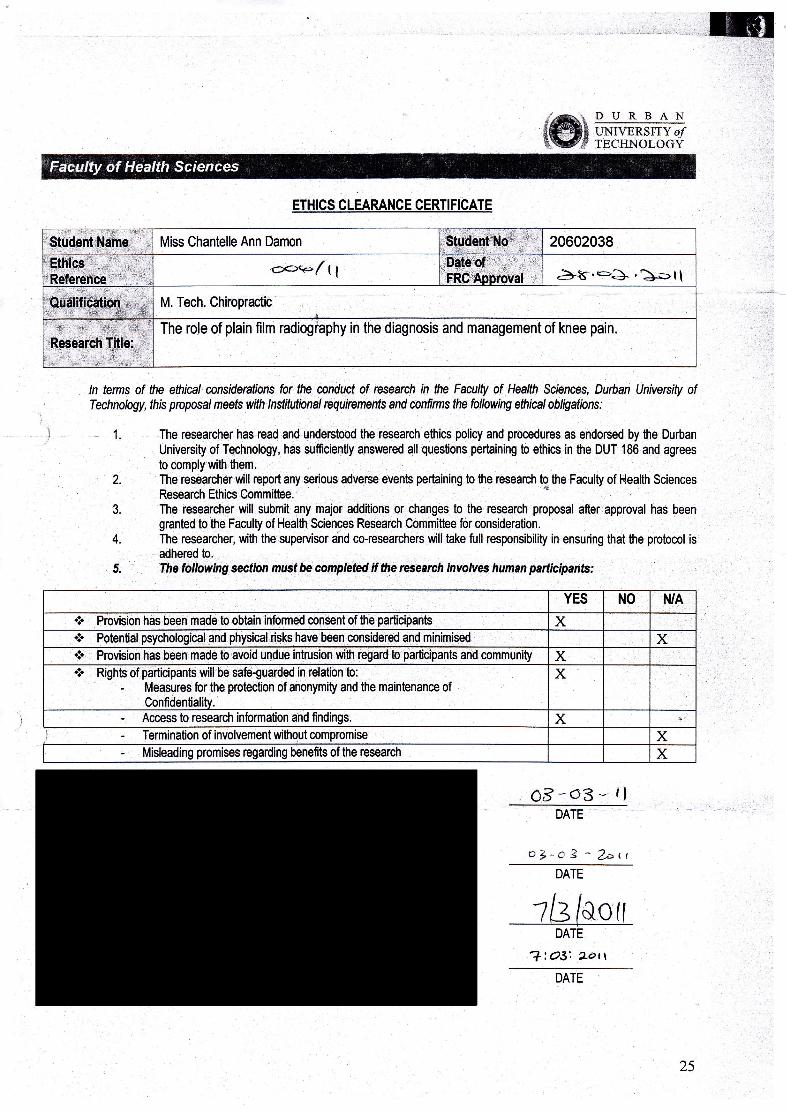
DURBA,NUNIYER,SITYo/TECHNOLOGY
ETHICS CLEARANCE CERTIFICATE
Miss Chantelle Ann Damon 20602038
eov111 5{t.P5'>.>tlM. Tech. Chiropractic
The role of plain film radiogiaphy in the diagnosis and management of knee pain.
ln terms of the ethical considerations for tln nnduct of research in the Faculty of Heafth Sciences, Duhan University ofTechnol4y, this proposal meets with lnstittrtiona[ re1uirements and confrrms the foilowlng ekical obligations:
1. The researcher has read and underslood the research ethics policy and procedures as endorsed by the Durban
University of Technology, has sufficiently answered all questions p6rtaining to etrics in the DUT 186 and agreesto comply wih frrem.
2. The researcher will report any serious adverse events pertaining to the research to the Faculty of Health Sciences
Research Ethics Committee. -3. The researcher will submit any major additions or changes to the research proposal after approval has been
granted to the Faculg of Healfr Sciences Research Committee for consideration.
4. The researcher, with the supervisor and co+esearchers will take full responsibili$ in ensuring that the protocol isadhered to.
5. The following section must be canpleted if the research fnvolves human pafticipanb:
03-03- rlDATE
07-e ? ^ ?.attDATE
rblaorrDATE
7io3'. Lotr
YES NO N/A
* Provision has been made to obtain informed consent of the participants X* Potential psychological,and physical dsks have been considered and minimised X* Provhion has been made to avoid undue intrusion wih regard to participants and community X{. Righb of participants will be safe1uarded in relation to:
- Measures for: the protection of anonymity and the maintenance ofConfidentialitv-
X
Access to research information and findings. X- Termination of involvement without compromise X
Misleading promises regarding benefits of the research X
DATE
25