Embed Size (px)
Citation preview

The Role of the Kidneys in Glucose Homeostasis:A New Path Toward Normalizing Glycaemia
R. A. DeFronzo1, J. A. Davidson2& S. Del Prato3
1University of Texas Health Science Center and Audie L. Murphy Memorial Veterans Hospital, San
Antonio, Texas, USA
2University of Texas Southwestern Medical School, Dallas, Texas, USA
3University of Pisa, Pisa, Italy
Address correspondence simultaneously to:
Ralph A. DeFronzo, MD J. A. Davidson
Diabetes Division Worldwide Initiative for Diabetes
Education
University of Texas Health Science Center P.O. Box 3709
7703 Floyd Curl Drive New York, NY 10163-3709
San Antonio, TX 78229 e-mail: [email protected]
Email: [email protected]
Keywords: clinical trials, diabetes, glucose homeostasis,glycosuria, hyperglycaemia, kidneys, renal
glucose transport, SGLT2, SGLT2 inhibitors, sodium-glucose cotransporters
Conflict of Interest: Drs. DeFronzo and Del Prato are on the Advisory Board of Boehringer-
Ingelheim. Dr Del Prato also serves as a consultant for Bristol Myers-Squibb and Astra Zeneca.Dr.
DeFronzo serves as a consultant for Bristol Myers Squibb. Dr. Davidson has no conflict of interest
with regard to companies producing SGLT2 inhibitors.
This article has been accepted for publication and undergone full peer review but has not been through the copyediting, typesetting, pagination and proofreading process which may lead to differences between this version and the Version of Record. Please cite this article as an ‘Accepted Article’, doi: 10.1111/j.1463-1326.2011.01511.x
Diabetes, Obesity and Metabolism © 2011 Blackwell Publishing Ltd

Abstract
The maintenance of normal glucose homeostasis requires a complex, highly integrated interaction
among the liver, muscle, adipocytes, pancreas and neuroendocrine system. Recent studies have
demonstrated that the kidneys also play a central role in glucose homeostasis by reabsorbing all of
the filtered glucose, an adaptive mechanism that ensures sufficient energy is available during fasting
periods. This mechanism becomes maladaptive in diabetes, however, as hyperglycaemia augments
the expression and activity of the sodium-glucose cotransporter (SGLT) 2 in the proximal tubule of
the kidney. As a result, glucose reabsorption may be increased by as much as 20% in individuals
with poorly controlled diabetes. SGLT2 is a low-affinity, high-capacity glucose transport protein that
reabsorbs 90% of filtered glucose, while the high-affinity, low-capacity SGLT1 transporter reabsorbs
the remaining 10%. SGLT2 represents a novel target for the treatment of diabetes. In animal studies,
SGLT2 inhibition reduces plasma glucose levels, resulting in improved beta cell function and
enhanced insulin sensitivity in liver and muscle. Human studies have confirmed the efficacy of
SLGT2 inhibitors in improving glucose control and reducing the A1c.Because the mechanism of
SGLT2 inhibition is independent of circulating insulin levels or insulin sensitivity, these agents can
be combined with all other antidiabetic classes, including exogenous insulin. Although the long-term
efficacy and safety of SGLT2 inhibitors remain under study, the class represents a novel therapeutic
objective with potential for the treatment of both type 2 and type 1 diabetes.

Introduction
Insulin resistance in muscle, liver and adipocytes and impaired insulin secretion are the core defects
in type 2 diabetes [1–4]. Excess glucose production by the liver and decreased glucose utilization by
insulin target tissues result in fasting and postprandial hyperglycaemia [1, 5]. The beta-cell
dysfunction and insulin resistance can be detected long before the development of overt diabetes [2].
As shown in Table 1, at least eight metabolic or hormonal abnormalities contribute to the
development of hyperglycaemia [4]. Most of these metabolic abnormalities have been well described
elsewhere and will not be detailed further here [1–10].
Until recently, little attention has been focused on the role of the kidney in glucose
homeostasis. However, along with the liver, this organ plays a vital role in ensuring that energy
needs are met during fasting periods. Approximately 180 liters of plasma per day are filtered by the
kidney, which works to maintain intravascular volume and acid-base, electrolyte and water balance
by reabsorbing water, sodium, chloride and bicarbonate and secreting hydrogen ions and potassium
produced by ingested foodstuffs. The kidney also plays a critical role in absorbing all of the filtered
glucose. With a glomerular filtration rate of 180 litres per day and a plasma glucose concentration of
5 mmol/l, the kidney filters approximately 162 grams (900 mmoles) of glucose per day, thereby
helping to maintain normal fasting plasma glucose (FPG) levels (~5.6 mmol/l) [11, 12]. The kidney
has developed a very efficient adaptive system involving the sodium-glucose cotransporter (SGLT) 2
and SGLT1 to reclaim all of the filtered glucose. When plasma glucose levels exceed the maximal
reabsorptive capacity of the renal SGLT transport system, glycosuria occurs.
Expression and activity of SGLT2 - the transport protein responsible for 80-90% of renal
glucose reabsorption[13,14] - are increased in type 2 diabetes [15]. As a result, a higher than normal

amount of glucose is reabsorbed by the kidneys into the bloodstream, thereby contributing to and
maintaining hyperglycaemia. Chronically elevated plasma glucose levels exacerbate insulin
resistance and beta-cell dysfunction (ie, glucotoxicity), further contributing to the abnormal glucose
homeostasis that characterizes type 2 diabetes [2, 4, 12].
The complex pathophysiology and progressive nature of type 2 diabetes often render
monotherapy ineffective with currently available agents [4, 16-18]. Solving this problem becomes a
more acute issue when considering the fact that, by year 2025, 380 million individuals worldwide are
projected to have diabetes, with the prevalence more than doubling in many regions [19]. These
management challenges leave a very large number of patients well above target glucose levels [19-
21]. As demonstrated by the recent 20-year follow-up of the United Kingdom Prospective Diabetes
Study, improved control of blood glucose in newly diagnosed patients significantly decreases the
long-term risk of both microvascular and macrovascular complications [22]. In contrast, three large
trials of intensive glucose control in with diabetes with diabetes and a long duration of disease failed
to show a macrovascular benefit [23-25]. The essential lesson to be learned from these trials is the
importance of early therapeutic intervention to preserve beta-cell function, increase insulin
sensitivity and prevent micro- and macrovascular complications [26]. To accomplish these goals, we
must continue to explore new therapeutic options as novel pathophysiologic mechanisms responsible
for type 2 diabetes are elucidated.

Renal Glucose Transport
SGLT2 is a high-capacity, low-affinity glucose transporter that occurs in the early convoluted
segment (S1) of the proximal tubule, where luminal glucose is abundant [13,14,27]. The SGLT2
transporter mediates 90% of renal glucose reabsorption by coupling glucose transport to the
electrochemical sodium gradient (Figure 1) [11-14]. First, sodium is absorbed across the luminal cell
membrane, creating an energy gradient that permits glucose to passively enter the cell. Then, an
adenosine triphosphatase (ATPase)-mediated sodium-potassium pump returns the sodium to the
bloodstream. This exchange alters the concentration gradient within the cell, and glucose diffuses to
the basolateral glucose transporter (GLUT) 2, through which it passes back into the bloodstream
[28].
The other 10% of renal glucose reabsorption occurs through SGLT1, a high-affinity, low-
capacity transport protein that is found in the more distal, straight section of the proximal tubule
(S3), where there is less luminal glucose [14,27]. SGLT1 also resides in the intestine, where it is
responsible for absorption of dietary glucose and galactose [11, 12]. Because SGLT1 resides in
intestinal as well as renal tissues, and because it is not specific for glucose alone, it is not considered
a viable target for therapeutic intervention.Inhibition of this transporter has the potential to cause
osmotic diarrhea and malabsorption. However, as long as clinically significant gastrointestinal side
effects are not observed, combined SGLT2/SGLT1 inhibition remains a therapeutic option.
In the kidney, the amount of glucose reabsorbed through the SGLT1 and SGLT2 transporters
is equal to the amount of glucose that is filtered by the glomerulus. Glucose reabsorption by the
proximal tubule increases linearly with increasing glucose concentration, up to a theoretical
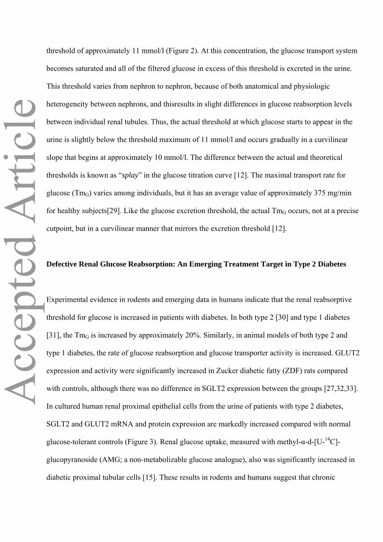
threshold of approximately 11 mmol/l (Figure 2). At this concentration, the glucose transport system
becomes saturated and all of the filtered glucose in excess of this threshold is excreted in the urine.
This threshold varies from nephron to nephron, because of both anatomical and physiologic
heterogeneity between nephrons, and thisresults in slight differences in glucose reabsorption levels
between individual renal tubules. Thus, the actual threshold at which glucose starts to appear in the
urine is slightly below the threshold maximum of 11 mmol/l and occurs gradually in a curvilinear
slope that begins at approximately 10 mmol/l. The difference between the actual and theoretical
thresholds is known as “splay” in the glucose titration curve [12]. The maximal transport rate for
glucose (TmG) varies among individuals, but it has an average value of approximately 375 mg/min
for healthy subjects[29]. Like the glucose excretion threshold, the actual TmG occurs, not at a precise
cutpoint, but in a curvilinear manner that mirrors the excretion threshold [12].
Defective Renal Glucose Reabsorption: An Emerging Treatment Target in Type 2 Diabetes
Experimental evidence in rodents and emerging data in humans indicate that the renal reabsorptive
threshold for glucose is increased in patients with diabetes. In both type 2 [30] and type 1 diabetes
[31], the TmG is increased by approximately 20%. Similarly, in animal models of both type 2 and
type 1 diabetes, the rate of glucose reabsorption and glucose transporter activity is increased. GLUT2
expression and activity were significantly increased in Zucker diabetic fatty (ZDF) rats compared
with controls, although there was no difference in SGLT2 expression between the groups [27,32,33].
In cultured human renal proximal epithelial cells from the urine of patients with type 2 diabetes,
SGLT2 and GLUT2 mRNA and protein expression are markedly increased compared with normal
glucose-tolerant controls (Figure 3). Renal glucose uptake, measured with methyl-α-d-[U-14C]-
glucopyranoside (AMG; a non-metabolizable glucose analogue), also was significantly increased in
diabetic proximal tubular cells [15]. These results in rodents and humans suggest that chronic
hyperglycaemiaupregulates SGLT2/GLUT2 transport expression and activity.
During evolution, the kidney developed an intricate system to reabsorb all of the filtered
glucose to conserve energy at a time when energy intake was sparse. From an evolutionary
standpoint, the increase in SGLT2 transport in response to hyperglycaemia can be viewed as an
adaptive response. However, in the patient with diabetes this adaptive response becomes
maladaptive, and glycosuria is not observed until the plasma glucose concentration increases to
levels that are substantially higher than 11 mmol/l - the glucose concentration threshold in non-
diabetic individuals. Thus, instead of allowing the kidney to excrete the excess filtered glucose in the
urine and correct the hyperglycaemia, the SGLT2 transporter works counterproductively to maintain
the elevated plasma glucose concentration [12].
Inhibition of SGLT2 transport ‘resets’ the system by lowering the threshold for
glycosuria(Figure 4), leading to correction of the hyperglycaemia. Normalization of the blood
glucose level ameliorates insulin resistance in muscle by augmenting insulin signaling, GLUT4 and
glycogen synthase activity [34,35]. In the liver, correction of hyperglycaemia decreases glucose-6-
phosphatase and PEP carboxykinase activity, leading to a decrease in gluconeogenesis and total
hepatic glucose production (HGP), with a resulting decrease in FPG concentration [36,37].
Correction of the hyperglycaemia also improves beta-cell function [34,38]. Collectively, the
deleterious effects of chronic hyperglycaemia on beta-cell function and on liver and muscle insulin
sensitivity are referred to as ‘glucotoxicity’ [34,39].
Phlorizin Studies in Experimental Models of Type 2 Diabetes
Initial evidence that reversing glucotoxicity by augmenting renal glucose excretion could lead to
improved glycaemic control has come from animal studies using phlorizin, a molecule that inhibits
both the SGLT1 and SGLT2 transporters (and therefore has not been studied for use in humans). In
partially pancreatectomized diabetic rats, phlorizin normalized both fasting and postprandial plasma
glucose concentrations. Withdrawal of phlorizin was associated with a return to the diabetic state
(Figure 5a) [40]. In the same study, correction of hyperglycaemia with phlorizin resulted in a marked
improvement in peripheral insulin sensitivity from 24.8±0.6 to 33.1±1.1 mg/kg·min (p < 0.001;
Figure 5b) [40]. In another study in rats with streptozotocin-induced diabetes,phlorizin treatment
restored glucose utilization to normal levels [41]. Improved first- and second-phase insulin secretion
also has been demonstrated after correction of the hyperglycaemia with phlorizin in diabetic rats
(Figure 5c) [42]. Discontinuation of phlorizin led to a return of insulin resistance and a decline in
beta-cell function [40,42]. Correction of hyperglycaemia with phlorizin in diabetic dogs has also
been shown to normalize the elevated plasma glucagon levels that are associated with the diabetic
state [43].
Similar improvements in beta-cell function and insulin sensitivity have been observed with T-
1095, an oral phlorizin derivative that no longer is in clinical development [37,44,45]. In
streptozocin-induced diabetic rats, both insulin-stimulated glucose disposal and the elevated basal
rate of hepatic glucose production were normalized, and insulin signaling in skeletal muscle and liver
was enhanced [44].
Animal Studies With Agents in Clinical Development
Phlorizin must be administered by injection and inhibits both SGLT1 and SGLT2, so it has served
primarily as a research tool. However, results obtained with phlorizin have provided the scientific
basis for the development of specific SGLT2 inhibitors for the treatment of type 2 diabetes (Table 2).
Dapagliflozin is the most advanced of these inhibitors in clinical development. In both normal and
ZDF rats, dapagliflozin in doses ranging from 0.1 to 10 mg/kg body weight markedly increased renal
glucose excretion and significantly decreased FPG by day 15 [46]. No change in FPG was observed
in normal rats treated with dapagliflozin because of a compensatory increase in HGP that maintained
normoglycaemia. In contrast, HGP decreased significantly in the dapagliflozin-treated vs. untreated
ZDF rats. Whole-body insulin-stimulated glucose disposal increased significantly with dapagliflozin
treatment [46]. ZDF rats fed a high-fat diet and treated with dapagliflozin also had improved
pancreatic function and improved insulin sensitivity [47].
Similar results have been seen with other SGLT2 inhibitors in development. Canagliflozin
(JNJ-28431754) lowers blood glucose levels and decreases body weight in obese diabetic animals
[48]. Finally, when compared with remogliflozin (which recently was discontinued) in a study of
mice and rats, BI 10773 had more potent inhibition of SGLT2 and produced significantly greater 24-
hour urinary glucose excretion [49].
Preclinical data also support the potential of ISIS 388626, an SGLT2 antisense
oligonucleotide that is highly specific for the renal SGLT2 transporter. In rats and dogs, this
compound decreased the SGLT2 mRNA and protein by approximately 80% without any effect on
SGLT1. FPG, postprandial glucose and glycatedhaemoglobin (HbA1c) were reduced significantly
with ISIS 388626, without changes in plasma or urine electrolytes [50]. Treatment of normal
cynomolgus monkeys increased glycosuria>1000-fold without inducing hypoglycaemia [51].
Clinical Evidence for SGLT2 Inhibition as Therapy for Type 2 Diabetes
On cursory review, a diabetes treatment strategy that increases glycosuria may seem counterintuitive.
However, in addition to animal data supporting the effectiveness of this approach, a human genetic
model has demonstrated its long-term safety. Familial renal glycosuria (FRG) results from a
mutation in the gene for SGLT2; 21 different mutations in 21 different families have been described
[52,53]. Whereas individuals with type A FRG have reduced levels of an abnormal SGLT2 protein,
which results in a lower TmG, individuals with type B FRG are characterized by SGLT2 transporters
with a diminished affinity for glucose, resulting in an exaggerated splay but a normal TmG [53].
Regardless of FRG type, affected individuals excrete as much as 100 grams of glucose per day in
their urine but nevertheless remain asymptomatic [11,52]. Blood glucose concentrations remain
normal due to an increased rate of HGP, which precisely counterbalances the amount of glucose that
is lost in the urine. Plasma volume and electrolyte composition remain normal because fluid and
electrolytes that are not absorbed in the proximal tubule are completely reabsorbed in more distal
parts of the nephron. These individuals have normal kidney and bladder function and no increased
incidence of diabetes or urinary tract infection [11, 12, 52,53].
The benign nature of FRG has established SGLT2 inhibition as a feasible approach to the
treatment of patients with diabetes. To date, the majority of available human trial reports on the
safety and efficacy of SGTL2 inhibitors have not revealed major adverse side effects. The most
commonly encountered side effect has been fungal infection of the genital organs. An increased
incidence of bacterial urinary tract infections also has been described in some studies (see subsequent
discussion).
Dapagliflozin is the SGLT2 inhibitor most advanced in clinical development; in humans [54]
it is rapidly absorbed with maximum plasma concentrations (Cmax) observed within 2 hours and half
life of ~17 hours (54). Dapagliflozin is highly protein bound (97-98%) and renal excretion is low (2-
4%) [54]. Approximately 0.1% of dapagliflozin is excreted as an inactive metabolite (54),which has
a bioavailability of 84%, with a half-life of 4.6 hours. In humans, the free dapagliflozinfraction is 4%
at a 10 μM plasma concentration. In Chinese hamster ovary cells expressing both SGLT1 and
SGLT2, dapagliflozin has a 1200-fold greater selectivity for SGLT2 vs. SGLT1, with a Ki of 1.1 nM
for SGLT2 and 1390 nM for SGLT1 [55].
In a 12-week, randomized, double-blind, placebo-controlled study involving 389 treatment-
naïve patients with type 2 diabetes, dapagliflozin significantly reduced HbA1c by 0.4% to 0.7%
across doses ranging from 2.5 mg to 50 mg (p < 0.01 vs. placebo; Figure 6)[56]. Metformin extended
release (XR) 1500 mg was used as an active comparator. Glycosuriaincreased in a dose-dependent
fashion by 52 g (289 mmoles)/day to 85 g(472 mmoles)/day in the dapagliflozin groups. This was
associated with a dose-related decrease in FPG, ranging from 1.1 mM to 1.7 mM, versus a 1.0 mM
decrease with metformin. Dapagliflozin caused a dose-related decline in the mean 3-hour
postprandial glucose area under the curve (AUC) from 12,215 to 8,913 mg/min/dl compared with
7,775 mg/min/dl for metformin [56]. HbA1c reductions were numerically similar between metformin
and the 2.5 mg and 5 mg doses of dapagliflozin, while dapagliflozin 10 mg and 50 mg yielded
slightly greater decreases in HbA1c
(Figure 6) [56]. In a large (n=546) 24-week trial in metformin-treated patients with type 2 diabetes,
dapagliflozin in doses of 2.5, 5, and 10 mg/day reduced the HbA1c by -0.67, -0.70, and -0.84%,
respectively, compared to placebo (0.3%) (all p<0.01) [57]. Body weight was reduced by 2.26,
3.10, and 2.96 kg, respectively, compared to controls (-0.87 kg) (all p<0.01) [57]. In 485 patients

with type 2 diabetes who were controlled by diet and exercise, dapagliflozin in doses of 2,5, 5.0, and
10 mg/day reduced the HbA1 by -0.58, -0.77, and -0.89% and body weight by -3.3, -2.8, and -3.2 kg
after 24 weeks [58]. In a subgroup of 74 patients with diabetes with HbA1c = 10.1-12.0% (87-108
mmol/mol), 24 weeks of dapagliflozin treatment reduced the HbA1c by 2.88% (8 mmol/mol) (5
mg/d) and 2.66% (6 mmol/mol) (10 mg/d) (58). In a preliminary study, Nauck et al. [59] compared
the efficacy of dapagliflozin (n=400) and glipizide (n=401) in metformin- treated patients with
T2DM and a starting HbA1c of 7.7% (61 mmol/mol). After 52 weeks the decrement in the HbA1c
was identical (-0.52%) in both treatment groups [59]. Dapagliflozin-treated subjects lost on average
3.2 kg, while glipizide-treated subjects gained 1.4 kg (p<0.0001). Both systolic (-4.3 versus +0.8
mmHg, p<0.001) and diastolic (-1.6 versus -0.4 mmHg, R=0.02) blood pressure declined more with
dapagliflozin. In a provocative study, Wilding et al randomized 71 insulin-treated (~50 units/d)
patients with type 2 diabetes who also were receiving an insulin sensitizer (metformin and/or
thiazolidinedione) to add on therapy with dapagliflozin (5 and 10 mg/d) or placebo [60]. Even
though the insulin dose was reduced by 50% at the start of therapy (the insulin sensitizer dose was
unchanged), after 12 weeks of dapagliflozin therapy, the HbA1c declined by 0.70 - 0.78% (p<0.01
vs. placebo). The placebo-subtracted reductions in body weight were 2.6 and 2.4 kg, respectively
(p<0.01 vs placebo). Both the increase in glycosuria and 50% reduction in insulin dose could have
contributed to the weight loss in dapagliflozin-treated subjects. Zhang et al [61] compared 151 early
stage (diabetes duration = 1 year) and 58 late stage (diabetes duration = 11 years) patients with type 2
diabetes randomly assigned to receive 10 or 20 mg/day of dapagliflozin for 12 weeks [61]. The late
stage diabetic group had a HbA1c = 8.4% (69 mmol/mol) was on large dose of insulin (> 50
units/day) plus metformin and a thiazolidinedione, and had long standing diabetes (mean = 11.1
years) compared to the early stage group (diabetes duration = 1.0 years, HbA1c = 7.6% (60
mmol/mol), no antidiabetic medications) [61]. The decline in HbA1c (0.5-0.7 vs 0.6-0.8%,
respectively) was similar in late and early stage patients with diabetes [61]. This is explained by the

unique mechanism of action of dapagliflozin on the kidney that is independent of the severity of
insulin resistance or beta cell failure. A greater reduction in body weight was observed in the late
stage diabetic group and this most likely is explained by the reduction in insulin dose since glucose
excretion was similar in both groups (Table 4).
In a 12-week study involving 71 patients with poorly controlled glycaemia despite therapy
with insulin plus metformin and/or a thiazolidinedione, the addition of dapagliflozin 10 mg and 20
mg significantly reduced HbA1c by 0.61% (confidence interval [95% CI] 0.9%, 0.4%) and 0.69% (CI
0.9%, 0.4%), respectively, compared with a 0.09% (CI 0.2%, 0.4%) reduction in the placebo group.
Significant decreases in FPG and postprandial glucose were also observed in this study [62].
There are a number of other SGLT2 inhibitors currently under development or in clinical
trials. In a phase 1 study, a single dose of sergliflozin (50-500 mg) caused a dose dependent increase
in glycosuria in both normal subjects and patients with T2DM (63,64). The 500 mg dose reduced the
mean plasma glucose concentration during the OGTT from 18.3 mM to 11.2 mM (63). More
prolonged treatment (14 days) with sergliflozin also induced dose-dependent glycosuria with modest
weight loss [64].
In a double blind, placebo-controlled, dose-ranging study in 451 metformin-treated T2DM
subjects canagliflozin in doses of 50, 100, 200, 300 mg/day for 12 weeks reduced the HbA1cby 0.7-
0.9% from baseline and 0.5-0.7% versus placebo in association with weight loss of 1.3-2.3% [65]. In
a 16 day trial canagliflozin improved beta cell function in patients with type 2 diabetes using a
model-based method to calculate insulin secretion [66]. In a small study involving 29 subjects with
T2DM who were sub-optimally controlled (HbA1c=8.4%) (69 mmol/mol) with insulin, canagliflozin
at 100 and 300 mg/day for 28 days reduced the HbA1cby 0.7% and 0.9%, respectively [67]. In a
single dose study BI10773 in doses ranging from 1 to 100 mg caused a dose –dependent increase in
urine glucose excretion in healthy male subjects [68]. At the 100 mg dose, BI10773 increased
urinary glucose excretion to 74 grams over 24 hours and reduced the plasma glucose excursion
during an OGTT. In a 12 week double-blind study, 361 Japanese patients with type 2 diabetes who
were treated with ASP1941 at doses ranging from 12.5 to 100 mg/day experienced a 0.9% reduction
in HbA1c at the two highest doses (50 and 100 mg/day) [69]. Body weight also was dose-
dependently reduced by up to 2 kg in the 100 mg/day dose. In a Phase 2A study, LX4211, which
inhibits SGLT2 and to a lesser extent SGLT1, at doses of 150 and 300 mg/day reduced the HbA1c by
1.2% but the starting A1c (8.2-8.5%) (66-70 mmol/mol) was higher than in most other studies and the
placebo decreased the HbA1c by 0.5% [70]. Sanofi-Aventis recently has initiated human trials with
AVE2268 [71]. In mice and rats this compound was shown to be highly selective for SGLT2 and
caused a significant dose-dependent increase in urinary glucose excretion and reduction in blood
glucose concentration during an OGTT [71]. Remogliflozin, which was developed by Kissei
Pharmaceuticals and GlaxoSmithKline, has been discontinued, apparently to make way for
development of the SGLT2 inhibitor (KGA-3235).
Because glycosuria translates into a loss of calories through the urine, SGLT2 inhibition
would be expected to cause weight loss, and this has been borne out in clinical trials with
dapagliflozin. When measured, dapagliflozin causes the urinary loss of 60-80 grams of glucose per
day, which equates to 240-320 cal/day or 2-3 pounds per month if this deficit is not offset by
increased caloric intake. Consistent with this, after 12 weeks of dapagliflozin treatment among drug-
naïve patients, mean body weight decreased by 2.5 kg to 3.4 kg, compared with a 1.7 kg loss in the
metformin group [56]. Similar weight loss has been observed in other dapagliflozin studies [57-
59,61]. In the study of insulin-treated patients, dapagliflozincaused a weight loss of 4.3-4.5 kg vs. 1.9
kg among patients receiving placebo [60].
The long term (beyond 12 months) durability of dapagliflozin’s effect on glycaemic control
and weight loss remains to be determined. It also is possible that after plasma glucose levels return to
the normal range, the effectiveness of SGLT2 inhibition may wane. However, since these agents
cause persistent glycosuriain individuals with normal glucose tolerance, it is reasonable to assume
that they will maintain their glycaemic efficacy in patients with type 2 patients with diabetes,
especially when coupled with agents such as metformin or incretin-based therapies, which reduce
HGP.
It is noteworthy that the increase in urine glucose excretion (60-80 grams/day) with all
SGLT2 inhibitors represents <50% of the filtered glucose load. The failure to observe a greater
inhibition of renal glucose reabsorption could be explained by: (i) inability of the SGLT2 inhibitor
to interact with the SGLT2 inhibitors because of their anatomical location; (ii) competitive inhibition
which progressively raises the local glucose concentration at the site of the SGLT2 transporter, thus
reducing its effectiveness; (iii) insufficiently high drug concentrations in the tubular lumen to inhibit
the SGLT2 inhibitor; (iv) in man, glucose transporters other SGLT2 are responsible for a much
greater fraction of glucose reabsorption than previously reported; (v) up-regulation of SGLT1 or
other glucose transporters. The later seems unlikely since the magnitude of glycosuria on days 1-3
versus day 14 after the start of dapagliflozin is similar [56].
Safety of SGLT2 Inhibitors No long-term safety data are available for the SGLT2 inhibitors. Possible safety/tolerability
considerations include the risk of urogenital infection, electrolyte imbalance, nocturia, intravascular
volume depletion and nephrotoxicity due to accumulation of advanced glycation end products within
the kidney. An increased incidence of vulva-vaginal infections in women and balanitis in males (~8-
10% with dapagliflozinvs 3-5% in subjects receiving placebo) has been observed [56-63]. In some
clinical studies, a small increase (3-5%) in the rate of urinary tract infections also has been reported.
The majority of these infections involved the lower urinary tract, i.e. cystitis, and responded to
standard therapy [59-61]. Other side effects have not been observed with dapagliflozin (Table 3),
and the long-term follow-up of individuals with FRG indicates that such side effects are unlikely to
be encountered with the SGLT2 inhibitorsas a result of the glycosuria per se. However, this does not
exclude the possibility that the molecule used to induce glycosuria or that the combination of
hyperglycemia plus glycosuria might be injurious to the kidney. In a 12-week trial with
dapagliflozin, serum magnesium increased slightly but significantly, while the serum uric acid
declined by approximately 1 mg/dl. There were no clinically relevant changes in serum sodium,
calcium,phosphate or potassium levels [56]. It is possible that the mechanism of action of these
agents may limit their use in patients with renal impairment. If the glomerular filtration rate is
significantly reduced, this would be expected to reduce the filtered glucose load and diminish their
glycaemic effect.
In a 12-week trial, small increases in blood urea nitrogen (BUN) and hematocrit have been
observed in dapagliflozin-treated patients [56,57], but serum creatinine did not change and the
change in BUN-creatinine ratio were not dose-dependent. No hypotension has been observed,
although dapagliflozin did yield decreases in systolic blood pressure of 3 to 7 mmHg, a potentially
beneficial result that warrants further study [56-59]. Hypoglycemia has not been observed with the
SGLT2 inhibitors [56-59].
Although,to date, there is no evidence that the SLGT2 inhibitors are associated with
deterioration in renal function, all published studies are of relatively short duration (6-12 months).
To the contrary and speculative at present, it is possible that SGLT2 inhibitor therapy may prevent
diabetic nephropathy. First, improved glycaemic control reduces the risk of diabetic complications
[72,73]. Second, by enhancing sodium delivery to the juxtaglomerular apparatus, SGLT2 inhibition
might have a renalprotective effect, independent of glucose reduction. In type 2 diabetes, increased
glucose and sodium absorption in the proximal tubule reduces the amount of sodium available for
delivery to the juxtaglomerular apparatus. As a result, the glomerulo-tubular feedback reflex is
activated, leading to increased renal plasma flow, elevated intra-glomerular pressure and increased
glomerular filtration rate. Together, these restore normal salt delivery to the juxtaglomerular
apparatus, but at the expense of increased intra-glomerular pressure. These changes in renal
hemodynamics lead to renal hypertrophy and eventually to the development of diabetic nephropathy
[74-76]. Normalization of the plasma glucose concentration with insulin reduces the filtered glucose
load and has been shown to reverse renal hyperfiltration and reduce kidney size [76]. SGLT2
inhibitors may prevent diabetic nephropathy, not only by reducing the plasma glucose concentration
and therefore the filtered glucose load, but also by increasing sodium delivery to the distal nephron,
thereby inhibiting the glomerulo-tubular feedback reflex.
Summary
Until recently, excessive renal glucose reabsorption has not been considered a pathophysiologic
derangement that contributes to the development of hyperglycaemia in individuals with diabetes. By
reducing glycosuria, enhanced proximal tubular glucose reabsorption helps maintain
hyperglycaemia, thereby contributing to insulin resistance in both liver and muscle and impairing
insulin secretion, the core defects of type 2 diabetes. By correcting hyperglycaemia and reducing
glucotoxicity, SGLT2 inhibitors may have a disease-modifying effect.
The pathogenesis of type 2 diabetes involves numerous defects in a wide variety of tissues.
No single antidiabetic agent can correct all of these metabolic disturbances, and effective antidiabetic
therapy will require multiple drugs used in combination. With a unique mechanism of action—
increased urinary glucose excretion—the SGLT2 inhibitors can be used as monotherapy as well as in
combination with currently available antidiabetic agents. The SGLT2 inhibitors carry little or no risk
of hypoglycaemia because they do not affect glucose counterregulatory mechanisms. In fact, because
the action of SGLT2 inhibitors is independent of insulin, this class has the potential to be combined
with exogenous insulin as adjunctive therapy for type 1 diabetes—although combining a glycosuric
agent with a fixed dose of insulin would be associated with a potential for hypoglycaemia. Because
increased glycosuriaresults in caloric loss, these glycosuric agents can be expected to yield weight
loss along with a reduction in plasma glucose levels. With these properties, the SGLT2 inhibitors
have potential for use throughout the continuum of diabetes treatment.
Acknowledgement
The authors have drafted this article on behalf of the Worldwide Initiative for Diabetes Education
(WorldWIDE). WorldWIDE had no input into writing the initial version of the manuscript or into
any revisions of the manuscript.WorldWIDE is a charitable foundation that provides training,
education and information to healthcare providers. The mission of WorldWIDE is to challenge and
shape the future management of diabetes in order to provide optimal treatment for all patients and to
enhance professional education for physicians and other diabetes professionals. Publication of the
article would help to achieve this goal.
WorldWIDE receives financial support from AstraZeneca, Bayer Healthcare, Bristol-Myers Squibb,
GlaxoSmithKline, Johnson & Johnson, Merck Sharp &Dohme, Novartis, Novo Nordisk, Pfizer Inc
and Sanofi-Aventis.
The authors acknowledge the editorial assistance of IntraMed Educational Group in the styling of
this manuscript.
References
1. DeFronzo RA. The triumvirate: beta-cell, muscle, liver: a collusion responsible for NIDDM.
Diabetes 1988; 37: 667–687.
2. Kahn SE. Zraika S, Utzschneider, Hull RL. The beta cell lesion in type 2 diabetes: there has
to be a primary functional abnormality. Diabetologia 2009; 52:1003-1012.
3. Boden G, Shulman GI. Free fatty acids in obesity and type 2 diabetes: defining their role in
the development of insulin resistance and beta-cell dysfunction. Eur J Clin Invest 2002; 32
(Suppl. 3):14–23.
4. DeFronzo RA. Banting Lecture. From the triumvirate to the ominous octet: a new paradigm
for the treatment of type 2 diabetes mellitus. Diabetes 2009; 58: 773–795.
5. Mitrakou A, Kelley D, Veneman T et al. Contribution of abnormal muscle and liver glucose
metabolism to postprandial hyperglycaemia in NIDDM. Diabetes 1990; 39: 1381–1390.
6. Kieffer TJ, Habener JF. The glucagon-like peptides. Endocr Rev 1999; 20: 876–913.
7. Kashyap S, Belfort R, Gastaldelli A et al. A sustained increase in plasma free fatty acids
impairs insulin secretion in nondiabetic subjects genetically predisposed to develop type 2
diabetes. Diabetes 2003; 52: 2461–2474.
8. DeFronzo RA, Mandarino LJ. Pathogenesis of type 2 diabetes mellitus. In: Goldfine ID,
Rushakoff RJ eds. Diabetes and Carbohydrate Metabolism. 2003. Available from
http://www.endotext.org/diabetes/diabetes6/diabetesframe6.htm. Accessed 2 March 2009.
9. Abdul-Ghani MA, Tripathy D, DeFronzo RA. Contributions of -cell dysfunction and insulin
resistance to the pathogenesis of impaired glucose tolerance and impaired fasting glucose.
Diabetes Care 2006; 29: 1130–1139.
10. Drucker DJ. Enhancing incretin action for the treatment of type 2 diabetes. Diabetes Care
2003; 26: 2929–2940.
11. Wright EM, Hirayama BA, Loo DF. Active sugar transport in health and disease. J Intern
Med 2007; 261: 32–43.
12. Abdul-Ghani MA, DeFronzo RA. Inhibition of renal glucose reabsorption: a novel strategy
for achieving glucose control in type 2 diabetes mellitus. EndocrPract 2008; 14: 782–790.
13. Vallon V, Platt KA, Cunard R, Schroth J, Whaley J, Thomson SC, Koepsell H, Reig T.
SGLT2 mediates glucose reabsorption in the early proximal tubule. J Am SocNephrol 2011;
22:104-112.
14. Wright EM, Loo DD, Hirayama BA. Biology of human sodium glucose transporters.
Physiol Rev 2011; 91:733-94.
15. Rahmoune H, Thompson PW, Ward JM, Smith CD, Hong G, Brown J. Glucose transporters
in human renal proximal tubular cells isolated from the urine of patients with non-insulin-
dependent diabetes. Diabetes 2005; 54: 3427–3434.
16. United Kingdom Prospective Diabetes Study Group. U.K. prospective study 16. Overview of
6 years’ therapy of type II diabetes: a progressive disease. Diabetes 1995; 44: 1249–1258.
17. Turner RC, Cull CA, Frighi V, Holman RR. Glycemic control with diet, sulfonylurea,
metformin, or insulin in patients with type 2 diabetes mellitus: progressive requirement for
multiple therapies (UKPDS 49). UK Prospective Diabetes Study (UKPDS) Group. JAMA
1999; 281: 2005–2012.
18. Kahn SE, Haffner SM, Heise MA et al. Glycemic durability of rosiglitazone, metformin, or
glyburide monotherapy. N Engl J Med 2006; 355: 2427–2443.
19. Diabetes Atlas Committee. Diabetes Atlas, 3rd ed. Brussels: International Diabetes
Federation, 2007.
20. American Association of Clinical Endocrinologists. State of diabetes complications in
America. Blood sugar levels. Available from
http://www.stateofdiabetes.com/blood_sugar_levels.html. Accessed 2 March 2009.
21. Hoerger TJ, Segel JE, Gregg EW, Saaddine JB. Is glycemic control improving in U.S. adults?
Diabetes Care 2008; 31: 81–86.
22. Holman RR, Paul SK, Bethel MA, Matthews DR, Neil HA. 10-Year follow-up of intensive
glucose control in type 2 diabetes. UKPDS 80. N Engl J Med 2008; 359: 1565–1576.
23. Gerstein HC, Miller ME, Byington RD et al. Effects of intensive glucose lowering in type 2
diabetes. N Engl J Med 2008; 358: 2545–2559.
24. Patel A, MacMahon S, Chalmers J et al, for the ADVANCE Collaborative Group. Intensive
blood glucose control and vascular outcomes in patients with type 2 diabetes. N Engl J Med
2008; 358: 2560–2572.
25. Duckworth W, Abraira C, Moritz T et al, for the VADT Investigators. Glucose control and

vascular complications in veterans with type 2 diabetes. N Engl J Med 2009; 360: 129–139.
26. Del Prato S. Megatrials in type 2 diabetes. From excitement to frustration? Diabetologia
2009; 52: 1219–1226.
27. Dominguez JH, Song B, Maianu L, Garvey WT, Qulali M. Gene expression of epithelial
glucose transporters: the role of diabetes mellitus. J Am SocNephrol 1994; 5 (Suppl. 1):
S29–S36.
28. Hediger MA, Rhoads DB. Molecular physiology of sodium-glucose cotransporters. Physiol
Rev 1994; 74: 993–1026.
29.
altin H. Renal Function: Mechanism preserving fluid and solute balance in health. Little,
Brown, and Company, Boston, 1983.
30.
arber SJ, Berger EY, Earle DP. Effect of diabetes and insulin of the maximum capacity of
the renal tubules to reabsorb glucose. J Clin Invest 1951; 30:125-129..
31. Mogensen CE.Maximum tubular reabsorption capacity for glucose and renal hemodynamics
during rapid hypertonic glucose infusion in normal and diabetic subjects.Scand J Clin Lab
Invest 1971; 28: 101–109.
32. Dominguez JH, Camp K, Maianu L, Feister H, Garvey WT. Molecular adaptations of
GLUT1 and GLUT2 in renal proximal tubules of diabetic rats. Am J Physiol 1994; 266 (2, Pt
2): F283–F290.
33. Kamran M, Peterson RG, Dominguez JH. Over-expression of GLUT2 gene in renal proximal
tubules of diabetic Zucker rats. J Am SocNephrol 1997; 8: 943–948.
34. Rossetti L, Giaccari A, DeFronzo RA. Glucose toxicity. Diabetes Care 1990; 13: 610–630
35. Kahn BB, Rossetti L, Lodish HF, Charron MJ. Decreased in vivo glucose uptake but normal
expression of GLUT1 and GLUT4 in skeletal muscle of diabetic rats. J Clin Invest 1991; 87:
2197–2206.
36. Mevorach M, Giacca A, Aharon Y, Hawkins M, Shamoon H, Rossetti L. Regulation of
endogenous glucose production by glucose per se is impaired in type 2 diabetes mellitus. J
Clin Invest 1998; 102: 744–753.
37. Oku A, Ueta K, Arakawa K et al. T-1095, an inhibitor of renal Na+-glucose cotransporters,
may provide a novel approach to treating diabetes. Diabetes 1999; 48: 1794–1800.
38. Kosaka K, Kuzuya T, Akanuma Y, Hagura R. Increase in insulin response after treatment of
overt maturity-onset diabetes is independent of the mode of treatment. Diabetologia 1980;
18: 23–28.
39. Yki-Jarvinen H. Glucose toxicity. Endocr Rev 1992; 13: 415–431.
40. Rossetti L, Smith D, Shulman GI, Papachristou D, DeFronzo RA. Correction of
hyperglycemia with phlorizin normalizes tissue sensitivity to insulin in diabetic rats. J Clin
Invest 1987; 79: 1510–1515.
41. Lisato G, Cusin I, Tiengo A, Del Prato S, Jeanrenaud B. The contribution of hyperglycaemia
and hypoinsulinaemia to the insulin resistance of streptozotocin-diabetic rats.Diabetologia
1992; 35: 310–315.
42. Rossetti L, Shulman GI, Zawalich W, DeFronzo RA. Effect of chronic hyperglycemia on in
vivo insulin secretion in partially pancreatectomized rats. J Clin Invest 1987; 80: 1037–1044.
43. Starke A, Grundy S, McGarry JD, Unger RH. Correction of hyperglycemia with phloridzin
restores the glucagon response to glucose in insulin-deficient dogs: implications for human
diabetes. ProcNatlAcadSci U S A; 1985; 82: 1544–1546.
44. Asano T, Ogihara T, Katagiri H et al. Glucose transporter and Na+/glucose cotransporter as
molecular targets of anti-diabetic drugs. Curr Med Chem 2004; 11: 2717–2724.
45. Nunoi K, Yasuda K, Adachi T et al. Beneficial effect of T-1095, a selective inhibitor of renal
Na+-glucose cotransporters, on metabolic index and insulin secretion in spontaneously
diabetic GK rats. ClinExpPharmacolPhysiol 2002; 29: 386–390.
46. Han S, Hagan DL, Taylor JR et al. Dapagliflozin, a selective SGLT2 inhibitor, improves
glucose homeostasis in normal and diabetic rats. Diabetes 2008; 57: 1723–1729.
47. MacDonald RF, Westgate L, Poucher SM, Mayers RM, Whaley JM. The SGLT2 inhibitor
dapagliflozin prevents the loss of pancreatic function in the high fat fed female (HFFF) ZDF rat.
Diabetes 2009; 58(Suppl. 2): 1468-P (Abstract).
48. Liang Y, Arakawa K, Martin T et al. JNJ-28431754/TA-7284, an SGLT2 inhibitor, lowers blood
glucose and reduces body weight in obese and type 2 diabetic animal models. Diabetes 2009; 58
(Suppl. 2): 534-P (Abstract).
49. Grempler R, Thomas L, Eckhardt M et al.In vitro properties and in vivo effect on urinary glucose
excretion of BI 10773, a novel selective SGLT2 inhibitor. Diabetes 2009; 58 (Suppl. 2): 521-P
(Abstract).
50. Wancewicz EV, Siwkowski A, Meibohm B et al. Long term safety and efficacy of ISIS
388626, an optimized SGLT2 antisense inhibitor, in multiple diabetic and euglycemic
species. Diabetes 2008; 57 (Suppl. 2): 334-OR (Abstract).
51. Bhanot S, Murray SF, Booten SL et al. ISIS 388626, an SGLT2 antisense drug,causes robust
and sustained glucosuriaglycosuria in multiple species and is safe andwell-tolerated. Diabetes
2009; 58 (Suppl. 2): 328-OR (Abstract).
52. Santer R, Calado J. Familial renal glucosuria and SLGT2: from a mendelian trait to a
therapeutic target. Clin J Am SocNephrol 2010; 5:133-141.
53. Santer R, Kinner M, Lassen CL et al. Molecular analysis of the SGLT2 gene in patients with
renal glycosuria. J Am SocNephrol 2003; 14: 2873–2882.
54. Komoroski B, Vachharajani N, Feng Y, Li L, Kornhauser D, Pfister M: Dapagliflozin, a
novel, selective SGLT2 inhibitor, improved glycemic control over 2 weeks in patients with
type 2 diabetes mellitus. ClinPharmacolTher 2009; 85:513-519.
55. Meng W, Ellsworth BA, Nirschl AA et al. Discovery of dapagliflozin: a potent, selective
renal sodium-dependent glucose cotransporter2 (SGLT2) inhibitor for the treatment of type 2
diabetes. J Med Chem 2008; 51: 1145–1149.
56. List JF, Woo V, Morales E, Tang W, Fiedorek FT. Sodium-glucose co-transport inhibition
with dapagliflozin in type 2 diabetes mellitus. Diabetes Care 2009; 32: 650–657.
57. Bailey CJ, Gross JL, Pieters A, Bastien A, List JF: Effect of dapagliflozin in patients with

type 2 diabetes who have inadequate glycaemic control with metformin: a randomised,
double-blind, placebo-controlled trial. Lancet 2010; 375:2223-2233.
58. Ferrannini E, Ramos SJ, Salsali A, Tang W, List JF: Dapagliflozinmonotherapy in type 2
patient with diabetess with inadequate glycemic control by diet and exercise: a randomized,
double-blind, placebo-controlled, phase 3 trial. Diabetes Care 2010; 33:2217-2224.
59. Nauck M, Del Prato, S, Rohwedder, K, Elze, M, Parikh, S Dapagliflozinvsglipizide in
patients with type 2 diabetes mellitus inadequately controlled on metformin: 52-week results
of a double-blind, randomised, controlled trial. Diabetologia 2010; 53[suppl1]:S1-S556.
60. Wilding JP, Norwood P, T'Joen C, Bastien A, List JF, Fiedorek FT: A study of dapagliflozin
in patients with type 2 diabetes receiving high doses of insulin plus insulin sensitizers:
applicability of a novel insulin-independent treatment. Diabetes Care 2009; 32:1656-1662.
61. Zhang L, Feng Y, List J, Kasichayanula S, Pfister M: Dapagliflozin treatment in patients with
different stages of type 2 diabetes mellitus: effects on glycaemic control and body weight.
Diabetes ObesMetab 2010; 12:510-516.
62. Wilding JPH Norwood P, T’Joen C, Bastien A, List JF, Fiedorek FT. Dapagliflozin pilot study in
insulin-resistant T2DM patients. Diabetes 2009; 58 (Suppl. 2): 482-P (Abstract).
63. Hussey E, Clark R, Amin M, Kipnes M, Semmes R, O'driscoll E, Leong J, Murphy S,
Dobbins R, Nunez D: Early clinical studies to assess safety, tolerability, pharmacokinetics
and pharmacodynamics of single dose of sergliflozin, a novel inhibitor of renal glucose
reabsorption in healthy volunteers and subjects with type 2 diabetes mellitus. Diabetes 2007;
56 (Suppl 1):A189.
64. Hussey E, Dobbins R, Stolz R, Stockman N, Semmes R, Murray S, Nunez D: A double-blind
randomized repeat dose study to assess safety, tolerability, pharmacokineticks and
pharmacodynamics of three times daily dosing of sergliflozin, a novel inhibitor of renal
glucose reabsorption in healthy overweight and obese subjects. Diabetes 2007; 56 (Suppl 1)
A491.
65. Rosenstock J, Arbit D, Usiskin K, Capuano G, Canovatchel W: Canagliflozin an inhibitor of
sodium glucose co-transporter 2 (SGLT2), improves glycemic control and lowers body
weight in subjects with type 2 diabetes (T2D) on metformin. Diabetes 2010; 59 (suppl
1):A21.
66. Polidori D, Zhao Y, Sha S, Canovatchel W: Canagliflozin treatment improves beta cell
function in subject with type 2 diabetes. Diabetes 2010; 59 (suppl 1):A176.
67. Schwartz S, Morrow L, Hompesch M, Devinent D, Skee D, Vandebosch A, Murphy J, M. P:
Canagliflozin improves glycemic control in subjects with type 2 diabetes (T2D) not optimally
controlled on stable doses of insulin. Diabetes 2010; 59 (Suppl 1) A154.
68. Koiwai K, Seman L, Yamamura N, Macha S, Taniguchi A, Negishi T, Sesoko S, Dugi KA:
Safety, tolerability, pharmacokinetics and pharmacodynamics of single doses of BI 10773, a
sodium-glucose co-transporter inhibitor (SGLT2), in Japanese healthy volunteers. Diabetes
2010; 59 (Suppl 1):A571.
69. Kashiwagi A, Utsuno A, Kazuta K, Yoshida S, Kageyama S: ASP1941, a novel, selective
SGLT2 inhibitor, was effective and safe in Japanese healthy volunteers and patients with type
2 diabetes mellitus. Diabetes 2010; 59 (suppl 1):A21.
70. Freiman J, Ruff DA, Frazier KS, Combs K, Turnage A, Shadoan M, Powell D, Zambrowicz
B, Brown P: LX4211, a dual SGLT2/SGLT1 inhibitor, shows rapid and significant
improvements in glycemic control over 28 days in patients with type 2 diabetes (T2DM).
Diabetes 2010; 59 (supp 1):17-LB.
71. Bickel M, Brummerhop H, Frick W, Glombik H, Herling AW, Heuer HO, Plettenburg O,
Theis S, Werner U, Kramer W: Effects of AVE2268, a substituted glycopyranoside, on
urinary glucose excretion and blood glucose in mice and rats. Arzneimittelforschung 2008;
58:574-580.
72. Diabetes Control and Complications Trial Research Group. The effect of intensive treatment
of diabetes on the development and progression of long-term complications in insulin-
dependent diabetes mellitus. N Engl J Med 1993; 329: 977–986.
73. United Kingdom Prospective Diabetes Study Group. Intensive blood-glucose control with
sulphonylureas or insulin compared with conventional treatment and risk of complications in
patients with type 2 diabetes (UKPDS 33). Lancet 1998; 352: 837–853.
74. Bank N, Aynedjian HS. Progressive increases in luminal glucose stimulate proximal sodium
absorption in normal and diabetic rats. J Clin Invest 1990; 86: 309–316.
75. Nelson RG, Bennett PH, Beck GJ et al, for The Diabetic Renal Disease Study Group.
Development and progression of renal disease in Pima Indians with non-insulin-dependent
diabetes mellitus. N Engl J Med 1996; 335: 1636–1642.
76. Tuttle KR, Bruton JL, Perusek MC, Lancaster JL, Kopp DT, DeFronzo RA. Effect of strict
glycemic control on renal hemodynamic response to amino acids and renal enlargement in

insulin-dependent diabetes mellitus. N Engl J Med 1991; 324: 1626–1632.
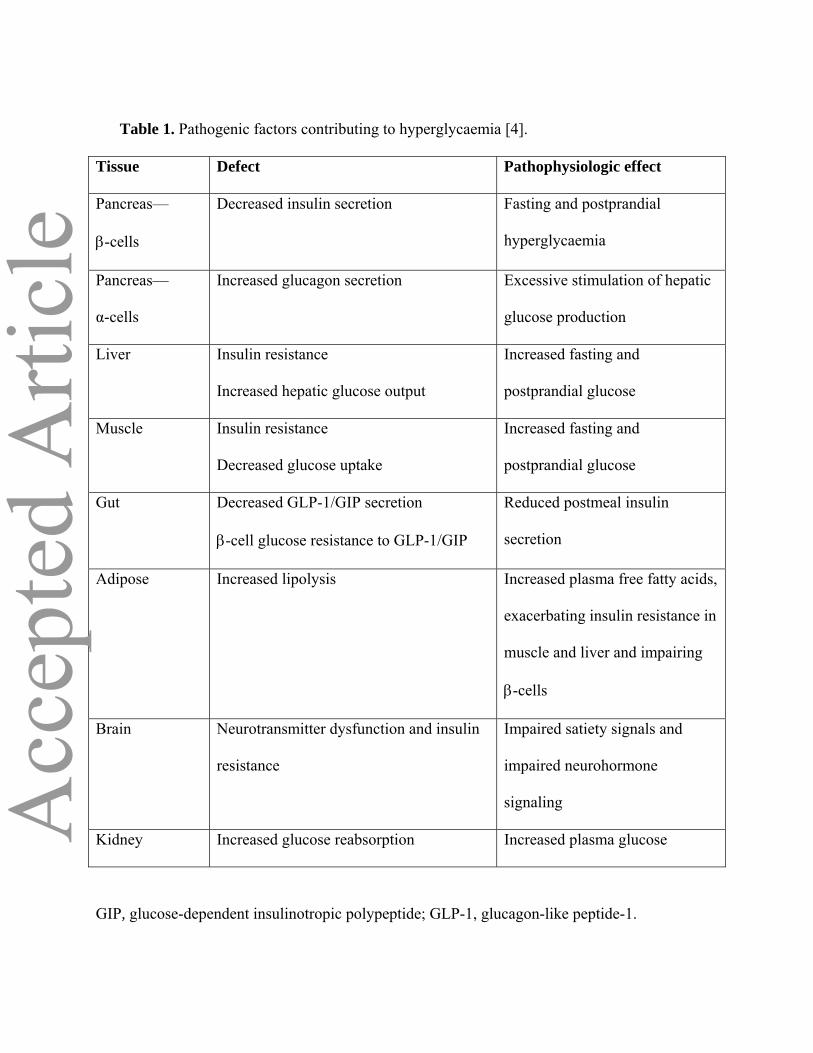
Table 1. Pathogenic factors contributing to hyperglycaemia [4].
Tissue Defect Pathophysiologic effect
Pancreas—
β-cells
Decreased insulin secretion Fasting and postprandial
hyperglycaemia
Pancreas—
α-cells
Increased glucagon secretion Excessive stimulation of hepatic
glucose production
Liver Insulin resistance
Increased hepatic glucose output
Increased fasting and
postprandial glucose
Muscle Insulin resistance
Decreased glucose uptake
Increased fasting and
postprandial glucose
Gut Decreased GLP-1/GIP secretion
β-cell glucose resistance to GLP-1/GIP
Reduced postmeal insulin
secretion
Adipose Increased lipolysis Increased plasma free fatty acids,
exacerbating insulin resistance in
muscle and liver and impairing
β-cells
Brain Neurotransmitter dysfunction and insulin
resistance
Impaired satiety signals and
impaired neurohormone
signaling
Kidney Increased glucose reabsorption Increased plasma glucose
GIP, glucose-dependent insulinotropic polypeptide; GLP-1, glucagon-like peptide-1.

Table 2. SGLT2 inhibitors in clinical development.
Clinical
development phase
Agent Manufacturer
Phase III Dapagliflozin AstraZeneca/Bristol-Myers Squibb
BI 10773 Boehringer Ingelheim
Canagliflozin (JNJ-
28431754)
Johnson & Johnson
Phase II AVE-2268 Sanofi-Aventis
Remogliflozin
(discontinued)
Sergliflozin (discontinued)
GlaxoSmithKline/Kissei
TS-033 Taisho
YM-543 Astellas/Kotobuki Pharmaceuticals
ISIS 388626 ISIS
Phase I CSG-452A Chugai/Roche
SAR-7226 Sanofi-Aventis
TA-7284 Mitsubishi Tanabe/Johnson &
Johnson
SGLT, sodium‐glucose cotransporter
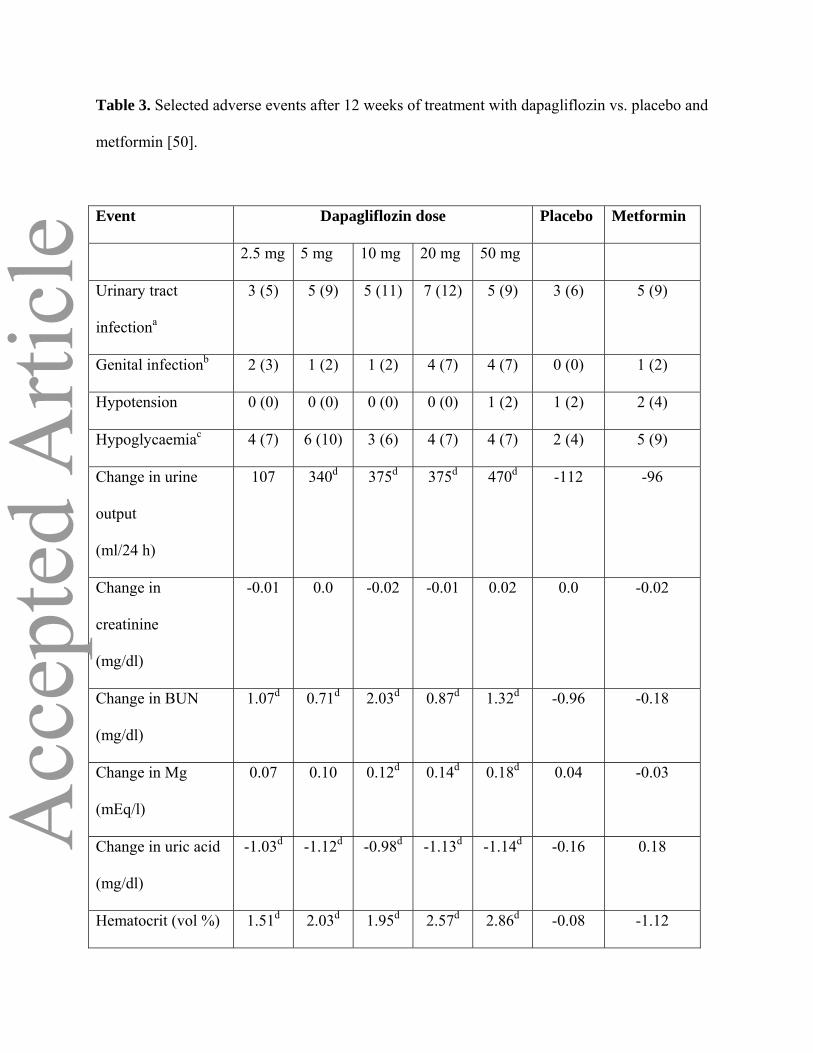
Table 3. Selected adverse events after 12 weeks of treatment with dapagliflozin vs. placebo and
metformin [50].
Event Dapagliflozin dose Placebo Metformin
2.5 mg 5 mg 10 mg 20 mg 50 mg
Urinary tract
infectiona
3 (5) 5 (9) 5 (11) 7 (12) 5 (9) 3 (6) 5 (9)
Genital infectionb 2 (3) 1 (2) 1 (2) 4 (7) 4 (7) 0 (0) 1 (2)
Hypotension 0 (0) 0 (0) 0 (0) 0 (0) 1 (2) 1 (2) 2 (4)
Hypoglycaemiac 4 (7) 6 (10) 3 (6) 4 (7) 4 (7) 2 (4) 5 (9)
Change in urine
output
(ml/24 h)
107 340d 375d 375d 470d -112 -96
Change in
creatinine
(mg/dl)
-0.01 0.0 -0.02 -0.01 0.02 0.0 -0.02
Change in BUN
(mg/dl)
1.07d 0.71d 2.03d 0.87d 1.32d -0.96 -0.18
Change in Mg
(mEq/l)
0.07 0.10 0.12d 0.14d 0.18d 0.04 -0.03
Change in uric acid
(mg/dl)
-1.03d -1.12d -0.98d -1.13d -1.14d -0.16 0.18
Hematocrit (vol %) 1.51d 2.03d 1.95d 2.57d 2.86d -0.08 -1.12

aIncludes cystitis and bacterial and fungal urinary tract infection.
bIncludes vulvovaginal mycotic infection, genital herpes, genital infection and penile infection.
cHypoglycaemia was not defined, but there was no reported fingerstick glucose value ≤2.8
mmol/l.
dp < 0.05 vs. placebo.
BUN, blood urea nitrogen; Mg, magnesium
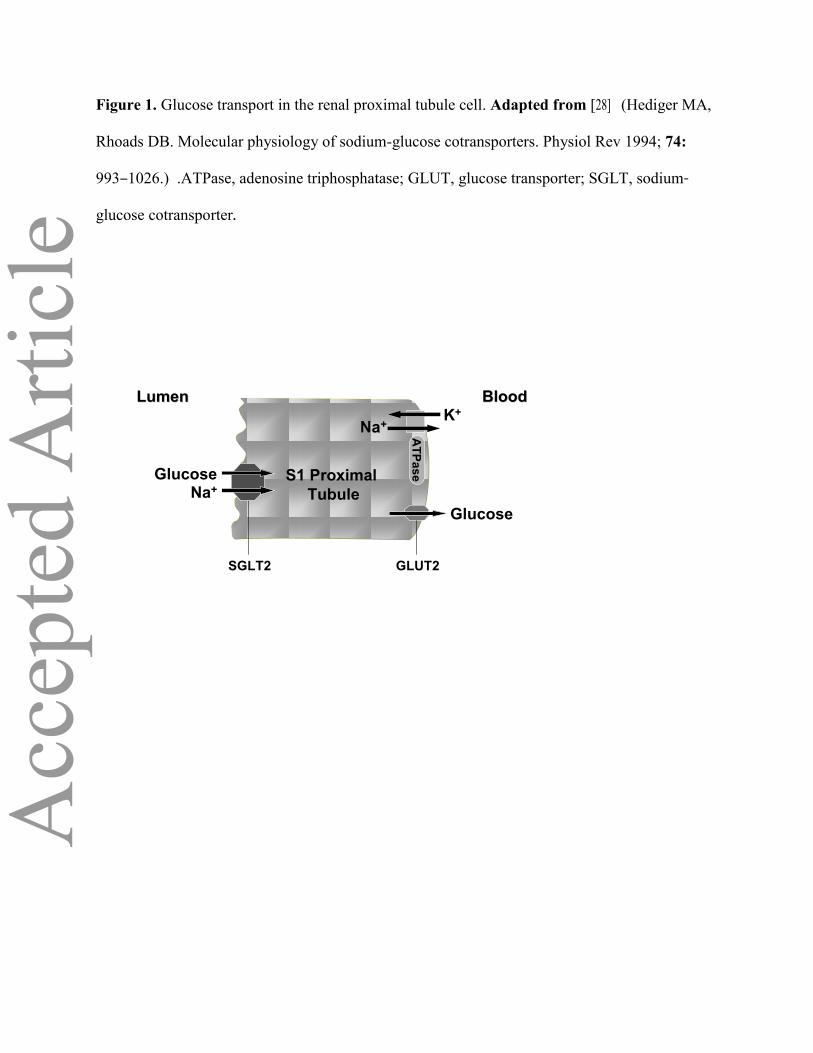
Figure 1. Glucose transport in the renal proximal tubule cell. Adapted from [28] (Hediger MA,
Rhoads DB. Molecular physiology of sodium-glucose cotransporters. Physiol Rev 1994; 74:
993–1026.) .ATPase, adenosine triphosphatase; GLUT, glucose transporter; SGLT, sodium-
glucose cotransporter.
Figure 1Figure 1
S1 Proximal Tubule
NaNa++K+
ATPase
Glucose
GLUT2
Glucose
SGLT2
BloodBloodLumenLumen
Na+

Figure 2. Renal glucose handling. Adapted from [12].
TmG, maximal transport rate for glucose
Plasma Glucose Concentration (mmol/l)
155
Glu
co
se F
lux
(mg
/min
)
Splay
Exc
retio
n
TmG
10
Actual Threshold
Rea
bsor
ptio
n
Theoretical threshold
Splay
0
100
200
300
400
Figure 2Figure 2
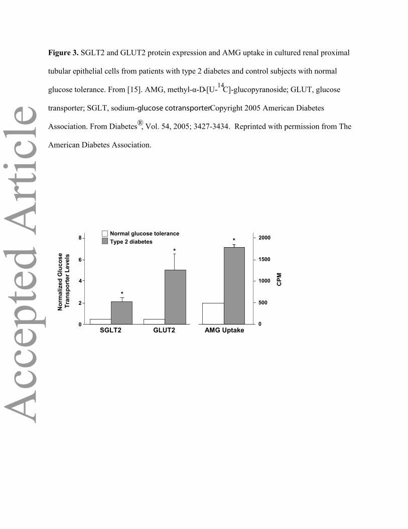
Figure 3. SGLT2 and GLUT2 protein expression and AMG uptake in cultured renal proximal
tubular epithelial cells from patients with type 2 diabetes and control subjects with normal
glucose tolerance. From [15]. AMG, methyl-α-D-[U-14C]-glucopyranoside; GLUT, glucose
transporter; SGLT, sodium-glucose cotransporter. Copyright 2005 American Diabetes
Association. From Diabetes®, Vol. 54, 2005; 3427-3434. Reprinted with permission from The
American Diabetes Association.
GLUT2 AMG UptakeSGLT2
Normal glucose toleranceType 2 diabetes
0
2
6
8
0
500
1000
1500
2000
Nor
mal
ized
Glu
cose
Tr
ansp
orte
r Lev
els
CPM
*
4
**
Figure 3Figure 3
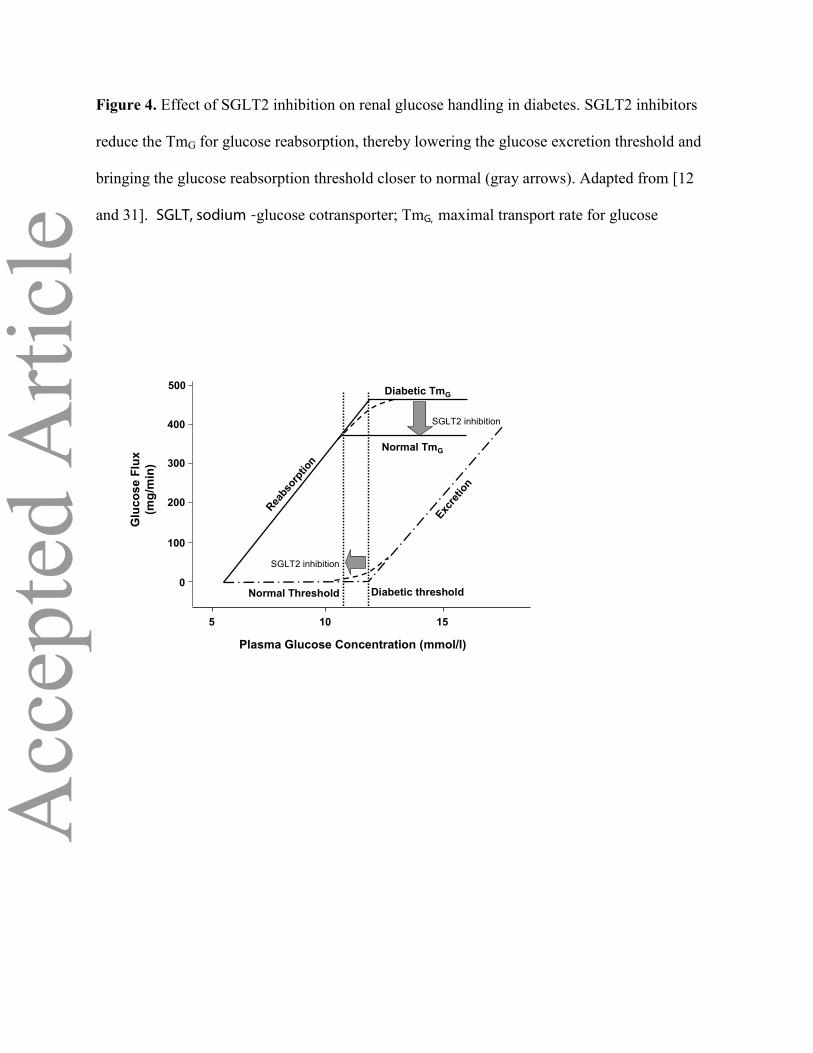
Figure 4. Effect of SGLT2 inhibition on renal glucose handling in diabetes. SGLT2 inhibitors
reduce the TmG for glucose reabsorption, thereby lowering the glucose excretion threshold and
bringing the glucose reabsorption threshold closer to normal (gray arrows). Adapted from [12
and 31]. SGLT, sodium -glucose cotransporter; TmG, maximal transport rate for glucose
Plasma Glucose Concentration (mmol/l)
Glu
cose
Flu
x (m
g/m
in)
Excre
tion
Diabetic TmG
Normal Threshold
Reabs
orpt
ion
Diabetic threshold
155 10
0
100
200
300
400
Figure 4Figure 4
Normal TmG
SGLT2 inhibition
SGLT2 inhibition
500
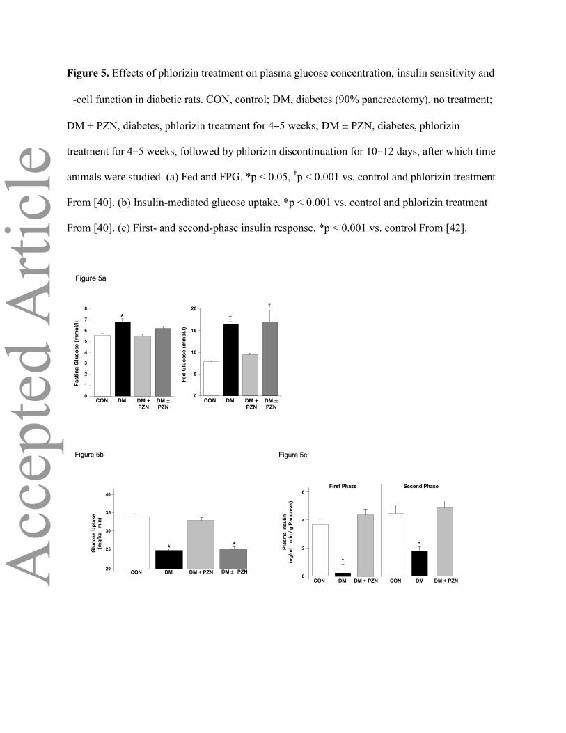
Figure 5. Effects of phlorizin treatment on plasma glucose concentration, insulin sensitivity and
-cell function in diabetic rats. CON, control; DM, diabetes (90% pancreactomy), no treatment;
DM + PZN, diabetes, phlorizin treatment for 4–5 weeks; DM ± PZN, diabetes, phlorizin
treatment for 4–5 weeks, followed by phlorizin discontinuation for 10–12 days, after which time
animals were studied. (a) Fed and FPG. *p < 0.05, †p < 0.001 vs. control and phlorizin treatment
From [40]. (b) Insulin-mediated glucose uptake. *p < 0.001 vs. control and phlorizin treatment
From [40]. (c) First- and second-phase insulin response. *p < 0.001 vs. control From [42].
0
5
10
15
20
0
1
2
3
4
5
6
7
8
Fast
ing
Glu
cose
(mm
ol/l)
CON
*
Fed
Glu
cose
(mm
ol/l)
†
†
Figure 5aFigure 5a
DM DM + PZN
DM ±PZN
CON DM DM + PZN
DM ±PZN
Glu
cose
Upt
ake
(mg/
kg ∙
min
)
DM ± PZNDM + PZNDMCON
**
20
25
30
35
40
Figure 5bFigure 5b
First PhaseFirst Phase Second PhaseSecond Phase
CON CONDM + PZN DM + PZNDM DM
6
0
4
*
*2Pl
asm
a In
sulin
(ng/
ml ∙
min
/ g
Panc
reas
)
Figure 5cFigure 5c
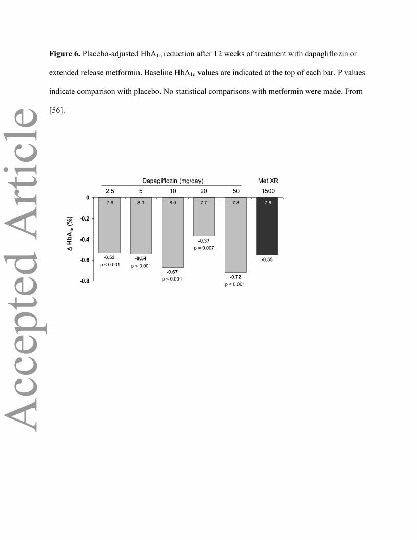
Figure 6. Placebo-adjusted HbA1c reduction after 12 weeks of treatment with dapagliflozin or
extended release metformin. Baseline HbA1c values are indicated at the top of each bar. P values
indicate comparison with placebo. No statistical comparisons with metformin were made. From
[56].
ΔH
bA1c
(%)
Figure 6Figure 6
-0.53 -0.54
-0.67
-0.37
-0.72
-0.55
-0.8
-0.6
-0.4
-0.2
0
Dapagliflozin (mg/day) Met XR
2.5 5 10 20 50 1500
7.6 8.0 8.0 7.7 7.8 7.6
p < 0.001 p < 0.001
p < 0.001p < 0.001
p = 0.007