Embed Size (px)
Citation preview
Washington University in St. LouisWashington University Open Scholarship
All Theses and Dissertations (ETDs)
January 2011
The Role of Viral Protein pUL21a in HumanCytomegalovirus InfectionAnthony FehrWashington University in St. Louis
Follow this and additional works at: https://openscholarship.wustl.edu/etd
This Dissertation is brought to you for free and open access by Washington University Open Scholarship. It has been accepted for inclusion in AllTheses and Dissertations (ETDs) by an authorized administrator of Washington University Open Scholarship. For more information, please [email protected].
Recommended CitationFehr, Anthony, "The Role of Viral Protein pUL21a in Human Cytomegalovirus Infection" (2011). All Theses and Dissertations (ETDs).105.https://openscholarship.wustl.edu/etd/105
WASHINGTON UNIVERSITY
Division of Biology and Biomedical Sciences
Program in Molecular Microbiology and Microbial Pathogenesis
Dissertation Examination Committee:
Dong Yu, Chair
Keril J. Blight
Anthony R. French
Ted H. Hansen
Herbert W. Virgin
David Wang
THE ROLE OF VIRAL PROTEIN pUL21a IN HUMAN CYTOMEGALOVIRUS
INFECTION
by
Anthony Roger Fehr
A dissertation presented to the
Graduate School of Arts and Sciences
of Washington University in
partial fulfillment of the
requirements for the degree
of Doctor of Philosophy
August 2011
Saint Louis, Missouri

ii
ABSTRACT OF THE DISSERTATION
The Role of Viral Protein pUL21a in Human Cytomegalovirus Infection
By
Anthony Roger Fehr
Doctor of Philosophy in Biology and Biomedical Sciences
(Molecular Microbiology and Microbial Pathogenesis)
Washington University in St. Louis, 2011
Dr. Dong Yu, Chairperson
The double-stranded DNA virus human cytomegalovirus (HCMV) is a ubiquitous
human pathogen that causes severe disease in immunocompromised individuals and
neonates. The biology of HCMV remains largely undefined and functions of many of the
roughly 200 putative HCMV genes are still unknown. One of these genes, UL21a, was
shown to be critical for efficient viral replication in two large-scale mutagenesis studies.
UL21a encodes for a putative protein that is 123 amino acids and has no homology to any
known proteins. The gene products of UL21a, its role during infection, and its function(s)
remain undefined.
Here, we characterized UL21a and demonstrated its role in HCMV infection. We
identified a single UL21a transcript which was expressed with early gene kinetics.
UL21a encoded a protein termed pUL21a which localized to the cytoplasm and
underwent proteasome-dependent degradation. UL21a was specifically required for
efficient viral replication, and the growth of a UL21a deletion virus was similar to that of
a stop codon mutant, suggesting it is pUL21a which facilitates viral replication.
To identify the role of pUL21a during virus infection, we analyzed fibroblasts
infected with equal amounts of wild-type and UL21a deletion virus for defects in
iii
multiple steps of the viral lifecycle. The UL21a deletion virus entered cells and initiated
viral gene expression efficiently; however, it synthesized viral DNA poorly and
accumulated several immediate-early (IE) transcripts at reduced levels at late times of
infection. The reduction in IE transcripts was dependent on the reduction in viral DNA
synthesis, showing that multiple IE transcripts are dependent on viral DNA synthesis for
their full expression. Finally, using complementing cells we show that it is the de novo
synthesis of pUL21a which facilitates viral DNA synthesis.
To determine the function(s) of pUL21a, we identified proteins that specifically
interacted with pUL21a. We found that pUL21a interacts with the Anaphase-Promoting
Complex (APC), a multi-subunit E3 ubiquitin ligase which targets multiple proteins for
proteasome-dependent degradation. The APC is critical for progression through mitosis
and the regulation of cellular DNA synthesis. We have found that pUL21a specifically
binds to the APC, and is required for the accumulation of APC substrates, degradation of
APC4 and APC5, and dissociation of the APC during HCMV infection. Finally, shRNA
knockdown of the APC activator Cdh1 and to a lesser degree APC8, significantly
restored late gene expression of the UL21a mutant virus, suggesting the APC has
antiviral activity for HCMV. Thus, we propose that one mechanism for pUL21a to
promote viral replication is to regulate the function of the APC.
iv
ACKNOWLEDGEMENTS
First, I must give a huge thanks to my thesis advisor Dong Yu. Dong has pushed
me to be better every day for the last 5 years and I have grown tremendously as a scientist
under his guidance. He is always there to discuss problems, formulate hypotheses, devise
experiments, and talk about all areas of science. He has trusted me to be independent in
my work and has provided all the resources needed to do excellent science. I also must
thank my thesis chair Herbert “Skip” Virgin and his lab for the incredible amount of
feedback on my project in our joint lab meetings. I also must acknowledge other
members of my committee and financial support/awards provided by the Departments of
Molecular Microbiology/Developmental Biology and the Division of Infectious Diseases.
I would like to thank all of the members of the Yu lab, Zhikang, Baoqin, Cris,
Travis, Yi Chieh, Anne, Carlos, Camille, and Nick. I have to especially thank Zhikang,
who I have learned a great deal from as he has been the senior member of the lab, and
Cris our manager, for his amazing dedication and work ethic makes all of my work
possible. I will miss the camaraderie of our group and how well we all work together.
I have to thank my parents, Roger and Chris, who have been more supportive than
any parents I know. They have made countless trips to St. Louis to hang out and help us
out. They are always interested in what I am doing, and are my biggest fans.
Finally, and most importantly, I have to thank my wife Rachel and my son Darin.
Their endless love and support drive me to be the best I can be. They are my best friends,
and no matter how my day went, I am always husband and dad when I get home. I would
not be where I am without them, and they deserve a lot of credit for this thesis work.

v
TABLE OF CONTENTS
Title Page ………………………………………………………………………………….i
Abstract of the Dissertation ……………………………………………………………....ii
Acknowledgements ………………………………………………………………………iv
Table of Contents ………………………………………………………………………....v
List of Tables.....................................................................................................................vi
List of Figures …………………………………………………………………………...vii
List of Abbreviations ………………………………………………………………...…..ix
Chapter I
Introduction ……………………………………………………………………………….1
Chapter II …………………………………………………………………………......…39
Human Cytomegalovirus Gene UL21a Encodes a Short-Lived Cytoplasmic Protein
and Facilitates Virus Replication in Fibroblasts
Chapter III …………………………………………………..…………………………...85
Human Cytomegalovirus Early Protein pUL21a Promotes Efficient Viral DNA
Synthesis and the Late Accumulation of Immediate-Early Transcripts
Chapter IV ……………………………………………………………………………...131
pUL21a Regulates the Anaphase-Promoting Complex and Targets APC4 and APC5
for Proteasome-Dependent Degradation
Chapter V ………………………………………………………………………...…….174
Summary and Future Directions
vi
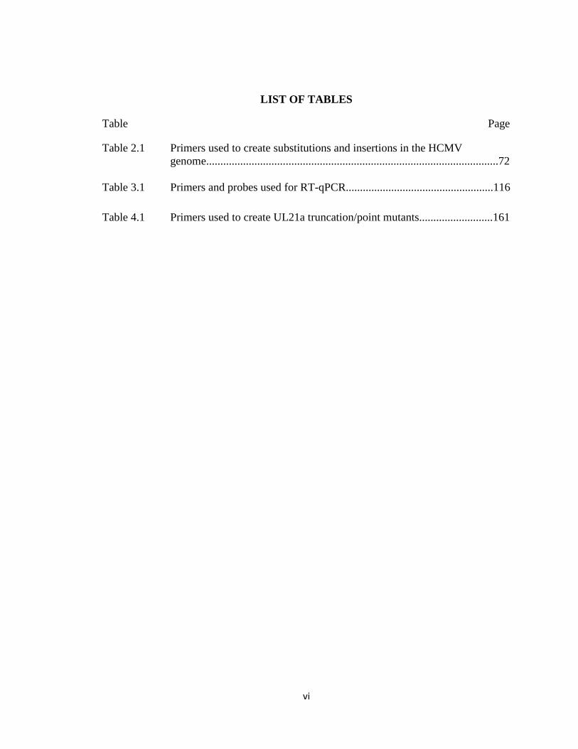
LIST OF TABLES
Table Page
Table 2.1 Primers used to create substitutions and insertions in the HCMV
genome.......................................................................................................72
Table 3.1 Primers and probes used for RT-qPCR....................................................116
Table 4.1 Primers used to create UL21a truncation/point mutants..........................161
vii
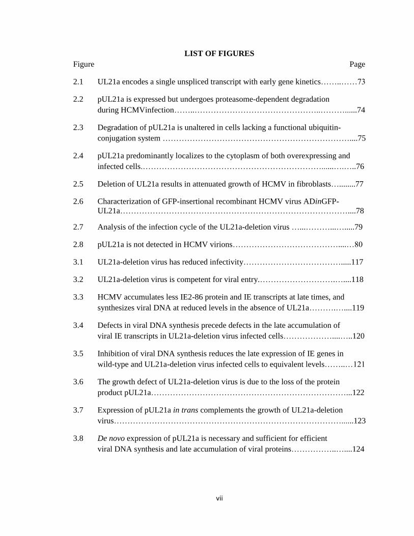
LIST OF FIGURES
Figure Page
2.1 UL21a encodes a single unspliced transcript with early gene kinetics……..……73
2.2 pUL21a is expressed but undergoes proteasome-dependent degradation
during HCMVinfection……..………………………………………..………......74
2.3 Degradation of pUL21a is unaltered in cells lacking a functional ubiquitin-
conjugation system ……………………………………………………………....75
2.4 pUL21a predominantly localizes to the cytoplasm of both overexpressing and
infected cells.…………………………………………………………......….…..76
2.5 Deletion of UL21a results in attenuated growth of HCMV in fibroblasts…........77
2.6 Characterization of GFP-insertional recombinant HCMV virus ADinGFP-
UL21a…………………………………………………………………………....78
2.7 Analysis of the infection cycle of the UL21a-deletion virus …...………...….....79
2.8 pUL21a is not detected in HCMV virions…………………………………....…80
3.1 UL21a-deletion virus has reduced infectivity……………………………….....117
3.2 UL21a-deletion virus is competent for viral entry.……………………….…....118
3.3 HCMV accumulates less IE2-86 protein and IE transcripts at late times, and
synthesizes viral DNA at reduced levels in the absence of UL21a……….…....119
3.4 Defects in viral DNA synthesis precede defects in the late accumulation of
viral IE transcripts in UL21a-deletion virus infected cells………………....…..120
3.5 Inhibition of viral DNA synthesis reduces the late expression of IE genes in
wild-type and UL21a-deletion virus infected cells to equivalent levels……..…121
3.6 The growth defect of UL21a-deletion virus is due to the loss of the protein
product pUL21a………………………………………………………………...122
3.7 Expression of pUL21a in trans complements the growth of UL21a-deletion
virus…………………………………………………………………………......123
3.8 De novo expression of pUL21a is necessary and sufficient for efficient
viral DNA synthesis and late accumulation of viral proteins……………..…....124
viii
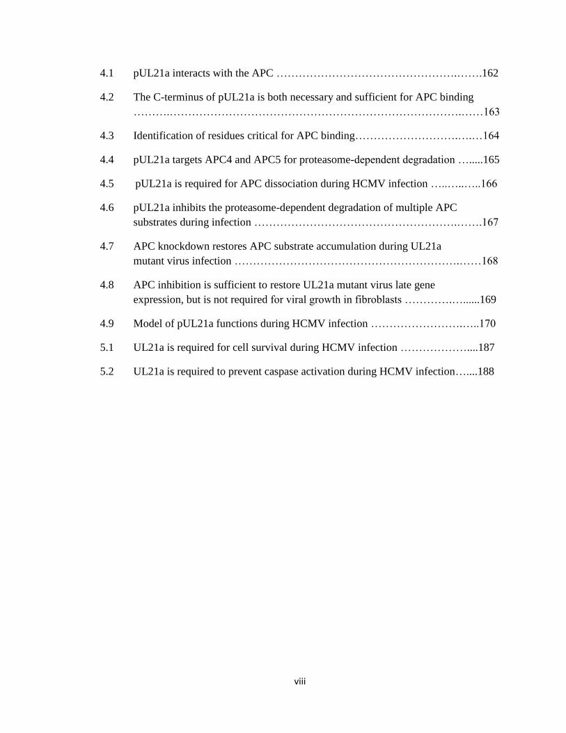
4.1 pUL21a interacts with the APC ………………………………………….…….162
4.2 The C-terminus of pUL21a is both necessary and sufficient for APC binding
……….…………………………………………………………………….……163
4.3 Identification of residues critical for APC binding……………………….….…164
4.4 pUL21a targets APC4 and APC5 for proteasome-dependent degradation ….....165
4.5 pUL21a is required for APC dissociation during HCMV infection …..…..…..166
4.6 pUL21a inhibits the proteasome-dependent degradation of multiple APC
substrates during infection ……………………………………………….…….167
4.7 APC knockdown restores APC substrate accumulation during UL21a
mutant virus infection …………………………………………………….……168
4.8 APC inhibition is sufficient to restore UL21a mutant virus late gene
expression, but is not required for viral growth in fibroblasts ………….…......169
4.9 Model of pUL21a functions during HCMV infection …………………….…..170
5.1 UL21a is required for cell survival during HCMV infection ………………....187
5.2 UL21a is required to prevent caspase activation during HCMV infection…....188
ix
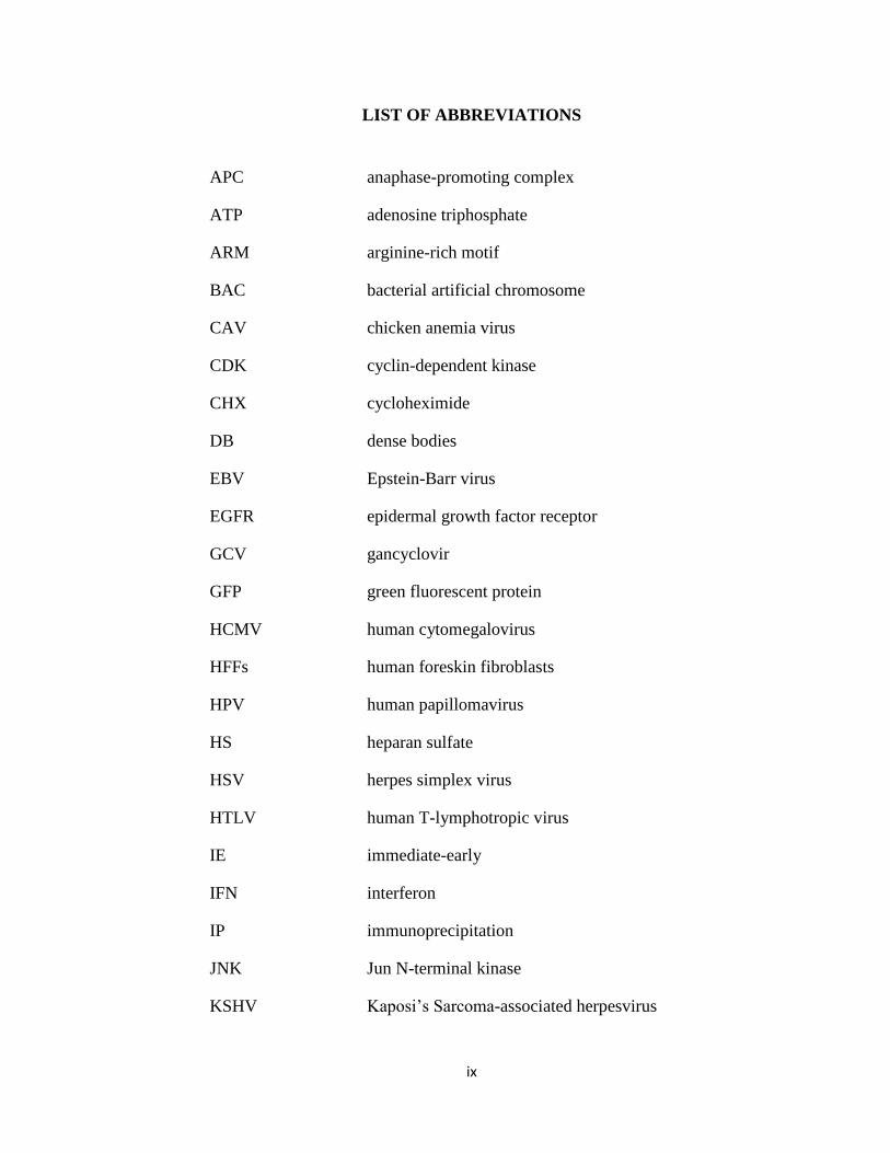
LIST OF ABBREVIATIONS
APC anaphase-promoting complex
ATP adenosine triphosphate
ARM arginine-rich motif
BAC bacterial artificial chromosome
CAV chicken anemia virus
CDK cyclin-dependent kinase
CHX cycloheximide
DB dense bodies
EBV Epstein-Barr virus
EGFR epidermal growth factor receptor
GCV gancyclovir
GFP green fluorescent protein
HCMV human cytomegalovirus
HFFs human foreskin fibroblasts
HPV human papillomavirus
HS heparan sulfate
HSV herpes simplex virus
HTLV human T-lymphotropic virus
IE immediate-early
IFN interferon
IP immunoprecipitation
JNK Jun N-terminal kinase
KSHV Kaposi’s Sarcoma-associated herpesvirus

x
MCMV murine cytomegalovirus
MCP major capsid protein
MHC major histocompatibility
MOI multiplicity of infection
NK natural killer cell
NIEP non-infectious enveloped particle
ORF open reading frame
PAA phosphonoacetic acid
PACR poxviral APC/cyclosome regulator
PDGFR-α platelet-derived growth factor receptor-α
PFKFB3 6-phosphofructo-2-kinase/fructose-2,6-bisphosphatase
isoform 3
PFU plaque forming unit
PLK-1 polo-like kinase-1
PP2A protein phosphatase 2A
qPCR quantitative polymerase chain reaction
RACE rapid amplification of cDNA ends
Rb retinoblastoma tumor suppressor protein
RRM2 ribonucleotide reductase subunit M2
RT reverse transcriptase
shRNA short hairpin RNA
siRNA small interfering RNA
TCID50 tissue culture infectious dose
TK thymidine kinase
TPR tetratricopeptide repeat

xi
UL unique-long
US unique-short
VZV Varicella-Zoster virus
Y2H Yeast-2-Hybrid

1
Chapter I
Introduction

2
Herpesviruses
Overview
Herpesviruses are a large class of viruses and are widespread throughout nature.
Most mammalian species have at least one known herpesvirus pathogen. There are eight
herpesvirsues which infect humans. These viruses are: herpes simplex virus type 1
(HSV-1), herpes simplex virus type 2 (HSV-2), varicella-zoster virus (VZV), Epstein-
Barr virus (EBV), human cytomegalovirus (HCMV), human herpesviruses 6A, 6B, and 7,
and Kaposi’s sarcoma-associated herpesvirus (KSHV) (95).
The relatedness between all herpesviruses suggests that they evolved from a
common ancestor, which likely lived many millions of years ago. In fact, herpesviruses
even share some features with bacteriophages, suggesting they may have evolved from
bacteriophages, a lineage which may have begun at the early stages of parasitism, nearly
a billion years ago.
Herpesviruses share at least four significant biological properties. They all
encode several proteins involved in nucleotide metabolism, DNA synthesis, and protein
processing. The synthesis of viral DNAs and capsid assembly occurs in the nucleus with
final envelopment of the virus occurring in the cytoplasm. Productive infection results in
the destruction of the host cell, and all herpesviruses studied to date are able to establish
latency in their natural host species. While herpesviruses have many similarities, there
are also several difference which distinguish them. Herpesviruses can vary widely in the
length of their replicative cycle, host cell tropism, and cell types in which they establish
latency, and disease manifestations. Based on these differences, the herpesviruses are
classified into three main subfamilies: Alphaherpesvirinae, Betaherpesvirinae, and
3
Gammaherpesvirinae. Alphaherpesvirinae have a short replication cycle, a broad species
and cell tropism,, and establish a latent infection in sensory ganglia. Betaherpesvirinae
have a relatively long replication cycle, a broad cell tropism but strict species tropism,
and they establish latency in secretory glands, lymphorecticular cells, kidneys, and other
tissues. Gammaherpesvirinae have a short replication cycle, replicate in epithelial and
fibroblast cells, and establish latency and can promote tumorigenesis of T or B
lymphocytes.
Structure/Genome
All Herpesviruses share a common structure. The virion consists of a core, the
capsid, a tegument layer, and an envelope. The core contains the viral DNA within a
proteinaceous spindle. The capsids for all herpesiviruses are very similar, with a 100 nm
diameter and a triangulation number of 16, and 960 copies of the capsid protein per
capsid. The tegument, the structure found between the capsid and the envelope, is
thought to have an ordered structure despite the lack of distinctive features. The
tegument proteins are added both in the nucleus following capsid formation and in the
cytoplasm prior to envelopment, and these proteins are often involved in helping the virus
establish a productive infection during the initial stages of the life cycle. The envelope is
a membrane layer around the virus, likely derived from the trans-Golgi or endosome, and
contains large numbers of glycoproteins.
Herpesviruses contain linear, double-stranded DNA genomes ranging in size from
120-250 kb. The genome circularizes upon entry into the nuclei of infected cells.
Herpesvirus genomes contain terminal and internal repeats in a variety of different
4
arrangements. In specific viruses like HSV or HCMV, multiple isomers of the genome
can be present due to inversion of the repeats. Herpesviruses can encode between 70 and
200 genes, but these numbers are likely underestimates, and do not take into account
small RNAs or protein products of less than 100 amino acids.
Lifecycle
Herpesviruses begin their lifecycle with glycoprotein attachment at the surface of
the cell. Some viruses enter by endocytosis while others fuse at the plasma membrane.
Regardless of their route of entry, all herpesviruses traffic to the nucleus where the
genome is released, circularizes, and begins transcription of gene products. Gene
expression is a classic regulatory cascade, with viral genes falling into separate groups
with respect to their order of expression. There are three major expression kinetic classes
for herpesvirus genes: immediate early, early, and late. Immediate-early (IE) genes are
expressed within a few hours of infection and primarily act as transactivators for the
production of early genes, which are required for DNA replication. Late genes encode
structural proteins important for encapsidation and packaging of the virion. There are
two sub-classes of late genes, leaky late and true late. Leaky late genes are augmented by
viral DNA synthesis while true late genes are completely dependent on viral DNA
synthesis for their expression.
Following capsid assembly in the nucleus, the virus goes through an
envelopment/deenvelopment step to transit through the nuclear membrane and egress into
the cytoplasm. Once in the cytoplasm, the capsid acquires its full complement of
tegument proteins and traffics to the assembly center where its envelope is acquired.
5
Once its envelope is acquired, the virus traffics to the membrane where it is subsequently
released to begin the cycle anew.
As mentioned previously, all herpesviruses establish latency as an alternative
lifestyle. During latency, the viral genome is maintained as an episome, and only a few
viral gene products are expressed. Latent genomes are able to reactivate and begin a
productive infection upon certain stress stimuli.
HCMV
Clinical Relevance
Human cytomegalovirus (HCMV), the prototypical betaherpesvirus, is a
ubiquitous pathogen that infects the majority of the world’s population. HCMV is usually
asymptomatic in immunocompetent individuals, except in rare cases where it causes
mononucleosis. However, HCMV can cause severe disease and death in
immunocompromised individuals such as AIDS patients and transplant recipients.
Importantly, HCMV is the most common viral cause of birth defects leading to mental
retardation, blindness, and hearing loss (64). In addition, HCMV infection is also a
possible risk factor in the development of vascular diseases such as atherosclerosis,
transplant vascular sclerosis, and coronary restenosis after angioplasty surgery (24, 50,
59, 65, 115, 120, 148). The economic burden to the U.S. health care system for this virus
is estimated at approximately 4 billion dollars annually, with a majority of the costs
attributed to long-term sequelae experienced by individuals who acquire congenital
HCMV disease (33). A comprehensive understanding of how HCMV interacts with the
6
host to establish both acute and latent infections will be critical for developing an
effective vaccine and novel therapeutics to combat HCMV disease (43, 102).
HCMV Pathogenesis
HCMV is transmitted by direct contact with bodily fluids of infected individuals.
The virus replicates in the mucosal epithelium at the site of inoculation. Hematopoietic
cells then disseminate the virus during a systemic viral infection to a wide range of cell
types, including epithelial, endothelial, fibroblast, macrophage, and dendritic cells. Virus
produced from these cells contributes to viral shedding following primary infection
which can last for several months in adults to several years in young children (64).
The immune response to HCMV is strong, broad, and long lasting. The immune
response lasts for many years and a large percentage of memory T cells in older
individuals can be devoted towards CMV antigens (122). Interestingly, despite the
strength of the immune response, pre-existing immunity does not prevent reinfection, but
does limit acute disease in both immunocompetent and immunocompromised individuals
(62, 133). The ability of CMV to continue viral shedding for several months in the face
of such a strong immune response is likely due to the many gene products encoded by
CMV to modulate both innate and adaptive immune responses (62).
Natural Killer (NK) cells are likely the first immune cells that limit viral
replication. Mice with NK cell deficiencies are highly susceptible to murine CMV
(MCMV) infection (101). In humans, a patient with NK cell deficiency suffered from
severe infection by HCMV, which suggests this response is also important to control
HCMV (64). Furthermore, multiple HCMV gene products, including UL16, UL40,
7
UL140, UL141, and UL142 all play a role in NK cell inhibition using several different
mechanisms (62, 140). Cell-mediated immunity and specifically CD8+ cytotoxic T cells
are also critical for control of HCMV. Adoptive transfer of CD8+ T cells specific for
CMV antigens into both mice and humans can protect from CMV disease (29, 46, 92).
HCMV also encodes numerous gene products designed to interfere with MHC I
expression and translocation to the cell surface, such as US2, US3, US6, and US11 (62).
Antibody response also plays a role during infection, likely by preventing transmission
between individuals, as most virus in the host remains cell-associated except in secretions
such as saliva and breast milk. Passive immunization with a high titer HCMV-specific
immunge globulin can prevent transplacental transmission during pregnancy (72).
Like all herpesviruses, HCMV establishes latency and has the ability to reactivate
from latency following immunosuppression; which profoundly affects its ability to
establish and cause disease. Latent virus is found primarily in hematopoietic cells which
are able to repress viral gene expression. Further differentiation into cells such as
macrophages and dendritic cells results in reactivation, suggesting there are
differentiation-specific factors that restrict viral gene regulation (88, 90, 91, 107, 111,
112). Study of HCMV latency has been hampered by the lack of a small animal model
and thus investigators have relied on cell culture based systems of experimental latency.
However, the MCMV surrogate model recapitulates the general process of latency and
reactivation and thus has provided unique insight into these processes. However, the
viral genes which mediate this process remain mostly elusive (79).
8
HCMV Virion
The HCMV virion is typical but somewhat larger than other herpesviruses (200-
300 nm) (reviewed in (64)). The envelope contains ~20 glycoproteins including the
conserved gB, gH:gL, and gM:gN complexes, as well as several G-coupled protein
receptors. It also contains a wide array of tegument proteins, most of which are
phosphorylated and highly immunogenic. The most abundant tegument proteins are the
pp65 and pp71 proteins. pp65 helps block interferon responses and pp71 acts as a viral
transactivator (VTA) (4, 8, 34). HCMV has a class E genome, which contains both
unique long (UL) and unique short (US) segments flanked by terminal and internal
repeats. HCMV is the largest known human virus in terms of genome size, with a
genome of ~240 kb, and encodes at least 166 ORFs, several microRNAs, and likely
several other unknown gene products (12, 16, 18, 26, 67, 68). Interestingly, HCMV
produces an abundant number of both dense bodies (DB) and non-infectious particles
(NIEP), in excess of several hundred more DB/NIEP than infectious virus. Dense bodies
are largely composed of the tegument protein pp65 surrounded by a cellular membrane
while NIEP’s are virions which are largely devoid of viral DNA. HCMV is the only
herpesvirus to produce DB’s and it is unclear why the virus produces so many DB’s and
NIEP’s.
Viral Replication in vitro
HCMV has a relatively slow replication cycle, taking 48-72 hours to begin
producing infectious virus in cell culture. Commonly used laboratory strains AD169 and
Towne, grow well in fibroblasts but are unable to replicate in additional cell types due to
9
mutations in the UL128-131 genes and large deletions in the ULb’ region. Clinical
isolates of the virus retain the ability to grow in endothelial, epithelial, macrophage, and
dendritic cells in vitro. However, except for U373MG cells, fully transformed cells are
non-permissive for HCMV replication due to unknown reasons.
HCMV binds to heparan sulfate (HS) on the cell surface but additional receptors
remain controversial. It was originally proposed that HCMV uses Epidermal Growth
Factor Receptor (EGFR) as a receptor, but this has recently been disputed (19, 39, 137).
More recently, cellular integrins and platelet-derived growth factor-α (PDGFR-α) have
been proposed to serve as a receptor for the virus (21, 114, 136). Regardless, it has been
shown that gB binds to HS and, along with gH:gL, plays a role in fusion (14). HCMV
enters the cellular cytoplasm by fusion with a cellular membrane; however, where this
occurs depends on the cell type (14). HCMV enters fibroblasts by fusion at the cell
membrane, while in epithelial and endothelial cells the virus is internalized via
endocytosis before fusion occurs in the cytoplasm (97). Translocation of the virion to the
nucleus occurs by a relatively uncharacterized process, but likely involves transport on
microtubules followed by uncoating and release of viral DNA at nuclear pores (74).
Based on studies of the HSV-1 homolog UL36, it is likely that the conserved UL48 and
UL47 proteins play an essential role in this process (5, 17, 143).
Upon entry into the nucleus, the viral genome is repressed by promyelocytic
leukemia protein (PML bodies) and requires the tegument protein pp71 to alleviate this
repression (98, 99, 124, 125). pp71 targets the PML protein Daxx for ubiquitin-
independent, proteaseome-dependent degradation which activates viral immediate-early
(IE) gene transcription (37, 99). In fact, co-expression of pp71 along with bacterial
10
artificial chromosome (BAC) transfection dramatically increases the ability to produce
infectious virus from BAC transfection (see below) (4). IE genes do not require viral
protein synthesis for transcription and thus are identified by treatment of infected cells
with the protein synthesis inhibitor cycloheximide. The virus expresses its IE genes by 2
to 4 h after viral entry and these genes persist throughout the infection. The primary
proteins encoded by the major immediate-early (MIE) transcript are IE1-72 and IE2-86,
which are produced by alternative splicing (117-119). These IE proteins are critical for
the establishment of a productive infection and must be down-regulated for the virus to
establish latency. The IE2-86 protein is essential for viral replication, while IE1-72 is
required at a low multiplicity of infection (MOI) (22, 25, 31, 57, 63, 100). Both proteins
transactivate viral promoters and also modulate the cellular environment to be conducive
for viral infection. Additional IE genes, which include TRS1, UL37x1, and US3, help
HCMV to overcome innate and adaptive cellular antiviral responses (3, 13, 23, 27, 28,
41, 87).
Transcription of early genes shortly follows IE gene expression, appearing at
between 4 and 12 hpi. True early gene transcription requires IE protein expression but
not viral DNA replication, and thus can be classified by sensitivity to cycloheximide but
resistance to DNA synthesis inhibitors such as phosphonoacetic acid (PAA). Early genes
encode DNA replication enzymes, such as UL44 (processivity factor) and UL54 (viral
DNA polymerase), as well as viral regulatory proteins that alter host cells creating a
favorable environment for replication. DNA replication begins at 14-16 hours post
infection and requires bi-directional transcription at the origin of lytic replication
(OriLyt) and at least 6 proteins specifically involved in replication. These proteins
11
include the viral polymerase (UL54), processivity factor (UL44), ssDNA binding protein
(UL57), and a trimeric helicase-primase complex (UL70/UL102/UL105). Other genes
known to be directly involved in viral DNA synthesis include UL84, UL112-113, UL98
(nuclease), UL114 (uracil DNA glycosidase), and IE2-86 (75). Viral DNA replication
then proceeds using a rolling-circle mechanism.
Late genes are expressed following the onset of viral DNA replication, and many
of them encode structural proteins, such as the major capsid protein (MCP) and pp28,
which are required for assembly and maturation of the virion (106). Other late-gene
products, such as pp71, are tegument proteins which can antagonize intrinsic cellular
defenses and help progeny virus initiate IE gene expression during subsequent infection
(98, 99). Capsid assembly and egress are similar to other herpesviruses, albeit the
mechanisms and genes used to achieve the same goal are slightly different.
HCMV Regulation of Apoptosis
Apoptosis can function as an innate antiviral defense and is especially
problematic for viruses with long replication cycles, such as CMV. Cellular events
leading to apoptosis are classified into 3 different pathways: the mitochondria-mediated
intrinsic pathway, the extra-cellular ligand-mediated extrinsic pathway, and the
endoplasmic reticulum (ER)-mediated pathway. These pathways lead to the activation of
caspase cascades, ultimately culminating in the activation of caspase-3 which leads to
cleavage of substrates such as PARP, which ultimately causes cell death. HCMV
encodes several factors which block the ability of the cell to initiate apoptosis. During
infection, pUL36 (vICA) blocks FAS-mediated apoptosis by inhibiting proteolytic
12
activation of caspase 8 (108). pUL37x1 (vMIA) prevents activation of caspase 9 by
sequestering Bax, and its interaction with GADD45 family members appears to play an
important role in this function (3, 23, 87, 110). pUL38 blocks ER stress induced
apoptosis by preventing Jun N-terminal kinase (JNK) phosphorylation and by increasing
expression of ATF4; however, the mechanism pUL38 uses to achieve this functions
remains unknown (127, 141). Interestingly, the 2.7 kb RNA encoded by HCMV can bind
to a mitochondrial enzyme complex and block Rotenone-induced cell death (89). In
overexpression, IE1 and IE2 block apoptosis by activating the Akt-mediated signaling
pathway (145, 149). The ability of HCMV to block apoptosis is clearly important for
replication, as growth defects of both UL37x1 and UL38 mutant viruses can be
significantly restored by the addition of caspase inhibitors (87, 127).
HCMV Regulation of the Cell Cycle
HCMV also has the ability to regulate cell cycle progression in order to create an
environment conducive to its own replication. HCMV has been shown to stimulate cells
from Go but prevent cellular DNA synthesis from occurring, ultimately coercing the cell
into maintaining itself at the G1/S boundary of the cell cycle (84). The prevailing theory
is that this stage of the cell cycle will provide substantial nutrients and nucleotides for the
virus to replicate, but those resources will not be diverted to helping the cell replicate its
own genome. However, this has not been experimentally proven, and preventing cellular
DNA synthesis may also promote viral replication in other ways, such as preventing a
robust DNA damage response.
13
One of the major cell cycle control proteins targeted by HCMV is the
retinoblastoma tumor-suppressor protein (Rb), which binds to E2F transcription factors in
its unphosphorylated form, thus inhibiting expression of genes required for DNA
replication. HCMV uses at least two mechanisms to inhibit Rb and stimulate cell cycle
progression from G0 to G1. First, the pp71 protein targets unphosphorylated Rb for
ubiquitin-independent, proteasome-dependent degradation (42). To eliminate any
residual Rb activity, the UL97 kinase phosphorylates Rb during infection, rendering it
inactive (36).
HCMV also inhibits the anaphase-promoting complex (APC), a large E3
ubiquitin-ligase important for progression through mitosis and proper entry into S phase
(130, 132, 139). As HCMV does not infect cells in the G2/M phase of the cell cycle, it
has been thought that the inhibition of the APC would advance cellular progression into S
phase prematurely. However, it has not been confirmed that APC inhibition helps the
virus promote S phase nor is it known whether it plays an important role during virus
infection.
HCMV also uses multiple mechanisms to prevent cellular DNA synthesis. IE2-86
inhibits cyclin A transcription and induces p16 expression, which is capable of
preventing Cdk activity (69, 73, 78, 104, 138). pUL69 blocks cellular DNA synthesis by
an undefined mechanism, and pUL117 blocks MCM loading and accumulation during
infection, which prevents licensing of the replication origins (30, 55, 84). Importantly, all
three of these proteins are necessary for the virus to prevent cellular DNA replication, as
the absence of any one protein results in a virus unable to prevent cellular DNA
synthesis.
14
BAC Genetic System
Development of Herpes/HCMV BACs
In the late 90’s, many herpesviruses were cloned as bacterial artificial
chromosomes (BACs) (reviewed in (1)). Prior to this technology, the only way to make
mutant viruses was to use homologous recombination in mammalian cells. Due to the
large genome size and slow-replication kinetics, this method of creating recombinant
viruses was especially difficult in the β-Herpesviruses. To circumvent this problem,
researchers inserted the genomes of these viruses into F-plasmids in Escherichia coli (E.
coli) which could be delivered into a suitable mammalian cell type in order to create
infectious virus. This method created a system where viral recombinants could be
created in the absence of viral growth, thus one can create a viral mutant independently of
its viral fitness. Furthermore, the viral genome could be characterized before
reconstitution of viral progeny, reducing the likelihood of spurious point mutations or
deletions occurring in the process of producing recombinant virus.
One disadvantage to the original BAC system was that a set of non-essential viral
genes had to be deleted to accommodate the large BAC vector sequence (~10 kb) and
maintain the viral genome within its packaging limit. It has been shown that simply
inserting the BAC vector sequence into the viral genome without removing any viral
sequence can result in an attenuated virus (144). To eliminate the need to delete viral
sequences, BACs were then created which had the BAC vector sequence flanked by loxP
sites and also expressed an intron-containing Cre recombinase (109, 144). In E. Coli, the
Cre recombinase will not be functional due to the intron, which allows maintenance of
the BAC vector sequence. Upon delivery into mammalian cells the Cre recombinase is

15
expressed, the BAC vector sequence is cleaved, and virus is produced that is full length
except for the addition of a small loxP site.
In HCMV, several strains have been cloned as BACs including the laboratory
strains Towne and AD169, and several clinical isolates including TR, TB40, and Merlin
(7, 57, 68, 116). The ability to create recombinant clinical BACs will be critical to the
future study of HCMV, as the laboratory strains contain large deletions and many point
mutations and thus does not behave precisely like virus acquired directly from patients.
HCMV Functional Profiling
Not long after the advent of the BAC, several groups set out to create a complete
library of ORF/gene mutations in HCMV and screen these mutants for their growth in
fibroblasts. The functions of many of the 166 HCMV ORFs remains unknown, and thus
these mutant libraries represented a significant step in the ability to understand the
biology of this virus at the molecular level. Complete mutant libraries were created in
both the Towne and AD169 strains (17, 143). These studies identified whether each gene
of these HCMV strains was essential (no growth of mutant virus), nonessential (mutant
virus grows like wild-type virus), or augmenting (mutant virus has >10 fold growth
defect) for growth on fibroblasts. The conclusions of these two studies largely agreed
with each other; and occasional differences have been attributed to the location of the
mutation (i.e. UL35, (54)) . In the AD169 strain, it was found that 41 genes were
essential, 88 non-essential, and 27 augmenting for replication in fibroblasts (143). Some
of these results have been confirmed in subsequent studies of individual viral genes. In
the Towne strain the nonessential genes were further screened for growth in endothelial

16
and epithelial cells and a subset were found to be tropism factors for these cell types (17).
However, the Towne strain does not grow well in these cell types. Therefore, it will be
critical to confirm these results in clinical strains which grow more readily in epithelial
and endothelial cells.
UL21a
The original annotation of the HCMV genome identified an open reading frame 3’
of UL20 and 5’ of UL21.5 as UL21 (11). Subsequently, Davison et. al. determined that
the UL21 ORF had no homology in the closely related chimpanzee cytomegalovirus.
Instead, gene UL21a, which encodes an alternative reading frame that starts 185 bp
downstream of the start codon and stops 29 bp downstream of the stop codon of the
putative UL21 ORF, was highly conserved in CCMV and therefore added to the
annotation of the HCMV genome (15). UL21a is completely conserved among all
sequenced isolates of HCMV, including AD169, Towne, Fix, TR, and Merlin (15-17, 67,
68). UL21a appears to be specific to primate CMVs, as homologs can be found in
chimpanzee and rhesus CMVs, but not in the more distantly related MCMV (93).
Despite this, no experimental evidence exists to suggest HCMV expresses UL21, UL21a,
or both.
UL21a is predicted to encode for a 123 amino acid protein, ~15 kDa in mass. It
shares no apparent homology with any viral or cellular proteins in the database, and thus
its function is likely unique to CMV biology. Both large-scale mutagenesis screenings
classified the previously uncharacterized UL21a locus as augmenting for viral replication
in fibroblasts. In AD169, two independent mutations resulted in viruses with a ~50 fold
17
growth defect (143). In the Towne strain, a complete deletion of the entire UL21 ORF
(encompassing most of UL21a) had a ~20,000 fold defect (17). It is likely that several
factors, most notably the strain of virus, is responsible for the discrepancy in these two
reports. Nonetheless, both studies suggest an important role for the UL21a locus in the
lifecycle of HCMV. Further experimental evidence will be required to confirm the
existence of the UL21a protein, its role in the viral lifecycle, and its function(s).
Anaphase Promoting Complex
Cellular proliferation and cell cycle progression depends on the specific protein
degradation of critical regulators. One class of critical regulators of the cell cycle, cyclins
(activating subunit of cyclin-dependent kinases), were found to be regulated by protein
degradation. The protein complex required for its degradation was identified in the mid-
90’s and termed the anaphase-promoting complex/cyclosome, or APC (reviewed in
(129)). Without the APC, cells cannot separate sister chromatids during anaphase, exit
mitosis, or properly enter S phase.
Many viruses modulate the cell cycle to their advantage, in order to provide the
proper resources for the virus to replicate their own genomes (32). As viral genomes are
limited in their protein coding capacity, viruses need to target cellular processes where
they can exert a maximal effect with relatively few activities. In fact, the study of viral
proteins led to the identification of p53 and Rb as critical proteins in the control of the
cell cycle. As the APC is another critical node in cell cycle regulation, it is not surprising
that in the past 10 years several viral proteins have been identified as regulators of this

18
complex. Here we will review what is known about the viral proteins that regulate the
APC and discuss the questions which need to be addressed in future research.
Background
The anaphase promoting complex (APC) is a large, multi-subunit E3 ubiquitin
ligase which targets multiple proteins for ubiquitin-dependent proteasome degradation
(reviewed in (77)). E3 ubiquitin ligases are a large class of proteins which catalyze the
final step in the ubiquitin transfer reaction, which is the transfer of an ubiquitin moiety
from an E2 ubiquitin ligase to a target protein. They serve as a platform for the binding
of an E2-ubiquitin and the target protein to facilitate the ability of a lysine residue of the
target protein to attack the thioester bond that links the carboxyl terminus of ubiquitin to
the active-site cysteine of the E2. Most substrates of the APC are involved in cell cycle
regulation, making the APC an essential regulator of the cell cycle. The APC performs
two important functions during the cell cycle, it promotes the separation of sister
chromatids during anaphase and then functions to prevent a premature entry into S phase
when cellular DNA is duplicated (77). As the APC is essential for cell cycle progression,
it has become a novel target for anti-cancer therapeutics (147).
The human APC is a 1-1.5 MDa complex, containing 11 subunits along with two
co-activators, which function at different stages of the cell cycle. The APC is a cullin-
RING E3 ubiquitin ligase, and APC2 and APC11 contain the cullin and RING domains,
respectively (123, 135). Another protein in this sub-complex is Doc1 (APC10) which is
thought to help bind substrates and bring substrate specificity to the complex (9, 10). On
the opposite side of the complex is another subcomplex, which contains several proteins
19
that carry multiple copies of the tetratricopeptide repeat (TPR) which are thought to
facilitate protein-protein interactions and help facilitate substrate binding and specificity
(APC3/6/7/8) (128). These two subcomplexes are held together by a bridge composed of
APC1/4/5. It is unknown why the APC is so large and is composed of so many subunits,
although it is likely that its size gives it large flexibility in its ability to target multiple
proteins for ubiquitin-conjugation.
The APC contains two separate co-activator proteins which function at different
times during the cell cycle. Cdc20 is the co-activator for the APC from metaphase
through anaphase while Cdh1 takes over at the end of mitosis and is active up until S
phase. Having two separate activator proteins allows the complex to have distinct
functions at different stages of the cell cycle. It is unknown how the activator proteins
function, but it is thought to provide binding interactions with the substrates (48).
The interaction of Cdc20 and Cdh1 with the APC is highly regulated, and this
regulation allows them to function at different points in the cell cycle (47, 49, 96, 105,
134, 146). Cdc20 can only associate with the APC when it has been phosphorylated by
several mitotic kinases. In contrast, Cdh1 is inactivated by phosphorylation, and thus
does not become active until cyclin-dependent kinase (CDK) activity has been
diminished late in mitosis. The Cdc20 complex (APCCdc20
) is also regulated by the
spindle-assembly checkpoint (SAC), which prevents the APC from initiating anaphase
until the spindle poles align (38). The APC can also be negatively regulated by the E2F
responsive protein early mitotic inhibitor-1 (Emi1) (35). Finally, there is a strong
feedback mechanism within the APC, as the APCCdh1
complex is capable of targeting
Cdc20 and Cdh1, as well as its own E2 enzyme, UbcH10, for proteasome-dependent
20
degradation (51, 83, 85, 103, 142). The loss of CDK activity as well as targeted
degradation of Cdc20 by APCCdh1
are thought to allow the transition from APCCdc20
to
APCCdh1
during the late stages of mitosis, while Cdh1 and UbcH10 degradation as well as
Emi1 induction and increasing CDK activity are thought to diminish APCCdh1
activity and
restore the APC to its inactive form during late G1/S phase.
The APC targets at least 30 proteins for polyubiquitination and subsequent
proteasomal degradation. There are at least 2 well-known recognition motifs which
target a protein to become ubiquitinated by the APC, although several others have
recently been discovered (2, 40, 52). The primary motifs recognized by the APC are the
D-box (RXXLXXXXN/D/E) and the KEN domain (20, 80). Some of the more well-
known substrates include the A and B – type cyclins and securin. The degradation of the
cyclins during mitosis reduces CDK activity which allows for disassembly of the mitotic
spindle, chromosome decondensation, reformation of the nuclear envelope, and the
formation of a cytokinetic groove (44, 70, 121). Securin is a mitotic regulator that binds
and inhibits separase, an enzyme which cleaves sister-chromatids, allowing for their
separation. Degradation of securin by APCCdc20
activates separase and allows anaphase
to initiate (reviewed in (71)). In addition, the APC has many targets involved in a variety
of functions including but not limited to: mitotic kinases (Aurora kinase A and B, polo-
like kinase (PLK-1)), proteins involved in the pre-replication complex (Cdc6 and
geminin), nucleotide biosynthesis (thymidine kinase (TK), ribonucleotide reductase
(RRM2), and deoxythymidylate kinase), glycolysis (6-phosphofructo-2-kinase/fructose-
2,6-bisphosphatase isoform 3 (PFKFB3)), and glutaminolysis (glutaminase 1) (56).
However, the role of APC degradation of many of these proteins in cell-cycle regulation

21
has not been formally determined. In addition, not all substrates of the APC are degraded
with equal kinetics. Some are ubiquitinated rapidly, while others take much longer to
obtain a large enough number of ubiquitin modifications to be targeted to the proteasome
(86). Furthermore, APC targeting of some substrates (i.e. CDC6) is prevented by post-
translational modifications (113).
Viral Modulation of the APC
As several viruses, especially DNA viruses, have been known to manipulate the
cell cycle, it is not surprising that several viral proteins have been shown to modulate the
function of the APC. Those viral proteins that have been shown to modulate the APC
include: Adenovirus E4orf4, Human T-lymphotropic virus (HTLV) tax, Human
Papillomavirus (HPV) E2, Chicken Anemia Virus (CAV) apoptin, Orf virus PACR
(poxvirus APC/cyclosome regulator), HCMV pUL97, and an additional undefined
HCMV protein.
Adenovirus E4orf4. E4orf4 has been shown to exert a G2/M phase block in the
cell cycle and causes apoptosis in overexpression. These functions are dependent on its
interaction with protein phosphatase 2A (PP2A) (reviewed in (94)). It has been published
that E4orf4 can both inhibit and activate the APC through its interaction with PP2A (45,
66). This contradictory data may be explained by the fact that PP2A may be able to
activate one form of the complex while inhibiting the other, as the APCCdc20
complex is
activated by phosphorylation and the APCCdh1
complex is inhibited by it. It was shown
that E4orf4 can bind to the APC but both the binding site and the mechanism it may use
to regulate the APC remains unknown. While these studies have shown altered
22
regulation of APC substrates in yeast expressing E4orf4, neither unequivocally
demonstrated that this was due to increased or decreased APC activity, nor did they
compare the activity of the APC in E4orf4 cells using an in vitro assay. E4orf4 has not
been shown to regulate the APC in mammalian cells or during virus infection.
Furthermore, E4orf4 is non-essential for growth of Adenovirus in human cancer or
primary cells in tissue culture, so it is unclear how the regulation of the APC by E4orf4
may impact viral replication (60). Thus there are still many unanswered questions about
the ability of E4orf4 to regulate the APC.
HTLV Tax. HTLV is the etiologic agent of T cell leukemia/lymphoma and many
pathological findings have shown that mitotic aberrations accompany HTLV-1 viral
replication and may promote cancer development. In fact, HTLV-1 transformed T cells
are delayed in their progression through the S/G2/M phases of the cell cycle. Liu et. al.
showed that this delay correlated with an increase in several APC substrates, and they
further showed that the APCCdc20
is prematurely activated in Tax-expressing cells (53).
Tax bound to the APCCdc20
, but its mechanism of action remains unknown. Tax-
expressing cells display mitotic aberrations which may be due to increased APC activity;
however this has not been experimentally proven and may be difficult to show as Tax has
several other functions. A Tax mutant which maintains all functions except its ability to
activate the APC will be critical to analyzing the contribution of APC activation to
mitotic aberrations induced by Tax. It would also help define the contribution of this
function in virus induced tumor formation as this area has not yet been explored.
HPV E2. Human Papillomaviruses are small DNA viruses which cause cervical
cancers. The different strains of HPV can be divided into a low-risk and a high risk
23
group for developing cervical cancer. Bellanger et. al. showed that HPV E2 proteins
from high-risk but not the low-risk group were capable of inducing a mitotic block and
subsequent apoptosis (6). Interestingly, those cells which overcame this block showed
large genomic instability. They went on to show that the high-risk E2 proteins bound to
the APC and promoted the accumulation of APC substrates. It is not known whether the
ability to inhibit the APC by E2 leads to the genomic instability or whether it could be
attributed to other functions of this protein. Also, E2 is required for viral DNA synthesis,
and thus APC regulation may be important for the ability of the virus to replicate its
genome (76). As with the Tax protein, a mutant E2 protein which maintains all functions
except its ability to inhibit the APC will be critical for further study.
CAV apoptin. Like E4orf4, apoptin induces a strong G2/M arrest and apoptosis
(126). Teodoro et. al. identified APC1 as a binding partner of apoptin using Co-IP-mass
spectrometry, although it is not clear if this is its direct binding partner or if its binds
another protein in this complex. As APC1 was the only protein they identified in their
pull-down, it is likely that this may be the direct binding partner. This group further
showed that apoptin expression caused an increase in APC substrates and showed
dramatic dissociation of the APC in the presence of apoptin. They mapped the binding
domain to the C-terminal 40 amino acids, which was also sufficient to induce apoptosis.
This suggests that the ability of apoptin to induce apoptosis may be an effect of its ability
to inhibit the APC. Finally they showed that the addition of APC1 siRNA’s induces a
G2/M arrest and apoptosis in a manner very similar to apoptin. It will be of interest to
identify the mechanism apoptin uses to dissociate the APC. Apoptin null CAV is
defective in DNA synthesis, and a point mutant which synthesizes viral DNA normally
24
does not produce viral particles (81). It would of interest to determine if inhibiting the
APC with siRNAs or a specific inhibitor could restore any level of DNA replication or
particle formation of these mutants.
Orf virus PACR. Recently, Mo et. al. identified a poxviral protein with
significant sequence homology to APC11 (61). The protein, which they named PACR
(poxviral APC/C regulator) is only present in the parapoxvirus family. It binds APC2 in
a manner that mimics APC11, but it lacks the catalytic residues, and thus it lacks any in
vitro ubiquitin ligase activity. Interestingly, swapping the catalytic domains of APC11
and PACR also switched their ubiquitin ligase activity in vitro. PACR overexpression
also exhibited a G2/M arrest with an increase in APC substrates, suggesting that PACR
was able to inhibit the APC by mimicking the function of APC11. The deletion of PACR
from the Orf virus genome results in a virus with a growth defect of nearly 2 logs. It is
unclear whether this defect is due to its ability to regulate the APC or whether it may
have additional functions which are responsible for this defect.
HCMV UL97. HCMV has been shown to modulate the function of the APC
during infection (131, 132, 139). One of the mechanisms it uses to regulate the APC is to
promote the phosphorylation of Cdh1. It was recently shown by Tran et. al. that the
UL97 protein kinase was directly responsible for Cdh1 phosphorylation during infection.
UL97 is a CDK mimic which phosphorylates Rb (36, 82), and thus it is not surprising
that it phosphorylates Cdh1 as Cdh1 is a normal target of CDKs. In a UL97 deletion
mutant virus, which does not cause phosphorylation of Cdh1 upon infection, APC
substrate accumulation was delayed compared to wild-type virus. However, by 24-36
hpi, the accumulation of APC substrates was nearly identical between wild-type and
25
mutant virus, suggesting that HCMV must have other mechanisms to inhibit the APC
(131). It is unknown whether the inhibition of the APC by UL97 plays a role in the
ability of the virus to replicate or cause disease.
Undefined HCMV Protein. In addition to UL97, it is clear that HCMV contains
at least one other factor which can regulate the APC. As shown by Tran et. al., in the
absence of UL97 APC substrates still accumulate to nearly the same levels as in wild-
type virus, and the complex dissociates as had been shown previously (131). They
further investigated the levels of numerous APC subunits and found that APC4 and
APC5 were degraded in a proteasome-dependent manner during infection. The
degradation of APC4 and APC5 likely lead to the dissociation of the complex as the APC
dissociation is prevented by proteasome inhibitors. Finally, it was shown that this
phenotype was likely due to an early viral protein as the phenotype was seen by 6 hpi and
viral tegument and IE protein expression were insufficient to target APC4 and APC5 for
degradation. The identification of this factor and whether its function is critical for viral
replication will be important questions to address in the future.
Why Target the APC?
What downstream effects of the APC may be important for viral replication? The
APC both promotes mitosis using the Cdc20 activator and helps maintain cells in G1
using the Cdh1 activator. It is largely assumed that most of the viruses that target the
APC target the Cdh1 complex to promote S phase and a proliferative state. In fact,
HCMV only infects cells in G1 phase, it is therefore unlikely that it would target the
mitotic version of the APC. However, this has not been experimentally verified. The
26
APC targets at least 30-40 proteins for ubiquitination and degradation, and thus inhibiting
this complex would stabilize the levels of numerous proteins, any one of which may be
important for viral replication and pathogenesis. Furthermore, viral proteins may be
targets of the APC. It has been shown that the bovine papillomavirus replicative helicase
E1 is targeted by the APC and several HCMV proteins contain the consensus D-box
which is recognized by the APC (58, 131). However, none of these HCMV proteins have
been shown to be regulated by proteasome-dependent proteolysis.
It is interesting to note that in both poxviruses and herpesviruses families, the only
viruses which have been found to modulate the APC (Parapoxviruses and HCMV) are
those that do not encode for their own thymidine kinase (TK) and ribonucleotide
reductase (RR) enzymes (61, 139). Both these enzymes are critical for the production of
deoxyribonucleotides. It seems plausible that APC regulation is one mechanism used by
viruses to accumulate a substantial concentration of nucleotides so they can efficiently
replicate their own genomes, but this has not been experimentally verified. It is also
possible that viruses regulate the APC to enhance the accumulation of other substrate
proteins such as geminin, cyclins A and B, PFKFB3, and glutaminase, to name a few. It
will be intriguing to see if any APC substrates appear in future siRNA screens for
proteins required for efficient replication of the viruses which regulate the APC.
In sum, multiple viral proteins, mostly from DNA viruses, have now been shown
to modulate the function of the APC. The limitation to these studies is that, except for
UL97, none of them have been shown to have this function during viral infection. Future
studies should be focused on identifying the mechanisms these proteins use to regulate
the APC, whether or not they perform these functions in the context of a viral infection,
27
and ultimately determine how the APC and its substrates may affect viral replication and
pathogenesis. Further study of these proteins promises not only to shed light on
mechanisms viruses use to promote replication, but will also be important to understand
the biology of the APC. Research on the mechanisms used by viral proteins to disrupt
the APC may lead to novel ideas for targeting the APC for anti-cancer therapy.
28
REFERENCES
1. Adler, H., M. Messerle, and U. H. Koszinowski. 2003. Cloning of herpesviral
genomes as bacterial artificial chromosomes. Rev Med Virol 13:111-21.
2. Araki, M., H. Yu, and M. Asano. 2005. A novel motif governs APC-dependent
degradation of Drosophila ORC1 in vivo. Genes Dev 19:2458-65.
3. Arnoult, D., L. M. Bartle, A. Skaletskaya, D. Poncet, N. Zamzami, P. U.
Park, J. Sharpe, R. J. Youle, and V. S. Goldmacher. 2004. Cytomegalovirus
cell death suppressor vMIA blocks Bax- but not Bak-mediated apoptosis by
binding and sequestering Bax at mitochondria. Proc Natl Acad Sci U S A
101:7988-93.
4. Baldick, C. J., Jr., A. Marchini, C. E. Patterson, and T. Shenk. 1997. Human
cytomegalovirus tegument protein pp71 (ppUL82) enhances the infectivity of
viral DNA and accelerates the infectious cycle. J Virol 71:4400-8.
5. Bechtel, J. T., and T. Shenk. 2002. Human cytomegalovirus UL47 tegument
protein functions after entry and before immediate-early gene expression. J Virol
76:1043-50.
6. Bellanger, S., S. Blachon, F. Mechali, C. Bonne-Andrea, and F. Thierry.
2005. High-risk but not low-risk HPV E2 proteins bind to the APC activators
Cdh1 and Cdc20 and cause genomic instability. Cell Cycle 4:1608-15.
7. Borst, E. M., G. Hahn, U. H. Koszinowski, and M. Messerle. 1999. Cloning of
the human cytomegalovirus (HCMV) genome as an infectious bacterial artificial
chromosome in Escherichia coli: a new approach for construction of HCMV
mutants. J Virol 73:8320-9.
8. Browne, E. P., and T. Shenk. 2003. Human cytomegalovirus UL83-coded pp65
virion protein inhibits antiviral gene expression in infected cells. Proc Natl Acad
Sci U S A 100:11439-44.
9. Carroll, C. W., M. Enquist-Newman, and D. O. Morgan. 2005. The APC
subunit Doc1 promotes recognition of the substrate destruction box. Curr Biol
15:11-8.
10. Carroll, C. W., and D. O. Morgan. 2002. The Doc1 subunit is a processivity
factor for the anaphase-promoting complex. Nat Cell Biol 4:880-7.
11. Chee, M. S., A. T. Bankier, S. Beck, R. Bohni, C. M. Brown, R. Cerny, T.
Horsnell, C. A. Hutchison, 3rd, T. Kouzarides, J. A. Martignetti, and et al. 1990. Analysis of the protein-coding content of the sequence of human
cytomegalovirus strain AD169. Curr Top Microbiol Immunol 154:125-69.
12. Chee, M. S., S. C. Satchwell, E. Preddie, K. M. Weston, and B. G. Barrell.
1990. Human cytomegalovirus encodes three G protein-coupled receptor
homologues. Nature 344:774-7.
13. Child, S. J., M. Hakki, K. L. De Niro, and A. P. Geballe. 2004. Evasion of
cellular antiviral responses by human cytomegalovirus TRS1 and IRS1. J Virol
78:197-205.
14. Compton, T., and A. Fiere. 2006. Early events in human cytomegalovirus
infection, p. 229-238. In A. Arvin, E. Mocarski, and P. S. Moore (ed.), Human
Herpesviruses: Biology, Therapy and Immunoprophylaxis. Cambridge Press,
Cambridge.
29
15. Davison, A. J., A. Dolan, P. Akter, C. Addison, D. J. Dargan, D. J. Alcendor,
D. J. McGeoch, and G. S. Hayward. 2003. The human cytomegalovirus genome
revisited: comparison with the chimpanzee cytomegalovirus genome. J Gen Virol
84:17-28.
16. Dolan, A., C. Cunningham, R. D. Hector, A. F. Hassan-Walker, L. Lee, C.
Addison, D. J. Dargan, D. J. McGeoch, D. Gatherer, V. C. Emery, P. D.
Griffiths, C. Sinzger, B. P. McSharry, G. W. Wilkinson, and A. J. Davison. 2004. Genetic content of wild-type human cytomegalovirus. J Gen Virol 85:1301-
12.
17. Dunn, W., C. Chou, H. Li, R. Hai, D. Patterson, V. Stolc, H. Zhu, and F. Liu.
2003. Functional profiling of a human cytomegalovirus genome. Proc Natl Acad
Sci U S A 100:14223-8.
18. Dunn, W., P. Trang, Q. Zhong, E. Yang, C. van Belle, and F. Liu. 2005.
Human cytomegalovirus expresses novel microRNAs during productive viral
infection. Cell Microbiol 7:1684-95.
19. Fairley, J. A., J. Baillie, M. Bain, and J. H. Sinclair. 2002. Human
cytomegalovirus infection inhibits epidermal growth factor (EGF) signalling by
targeting EGF receptors. J Gen Virol 83:2803-10.
20. Fang, G., H. Yu, and M. W. Kirschner. 1998. Direct binding of CDC20 protein
family members activates the anaphase-promoting complex in mitosis and G1.
Mol Cell 2:163-71.
21. Feire, A. L., H. Koss, and T. Compton. 2004. Cellular integrins function as
entry receptors for human cytomegalovirus via a highly conserved disintegrin-like
domain. Proc Natl Acad Sci U S A 101:15470-5.
22. Gawn, J. M., and R. F. Greaves. 2002. Absence of IE1 p72 protein function
during low-multiplicity infection by human cytomegalovirus results in a broad
block to viral delayed-early gene expression. J Virol 76:4441-55.
23. Goldmacher, V. S., L. M. Bartle, A. Skaletskaya, C. A. Dionne, N. L.
Kedersha, C. A. Vater, J. W. Han, R. J. Lutz, S. Watanabe, E. D. Cahir
McFarland, E. D. Kieff, E. S. Mocarski, and T. Chittenden. 1999. A
cytomegalovirus-encoded mitochondria-localized inhibitor of apoptosis
structurally unrelated to Bcl-2. Proc Natl Acad Sci U S A 96:12536-41.
24. Grattan, M. T., C. E. Moreno-Cabral, V. A. Starnes, P. E. Oyer, E. B.
Stinson, and N. E. Shumway. 1989. Cytomegalovirus infection is associated
with cardiac allograft rejection and atherosclerosis. Jama 261:3561-6.
25. Greaves, R. F., and E. S. Mocarski. 1998. Defective growth correlates with
reduced accumulation of a viral DNA replication protein after low-multiplicity
infection by a human cytomegalovirus ie1 mutant. J Virol 72:366-79.
26. Grey, F., A. Antoniewicz, E. Allen, J. Saugstad, A. McShea, J. C. Carrington,
and J. Nelson. 2005. Identification and characterization of human
cytomegalovirus-encoded microRNAs. J Virol 79:12095-9.
27. Hakki, M., and A. P. Geballe. 2005. Double-stranded RNA binding by human
cytomegalovirus pTRS1. J Virol 79:7311-8.
28. Hakki, M., E. E. Marshall, K. L. De Niro, and A. P. Geballe. 2006. Binding
and nuclear relocalization of PKR by human cytomegalovirus TRS1. J Virol.
30
29. Hakki, M., S. R. Riddell, J. Storek, R. A. Carter, T. Stevens-Ayers, P.
Sudour, K. White, L. Corey, and M. Boeckh. 2003. Immune reconstitution to
cytomegalovirus after allogeneic hematopoietic stem cell transplantation: impact
of host factors, drug therapy, and subclinical reactivation. Blood 102:3060-7.
30. Hayashi, M. L., C. Blankenship, and T. Shenk. 2000. Human cytomegalovirus
UL69 protein is required for efficient accumulation of infected cells in the G1
phase of the cell cycle. Proc Natl Acad Sci U S A 97:2692-6.
31. Heider, J. A., Y. Yu, T. Shenk, and J. C. Alwine. 2002. Characterization of a
human cytomegalovirus with phosphorylation site mutations in the immediate-
early 2 protein. J Virol 76:928-32.
32. Heilman, D. W., M. R. Green, and J. G. Teodoro. 2005. The anaphase
promoting complex: a critical target for viral proteins and anti-cancer drugs. Cell
Cycle 4:560-3.
33. Heineman, T. (ed.). 2007. Human cytomegalovirus vaccines. Cambridge
University Press.
34. Homer, E. G., A. Rinaldi, M. J. Nicholl, and C. M. Preston. 1999. Activation
of herpesvirus gene expression by the human cytomegalovirus protein pp71. J
Virol 73:8512-8.
35. Hsu, J. Y., J. D. Reimann, C. S. Sorensen, J. Lukas, and P. K. Jackson. 2002.
E2F-dependent accumulation of hEmi1 regulates S phase entry by inhibiting
APC(Cdh1). Nat Cell Biol 4:358-66.
36. Hume, A. J., J. S. Finkel, J. P. Kamil, D. M. Coen, M. R. Culbertson, and R.
F. Kalejta. 2008. Phosphorylation of retinoblastoma protein by viral protein with
cyclin-dependent kinase function. Science 320:797-9.
37. Hwang, J., and R. F. Kalejta. 2007. Proteasome-dependent, ubiquitin-
independent degradation of Daxx by the viral pp71 protein in human
cytomegalovirus-infected cells. Virology 367:334-8.
38. Hwang, L. H., L. F. Lau, D. L. Smith, C. A. Mistrot, K. G. Hardwick, E. S.
Hwang, A. Amon, and A. W. Murray. 1998. Budding yeast Cdc20: a target of
the spindle checkpoint. Science 279:1041-4.
39. Isaacson, M. K., A. L. Feire, and T. Compton. 2007. Epidermal growth factor
receptor is not required for human cytomegalovirus entry or signaling. J Virol
81:6241-7.
40. Jin, L., A. Williamson, S. Banerjee, I. Philipp, and M. Rape. 2008. Mechanism
of ubiquitin-chain formation by the human anaphase-promoting complex. Cell
133:653-65.
41. Jun, Y., E. Kim, M. Jin, H. C. Sung, H. Han, D. E. Geraghty, and K. Ahn.
2000. Human cytomegalovirus gene products US3 and US6 down-regulate
trophoblast class I MHC molecules. J Immunol 164:805-11.
42. Kalejta, R. F., J. T. Bechtel, and T. Shenk. 2003. Human cytomegalovirus pp71
stimulates cell cycle progression by inducing the proteasome-dependent
degradation of the retinoblastoma family of tumor suppressors. Mol Cell Biol
23:1885-95.
43. Khanna, R., and D. J. Diamond. 2006. Human cytomegalovirus vaccine: time to
look for alternative options. Trends Mol Med 12:26-33.
31
44. King, R. W., J. M. Peters, S. Tugendreich, M. Rolfe, P. Hieter, and M. W.
Kirschner. 1995. A 20S complex containing CDC27 and CDC16 catalyzes the
mitosis-specific conjugation of ubiquitin to cyclin B. Cell 81:279-88.
45. Kornitzer, D., R. Sharf, and T. Kleinberger. 2001. Adenovirus E4orf4 protein
induces PP2A-dependent growth arrest in Saccharomyces cerevisiae and interacts
with the anaphase-promoting complex/cyclosome. J Cell Biol 154:331-44.
46. Koszinowski, U. H., M. Del Val, and M. J. Reddehase. 1990. Cellular and
molecular basis of the protective immune response to cytomegalovirus infection.
Curr Top Microbiol Immunol 154:189-220.
47. Kraft, C., F. Herzog, C. Gieffers, K. Mechtler, A. Hagting, J. Pines, and J. M.
Peters. 2003. Mitotic regulation of the human anaphase-promoting complex by
phosphorylation. EMBO J 22:6598-609.
48. Kraft, C., H. C. Vodermaier, S. Maurer-Stroh, F. Eisenhaber, and J. M.
Peters. 2005. The WD40 propeller domain of Cdh1 functions as a destruction box
receptor for APC/C substrates. Mol Cell 18:543-53.
49. Kramer, E. R., N. Scheuringer, A. V. Podtelejnikov, M. Mann, and J. M.
Peters. 2000. Mitotic regulation of the APC activator proteins CDC20 and CDH1.
Mol Biol Cell 11:1555-69.
50. Kuvin, J. T., and C. D. Kimmelstiel. 1999. Infectious causes of atherosclerosis.
Am Heart J 137:216-26.
51. Listovsky, T., Y. S. Oren, Y. Yudkovsky, H. M. Mahbubani, A. M. Weiss, M.
Lebendiker, and M. Brandeis. 2004. Mammalian Cdh1/Fzr mediates its own
degradation. EMBO J 23:1619-26.
52. Littlepage, L. E., and J. V. Ruderman. 2002. Identification of a new APC/C
recognition domain, the A box, which is required for the Cdh1-dependent
destruction of the kinase Aurora-A during mitotic exit. Genes Dev 16:2274-85.
53. Liu, B., S. Hong, Z. Tang, H. Yu, and C. Z. Giam. 2005. HTLV-I Tax directly
binds the Cdc20-associated anaphase-promoting complex and activates it ahead of
schedule. Proc Natl Acad Sci U S A 102:63-8.
54. Liu, Y., and B. J. Biegalke. 2002. The human cytomegalovirus UL35 gene
encodes two proteins with different functions. J Virol 76:2460-8.
55. Lu, M., and T. Shenk. 1999. Human cytomegalovirus UL69 protein induces
cells to accumulate in G1 phase of the cell cycle. J Virol 73:676-83.
56. Manchado, E., M. Eguren, and M. Malumbres. 2010. The anaphase-promoting
complex/cyclosome (APC/C): cell-cycle-dependent and -independent functions.
Biochem Soc Trans 38:65-71.
57. Marchini, A., H. Liu, and H. Zhu. 2001. Human cytomegalovirus with IE-2
(UL122) deleted fails to express early lytic genes. J Virol 75:1870-8.
58. Mechali, F., C. Y. Hsu, A. Castro, T. Lorca, and C. Bonne-Andrea. 2004.
Bovine papillomavirus replicative helicase E1 is a target of the ubiquitin ligase
APC. J Virol 78:2615-9.
59. Melnick, J. L., E. Adam, and M. E. Debakey. 1993. Cytomegalovirus and
atherosclerosis. Eur Heart J 14 Suppl K:30-8.
60. Miron, M. J., P. Blanchette, P. Groitl, F. Dallaire, J. G. Teodoro, S. Li, T.
Dobner, and P. E. Branton. 2009. Localization and importance of the
adenovirus E4orf4 protein during lytic infection. J Virol 83:1689-99.

32
61. Mo, M., S. B. Fleming, and A. A. Mercer. 2009. Cell cycle deregulation by a
poxvirus partial mimic of anaphase-promoting complex subunit 11. Proc Natl
Acad Sci U S A 106:19527-32.
62. Mocarski, E. S., Jr. 2004. Immune escape and exploitation strategies of
cytomegaloviruses: impact on and imitation of the major histocompatibility
system. Cell Microbiol 6:707-17.
63. Mocarski, E. S., G. W. Kemble, J. M. Lyle, and R. F. Greaves. 1996. A
deletion mutant in the human cytomegalovirus gene encoding IE1(491aa) is
replication defective due to a failure in autoregulation. Proc Natl Acad Sci U S A
93:11321-6.
64. Mocarski, E. S., T. Shenk, and R. F. Pass (ed.). 2007. Cytomegaloviruses, 5th
ed, vol. 2. Lippincott Williams & Wilkins, Philadelphia.
65. Muhlestein, J. B., B. D. Horne, J. F. Carlquist, T. E. Madsen, T. L. Bair, R.
R. Pearson, and J. L. Anderson. 2000. Cytomegalovirus seropositivity and C-
reactive protein have independent and combined predictive value for mortality in
patients with angiographically demonstrated coronary artery disease. Circulation
102:1917-23.
66. Mui, M. Z., D. E. Roopchand, M. S. Gentry, R. L. Hallberg, J. Vogel, and P.
E. Branton. 2010. Adenovirus protein E4orf4 induces premature APCCdc20
activation in Saccharomyces cerevisiae by a protein phosphatase 2A-dependent
mechanism. J Virol 84:4798-809.
67. Murphy, E., I. Rigoutsos, T. Shibuya, and T. E. Shenk. 2003. Reevaluation of
human cytomegalovirus coding potential. Proc Natl Acad Sci U S A 100:13585-
90.
68. Murphy, E., D. Yu, J. Grimwood, J. Schmutz, M. Dickson, M. A. Jarvis, G.
Hahn, J. A. Nelson, R. M. Myers, and T. E. Shenk. 2003. Coding potential of
laboratory and clinical strains of human cytomegalovirus. Proc Natl Acad Sci U S
A 100:14976-81.
69. Murphy, E. A., D. N. Streblow, J. A. Nelson, and M. F. Stinski. 2000. The
human cytomegalovirus IE86 protein can block cell cycle progression after
inducing transition into the S phase of permissive cells. J Virol 74:7108-18.
70. Murray, A. W., M. J. Solomon, and M. W. Kirschner. 1989. The role of cyclin
synthesis and degradation in the control of maturation promoting factor activity.
Nature 339:280-6.
71. Nasmyth, K. 2001. Disseminating the genome: joining, resolving, and separating
sister chromatids during mitosis and meiosis. Annu Rev Genet 35:673-745.
72. Nigro, G., S. P. Adler, R. La Torre, and A. M. Best. 2005. Passive
immunization during pregnancy for congenital cytomegalovirus infection. N Engl
J Med 353:1350-62.
73. Noris, E., C. Zannetti, A. Demurtas, J. Sinclair, M. De Andrea, M. Gariglio,
and S. Landolfo. 2002. Cell cycle arrest by human cytomegalovirus 86-kDa IE2
protein resembles premature senescence. J Virol 76:12135-48.
74. Ogawa-Goto, K., K. Tanaka, W. Gibson, E. Moriishi, Y. Miura, T. Kurata, S.
Irie, and T. Sata. 2003. Microtubule network facilitates nuclear targeting of
human cytomegalovirus capsid. J Virol 77:8541-7.
33
75. Pari, G. S., and D. G. Anders. 1993. Eleven loci encoding trans-acting factors
are required for transient complementation of human cytomegalovirus oriLyt-
dependent DNA replication. J Virol 67:6979-88.
76. Penrose, K. J., and A. A. McBride. 2000. Proteasome-mediated degradation of
the papillomavirus E2-TA protein is regulated by phosphorylation and can
modulate viral genome copy number. J Virol 74:6031-8.
77. Peters, J. M. 2006. The anaphase promoting complex/cyclosome: a machine
designed to destroy. Nat Rev Mol Cell Biol 7:644-56.
78. Petrik, D. T., K. P. Schmitt, and M. F. Stinski. 2006. Inhibition of cellular
DNA synthesis by the human cytomegalovirus IE86 protein is necessary for
efficient virus replication. J Virol 80:3872-83.
79. Petrucelli, A., M. Rak, L. Grainger, and F. Goodrum. 2009. Characterization
of a novel Golgi apparatus-localized latency determinant encoded by human
cytomegalovirus. J Virol 83:5615-29.
80. Pfleger, C. M., and M. W. Kirschner. 2000. The KEN box: an APC recognition
signal distinct from the D box targeted by Cdh1. Genes Dev 14:655-65.
81. Prasetyo, A. A., T. Kamahora, A. Kuroishi, K. Murakami, and S. Hino. 2009.
Replication of chicken anemia virus (CAV) requires apoptin and is complemented
by VP3 of human torque teno virus (TTV). Virology 385:85-92.
82. Prichard, M. N., E. Sztul, S. L. Daily, A. L. Perry, S. L. Frederick, R. B. Gill,
C. B. Hartline, D. N. Streblow, S. M. Varnum, R. D. Smith, and E. R. Kern. 2008. Human cytomegalovirus UL97 kinase activity is required for the
hyperphosphorylation of retinoblastoma protein and inhibits the formation of
nuclear aggresomes. J Virol 82:5054-67.
83. Prinz, S., E. S. Hwang, R. Visintin, and A. Amon. 1998. The regulation of
Cdc20 proteolysis reveals a role for APC components Cdc23 and Cdc27 during S
phase and early mitosis. Curr Biol 8:750-60.
84. Qian, Z., V. Leung-Pineda, B. Xuan, H. Piwnica-Worms, and D. Yu. 2010.
Human cytomegalovirus protein pUL117 targets the mini-chromosome
maintenance complex and suppresses the cellular DNA synthesis. PLoS
Pathogens 6:e1000814.
85. Rape, M., and M. W. Kirschner. 2004. Autonomous regulation of the anaphase-
promoting complex couples mitosis to S-phase entry. Nature 432:588-95.
86. Rape, M., S. K. Reddy, and M. W. Kirschner. 2006. The processivity of
multiubiquitination by the APC determines the order of substrate degradation.
Cell 124:89-103.
87. Reboredo, M., R. F. Greaves, and G. Hahn. 2004. Human cytomegalovirus
proteins encoded by UL37 exon 1 protect infected fibroblasts against virus-
induced apoptosis and are required for efficient virus replication. J Gen Virol
85:3555-67.
88. Reeves, M., P. Sissons, and J. Sinclair. 2005. Reactivation of human
cytomegalovirus in dendritic cells. Discov Med 5:170-4.
89. Reeves, M. B., A. A. Davies, B. P. McSharry, G. W. Wilkinson, and J. H.
Sinclair. 2007. Complex I binding by a virally encoded RNA regulates
mitochondria-induced cell death. Science 316:1345-8.
34
90. Reeves, M. B., P. J. Lehner, J. G. Sissons, and J. H. Sinclair. 2005. An in vitro
model for the regulation of human cytomegalovirus latency and reactivation in
dendritic cells by chromatin remodelling. J Gen Virol 86:2949-54.
91. Reeves, M. B., P. A. MacAry, P. J. Lehner, J. G. Sissons, and J. H. Sinclair.
2005. Latency, chromatin remodeling, and reactivation of human cytomegalovirus
in the dendritic cells of healthy carriers. Proc Natl Acad Sci U S A 102:4140-5.
92. Riddell, S. R., and P. D. Greenberg. 1997. T cell therapy of human CMV and
EBV infection in immunocompromised hosts. Rev Med Virol 7:181-192.
93. Rivailler, P., A. Kaur, R. P. Johnson, and F. Wang. 2006. Genomic sequence
of rhesus cytomegalovirus 180.92: insights into the coding potential of rhesus
cytomegalovirus. J Virol 80:4179-82.
94. Robert, A., M. J. Miron, C. Champagne, M. C. Gingras, P. E. Branton, and
J. N. Lavoie. 2002. Distinct cell death pathways triggered by the adenovirus early
region 4 ORF 4 protein. J Cell Biol 158:519-28.
95. Roizman, B., and P. E. Pellett. 2001. The family herpesviridae: a brief
introduction, p. 2381-2397. In D. M. Knipe, P. M. Howley, D. E. Griffin, R. A.
Lamb, M. A. Martin, B. Roizman, and S. E. Straus (ed.), Fields Virology, vol. 2.
Lippincott-Raven, Philadelphia, Pa.
96. Rudner, A. D., and A. W. Murray. 2000. Phosphorylation by Cdc28 activates
the Cdc20-dependent activity of the anaphase-promoting complex. J Cell Biol
149:1377-90.
97. Ryckman, B. J., M. A. Jarvis, D. D. Drummond, J. A. Nelson, and D. C.
Johnson. 2006. Human cytomegalovirus entry into epithelial and endothelial cells
depends on genes UL128 to UL150 and occurs by endocytosis and low-pH
fusion. J Virol 80:710-22.
98. Saffert, R. T., and R. F. Kalejta. 2007. Human cytomegalovirus gene expression
is silenced by Daxx-mediated intrinsic immune defense in model latent infections
established in vitro. J Virol 81:9109-20.
99. Saffert, R. T., and R. F. Kalejta. 2006. Inactivating a cellular intrinsic immune
defense mediated by Daxx is the mechanism through which the human
cytomegalovirus pp71 protein stimulates viral immediate-early gene expression. J
Virol 80:3863-71.
100. Sanders, R. L., C. L. Clark, C. S. Morello, and D. H. Spector. 2008.
Development of cell lines that provide tightly controlled temporal translation of
the human cytomegalovirus IE2 proteins for complementation and functional
analyses of growth-impaired and nonviable IE2 mutant viruses. J Virol 82:7059-
77.
101. Scalzo, A. A. 2002. Successful control of viruses by NK cells--a balance of
opposing forces? Trends Microbiol 10:470-4.
102. Schleiss, M. R. 2008. Cytomegalovirus vaccine development. Curr Top
Microbiol Immunol 325:361-82.
103. Shirayama, M., W. Zachariae, R. Ciosk, and K. Nasmyth. 1998. The Polo-like
kinase Cdc5p and the WD-repeat protein Cdc20p/fizzy are regulators and
substrates of the anaphase promoting complex in Saccharomyces cerevisiae.
EMBO J 17:1336-49.
35
104. Shlapobersky, M., R. Sanders, C. Clark, and D. H. Spector. 2006. Repression
of HMGA2 gene expression by human cytomegalovirus involves the IE2 86-
kilodalton protein and is necessary for efficient viral replication and inhibition of
cyclin A transcription. J Virol 80:9951-61.
105. Shteinberg, M., Y. Protopopov, T. Listovsky, M. Brandeis, and A. Hershko.
1999. Phosphorylation of the cyclosome is required for its stimulation by
Fizzy/cdc20. Biochem Biophys Res Commun 260:193-8.
106. Silva, M. C., Q. C. Yu, L. Enquist, and T. Shenk. 2003. Human
cytomegalovirus UL99-encoded pp28 is required for the cytoplasmic
envelopment of tegument-associated capsids. J Virol 77:10594-605.
107. Sissons, J. G., M. Bain, and M. R. Wills. 2002. Latency and reactivation of
human cytomegalovirus. J Infect 44:73-7.
108. Skaletskaya, A., L. M. Bartle, T. Chittenden, A. L. McCormick, E. S.
Mocarski, and V. S. Goldmacher. 2001. A cytomegalovirus-encoded inhibitor
of apoptosis that suppresses caspase-8 activation. Proc Natl Acad Sci U S A
98:7829-34.
109. Smith, G. A., and L. W. Enquist. 1999. Construction and transposon
mutagenesis in Escherichia coli of a full-length infectious clone of pseudorabies
virus, an alphaherpesvirus. J Virol 73:6405-14.
110. Smith, G. B., and E. S. Mocarski. 2005. Contribution of GADD45 family
members to cell death suppression by cellular Bcl-xL and cytomegalovirus vMIA.
J Virol 79:14923-32.
111. Soderberg-Naucler, C., K. N. Fish, and J. A. Nelson. 1997. Reactivation of
latent human cytomegalovirus by allogeneic stimulation of blood cells from
healthy donors. Cell 91:119-26.
112. Soderberg-Naucler, C., D. N. Streblow, K. N. Fish, J. Allan-Yorke, P. P.
Smith, and J. A. Nelson. 2001. Reactivation of latent human cytomegalovirus in
CD14(+) monocytes is differentiation dependent. J Virol 75:7543-54.
113. Song, L., and M. Rape. 2011. Substrate-specific regulation of ubiquitination by
the anaphase-promoting complex. Cell Cycle 10:52-6.
114. Soroceanu, L., A. Akhavan, and C. S. Cobbs. 2008. Platelet-derived growth
factor-alpha receptor activation is required for human cytomegalovirus infection.
Nature 455:391-5.
115. Speir, E., R. Modali, E. S. Huang, M. B. Leon, F. Shawl, T. Finkel, and S. E.
Epstein. 1994. Potential role of human cytomegalovirus and p53 interaction in
coronary restenosis. Science 265:391-4.
116. Stanton, R. J., K. Baluchova, D. J. Dargan, C. Cunningham, O. Sheehy, S.
Seirafian, B. P. McSharry, M. L. Neale, J. A. Davies, P. Tomasec, A. J.
Davison, and G. W. Wilkinson. Reconstruction of the complete human
cytomegalovirus genome in a BAC reveals RL13 to be a potent inhibitor of
replication. J Clin Invest 120:3191-208.
117. Stenberg, R. M., A. S. Depto, J. Fortney, and J. A. Nelson. 1989. Regulated
expression of early and late RNAs and proteins from the human cytomegalovirus
immediate-early gene region. J Virol 63:2699-708.
118. Stenberg, R. M., P. R. Witte, and M. F. Stinski. 1985. Multiple spliced and
unspliced transcripts from human cytomegalovirus immediate-early region 2 and
36
evidence for a common initiation site within immediate-early region 1. J Virol
56:665-75.
119. Stinski, M. F., D. R. Thomsen, R. M. Stenberg, and L. C. Goldstein. 1983.
Organization and expression of the immediate early genes of human
cytomegalovirus. J Virol 46:1-14.
120. Streblow, D. N., S. L. Orloff, and J. A. Nelson. 2001. Do pathogens accelerate
atherosclerosis? J Nutr 131:2798S-2804S.
121. Sudakin, V., D. Ganoth, A. Dahan, H. Heller, J. Hershko, F. C. Luca, J. V.
Ruderman, and A. Hershko. 1995. The cyclosome, a large complex containing
cyclin-selective ubiquitin ligase activity, targets cyclins for destruction at the end
of mitosis. Mol Biol Cell 6:185-97.
122. Sylwester, A. W., B. L. Mitchell, J. B. Edgar, C. Taormina, C. Pelte, F.
Ruchti, P. R. Sleath, K. H. Grabstein, N. A. Hosken, F. Kern, J. A. Nelson,
and L. J. Picker. 2005. Broadly targeted human cytomegalovirus-specific CD4+
and CD8+ T cells dominate the memory compartments of exposed subjects. J Exp
Med 202:673-85.
123. Tang, Z., B. Li, R. Bharadwaj, H. Zhu, E. Ozkan, K. Hakala, J. Deisenhofer,
and H. Yu. 2001. APC2 Cullin protein and APC11 RING protein comprise the
minimal ubiquitin ligase module of the anaphase-promoting complex. Mol Biol
Cell 12:3839-51.
124. Tavalai, N., P. Papior, S. Rechter, M. Leis, and T. Stamminger. 2006.
Evidence for a role of the cellular ND10 protein PML in mediating intrinsic
immunity against human cytomegalovirus infections. J Virol 80:8006-18.
125. Tavalai, N., P. Papior, S. Rechter, and T. Stamminger. 2008. Nuclear domain
10 components promyelocytic leukemia protein and hDaxx independently
contribute to an intrinsic antiviral defense against human cytomegalovirus
infection. J Virol 82:126-37.
126. Teodoro, J. G., D. W. Heilman, A. E. Parker, and M. R. Green. 2004. The
viral protein Apoptin associates with the anaphase-promoting complex to induce
G2/M arrest and apoptosis in the absence of p53. Genes Dev 18:1952-7.
127. Terhune, S., E. Torigoi, N. Moorman, M. Silva, Z. Qian, T. Shenk, and D.
Yu. 2007. Human cytomegalovirus UL38 protein blocks apoptosis. J Virol
81:3109-23.
128. Thornton, B. R., T. M. Ng, M. E. Matyskiela, C. W. Carroll, D. O. Morgan,
and D. P. Toczyski. 2006. An architectural map of the anaphase-promoting
complex. Genes Dev 20:449-60.
129. Thornton, B. R., and D. P. Toczyski. 2006. Precise destruction: an emerging
picture of the APC. Genes Dev 20:3069-78.
130. Tran, K., J. P. Kamil, D. M. Coen, and D. H. Spector. 2011. Inactivation and
disassembly of the anaphase-promoting complex during human cytomegalovirus
infection is associated with degradation of the APC5 and APC4 subunits and does
not require UL97-mediated phosphorylation of Cdh1. J Virol 84:10832-43.
131. Tran, K., J. P. Kamil, D. M. Coen, and D. H. Spector. 2010. Inactivation and
disassembly of the anaphase-promoting complex during human cytomegalovirus
infection is associated with degradation of the APC5 and APC4 subunits and does
not require UL97-mediated phosphorylation of Cdh1. J Virol 84:10832-43.
37
132. Tran, K., J. A. Mahr, J. Choi, J. G. Teodoro, M. R. Green, and D. H.
Spector. 2008. Accumulation of substrates of the anaphase-promoting complex
(APC) during human cytomegalovirus infection is associated with the
phosphorylation of Cdh1 and the dissociation and relocalization of APC subunits.
J Virol 82:529-37.
133. Tu, W., L. Potena, P. Stepick-Biek, L. Liu, K. Y. Dionis, H. Luikart, W. F.
Fearon, T. H. Holmes, C. Chin, J. P. Cooke, H. A. Valantine, E. S. Mocarski,
and D. B. Lewis. 2006. T-cell immunity to subclinical cytomegalovirus infection
reduces cardiac allograft disease. Circulation 114:1608-15.
134. Visintin, R., K. Craig, E. S. Hwang, S. Prinz, M. Tyers, and A. Amon. 1998.
The phosphatase Cdc14 triggers mitotic exit by reversal of Cdk-dependent
phosphorylation. Mol Cell 2:709-18.
135. Vodermaier, H. C., C. Gieffers, S. Maurer-Stroh, F. Eisenhaber, and J. M.
Peters. 2003. TPR subunits of the anaphase-promoting complex mediate binding
to the activator protein CDH1. Curr Biol 13:1459-68.
136. Wang, X., D. Y. Huang, S. M. Huong, and E. S. Huang. 2005. Integrin
alphavbeta3 is a coreceptor for human cytomegalovirus. Nat Med 11:515-21.
137. Wang, X., S. M. Huong, M. L. Chiu, N. Raab-Traub, and E. S. Huang. 2003.
Epidermal growth factor receptor is a cellular receptor for human
cytomegalovirus. Nature 424:456-61.
138. Wiebusch, L., J. Asmar, R. Uecker, and C. Hagemeier. 2003. Human
cytomegalovirus immediate-early protein 2 (IE2)-mediated activation of cyclin E
is cell-cycle-independent and forces S-phase entry in IE2-arrested cells. J Gen
Virol 84:51-60.
139. Wiebusch, L., M. Bach, R. Uecker, and C. Hagemeier. 2005. Human
cytomegalovirus inactivates the G0/G1-APC/C ubiquitin ligase by Cdh1
dissociation. Cell Cycle 4:1435-9.
140. Wills, M. R., O. Ashiru, M. B. Reeves, G. Okecha, J. Trowsdale, P. Tomasec,
G. W. Wilkinson, J. Sinclair, and J. G. Sissons. 2005. Human cytomegalovirus
encodes an MHC class I-like molecule (UL142) that functions to inhibit NK cell
lysis. J Immunol 175:7457-65.
141. Xuan, B., Z. Qian, E. Torigoi, and D. Yu. 2009. Human cytomegalovirus
protein pUL38 induces ATF4 expression, inhibits persistent JNK
phosphorylation, and suppresses endoplasmic reticulum stress-induced cell death.
J Virol 83:3463-74.
142. Yamanaka, A., S. Hatakeyama, K. Kominami, M. Kitagawa, M. Matsumoto,
and K. Nakayama. 2000. Cell cycle-dependent expression of mammalian E2-C
regulated by the anaphase-promoting complex/cyclosome. Mol Biol Cell
11:2821-31.
143. Yu, D., M. C. Silva, and T. Shenk. 2003. Functional map of human
cytomegalovirus AD169 defined by global mutational analysis. Proc Natl Acad
Sci U S A 100:12396-401.
144. Yu, D., G. A. Smith, L. W. Enquist, and T. Shenk. 2002. Construction of a self-
excisable bacterial artificial chromosome containing the human cytomegalovirus
genome and mutagenesis of the diploid TRL/IRL13 gene. J Virol 76:2316-28.

38
145. Yu, Y., and J. C. Alwine. 2002. Human cytomegalovirus major immediate-early
proteins and simian virus 40 large T antigen can inhibit apoptosis through
activation of the phosphatidylinositide 3'-OH kinase pathway and the cellular
kinase Akt. J Virol 76:3731-8.
146. Zachariae, W., M. Schwab, K. Nasmyth, and W. Seufert. 1998. Control of
cyclin ubiquitination by CDK-regulated binding of Hct1 to the anaphase
promoting complex. Science 282:1721-4.
147. Zeng, X., F. Sigoillot, S. Gaur, S. Choi, K. L. Pfaff, D. C. Oh, N. Hathaway,
N. Dimova, G. D. Cuny, and R. W. King. 2010. Pharmacologic inhibition of the
anaphase-promoting complex induces a spindle checkpoint-dependent mitotic
arrest in the absence of spindle damage. Cancer Cell 18:382-95.
148. Zhou, Y. F., M. B. Leon, M. A. Waclawiw, J. J. Popma, Z. X. Yu, T. Finkel,
and S. E. Epstein. 1996. Association between prior cytomegalovirus infection
and the risk of restenosis after coronary atherectomy. N Engl J Med 335:624-30.
149. Zhu, H., Y. Shen, and T. Shenk. 1995. Human cytomegalovirus IE1 and IE2
proteins block apoptosis. J Virol 69:7960-70.

39
Chapter II
Human Cytomegalovirus Gene UL21a Encodes a Short-lived Cytoplasmic Protein
and Facilitates Virus Replication in Fibroblasts
This chapter contains data published in Journal of Virology:
Fehr A. R. and D. Yu. 2010. Human cytomegalovirus gene UL21a encodes a short-lived
cytoplasmic protein and facilitates virus replication in fibroblasts. J Virol. 84:291-302.

40
ABSTRACT
The human cytomegalovirus (HCMV) gene UL21a was recently annotated by its
conservation in chimpanzee cytomegalovirus. Two large-scale mutagenic analyses
showed that mutations in overlapping UL21a/UL21 resulted in a severe defect of virus
growth in fibroblasts. Here we characterized UL21a and demonstrated its role in HCMV
infection. We mapped a UL21a-specific transcript of ~600 bp that was expressed with
early kinetics. UL21a encoded pUL21a, a protein of ~15 kDa, which was expressed and
localized predominantly to the cytoplasm during HCMV infection or when expressed
alone. Interestingly, pUL21a was drastically stabilized in the presence of proteasome
inhibitor MG132, but its instability was independent of a functional ubiquitin-mediated
pathway, suggesting that pUL21a underwent proteasome-dependent, ubiquitin-
independent degradation. A UL21a-deletion virus was attenuated in primary human
newborn foreskin fibroblasts (HFFs) and embryonic lung fibroblasts (MRC-5) while a
marker-rescued virus and mutant viruses lacking the neighboring or overlapping genes
UL20, UL21, or UL21.5-UL23 replicated at wild-type levels. The growth defect of
UL21a-deficient virus in MRC-5 cells was more pronounced than that in HFFs. At high
multiplicity of infection, the UL21a-deletion virus synthesized viral proteins with wild-
type kinetics but had a 2-3 fold defect in viral DNA replication. More importantly,
although pUL21a was not detected in the virion, progeny virions produced by the mutant
virus were ~10 times less infectious than wild-type virus, suggesting that UL21a is
required for HCMV to establish efficient productive infection. We conclude that UL21a
encodes a short-lived cytoplasmic protein and facilitates HCMV replication in
fibroblasts.

41
INTRODUCTION
Human cytomegalovirus (HCMV), the prototypic β-herpesviruses, is a ubiquitous
pathogen that infects the majority of the world’s population. HCMV is usually
asymptomatic in immunocompetent individuals, except in rare cases where it causes
mononucleosis. However, HCMV can cause severe disease and death in
immunocompromised individuals such as AIDS patients and transplant recipients.
Importantly, HCMV is the most common viral cause of birth defects leading to mental
retardation, blindness, and hearing loss (5). In addition, HCMV infection is also a
possible risk factor in the development of vascular diseases such as atherosclerosis,
transplant vascular sclerosis, and coronary restenosis after angioplasty surgery (17, 21,
23, 26, 34, 35, 46).
HCMV contains a 240-kb double-stranded DNA genome that encodes at least 166
putative open reading frames (ORFs) and several miRNAs (8, 12, 13, 15, 18, 28, 29).
With the advent of the infectious bacterial artificial chromosome (BAC) clone-based
genetic system for HCMV (3, 44), the functions of many HCMV genes have started to be
elucidated. Genome-scale mutagenesis approaches have been used to delineate the
functions of genes encoded by HCMV (14, 43). These systematic studies have identified
a subset of candidate viral genes that are important for HCMV to establish infection in
tissue culture models of primary human cells including fibroblasts. Nonetheless,
products of more than half of the annotated viral genes have not been experimentally
identified and characterized (25).
42
Little is known about the gene products produced from the viral genomic region
where UL21a resides. UL21.5 is the only gene within this region that has been
characterized in detail. UL21.5 encodes a late transcript that is 400-500 bp in length,
spliced, and incorporated into virions (4, 32) (Fig. 2.2A). The protein product of UL21.5,
pUL21.5, is a soluble receptor decoy for CC chemokines, selectively binds to RANTES,
and prevents binding with its cognate receptors (27, 38). UL23 is a member of the US22
gene family and encodes a tegument protein (1). In addition, HCMV also encodes a
miRNA UL22A-1 with unknown targets that is expressed with early gene kinetics from a
locus adjacent to UL21.5 (15, 18). However, no gene products emanating from UL21 or
UL21a have been identified.
The original annotation of the HCMV genome recognized the putative gene UL21
that was predicted to encode the largest ORF between neighboring genes UL20 and
UL21.5 (8). Subsequently, an elegant comparative analysis by Davison and coworkers
showed that the UL21 ORF had no homologue in the closely related chimpanzee
cytomegalovirus (CCMV) (12). Instead, gene UL21a, which encodes an alternative
reading frame that starts 185 bp downstream of the start codon and stops 29 bp
downstream of the stop codon of the putative UL21 ORF, was highly conserved in
CCMV and therefore added to the annotation of the HCMV genome (12) (Fig. 2.1 and
data not shown). Furthermore, UL21a is entirely conserved among all HCMV strains
that have been sequenced, including AD169, Towne, Toledo, PH, Merlin, and TR (12-14,
28, 29). However, no experimental evidence has been documented as to whether HCMV
expresses UL21a, UL21, or both.
43
UL21a appears specific to primate CMVs as its homologues are found in CCMV
and Rhesus CMV (RhCMV) (33), but are not present in the more distantly related viruses
such as murine CMV (MCMV) (data not shown). UL21a is predicted to encode a protein
of 123 amino acids (aa) with the calculated molecular weight of 14.3 kDa. It shares no
apparent homology with any known cellular or viral proteins in the database, suggesting
that the function of this protein is likely to be unique to CMV biology. Two large-scale
mutagenic analyses started to reveal the importance of this previously uncharacterized
UL21a/UL21 locus in HCMV infection (14, 43). In the AD169 strain, two independent
insertional mutations within UL21a resulted in a growth defect of ~50 fold in human
foreskin fibroblasts (HFFs) (43). In the Towne strain, a substitution mutation targeting
UL21 simultaneously deleted the overlapping UL21a, and a more severe growth defect of
> 20,000 fold was observed with this mutant virus (14). It is possible that several factors,
such as the nature of the mutations, the difference of virus strains, and the assays for
growth analysis, are responsible for the different degrees of growth attenuation seen in
mutant AD169 and Towne viruses. Nonetheless, these studies suggest an important role
of the UL21a/UL21 locus for the virus to establish efficient infection.
In this study, we mapped the transcript arising from the UL21a locus and
characterized its protein product, termed pUL21a. Analysis of recombinant HCMV virus
lacking UL21a, UL21, or its neighboring genes indicates that UL21a, but not UL21 or
other neighboring genes, is required for HCMV growth in primary human fibroblasts.
Although we could not detect pUL21a in the virion, the UL21a-deletion virus produced
progeny that are ~10 times less infectious than wild-type virus, suggesting that UL21a is
required for HCMV to initiate efficient productive infection.
44
MATERIALS AND METHODS
Plasmids and antibodies. pYD-C235 is a pLPCX-derived retroviral vector (Clontech)
that expresses a DsRed gene driven by an internal ribosome entry site 2 (IRES2) (36).
pYD-C423, pYD-C428, and pYD-C429 were created by amplifying the UL21a, UL99
(encoding pp28), and UL44 coding sequences and cloning the PCR products upstream of
the IRES2 of pYD-C235, respectively. pYD-C255 contained a GalK/kanamycin dual-
expression cassette that was used for the first step of linear recombination (31) (see
below).
To generate a polyclonal antibody to pUL21a, the entire UL21a coding sequence
was cloned upstream of a six-His tag in the expression vector pET-22b (Novagen). The
His-tagged fusion protein was produced in Eschericia coli, purified using Ni-NTA
agarose beads (Qiagen), and used as an immunogen to generate the rabbit antisera
(Covance). The rabbit antisera were subsequently affinity-purified using the UL21a
recombinant protein (Covance), resulting in the high titer, highly specific anti-pUL21a
polyclonal antibody that was tested for its specific interaction with the immunogen or the
virus-produced pUL21a by immunoblot assay. Additional primary antibodies used in this
study included: anti-β-actin (clone AC15; Abcam); anti-p53 (clone OP-03, Calbiochem);
anti-UL44 (clone 10D8; Virusys); anti-MCP (major capsid protein) (generous gift from
Wade Gibson, John Hopkins University); anti-IE1, anti-pp65, anti-pUL69, and anti-pp28
(36) (generous gifts from Thomas Shenk, Princeton University).
Cells and viruses. Primary human newborn foreskin fibroblasts (HFFs), embryonic lung
fibroblasts (MRC-5), Hela cells, and mouse ts20 cells (10) (a generous gift from Harvey
45
Ozer, UMDNJ-New Jersey Medical School) were propagated in Dulbecco’s modified
Eagle’s medium (DMEM) supplemented with 10% fetal calf serum, non-essential amino
acids, and penicillin/streptomycin. For transient expression, Hela cells were transfected
using lipofectamine (Invitrogen) and HFFs were electroporated using Amaxa
technologies (Lonza) with plasmids expressing the proteins of interest according to the
manufacturers’ instructions.
Various BAC-HCMV clones were constructed to reconstitute recombinant
HCMV viruses. The BAC-HCMV clone pAD-GFP was used as the parental clone to
produce wild-type virus (ADwt). pAD-GFP carries the full-length genome of HCMV
strain AD169 and contains a simian virus 40 (SV40) early promoter-driven GFP gene in
place of the viral US4-US6 region (36, 44). All other recombinant BAC clones were
constructed using a linear recombination protocol in the bacterial strain SW102 (31). A
GalK/kanamycin dual marker cassette was amplified by PCR from pYD-C255 with a pair
of 70-bp primers that had 5’-terminal 50-bp sequences homologous to the viral sequences
of targeted sites (See Table 2.1 for all primers). The marker cassette was subsequently
recombined into pAD-GFP at the locus of interest by linear recombination to generate
substitution or insertional mutant BAC-HCMV clones. Resulting transformants were
selected on kanamycin-containing LB plates to identify clones carrying the marker
cassette. One of the substitution mutant BAC-HCMV clones, pADsubUL21a, carried the
marker cassette in place of UL21a. To create a UL21a marker rescue BAC-HCMV clone
(pADrevUL21a), the UL21a sequence was amplified by PCR and subsequently
recombined into pADsubUL21a to replace the marker cassette by linear recombination.
The resulting recombinants were selected on 2-deoxy-galactose (DOG)-containing
46
minimal media plates for the loss of GalK/kanamycin. To create a UL21a clean-deletion
BAC-HCMV clone (pADdlUL21a), a pair of overlapping 70-bp primers were generated
that together spanned 50 bps both upstream and downstream of the UL21a ORF. The
PCR fragment generated from these primers was recombined into pADsubUL21a to
replace the marker cassette by linear recombination. To insert the GFP-S tag at the N-
terminus of the UL21a ORF in the viral genome, the GalK/kanamycin marker was
amplified by PCR and inserted immediately downstream of the UL21a start codon by
linear recombination in pAD/Cre, a BAC clone carrying the full-length AD169 genome
without GFP, resulting in the clone ADinGalK/kan-UL21a. The GFP-S tag sequence was
amplified from the plasmid pIC113 (9) and recombined into the viral genome of
ADinGalK/kan-UL21a, in frame at the N terminus of the UL21a ORF, to replace the
marker sequence by linear recombination, resulting in ADinGFP-UL21a. All
recombinant BACs were verified by restriction digestion, Southern blot, PCR, and direct
sequencing analysis.
To reconstitute virus, 2-4 µg of the BAC-HCMV DNA and 1 µg of the pp71-
expression plasmid were transfected into MRC-5 fibroblasts by electroporation (44).
Culture medium was changed 24 hours later, and virus was harvested by collecting cell-
free culture supernatant when the entire monolayer of infected cells was lysed.
Alternatively, virus was also produced by collecting cell-free culture supernatant from
HFFs infected at a multiplicity of infection (MOI) of 0.005. Virus-containing culture
supernatants were then purified by ultra-centrifugation through 20% D-sorbitol cushion at
an average rcf of 53,000 x g for 1 hr, resuspended in DMEM with 10% fetal calf serum,
and saved as viral stocks. HCMV virus titers were determined in duplicate by plaque
47
assay (31), and their DNA content was determined by realtime-quantitative PCR (see
below).
Analysis of viral growth kinetics. HFFs or MRC-5 cells were seeded in 12-well plates
overnight to produce a subconfluent monolayer. Cells were then inoculated with
recombinant HCMV viruses for 1 hr at an MOI of 0.001 for multi-step growth analysis or
1 for single-step growth analysis. Inoculum was removed, infected monolayers were
rinsed with PBS, and fresh medium was replenished. At various times postinfection, cell-
free virus was collected by harvesting medium from infected cultures. In addition,
infected cells were pelleted, lysed by a freeze-thaw cycle followed by sonication, cell
debris was cleared by low-speed centrifugation, and supernatants were saved as cell-
associated virus samples. Titers of each virus sample were determined by plaque assay.
Moreover, plaque size was measured using Image J software (NIH), and the P value
associated with Student's paired t-test with a two-tailed distribution was calculated by
scoring at least 50 plaques per infection.
Analysis of DNA, RNA, and proteins. To prepare virion DNA for
quantification by realtime-quantitative PCR, 100 µl of cell-free virus (or 10µl of purified
virus) was treated with DNase I (30 U, Roche) at 37°C for 30 min to remove
contaminating DNA, followed by incubation at 75°C for 20 min to stop the reaction. The
samples were then incubated overnight at 55°C in lysis buffer (400 mM NaCl, 10 mM
Tris [pH 8.0], 10 mM EDTA, 0.1 mg/ml proteinase K, 0.2% sodium dodecyl sulfate
[SDS]), and DNA was extracted with phenol-chloroform and treated with 20 µg/ml
RNase A for 1 hr at 37°C. DNA was extracted again with phenol-chloroform,

48
precipitated with ethanol, and re-suspended in nuclease-free water (Ambion). To prepare
intra-cellular DNA from infected cells for quantification, HCMV-infected HFFs were
collected at various times postinfection, re-suspended in lysis buffer, and incubated at
55°C overnight. DNA was extracted with phenol-chloroform, treated with 100 µg/ml
RNase A for 1 h at 37°C, extracted again with phenol-chloroform, precipitated with
ethanol, and re-suspended in nuclease-free water.
Viral DNA was quantified by realtime-quantitative PCR as previously described
(31) using a TaqMan probe (Applied Biosystems) and primers specific for the HCMV
UL54 gene (30). Cellular DNA was quantified with SYBR Green PCR Master Mix
(Clonetech) and a primer pair specific for the human β-actin gene (5’-CTC CAT CCT
GGC CTC GCT GT-3’ and 5’-GCT GTC ACC TTC ACC GTT CC-3’). The
accumulation of viral DNA was normalized by dividing UL54 gene equivalents by β-
actin equivalents. The accumulation of wild-type viral DNA at 2 hours postinfection (2
hpi) was arbitrarily set as 1.
mRNA transcripts expressed during HCMV infection were analyzed by Northern
blotting and Rapid Amplification of cDNA Ends (RACE) as previously described (31,
44). For Northern blot analysis, single-stranded DNA probes were prepared by using
PCR-generated templates and a Strip-EZ PCR kit (Ambion) according to the
manufacturer’s instructions. Primer pairs used to generate the templates were as follows:
5’-ATG GGA GGT AGC CCT GTT CC-3’ and 5’-TTA AAA CTG GTC CCA ATG
TTC TT-3’ (for the UL21a probe); 5’-GTA GCC TAC ACT TTG GCC ACC-3’ and 5’-
TTA CTG GTC AGC CTT GCT TCT A-3’ (for the UL123 probe); 5’-GCG CGC CAG
49
TAC TTT AAC ACA G-3’ and 5’-TCA CGA GTT AAA TAA CAT GGA TTG-3’ (for
the UL86 probe). Primers used to generate the 32
P-labeled strand-specific anti-sense
probes were as follows: 5’-TCGTCCAGCAGTAGCACCAGCGGATTGG-3’ (for the
UL21a probe), 5’-TTA CTG GTC AGC CTT GCT TCT A-3’ (for the UL123 probe), and
5’-TCA CGA GTT AAA TAA CAT GGA TTG-3’ (for the UL86 probe). RACE
analysis was performed by using SMARTTM
PCR cDNA Synthesis Kit (Clontech).
Gene-specific primers 5’-TCG TCC AGC AGT AGC ACC AGC GGA TTG G-3’ and
5’-ATC TGT GCG CAT GGA CTT TCG GGC TCG C-3’ were used for 5’ and 3’
RACE, respectively (Fig. 1A).
To analyze protein accumulation by immunoblotting, total cell extracts were
prepared by lysing PBS-washed infected cells in sodium dodecyl sulfate (SDS)-
containing sample buffer. Virion proteins were prepared by purifying virions by ultra-
centrifugation through sorbitol cushion and re-suspending in SDS-containing sample
buffer. Proteins were resolved by electrophoresis on a SDS-containing polyacrylamide
gel, transferred to PVDF membranes, hybridized with primary antibodies, reacted with
HRP-conjugated secondary antibodies, and visualized by SuperSignal West Pico
Chemiluminescent Substrate (Thermo Scientific) (42).
To analyze intra-cellular protein localization by immunofluorescence, cells grown
on glass coverslips were fixed in 2% paraformaldehyde, permeabilized with 0.1% Triton
X-100 for 5 min, incubated with the primary antibody, and subsequently labeled with the
secondary antibody. Labeled cells were counterstained with TO-PRO-3 (Molecular
Probes) to visualize the nuclei and then mounted on slides with Prolong Gold antifade
50
reagent (Molecular Probes). Images were captured using Zeiss LSM Image software
with a Zeiss LSM 510 META confocal laser scanning microscope (31).
Proteasome inhibition assays. Protease inhibitors Z-Leu-Leu-Leu-CHO (MG132), N-
acetyl-Leu-Leu-Met (ALLM), and N-acetyl-Leu-Leu-Nle-CHO (ALLN) (Calbiochem)
were resuspended in DMSO (Sigma) at the concentration of 10mM as stock solutions. In
over-expression experiments, protease inhibitors were added to culture medium 48 hours
after Hela cells were transfected with expression plasmids, and cell lysates were collected
24 hours later for immunoblot analysis. In HCMV infection experiments, MG132 was
added to culture medium of infected HFFs 10 hours before cell lysates were collected.
RESULTS
UL21a encodes a single transcript with early gene expression kinetics.
No products have been previously identified from the UL21/UL21a gene locus.
As UL21a is highly conserved at the amino acid level among primate CMVs we
hypothesized that this gene encoded a protein product.
We carried out Northern blot analysis to assess potential transcripts arising from
the UL21a gene locus during HCMV infection (Fig. 2.1B). Using a UL21a-specific anti-
sense probe, we detected a single specific transcript of ~600 bp during wild-type virus
infection. The accumulation of this transcript was evident as early as 8 hours
postinfection (hpi) and became more abundant by 24 hpi. The transcript was absent in
infection of ADsubUL21a, the recombinant virus in which the entire UL21a putative
coding sequence was replaced by a marker sequence (Fig. 2.5A and data not shown),
51
indicating its UL21a-specific origin. The expression kinetics of the transcript was
determined by inhibition assays for protein translation and DNA replication.
Cycloheximide (CHX), a protein synthesis inhibitor, increased the accumulation of the
immediate-early transcript of UL123 whereas it significantly reduced the accumulation of
the UL21a transcript at 8 and 24 hpi, ruling out UL21a as an immediate-early gene.
Phosphonoacetic acid (PAA), a DNA synthesis inhibitor, blocked the accumulation of the
8-kb transcript of the late gene UL86 (7) but did not decrease production of the UL21a
transcript at 48 hpi, ruling out UL21a as a late or leaky late gene. Together with its
temporal expression pattern, these results indicate that UL21a is transcribed with early
gene kinetics.
To map the ends of the UL21a transcript, we performed both 5’- and 3’- RACE
analysis of HCMV-infected HFFs using two UL21a-specific primers that would produce
overlapping RACE products (Fig. 2.1A). Both RACE reactions produced a single band,
providing additional evidence that UL21a encodes a single transcript (Fig. 2.21C).
Sequence analysis of RACE products indicated that the UL21a transcript was unspliced
and initiated 23 bp upstream of the start codon. All of the three 3’-RACE products
terminated at different locations within 30-75 bp downstream of the stop codon,
suggesting that the transcript can be polyadenylated at multiple sites within this region
(Fig. 1A). Notably, 5’-RACE analysis did not detect a product that could encode the N-
terminal portion of the putative UL21 ORF (Fig. 2.1A), even after extending the reaction
for an additional 10 cycles (data not shown). Therefore, we mapped the UL21a transcript
but found no evidence for the presence of a UL21-specific transcript.
52
pUL21a is expressed but targeted for degradation in a proteasome-dependent
manner during HCMV infection.
To directly detect the protein products encoded by UL21a, we created a rabbit
polyclonal antibody against a recombinant peptide derived from the UL21a coding
sequence. The anti-sera were affinity-purified with the UL21a recombinant peptide to
generate a specific polyclonal antibody. The antibody was sensitive as it could detect the
recombinant protein at the level of 10-8
nanomoles when used in immunoblot analysis
(data not shown). Importantly, this antibody detected a single protein, termed pUL21a,
with an apparent molecular weight of ~15 kDa in the lysates of cells infected with wild-
type virus but not in mock-infected or cells infected with UL21a-deletion virus (Fig.
2.2A). Therefore, pUL21a was encoded by UL21a, was expressed at the predicted size,
and was lost in the deletion mutant virus. The accumulation of pUL21a became visible
10 hpi, peaked at 24-48 hpi, and then gradually decreased. Despite the strong sensitivity
of the antibody, the signal detected in infected cell lysates was weak, even at 24 hpi,
suggesting low levels of pUL21a accumulated during HCMV infection.
We were intrigued by the apparent low levels of pUL21a because the
accumulation of the UL21a transcript appeared abundant during infection (Fig. 2.1B),
and thus hypothesized that pUL21a was targeted for degradation. We tested this
hypothesis by expressing pUL21a in the presence or absence of protease inhibitors.
MG132 is a specific inhibitor of the proteasome, ALLN is a calpain inhibitor that also
acts as a weak inhibitor of the proteasome, and ALLM is a calpain inhibitor that does not
inhibit the proteasome at the concentrations used in this study (22). When proteins were
expressed from the HCMV IE promoter in Hela cells in a transient expression
53
experiment, the proteasome inhibitor MG132 drastically enhanced the accumulation of
pUL21a whereas the calpain inhibitor ALLM had a minimal effect (Fig. 2.2B). ALLN
increased the accumulation of pUL21a to some extent, consistent with its weak inhibitory
activity to the proteasome. As a control, all three inhibitors had a minimal effect on the
accumulation of another HCMV protein, pp28. The degradation of pUL21a could be
markedly inhibited by MG132 at a concentration as low as 0.5 M (Fig. 2.2C). At the
concentration of 2.5 M, treatment with MG132 for as little as 5 hours had a clear effect
in the rescue of pUL21a accumulation (Fig. 2.2C). Interestingly, when pUL21a
accumulation was stabilized, a second, faster-migrating protein band was also detected by
the -pUL21a antibody, suggesting that an additional form of pUL21a might be
produced. These results indicate that the proteasome-mediated degradation of pUL21a is
independent of other viral factors. Furthermore, such degradation appears to be pUL21a-
specific as the viral protein pp28 was stable independent of proteasome inhibition.
We then tested whether pUL21a was also subject to rapid degradation in the
context of viral infection. HFFs were infected with ADwt or ADsubUL21a, treated with
MG132 or DMSO control at 38 hpi, and analyzed for the accumulation of viral proteins
at 48 hpi by immunoblot analysis (Fig. 2.2D). Again, while MG132 had a marginal
effect on the accumulation of HCMV proteins IE1 and pUL44, it drastically increased the
accumulation of pUL21a during HCMV infection. Therefore pUL21a is targeted for
proteasome-dependent degradation during HCMV infection.
Degradation of pUL21a is unaltered in cells lacking a functional ubiquitin-
conjugation system.
54
The most common mechanism by which proteins are targeted to the 26S
proteasome is via polyubiquitination at internal lysine residues, and in some cases,
ubiquitination can occur at cysteine, serine, and threonine residues (6, 40), or at the N
terminus of the protein (2, 11). The UL21a ORF contains no lysines or cysteines, and
furthermore, we could not detect pUL21a-ubiquitin conjugates in cells treated with
MG132 (data not shown). We hypothesized that pUL21a degradation was ubiquitin-
independent, a mechanism that has been recently reported for a limited number of
proteins (24, 45). To test this hypothesis, we transiently transfected pUL21a-expressing
vector into mouse ts20 cells that carried a temperature-sensitive E1 ubiquitin-activating
enzyme (10), and examined the accumulation of pUL21a at both permissive (35 oC) and
restrictive (39oC ) temperatures. In these cells, the ubiquitin pathway was disabled at the
restrictive temperature, thus stabilizing proteins, such as p53, that were targeted for the
ubiquitin-mediated degradation (10). In this experiment, the ubiquitin pathway was
active at 35°C, reducing p53 to the undetectable level, and as anticipated, MG132
treatment stabilized p53 (Fig. 2.3). At 39oC, p53 was stabilized and MG132 treatment
had no effect on its accumulation, indicating that the ubiquitin-conjugation pathway was
indeed disabled at this restrictive temperature. In contrast, the pUL21a level was equally
low at both temperatures but the protein level was greatly enhanced by MG132,
suggesting that the degradation of pUL21a is proteasome-dependent and ubiquitin-
independent.
pUL21a specifically localizes to the cytoplasm.
Next we sought to determine the intra-cellular localization of pUL21a by indirect
immunofluorescence. To determine the localization pattern of pUL21a in the absence of
55
any other viral proteins, Hela or HFF cells were transfected with the plasmid expressing
both a CMV IE promoter-driven viral protein (i.e., pUL21a, pp28, or pUL44) and IRES-
driven dsRed. The over-expressed pUL21a was detected throughout the cytoplasm by
the -pUL21a antibody but was almost completely excluded from the nucleus of dsRed-
positive cells (Fig. 2.4A and data not shown). The antibody was specific to pUL21a as it
did not cross-react with other over-expressed viral proteins (i.e., pp28 and pUL44). We
then analyzed the localization of pUL21a during virus infection. Unfortunately, the low
level of pUL21a accumulation during infection relative to that in over-expression (data
not shown) and perhaps also the low sensitivity of -pUL21a antibody to the non-
denatured protein prevented us from detecting the native pUL21a during infection by
immunofluorescence analysis, even in the presence of MG132 (Fig. 2.4B and data not
shown). To circumvent this issue, we created a recombinant HCMV virus, ADinGFP-
UL21a, which accumulated a functional, GFP-S tagged variant of pUL21a at much
higher levels than the native protein during infection by an unknown mechanism (Fig.
2.6B). Similar to what was observed in over-expression experiments, the tagged UL21a
protein was detected predominantly in the cytoplasm by the -pUL21a antibody during
the entire course of ADinGFP-UL21a infection (Fig. 2.4B). In a few infected and
transfected cells, some pUL21a-specific staining was also observed in the nucleus (data
not shown). pUL21a is only 123-aa long with the apparent molecular weight of ~15 kDa,
a size that would allow it to pass through nuclear pores freely. The low level of pUL21a
staining in the nucleus of these cells might represent the background level of passive
diffusion of the protein through the nuclear pores. Nonetheless, even in these cells, the
distribution of pUL21a in the cytoplasm was much more abundant than that in the
56
nucleus. In both over-expression and infection experiments, pUL21a distribution
appeared to be diffuse with no apparent co-localization with cytoplasmic organelles.
UL21a facilitates HCMV replication in fibroblasts.
Previously, two genome-wide mutagenic analyses tentatively classified
overlapping UL21a/UL21 as augmenting genes because mutations in this locus resulted
in attenuated growth of HCMV in fibroblasts (14, 43). As the transcript and the protein
product arising from UL21a have now been identified, we created mutant viruses where
UL21a, UL21, and its neighboring genes were individually deleted, and determined
whether the UL21a gene was directly required for growth of the virus.
We constructed three deletion mutant BAC-HCMV clones (pADsubUL20,
pADsubUL21a, and pADsubUL21.5-UL23) using the linear recombination-based BAC
mutagenesis approach (Fig. 2.5A) (see Materials and Methods). Each of these clones was
derived from the parental BAC clone (pAD-GFP) that carries the genome of the HCMV
AD169 strain, expresses an SV40 early promoter-driven GFP gene, and reconstitutes a
wild-type control virus ADwt (31, 44). The mutant BAC clones were used to generate
mutant viruses, namely ADsub UL20, ADsubUL21a, and ADsubUL21.5-UL23,
respectively, upon transfection into human fibroblasts. In pADsubUL21a or
pADsubUL20, the entire coding sequence of UL21a or UL20 was replaced by a
GalK/kanamycin marker cassette. In pADsubUL21.5-UL23, the viral genomic sequence
encoding the C-terminal half of the UL21.5 ORF (i.e., aa 64-103), the miRNA UL22A-1,
and the C-terminal half of the UL23 ORF (i.e., aa 121-284) was replaced by the marker
cassette. In addition, the UL21a gene in ADsubUL21a was subsequently repaired,

57
producing the marker-rescued BAC clone, pADrevUL21a, that was used to generate the
marker-rescued virus, ADrevUL21a (Fig. 2.5A). All recombinant BAC clones were
examined for integrity and desired alterations using EcoR I endonuclease digestion,
Southern blotting, PCR, and direct sequencing analysis (data not shown). All other
recombinant BAC clones used in this study were examined with the same level of
analysis and were shown to carry the intact viral genome and contain the precise
modification at the correct locus.
We then tested the roles of neighboring genes UL20, UL21a/UL21, and UL21.5-
UL23, in HCMV growth in HFFs. The first indication of the importance of UL21a in
HCMV growth was the observation that the UL21a-deletion virus ADsubUL21a
generated much smaller plaques and produced progeny virus with titers about 30 fold
lower than those by wild-type virus (data not shown). On average, the size of plaque
produced by ADsubUL21a was no more than 40% of those produced by ADwt at 12 days
postinfection (dpi) (Fig. 2.5B). A multi-step growth analysis demonstrated that two
independent isolates of UL21a-deletion virus were attenuated in growth, producing ~30
fold less infectious virus than wild-type virus by 16 dpi (Fig. 2.5C). The marker-rescued
virus ADrevUL21a replicated indistinguishably from wild-type virus. Furthermore,
ADdlUL21a, a mutant virus in which UL21a was deleted without the insertion of
GalK/kanamycin marker sequence, replicated indistinguishably from ADsubUL21a virus
(data not shown). In contrast, mutant viruses lacking neighboring genes UL20
(ADsubUL20) or UL21.5-UL23 (ADsubUL21.5-UL23) replicated at wild-type levels,
consistent with previous reports (Fig. 2.5C) (14, 38, 43). These results indicated that
UL21a-deletion virus is markedly attenuated in growth. This defect is the direct result of
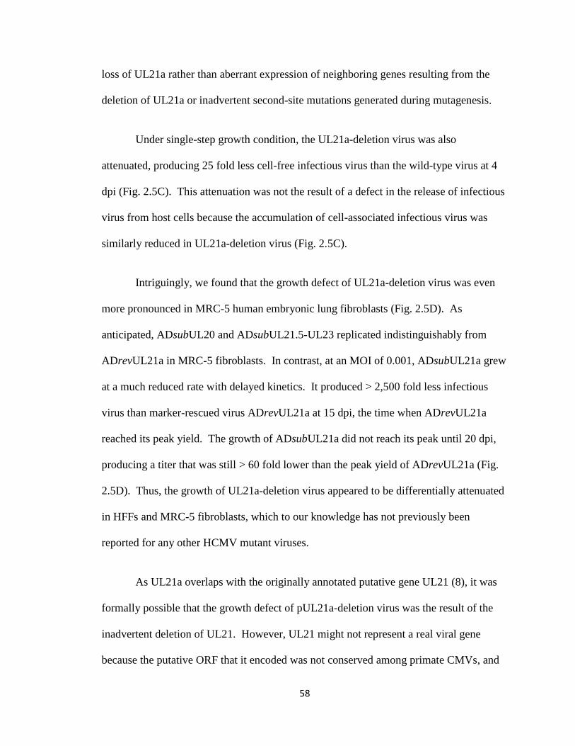
58
loss of UL21a rather than aberrant expression of neighboring genes resulting from the
deletion of UL21a or inadvertent second-site mutations generated during mutagenesis.
Under single-step growth condition, the UL21a-deletion virus was also
attenuated, producing 25 fold less cell-free infectious virus than the wild-type virus at 4
dpi (Fig. 2.5C). This attenuation was not the result of a defect in the release of infectious
virus from host cells because the accumulation of cell-associated infectious virus was
similarly reduced in UL21a-deletion virus (Fig. 2.5C).
Intriguingly, we found that the growth defect of UL21a-deletion virus was even
more pronounced in MRC-5 human embryonic lung fibroblasts (Fig. 2.5D). As
anticipated, ADsubUL20 and ADsubUL21.5-UL23 replicated indistinguishably from
ADrevUL21a in MRC-5 fibroblasts. In contrast, at an MOI of 0.001, ADsubUL21a grew
at a much reduced rate with delayed kinetics. It produced > 2,500 fold less infectious
virus than marker-rescued virus ADrevUL21a at 15 dpi, the time when ADrevUL21a
reached its peak yield. The growth of ADsubUL21a did not reach its peak until 20 dpi,
producing a titer that was still > 60 fold lower than the peak yield of ADrevUL21a (Fig.
2.5D). Thus, the growth of UL21a-deletion virus appeared to be differentially attenuated
in HFFs and MRC-5 fibroblasts, which to our knowledge has not previously been
reported for any other HCMV mutant viruses.
As UL21a overlaps with the originally annotated putative gene UL21 (8), it was
formally possible that the growth defect of pUL21a-deletion virus was the result of the
inadvertent deletion of UL21. However, UL21 might not represent a real viral gene
because the putative ORF that it encoded was not conserved among primate CMVs, and
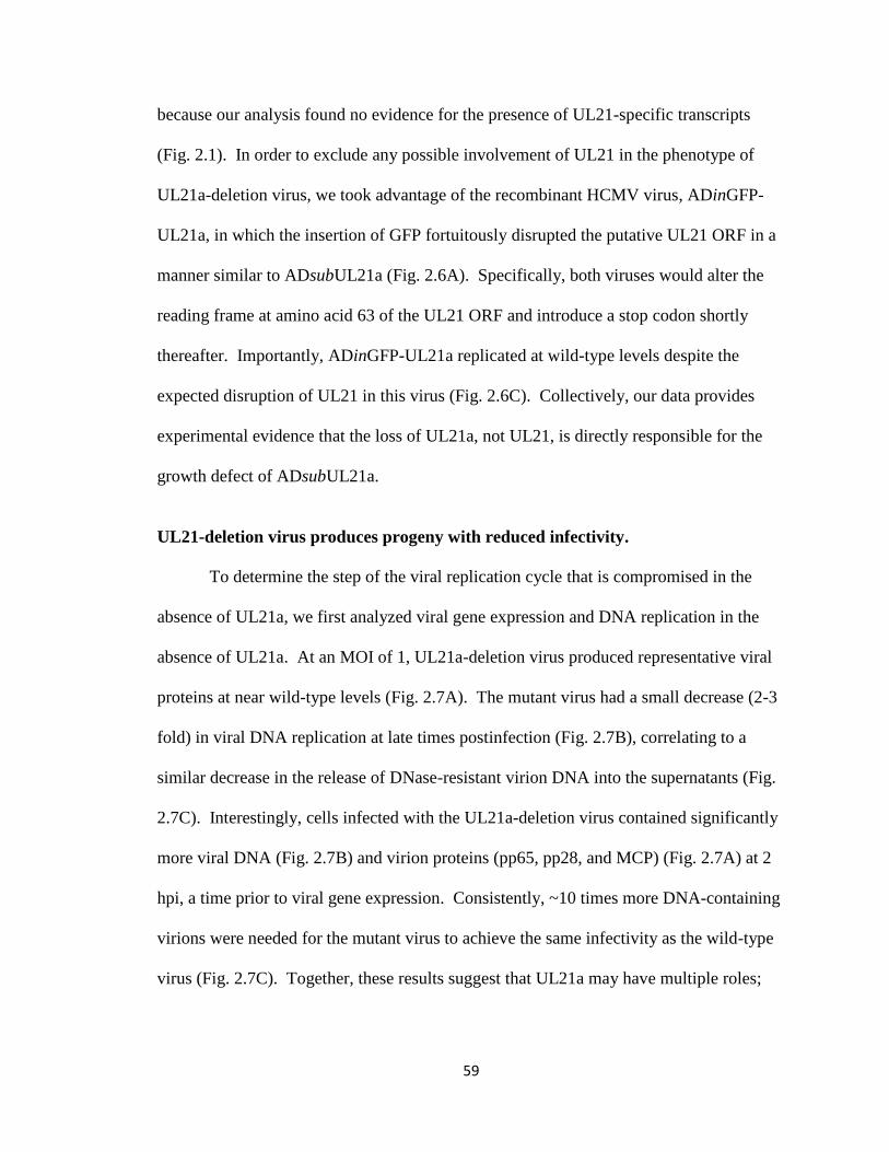
59
because our analysis found no evidence for the presence of UL21-specific transcripts
(Fig. 2.1). In order to exclude any possible involvement of UL21 in the phenotype of
UL21a-deletion virus, we took advantage of the recombinant HCMV virus, ADinGFP-
UL21a, in which the insertion of GFP fortuitously disrupted the putative UL21 ORF in a
manner similar to ADsubUL21a (Fig. 2.6A). Specifically, both viruses would alter the
reading frame at amino acid 63 of the UL21 ORF and introduce a stop codon shortly
thereafter. Importantly, ADinGFP-UL21a replicated at wild-type levels despite the
expected disruption of UL21 in this virus (Fig. 2.6C). Collectively, our data provides
experimental evidence that the loss of UL21a, not UL21, is directly responsible for the
growth defect of ADsubUL21a.
UL21-deletion virus produces progeny with reduced infectivity.
To determine the step of the viral replication cycle that is compromised in the
absence of UL21a, we first analyzed viral gene expression and DNA replication in the
absence of UL21a. At an MOI of 1, UL21a-deletion virus produced representative viral
proteins at near wild-type levels (Fig. 2.7A). The mutant virus had a small decrease (2-3
fold) in viral DNA replication at late times postinfection (Fig. 2.7B), correlating to a
similar decrease in the release of DNase-resistant virion DNA into the supernatants (Fig.
2.7C). Interestingly, cells infected with the UL21a-deletion virus contained significantly
more viral DNA (Fig. 2.7B) and virion proteins (pp65, pp28, and MCP) (Fig. 2.7A) at 2
hpi, a time prior to viral gene expression. Consistently, ~10 times more DNA-containing
virions were needed for the mutant virus to achieve the same infectivity as the wild-type
virus (Fig. 2.7C). Together, these results suggest that UL21a may have multiple roles;
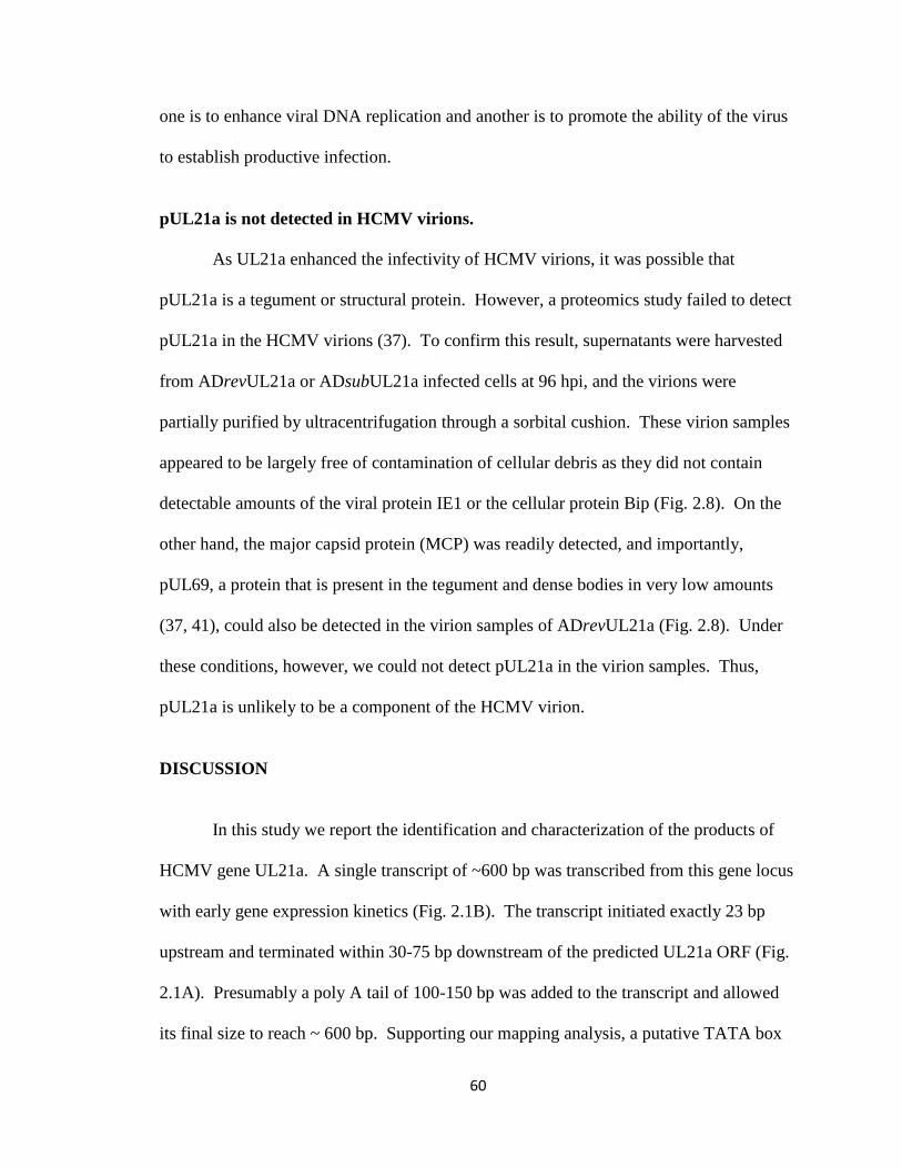
60
one is to enhance viral DNA replication and another is to promote the ability of the virus
to establish productive infection.
pUL21a is not detected in HCMV virions.
As UL21a enhanced the infectivity of HCMV virions, it was possible that
pUL21a is a tegument or structural protein. However, a proteomics study failed to detect
pUL21a in the HCMV virions (37). To confirm this result, supernatants were harvested
from ADrevUL21a or ADsubUL21a infected cells at 96 hpi, and the virions were
partially purified by ultracentrifugation through a sorbital cushion. These virion samples
appeared to be largely free of contamination of cellular debris as they did not contain
detectable amounts of the viral protein IE1 or the cellular protein Bip (Fig. 2.8). On the
other hand, the major capsid protein (MCP) was readily detected, and importantly,
pUL69, a protein that is present in the tegument and dense bodies in very low amounts
(37, 41), could also be detected in the virion samples of ADrevUL21a (Fig. 2.8). Under
these conditions, however, we could not detect pUL21a in the virion samples. Thus,
pUL21a is unlikely to be a component of the HCMV virion.
DISCUSSION
In this study we report the identification and characterization of the products of
HCMV gene UL21a. A single transcript of ~600 bp was transcribed from this gene locus
with early gene expression kinetics (Fig. 2.1B). The transcript initiated exactly 23 bp
upstream and terminated within 30-75 bp downstream of the predicted UL21a ORF (Fig.
2.1A). Presumably a poly A tail of 100-150 bp was added to the transcript and allowed
its final size to reach ~ 600 bp. Supporting our mapping analysis, a putative TATA box

61
was found 55 bp upstream and a consensus polyA site immediately followed the stop
codon of the UL21a ORF. The UL21a-specific polyclonal antibody allowed the
detection of a protein, pUL21a, with an apparent molecular weight of ~15 kDa, which
was only present in infection with wild-type virus but lost in infection with UL21a-
deletion virus (Fig. 2.2A). pUL21a was detected as early as 10 hpi and reached its peak
accumulation by 24-48 hpi (Fig. 2.2A), and therefore its expression was consistent with
early gene kinetics of UL21a (Fig. 2.1B). Thus, our study provides the first experimental
evidence for the presence of viral gene UL21a and its product pUL21a. pUL21a was
likely the sole product encoded by the UL21a locus because our sequence analysis found
no evidence for the presence of miRNAs, other non-coding RNAs, or small proteins
predicted to be encoded by the UL21a transcript (data not shown). In addition, extensive
third-base wobble was found in the conserved regions of the UL21a protein homologues
in CCMV and RhCMV (data not shown), suggesting that it was the UL21a protein
sequence that was maintained by evolutionary selection pressure.
UL21a was recently added to the annotation of the HCMV genome only based on
the presence of its homologue but not the originally annotated UL21 in chimpanzee CMV
(12). UL21 overlaps with UL21a, and encodes the largest putative ORF (i.e., 175 aa) in
this region, an essential criteria used in the original annotation (8). However, both
UL21a and UL21 are conserved among all sequenced isolates of HCMV (29), and have
equal likelihoods to encode proteins as predicted by a Bio-Dictionary-based Gene Finder
algorithm (28). In this study, we mapped the UL21a-specific transcript and identified the
protein pUL21a but found no evidence for the presence of a UL21 transcript (Fig. 2.1).
Furthermore, a GFP insertion that disrupted the putative UL21 ORF, but preserved the

62
UL21a ORF, replicated as efficiently as wild-type virus (data not shown), ruling out any
potential role of UL21, if it exists, in HCMV growth in fibroblasts. These results lead us
to favor the view that UL21a, and not UL21, is expressed and required for efficient
HCMV growth in fibroblasts.
UL21a was important for HCMV to establish efficient infection because the
deletion virus was attenuated in growth, particularly in MRC-5 fibroblasts as compared to
that in HFFs (Fig. 2.5). The basis for such an elevated defect in UL21a-deletion virus
growth in MRC-5 cells is unclear but it is likely due to physiological differences between
these two cell types. For instance, MRC-5 are embryonic lung fibroblasts that proliferate
more rapidly than HFFs and are able to grow up to > 30 passages, whereas HFFs are
newborn foreskin fibroblasts that generally undergo senescence much sooner. In
addition, an unknown difference between the two cell types, such as differential
expression of a specific gene or a set of genes, may also targeted by UL21a and
contribute to, at least in part, the difference that we observed. Nonetheless, analysis of
the life cycle of UL21a-deletion virus in HFFs under a high MOI condition reveals a
minor defect in viral DNA replication at later times (2-3 fold) and a pronounced
reduction in infectivity of progeny produced (~10 fold) (Fig. 2.7). Intriguingly, pUL21a
does not appeared to be a tegument or structural protein (Fig. 2.8) (37), raising the
question of how pUL21a enhances the infectivity of the virion. It is conceivable that
pUL21a may help proper tegumentation of virion particles. Alternatively, pUL21a may
promote efficient viral gene expression or DNA replication, a function that can be
compensated for by the addition of excess virions. We are currently searching for viral or

63
cellular factors that pUL21a may interact with and investigating the mechanistic basis of
its function.
pUL21a underwent proteasome-dependent degradation during infection (Fig. 2.2)
despite its important role in HCMV growth (Fig. 2.5). pUL21a degradation was blocked
by the treatment of the proteasome inhibitor MG132 and was independent of any other
viral factors. The absence of any lysines or cysteines in the pUL21a sequence and the
failure to detect pUL21a-ubiquitin conjugates (data not shown) led us to hypothesize that
the degradation of pUL21a might be ubiquitin-independent as shown for the parvovirus
minute virus NS2 protein and the HCV F protein (24, 45). Our hypothesis was supported
by the fact that pUL21a was efficiently degraded in cells that lacked a functional
ubiquitin-conjugation system (Fig. 2.3). Interestingly, HCMV contains a virus-encoded
deubiquitinating protease within its virion, which may favor ubiquitin-independent
protein degradation in infected cells (39). Notably, the HCMV protein pp71 directs
cellular proteins Rb and hDaxx for ubiquitin-independent proteasome-dependent
degradation shortly after infection (19, 20). How would then pUL21a be targeted to the
proteasome? pUL21a is rich in prolines (21/123= 17%) that are dispersed but are more
predominant at the C-terminus (data not shown), reminiscent of a relatively unstructured
protein. We speculate that pUL21a may be recognized as a mis-folded protein in the
cytoplasm and targeted to the proteasome for degradation. In accordance with this
hypothesis, an N-terminal GFP tag, which would presumably add significant structure to
the protein, dramatically stabilized levels of pUL21a (Fig. 2.6B).
64
We detected a second, faster-migrating form of the UL21a-specific protein during
pUL21a overexpression in the presence of MG132 or during HCMV infection where
pUL21a was stabilized by the N-terminal GFP tag (Fig. 2.2 and 2.6B). It is possible that
one of the forms of pUL21a results from post-translational modification, such as
phosphorylation, as the protein contains multiple serine/threonine residues at its N-
terminus. In addition, the faster-migrating form may be derived from alternative
translation as the UL21a ORF contains multiple methionines. Finally, the faster-
migrating form may represent an intermediate product of proteasome degradation that is
stabilized by MG132.
What would be the benefit for the virus to target pUL21a to the proteasome?
Perhaps the binding of pUL21a to the proteasome is important to regulate general protein
degradation during HCMV infection as proposed for the HCV F protein (45), or perhaps
pUL21a interacts with certain antiviral factors and directs them for degradation. This
would be reminiscent of the HIV protein Vif that binds to APOBEC3G, leading to poly-
ubiquitination and degradation of both proteins (16). Studies are currently under way to
test these hypotheses.
65
ACKNOWLEDGEMENTS
We thank Herbert Virgin and the members of his laboratory for helpful
discussions and invaluable advice, Harvey Ozer for ts20 cells, Wade Gibson and Thomas
Shenk for the antibodies, and members of Yu lab for their critical reading of the
manuscript.
This work was supported by a Public Health Service grant (CA-120768) from the
National Cancer Institute and in part by a Grant from the American Heart Association
(0755804Z). D.Y. holds an Investigators in the Pathogenesis of Infectious Disease
Award from the Burroughs Wellcome Fund. A.R.F. was a Morse/Berg Fellow of the
Department of Molecular Microbiology, Washington University School of Medicine, and
is supported by Institutional Training Grant T32-AI007172.

66
FIGURE LEGENDS
Figure 2.1. UL21a encodes a single unspliced transcript with early gene kinetics. (A)
The HCMV genomic region spanning UL20-UL23. The top panel shows the schematic
structure of the viral genomic region. Annotated viral open reading frames (ORF) are
indicated by open boxed arrows. The HCMV-encoded micro RNA UL22A-1 is indicated
by the black boxed arrow. Also shown are the transcripts from this region that have been
identified in previous studies or in this study. The bottom panel shows the genomic
sequence where UL21a resides with the sense-strand sequence depicted. The mapped
start and termination sites of the UL21a transcript are indicated. The UL21a ORF is
highlighted in gray. Also shown are the putative TATA box, poly A site (both indicated
with lines), the start and stop codons of the putative UL21 ORF (both indicated with
boxes), and gene specific primers used for 5’ or 3’ RACE (both indicated with arrows).
(B) Northern blot analysis of transcripts arising from the UL21a/UL21 gene locus. HFFs
were either mock-infected or infected with wild-type virus (ADwt) or UL21a-deletion
virus (ADsubUL21a) at an MOI of 1 in the presence or absence of 100μg/ml CHX or
200μg/ml PAA. Cells were harvested at indicated times, total RNA was isolated, and the
UL21a-specific transcript or UL123 (encoding IE1) and UL86 (encoding MCP) control
transcripts were analyzed by Northern blotting using UL21a, UL123, and UL86 strand-
specific anti-sense probes. Also shown is the ethidium bromide-staining of 18S rRNA as
the loading control. Molecular size markers (in kb) are indicated. (C) RACE analysis of
UL21a-specific transcripts. HFFs were infected with ADwt at an MOI of 1, cells were
harvested at 40 hpi, total RNA were isolated, cDNA was generated by reverse
transcription (RT), and 5’ or 3’ portions of the sequences of the UL21a-specific
67
transcripts were amplified by 5’ or 3’ RACE using universal primer mix (UPM) and
UL21a gene specific primers (GSP) (see Fig. 2A and Materials and Methods),
respectively. RACE products were analyzed by agarose gel electrophoresis, individual
product was cloned, their sequences were determined, and the assembled UL21a
transcript was indicated in Fig. 2A. Molecular size markers (in bp) are indicated.
Figure 2.2. pUL21a is expressed but undergoes proteasome-dependent degradation
during HCMV infection. (A) Kinetics of pUL21a accumulation in HCMV infection.
HFFs were infected with ADwt or ADsubUL21a at an MOI of 1, cells were collected at
indicated times, and total lysates were analyzed by immunoblotting using an affinity-
purified rabbit polyclonal antibody raised against a UL21a recombinant protein. (Blotting
was performed under a long exposure to reveal the presence of pUL21a at 10 hpi). The
antibody to viral protein IE1 or cellular protein actin was used as infection or loading
control, respectively. (B) Proteasome inhibitor stabilizes the accumulation of over-
expressed pUL21a. Hela cells were transfected with vector expressing pUL21a (pLP-
pUL21a) or another HCMV protein pp28 (pLP-pp28), and protease inhibitor as indicated
or DMSO control was added at 48 hours to the final concentration of 10 μM. Total cell
lysates were collected 24 hours later and analyzed by immunoblotting using the antibody
to pUL21a or pp28 (Blotting was performed under a short exposure to reveal
differentiated levels of pUL21a in the presence or absence of MG132). (C) Dose- and
time-dependent effects of proteasome inhibitor on the accumulation of over-expressed
pUL21a. Hela cells transfected with pLP-pUL21a were incubated with MG132 at
different concentration as indicated (μM) for 24 hours (left panel) or with 2.5 μM MG132
for different times as indicated (right panel), and cell lysates were prepared for
68
immunoblot analysis using the -pUL21a antibody (short exposure). (D) HFFs were
infected with indicated HCMV virus at an MOI of 1, MG132 or DMSO control was
added at 38 hpi to the final concentration of 10 μM, cells were collected at 48 hpi, and
total lysates were analyzed by immunoblotting using the antibody to viral proteins IE1,
pUL44, or pUL21a (short exposure).
Figure 2.3. Degradation of pUL21a is unaltered in cells lacking a functional ubiquitin-
conjugation system. Mouse ts20 cells were transfected with pLP-pUL21a or the empty
vector pLP, and cultured at 35°C. 24 hours later, DMSO control or MG132 was added to
indicated cell culture at the final concentration of 10 μM, one set of cells were kept at
35°C, and the second set were shifted to 39°C. Total cell lysates were prepared after 20
hours of drug treatment and analyzed by immunoblotting for accumulation of p53 and
pUL21a. The antibody to actin was used as a loading control.
Figure 2.4. pUL21a predominantly localizes to the cytoplasm of both overexpressing
and infected cells. (A) Hela cells (left panel) or HFFs (right panel) were transfected with
plasmid expressing pUL21a or control viral proteins (pp28 or pUL44), seeded onto
coverslips at 48 hours post-transfection, and analyzed for the subcellular localization of
pUL21a 24 hours later by immunofluorescence using the -pUL21a antibody. Cells
were also counterstained with TO-PRO3 to visualize the nuclei of the cells. Scale bars
(10 m) are shown. (B) HFFs were infected with ADsubUL21a, ADwt, or ADinGFP-
UL21a at an MOI of 0.1, and then analyzed by immunofluorescence using the -pUL21a
antibody at indicated times postinfection.
69
Figure 2.5. Deletion of UL21a results in attenuated growth of HCMV in fibroblasts. (A)
Schematic structure of the viral genomic region in recombinant BAC-HCMV clones
where genetic alterations were introduced. Viral ORFs and known miRNAs are indicated
by open and black boxed arrows, respectively. The boxes below the first line represent
the locations of viral sequences that were replaced by the GalK/kanamycin marker
sequence in the substitution mutants, as indicated. Note that the UL21a sequence was
reintroduced into pADsubUL21a to replace the marker sequence and generate the
marker-rescued clone pADrevUL21a. (B) Representative images of plaques (as indicated
by virus-driven GFP expression) from HFFs that were infected with wild-type or UL21a-
deletion virus, overlaid with agarose, and examined under a fluorescent microscope at 12
dpi. Scale bars (0.1 mm) are shown. Average sizes of 50 plaques from wild type- or
mutant- virus infection, and the P value by Student's paired t-test are also shown. (C-D)
Growth kinetic analysis of recombinant HCMV viruses. HFFs (C) or MRC-5 primary
human embryonic lung fibroblasts (D) were infected with indicated viruses at an MOI of
0.001 or 1, cell-free and cell-associated viruses were collected at different days
postinfection, and their yields were determined by plaque assay.
Figure 2.6. Characterization of GFP-insertional recombinant HCMV virus ADinGFP-
UL21a. (A) A GFP-S tag was inserted in-frame at the N-terminus of the UL21a ORF by
linear recombination-based BAC mutagenesis, resulting in the recombinant virus
ADinGFP-UL21a. The insertion of the GFP-S tag concurrently disrupted and resulted in
the premature termination of the overlapping UL21 ORF. Shown are predicted
sequences and positions of amino acids encoded by the putative UL21 gene in ADwt,
ADsubUL21a, and ADinGFP-UL21a. Amino acid changes of the UL21 ORF in

70
ADsubUL21a and ADinGFP-UL21a are highlighted in gray, and stop codons are
indicated by *. (B) HFFs were mock-infected, infected with ADwt or ADinGFP-UL21a
at an MOI of 1, and total cell lysates were prepared at 24 hpi and analyzed by
immunoblotting. The blot hybridized to the -pUL21a antibody was over-exposed in
order to visualize the accumulation of both native and GFP-tagged pUL21a. The
antibody to viral protein IE1 or cellular protein actin was used as infection or loading
control, respectively. (C) HFFs were infected with indicated viruses at an MOI of 0.001,
culture medium was collected at different days postinfection, and the yield of cell-free
virus produced was quantified by plaque assay.
Figure 2.7. Analysis of the infection cycle of the UL21a-deletion virus. (A) Analysis of
viral gene expression. HFFs were infected with ADwt or ADsubUL21a at an MOI of 1,
total cell lysates were collected at indicated times, and were analyzed by immunoblotting
with antibodies against IE1, pUL44, pp65, pp28, MCP, and pUL21a. (Note the
difference in tegument proteins at 2 hpi.) (B) Analysis of viral DNA replication. HFFs
were infected as described above, the total cell-associated DNA was isolated at indicated
times, and the accumulation of viral genomes was determined by realtime quantitative
PCR. (Note the difference in viral DNA at 2 hpi.) (C) Analysis of the infectivity of
progeny virions. HFFs were infected as described as above, cell-free virus was collected
at 72 and 96 hpi, and same volumes of viral samples were measured for their viral
genome copies by realtime quantitative PCR (top panel) and for their infectivity by
plaque assay (middle panel). The viral genome copy normalized by PFU was presented
in the bottom panel. The relative genome copy per PFU of wild-type virus was set to 1.
71
Figure 2.8. pUL21a is not detected in HCMV virions. 5X10⁶ HFFs were infected with
ADrevUL21a or ADsubUL21a at an MOI of 0.2, and total cell lysates and cell-free
virions were collected at 96 hpi. Cell-free virions were then partially purified by
ultracentrifugation through a sorbitol cushion and quantified for their genome copies by
realtime-quantitative PCR. Cell lysates from 3.75x10⁵ cells and virions from 6x10
9
genome copies were analyzed by immunoblotting for MCP, pUL69, IE1, and Bip. For
pUL21a, twice as many virions (1.2X10¹⁰
genome copies) were used in the experiment.

72
TABLE 2.1. Primers used to create substitutions and insertions in the HCMV genome
Targeted
HCMV gene
Type of
genetic alteration Primer pairs used to introduce alteration
a
Resulting recombinant
HCMV virus
UL21a Substitution 5’-agccatgcagcgtgcggcgcctctctcatggatccactgtcac
cgtcgcg CCTGTTGACAATTAATCATCG-3’
5’-atacagacttttatatgatccttgtacagatgtaaataaaatgttttt
at CTCAGCAAAAGTTCGATTTA-3’
ADsubUL21a
UL20 Substitution 5’-tggaacggtctttatatatacaaacgccgttatgttcagtgtcc
ggcaagCCTGTTGACAATTAATCATCG - 3’
5’-tatggaaaatatgtagtccgtaccgcttggggctcaaagttca
aagtccgCTCAGCAAAAGTTCGATTTA-3’
ADsubUL20
UL21.5-23 Substitution 5’-gcaagaacgtaactctcagtcagggggggtccaccaccgac
ggagacgaaCCTGTTGACAATTAATCATCG-3’
5’-gaccgaccacatctactctgactcgttgacctttgtggccgaga
gcatca CTCAGCAAAAGTTCGATTTA-3’
ADsubUL21.5-23
UL21a N-terminal
Insertion
5’-agccatgcagcgtgcggcgcctctctcatggatccactgtcac
cgtcgcg CCTGTTGACAATTAATCATCG-3’
5’-ggcatgagtccttgagtgacggtggtgagctggggaacagg
gctacctcc CTCAGCAAAAGTTCGATTTA-3’
ADinGalK/kan-
UL21a
UL21a N-terminal
Insertion
5’-agccatgcagcgtgcggcgcctctctcatggatccactgtcac
cgtcgcg ATGGTGAGCAAGGGCGAGGAG-3’
5’-ggcatgagtccttgagtgacggtggtgagctggggaacagg
gctacctcc CTCGAGACTAGTACCTCCACC-3’
ADinGFP-UL21a
a Virus specific sequences are in lower case, GalK-kanamycin marker specific sequences
are in upper case, GFP-S tag specific sequences are underlined and in upper case.
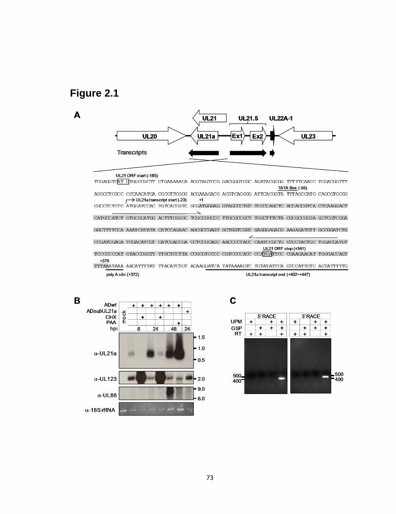
73
Figure 2.1

74
Figure 2.2

75
Figure 2.3
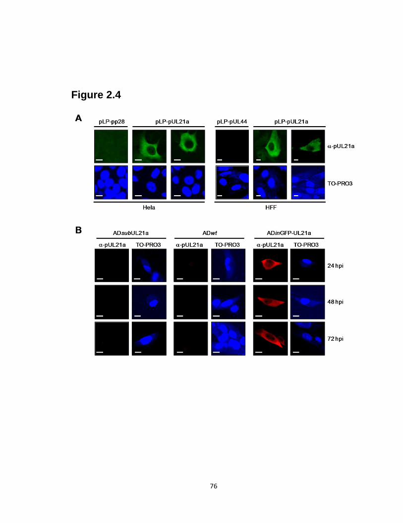
76
Figure 2.4
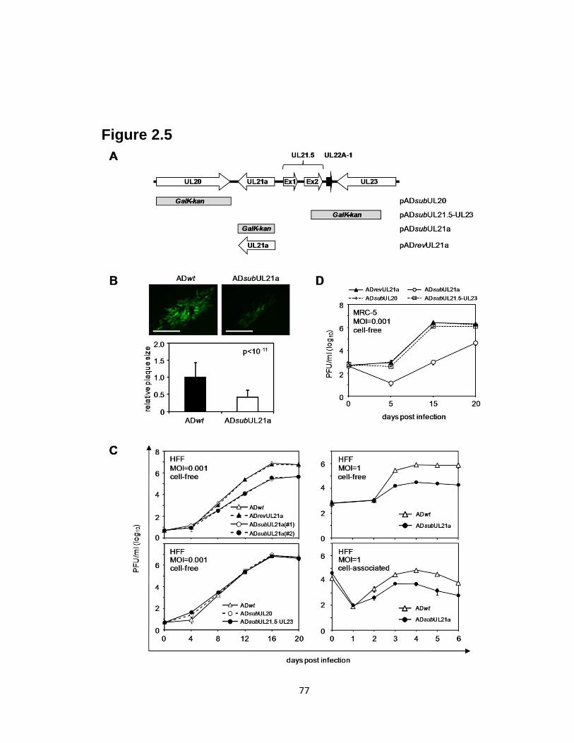
77
Figure 2.5

78
Figure 2.6

79
Figure 2.7
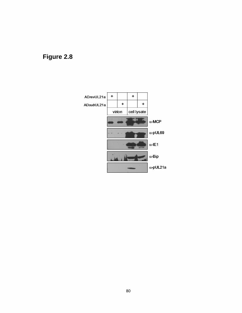
80
Figure 2.8
81
REFERENCES
1. Adair, R., E. R. Douglas, J. B. Maclean, S. Y. Graham, J. D. Aitken, F. E.
Jamieson, and D. J. Dargan. 2002. The products of human cytomegalovirus
genes UL23, UL24, UL43 and US22 are tegument components. J Gen Virol
83:1315-24.
2. Ben-Saadon, R., I. Fajerman, T. Ziv, U. Hellman, A. L. Schwartz, and A.
Ciechanover. 2004. The tumor suppressor protein p16(INK4a) and the human
papillomavirus oncoprotein-58 E7 are naturally occurring lysine-less proteins that
are degraded by the ubiquitin system. Direct evidence for ubiquitination at the N-
terminal residue. J Biol Chem 279:41414-21.
3. Borst, E. M., G. Hahn, U. H. Koszinowski, and M. Messerle. 1999. Cloning of
the human cytomegalovirus (HCMV) genome as an infectious bacterial artificial
chromosome in Escherichia coli: a new approach for construction of HCMV
mutants. J Virol 73:8320-9.
4. Bresnahan, W. A., and T. Shenk. 2000. A subset of viral transcripts packaged
within human cytomegalovirus particles. Science 288:2373-6.
5. Britt, W. J., and C. A. Alford (ed.). 1996. Cytomegalovirus, 3rd ed. Lippincott-
Raven, Philadelphia.
6. Cadwell, K., and L. Coscoy. 2005. Ubiquitination on nonlysine residues by a
viral E3 ubiquitin ligase. Science 309:127-30.
7. Chee, M., S. A. Rudolph, B. Plachter, B. Barrell, and G. Jahn. 1989.
Identification of the major capsid protein gene of human cytomegalovirus. J Virol
63:1345-53.
8. Chee, M. S., A. T. Bankier, S. Beck, R. Bohni, C. M. Brown, R. Cerny, T.
Horsnell, C. A. Hutchison, 3rd, T. Kouzarides, J. A. Martignetti, and et al. 1990. Analysis of the protein-coding content of the sequence of human
cytomegalovirus strain AD169. Curr Top Microbiol Immunol 154:125-69.
9. Cheeseman, I. M., and A. Desai. 2005. A combined approach for the
localization and tandem affinity purification of protein complexes from
metazoans. Sci STKE 2005:pl1.
10. Chowdary, D. R., J. J. Dermody, K. K. Jha, and H. L. Ozer. 1994.
Accumulation of p53 in a mutant cell line defective in the ubiquitin pathway. Mol
Cell Biol 14:1997-2003.
11. Ciechanover, A., and R. Ben-Saadon. 2004. N-terminal ubiquitination: more
protein substrates join in. Trends Cell Biol 14:103-6.
12. Davison, A. J., A. Dolan, P. Akter, C. Addison, D. J. Dargan, D. J. Alcendor,
D. J. McGeoch, and G. S. Hayward. 2003. The human cytomegalovirus genome
revisited: comparison with the chimpanzee cytomegalovirus genome. J Gen Virol
84:17-28.
13. Dolan, A., C. Cunningham, R. D. Hector, A. F. Hassan-Walker, L. Lee, C.
Addison, D. J. Dargan, D. J. McGeoch, D. Gatherer, V. C. Emery, P. D.
Griffiths, C. Sinzger, B. P. McSharry, G. W. Wilkinson, and A. J. Davison. 2004. Genetic content of wild-type human cytomegalovirus. J Gen Virol 85:1301-
12.
82
14. Dunn, W., C. Chou, H. Li, R. Hai, D. Patterson, V. Stolc, H. Zhu, and F. Liu.
2003. Functional profiling of a human cytomegalovirus genome. Proc Natl Acad
Sci U S A 100:14223-8.
15. Dunn, W., P. Trang, Q. Zhong, E. Yang, C. van Belle, and F. Liu. 2005.
Human cytomegalovirus expresses novel microRNAs during productive viral
infection. Cell Microbiol 7:1684-95.
16. Goila-Gaur, R., and K. Strebel. 2008. HIV-1 Vif, APOBEC, and intrinsic
immunity. Retrovirology 5:51.
17. Grattan, M. T., C. E. Moreno-Cabral, V. A. Starnes, P. E. Oyer, E. B.
Stinson, and N. E. Shumway. 1989. Cytomegalovirus infection is associated
with cardiac allograft rejection and atherosclerosis. Jama 261:3561-6.
18. Grey, F., A. Antoniewicz, E. Allen, J. Saugstad, A. McShea, J. C. Carrington,
and J. Nelson. 2005. Identification and characterization of human
cytomegalovirus-encoded microRNAs. J Virol 79:12095-9.
19. Hwang, J., and R. F. Kalejta. 2007. Proteasome-dependent, ubiquitin-
independent degradation of Daxx by the viral pp71 protein in human
cytomegalovirus-infected cells. Virology 367:334-8.
20. Kalejta, R. F., and T. Shenk. 2003. Proteasome-dependent, ubiquitin-
independent degradation of the Rb family of tumor suppressors by the human
cytomegalovirus pp71 protein. Proc Natl Acad Sci U S A 100:3263-8.
21. Kuvin, J. T., and C. D. Kimmelstiel. 1999. Infectious causes of atherosclerosis.
Am. Heart J. 137:216-226.
22. McCormick, A. L., C. D. Meiering, G. B. Smith, and E. S. Mocarski. 2005.
Mitochondrial cell death suppressors carried by human and murine
cytomegalovirus confer resistance to proteasome inhibitor-induced apoptosis. J
Virol 79:12205-17.
23. Melnick, J. L., E. Adam, and M. E. Debakey. 1993. Cytomegalovirus and
atherosclerosis. Eur Heart J 14 Suppl K:30-8.
24. Miller, C. L., and D. J. Pintel. 2001. The NS2 protein generated by the
parvovirus minute virus of mice is degraded by the proteasome in a manner
independent of ubiquitin chain elongation or activation. Virology 285:346-55.
25. Mocarski, E. S., and C. T. Courcelle (ed.). 2001. Cytomegaloviruses and their
replication, 4th ed, vol. 2. Lippincott-Raven, Philadelphia, Pa.
26. Muhlestein, J. B., B. D. Horne, J. F. Carlquist, T. E. Madsen, T. L. Bair, R.
R. Pearson, and J. L. Anderson. 2000. Cytomegalovirus seropositivity and C-
reactive protein have independent and combined predictive value for mortality in
patients with angiographically demonstrated coronary artery disease. Circulation
102:1917-1923.
27. Mullberg, J., M. L. Hsu, C. T. Rauch, M. J. Gerhart, A. Kaykas, and D.
Cosman. 1999. The R27080 glycoprotein is abundantly secreted from human
cytomegalovirus-infected fibroblasts. J Gen Virol 80 ( Pt 2):437-40.
28. Murphy, E., I. Rigoutsos, T. Shibuya, and T. E. Shenk. 2003. Reevaluation of
human cytomegalovirus coding potential. Proc Natl Acad Sci U S A 100:13585-
90.
29. Murphy, E., D. Yu, J. Grimwood, J. Schmutz, M. Dickson, M. A. Jarvis, G.
Hahn, J. A. Nelson, R. M. Myers, and T. E. Shenk. 2003. Coding potential of
83
laboratory and clinical strains of human cytomegalovirus. Proc Natl Acad Sci U S
A 100:14976-81.
30. Petrik, D. T., K. P. Schmitt, and M. F. Stinski. 2006. Inhibition of cellular
DNA synthesis by the human cytomegalovirus IE86 protein is necessary for
efficient virus replication. J Virol 80:3872-83.
31. Qian, Z., B. Xuan, T. T. Hong, and D. Yu. 2008. The full-length protein
encoded by human cytomegalovirus gene UL117 is required for the proper
maturation of viral replication compartments. J Virol 82:3452-65.
32. Rawlinson, W. D., and B. G. Barrell. 1993. Spliced transcripts of human
cytomegalovirus. J Virol 67:5502-13.
33. Rivailler, P., A. Kaur, R. P. Johnson, and F. Wang. 2006. Genomic sequence
of rhesus cytomegalovirus 180.92: insights into the coding potential of rhesus
cytomegalovirus. J Virol 80:4179-82.
34. Speir, E., R. Modali, E. S. Huang, M. B. Leon, F. Shawl, T. Finkel, and S. E.
Epstein. 1994. Potential role of human cytomegalovirus and p53 interaction in
coronary restenosis. Science 265:391-4.
35. Streblow, D. N., S. L. Orloff, and J. A. Nelson. 2001. Do pathogens accelerate
atherosclerosis? J Nutr 131:2798S-2804S.
36. Terhune, S., E. Torigoi, N. Moorman, M. Silva, Z. Qian, T. Shenk, and D.
Yu. 2007. Human cytomegalovirus UL38 protein blocks apoptosis. J Virol
81:3109-23.
37. Varnum, S. M., D. N. Streblow, M. E. Monroe, P. Smith, K. J. Auberry, L.
Pasa-Tolic, D. Wang, D. G. Camp, 2nd, K. Rodland, S. Wiley, W. Britt, T.
Shenk, R. D. Smith, and J. A. Nelson. 2004. Identification of proteins in human
cytomegalovirus (HCMV) particles: the HCMV proteome. J Virol 78:10960-6.
38. Wang, D., W. Bresnahan, and T. Shenk. 2004. Human cytomegalovirus
encodes a highly specific RANTES decoy receptor. Proc Natl Acad Sci U S A
101:16642-7.
39. Wang, J., A. N. Loveland, L. M. Kattenhorn, H. L. Ploegh, and W. Gibson.
2006. High-molecular-weight protein (pUL48) of human cytomegalovirus is a
competent deubiquitinating protease: mutant viruses altered in its active-site
cysteine or histidine are viable. J Virol 80:6003-12.
40. Wang, X., R. A. Herr, W. J. Chua, L. Lybarger, E. J. Wiertz, and T. H.
Hansen. 2007. Ubiquitination of serine, threonine, or lysine residues on the
cytoplasmic tail can induce ERAD of MHC-I by viral E3 ligase mK3. J Cell Biol
177:613-24.
41. Winkler, M., and T. Stamminger. 1996. A specific subform of the human
cytomegalovirus transactivator protein pUL69 is contained within the tegument of
virus particles. J Virol 70:8984-7.
42. Xuan, B., Z. Qian, E. Torigoi, and D. Yu. 2009. Human cytomegalovirus
protein pUL38 induces ATF4 expression, inhibits persistent JNK
phosphorylation, and suppresses endoplasmic reticulum stress-induced cell death.
J Virol 83:3463-74.
43. Yu, D., M. C. Silva, and T. Shenk. 2003. Functional map of human
cytomegalovirus AD169 defined by global mutational analysis. Proc Natl Acad
Sci U S A 100:12396-401.
84
44. Yu, D., G. A. Smith, L. W. Enquist, and T. Shenk. 2002. Construction of a self-
excisable bacterial artificial chromosome containing the human cytomegalovirus
genome and mutagenesis of the diploid TRL/IRL13 gene. J Virol 76:2316-28.
45. Yuksek, K., W. L. Chen, D. Chien, and J. H. Ou. 2009. Ubiquitin-independent
degradation of hepatitis C virus F protein. J Virol 83:612-21.
46. Zhou, Y. F., M. B. Leon, M. A. Waclawiw, J. J. Popma, Z. X. Yu, T. Finkel,
and S. E. Epstein. 1996. Association between prior cytomegalovirus infection
and the risk of restenosis after coronary atherectomy. N Engl J Med 335:624-30.

85
Chapter III
Human cytomegalovirus early protein pUL21a promotes efficient viral DNA
synthesis and the late accumulation of immediate-early transcripts
This chapter contains data published in Journal of Virology:
Fehr A. R. and D. Yu. 2011. Human cytomegalovirus early protein pUL21a facilitates
efficient viral DNA synthesis and the late accumulation of immediate-early transcripts. J
Virol. 85:663-674
86
ABSTRACT
We have previously reported that a newly annotated gene of human
cytomegalovirus (HCMV), UL21a, encodes an early viral protein termed pUL21a. Most
notably, the virions of UL21a-deletion virus had markedly reduced infectivity, indicating
that UL21a is required to establish an efficient productive infection. In this report, we
infected fibroblasts with equal numbers of DNA-containing viral particles and identified
where in the viral life cycle pUL21a acted. The UL21a-deletion virus entered cells and
initiated viral gene expression efficiently; however, it synthesized viral DNA poorly and
accumulated several immediate-early (IE) transcripts at reduced levels at late times of
infection. The defect in viral DNA synthesis preceded that in gene expression, and
inhibition of viral DNA synthesis reduced the late accumulation of IE transcripts in both
wild-type and mutant virus infected cells to equivalent levels. This suggests that reduced
viral DNA synthesis is the cause of reduced IE gene expression in the absence of UL21a.
The growth of UL21a-deletion virus was similar to that of recombinant HCMV where
pUL21a expression was abrogated by stop-codon mutations, and the defect was rescued
in pUL21a-expressing fibroblasts. pUL21a expression in trans was sufficient to restore
viral DNA synthesis and gene expression of mutant virus produced from normal
fibroblasts whereas mutant virus produced from complementing cells still exhibited the
defect in normal fibroblasts. Thus, pUL21a does not promote the functionality of HCMV
virions; rather, its de novo synthesis facilitates viral DNA synthesis, which is necessary
for the late accumulation of IE transcripts and establishment of a productive infection.
87
INTRODUCTION
Human cytomegalovirus (HCMV), the prototypic β-herpesviruses, is a ubiquitous
pathogen that infects 50-90% of the world’s population. Upon primary infection, HCMV
establishes a lifelong latent or persistent/recurrent infection within its host. Though
asymptomatic in most immunocompetent individuals, HCMV can cause severe disease
and death in immunocompromised individuals, including AIDS patients and organ
transplant recipients. HCMV is also the most common viral cause of birth defects,
leading to hearing loss, blindness, and mental retardation in congenitally and perinatally
infected infants (32). The economic burden to the US health care system for this virus is
estimated at approximately four billion dollars annually with a majority of the costs
attributed to long-term sequelae experienced by individuals who acquire congenital
HCMV disease (19). A comprehensive understanding of how HCMV interacts with the
host to establish both acute and latent infections will be critical for developing an
effective vaccine and novel therapeutics to combat HCMV disease (24, 59).
HCMV expresses its genes in a highly regulated temporal cascade during a
productive infection (32). The virus first expresses its immediate-early genes, which
appear 2-4 hours after viral entry and persist throughout the infection. The products of
the major immediate-early (MIE) transcript are critical for the establishment of a
productive infection, and must be downregulated for the virus to establish latency (32).
The primary proteins encoded by the MIE transcript are IE1-72 and IE2-86, which are
produced by alternative splicing (62, 64, 66). IE1-72 is abundantly expressed during the
first few hours of infection. Its abundance then only undergoes a limited increase
throughout the remainder of the infection (63). In contrast, the accumulation of IE2-86 is
88
low during the first 12 hours of infection but it increases considerably between 24 and 72
hpi (62, 64). The reason for this differential MIE expression is unclear, but a specific
inhibitor of the cyclin-dependent kinases (CDK) causes a shift in the ratio of IE1/IE2
during the first 12 hours of infection (55), suggesting that CDK activity may play a role
in this regulation. The IE2-86 protein is essential for viral replication while IE1-72 is
only required at a low MOI (11, 14, 18, 29, 31, 58). Both proteins transactivate viral
promoters and also modulate the cellular environment to be more conducive for viral
infection. Additional IE genes, which include TRS1, UL37x1, and US3, help HCMV to
overcome innate and adaptive cellular antiviral responses (1, 4, 5, 13, 17, 23, 44).
Transcription of early genes shortly follows IE gene expression, appearing
between 4 and 12 hpi. These genes encode DNA replication enzymes, such as UL44
(processivity factor) and UL54 (viral DNA polymerase), as well as viral regulatory
proteins that alter host cells for a cellular environment conducive to viral replication.
Late genes are expressed following the onset of viral DNA replication and many of them
encode structural proteins, such as the major capsid protein (MCP) and pp28, which are
required for assembly and maturation of the virion. Other late genes, such as pp71, are
tegument proteins which can antagonize intrinsic cellular defenses and help progeny
virus initiate IE gene expression during subsequent infection (20, 51, 52).
HCMV contains a 240-kb double-stranded DNA genome that encodes at least 166
putative open reading frames (ORFs) and several miRNAs (3, 7, 9, 15, 34, 35). With the
advent of the infectious bacterial artificial chromosome (BAC) clone-based genetic
system for HCMV (2, 73), the functions of HCMV genes have started to be elucidated.
Genome-scale mutagenesis approaches have identified a subset of candidate viral genes
89
that are important for HCMV to establish infection in tissue culture models of primary
human cells including fibroblasts (8, 72). More detailed investigations have begun to
uncover the functions of selected viral genes. We have recently identified a 15-kDa viral
protein, pUL21a, which is expressed with early kinetics from the newly annotated
HCMV gene UL21a (10). pUL21a predominantly localizes to the cytoplasm and
undergoes rapid degradation in a ubiquitin-independent, proteasome-dependent manner
(10). Analysis of mutant HCMV virus lacking the UL21a coding sequence reveals a
minor defect in viral DNA replication (~2-3 fold) and a more pronounced reduction in
infectivity of progeny viruses (~10 fold), suggesting that UL21a is required for the
establishment of a productive infection (10). It is possible that UL21a promotes the
proper tegumentation of the HCMV virion, enabling the virus to effectively initiate
productive infection. Alternatively, it may act at the later stages of infection, such as
viral gene expression or DNA replication, to drive the progression of the viral life cycle.
In this study, we sought to determine how UL21a drives HCMV infection by
analyzing fibroblasts infected with equal numbers of DNA-containing virions of
recombinant viruses. We report that UL21a is not required for HCMV to establish the
initial events of productive infection, but it facilitates efficient viral DNA synthesis and
the late accumulation of several viral IE transcripts. Moreover, we present evidence that
DNA synthesis is the cause and not a consequence of the late accumulation of the IE
transcripts. We show that pUL21a, the protein product of UL21a, carries out the function
of this gene locus. pUL21a expression in trans restores viral DNA synthesis and lytic
gene expression of mutant virus that is originally produced from normal fibroblasts.
However, mutant virus produced from complementing cells still exhibited reduced lytic
90
gene expression in normal fibroblasts. Collectively, our results indicate that HCMV
virions function normally independent of pUL21a, but the de novo synthesis of this
protein facilitates viral DNA synthesis, which is necessary for the efficient late
accumulation of IE transcripts and the establishment of a productive infection.
MATERIALS AND METHODS
Plasmids and antibodies. pYD-C160 and pYD-C476 are retroviral vectors derived
from pRetro-EBNA. pYD-C160, which carries the coding sequence of the green
fluorescent protein (GFP), was previously described (69), and pYD-C476 was created by
PCR amplifying the coding sequence of the GFP-UL21a fusion protein from pADinGFP-
UL21a (10) and inserting it into the multiple cloning site of pRetro-EBNA.
The primary antibodies used in the present study included anti-β actin (AC-15,
Abcam); anti-IE2 (mAB8140, Chemicon); anti-acetyl histone H3 (DAM1422332,
Upstate); anti-Bip/Grp 78 (H-129, Santa Cruz); anti-UL21a (10); anti-UL38 (69); and
anti-IE1, anti-pp28 (60), and anti-TRS1 (49) (generous gifts from Thomas Shenk,
Princeton University).
Cells and Viruses. Primary embryonic lung fibroblasts (MRC-5) and human newborn
foreskin fibroblasts (HFFs) were propagated with Dulbecco’s modified Eagle medium
(DMEM) supplemented with 10% fetal bovine serum, non-essential amino acids, and
penicillin-streptomycin. To create MRC-5 cells expressing GFP (MRC-5-GFP) or the
GFP/UL21a fusion protein (MRC-5-GFP/UL21a), retrovirus stocks were prepared by
transfecting the retroviral vector pYD-C160 or pYD-C476 into Phoenix-Ampho cells
(25) using lipofectamine (Invitrogen) according to the manufacturers’ instructions.

91
MRC-5 cells were transduced with retrovirus three times and then let recover for 72
hours to generate a pool of cells expressing the protein of interest.
Five BAC-HCMV clones were used in the present study to reconstitute
recombinant HCMV viruses. pAD-GFP carried the GFP-tagged genome of the HCMV
AD169 strain and was used to produce wild-type virus ADwt (73). pADsubUL21a
carried a GalK/kanamycin dual mutagenic cassette in place of the entire UL21a coding
sequence as previously described (10). pADpmpUL21a carried stop-codon mutations at
Gln8 and Thr
39 engineered to specifically disrupt pUL21a expression (Fig. 3.6). It was
constructed by replacing the GalK/kanamycin cassette in pADsubUL21a with the PCR
fragment of the UL21a gene that contained the desired mutations via linear
recombination as previously described (43). These mutations were first generated by
using a QuickChange XL kit (Stratagene) with two primer pairs containing the desired
mutations and the template plasmid pYD-C417 carrying the HCMV UL21a sequence,
resulting in the plasmid pYD-C478 carrying these mutations. The primer pairs are as
follows: 1st pair (Q8X), 5’-GGTAGCCCTGTTCCCTAGCTCACCACCGTCACT-3’ and
5’-AGTGACGGTGGTGAGCTAGGGAACAGGGCTACC-3’; 2nd
pair (R39X), 5’-
GCCTCCGACGAGCTCACGGCGCTAGAAAGC-3’and 5’-
GCTTTCTACGCGCCGTGAGCTCGTCGGAGGC-3’. pADrevpUL21a was a marker-
rescued clone and was created by replacing the mutant UL21a gene in pADpmpUL21a
with a wild-type copy of UL21a via two-step linear recombination as previously
described (43). pTR is a BAC clone carrying the genome of a clinical HCMV isolate TR
that was originally isolated from the ocular vitreous fluid of a patient with advanced HIV
with CMV retinitis (61) and was used to reconstitute HCMV TR virus (35). All
92
recombinant BACs were verified by restriction digestion, PCR analysis, and direct
sequencing.
To reconstitute virus, 2 µg of the BAC-HCMV DNA and 1 µg of the pp71-
expressing plasmid were transfected into MRC-5 cells by electroporation. Culture
medium was changed 24 hours later, and virus stock was prepared by harvesting cell-free
culture supernatant when the entire monolayer of cells was lysed. Alternatively, virus
stocks were produced by collecting cell-free culture medium from infection of HFFs at
the multiplicity of infection (MOI) of 0.05. Virus-containing culture supernatants were
then purified by ultracentrifugation through a 20% D-sorbitol cushion at an average
relative centrifugal force of 64,000 x g for 1 hour, resuspended in DMEM with 10% fetal
calf serum, and saved as viral stocks. HCMV virus titers were determined in duplicate in
HFFs by Tissue Culture Infectious Dose 50 (TCID50) assay and their DNA content was
determined by real-time quantitative PCR (qPCR) (see below).
Cellular fractionation. Nuclear and cytoplasmic fractions were derived from infected
cells at 2 hours post-infection as previously described (27). The cell pellet was
resuspended in RSB (10 mM NaCl, 3 mM MgCl2, 10 mM Tris-HCl [pH 7.4]), and 20X
detergent (10% sodium deoxycholate and 20% Tween 40 in RSB) was added to a 1X
final concentration. Samples were then vortexed and incubated on ice for 10 min. Nuclei
were pelleted by centrifugation at 1,000 x g for 3 min at 4°C. The cytoplasmic
(supernatant) and nuclear (pellet) fractions were then processed for total DNA and
proteins as stated below.
Analysis of virion and intracellular DNA. Virion DNA was prepared as previously
described (10). Briefly, to prepare virion DNA, 100 µl of cell-free virus (or 10 µl of
93
purified virus) was treated with DNase I (30 U; Roche) at 37°C for 30 min, followed by
incubation at 75°C for 20 min to stop the reaction. The samples were then incubated at
55°C overnight in lysis buffer (400 mM NaCl, 10 mM Tris [pH 8.0], 10 mM EDTA, 0.1
mg of proteinase K/ml, 0.2% sodium dodecyl sulfate [SDS]). Virion DNA was extracted
with phenol-chloroform and treated with RNase A at 20 µg/ml at 37°C for 1 hour. DNA
was extracted again with phenol-chloroform, precipitated with ethanol, and resuspended
in nuclease-free water (Ambion). To prepare intracellular DNA, HCMV-infected MRC-5
cells were collected at various times post-infection, resuspended in lysis buffer, and
incubated at 55°C overnight. DNA was extracted with phenol-chloroform, treated with
RNase A, extracted again with phenol-chloroform, precipitated with ethanol, and
resuspended in nuclease-free water. Viral DNA was quantified by qPCR as previously
described by using a TaqMan probe (Applied Biosystems) and primers specific for the
HCMV UL54 gene (41). Cellular DNA was quantified with SYBR green PCR Master
Mix (Clonetech) and a primer pair specific for the human β-actin gene as previously
described (43). The accumulation of viral DNA was normalized by dividing UL54 gene
equivalents by β-actin equivalents.
Protein analysis. Protein accumulation and subcellular localization were determined by
immunoblotting and immunofluorescence, respectively. For immunoblotting, total cell
extracts were prepared by lysing phosphate-buffered saline-washed infected cells in SDS-
containing sample buffer. Proteins were resolved by electrophoresis on a SDS-containing
polyacrylamide gel, transferred to a polyvinylidene difluoride membranes, hybridized
with a primary antibody, reacted with the horseradish peroxidase-conjugated secondary
antibody, and visualized by using SuperSignal West Pico chemiluminescent substrate

94
(Thermo Scientific). For immunofluorescence, cells grown on glass coverslips were
fixed in 2% paraformaldehyde, permeabilized with 0.1% Triton X-100 for 5 min,
incubated with a primary antibody, and subsequently labeled with the AlexaFluor 488
secondary antibody (Invitrogen). Labeled cells were counterstained with TO-PRO-3
(Molecular Probes) to visualize the nuclei and then mounted on slides with Prolong Gold
antifade reagent (Molecular Probes). Images were captured using Zeiss LSM Image
software with a Zeiss LSM 510 META confocal laser scanning microscope.
Reverse transcription coupled-quantitative PCR analysis. Total RNA was extracted
by using the TRIzol reagent (Invitrogen) and treated with the Turbo DNA-free reagent
(Ambion) to remove genomic DNA contaminants. cDNA was reverse transcribed from
total RNA with random hexamer primers using the High Capacity cDNA reverse
transcription kit (Applied Biosystems). cDNA was quantified by qPCR using a Taqman
probe and primers specific for the viral gene IE1 or UL37. Alternatively, cDNA was
quantified using SYBR Advantage qPCR Premix (Clontech) and primers for the viral
genes TRS1, IE2, exon 3 of the major immediate-early (MIE) transcript, or the human
GAPDH (glyceraldehyde-3-phosphate dehydrogenase) gene. Primers and probes specific
for IE1 and IE2 spanned the splice junction of these genes as previously reported (70).
All of the primer pairs and Taqman probes are listed in Table 3.1. cDNA from infected
cells was used to generate a standard curve for each gene examined. The standard curve
was then used to calculate the relative amount of specific RNA present in a sample. The
amounts of TRS1, UL37, IE2-86, IE1-72, and the MIE transcript were normalized using
GAPDH as an internal control.
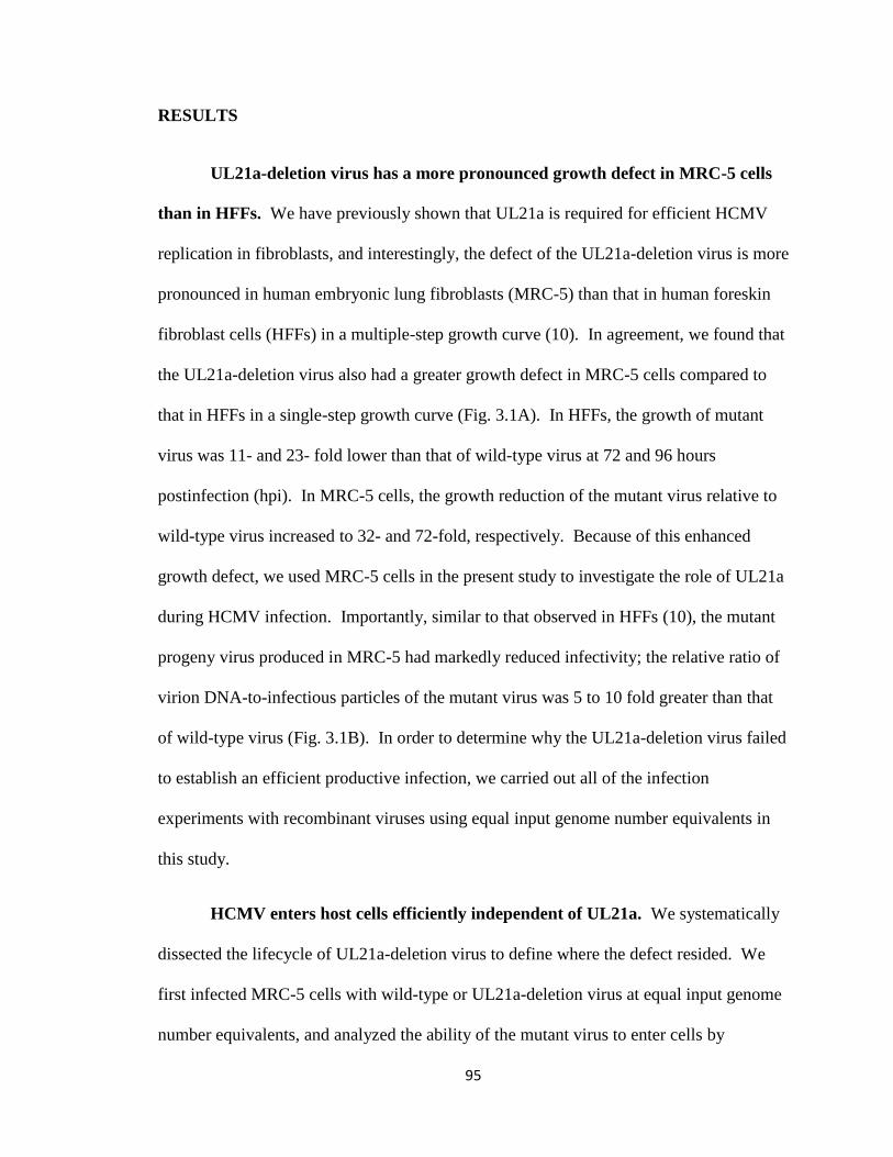
95
RESULTS
UL21a-deletion virus has a more pronounced growth defect in MRC-5 cells
than in HFFs. We have previously shown that UL21a is required for efficient HCMV
replication in fibroblasts, and interestingly, the defect of the UL21a-deletion virus is more
pronounced in human embryonic lung fibroblasts (MRC-5) than that in human foreskin
fibroblast cells (HFFs) in a multiple-step growth curve (10). In agreement, we found that
the UL21a-deletion virus also had a greater growth defect in MRC-5 cells compared to
that in HFFs in a single-step growth curve (Fig. 3.1A). In HFFs, the growth of mutant
virus was 11- and 23- fold lower than that of wild-type virus at 72 and 96 hours
postinfection (hpi). In MRC-5 cells, the growth reduction of the mutant virus relative to
wild-type virus increased to 32- and 72-fold, respectively. Because of this enhanced
growth defect, we used MRC-5 cells in the present study to investigate the role of UL21a
during HCMV infection. Importantly, similar to that observed in HFFs (10), the mutant
progeny virus produced in MRC-5 had markedly reduced infectivity; the relative ratio of
virion DNA-to-infectious particles of the mutant virus was 5 to 10 fold greater than that
of wild-type virus (Fig. 3.1B). In order to determine why the UL21a-deletion virus failed
to establish an efficient productive infection, we carried out all of the infection
experiments with recombinant viruses using equal input genome number equivalents in
this study.
HCMV enters host cells efficiently independent of UL21a. We systematically
dissected the lifecycle of UL21a-deletion virus to define where the defect resided. We
first infected MRC-5 cells with wild-type or UL21a-deletion virus at equal input genome
number equivalents, and analyzed the ability of the mutant virus to enter cells by
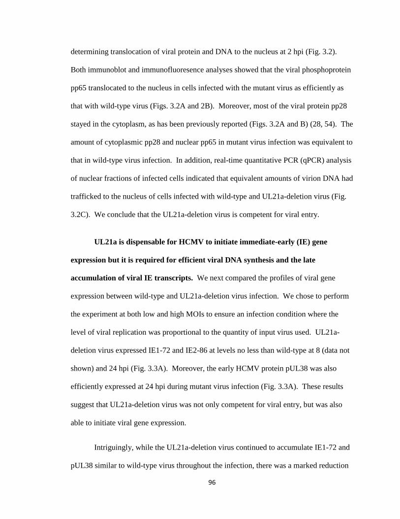
96
determining translocation of viral protein and DNA to the nucleus at 2 hpi (Fig. 3.2).
Both immunoblot and immunofluoresence analyses showed that the viral phosphoprotein
pp65 translocated to the nucleus in cells infected with the mutant virus as efficiently as
that with wild-type virus (Figs. 3.2A and 2B). Moreover, most of the viral protein pp28
stayed in the cytoplasm, as has been previously reported (Figs. 3.2A and B) (28, 54). The
amount of cytoplasmic pp28 and nuclear pp65 in mutant virus infection was equivalent to
that in wild-type virus infection. In addition, real-time quantitative PCR (qPCR) analysis
of nuclear fractions of infected cells indicated that equivalent amounts of virion DNA had
trafficked to the nucleus of cells infected with wild-type and UL21a-deletion virus (Fig.
3.2C). We conclude that the UL21a-deletion virus is competent for viral entry.
UL21a is dispensable for HCMV to initiate immediate-early (IE) gene
expression but it is required for efficient viral DNA synthesis and the late
accumulation of viral IE transcripts. We next compared the profiles of viral gene
expression between wild-type and UL21a-deletion virus infection. We chose to perform
the experiment at both low and high MOIs to ensure an infection condition where the
level of viral replication was proportional to the quantity of input virus used. UL21a-
deletion virus expressed IE1-72 and IE2-86 at levels no less than wild-type at 8 (data not
shown) and 24 hpi (Fig. 3.3A). Moreover, the early HCMV protein pUL38 was also
efficiently expressed at 24 hpi during mutant virus infection (Fig. 3.3A). These results
suggest that UL21a-deletion virus was not only competent for viral entry, but was also
able to initiate viral gene expression.
Intriguingly, while the UL21a-deletion virus continued to accumulate IE1-72 and
pUL38 similar to wild-type virus throughout the infection, there was a marked reduction
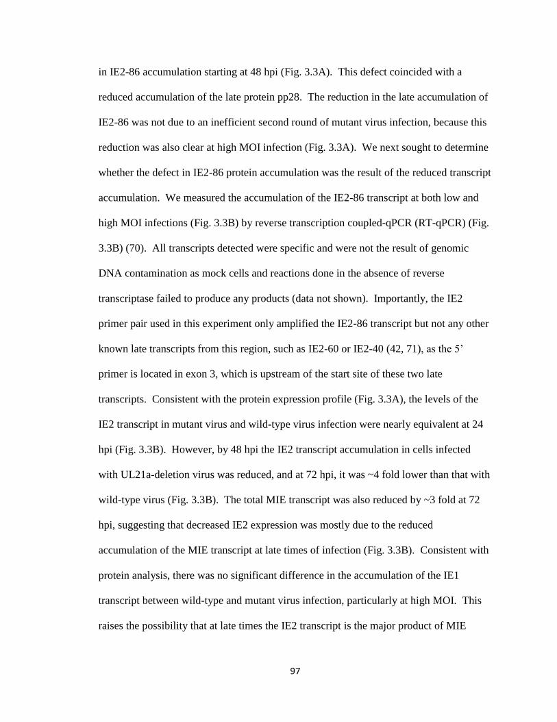
97
in IE2-86 accumulation starting at 48 hpi (Fig. 3.3A). This defect coincided with a
reduced accumulation of the late protein pp28. The reduction in the late accumulation of
IE2-86 was not due to an inefficient second round of mutant virus infection, because this
reduction was also clear at high MOI infection (Fig. 3.3A). We next sought to determine
whether the defect in IE2-86 protein accumulation was the result of the reduced transcript
accumulation. We measured the accumulation of the IE2-86 transcript at both low and
high MOI infections (Fig. 3.3B) by reverse transcription coupled-qPCR (RT-qPCR) (Fig.
3.3B) (70). All transcripts detected were specific and were not the result of genomic
DNA contamination as mock cells and reactions done in the absence of reverse
transcriptase failed to produce any products (data not shown). Importantly, the IE2
primer pair used in this experiment only amplified the IE2-86 transcript but not any other
known late transcripts from this region, such as IE2-60 or IE2-40 (42, 71), as the 5’
primer is located in exon 3, which is upstream of the start site of these two late
transcripts. Consistent with the protein expression profile (Fig. 3.3A), the levels of the
IE2 transcript in mutant virus and wild-type virus infection were nearly equivalent at 24
hpi (Fig. 3.3B). However, by 48 hpi the IE2 transcript accumulation in cells infected
with UL21a-deletion virus was reduced, and at 72 hpi, it was ~4 fold lower than that with
wild-type virus (Fig. 3.3B). The total MIE transcript was also reduced by ~3 fold at 72
hpi, suggesting that decreased IE2 expression was mostly due to the reduced
accumulation of the MIE transcript at late times of infection (Fig. 3.3B). Consistent with
protein analysis, there was no significant difference in the accumulation of the IE1
transcript between wild-type and mutant virus infection, particularly at high MOI. This
raises the possibility that at late times the IE2 transcript is the major product of MIE
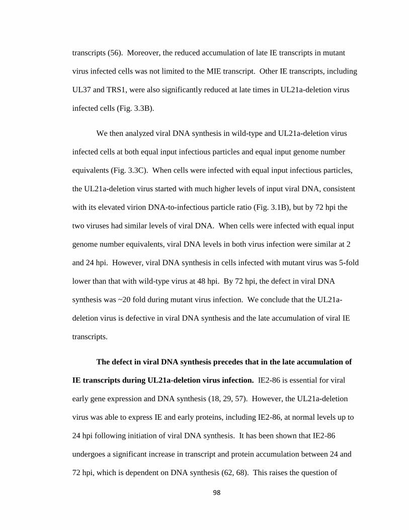
98
transcripts (56). Moreover, the reduced accumulation of late IE transcripts in mutant
virus infected cells was not limited to the MIE transcript. Other IE transcripts, including
UL37 and TRS1, were also significantly reduced at late times in UL21a-deletion virus
infected cells (Fig. 3.3B).
We then analyzed viral DNA synthesis in wild-type and UL21a-deletion virus
infected cells at both equal input infectious particles and equal input genome number
equivalents (Fig. 3.3C). When cells were infected with equal input infectious particles,
the UL21a-deletion virus started with much higher levels of input viral DNA, consistent
with its elevated virion DNA-to-infectious particle ratio (Fig. 3.1B), but by 72 hpi the
two viruses had similar levels of viral DNA. When cells were infected with equal input
genome number equivalents, viral DNA levels in both virus infection were similar at 2
and 24 hpi. However, viral DNA synthesis in cells infected with mutant virus was 5-fold
lower than that with wild-type virus at 48 hpi. By 72 hpi, the defect in viral DNA
synthesis was ~20 fold during mutant virus infection. We conclude that the UL21a-
deletion virus is defective in viral DNA synthesis and the late accumulation of viral IE
transcripts.
The defect in viral DNA synthesis precedes that in the late accumulation of
IE transcripts during UL21a-deletion virus infection. IE2-86 is essential for viral
early gene expression and DNA synthesis (18, 29, 57). However, the UL21a-deletion
virus was able to express IE and early proteins, including IE2-86, at normal levels up to
24 hpi following initiation of viral DNA synthesis. It has been shown that IE2-86
undergoes a significant increase in transcript and protein accumulation between 24 and
72 hpi, which is dependent on DNA synthesis (62, 68). This raises the question of
99
whether the reduced IE2-86 expression in UL21a-deletion virus infected cells is the cause
or a consequence of defective viral DNA synthesis. To address this question, we first
examined the temporal relationship between the defect in viral DNA synthesis and that in
MIE transcript accumulation. In this experiment, a reduction in viral DNA synthesis was
clearly evident at 32 hpi, and that reduction reached 4-fold by 40 hpi in mutant virus
infection (Fig. 3.4A). In contrast, the difference of MIE expression was minimal between
wild-type and mutant virus infection, particularly at 32 hpi (Fig. 3.4B). This result led us
to hypothesize that the lack of pUL21a results in reduced viral DNA synthesis, which is
the cause of the reduced late accumulation of IE transcripts.
Inhibition of viral DNA synthesis reduces the late accumulation of IE
transcripts in wild-type and UL21a-deletion virus infected cells to equivalent levels.
To directly test the hypothesis that the late accumulation of IE transcripts is dependent on
UL21a-mediated viral DNA synthesis during HCMV infection, we infected MRC-5 cells
with wild-type and UL21a-deletion virus at a high MOI in the presence or absence of the
viral DNA synthesis inhibitor phosphonoacetic acid (PAA). The accumulation of IE2-86
and the late protein pp28 were reduced to undetectable levels in both wild-type and
mutant virus infected cells in the presence of PAA, particularly at 48 and 72 hpi (Fig.
3.5A). Conversely, the expression levels of IE1-72 and early proteins pUL38 and
pUL21a were minimally affected. Therefore, the efficient accumulation of IE2-86
protein at late times is dependent on viral DNA synthesis. This effect of viral DNA
synthesis on late IE2 expression occurred primarily at the transcriptional level as PAA
reduced the accumulation of IE2 transcript by ~4 and ~9-fold during wild-type virus
infection at 24 and 72 hpi, respectively (Fig. 3.5B). Importantly, IE2 was not the only IE
100
gene whose transcript accumulation was affected by inhibition of viral DNA synthesis.
This effect was global as PAA also markedly reduced transcript accumulation of two
other IE genes, UL37 and TRS1. As the effect of PAA on IE expression was evident as
early as 24 hpi at high MOI infection, it was unlikely the result of reduced secondary
infection. Moreover, PAA reduced all the IE transcripts examined to equivalent levels in
wild-type and UL21a-deletion virus infection, even at 72 hpi, when the difference in IE
expression is most notable between these two virus infections (Fig. 3.5B). Collectively,
these results suggest that the reduced late IE transcript accumulation in the absence of
UL21a is a consequence of reduced viral DNA synthesis.
Finally, we analyzed viral gene expression in the presence of an additional viral
DNA synthesis inhibitor, gancyclovir (GCV), to rule out any potential off-target effects
of PAA. In addition, we also examined the gene expression profile of an HCMV clinical
isolate to determine whether viral DNA synthesis-dependent late IE gene expression was
limited to the laboratory strains of HCMV. MRC-5 cells were infected with either ADwt
(Fig. 3.5C, left panel) or the BAC-derived clinical HCMV isolate TR (Fig. 5C, right
panel) and treated with PAA or GCV. As expected, neither drug reduced the levels of the
early proteins pUL38 or pUL21a in either viral strain. Importantly, both PAA and GCV
significantly reduced the level of IE2-86 and the late protein pp28 in cells infected with
ADwt, indicating that reduced IE2-86 levels were not due to any potential off-target
effects of PAA (Fig. 3.5C). During TR infection, GCV had a minimal effect on viral
protein expression at all times examined. This is consistent with the fact that TR was
isolated from drug-resistant patient with advanced HIV with CMV retinitis (35, 61),
though this has not been previously shown in tissue culture. Nonetheless, PAA reduced
101
the accumulation of IE2-86 and pp28 at 72 hpi in TR virus infected cells, indicating that
DNA synthesis-dependent late IE gene expression was not strain specific.
The defect of UL21a-deletion virus is due to the loss of the protein product
pUL21a. Previously, using various recombinant viruses, we were able to show that the
defect of the UL21a-deletion virus was due to the loss of the UL21a gene but not the
overlapping UL21 ORF or altered regulation of the neighboring genes (10). However, it
is formally possible that the defect of the mutant virus was due to other unidentified
functional products or DNA sequence within the UL21a gene rather than the identified
protein product, pUL21a. To rule out this possibility, we created a recombinant virus,
ADpmpUL21a, where we introduced two stop-codon point mutations at residues Gln8
and Arg39
of the UL21a ORF to abrogate the expression of pUL21a (Fig. 6A). In
addition, we also created a specific marker-rescued virus for this mutant, ADrevpUL21a
(Fig. 3.6A). We included two stop-codon mutations as the virus was able to produce a
truncated protein when only one stop-codon mutation was inserted at Gln8 (data not
shown). ADpmpUL21a did not express pUL21a (Fig. 3.6C) and grew indistinguishably
from ADsubUL21a in a low MOI growth curve in MRC-5 cells, while the growth of
ADrevpUL21a was nearly identical to that of ADrevUL21a, which was previously shown
to grow like wild-type (Fig. 3.6B) (10). Additionally, when infecting with equal input
genome number equivalents, ADpmpUL21a produced markedly reduced amounts of IE2-
86 protein compared to the marker-rescued virus in a manner indistinguishable from the
UL21a-deletion virus (Fig. 3.6C). We conclude that the phenotype of the UL21a-
deletion virus is due to the loss of pUL21a and not the loss of any other unidentified
functional gene products or DNA sequence.
102
De novo expression of pUL21a is necessary and sufficient for efficient viral
gene expression. pUL21a may facilitate viral DNA synthesis directly or it may be
required for the proper composition of the virion, enabling it to initiate efficient viral
DNA synthesis. To differentiate these potential mechanisms, we first attempted to create
an inducible expression system to express pUL21a. Multiple approaches were used,
including expressing pUL21a within the virus from a tetracycline-inducible promoter or
tagging pUL21a with a destabilization domain, ddFKBP (12, 50). However, neither
system was able to efficiently regulate the production of pUL21a, possibly due to the
highly unstable nature of the protein (data not shown) (10). Instead, we developed a
complementation system in which we expressed a functional GFP/UL21a fusion protein
(10) in MRC-5 cells by retroviral transduction. The GFP/UL21a fusion protein was
abundantly expressed in these cells and appeared as two bands (Fig. 3.8B), possibly due
to partial degradation or posttranslational modification as we previously reported (10).
Overexpression of the GFP/UL21a protein (pGFP/UL21a) complemented growth of the
UL21a-deletion virus by greater than 200-fold (Fig. 3.7A). A difference of 10-20 fold in
titer of progeny virus remained between ADwt and ADsubUL21a at 12-18 dpi when they
were grown in pGFP/UL21a-expressing cells. One likely reason for this difference is that
we titered progeny virus in normal fibroblasts in which the mutant virus had reduced
infectivity (Fig. 3.1B). To test this hypothesis, the titers of wild-type and mutant virus
were determined on cells expressing GFP/UL21a or control cells expressing GFP only.
When virus collected from pGFP/UL21a-expressing cells at 12 dpi was titered on non
complementing GFP-expressing cells, there was a 10-fold difference in titer between
ADwt and ADsubUL21a, similar to that on untransduced cells (Figs. 3.7A-B). When

103
these viruses were titered on GFP/UL21a cells, there was no difference between ADwt
and ADsubUL21a (Fig. 3.7B). Thus, we restored the ability of the mutant virus to
replicate using pGFP/UL21a-expressing complementing cells.
To test the hypothesis that de novo expression of pUL21a facilitates efficient viral
replication, we performed two distinct experiments. First, we infected MRC-5 cells
expressing either GFP or GFP/UL21a with wild-type or UL21a-deletion virus that was
produced from normal MRC-5 cells. The expression of pGFP/UL21a but not GFP
restored the ability of the UL21a-deletion virus to efficiently synthesize viral DNA and
produce IE2-86 and pp28, particularly at late times post infection (Figs. 3.8A-B).
In the second experiment, we infected normal MRC-5 cells with wild-type or
UL21a-deletion virus that was produced from GFP or pGFP/UL21a-expressing cells. We
found that even when UL21a-deletion virus was produced on complementing cells, it still
expressed IE2-86 and pp28 at markedly reduced levels similar to the mutant virus that
was produced on GFP control cells (Fig. 3.8C). Therefore, pUL21a is unlikely to be
required for the functionality of HCMV virions. Together, these two sets of experiments
suggest that the de novo expression of pUL21a is both necessary and sufficient for
efficient viral lytic replication.
Collectively, our data indicate that de novo pUL21a expression facilitates HCMV
DNA synthesis, leading to efficient late expression of viral IE genes.
DISCUSSION
In a previous study, we have found that the HCMV gene UL21a encodes a small
15-kDa protein, pUL21a, which predominantly localizes to the cytoplasm (10). The
104
UL21a-deletion virus has an elevated virion particle-to-infectious virus ratio, suggesting
that UL21a is required for HCMV to establish an efficient productive infection (10). In
the present study we determined why the mutant virus was unable to establish a
productive infection by performing infection analysis using equal numbers of DNA-
containing virion particles instead of infectious particles. The UL21a-deletion virus
entered host cells and initiated its gene expression program like the wild-type virus.
However, by 48-72 hpi the mutant virus failed to synthesize its DNA efficiently and
expressed its IE genes at significantly reduced levels, even though the IE1-72 transcript
and protein were not affected (Fig. 3.3).
Analysis of the UL21a mutant virus infection also provided evidence to clarify the
relationship of viral DNA synthesis and late accumulation of viral IE gene products. The
immediate-early expression of the viral IE2-86 protein is required for early gene
expression and viral DNA synthesis (18, 29, 57). However, a couple of earlier studies
have also suggested that the late accumulation of the IE2-86 protein and transcript is
dependent on viral DNA synthesis (62, 68). Stenberg and coworkers showed that the late
IE2-86 accumulation was eliminated in a temperature sensitive mutant of HCMV which
could not synthesize viral DNA at the restrictive temperature (62). Nonetheless, this
mutation in the viral genome has not been mapped, leaving open the possibility that an
additional defect prior to DNA synthesis controls the late IE2-86 accumulation. Tenney
and coworkers showed that the late accumulation of the IE2-86 specific transcript was
sensitive to the viral DNA synthesis inhibitor, phosphonoformic acid (PFA) (67).
Nonetheless, it was unclear whether viral DNA synthesis promotes the late expression of
only IE2 or the entire IE gene class. In our study, the reduced levels of IE proteins,
105
including IE2-86, seen in UL21a mutant virus were not detected until after 24 hpi, a time
when viral DNA synthesis has already initiated. In fact, we could detect reduced DNA
synthesis before reduced IE transcript accumulation in mutant virus infection (Fig. 3.4).
Importantly, PAA reduced the expression of IE genes in wild-type and UL21a-deletion
virus infected cells to equivalent levels (Fig. 3.5). Collectively, these data strongly
suggests that pUL21a facilitates viral DNA synthesis, which then enhances expression of
IE genes, including IE2, at late times during infection.
Inhibition of viral DNA synthesis also reduced the expression of additional IE
genes, such as total MIE, TRS1, and UL37, but it had a much smaller or negligible effect
on expression of IE1 and the true early gene UL38 (Fig. 3.5). Our result on TRS1
expression is consistent with previous reports that the accumulation of TRS1 transcript
and protein dramatically increases between 24 and 48 hpi (30, 36). Our result on UL37
expression is somewhat different from an earlier report showing no detectable reduction
of UL37 transcript levels in the presence of PFA (67). The reason for this discrepancy is
unclear but it is possibly due to different assays used in these studies. We used the more
sensitive RT-qPCR method instead of Northern blot for transcript analysis. Moreover,
we used GAPDH instead of total RNA as an internal control as the GAPDH transcript
remains constant throughout infection (33, 47) and is a widely used control for qPCR
analysis of HCMV infection (22, 53). Nonetheless, to our knowledge, this is the first
report showing that the efficient transcript accumulation of multiple IE genes, in addition
to IE2, is dependent on viral DNA synthesis.
Why does inhibition of viral DNA synthesis by PAA treatment or infection in the
absence of pUL21a have such a drastic effect on IE2 expression, but have little-to-

106
negligible effect on IE1 expression (Fig. 3.5B)? It has clearly been shown by several
groups that IE1 and IE2 proteins accumulate with different kinetics during the viral
lifecycle. Despite being produced from the same pre-mRNA, IE1 largely accumulates
during the very early stages of infection whereas IE2 primarily accumulates between 24
and 72 hpi, and its accumulation is dependent on DNA synthesis (62, 68) (Fig. 3.5B).
These data suggest that the onset of viral DNA synthesis triggers a viral event that results
in a switch from producing IE1 to IE2 transcript. This could be due to alternative
splicing or altered stability of one of the MIE transcripts. Interestingly, it has been
reported that CDK inhibitors reverse the accumulation of IE1 and IE2 at early stages of
infection (55), suggesting that CDK activity may be promoting IE1 accumulation early
during infection. CDKs appeared to accomplish this by regulating splicing, as the
inhibitors had the similar effect on another spliced IE transcript, UL37, but not on
unspliced viral genes (55). It is intriguing to speculate that viral DNA synthesis may
somehow alter one or more CDK activities, which results in a change of the splicing
pattern of the MIE transcript, and the switch from IE1 to IE2 production.
How does pUL21a augment viral DNA synthesis? Our analysis of the mutant
virus infection in complementing cells indicates that the de novo expression of pUL21a is
necessary and sufficient for the establishment of a productive infection (Fig. 3.8).
Moreover, the mutant virus was competent for entry and initiation of gene expression
(Figs. 3.2 and 3.3). These results, together with the fact that pUL21a is undetectable in
the HCMV virion nor does it localize to cytoplasmic viral assembly centers (10), argue
strongly against its potential involvement in virion assembly. pUL21a has a diffuse
cytopasmic distribution in both infected and overexpressing cells and shares no
107
homology to any known DNA replication proteins. Furthermore, it is not expressed from
genomic loci required for transient complementation of HCMV oriLyt dependent
replication (39), suggesting that this protein is unlikely to act directly as a core DNA
replication enzyme to synthesize viral DNA. pUL21a may play other roles, for instance,
by increasing concentrations of dNTPs within infected cells, enhancing accumulation of
viral and cellular DNA replication enzymes required for viral DNA synthesis, shuttling
critical factors in or out of the nucleus, or by blocking cellular antiviral defenses against
viral DNA synthesis. We are currently testing these potential roles for pUL21a,
identifying viral or cellular factors that it may interact with, and investigating the
mechanistic basis of its function.
Little is known about regulation of immediate-early gene expression by viral
DNA synthesis in herpesvirus infection. It is well understood that the expression of
subsets of early (β2, delayed-early) and late (γ1, early-late) viral genes are augmented by
viral DNA replication during lytic infection of herpesviruses (48). Early evidence
suggested that expression of the IE2 gene could be also augmented by viral DNA
synthesis in HCMV infection (62, 68). In addition, the expression of the IE1 protein in
MCMV infection and the expression of an IE protein ICP-27 during HSV-1 infection of
trigeminal ganglion and rat superior cervical ganglia neurons have also been shown to be
enhanced by viral DNA synthesis (26, 38, 45). For HSV-1, the studies led to the
hypothesis that DNA synthesis represents a critical decision point in the choice between a
latent and lytic infection. In neurons, HSV-1 expresses limited IE genes during the initial
phase of the infection. If robust viral DNA synthesis follows, the accumulation of the IE
genes is dramatically increased, allowing the virus to establish a lytic infection.
108
Conversely, such as in the case of a TK mutant or in the presence of PAA, viral DNA
synthesis is inhibited, no additional IE genes are produced, and the virus will establish a
latent infection (26, 38). It is intriguing to speculate that DNA synthesis may also be
critical for HCMV to establish a latent or lytic infection in appropriate cell types, such as
in CD34+ cells or undifferentiated THP-1 cells. When these cells were treated with
histone deacetylase (HDAC) inhibitors, HCMV initiated IE1-72 expression and
progressed to early gene expression, but the infection aborted prior to late gene
expression (51, 53). Thus, in addition to a block at IE gene expression, HCMV perhaps
is also unable to initiate efficient viral DNA replication in these cells, both of which may
contribute to the establishment of its latency.
Finally, it will be important to identify the function(s) of the late accumulating IE
proteins, particularly the IE2-86 protein. It is clear that expression of IE2-86 at
immediate early times is essential for early viral gene expression and viral DNA
synthesis in HCMV infection (18, 29, 57). Almost paradoxically, the greatest increase in
IE2-86 accumulation comes at late times of infection, and it is not entirely clear what, if
any, role this late accumulating IE2-86 protein plays during viral replication. It has been
shown that IE2-86 is involved in auto-repression of the MIEP during the late stages of
infection by its interactions with histone modifying enzymes (46). Mutation within the
cis repression sequence (crs) in the MIEP increases the abundance of both IE transcripts
and protein during late times of infection (46). However, mutations within the crs also
resulted in defects in the early events of viral infection, including reduced MIE gene
expression and viral DNA replication, thus complicating the analysis defining the precise
role of this IE auto-repression during HCMV infection (21). Another potential function
109
for the newly synthesized IE2-86 is to transactivate the late promoters of HCMV. Late
promoters remain largely associated with repressive chromatin marks and only begin to
become acetylated starting at 24 hpi (6, 16). Binding of IE2-86 to HDACs 1, 2, 3, and
multiple histone methyltransferases (HMTs) during the late stages of infection may
activate transcription from late promoters (37, 40, 46). However, it is also possible that
IE2-86 transactivates late promoters through its interactions with many other viral and
cellular partners, or that the late accumulating IE2-86 is responsible for one of the many
other functions ascribed to this protein (65). In any event, it is sensible for the virus to
couple DNA synthesis with expression of transactivator proteins that are required for late
gene expression. This will ensure that late proteins, such as capsid and tegument
proteins, will accumulate along with viral DNA synthesis, enhancing the production of
infectious viral particles and facilitating the ability of the virus to spread to new host
cells.

110
ACKNOWLEGEMENTS
We thank Herbert Virgin and the members of his laboratory for helpful discussion
and invaluable advice, Caroline Kulesza (University of Colorado School of Medicine) for
the nuclear fractionation protocol, Thomas Shenk for the antibodies, and members of the
Yu lab for critical reading of the manuscript.
This study was supported by a Public Health Service grant (CA-120768), and in
part by a Grant from the American Heart Association (09GRNT2290199) and a grant
from Children’s Discovery Institute at Washington University. D. Y. holds an
Investigators in the Pathogenesis of Infectious Disease award from the Burroughs
Wellcome Fund. A.R.F. was a Morse/Berg Fellow of the Department of Molecular
Microbiology, Washington University School of Medicine, and is supported by
Institutional Training Grant T32-AI007172.

111
FIGURE LEGEND
Figure 3.1. UL21a-deletion virus has reduced infectivity. (A) Growth of UL21a-
deletion virus in HFF and MRC-5 fibroblasts. HFFs or MRC-5 fibroblasts were infected
with wild-type virus (ADwt) or UL21a-deletion virus (ADsubUL21a) at an MOI of 5,
cell-free viruses were collected at 72 and 96 hours postinfection (hpi), and their yields
were determined by TCID50 assay. Data are presented as the fold reduction in growth of
ADsubUL21a relative to ADwt in each cell type. (B) Relative infectivity of progeny
virions. MRC-5 cells were infected and cell-free virus was collected as described in (A).
The same volumes of viral samples were measured for their viral genome copies by real-
time quantitative PCR (qPCR) and for their infectivity by TCID50 assay. The result is
presented as the viral genome copy number per TCID50 unit. The relative genome copy
per TCID50 unit of wild-type virus at 72 hpi was set to 1. Shown is a representative of at
least 2 independent experiments.
Figure 3.2. UL21a-deletion virus is competent for viral entry. (A-B) Translocation of
tegument proteins pp65 and pp28 delivered into infected cells. MRC-5 cells were
infected with ADwt and ADsubUL21a at an input genome number equivalent to 1 or 10
TCID50 units of wild-type virus/cell, collected at 2 hpi, and analyzed by immunoblotting
(A) or immunofluorescence (B), respectively. For immunoblotting, total lysate ("T"),
nuclear ("N"), and cytoplasmic ("C") fractions were prepared and analyzed as indicated.
Acetyl-Histone H3 and Bip were used as nuclear and cytoplasmic fractionation controls,
respectively. For immunofluorescence, the staining patterns of nuclei and viral antigens
were shown as blue and green, respectively. Scale bar = 10 m. (C) Nuclear
translocation of virion DNA. Intracellular DNA was isolated from both total and nuclear
112
fractions at 2 hpi. The quantity of viral genomes in each fraction was determined by
qPCR using primers specific for viral UL54 gene and viral genome copies were
normalized to cellular genomes using β-actin primers. The percentage of viral DNA
translocated into the nucleus was determined by dividing viral genome equivalents in the
nuclear fraction by those in the total cell lysate. Shown is a representative of at least 2
independent experiments.
Figure 3.3. HCMV accumulates less IE2-86 protein and IE transcripts at late times, and
synthesizes viral DNA at reduced levels in the absence of UL21a. (A) Analysis of viral
protein accumulation. MRC-5 cells were infected with ADwt or ADsubUL21a at an
input genome number equivalent to 0.25 (left panel) or 3 (right panel) TCID50 units of
wild-type virus/cell. Cells were collected at indicated times, and total cell lysates were
analyzed by immunoblotting with indicated antibodies. (B) Analysis of viral transcript
accumulation. MRC-5 cells were infected with ADwt or ADsubUL21a at an input
genome number equivalent to 0.25 (left panel) or 3 (right panel) TCID50 units of wild-
type virus/cell. Total RNA was collected at indicated times, amounts of selected viral IE
transcripts were measured by reverse transcription-coupled quantitative PCR (RT-qPCR),
and normalized to that of GAPDH. Also shown are the genomic structure of the MIE
locus and the locations of RT-PCR primers for analyzing IE1, IE2 and total IE
transcripts. The normalized amount of viral transcript at 24 hpi during wild-type virus
infection was set to 1. (C) Analysis of viral DNA synthesis. MRC-5 cells were infected
with ADwt or ADsubUL21a at either an input genome number equivalent to 0.25 TCID50
units of wild-type virus/cell or an MOI of 0.05. Total intracellular DNA was collected,
viral DNA was measured by qPCR, and the amount was normalized to cellular genome

113
copies with primers to β-actin. The normalized amount of viral DNA at 2 hpi during
wild-type virus infection was set to 1. Shown is a representative of at least 2 independent
experiments.
Figure 3.4. Defects in viral DNA synthesis precede defects in the late accumulation of
viral IE transcripts in UL21a-deletion virus infected cells. (A) Analysis of viral DNA
synthesis. MRC-5 cells were infected with ADwt or ADsubUL21a at an input genome
number equivalent to 0.25 TCID50 units of wild-type virus/cell. Total intracellular DNA
was collected at indicated times and analyzed by qPCR as described in legend to Fig. 3C.
The normalized amount of viral DNA at 24 hpi during wild-type virus infection was set
to 1. (B) Analysis of viral transcript accumulation. MRC-5 cells were infected as
described in (A), total RNA was collected, and amounts of MIE transcript were analyzed
by RT-qPCR as described in legend to Fig. 3B. The normalized amount of viral
transcript at 24 hpi during wild-type virus infection was set to 1. Shown is a
representative of at least 2 independent experiments. n.s., P>0.05; *, P<0.05 by
Student’s t test.
Figure 3.5. Inhibition of viral DNA synthesis reduces the late expression of IE genes in
wild-type and UL21a-deletion virus infected cells to equivalent levels. (A) Analysis of
viral protein accumulation in the presence of PAA. MRC-5 cells were infected with
ADwt or ADsubUL21a at an input genome number equivalent to 3 TCID50 units of wild-
type virus/cell in the presence or absence of PAA (100 µg/ml). Cells were collected at
indicated times, and total cell lysate was analyzed by immunoblotting with indicated
antibodies. (B) Analysis of viral transcript accumulation. MRC-5 cells were infected in
the presence ("+") or absence ("-") of PAA as in (A), total RNA was collected and
114
selected IE transcripts were analyzed by RT-qPCR and normalized to that of GAPDH.
The normalized amount of viral transcript at 24 hpi during wild-type virus infection was
set to 1. n.s., P>0.05; *, P<0.05 by Student’s t test. (C) Viral protein accumulation in
presence of PAA or gancyclovir (GCV) during infection of HCMV strains AD169 and
TR. MRC-5 cells were infected with ADwt (left panel) or TR (right panel) at an MOI of
2 with addition of no drug, PAA (100 µg/ml), or GCV (30 µg/ml). Cells were harvested
at indicated times and analyzed by immunoblotting with indicated antibodies. Shown is a
representative of at least 2 independent experiments.
Figure 3.6. The growth defect of UL21a-deletion virus is due to the loss of the protein
product pUL21a. (A) Diagram of the UL21a locus in UL21a-deletion virus
(ADsubUL21a), point mutant virus in which 2 stop-codon mutations were introduced into
the UL21a ORF (ADpmpUL21a), and its marker-rescued virus (ADrevpUL21a). (B)
Growth kinetic analysis of recombinant HCMV viruses ADpmpUL21a and
ADrevpUL21a. MRC-5 cells were infected with indicated viruses at an MOI of 0.01,
cell-free viruses were collected at different days post infection, and their yields were
determined by TCID50. (C) Analysis of viral protein accumulation. MRC-5 cells were
infected with ADpmpUL21a or ADrevpUL21a at an input genome number equivalent to
0.25 TCID50 units of wild-type virus/cell, cells were collected at indicated times, and total
lysate was analyzed by immunoblotting. Shown is a representative of at least 2
independent experiments.
Figure 3.7. Expression of pUL21a in trans complements the growth of UL21a-deletion
virus. MRC-5 cells were transduced with retrovirus expressing GFP (MRC5-GFP) or
expressing the GFP/UL21a fusion protein (MRC5-GFP/UL21a), and then infected with
115
ADwt and ADsubUL21a at an MOI of 0.2. Cell-free viruses were collected at different
days postinfection, and their yields were determined by TCID50 in HFFs (A). In addition,
ADwt and ADsubUL21a collected at 12 dpi from MRC5-GFP/UL21a cells were also
titered on both MRC5-GFP and MRC5-GFP/UL21a cells to compare their titers
measured on complementing and non-complementing cells (B).
Figure 3.8. De novo expression of pUL21a is necessary and sufficient for efficient viral
DNA synthesis and late accumulation of viral proteins. Viral DNA synthesis (A) and
protein accumulation (B) in GFP- or GFP/UL21a- expressing cells infected with
recombinant virus that is generated in non-complementing cells. MRC5-GFP cells or
MRC5-GFP/UL21a cells were infected with ADwt and ADsubUL21a virus at an input
genome number equivalent to 2 TCID50 units of wild-type virus/cell. Infected cells were
collected at indicated times and analyzed for viral DNA by qPCR (A) and viral proteins
by immunoblotting (B). (C) Viral protein accumulation in normal MRC-5 cells infected
with recombinant virus that is generated in GFP- or GFP/UL21a- expressing cells.
Normal MRC-5 cells were infected with ADwt or ADsubUL21a virus that was produced
from either GFP ("MRC5-GFP") or GFP/UL21a ("MRC5-GFP/UL21a") expressing cells
at an input genome number equivalent to 2 TCID50 units of wild-type virus/cell. Infected
cells were collected at indicated times and analyzed for viral proteins by immunoblotting.
Shown is a representative of at least 2 independent experiments.
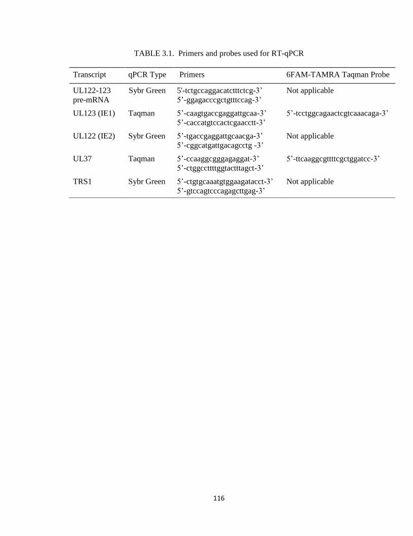
116
TABLE 3.1. Primers and probes used for RT-qPCR
Transcript qPCR Type Primers 6FAM-TAMRA Taqman Probe
UL122-123
pre-mRNA
Sybr Green 5'-tctgccaggacatctttctcg-3’
5’-ggagacccgctgtttccag-3’
Not applicable
UL123 (IE1) Taqman
5’-caagtgaccgaggattgcaa-3’
5’-caccatgtccactcgaacctt-3’
5’-tcctggcagaactcgtcaaacaga-3’
UL122 (IE2) Sybr Green 5’-tgaccgaggattgcaacga-3’
5’-cggcatgattgacagcctg -3’
Not applicable
UL37 Taqman
5’-ccaaggcgggagaggat-3’
5’-ctggccttttggtactttagct-3’
5’-ttcaaggcgttttcgctggatcc-3’
TRS1 Sybr Green 5’-ctgtgcaaatgtggaagatacct-3’
5’-gtccagtcccagagcttgag-3’
Not applicable
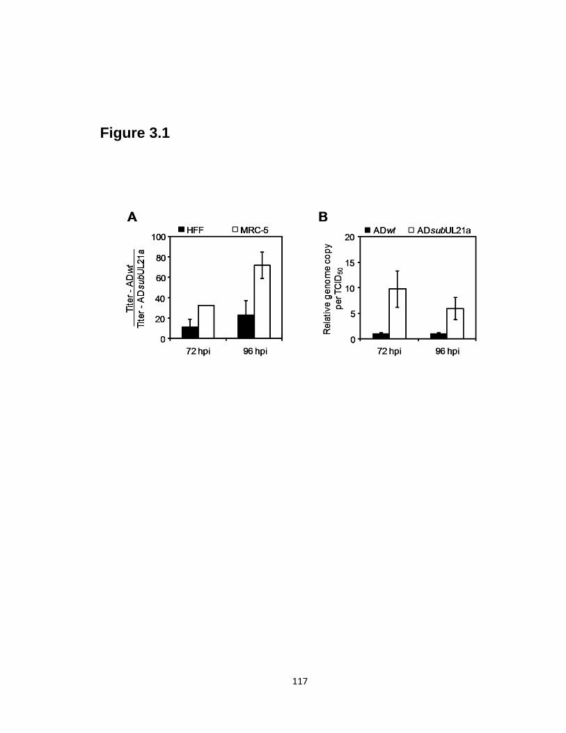
117
Figure 3.1
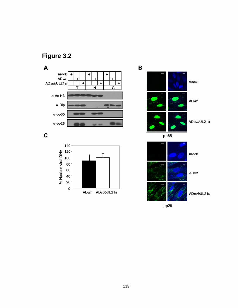
118
Figure 3.2
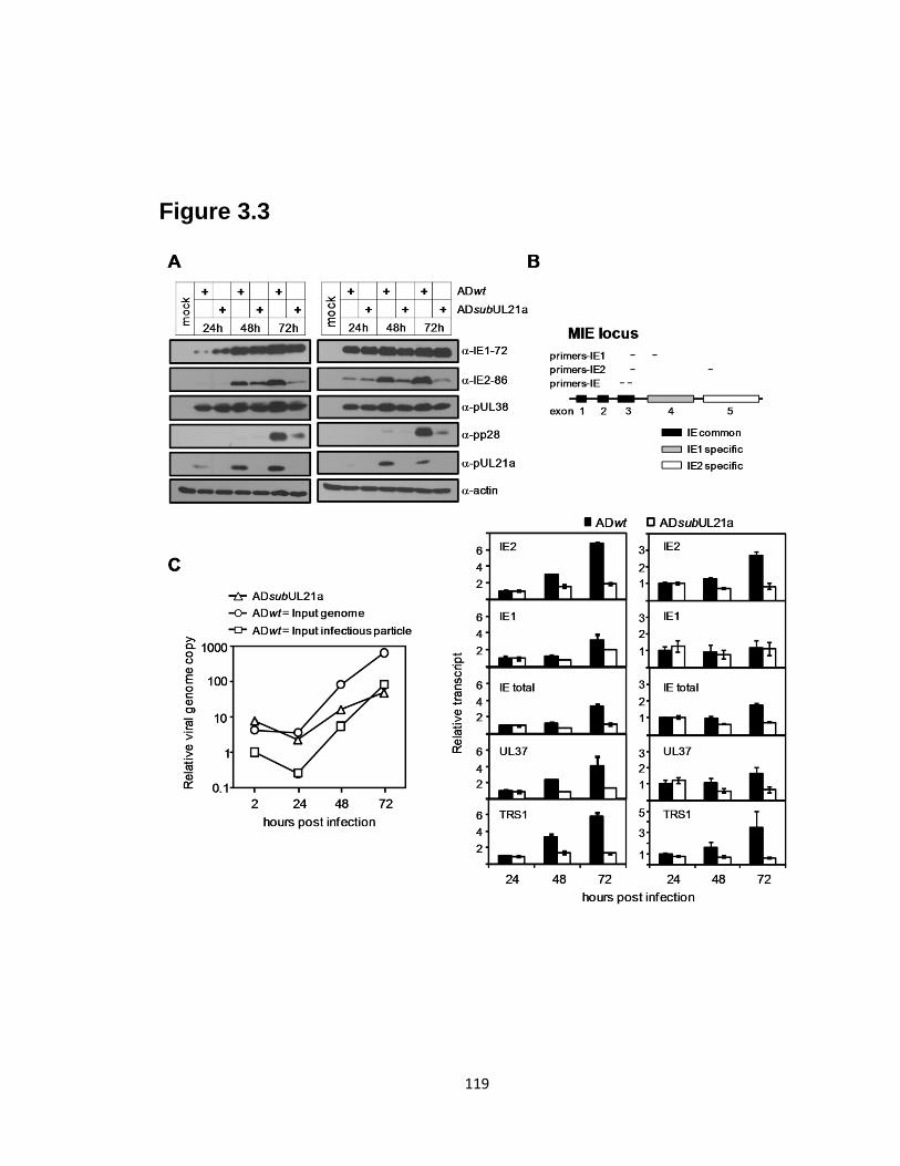
119
Figure 3.3
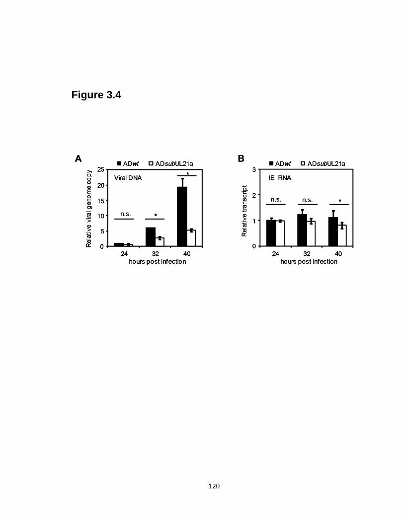
120
Figure 3.4
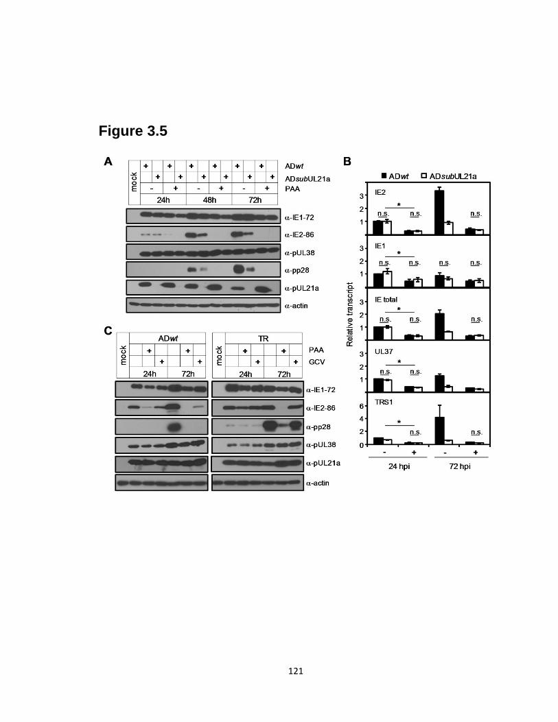
121
Figure 3.5
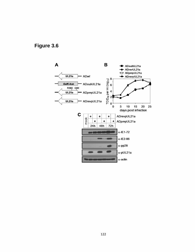
122
Figure 3.6
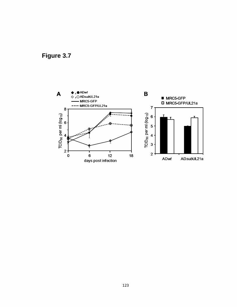
123
Figure 3.7

124
Figure 3.8
125
REFERENCES
1. Arnoult, D., L. M. Bartle, A. Skaletskaya, D. Poncet, N. Zamzami, P. U.
Park, J. Sharpe, R. J. Youle, and V. S. Goldmacher. 2004. Cytomegalovirus
cell death suppressor vMIA blocks Bax- but not Bak-mediated apoptosis by
binding and sequestering Bax at mitochondria. Proc Natl Acad Sci U S A
101:7988-93.
2. Borst, E. M., G. Hahn, U. H. Koszinowski, and M. Messerle. 1999. Cloning of
the human cytomegalovirus (HCMV) genome as an infectious bacterial artificial
chromosome in Escherichia coli: a new approach for construction of HCMV
mutants. J Virol 73:8320-9.
3. Chee, M. S., S. C. Satchwell, E. Preddie, K. M. Weston, and B. G. Barrell.
1990. Human cytomegalovirus encodes three G protein-coupled receptor
homologues. Nature 344:774-7.
4. Child, S. J., M. Hakki, K. L. De Niro, and A. P. Geballe. 2004. Evasion of
cellular antiviral responses by human cytomegalovirus TRS1 and IRS1. J Virol
78:197-205.
5. Child, S. J., S. Jarrahian, V. M. Harper, and A. P. Geballe. 2002.
Complementation of vaccinia virus lacking the double-stranded RNA-binding
protein gene E3L by human cytomegalovirus. J Virol 76:4912-8.
6. Cuevas-Bennett, C., and T. Shenk. 2008. Dynamic histone H3 acetylation and
methylation at human cytomegalovirus promoters during replication in
fibroblasts. J Virol 82:9525-36.
7. Dolan, A., C. Cunningham, R. D. Hector, A. F. Hassan-Walker, L. Lee, C.
Addison, D. J. Dargan, D. J. McGeoch, D. Gatherer, V. C. Emery, P. D.
Griffiths, C. Sinzger, B. P. McSharry, G. W. Wilkinson, and A. J. Davison. 2004. Genetic content of wild-type human cytomegalovirus. J Gen Virol 85:1301-
12.
8. Dunn, W., C. Chou, H. Li, R. Hai, D. Patterson, V. Stolc, H. Zhu, and F. Liu.
2003. Functional profiling of a human cytomegalovirus genome. Proc Natl Acad
Sci U S A 100:14223-8.
9. Dunn, W., P. Trang, Q. Zhong, E. Yang, C. van Belle, and F. Liu. 2005.
Human cytomegalovirus expresses novel microRNAs during productive viral
infection. Cell Microbiol 7:1684-95.
10. Fehr, A. R., and D. Yu. 2010. Human cytomegalovirus gene UL21a encodes a
short-lived cytoplasmic protein and facilitates virus replication in fibroblasts. J
Virol 84:291-302.
11. Gawn, J. M., and R. F. Greaves. 2002. Absence of IE1 p72 protein function
during low-multiplicity infection by human cytomegalovirus results in a broad
block to viral delayed-early gene expression. J Virol 76:4441-55.
12. Glass, M., A. Busche, K. Wagner, M. Messerle, and E. M. Borst. 2009.
Conditional and reversible disruption of essential herpesvirus proteins. Nat
Methods 6:577-9.
13. Goldmacher, V. S., L. M. Bartle, A. Skaletskaya, C. A. Dionne, N. L.
Kedersha, C. A. Vater, J. W. Han, R. J. Lutz, S. Watanabe, E. D. Cahir
McFarland, E. D. Kieff, E. S. Mocarski, and T. Chittenden. 1999. A
126
cytomegalovirus-encoded mitochondria-localized inhibitor of apoptosis
structurally unrelated to Bcl-2. Proc Natl Acad Sci U S A 96:12536-41.
14. Greaves, R. F., and E. S. Mocarski. 1998. Defective growth correlates with
reduced accumulation of a viral DNA replication protein after low-multiplicity
infection by a human cytomegalovirus ie1 mutant. J Virol 72:366-79.
15. Grey, F., A. Antoniewicz, E. Allen, J. Saugstad, A. McShea, J. C. Carrington,
and J. Nelson. 2005. Identification and characterization of human
cytomegalovirus-encoded microRNAs. J Virol 79:12095-9.
16. Groves, I. J., M. B. Reeves, and J. H. Sinclair. 2009. Lytic infection of
permissive cells with human cytomegalovirus is regulated by an intrinsic 'pre-
immediate-early' repression of viral gene expression mediated by histone post-
translational modification. J Gen Virol 90:2364-74.
17. Hakki, M., E. E. Marshall, K. L. De Niro, and A. P. Geballe. 2006. Binding
and nuclear relocalization of PKR by human cytomegalovirus TRS1. J Virol.
18. Heider, J. A., W. A. Bresnahan, and T. E. Shenk. 2002. Construction of a
rationally designed human cytomegalovirus variant encoding a temperature-
sensitive immediate-early 2 protein. Proc Natl Acad Sci U S A 99:3141-6.
19. Heineman, T. (ed.). 2007. Human cytomegalovirus vaccines. Cambridge
University Press.
20. Hwang, J., and R. F. Kalejta. 2007. Proteasome-dependent, ubiquitin-
independent degradation of Daxx by the viral pp71 protein in human
cytomegalovirus-infected cells. Virology 367:334-8.
21. Isomura, H., M. F. Stinski, A. Kudoh, S. Nakayama, T. Murata, Y. Sato, S.
Iwahori, and T. Tsurumi. 2008. A cis element between the TATA Box and the
transcription start site of the major immediate-early promoter of human
cytomegalovirus determines efficiency of viral replication. J Virol 82:849-58.
22. Juckem, L. K., K. W. Boehme, A. L. Feire, and T. Compton. 2008.
Differential initiation of innate immune responses induced by human
cytomegalovirus entry into fibroblast cells. J Immunol 180:4965-77.
23. Jun, Y., E. Kim, M. Jin, H. C. Sung, H. Han, D. E. Geraghty, and K. Ahn.
2000. Human cytomegalovirus gene products US3 and US6 down-regulate
trophoblast class I MHC molecules. J Immunol 164:805-11.
24. Khanna, R., and D. J. Diamond. 2006. Human cytomegalovirus vaccine: time to
look for alternative options. Trends Mol Med 12:26-33.
25. Kinsella, T. M., and G. P. Nolan. 1996. Episomal vectors rapidly and stably
produce high-titer recombinant retrovirus. Hum Gene Ther 7:1405-13.
26. Kosz-Vnenchak, M., J. Jacobson, D. M. Coen, and D. M. Knipe. 1993.
Evidence for a novel regulatory pathway for herpes simplex virus gene expression
in trigeminal ganglion neurons. J Virol 67:5383-93.
27. Kulesza, C. A., and T. Shenk. 2004. Human cytomegalovirus 5-kilobase
immediate-early RNA is a stable intron. J Virol 78:13182-9.
28. Landini, M. P., B. Severi, G. Furlini, and L. Badiali De Giorgi. 1987. Human
cytomegalovirus structural components: intracellular and intraviral localization of
p28 and p65-69 by immunoelectron microscopy. Virus Res 8:15-23.
29. Marchini, A., H. Liu, and H. Zhu. 2001. Human cytomegalovirus with IE-2
(UL122) deleted fails to express early lytic genes. J Virol 75:1870-8.

127
30. Marshall, E. E., C. J. Bierle, W. Brune, and A. P. Geballe. 2009. Essential role
for either TRS1 or IRS1 in human cytomegalovirus replication. J Virol 83:4112-
20.
31. Mocarski, E. S., G. W. Kemble, J. M. Lyle, and R. F. Greaves. 1996. A
deletion mutant in the human cytomegalovirus gene encoding IE1(491aa) is
replication defective due to a failure in autoregulation. Proc Natl Acad Sci U S A
93:11321-6.
32. Mocarski, E. S., T. Shenk, and R. F. Pass (ed.). 2007. Cytomegaloviruses, 5th
ed, vol. 2. Lippincott Williams & Wilkins, Philadelphia.
33. Munger, J., S. U. Bajad, H. A. Coller, T. Shenk, and J. D. Rabinowitz. 2006.
Dynamics of the cellular metabolome during human cytomegalovirus infection.
PLoS Pathog 2:e132.
34. Murphy, E., I. Rigoutsos, T. Shibuya, and T. E. Shenk. 2003. Reevaluation of
human cytomegalovirus coding potential. Proc Natl Acad Sci U S A 100:13585-
90.
35. Murphy, E., D. Yu, J. Grimwood, J. Schmutz, M. Dickson, M. A. Jarvis, G.
Hahn, J. A. Nelson, R. M. Myers, and T. E. Shenk. 2003. Coding potential of
laboratory and clinical strains of human cytomegalovirus. Proc Natl Acad Sci U S
A 100:14976-81.
36. Nevels, M., W. Brune, and T. Shenk. 2004. SUMOylation of the human
cytomegalovirus 72-kilodalton IE1 protein facilitates expression of the 86-
kilodalton IE2 protein and promotes viral replication. J Virol 78:7803-12.
37. Nevels, M., C. Paulus, and T. Shenk. 2004. Human cytomegalovirus immediate-
early 1 protein facilitates viral replication by antagonizing histone deacetylation.
Proc Natl Acad Sci U S A 101:17234-9.
38. Nichol, P. F., J. Y. Chang, E. M. Johnson, Jr., and P. D. Olivo. 1996. Herpes
simplex virus gene expression in neurons: viral DNA synthesis is a critical
regulatory event in the branch point between the lytic and latent pathways. J Virol
70:5476-86.
39. Pari, G. S., and D. G. Anders. 1993. Eleven loci encoding trans-acting factors
are required for transient complementation of human cytomegalovirus oriLyt-
dependent DNA replication. J Virol 67:6979-88.
40. Park, J. J., Y. E. Kim, H. T. Pham, E. T. Kim, Y. H. Chung, and J. H. Ahn.
2007. Functional interaction of the human cytomegalovirus IE2 protein with
histone deacetylase 2 in infected human fibroblasts. J Gen Virol 88:3214-23.
41. Petrik, D. T., K. P. Schmitt, and M. F. Stinski. 2006. Inhibition of cellular
DNA synthesis by the human cytomegalovirus IE86 protein is necessary for
efficient virus replication. J Virol 80:3872-83.
42. Puchtler, E., and T. Stamminger. 1991. An inducible promoter mediates
abundant expression from the immediate-early 2 gene region of human
cytomegalovirus at late times after infection. J Virol 65:6301-6.
43. Qian, Z., B. Xuan, T. T. Hong, and D. Yu. 2008. The full-length protein
encoded by human cytomegalovirus gene UL117 is required for the proper
maturation of viral replication compartments. J Virol 82:3452-65.
44. Reboredo, M., R. F. Greaves, and G. Hahn. 2004. Human cytomegalovirus
proteins encoded by UL37 exon 1 protect infected fibroblasts against virus-
128
induced apoptosis and are required for efficient virus replication. J Gen Virol
85:3555-67.
45. Reddehase, M. J., M. R. Fibi, G. M. Keil, and U. H. Koszinowski. 1986. Late-
phase expression of a murine cytomegalovirus immediate-early antigen
recognized by cytolytic T lymphocytes. J Virol 60:1125-9.
46. Reeves, M., J. Murphy, R. Greaves, J. Fairley, A. Brehm, and J. Sinclair.
2006. Autorepression of the human cytomegalovirus major immediate-early
promoter/enhancer at late times of infection is mediated by the recruitment of
chromatin remodeling enzymes by IE86. J Virol 80:9998-10009.
47. Reinhardt, B., P. Schaarschmidt, A. Bossert, A. Luske, G. Finkenzeller, T.
Mertens, and D. Michel. 2005. Upregulation of functionally active vascular
endothelial growth factor by human cytomegalovirus. J Gen Virol 86:23-30.
48. Roizman, B., and P. E. Pellett. 2001. The family herpesviridae: a brief
introduction, p. 2381-2397. In D. M. Knipe, P. M. Howley, D. E. Griffin, R. A.
Lamb, M. A. Martin, B. Roizman, and S. E. Straus (ed.), Fields Virology, vol. 2.
Lippincott-Raven, Philadelphia, Pa.
49. Romanowski, M. J., and T. Shenk. 1997. Characterization of the human
cytomegalovirus irs1 and trs1 genes: a second immediate-early transcription unit
within irs1 whose product antagonizes transcriptional activation. J Virol 71:1485-
96.
50. Rupp, B., Z. Ruzsics, T. Sacher, and U. H. Koszinowski. 2005. Conditional
cytomegalovirus replication in vitro and in vivo. J Virol 79:486-94.
51. Saffert, R. T., and R. F. Kalejta. 2007. Human cytomegalovirus gene expression
is silenced by Daxx-mediated intrinsic immune defense in model latent infections
established in vitro. J Virol 81:9109-20.
52. Saffert, R. T., and R. F. Kalejta. 2006. Inactivating a cellular intrinsic immune
defense mediated by Daxx is the mechanism through which the human
cytomegalovirus pp71 protein stimulates viral immediate-early gene expression. J
Virol 80:3863-71.
53. Saffert, R. T., R. R. Penkert, and R. F. Kalejta. 2010. Cellular and viral control
over the initial events of human cytomegalovirus experimental latency in CD34+
cells. J Virol 84:5594-604.
54. Sanchez, V., K. D. Greis, E. Sztul, and W. J. Britt. 2000. Accumulation of
virion tegument and envelope proteins in a stable cytoplasmic compartment
during human cytomegalovirus replication: characterization of a potential site of
virus assembly. J Virol 74:975-86.
55. Sanchez, V., A. K. McElroy, J. Yen, S. Tamrakar, C. L. Clark, R. A.
Schwartz, and D. H. Spector. 2004. Cyclin-Dependent Kinase Activity Is
Required at Early Times for Accurate Processing and Accumulation of the
Human Cytomegalovirus UL122-123 and UL37 Immediate-Early Transcripts and
at Later Times for Virus Production. J Virol 78:11219-32.
56. Sanchez, V., and D. H. Spector. 2008. Subversion of cell cycle regulatory
pathways. Curr Top Microbiol Immunol 325:243-62.
57. Sanders, R. L., C. L. Clark, C. S. Morello, and D. H. Spector. 2008.
Development of cell lines that provide tightly controlled temporal translation of
the human cytomegalovirus IE2 proteins for complementation and functional
129
analyses of growth-impaired and nonviable IE2 mutant viruses. J Virol 82:7059-
77.
58. Sanders, R. L., C. J. Del Rosario, E. A. White, and D. H. Spector. 2008.
Internal deletions of IE2 86 and loss of the late IE2 60 and IE2 40 proteins
encoded by human cytomegalovirus affect the levels of UL84 protein but not the
amount of UL84 mRNA or the loading and distribution of the mRNA on
polysomes. J Virol 82:11383-97.
59. Schleiss, M. R. 2008. Cytomegalovirus vaccine development. Curr Top
Microbiol Immunol 325:361-82.
60. Silva, M. C., Q. C. Yu, L. Enquist, and T. Shenk. 2003. Human
cytomegalovirus UL99-encoded pp28 is required for the cytoplasmic
envelopment of tegument-associated capsids. J Virol 77:10594-605.
61. Smith, I. L., I. Taskintuna, F. M. Rahhal, H. C. Powell, E. Ai, A. J. Mueller,
S. A. Spector, and W. R. Freeman. 1998. Clinical failure of CMV retinitis with
intravitreal cidofovir is associated with antiviral resistance. Arch Ophthalmol
116:178-85.
62. Stenberg, R. M., A. S. Depto, J. Fortney, and J. A. Nelson. 1989. Regulated
expression of early and late RNAs and proteins from the human cytomegalovirus
immediate-early gene region. J Virol 63:2699-708.
63. Stenberg, R. M., and M. F. Stinski. 1985. Autoregulation of the human
cytomegalovirus major immediate-early gene. J Virol 56:676-82.
64. Stenberg, R. M., P. R. Witte, and M. F. Stinski. 1985. Multiple spliced and
unspliced transcripts from human cytomegalovirus immediate-early region 2 and
evidence for a common initiation site within immediate-early region 1. J Virol
56:665-75.
65. Stinski, M. F., and D. T. Petrik. 2008. Functional roles of the human
cytomegalovirus essential IE86 protein. Curr Top Microbiol Immunol 325:133-
52.
66. Stinski, M. F., D. R. Thomsen, R. M. Stenberg, and L. C. Goldstein. 1983.
Organization and expression of the immediate early genes of human
cytomegalovirus. J Virol 46:1-14.
67. Tenney, D. J., and A. M. Colberg-Poley. 1991. Expression of the human
cytomegalovirus UL36-38 immediate early region during permissive infection.
Virology 182:199-210.
68. Tenney, D. J., and A. M. Colberg-Poley. 1991. Human cytomegalovirus UL36-
38 and US3 immediate-early genes: temporally regulated expression of nuclear,
cytoplasmic, and polysome-associated transcripts during infection. J Virol
65:6724-34.
69. Terhune, S., E. Torigoi, N. Moorman, M. Silva, Z. Qian, T. Shenk, and D.
Yu. 2007. Human cytomegalovirus UL38 protein blocks apoptosis. J Virol
81:3109-23.
70. White, E. A., C. L. Clark, V. Sanchez, and D. H. Spector. 2004. Small internal
deletions in the human cytomegalovirus IE2 gene result in nonviable recombinant
viruses with differential defects in viral gene expression. J Virol 78:1817-30.
71. White, E. A., C. J. Del Rosario, R. L. Sanders, and D. H. Spector. 2007. The
IE2 60-kilodalton and 40-kilodalton proteins are dispensable for human

130
cytomegalovirus replication but are required for efficient delayed early and late
gene expression and production of infectious virus. J Virol 81:2573-83.
72. Yu, D., M. C. Silva, and T. Shenk. 2003. Functional map of human
cytomegalovirus AD169 defined by global mutational analysis. Proc Natl Acad
Sci U S A 100:12396-401.
73. Yu, D., G. A. Smith, L. W. Enquist, and T. Shenk. 2002. Construction of a self-
excisable bacterial artificial chromosome containing the human cytomegalovirus
genome and mutagenesis of the diploid TRL/IRL13 gene. J Virol 76:2316-28.

131
Chapter IV
HCMV pUL21a Regulates the Anaphase Promoting Complex and Targets APC4
and APC5 for Proteasome-Dependent Degradation
132
ABSTRACT
HCMV encodes several proteins which manipulate important cell cycle regulators
in order facilitate viral replication. Recently, HCMV was shown to regulate the anaphase-
promoting complex/cyclosome (APC), a multi-subunit E3 ubiquitin ligase which is
critical for cell-cycle progression. HCMV regulates the APC in part by targeting two
subunits, APC4 and APC5, for proteasome-dependent degradation and causing
dissociation of the complex. However, the viral protein which mediates this activity has
not been identified. Here we used a focused proteomics approach and found that the
HCMV protein pUL21a interacted with the APC during infection and mapped the
interaction domain. Importantly, we found that pUL21a was required for the degradation
of APC4 and APC5, dissociation of the APC, and accumulation of APC substrates during
infection. Finally, knockdown of APC components Cdh1 and APC8 was able to
significantly restore late protein expression in the absence of pUL21a, suggesting that the
APCCdh1
has anti-viral activity during HCMV infection. Thus, pUL21a appears to
promote viral replication, at least in part, by regulating the function of the APC.
133
INTRODUCTION
Protein degradation plays a role in many different cellular processes, including
cell cycle progression, innate immunity, antigen presentation, and the turnover of
denatured, unfolded, or oxidized proteins. Most protein degradation is carried out by the
ubiquitin-proteasome system (UPS). Ubiquitin is conjugated to target proteins using a
cascade of ubiquitin conjugating enzymes, which attaches a poly-ubiquitin chain to a
lysine residue of the target protein. These ubiquitin molecules target the protein to the
26S proteasome where it is degraded. As the UPS is a powerful tool for regulating protein
levels, it is no surprise that many viruses have co-opted the UPS for their own benefit. On
one hand, many viruses target cellular proteins for degradation either by encoding their
own E3 ubiquitin ligase, or by targeting proteins to a cellular E3 ubiquitin ligase, thus
limiting their usefulness in fighting off infection. Classic examples of viral E3 ligases
include HSV-1 ICP0 (3, 7) and the KSHV proteins K3 and K5 (reviewed in (34)), while
numerous proteins, including HIV-1 vpr and vif (reviewed in (4, 22)), paramyxovirus V
(6), HPV E6 and E7 (reviewed in (2)), and HCMV pp71 (14) and UL27 (28) proteins,
target cellular proteins for proteasome-dependent degradation. In fact, pharmacological
inhibition of the proteasome often restricts multiple stages of the virus lifecycle,
suggesting viruses rely on the UPS for replication (5, 10, 27, 33, 35). On the other hand,
it is likely that viruses must also inhibit cellular E3 ligases in order to efficiently replicate
as ubiquitination regulates many important cellular processes centreal to virus infection.
The SV40 large T antigen inhibits the SCFfbw7
ubiquitin ligase which increases cyclin E
levels (36), and influenza virus NS1 inhibits TRIM 25 mediated ubiquitination of RIG-I,
thereby attenuating interferon production (11).

134
The anaphase-promoting complex/cyclosome (APC) is a large cullin-ring E3
ubiquitin ligase which is highly conserved across all eukaryotes (reviewed in (23)). It
consists of at least 11 subunits and 2 co-activator proteins which act at different stages of
the cell cycle. It is composed of 3 separate subcomplexes, including the cullin-ring ligase
domain (APC2/APC11/APC10), the specificity arm (APC3/6/7/8), and the bridge
(APC1/4/5). The primary functions of the APC are to promote progression through
mitosis and to help cells maintain G1/G0 until the cell is ready for DNA synthesis. The
APC targets >40 proteins for polyubiquitination by interacting with specific elements
such as the D-box, KEN-box, and others. Many of these targets, such as the A- and B-
type cyclins, are important cell cycle regulators. Due to its essential role in cell cycle
progression, the APC has become a novel target for anti-cancer therapeutics (38).
Many viruses modulate the host cell cycle to establish optimal conditions for viral
replication. As such, several viruses appear to target the APC, possibly to direct the cell
from G0 or G1 into S phase, an environment which is potentially more suitable for viral
replication. Viral proteins from adenoviruses, chicken anemia virus (CAV), human
papillomaviruses (HPV), human T-lymphotropic virus (HTLV), parapoxviruses, and
human cytomegalovirus (HCMV) have been shown to alter the function of the APC (1,
15, 16, 19, 21, 29, 31). However, the mechanisms these viral proteins use to disarm the
APC are largely unknown, and most have only been studied in overexpression. To date, it
has not been definitively shown that the APC can act in an anti- or pro-viral fashion.
HCMV, the prototypic β-herpesvirus, is a globally important opportunistic
pathogen which causes severe diseases in immunocompromised individuals and is the
leading viral cause of birth defects. This virus stimulates cell cycle progression of
135
quiescent cells but also blocks host DNA synthesis, arresting host cells in a pseudo-G1/S
phase (26). HCMV promotes cell cycle progression at least in part by targeting Rb for
proteasome-dependent degradation and inactivating phosphorylation (13, 14). HCMV has
recently been shown to target the APC using multiple mechanisms, which may be another
means by which the virus stimulates cell cycle progression from G0/G1 to a pseudo-G1/S
phase (31, 32, 37). The HCMV UL97 kinase can phosphorylate Cdh1, which should
render it mostly inactive. However, UL97 mutant virus showed only slightly increased
levels of APC activity during the early stages of infection, and by 24 -48 hpi the complex
became inactive, suggesting that HCMV can inhibit the APC even in the absence of Cdh1
phosphorylation. It was also shown that HCMV causes dissociation of the APC during
infection. This dissociation is likely mediated by the proteasome-dependent degradation
of two APC subunits, APC 4 and APC5, observed as early as 6-8 hours after infection.
Using a variety of drug treatments, it was shown that the dissociation of the APC and
degradation of APC4 and APC5 were due to an unknown early viral protein (31).
Our previous studies of the HCMV protein pUL21a have shown that it is an early
protein which is specifically required for efficient viral DNA synthesis (8, 9). Here, using
a focused proteomics approach, we found that pUL21a interacted with the APC and
mapped the APC binding domain of pUL21a to its C-terminus. Additionally, pUL21a
was required for APC4 and APC5 degradation, APC dissociation, and APC substrate
accumulation during infection. Finally, shRNA knockdown of the APC significantly
restored viral protein expression in the absence of pUL21a, showing for the first time that
the APC can act as an inhibitor of viral replication.
136
MATERIALS AND METHODS
Plasmids and Reagents. pYD-C235 is a pLPCX-derived retroviral vector (Clontech)
that expresses a DsRed gene driven by an internal ribosome entry site 2 (IRES2). pYD-
C474 was created by PCR amplifying the coding sequence of the pGFP-UL21a fusion
protein from pADgfpUL21a and ligating it into the multiple cloning site of pYD-C235.
pYD-C580 was created by PCR amplifying a version of UL21a carrying two stop-codon
mutations at the N-terminus and inserting it in place of wild-type UL21a in pYD-C474
(8). All truncation mutants of pGFP-UL21a were derived in pYD-C235 while point
mutants were derived in pYD-C474. All primers used to create these mutants are listed in
Table 1. All point mutants were created using a QuickChange XL kit (Stratagene). pYD-
C160 and pYD-C175 are pEBNA derived retroviral vectors expressing GFP and UL21a,
respectively. pYD-C648 and pYD-C649 are pLKO lentiviral vectors containing the GFP-
TetR and CMV-TetO2 constructs, respectively (generous gifts from Roger Everett,
University of Glasgow Centre for Viral Research) (7). YD-C663 was created by PCR
amplifying the UL21a sequence from pYD-C175 and ligating it into the multiple cloning
site of YD-C649. All pLKO derived shRNA lentiviral vectors were purchased from the
Washington University Children’s Discovery Institute/Genome Center.
The primary antibodies used in the present study included anti-β actin (AC-15,
Abcam); anti-HA (HA.11, Covance); anti-GFP (3E6 and A6455, Invitrogen); anti-APC3
(AF3.1, Santa Cruz and 610454, BD); anti-APC8 (6114, Biolegend); anti-APC4 (A301-
176A, Bethyl laboratories); anti-APC5 (A301-026A, Bethyl laboratories); anti-geminin
(sc-13015, Santa Cruz); anti-thymidine kinase (sc-56967, Santa Cruz); anti-Cdh1 (DH01,
Calbiochem); anti-cyclin B1 (ms868 P1, Thermo-Scientific); anti-UL21a (9); anti-IE2
137
(mAB8140, Chemicon); and anti-IE1 and anti-pp28 (generous gifts from Thomas Shenk,
Princeton University) (30). Phosphonoacetic acid (PAA), MG132, tetracycline, and
polyethylenimine (PEI) were purchased from Sigma-Aldrich. Lipofectamine 2000 and
Protein A conjugated Dynal® beads were purchased from Invitrogen.
Cells and Viruses. Primary embryonic lung fibroblasts (MRC-5), human newborn
foreskin fibroblasts (HFFs), 293T, and Hela cells were propagated with Dulbecco’s
modified Eagle medium (DMEM) supplemented with 10% fetal bovine serum, non-
essential amino acids, and penicillin-streptomycin. 293T cells were transfected using
either lipofectamine or PEI according to the manufacturers’ instructions. For shRNA
knockdown, pLKO derived shRNA plasmids were transfected into 293T cells along with
packaging plasmids. Lentivirus was collected at 48 and 72 hours and then applied to
MRC-5 cells. Those lentiviruses giving the strongest knockdown at 48 hours post-
transfection were chosen for further study. The shRNA sequence for Cdh1 knockdown
was 5’-CCAGTCAGAACCGGAAAGCCA-3’ and the shRNA sequence for APC8
knockdown was 5’-GCAGGAGGTAATATGCTATAA-3’. To create GFP-TetR stable
Hela cells, GFP-TetR lentivirus was produced as described for shRNA lentivirus. The
virus was then added to Hela cells which were sorted for GFP expression 48 hours later.
GFP positive cells were collected, grown in the presence of G418 (500 µg/ml), and
frozen as a stable cell line. TetO constructs were then transduced into this cell line,
selected with puromycin, and tested for the ability to be regulated by tetracycline (1
µg/ml).
Various recombinant HCMV AD169 viruses were reconstituted from transfection
of corresponding BAC-HCMV clones as described. Viral stocks were prepared by ultra-

138
centrifugation of infected culture supernatant through 20% D-sorbitol cushion and re-
suspending pelleted virus in serum-free medium. BAC-HCMV clones used in the present
study include: pAD-GFP, which carried the GFP-tagged genome of the HCMV AD169
strain and was used to produce wild-type virus ADgfp (30); pADgfpUL21a, which carries
an N-terminally GFP-tagged version of pUL21a as previously described (9);
pADsubUL21a, which carries a GalK/kanamycin dual mutagenic cassette in place of the
entire UL21a coding sequence as previously described (9); pADinUL21astop
, which
carries a mutant version of UL21a containing two stop-codons in the N-terminus of
UL21a as previously described (8); finally, pADgfpUL21aPR-AA
, pADgfpUL21aPH-AA
,
pADinUL21aPR-AA
, and pADinUL21aPH-AA
, were constructed by replacing the
GalK/kanamycin cassette in pADsubUL21a with a PCR fragment of the GFP tagged or
native UL21a gene that contained the desired mutations via linear recombination. GFP-
tagged proteins were introduced in pADsubUL21a within an HCMV AD169 BAC which
does not contain GFP. All BACs were confirmed by restriction digestion, PCR, and
sequencing. HCMV virus titers were determined in duplicate in HFFs by Tissue Culture
Infectious Dose 50 (TCID50) assays and DNA content was determined by real-time
quantitative PCR (qPCR) as described previously (9).
HCMV Infection. MRC-5 cells synchronized by contact inhibition were reseeded at sub-
confluency with serum-containing medium for 24 hours. Cells were then inoculated with
recombinant HCMV viruses for 1 hr at equal virion DNA content with a wild-type MOI
of 3, unless otherwise indicated. Then, inoculum was removed, infected monolayers were
rinsed with PBS, and fresh medium was replenished. When needed, PAA (100 µg/ml)
was added immediately following infection and MG132 (10 µM) was added 14 hours

139
prior to harvest. For viral growth analysis, cell-free virus was collected by harvesting
medium from infected cultures and viral titers were determined by TCID50. Cells were
analyzed for protein or RNA as described below. For shRNA knockdown experiments,
MRC-5 cells were transduced with lentivirus 24 hours following reseeding. They were
then incubated in fresh media for 48 hours and infected as described above.
Immunoprecipitation (IP). Frozen cell pellets were lysed in NP-40 lysis buffer (0.5%
NP-40, 50 mM Tris-Cl pH 8.0, 125 mM NaCl, supplemented with protease and
phosphatase inhibitors) using an end-over-end rotator at 4˚C for 30 minutes. Lysates
were centrifuged at 16,000 x g for 15 min, and supernatants were collected. Lysates were
then incubated with protein A dynal-beads coupled to 1 µg anti-HA (HA.11, Covance), 1
µg anti-GFP (3E6, Invitrogen) or 2 µg anti-APC3 (AF3.1, Santa Cruz) monoclonal
antibodies for 1 hour at 4˚C. Beads were washed with PBS and eluted in reducing sample
buffer by boiling for 5 minutes. Cell lysates (pre-IP) were also collected and boiled in
reducing sample buffer.
Protein Analysis. Protein accumulation was determined by immunoblotting. Total cell
or pre-IP extracts were collected, washed, and lysed in SDS-containing sample buffer
containing protease and phosphatase inhibitors. Proteins were resolved by
electrophoresis on a SDS-containing polyacrylamide gel, transferred to a polyvinylidene
difluoride (PVDF) membrane, hybridized with a primary antibody, reacted with the
horseradish peroxidase-conjugated secondary antibody, and visualized using SuperSignal
West Pico chemiluminescent substrate (Thermo Scientific).
140
Reverse Transcription Coupled-Quantitative PCR Analysis (RT-qPCR). Total RNA
was extracted withTRIzol (Invitrogen) and treated with Turbo DNA-free reagent
(Ambion) to remove genomic DNA contaminants. cDNA was reverse transcribed from
total RNA with random hexamer primers using the High Capacity cDNA reverse
transcription kit (Applied Biosystems). cDNA was quantified using SYBR Advantage
qPCR Premix (Clontech) and primers for the cellular genes geminin, APC4, APC5, and
GAPDH (glyceraldehyde-3-phosphate dehydrogenase) as an internal control. Primers are
as follows: geminin forward (F) 5’-GCCTTCTGCATCTGGATCTCTT-3’ and reverse
(R) 5’-CGATGTTTCCTTTTGGACAAGC-3’ (31); APC4 F 5’-
ATTCTCGTCCTTGGAGGAAGCTCT-3’ and R 5’-
TTCTGGCCATCCGAGTTACTTCAG-3’ (31); APC5 F 5’-
GTGCCATGTTCTTAGTGGCCAAGT-3’ and R 5’-
GATGCGCTCTTTGCAGTCAACCTT-3’ (31); GAPDH F 5’-
CTGTTGCTGTAGCCAAATTCGT-3’ and R 5’-ACCCACTCCTCCACCTTTGAC-3’
(8). cDNA from infected cells was used to generate a standard curve for each gene
examined. The standard curve was then used to calculate the relative amount of specific
RNA present in a sample.

141
RESULTS
pUL21a interacts with the anaphase-promoting complex (APC)
HCMV gene UL21a encodes a 15 kDa protein expressed with early gene kinetics
which facilitates efficient viral DNA synthesis (8, 9). However, the protein contains no
identifiable domains or significant homology with any known protein, thus its function(s)
remains unknown. To identify potential functions for this protein we took a proteomics
approach to identify its interacting partners. We used a recombinant virus which
expresses a GFP-tagged UL21a in its normal locus to identify its binding partners. This
virus grows with wild-type kinetics, and importantly, the pGFP-UL21a fusion protein
appears to be more stable and thus is present at much higher levels than native pUL21a,
making it feasible to detect interacting proteins (9). We infected cells with HCMV
expressing either free GFP (ADgfp) or pGFP-UL21a fusion protein (ADgfpUL21a) at an
MOI of 5, collected cells at 48 hpi, and immunoprecipitated (IP’d) lysates using a rapid
one-step immunoaffinity purification on magnetic beads coated with protein A and
coupled to GFP antibody. The eluted fraction was resolved by electrophoresis, silver
stained, and UL21a-specific bands were identified by mass spectrometry. At least 7
specific bands were detected upon silver staining the eluted protein fraction (Fig. 4.1A),
and the two bands depicted with arrows were identified as APC subunits APC3 and
APC8, both of which ran at the expected molecular weights of ~97 and ~65 kDa,
respectively (Fig. 4.1A and data not shown). To confirm these interactions, we collected
HCMV infected cells and immunoprecipitated lysates with both the GFP antibody and
APC3 antibody and analyzed eluted proteins by immunoblot (Fig. 4.1B). Free GFP and
pGFP-UL21a fusion proteins were expressed at similar levels, but only pGFP-UL21a

142
pulled down APC3 and APC8 during infection. The fact that pGFP-UL21a appears to
pull-down multiple subunits of the APC strongly suggests that it binds the complex, and
not just a free subunit. In the reciprocal IP, APC3 pulled down APC8 and GFP-UL21a,
but could not pull-down free GFP. These interactions are specific; neither GFP nor APC3
antibody could pull down cellular PCNA, and HA antibody could not pull down any of
the proteins detected here (data not shown).
To show that these interactions were not an artifact of the GFP tag, IPs were
performed on both ADgfp (wild-type) and ADsubUL21a (UL21a mutant) virus in the
presence or absence of proteasome inhibitor, MG132 (Fig. 4.1C). MG132 dramatically
stabilizes the levels of pUL21a, thus increasing the likelihood that we could detect an
APC-pUL21a interaction (9). In the presence of MG132, APC3 pulled down native
pUL21a, but not PCNA or the viral processivity factor UL44. Addition of MG132 is
necessary as pUL21a is highly unstable (Fig. 4.1C). Otherwise it could not accumulate to
levels that would readily allow defection of its interaction proteins.
To test if pUL21a was sufficient to bind the APC in the absence of other viral
proteins, we transfected gfpUL21awt
and gfpUL21astop
(UL21a containing two stop-
codons at the N-terminus of the protein) expressing plasmids into 293T cells and
performed the Co-IP-Western blot as described previously (Fig. 4.1D). pGFP-UL21awt
and pGFP-UL21astop
fusion proteins were expressed at equal levels, but only pGFP-
UL21awt
was able to pull-down both APC3 and APC8. Furthermore, APC3 was able to
pull-down pGFP-UL21awt
but not pGFP-UL21astop
, confirming the interaction. We
conclude that pUL21a interacts with the APC during infection and in overexpression.
143
The C-terminus of pUL21a is necessary and sufficient for APC binding
To dissect the mechanism for pUL21a to interact with the APC, we first identified
its interaction domain. Based on sequence alignment of pUL21a with its homologues in
chimpanzee CMV (CCMV) and Rhesus CMV (RhCMV), we divided the 123-amino acid
UL21a coding sequence into three regions (N, M, and C) (Fig. 4.2A). The N-terminus
contains a highly conserved arginine-rich motif (ARM). The middle region has limited
conservation, and the C-terminus contains several conserved amino acids along with a
highly hydrophobic stretch of amino acids (LVLLL). Based on this analysis, we created
individual truncation mutants fused to GFP, overexpressed them in 293T cells and
analyzed their ability to immunoprecipitate the APC (Fig. 4.2B-C). All truncation
mutants were expressed at similar levels, and could be efficiently pulled-down with the
GFP antibody (Fig. 4.2C and data not shown). As shown previously, the full-length
pGFP-UL21a fusion protein was able to pull-down both APC3 and APC8, while a stop-
codon fusion protein did not. Only the C-terminal fragment of pUL21a could consistently
pull-down both APC3 and APC8. To show the C-terminus is also necessary for binding,
we made a fusion protein with a combination of the N and M fragments of pUL21a,
which was also unable to pull-down the APC. Thus the C-terminus of pUL21a was both
necessary and sufficient for APC binding.
To identify residues critical for APC binding, we made alanine substitution
mutants of conserved residues within the C-terminus of pGFP-UL21a. We created a total
of 5 different mutations covering 12 conserved residues, with each mutation spanning 2-3
consecutive conserved residues (i.e. PL-AA) (Fig. 4.2A). As a control we made an
alanine mutation of the non-conserved proline-histidine residues at amino acids 111-112
144
of the HCMV protein (Fig.2A). Once again, all mutants were stable and were efficiently
pulled-down with the GFP antibody (Fig. 4.3A and data not shown). None of the first
four mutants, including those that disrupt the hydrophobic stretch of amino acids, had a
noticeable effect on APC binding. However, the ability of the most C-terminal conserved
mutant, PR109-110AA to pull down APC 3 and APC 8 was markedly compromised.
Importantly, mutation of the adjoining non-conserved residues, PH111-112AA, had no
affect on pUL21a binding to the APC. As proline-histidine have nearly identical
biological properties as proline-arginine, it is unlikely that the PR-AA mutant would
disrupt the overall structure of the protein while the PH-AA mutant would not. We
confirmed that these residues were important for APC binding in the context of infection
by inserting both the GFP-tagged and native forms of these proteins into recombinant
AD169 BACs (ADgfpUL21aPR-AA
, ADgfpUL21aPH-AA
, ADinUL21aPR-AA
, and
ADinUL21aPH-AA
) and assayed their ability to interact with the APC during infection.
The GFP-UL21aPH-AA
mutant protein interacted with APC3 and APC8 and was pulled-
down with APC3 antibody while the GFP-UL21aPR-AA
mutant was not (Fig. 4.3B).
Finally, in the presence of MG132, APC3 antibody pulled-down pUL21aPH-AA
but not
pUL21aPR-AA
(Fig. 4.3C).
pUL21a is required for APC dissociation and proteasome-dependent degradation of
APC4 and APC5
It has previously been shown that HCMV induces APC dissociation and APC4
and APC5 proteasome-dependent degradation, beginning as early as 6-8 hpi (31). To test
whether pUL21a may be required for these activities, we infected cells with various
recombinant HCMV viruses and analyzed cell lysates for APC subunit accumulation and
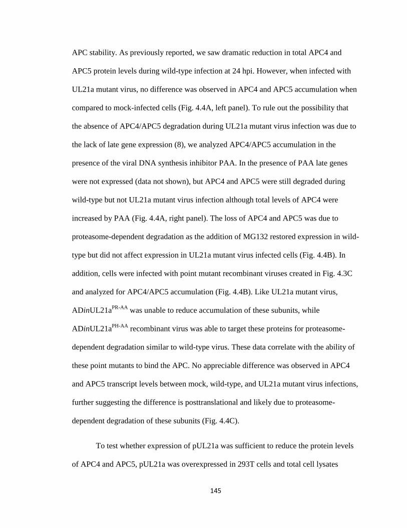
145
APC stability. As previously reported, we saw dramatic reduction in total APC4 and
APC5 protein levels during wild-type infection at 24 hpi. However, when infected with
UL21a mutant virus, no difference was observed in APC4 and APC5 accumulation when
compared to mock-infected cells (Fig. 4.4A, left panel). To rule out the possibility that
the absence of APC4/APC5 degradation during UL21a mutant virus infection was due to
the lack of late gene expression (8), we analyzed APC4/APC5 accumulation in the
presence of the viral DNA synthesis inhibitor PAA. In the presence of PAA late genes
were not expressed (data not shown), but APC4 and APC5 were still degraded during
wild-type but not UL21a mutant virus infection although total levels of APC4 were
increased by PAA (Fig. 4.4A, right panel). The loss of APC4 and APC5 was due to
proteasome-dependent degradation as the addition of MG132 restored expression in wild-
type but did not affect expression in UL21a mutant virus infected cells (Fig. 4.4B). In
addition, cells were infected with point mutant recombinant viruses created in Fig. 4.3C
and analyzed for APC4/APC5 accumulation (Fig. 4.4B). Like UL21a mutant virus,
ADinUL21aPR-AA
was unable to reduce accumulation of these subunits, while
ADinUL21aPH-AA
recombinant virus was able to target these proteins for proteasome-
dependent degradation similar to wild-type virus. These data correlate with the ability of
these point mutants to bind the APC. No appreciable difference was observed in APC4
and APC5 transcript levels between mock, wild-type, and UL21a mutant virus infections,
further suggesting the difference is posttranslational and likely due to proteasome-
dependent degradation of these subunits (Fig. 4.4C).
To test whether expression of pUL21a was sufficient to reduce the protein levels
of APC4 and APC5, pUL21a was overexpressed in 293T cells and total cell lysates
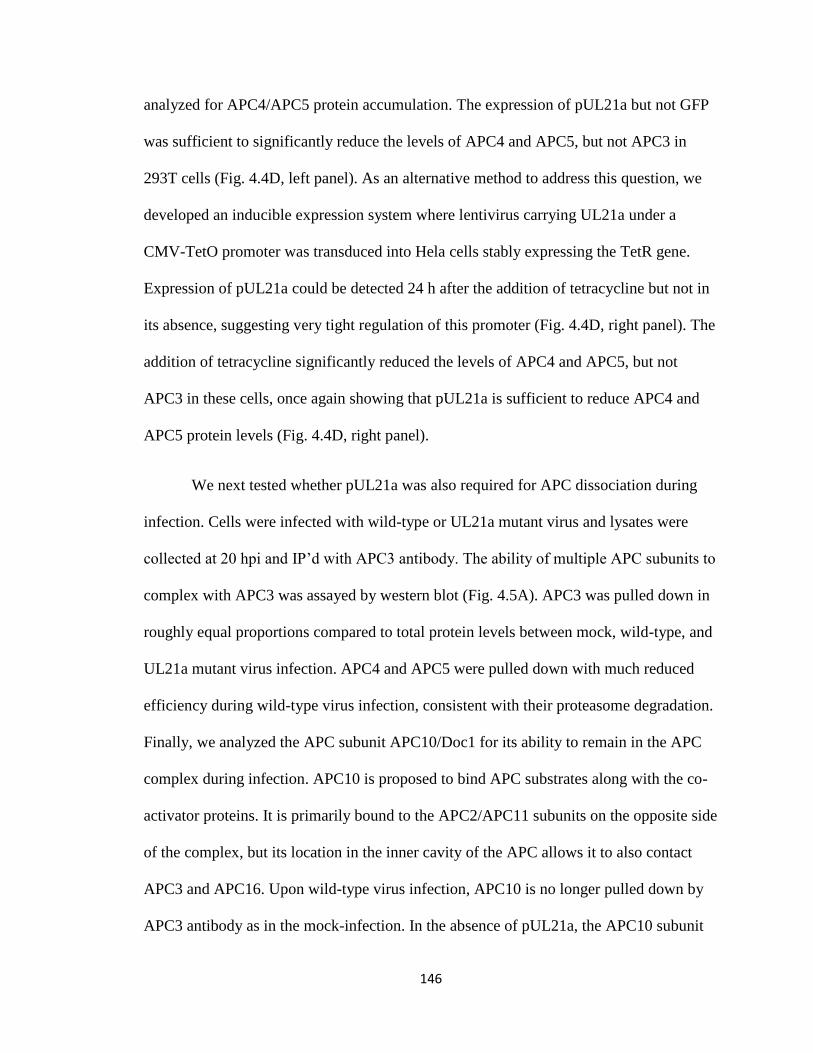
146
analyzed for APC4/APC5 protein accumulation. The expression of pUL21a but not GFP
was sufficient to significantly reduce the levels of APC4 and APC5, but not APC3 in
293T cells (Fig. 4.4D, left panel). As an alternative method to address this question, we
developed an inducible expression system where lentivirus carrying UL21a under a
CMV-TetO promoter was transduced into Hela cells stably expressing the TetR gene.
Expression of pUL21a could be detected 24 h after the addition of tetracycline but not in
its absence, suggesting very tight regulation of this promoter (Fig. 4.4D, right panel). The
addition of tetracycline significantly reduced the levels of APC4 and APC5, but not
APC3 in these cells, once again showing that pUL21a is sufficient to reduce APC4 and
APC5 protein levels (Fig. 4.4D, right panel).
We next tested whether pUL21a was also required for APC dissociation during
infection. Cells were infected with wild-type or UL21a mutant virus and lysates were
collected at 20 hpi and IP’d with APC3 antibody. The ability of multiple APC subunits to
complex with APC3 was assayed by western blot (Fig. 4.5A). APC3 was pulled down in
roughly equal proportions compared to total protein levels between mock, wild-type, and
UL21a mutant virus infection. APC4 and APC5 were pulled down with much reduced
efficiency during wild-type virus infection, consistent with their proteasome degradation.
Finally, we analyzed the APC subunit APC10/Doc1 for its ability to remain in the APC
complex during infection. APC10 is proposed to bind APC substrates along with the co-
activator proteins. It is primarily bound to the APC2/APC11 subunits on the opposite side
of the complex, but its location in the inner cavity of the APC allows it to also contact
APC3 and APC16. Upon wild-type virus infection, APC10 is no longer pulled down by
APC3 antibody as in the mock-infection. In the absence of pUL21a, the APC10 subunit

147
remains in the APC complex and is present upon pulldown with APC3 (Fig. 4.5A, right
panel). The integrity of the APC during wild-type virus infection is largely restored upon
addition of MG132, although total protein levels are reduced likely due to increased cell
death (Fig. 4.5A, right panel and data not shown). These data are recapitulated using
point mutant recombinant viruses ADinUL21aPR-AA
and ADinUL21aPH-AA
which act like
UL21a mutant and wild-type virus, respectively (Fig. 4.5B).
APC activity is regulated by pUL21a
To determine if pUL21a-dependent APC dissociation resulted in reduced APC
activity, we first analyzed the accumulation of multiple APC substrates during wild-type
and UL21a mutant virus infection (Fig. 4.6A). Both geminin and the APC co-activator
protein Cdh1 (which is also a substrate of the APC) were dramatically stabilized by wild-
type virus infection compared to mock by at least 24 hpi, as previously described. The
accumulation of these proteins was significantly reduced during UL21a mutant virus
infection, suggesting increased APC activity. This phenotype was maintained and even
increased throughout the infection. There was no difference in geminin transcript
accumulation between wild-type and mutant virus, suggesting these differences in protein
levels are not due to transcript regulation (Fig. 4.6B). Thymidine kinase (TK) was also
consistently reduced during UL21a mutant virus infection, but this was not apparent until
later times of infection (48-72 hpi). Interestingly, some substrates such as cyclin B1, were
not reduced and in some experiments even increased during UL21a mutant virus
infection. It is possible that the level of APC inhibition by UL97-mediated Cdh1
phosphorylation is sufficient for certain APC substrates. Alternatively the cyclin B1
transcript may be mis-regulated during UL21a mutant virus infection. The addition of
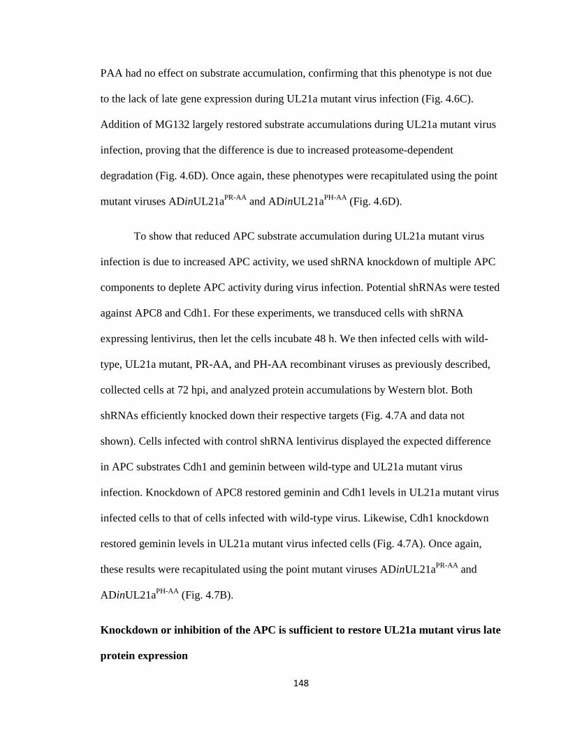
148
PAA had no effect on substrate accumulation, confirming that this phenotype is not due
to the lack of late gene expression during UL21a mutant virus infection (Fig. 4.6C).
Addition of MG132 largely restored substrate accumulations during UL21a mutant virus
infection, proving that the difference is due to increased proteasome-dependent
degradation (Fig. 4.6D). Once again, these phenotypes were recapitulated using the point
mutant viruses ADinUL21aPR-AA
and ADinUL21aPH-AA
(Fig. 4.6D).
To show that reduced APC substrate accumulation during UL21a mutant virus
infection is due to increased APC activity, we used shRNA knockdown of multiple APC
components to deplete APC activity during virus infection. Potential shRNAs were tested
against APC8 and Cdh1. For these experiments, we transduced cells with shRNA
expressing lentivirus, then let the cells incubate 48 h. We then infected cells with wild-
type, UL21a mutant, PR-AA, and PH-AA recombinant viruses as previously described,
collected cells at 72 hpi, and analyzed protein accumulations by Western blot. Both
shRNAs efficiently knocked down their respective targets (Fig. 4.7A and data not
shown). Cells infected with control shRNA lentivirus displayed the expected difference
in APC substrates Cdh1 and geminin between wild-type and UL21a mutant virus
infection. Knockdown of APC8 restored geminin and Cdh1 levels in UL21a mutant virus
infected cells to that of cells infected with wild-type virus. Likewise, Cdh1 knockdown
restored geminin levels in UL21a mutant virus infected cells (Fig. 4.7A). Once again,
these results were recapitulated using the point mutant viruses ADinUL21aPR-AA
and
ADinUL21aPH-AA
(Fig. 4.7B).
Knockdown or inhibition of the APC is sufficient to restore UL21a mutant virus late
protein expression
149
To determine whether APC inhibition by UL21a is beneficial for viral replication,
we used both proteasome inhibition and shRNA knockdown of APC components and
determined if it could restore viral late gene expression in UL21a mutant virus. We
previously showed that the UL21a mutant virus is defective in viral DNA synthesis and
the late expression of both IE2-86 and pp28, so we used these proteins as markers for
viral replication (8). Inhibition of the proteasome with MG132 even for only 14 hours
(58-72 hpi) significantly restored expression of pp28 and IE2-86 (Fig. 4.8A).
Additionally, knockdown of Cdh1 and to a lesser extent APC8 was clearly able to
increase pp28 and IE2-86 expression (Fig. 4.8B). This data suggests that Cdh1 and likely
the APCCdh1
complex is limiting the ability of the UL21a mutant virus to replicate.
The failure to regulate the APC alone is not sufficient to negatively affect HCMV
growth in human fibroblasts.
To determine whether APC regulation was also necessary for efficient virus
growth, we performed growth curve analysis with an HCMV pUL21a APC binding
mutant, ADinUL21aPR-AA
. We have shown in multiple assays that this mutant is not only
unable to bind the APC, but is also defective in targeting APC4/APC5 for degradation
and in regulating the APC. However, in a multi-step growth curve, this mutant virus grew
with equivalent kinetics to ADwt and ADinUL21aPH-AA
recombinant viruses, while
ADinUL21astop
had a severe growth defect (Fig. 4.8C). This data suggests that the failure
to regulate the APC alone is not sufficient to negatively affect HCMV growth in human
fibroblasts, and that pUL21a likely has additional functions to promote growth of HCMV
in human fibroblasts.

150
DISCUSSION
In warfare, bridge destruction is a common way to limit the ability of an enemy to
obtain supplies, maintain communication, and mobilize. Here we describe a viral protein
whose function is to destroy an important molecular bridge, likely aiding the virus in its
attempt to overtake the host cell. We found that the HCMV protein pUL21a interacts
with the anaphase-promoting complex (APC), regulates it, and promotes its dissociation,
likely by targeting components of the APC bridge (APC4/APC5) for proteasome-
dependent degradation. In fact, when cells infected with wild-type virus were treated with
the proteasome inhibitor MG132, APC4 and APC5 were stabilized, and the APC
remained intact (Fig. 4.5). Under these conditions, pUL21a was clearly expressed,
suggesting that the lack of dissociation was not due to the lack of pUL21a expression.
Thus, the evidence strongly suggests that proteasome-dependent degradation of APC4
and APC5 causes the dissociation of the APC during wild-type virus infection.
How does pUL21a target these proteins for proteasome-dependent degradation? It
is unlikely that pUL21a itself would act as an E3 ligase as it contains no domain or motif
which could have E3 ligase activity. We hypothesize that pUL21a either directly binds to
APC4/APC5 and targets them to the proteasome directly, or it recruits a cellular E3 ligase
to directly ubiquitinate these proteins. To identify the mechanism, we are currently
working to identify the direct binding partner for pUL21a, and also determining whether
APC4 and APC5 are ubiquitinated during wild-type virus infection.
Several other viral proteins have been shown to regulate the APC using a variety
of mechanisms. Some, like HPV E2, appear to bind and inhibit the Cdh1 activator protein

151
(1), while the Orf virus protein PACR appears to function as an APC11 mimic, but
without the enzymatic activity of a cullin-RING ligase (19, 20). The viral protein whose
mechanism appears to be most closely related to pUL21a is the CAV protein apoptin,
which likely binds the APC at the bridge and causes its dissociation (29). However, it is
presently unclear whether apoptin targets APC subunits for proteasome-dependent
degradation or if it simply dissociates the complex by preventing the association of two
specific subunits within the bridge of the APC. The fact that two independent viral
proteins have devised similar mechanisms to disrupt the APC suggests that targeting the
bridge of the APC may be an efficient means to disable the activity of this complex.
Most viral proteins which regulate the APC have only been studied in
overexpression. Recently, the UL97 kinase of HCMV was shown to phosphorylate Cdh1
and partially inhibit the complex during infection (31). APC substrate accumulation was
slightly delayed during a UL97 mutant virus infection, however all substrates eventually
accumulated at wild-type levels. It is unknown whether the phosphorylation of Cdh1 by
UL97 has an effect on viral replication. Deletion of PACR, the parapoxvirus APC11
mimic, or apoptin from CAV, resulted in viruses with a substantial growth defect in
tissue culture (19, 24). However, it has not been clarified whether PACR or apoptin
inhibit the APC during infection. Furthermore, it is unknown whether the defects of these
mutant viruses are due to an increase in APC activity. Here, we showed that a defect in
late viral gene expression could be partially restored by targeted knockdown of the APC
(Fig.4.8B). While it is likely that the APC restricts viral gene expression by inhibiting
viral DNA synthesis as this is the specific defect of the UL21a mutant virus, we cannot
rule out the possibility that both pp28 and IE2-86 are substrates of the APC. Regardless,
152
our study is the first to show that knockdown of APC components can result in increased
viral gene expression, suggesting that the APC has antiviral activities for HCMV which
can be overcome by expression of pUL21a. Despite this, mutation of the APC binding
domain in pUL21a did not affect the ability of HCMV to replicate in tissue culture like a
complete deletion of pUL21a does. This data suggests that pUL21a must have an
additional function which is critical for replication of the virus (see below).
What would be the benefit for the virus to disable the APC? The APC targets >40
proteins for proteasome-dependent degradation, and any of these proteins could be
important for viral replication. Alternatively, the virus may inhibit the APC to allow entry
of the cell into a different stage of the cell cycle where activity of other proteins (i.e.
CDKs) may be crucial for viral replication. It is interesting to note that in both poxvirus
and herpesvirus families, the only viruses which have been found to modulate the APC
(Parapoxviruses and HCMV) are those that do not encode for their own thymidine kinase
(TK) and ribonucleotide reductase subunit M2 (RRM2) enzymes. Both of these enzymes
are critical for the production of deoxyribonucleotides. This correlates well with the fact
that the UL21a deletion virus has a large defect in viral DNA synthesis (8). It seems
plausible that APC regulation is one mechanism used by HCMV and other viruses to
accumulate a substantial concentration of nucleotides so they can efficiently replicate
their own genomes. This hypothesis has previously been proposed but has not been
experimentally verified to date (19, 37). However, the APC also targets proteins involved
in cellular DNA replication, glycolysis/glutaminolysis, and multiple kinases/cyclins, all
of which could impact viral replication (17). Also, viral proteins have been shown to be
targets of the APC (18), and several HCMV proteins contain a putative D-box,
153
suggesting the APC may be able to degrade viral proteins as well (31). Future work will
need to address which substrates of the APC may be important for viral replication.
Based on these results and observations, we propose a model whereby pUL21a
disrupts the APC in order to increase the accumulation of at least one APC substrate (Fig.
4.9). The increased substrate protein(s) will help the cell modulate its nucleotide
pool/energy level/kinase activity, which in turn enhances the ability of the virus to
synthesize its DNA and produce late viral proteins. However, based on the fact that a PR-
AA point mutant cannot regulate the APC but still grows with wild-type kinetics, it is
likely that pUL21a also has at least one additional function. This function may be
redundant with APC inhibition, based on the data showing that knockdown of Cdh1 or
treatment with MG132 significantly restored late viral gene expression during mutant
virus infection. In dividing cells, only one of these two functions may be necessary for
efficient viral DNA replication; however, perhaps in terminally differentiated cells such
as neurons, both functions are necessary for efficient viral DNA replication as nucleotide
or energy pools in these cells would be much lower. It will be of interest to test the ability
of UL21a mutant viruses to grow in these cell types.
Study of the mechanism pUL21a uses to disarm the APC may have broad impacts
in cancer and neuronal disease. Due to its essential role in cell cycle progression, the
APC is a novel anti-cancer target (12). Recently drugs have been developed targeting this
complex and may be useful therapeutics (38). In fact, our preliminary data shows that
overexpression of pUL21a can prevent transformed cell lines from growing (data not
shown). Thus, study of pUL21a may shed light on novel mechanisms to target the APC
154
for drug development, provide useful information on how the APC is assembled, and
give further impetus to target the APC for anti-cancer therapies.
Finally, several studies have recently highlighted the importance of APCCdh1
in
neuronal development (reviewed in (25)). Congenital HCMV disease leads to neuronal
defects and severe complications such as blindness, hearing loss, and mental retardation.
HCMV infects neuronal precursor cells, differentiated neurons, and astroglia, and
deregulation of neuronal cells during HCMV infection is likely linked to the ability of
this virus to cause neuronal defects. It is interesting to speculate that inhibition of the
APC by pUL21a may be one of the factors leading to neuronal disease in congenitally
infected infants. Thus, study of pUL21a and the regulation of the APC during HCMV
infection may uncover new insights into the molecular basis of HCMV pathogenesis.

155
ACKNOWLEDGEMENTS
We especially thank Scott Terhune and John Savaryn for the identification of
pUL21a interacting partners by mass spectrometry. This project would not be possible
without their contribution. We would also like to thank Herbert Virgin and the members
of his laboratory for helpful discussion and invaluable advice, Roger Everett for the
pLKO GFP-TetR and CMV-TetO plasmids, Thomas Shenk for the antibodies, the
Children’s Discovery Institute/Genome Center at Washington University for the shRNA
lentiviral vectors, and members of the Yu lab for critical reading of the manuscript.
This study was supported by a Public Health Service grant (CA-120768), and in
part by a Grant from the American Heart Association (09GRNT2290199) and a grant
from Children’s Discovery Institute at Washington University. D. Y. holds an
Investigators in the Pathogenesis of Infectious Disease award from the Burroughs
Wellcome Fund. A.R.F. was a Morse/Berg Fellow of the Department of Molecular
Microbiology, Washington University School of Medicine, and was supported by
Institutional Training Grant T32-AI007172.
156
FIGURE LEGENDS
Figure 4.1. pUL21a interacts with the APC. (A) Identification of pUL21a interacting
partners. MRC-5 cells were infected with ADgfp or ADgfp-UL21a at an MOI of 5,
collected at 48 hpi, and immunoprecipitated (IP’d) with GFP antibody. Eluted proteins
were run on an SDS-containing polyacrylamide gel and silver stained. The bands
indicated with an arrow were identified by mass spectrometry as APC3 (100 kDa) and
APC8 (65 kDa). (B-C) pUL21a immunoprecipitates the APC during infection. (B) MRC-
5 cells were infected as described in A and lysates were IP’d with both GFP and APC3
antibodies. Cell lysates and IP’d proteins were analyzed by immunoblotting with
indicated antibodies. GFP blots were cropped to save space but are from the same lane
and exposed film. (C) MRC-5 cells were infected with ADgfp or ADsubUL21a at an
input genome number equivalent to 3 TCID50 units of wild-type virus/cell (as described
in Materials and Methods) in the presence (+) or absence (-) of 10µM MG132. Cells were
collected at 24 hpi and lysates were IP’d with APC3 antibody. Cell lysates and IP’d
proteins were analyzed by immunoblotting with indicated antibodies. (D) pUL21a
interacts with the APC when overexpressed in 293T cells. 293T cells were transfected
with GFP-UL21a or GFP-UL21astop
expressing plasmids. The GFP-UL21astop
protein
contains 2 stop-codons at the N-terminus of UL21a and thus does not express pUL21a.
Cells were collected at 72 hours post-transfection (hpt) and cell lysates were IP’d as in B.
Cell lysates and IP’d proteins were analyzed by immunoblotting with indicated
antibodies. PCNA and pUL44 were used as cellular and viral negative controls,
respectively. * indicates non-specific cross-reacting bands.
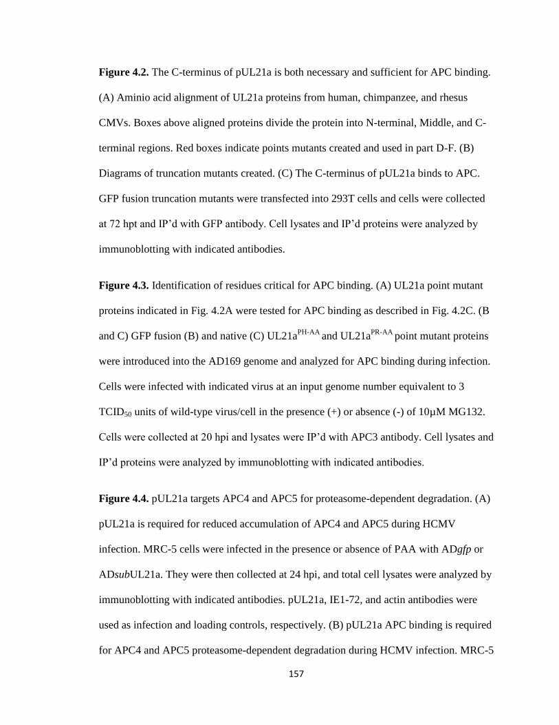
157
Figure 4.2. The C-terminus of pUL21a is both necessary and sufficient for APC binding.
(A) Aminio acid alignment of UL21a proteins from human, chimpanzee, and rhesus
CMVs. Boxes above aligned proteins divide the protein into N-terminal, Middle, and C-
terminal regions. Red boxes indicate points mutants created and used in part D-F. (B)
Diagrams of truncation mutants created. (C) The C-terminus of pUL21a binds to APC.
GFP fusion truncation mutants were transfected into 293T cells and cells were collected
at 72 hpt and IP’d with GFP antibody. Cell lysates and IP’d proteins were analyzed by
immunoblotting with indicated antibodies.
Figure 4.3. Identification of residues critical for APC binding. (A) UL21a point mutant
proteins indicated in Fig. 4.2A were tested for APC binding as described in Fig. 4.2C. (B
and C) GFP fusion (B) and native (C) UL21aPH-AA
and UL21aPR-AA
point mutant proteins
were introduced into the AD169 genome and analyzed for APC binding during infection.
Cells were infected with indicated virus at an input genome number equivalent to 3
TCID50 units of wild-type virus/cell in the presence (+) or absence (-) of 10µM MG132.
Cells were collected at 20 hpi and lysates were IP’d with APC3 antibody. Cell lysates and
IP’d proteins were analyzed by immunoblotting with indicated antibodies.
Figure 4.4. pUL21a targets APC4 and APC5 for proteasome-dependent degradation. (A)
pUL21a is required for reduced accumulation of APC4 and APC5 during HCMV
infection. MRC-5 cells were infected in the presence or absence of PAA with ADgfp or
ADsubUL21a. They were then collected at 24 hpi, and total cell lysates were analyzed by
immunoblotting with indicated antibodies. pUL21a, IE1-72, and actin antibodies were
used as infection and loading controls, respectively. (B) pUL21a APC binding is required
for APC4 and APC5 proteasome-dependent degradation during HCMV infection. MRC-5
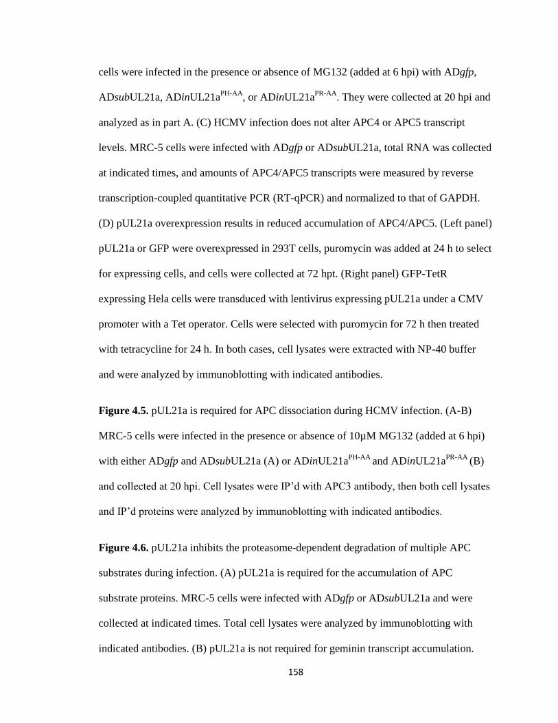
158
cells were infected in the presence or absence of MG132 (added at 6 hpi) with ADgfp,
ADsubUL21a, ADinUL21aPH-AA
, or ADinUL21aPR-AA
. They were collected at 20 hpi and
analyzed as in part A. (C) HCMV infection does not alter APC4 or APC5 transcript
levels. MRC-5 cells were infected with ADgfp or ADsubUL21a, total RNA was collected
at indicated times, and amounts of APC4/APC5 transcripts were measured by reverse
transcription-coupled quantitative PCR (RT-qPCR) and normalized to that of GAPDH.
(D) pUL21a overexpression results in reduced accumulation of APC4/APC5. (Left panel)
pUL21a or GFP were overexpressed in 293T cells, puromycin was added at 24 h to select
for expressing cells, and cells were collected at 72 hpt. (Right panel) GFP-TetR
expressing Hela cells were transduced with lentivirus expressing pUL21a under a CMV
promoter with a Tet operator. Cells were selected with puromycin for 72 h then treated
with tetracycline for 24 h. In both cases, cell lysates were extracted with NP-40 buffer
and were analyzed by immunoblotting with indicated antibodies.
Figure 4.5. pUL21a is required for APC dissociation during HCMV infection. (A-B)
MRC-5 cells were infected in the presence or absence of 10µM MG132 (added at 6 hpi)
with either ADgfp and ADsubUL21a (A) or ADinUL21aPH-AA
and ADinUL21aPR-AA
(B)
and collected at 20 hpi. Cell lysates were IP’d with APC3 antibody, then both cell lysates
and IP’d proteins were analyzed by immunoblotting with indicated antibodies.
Figure 4.6. pUL21a inhibits the proteasome-dependent degradation of multiple APC
substrates during infection. (A) pUL21a is required for the accumulation of APC
substrate proteins. MRC-5 cells were infected with ADgfp or ADsubUL21a and were
collected at indicated times. Total cell lysates were analyzed by immunoblotting with
indicated antibodies. (B) pUL21a is not required for geminin transcript accumulation.
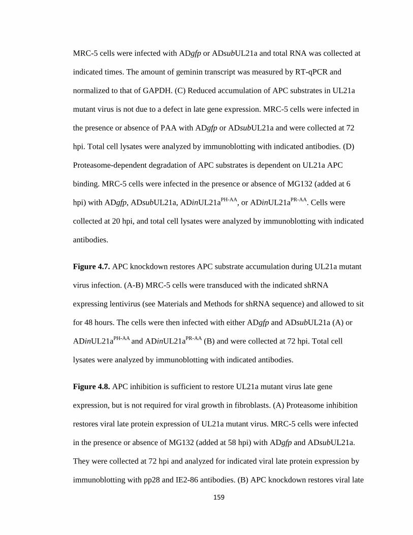
159
MRC-5 cells were infected with ADgfp or ADsubUL21a and total RNA was collected at
indicated times. The amount of geminin transcript was measured by RT-qPCR and
normalized to that of GAPDH. (C) Reduced accumulation of APC substrates in UL21a
mutant virus is not due to a defect in late gene expression. MRC-5 cells were infected in
the presence or absence of PAA with ADgfp or ADsubUL21a and were collected at 72
hpi. Total cell lysates were analyzed by immunoblotting with indicated antibodies. (D)
Proteasome-dependent degradation of APC substrates is dependent on UL21a APC
binding. MRC-5 cells were infected in the presence or absence of MG132 (added at 6
hpi) with ADgfp, ADsubUL21a, ADinUL21aPH-AA
, or ADinUL21aPR-AA
. Cells were
collected at 20 hpi, and total cell lysates were analyzed by immunoblotting with indicated
antibodies.
Figure 4.7. APC knockdown restores APC substrate accumulation during UL21a mutant
virus infection. (A-B) MRC-5 cells were transduced with the indicated shRNA
expressing lentivirus (see Materials and Methods for shRNA sequence) and allowed to sit
for 48 hours. The cells were then infected with either ADgfp and ADsubUL21a (A) or
ADinUL21aPH-AA
and ADinUL21aPR-AA
(B) and were collected at 72 hpi. Total cell
lysates were analyzed by immunoblotting with indicated antibodies.
Figure 4.8. APC inhibition is sufficient to restore UL21a mutant virus late gene
expression, but is not required for viral growth in fibroblasts. (A) Proteasome inhibition
restores viral late protein expression of UL21a mutant virus. MRC-5 cells were infected
in the presence or absence of MG132 (added at 58 hpi) with ADgfp and ADsubUL21a.
They were collected at 72 hpi and analyzed for indicated viral late protein expression by
immunoblotting with pp28 and IE2-86 antibodies. (B) APC knockdown restores viral late
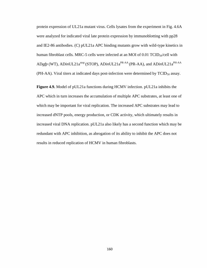
160
protein expression of UL21a mutant virus. Cells lysates from the experiment in Fig. 4.6A
were analyzed for indicated viral late protein expression by immunoblotting with pp28
and IE2-86 antibodies. (C) pUL21a APC binding mutants grow with wild-type kinetics in
human fibroblast cells. MRC-5 cells were infected at an MOI of 0.01 TCID50/cell with
ADgfp (WT), ADinUL21astop
(STOP), ADinUL21aPR-AA
(PR-AA), and ADinUL21aPH-AA
(PH-AA). Viral titers at indicated days post-infection were determined by TCID50 assay.
Figure 4.9. Model of pUL21a functions during HCMV infection. pUL21a inhibits the
APC which in turn increases the accumulation of multiple APC substrates, at least one of
which may be important for viral replication. The increased APC substrates may lead to
increased dNTP pools, energy production, or CDK activity, which ultimately results in
increased viral DNA replication. pUL21a also likely has a second function which may be
redundant with APC inhibition, as abrogation of its ability to inhibit the APC does not
results in reduced replication of HCMV in human fibroblasts.
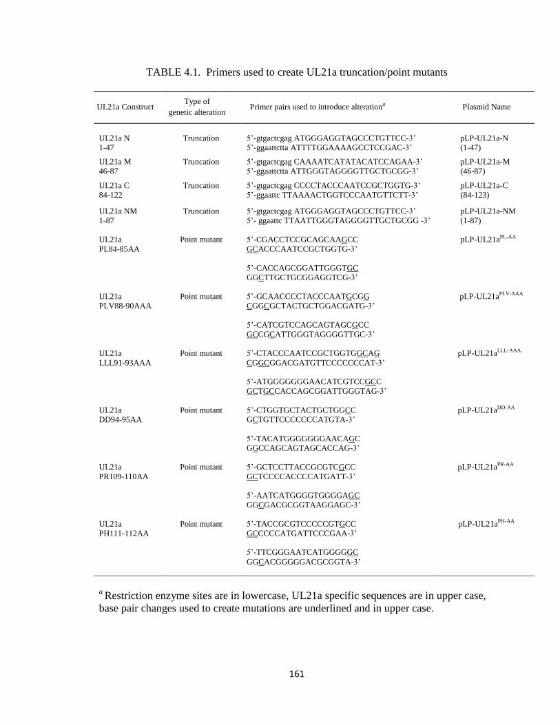
161
TABLE 4.1. Primers used to create UL21a truncation/point mutants
UL21a Construct Type of
genetic alteration Primer pairs used to introduce alterationa Plasmid Name
UL21a N
1-47
Truncation
5’-gtgactcgag ATGGGAGGTAGCCCTGTTCC-3’
5’-ggaattctta ATTTTGGAAAAGCCTCCGAC-3’
pLP-UL21a-N
(1-47)
UL21a M
46-87
Truncation 5’-gtgactcgag CAAAATCATATACATCCAGAA-3’
5’-ggaattctta ATTGGGTAGGGGTTGCTGCGG-3’
pLP-UL21a-M
(46-87)
UL21a C
84-122
Truncation 5’-gtgactcgag CCCCTACCCAATCCGCTGGTG-3’
5’-ggaattc TTAAAACTGGTCCCAATGTTCTT-3’
pLP-UL21a-C
(84-123)
UL21a NM
1-87
Truncation 5’-gtgactcgag ATGGGAGGTAGCCCTGTTCC-3’
5’- ggaattc TTAATTGGGTAGGGGTTGCTGCGG -3’
pLP-UL21a-NM
(1-87)
UL21a
PL84-85AA
UL21a
PLV88-90AAA
UL21a
LLL91-93AAA
UL21a
DD94-95AA
UL21a
PR109-110AA
UL21a
PH111-112AA
Point mutant
Point mutant
Point mutant
Point mutant
Point mutant
Point mutant
5’-CGACCTCCGCAGCAAGCC
GCACCCAATCCGCTGGTG-3’
5’-CACCAGCGGATTGGGTGC
GGCTTGCTGCGGAGGTCG-3’
5’-GCAACCCCTACCCAATGCGG
CGGCGCTACTGCTGGACGATG-3’
5’-CATCGTCCAGCAGTAGCGCC
GCCGCATTGGGTAGGGGTTGC-3’
5’-CTACCCAATCCGCTGGTGGCAG
CGGCGGACGATGTTCCCCCCCAT-3’
5’-ATGGGGGGGAACATCGTCCGCC
GCTGCCACCAGCGGATTGGGTAG-3’
5’-CTGGTGCTACTGCTGGCC
GCTGTTCCCCCCCATGTA-3’
5’-TACATGGGGGGGAACAGC
GGCCAGCAGTAGCACCAG-3’
5’-GCTCCTTACCGCGTCGCC
GCTCCCCACCCCATGATT-3’
5’-AATCATGGGGTGGGGAGC
GGCGACGCGGTAAGGAGC-3’
5’-TACCGCGTCCCCCGTGCC
GCCCCCATGATTCCCGAA-3’
5’-TTCGGGAATCATGGGGGC
GGCACGGGGGACGCGGTA-3’
pLP-UL21aPL-AA
pLP-UL21aPLV-AAA
pLP-UL21aLLL-AAA
pLP-UL21aDD-AA
pLP-UL21aPR-AA
pLP-UL21aPH-AA
a Restriction enzyme sites are in lowercase, UL21a specific sequences are in upper case,
base pair changes used to create mutations are underlined and in upper case.
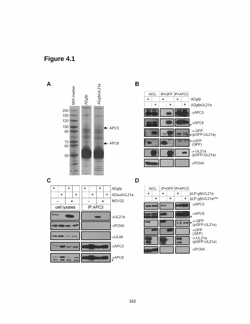
162
Figure 4.1
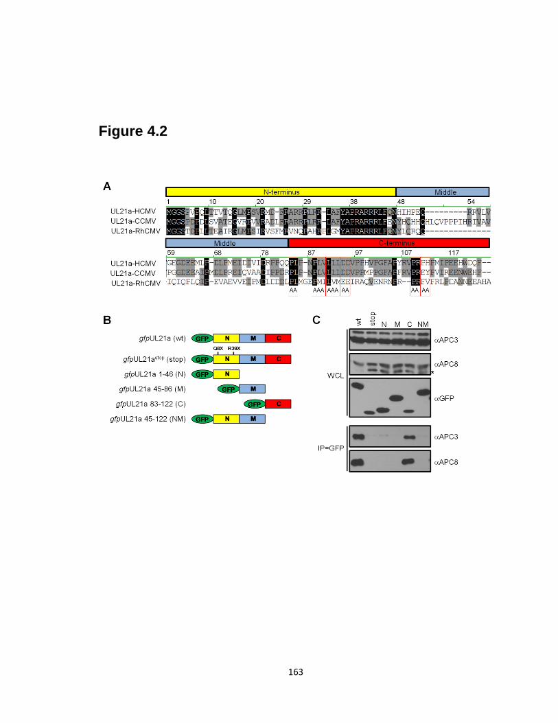
163
Figure 4.2
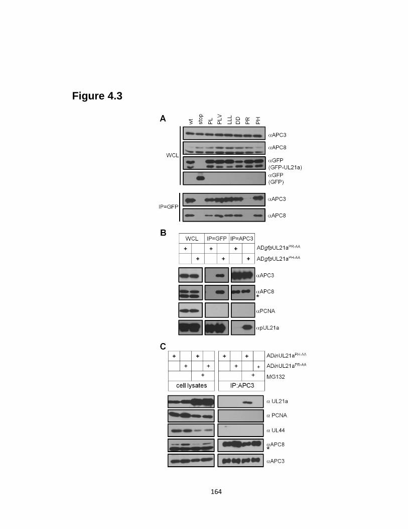
164
Figure 4.3
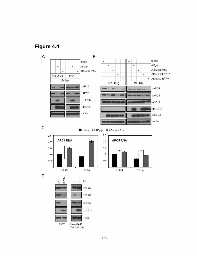
165
Figure 4.4
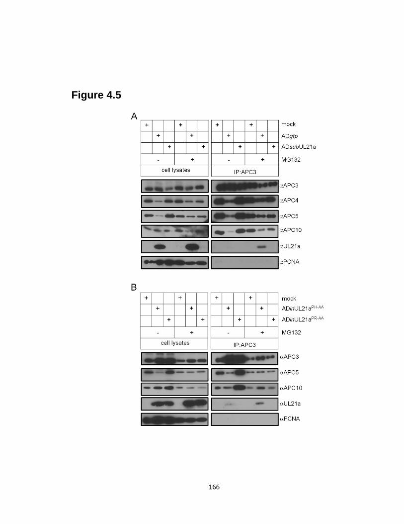
166
Figure 4.5
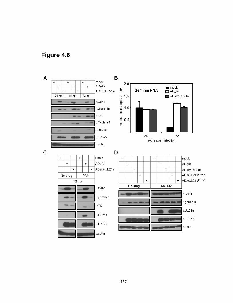
167
Figure 4.6
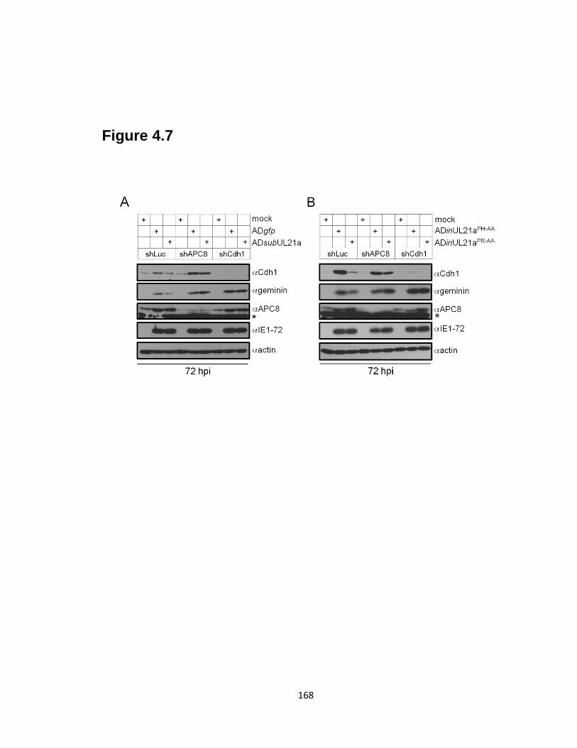
168
Figure 4.7
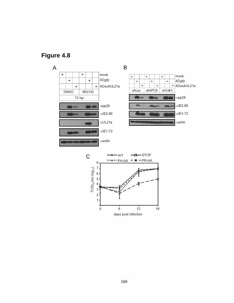
169
Figure 4.8
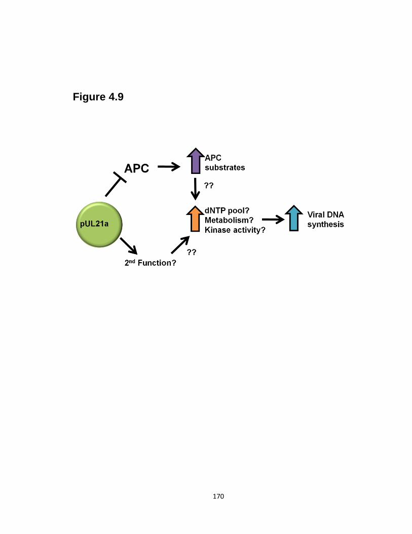
170
Figure 4.9
171
REFERENCES
1. Bellanger, S., S. Blachon, F. Mechali, C. Bonne-Andrea, and F. Thierry.
2005. High-risk but not low-risk HPV E2 proteins bind to the APC activators
Cdh1 and Cdc20 and cause genomic instability. Cell Cycle 4:1608-15.
2. Blanchette, P., and P. E. Branton. 2009. Manipulation of the ubiquitin-
proteasome pathway by small DNA tumor viruses. Virology 384:317-23.
3. Boutell, C., S. Sadis, and R. D. Everett. 2002. Herpes simplex virus type 1
immediate-early protein ICP0 and is isolated RING finger domain act as ubiquitin
E3 ligases in vitro. J Virol 76:841-50.
4. Casey, L., X. Wen, and C. M. de Noronha. 2010. The functions of the HIV1
protein Vpr and its action through the DCAF1.DDB1.Cullin4 ubiquitin ligase.
Cytokine 51:1-9.
5. Contin, R., F. Arnoldi, M. Mano, and O. R. Burrone. 2011. Rotavirus
replication requires a functional proteasome for effective assembly of viroplasms.
J Virol 85:2781-92.
6. Didcock, L., D. F. Young, S. Goodbourn, and R. E. Randall. 1999. The V
protein of simian virus 5 inhibits interferon signalling by targeting STAT1 for
proteasome-mediated degradation. J Virol 73:9928-33.
7. Everett, R. D., C. Boutell, C. McNair, L. Grant, and A. Orr. 2010.
Comparison of the biological and biochemical activities of several members of
the alphaherpesvirus ICP0 family of proteins. J Virol 84:3476-87.
8. Fehr, A. R., and D. Yu. 2011. Human cytomegalovirus early protein pUL21a
promotes efficient viral DNA synthesis and the late accumulation of immediate-
early transcripts. J Virol 85:663-74.
9. Fehr, A. R., and D. Yu. 2010. Human cytomegalovirus gene UL21a encodes a
short-lived cytoplasmic protein and facilitates virus replication in fibroblasts. J
Virol 84:291-302.
10. Fernandez-Garcia, M. D., L. Meertens, M. Bonazzi, P. Cossart, F. Arenzana-
Seisdedos, and A. Amara. 2011. Appraising the roles of CBLL1 and the
ubiquitin/proteasome system for flavivirus entry and replication. J Virol 85:2980-
9.
11. Gack, M. U., R. A. Albrecht, T. Urano, K. S. Inn, I. C. Huang, E. Carnero,
M. Farzan, S. Inoue, J. U. Jung, and A. Garcia-Sastre. 2009. Influenza A virus
NS1 targets the ubiquitin ligase TRIM25 to evade recognition by the host viral
RNA sensor RIG-I. Cell Host Microbe 5:439-49.
12. Heilman, D. W., M. R. Green, and J. G. Teodoro. 2005. The anaphase
promoting complex: a critical target for viral proteins and anti-cancer drugs. Cell
Cycle 4:560-3.
13. Hume, A. J., J. S. Finkel, J. P. Kamil, D. M. Coen, M. R. Culbertson, and R.
F. Kalejta. 2008. Phosphorylation of retinoblastoma protein by viral protein with
cyclin-dependent kinase function. Science 320:797-9.
14. Kalejta, R. F., J. T. Bechtel, and T. Shenk. 2003. Human cytomegalovirus pp71
stimulates cell cycle progression by inducing the proteasome-dependent
degradation of the retinoblastoma family of tumor suppressors. Mol Cell Biol
23:1885-95.
172
15. Kornitzer, D., R. Sharf, and T. Kleinberger. 2001. Adenovirus E4orf4 protein
induces PP2A-dependent growth arrest in Saccharomyces cerevisiae and interacts
with the anaphase-promoting complex/cyclosome. J Cell Biol 154:331-44.
16. Liu, B., S. Hong, Z. Tang, H. Yu, and C. Z. Giam. 2005. HTLV-I Tax directly
binds the Cdc20-associated anaphase-promoting complex and activates it ahead of
schedule. Proc Natl Acad Sci U S A 102:63-8.
17. Manchado, E., M. Eguren, and M. Malumbres. 2010. The anaphase-promoting
complex/cyclosome (APC/C): cell-cycle-dependent and -independent functions.
Biochem Soc Trans 38:65-71.
18. Mechali, F., C. Y. Hsu, A. Castro, T. Lorca, and C. Bonne-Andrea. 2004.
Bovine papillomavirus replicative helicase E1 is a target of the ubiquitin ligase
APC. J Virol 78:2615-9.
19. Mo, M., S. B. Fleming, and A. A. Mercer. 2009. Cell cycle deregulation by a
poxvirus partial mimic of anaphase-promoting complex subunit 11. Proc Natl
Acad Sci U S A 106:19527-32.
20. Mo, M., S. B. Fleming, and A. A. Mercer. 2010. Orf virus cell cycle regulator,
PACR, competes with subunit 11 of the anaphase promoting complex for
incorporation into the complex. J Gen Virol 91:3010-5.
21. Mui, M. Z., D. E. Roopchand, M. S. Gentry, R. L. Hallberg, J. Vogel, and P.
E. Branton. 2010. Adenovirus protein E4orf4 induces premature APCCdc20
activation in Saccharomyces cerevisiae by a protein phosphatase 2A-dependent
mechanism. J Virol 84:4798-809.
22. Niewiadomska, A. M., and X. F. Yu. 2009. Host restriction of HIV-1 by
APOBEC3 and viral evasion through Vif. Curr Top Microbiol Immunol 339:1-25.
23. Peters, J. M. 2006. The anaphase promoting complex/cyclosome: a machine
designed to destroy. Nat Rev Mol Cell Biol 7:644-56.
24. Prasetyo, A. A., T. Kamahora, A. Kuroishi, K. Murakami, and S. Hino. 2009.
Replication of chicken anemia virus (CAV) requires apoptin and is complemented
by VP3 of human torque teno virus (TTV). Virology 385:85-92.
25. Puram, S. V., and A. Bonni. 2011. Novel functions for the anaphase-promoting
complex in neurobiology. Semin Cell Dev Biol.
26. Qian, Z., V. Leung-Pineda, B. Xuan, H. Piwnica-Worms, and D. Yu. 2010.
Human cytomegalovirus protein pUL117 targets the mini-chromosome
maintenance complex and suppresses the cellular DNA synthesis. PLoS
Pathogens 6:e1000814.
27. Raaben, M., C. C. Posthuma, M. H. Verheije, E. G. te Lintelo, M. Kikkert, J.
W. Drijfhout, E. J. Snijder, P. J. Rottier, and C. A. de Haan. 2010. The
ubiquitin-proteasome system plays an important role during various stages of the
coronavirus infection cycle. J Virol 84:7869-79.
28. Reitsma, J. M., J. P. Savaryn, K. Faust, H. Sato, B. D. Halligan, and S. S.
Terhune. 2011. Antiviral inhibition targeting the HCMV kinase pUL97 requires
pUL27-dependent degradation of Tip60 acetyltransferase and cell-cycle arrest.
Cell Host Microbe 9:103-14.
29. Teodoro, J. G., D. W. Heilman, A. E. Parker, and M. R. Green. 2004. The
viral protein Apoptin associates with the anaphase-promoting complex to induce
G2/M arrest and apoptosis in the absence of p53. Genes Dev 18:1952-7.
173
30. Terhune, S., E. Torigoi, N. Moorman, M. Silva, Z. Qian, T. Shenk, and D.
Yu. 2007. Human cytomegalovirus UL38 protein blocks apoptosis. J Virol
81:3109-23.
31. Tran, K., J. P. Kamil, D. M. Coen, and D. H. Spector. 2010. Inactivation and
disassembly of the anaphase-promoting complex during human cytomegalovirus
infection is associated with degradation of the APC5 and APC4 subunits and does
not require UL97-mediated phosphorylation of Cdh1. J Virol 84:10832-43.
32. Tran, K., J. A. Mahr, J. Choi, J. G. Teodoro, M. R. Green, and D. H.
Spector. 2008. Accumulation of substrates of the anaphase-promoting complex
(APC) during human cytomegalovirus infection is associated with the
phosphorylation of Cdh1 and the dissociation and relocalization of APC subunits.
J Virol 82:529-37.
33. Tran, K., J. A. Mahr, and D. H. Spector. 2010. Proteasome subunits relocalize
during human cytomegalovirus infection, and proteasome activity is necessary for
efficient viral gene transcription. J Virol 84:3079-93.
34. Wang, X., R. A. Herr, and T. Hansen. 2008. Viral and cellular MARCH
ubiquitin ligases and cancer. Semin Cancer Biol 18:441-50.
35. Wang, Y. E., A. Park, M. Lake, M. Pentecost, B. Torres, T. E. Yun, M. C.
Wolf, M. R. Holbrook, A. N. Freiberg, and B. Lee. 2010. Ubiquitin-regulated
nuclear-cytoplasmic trafficking of the Nipah virus matrix protein is important for
viral budding. PLoS Pathog 6:e1001186.
36. Welcker, M., and B. E. Clurman. 2005. The SV40 large T antigen contains a
decoy phosphodegron that mediates its interactions with Fbw7/hCdc4. J Biol
Chem 280:7654-8.
37. Wiebusch, L., M. Bach, R. Uecker, and C. Hagemeier. 2005. Human
cytomegalovirus inactivates the G0/G1-APC/C ubiquitin ligase by Cdh1
dissociation. Cell Cycle 4:1435-9.
38. Zeng, X., F. Sigoillot, S. Gaur, S. Choi, K. L. Pfaff, D. C. Oh, N. Hathaway,
N. Dimova, G. D. Cuny, and R. W. King. 2010. Pharmacologic inhibition of the
anaphase-promoting complex induces a spindle checkpoint-dependent mitotic
arrest in the absence of spindle damage. Cancer Cell 18:382-95.
174
Chapter V
Summary and Future Directions
175
Summary
Two large-scale mutagenesis screens identified the previously uncharacterized
HCMV gene UL21a as being important but not essential for virus replication (4, 26).
However, no experimental evidence had been documented as to whether HCMV
expresses UL21a. Here, we began the study of UL21a and extensively characterized its
transcript and protein products. We found that UL21a encodes an early transcript and an
~15 kDa protein product (6). The UL21a protein, termed pUL21a, was found to be
degraded in a proteasome-dependent, but ubiquitin-independent manner (6). It localized
to the cytoplasm, but was not found in the HCMV virion (6). Using multiple HCMV
recombinant viruses and a complementing cell line, we further confirmed that pUL21a,
and not the neighboring genes or other gene products within UL21a, was important for
viral replication (5, 6). The UL21a mutant virus possessed a cell-type specific defect, as
the mutant had as much as a 1000-fold defect on MRC-5 cells whereas the defect never
got higher than 30-fold on HFFs (5, 6). The UL21a mutant virus had a high particle-to-
PFU ratio, and when normalized to equal viral genome copies, it had a dramatic defect in
viral DNA synthesis, but not early gene expression (5). Then, using a complementing
cell line, we showed that it was de novo synthesis of pUL21a and not an indirect effect on
the virion which caused this defect in DNA synthesis (5).
The current dogma in HCMV suggests that IE genes are expressed early and
remain at similar levels throughout the infection cycle. However, we found that most, if
not all, IE genes are regulated by viral DNA synthesis (5). There were some indications
in the literature in the late 80’s and early 90’s that this was the case, as researchers found
that the accumulation of the IE2-86 transcript and protein was inhibited by
176
phosphonoacetic acid (PAA) (20, 21). However, this data has largely been forgotten
(14). Our study was the first to extensively analyze multiple IE transcripts in the
presence or absence of PAA. All transcripts analyzed, including IE1-72, IE2-86,
UL37x1, and TRS1, were inhibited by PAA at least 2-fold (5). Furthermore, the protein
products of all but IE1-72 were profoundly affected as well. The lack of a strong effect
on IE1-72 protein expression is likely why this regulation has gone unnoticed as most
researchers use IE1-72 protein expression as a marker for IE gene expression.
UL21a contains no identifiable domains or significant homology with any known
proteins. Even though we had extensively characterized the UL21a gene, there was no
indication to its actual function(s). To identify potential functions for this protein we
took a proteomics approach to identify its interacting partners. We found that pUL21a
interacts with the anaphase-promoting complex (APC), a large E3 ubiquitin-ligase
involved primarily in cell-cycle progression. We further mapped the APC binding
domain to the C-terminus of pUL21a and identified two amino acids (PR108-109) which
were critical for this interaction. HCMV inhibits the APC by both phosphorylating the
co-activator Cdh1, and by targeting two subunits, APC4 and APC5, for proteasome-
dependent degradation (22, 23). The UL97 kinase was shown to phosphorylate Cdh1, but
the gene targeting APC4 and APC5 for degradation remained unknown (22). Here we
showed that pUL21a was both necessary and sufficient for APC4 and APC5 degradation
and was required for APC complex dissociation during infection. The degradation of
APC4 and APC5 was likely the cause of the dissociation, as proteasome inhibition could
restore the integrity of the APC. The degradation of APC4 and APC5 was dependent on
the ability of pUL21a to bind to the APC, as the PR108-109AA point mutant could not
177
induce their degradation and acted in all assays like a UL21a deletion virus. In the
absence of UL21a, increased APC activity could be detected by the increased degradation
of its substrates. The level of APC substrate accumulation during UL21a mutant virus
infection could be restored to wild-type levels by shRNA knockdown of APC
components, showing that decreased accumulation of these substrates was due to APC
activity. Most importantly, we showed that knockdown of APC8 and the co-activator
Cdh1 and proteasome inhibition could at least partially restore late viral gene expression
during UL21a mutant virus infection. This indicates for the first time that Cdh1, and
likely APCCdh1
, may have anti-viral functions.
Implications
The work described here has many implications for HCMV and virology research.
We are the first to identify and characterize the UL21a ORF, and have identified its role
during the viral lifecycle. We showed that the protein is degraded in a proteasome-
dependent, but ubiquitin-independent manner. Ubiquitin-independent degradation is
somewhat rare but has been reported for a few viral proteins, including the parvovirus
minute virus NS2 protein (11), the HCV F protein (27), and the MCMV m140 and m141
proteins (1). Interestingly, pp71 targets the cellular proteins Rb and Daxx for
proteasome-dependent ubiquitin-independent degradation (7, 8). It is unknown why the
virus appears to favor ubiquitin-independent mechanisms for proteasome degradation, but
it could be because the viral encodes its own deubiquitinase which may reduce the
effectiveness of ubiquitination (24).

178
We found that UL21a was required for viral DNA synthesis, which was not
unexpected as most early genes are important for viral DNA synthesis. However, the
additional finding that IE genes are regulated by viral DNA synthesis was intriguing as
the dogma in herpesvirus biology is that IE genes are expressed at high levels early and
stay elevated throughout infection. However, our study was not the first to identify this
type of regulation. Two previous studies had shown that IE2-86 protein and transcript
were inhibited by PAA (20, 21), and others have shown that the MCMV IE-1 and HSV-1
IE expression in trigeminal ganglion can be inhibited by PAA (9, 15, 19). Currently, it is
unknown whether the late accumulation of IE proteins, such as IE2-86, has any function
in promoting replication of the virus. Our lab has recently identified a novel late gene
regulator, UL79, in HCMV (17). In the absence of UL79, viral DNA replication and
IE2-86 production are normal, but no late genes are expressed. Perhaps IE2-86 or other
IE genes are increased at late times in order to activate expression of UL79 and other late
gene regulators, ultimately promoting viral replication. Clearly, further experiments are
needed to clarify this issue.
The most significant work of this thesis is the identification of pUL21a as a novel
regulator of the APC. We have clearly shown that pUL21a is both necessary and
sufficient for APC4 and APC5 proteasome-dependent degradation. During HCMV
infection, this likely leads to the dissociation of the complex, as APC4, APC5, and
APC10 were not pulled down with an APC3 antibody during wild-type infection as
efficiently as during mock or UL21a mutant virus infection. Proteasome inhibitors
restored the integrity of the complex without dramatically affecting pUL21a expression,
which strongly implicates APC4 and APC5 degradation as the cause of the dissociation.
179
However, this cannot be formally proven without the ability to overexpress non-
degradable forms of APC4 and APC5, which may prove difficult.
pUL21a is only one of a handful of viral proteins which have been shown to
regulate the APC (13). Only pUL21a and the UL97 protein kinase of HCMV have been
shown to inhibit this complex during virus infection. The fact that two viral proteins are
needed to inhibit this complex during HCMV infection strongly implies that the complex
is detrimental to virus replication. Knockdown of APC8 and especially Cdh1, was able
to restore some late viral protein expression in a UL21a mutant virus. This is further
supported by the observation that the proteasome inhibitor MG132 also significantly
restored late viral protein expression in a UL21a mutant virus. This data shows for the
first time that Cdh1, and likely the entire APC complex, may act in an antiviral manner
during HCMV infection. Further work will be needed to clarify the role of the APC
during infection in fibroblasts and other cell types infected by HCMV.
Finally, it is clear that pUL21a must have additional functions other than to inhibit
the APC. A pUL21a point mutant (PR108-109AA) which was unable to bind and
regulate the APC grew with wild-type virus kinetics, unlike a complete deletion virus.
The fact that knockdown of multiple APC subunits and the use of proteasome inhibitors
significantly restored late gene expression in a UL21a mutant implies that its functions
may be redundant (See Fig. 4.9). Perhaps in fibroblasts only one of these two functions
is sufficient to promote efficient replication, but in a terminally differentiated cell, such
as a neuron, both functions may be needed. It is currently unknown what this additional

180
function(s) may be, but pUL21a does have an Arginine Rich Motif (ARM) which in other
viral proteins has been shown to bind RNA (3).
Future Directions
There are many different future experiments which may follow this work. The
most pressing issues will be to clarify the role of the APC during virus infection of
multiple cell types, identify the APC substrates which may be important for viral
replication, determine the mechanism pUL21a uses to target APC4 and APC5 for
proteasome-dependent degradation, and continue identification of additional interacting
partners to discover novel functions for pUL21a.
I. Clarify the role of APC inhibition during HCMV infection
We have shown that knockdown of APC components restores some late viral
gene expression during UL21a mutant virus infection. However, this may not be the
most effective means to inhibit this complex due to residual expression of these proteins
and the possibility that they may be multi-functional. Recently, a novel pharmacologic
inhibitor of the APC was described (proTAME), and we have obtained this compound.
In the near future we will test whether this compound can restore viral DNA synthesis,
late gene expression, and virus production during UL21a mutant virus infection. We will
also create the UL21aPR-AA
point mutant virus in an HCMV clinical strain, TR, which will
allow us to identify cell types where pUL21a inhibition of the APC may be required for
viral replication. We will test the growth of this virus in multiple cell types, including
endothelial cells, epithelial cells, macrophages, and neuronal cells, then confirm that
wild-type pUL21a but not pUL21aPR-AA
can regulate the APC in these cell types.

181
Why does the virus inhibit the APC? The APC targets > 40 cellular proteins for
ubiquitination and subsequent proteasome degradation, thereby limiting their function. It
is likely that one or more of these proteins are important for viral replication.
Interestingly, two substrates of the APC are thymidine kinase (TK) and ribonucleotide
reductase M2 (RRM2), which are critical enzymes for nucleotide biosynthesis. All α-
and γ-herpesviruses encode their own versions of these enzymes while HCMV and other
β-herpesviruses do not (25). Thus HCMV must rely on these cellular enzymes for
nucleotide biosynthesis, and it is possible that APC inhibition is at least one mechanism it
uses to achieve this goal. In fact, a similar phenomenon has been described in poxviruses
(12). To test this hypothesis we plan on overexpressing TK and RRM2 from HSV-2
alone or in combination and testing whether one or both can restore viral DNA synthesis
in the UL21a mutant virus. We will use the HSV enzymes because they do not contain
an APC recognition domain and thus are not targeted for proteasome degradation. We
realize the APC targets multiple substrates for degradation, and many have the capability
to affect viral replication. If TK and RRM2 are not found to be important for viral
growth, we may systematically knockdown each known substrate of the APC and see
which ones are important for viral replication. One problem to this experiment is the
possibility that multiple APC substrates may be redundant in their functions, and thus
knocking down one may not produce a phenotype. Another issue is that not all APC
substrates have been described, and new ones are published nearly every month, so it is
likely we may leave out the critical regulators because they have not been discovered.
II. Determine the mechanism pUL21a uses to target APC4/APC5 for degradation
182
To fully understand how pUL21a inhibits the APC, it will be important to know
how pUL21a targets APC components for degradation. The first major step to answering
this question will be to identify the direct binding partner for pUL21a. We have shown
that pUL21a binds to the APC complex, but it unknown whether it binds directly to one
of the subunits or if it binds through an adapter protein. We hypothesize that pUL21a
either directly binds to APC4 and APC5 or to the directly adjacent subunits (APC8 and
APC1). We are currently setting up a yeast-2-hybrid (Y2H) assay to test whether these
proteins can bind directly to pUL21a. If neither of these proteins works, we will continue
to clone additional APC subunits into the Y2H vectors. Once we have identified a
candidate, we will create recombinant proteins and determine whether these proteins bind
in vitro.
The second part of this project will be to determine whether APC4 and APC5 are
ubiquitinated during infection. Previous published data suggested that E1 enzymes were
necessary for APC4/APC5 degradation; however this data is somewhat controversial, as
there was no way to determine whether the inhibitor was affecting the expression of
pUL21a (22). Regardless, it has not been directly shown that they are ubiquitinated. If
they are ubiquitinated, it would be important to identify the E3 ubiquitin ligase
responsible. It is highly unlikely that pUL21a could act as an E3 ubiquitin ligase since it
contains none of the sequence domains necessary for this enzymatic activity, thus it
would likely recruit a cellular E3 ligase. Genetic or biochemical experiments would be
used to identify this E3 ubiquitin ligase.
Alternatively, pUL21a may target these proteins directly as pUL21a is also
degraded in a proteasome-dependent manner. If pUL21a can directly target APC4/APC5
183
for degradation, we will need to define the biochemistry of this activity. The GFP-tagged
UL21a protein is able to bind the APC, but it cannot target these proteins for degradation,
suggesting that binding is not sufficient (data not shown). Targeted mutagenesis of
conserved residues within pUL21a should be able to identify additional sequence
important for this activity. In fact, we have recently created a mutant in the arginine rich
motif (ARM) of pUL21a which grows like a null virus, suggesting this region is clearly
important for one of its activities. It will be important to characterize this mutant and
determine whether it can regulate the APC. Alternatively, this region may be important
for other functions of this protein (see below).
III. Characterize the cell death inhibitory activity of pUL21a
In addition to the data in chapters 2-4, we have data which shows pUL21a is
required to prevent cell death during HCMV infection (Fig. 5.1). This cell death was
likely due to increased apoptosis as increased levels of cleaved caspase-3 and PARP
could be detected, which could be inhibited by caspase inhibitors (Fig. 5.2). It is
unknown whether the increased levels of apoptosis restrict viral growth. It has also not
yet been determined whether the increased apoptosis is caused by increased APC activity.
To test this, we can infect with our APC binding deficient mutant, ADinUL21aPR-AA
, and
test whether it causes increased cell death. Alternatively we could knockdown or inhibit
the APC and determine whether cell survival can be restored. If cell death is dependent
on APC activity, we could identify those substrates which can promote cell survival using
similar methods outlined in part I. If cell death is not dependent on APC activity, it will
be important to identify a second function for pUL21a and determine whether this
function promotes cell survival.
184
IV. Identify additional pUL21a function(s)
As mentioned previously, it is highly likely that pUL21a is a multi-functional
protein. To identify its additional function(s) we are continuing to use Co-IP to identify
additional binding partners of pUL21a. Also, as stated above, we have created a point
mutant with the ARM removed. This virus grows like UL21a deletion virus suggesting
this region is critical for pUL21a function(s). ARMs in other viral proteins have been
shown to mediate RNA binding, and in certain cases may help viruses escape RNA
sensors in the cytoplasm of its host cell. It may be of interest to test whether this region
confers RNA binding ability upon pUL21a and whether it is required to prevent a robust
interferon response.
V. Determine the requirement of Cdh1 for viral replication
Surprisingly, while knockdown of Cdh1 is capable of restoring viral late gene
expression to the UL21a mutant virus, it also has a strong negative effect on production
of infectious virus. In our preliminary studies, knockdown of Cdh1 reduces HCMV titers
in a single-step growth curve by as much as 2 logs, while knockdown of APC8 had a
much less dramatic effect (data not shown). This suggests that Cdh1 may have APC
independent function(s) which are critical for viral replication, but also could be due to a
different level of knockdown of each protein. It will be important to confirm these results
using additional methods (additional shRNAs, APC inhibitors) and to fully characterize
the ability of Cdh1 to inhibit viral replication. This data may have important
consequences both for HCMV and cell biology as Cdh1 has never been shown to have
APC independent functions.

185
Concluding Remarks
We have now extensively characterized the UL21a gene, described its role during
HCMV infection, and have identified at least one of its functions, to regulate the
anaphase-promoting complex. This work will provide significant new insight into the
mechanisms HCMV uses to facilitate its intra-cellular replication and may provide novel
targets for anti-viral therapies.
The APC has been shown to be critical for neuronal development (18), and
HCMV is largely known for its ability to cause neuronal defects in congenitally infected
infants. HCMV infects many types of neuronal cells in vitro, including differentiated
neurons, astrocytes, and neuronal precursor cells (2, 10, 16). It’s possible that the ability
of HCMV to inhibit the APC plays a role in the ability of the virus to infect neuronal cells
and cause neuronal defects. Further mechanistic study of pUL21a and its ability to
inhibit the APC may lead to novel insights into the molecular pathogenesis of HCMV.
The APC is a critical mediator of cell cycle progression, as inhibitors of the APC
prevent cells from progressing past the G2/M phase of the cell cycle. Further
mechanistic study should provide unique insights into the biology of the APC and may
lead us to new methods to target the APC for anti-cancer therapeutics. Over the last 50
years viruses have provided a number of unique tools which have allowed for
groundbreaking discoveries in cell biology, such as the discovery of prominent cell cycle
regulators such as p53 and Rb. Study of pUL21a will shed new light on cell biology, cell
cycle regulation, and will highlight additional ways HCMV has continued to evolve and
interact with its host in order to facilitate its own replication.
186
FIGURE LEGENDS
Figure 5.1. UL21a is required for cell survival during HCMV infection. (A-B) UL21a
mutant virus induces cell death. (A) MRC-5 cells were infected with ADwt or
ADsubUL21a at an equivalent MOI of 2 TCID50/cell (left two columns) or equivalent
genome copies (ADwt MOI equivalent to 10) (right two columns). Bright-field and
fluorescent microscopy was performed and pictures were taken with a Leica microscope
at 96 hpi. (B) MRC-5 cells were infected as in A and cell death was quantified using an
ATP assay (Promega) at the indicated times post-infection. (C) UL21a mutant virus
induced cell death requires viral gene expression but not DNA replication. MRC-5 cells
were infected as in B in the presence of PAA (100 µg/ml) or with UV-inactivated virus.
Cell death was quantified as in B at 120 hpi. (D) Expression of pGFP-UL21a can prevent
UL21a mutant virus induced cell death. MRC-5 cells were transduced 3X with pGFP-
UL21a expressing retrovirus, allowed to rest for 72 h, then infected with equivalent
genome copies as in part A and cell death was measured at indicated times post infection
using the ATP assay.
Figure 5.2. UL21a is required to prevent caspase activation during HCMV infection.
(A-B) UL21a mutant virus induces cleavage of caspase-3 and PARP which is dependent
on caspase activity. (A) MRC-5 cells were infected as in Fig. 5.1D and whole cell lysates
were collected at indicated times and analyzed by immunoblotting with indicated
antibodies. (B) MRC-5 cells were infected as in A and treated with DMSO or ZVAD-
FMK (10µM). Whole cell lysates were collected at indicated times and analyzed by
immunoblotting with indicated antibodies.
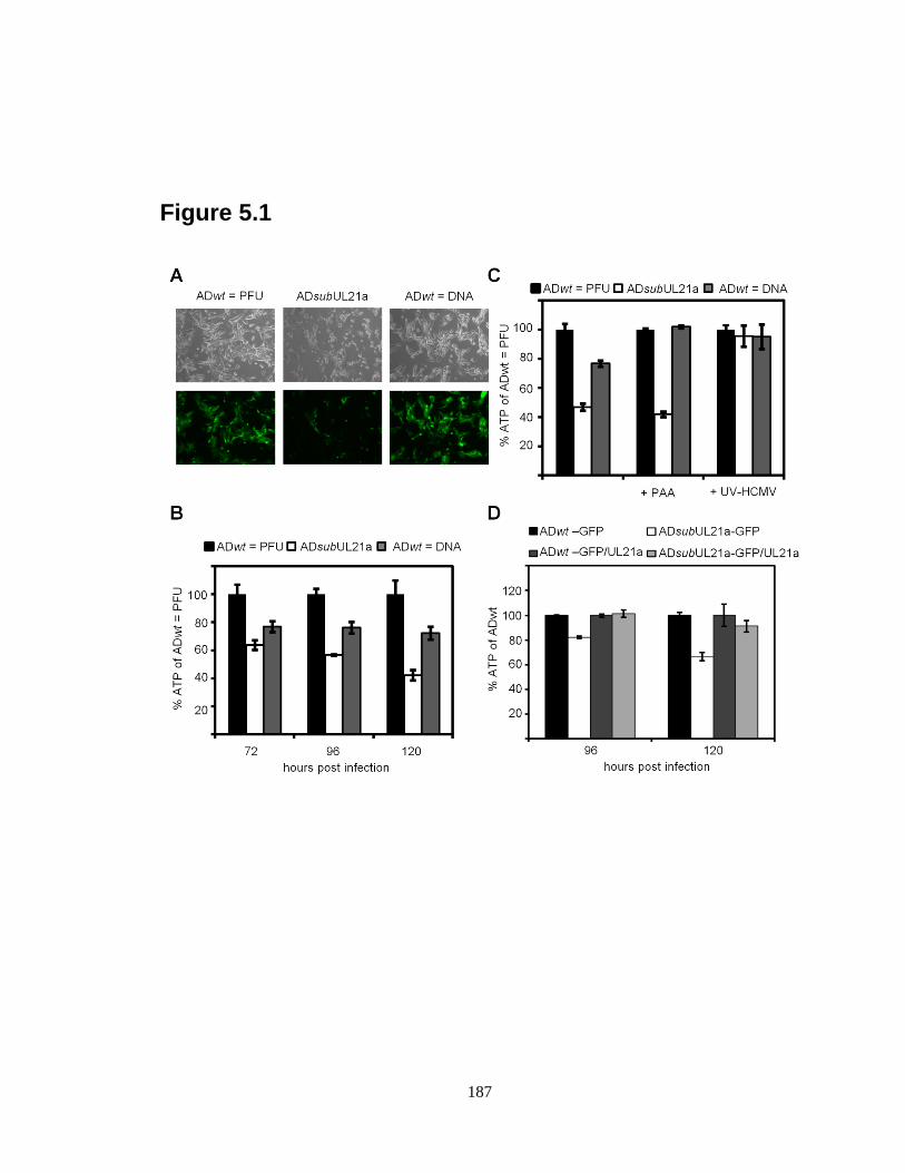
187
Figure 5.1

188
Figure 5.2
189
REFERENCES
1. Bolin, L. L., L. K. Hanson, J. S. Slater, J. A. Kerry, and A. E. Campbell.
2010. Murine cytomegalovirus US22 protein pM140 protects its binding partner,
pM141, from proteasome-dependent but ubiquitin-independent degradation. J
Virol 84:2164-8.
2. Cheeran, M. C., S. Hu, H. T. Ni, W. Sheng, J. M. Palmquist, P. K. Peterson,
and J. R. Lokensgard. 2005. Neural precursor cell susceptibility to human
cytomegalovirus diverges along glial or neuronal differentiation pathways. J
Neurosci Res 82:839-50.
3. Chien, C. Y., R. Tejero, Y. Huang, D. E. Zimmerman, C. B. Rios, R. M.
Krug, and G. T. Montelione. 1997. A novel RNA-binding motif in influenza A
virus non-structural protein 1. Nat Struct Biol 4:891-5.
4. Dunn, W., C. Chou, H. Li, R. Hai, D. Patterson, V. Stolc, H. Zhu, and F. Liu.
2003. Functional profiling of a human cytomegalovirus genome. Proc Natl Acad
Sci U S A 100:14223-8.
5. Fehr, A. R., and D. Yu. 2011. Human cytomegalovirus early protein pUL21a
promotes efficient viral DNA synthesis and the late accumulation of immediate-
early transcripts. J Virol 85:663-74.
6. Fehr, A. R., and D. Yu. 2010. Human cytomegalovirus gene UL21a encodes a
short-lived cytoplasmic protein and facilitates virus replication in fibroblasts. J
Virol 84:291-302.
7. Hwang, J., and R. F. Kalejta. 2007. Proteasome-dependent, ubiquitin-
independent degradation of Daxx by the viral pp71 protein in human
cytomegalovirus-infected cells. Virology 367:334-8.
8. Kalejta, R. F., and T. Shenk. 2003. Proteasome-dependent, ubiquitin-
independent degradation of the Rb family of tumor suppressors by the human
cytomegalovirus pp71 protein. Proc Natl Acad Sci U S A 100:3263-8.
9. Kosz-Vnenchak, M., J. Jacobson, D. M. Coen, and D. M. Knipe. 1993.
Evidence for a novel regulatory pathway for herpes simplex virus gene expression
in trigeminal ganglion neurons. J Virol 67:5383-93.
10. Luo, M. H., P. H. Schwartz, and E. A. Fortunato. 2008. Neonatal neural
progenitor cells and their neuronal and glial cell derivatives are fully permissive
for human cytomegalovirus infection. J Virol 82:9994-10007.
11. Miller, C. L., and D. J. Pintel. 2001. The NS2 protein generated by the
parvovirus minute virus of mice is degraded by the proteasome in a manner
independent of ubiquitin chain elongation or activation. Virology 285:346-55.
12. Mo, M., S. B. Fleming, and A. A. Mercer. 2009. Cell cycle deregulation by a
poxvirus partial mimic of anaphase-promoting complex subunit 11. Proc Natl
Acad Sci U S A 106:19527-32.
13. Mo, M., S. B. Fleming, and A. A. Mercer. 2010. Changing pace: viral mimicry
of an anaphase promoting complex subunit. Cell Cycle 9:632-3.
14. Mocarski, E. S., T. Shenk, and R. F. Pass (ed.). 2007. Cytomegaloviruses, 5th
ed, vol. 2. Lippincott Williams & Wilkins, Philadelphia.
15. Nichol, P. F., J. Y. Chang, E. M. Johnson, Jr., and P. D. Olivo. 1996. Herpes
simplex virus gene expression in neurons: viral DNA synthesis is a critical
190
regulatory event in the branch point between the lytic and latent pathways. J Virol
70:5476-86.
16. Odeberg, J., N. Wolmer, S. Falci, M. Westgren, A. Seiger, and C. Soderberg-
Naucler. 2006. Human cytomegalovirus inhibits neuronal differentiation and
induces apoptosis in human neural precursor cells. J Virol 80:8929-39.
17. Perng, Y. C., Z. Qian, A. R. Fehr, B. Xuan, and D. Yu. 2011. Human
cytomegalovirus gene UL79 is required for the accumulation of late viral
transcripts. J Virol.
18. Puram, S. V., and A. Bonni. 2011. Novel functions for the anaphase-promoting
complex in neurobiology. Semin Cell Dev Biol.
19. Reddehase, M. J., M. R. Fibi, G. M. Keil, and U. H. Koszinowski. 1986. Late-
phase expression of a murine cytomegalovirus immediate-early antigen
recognized by cytolytic T lymphocytes. J Virol 60:1125-9.
20. Stenberg, R. M., A. S. Depto, J. Fortney, and J. A. Nelson. 1989. Regulated
expression of early and late RNAs and proteins from the human cytomegalovirus
immediate-early gene region. J Virol 63:2699-708.
21. Tenney, D. J., and A. M. Colberg-Poley. 1991. Human cytomegalovirus UL36-
38 and US3 immediate-early genes: temporally regulated expression of nuclear,
cytoplasmic, and polysome-associated transcripts during infection. J Virol
65:6724-34.
22. Tran, K., J. P. Kamil, D. M. Coen, and D. H. Spector. 2010. Inactivation and
disassembly of the anaphase-promoting complex during human cytomegalovirus
infection is associated with degradation of the APC5 and APC4 subunits and does
not require UL97-mediated phosphorylation of Cdh1. J Virol 84:10832-43.
23. Tran, K., J. A. Mahr, J. Choi, J. G. Teodoro, M. R. Green, and D. H.
Spector. 2008. Accumulation of substrates of the anaphase-promoting complex
(APC) during human cytomegalovirus infection is associated with the
phosphorylation of Cdh1 and the dissociation and relocalization of APC subunits.
J Virol 82:529-37.
24. Wang, J., A. N. Loveland, L. M. Kattenhorn, H. L. Ploegh, and W. Gibson.
2006. High-molecular-weight protein (pUL48) of human cytomegalovirus is a
competent deubiquitinating protease: mutant viruses altered in its active-site
cysteine or histidine are viable. J Virol 80:6003-12.
25. Wiebusch, L., M. Bach, R. Uecker, and C. Hagemeier. 2005. Human
cytomegalovirus inactivates the G0/G1-APC/C ubiquitin ligase by Cdh1
dissociation. Cell Cycle 4:1435-9.
26. Yu, D., M. C. Silva, and T. Shenk. 2003. Functional map of human
cytomegalovirus AD169 defined by global mutational analysis. Proc Natl Acad
Sci U S A 100:12396-401.
27. Yuksek, K., W. L. Chen, D. Chien, and J. H. Ou. 2009. Ubiquitin-independent
degradation of hepatitis C virus F protein. J Virol 83:612-21.