Embed Size (px)
Citation preview

JOURNAL OF VIROLOGY, Mar. 2005, p. 2754–2767 Vol. 79, No. 50022-538X/05/$08.00�0 doi:10.1128/JVI.79.5.2754–2767.2005Copyright © 2005, American Society for Microbiology. All Rights Reserved.
Human Cytomegalovirus Labeled with Green Fluorescent Protein forLive Analysis of Intracellular Particle Movements
Kerstin Laib Sampaio,1 Yolaine Cavignac,1 York-Dieter Stierhof,2 and Christian Sinzger1*Institute of Medical Virology1 and Center for Plant Molecular Biology,2 University of Tubingen, Tubingen, Germany
Received 23 January 2004/Accepted 12 October 2004
Human cytomegalovirus (HCMV) replicates in the nuclei of infected cells. Successful replication thereforedepends on particle movements between the cell cortex and nucleus during entry and egress. To visualizeHCMV particles in living cells, we have generated a recombinant HCMV expressing enhanced green fluores-cent protein (EGFP) fused to the C terminus of the capsid-associated tegument protein pUL32 (pp150). Theresulting UL32-EGFP-HCMV was analyzed by immunofluorescence, electron microscopy, immunoblotting,confocal microscopy, and time-lapse microscopy to evaluate the growth properties of this virus and thedynamics of particle movements. UL32-EGFP-HCMV replicated similarly to wild-type virus in fibroblastcultures. Green fluorescent virus particles were released from infected cells. The fluorescence stayed associatedwith particles during viral entry, and fluorescent progeny particles appeared in the nucleus at 44 h afterinfection. Surprisingly, strict colocalization of pUL32 and the major capsid protein pUL86 within nuclearinclusions indicated that incorporation of pUL32 into nascent HCMV particles occurred simultaneously withor immediately after assembly of the capsid. A slow transport of nuclear particles towards the nuclear marginwas demonstrated. Within the cytoplasm, most particles performed irregular short-distance movements, whilea smaller fraction of particles performed centripetal and centrifugal long-distance movements. Althoughnumerous particles accumulated in the cytoplasm, release of particles from infected cells was a rare event,consistent with a release rate of about 1 infectious unit per h per cell in HCMV-infected fibroblasts ascalculated from single-step growth curves. UL32-EGFP-HCMV will be useful for further investigations into theentry, maturation, and release of this virus.
Like all herpesviruses, human cytomegalovirus (HCMV)replicates in the nucleus of the infected cell (18). This aspect ofthe herpesviral replication strategy entails the requirement forvarious particle movements during the replicative cycle, inparticular translocation of penetrated virus particles from thecell cortex towards the nucleus, egress of newly synthesizedvirus particles out of the nucleus, and translocation of tegu-mented and enveloped virus progeny towards the periphery ofthe cell in order to release infectious virus to surrounding cells(5, 14, 17, 19, 28). For HCMV, the importance of intracellulartransport of virus particles was underscored by the finding thatcell tropism variants of HCMV are discriminated by the strain-dependent efficiency of nuclear translocation (25). By nature,translocation is a dynamic process, which can be analyzed onlyinsufficiently by biochemical analyses of lysates of infected cellsor by immunodetection of particles in fixed-cell preparations.At least the dynamic aspects of interactions between virusparticles and target cells are best studied in live-imaging ap-proaches that have been enabled by the development of greenfluorescent virus variants. Recombinant fluorescent viruseshave been reported for herpes simplex virus and pseudorabiesvirus, the live imaging of which provided new insights into thenature of particle movements within relevant target cells (4, 6,12, 21, 22, 27). Green fluorescent cytomegalovirus has alsobeen reported (8, 10, 11, 16, 20, 23, 30); however, to date onlynonstructural proteins have been tagged, and therefore these
variants did not produce green fluorescent progeny virions.Live images of interactions between HCMV particles and theirtarget cells are therefore not available to date. In order toobtain fluorescence tags that stay associated with HCMV par-ticles during various intracellular translocation steps, the fluo-rescent protein has to be fused to viral capsid proteins orcapsid-associated tegument proteins. One tegument proteinknown to be strongly associated with the capsid is the phos-phoprotein pp150 (pUL32) (1, 2). Here we report the success-ful generation of a recombinant HCMV variant with enhancedgreen fluorescent protein (EGFP) fused to the C-terminal endof pUL32, resulting in green fluorescent virus particles. Possi-ble applications of this green fluorescent HCMV variant in-clude analyses of viral entry, nuclear translocation, nuclearegress after viral replication, dynamics of tegumentation, andviral egress. Examples of such applications are given in thisstudy.
MATERIALS AND METHODS
Cells and viruses. All experiments were carried out with human foreskinfibroblasts (HFF) isolated from foreskins of infants by trypsin treatment andwere used for experiments at passage 10 to 25. HFF were cultured in minimalessential medium (MEM) (Gibco, Karlsruhe, Germany) containing 5% fetal calfserum, 2.4 mmol of glutamine per liter, and 100 �g of gentamicin per ml(MEM5).
HCMV strain TB40 (26) was used for the generation of recombinant UL32-EGFP-HCMV. For the preparation of virus stocks, HFF were infected at amultiplicity of infection (MOI) (infectious units per cell) of 0.1. Supernatants ofinfected cultures were harvested at 6 days postinfection (p.i.) and stored at�80°C after removal of cell debris by centrifugation for 10 min at 2,800 � g. Theinfectious titer in HCMV preparations was determined by 50% tissue cultureinfective dose (TCID50) assays (15) in HFF on 96-well plates. For supernatant-mediated infection of cell cultures, the medium was replaced by infectious
* Corresponding author. Mailing address: Institut fur MedizinischeVirologie, Universitat Tubingen, Elfriede-Aulhorn-Strasse 6, D-72076Tubingen, Germany. Phone: 7071 2987459. Fax: 7071 295790. E-mail:[email protected].
2754
Dow
nloa
ded
from
http
s://j
ourn
als.
asm
.org
/jour
nal/j
vi o
n 13
Feb
ruar
y 20
22 b
y 59
.168
.65.
144.

supernatant at the indicated MOI, incubated for 2 h at 37°C, and finally replacedwith fresh medium (MEM5).
For single-step and multistep growth curves, HFF seeded in six-well plates (5� 105 cells per well) were infected with cell-free HCMV preparations at a virusconcentration of 2.5 � 105 TCID50s/ml (MOI � 1) or 2 � 104 TCID50s/ml (MOI� 0.08), respectively. After 2 h of incubation, the cultures were washed six timeswith medium to remove residual virus and were then cultured for 10 or 16 days,respectively. Starting at day 1 p.i., supernatant was removed daily from infectedcultures and replaced by fresh medium; the supernatant samples were centri-fuged at 2,800 � g for 10 min and stored at �80°C prior to determination of theinfectious titer by TCID50 assay.
For analysis of virus adsorption, cells were infected with virus preparations at4°C for the indicated time intervals.
The EGFP-tagged recombinant HCMV described in this publication will beavailable at the American Type Culture Collection as ATCC VR1578.
Construction of the recombination plasmid. Genomic DNA was preparedfrom late-stage infected cells (4 days p.i.) by phenol-chloroform extraction. Thecomplete HCMV-UL32 open reading frame, excluding the stop codon, wasamplified by PCR with the oligonucleotides 5�-CGGATCCTCCGTGTTCTTAATCTTCTCGA and 3�-AAGCTAGCATGAGTTTGCAGTTTATCGGTC, whichwere partial mismatch primers changing the last amino acid of pUL32 from Glu(E) to Asp (D) and introducing one BamHI site and one NheI site at the endsof the amplification product. The amplification product was cloned into the PCRcloning vector pDrive (Qiagen, Hilden, Germany). The BamHI/NheI fragmentcontaining the UL32 open reading frame was excised by using the restriction sitesintegrated into the primers (indicated by boldface) and was then cloned into theBamHI/NheI site of the EGFP-N1 vector (BD Clontech, Heidelberg, Germany).The resulting UL32-EGFP open reading frame contains five additional codonsbetween the last UL32 codon and the first EGFP codon. To obtain a recombi-nation plasmid, a 1.6-kbp fragment containing part of the UL31 open readingframe was amplified by PCR with the oligonucleotides 5�-AACTTAAGGAGGGGAGACGAGGACGACAGG and 3�-GGTCTAGAAACACACACGCAGACGTACTTT, which were partial mismatch primers introducing one AflII site andone XbaI site at the ends of the amplification product. The PCR products werecloned into the PCR cloning vector pDrive (Qiagen). AflII/XbaI fragmentscontaining part of the UL31 sequence were excised by using the restriction sitesintegrated into the primers (indicated by boldface), and the UL31 fragment wascloned into the AflII/XbaI site of the UL32-EGFP construct. This resulted in arecombination plasmid that was suitable for introduction of a pUL32-EGFPfusion gene at the orthotopic site of the HCMV genome by homologous recom-bination. This recombination plasmid was designated UL32-EGFP/UL31.
Generation and plaque purification of recombinant HCMV. The UL32-EGFPrecombination plasmid UL32-EGFP/UL31 was cleaved with NheI and AflII,ethanol precipitated, and dissolved in H2O. The cleaved construct was microin-jected into the nuclei of HCMV-TB40-infected HFF at 2 days p.i. by using amicromanipulation system (InjectMan and Femtojet; Eppendorf, Hamburg,Germany). At 4 days p.i. cells were cocultured with noninfected HFF at a ratioof 1/2,000 in 96-well plates and screened subsequently for EGFP-expressing fociof infected cells. Virus supernatant from wells containing green fluorescent fociwas plaque purified three times by limiting-dilution infections in 96-well plateswith centrifugal enhancement. To enrich for recombinant virus, wells that dis-played single standing green fluorescent foci were preferred for the subsequentstep of purification. A virus preparation was considered pure when all fociresulting from limiting-dilution infections with this preparation displayed EGFPexpression, indicating that no wild-type virus remained in the preparation. Inbrief, this was tested by acetone fixation of the respective 96-well plate followedby double immunofluorescence against pUL32 and EGFP. The viral tegumentprotein pUL32 was detected by using monoclonal antibody (MAb) XP1 (DadeBehring, Schwalbach, Germany) and Cy3-labeled goat anti-mouse immunoglob-ulin (Ig) antibodies (Jackson ImmunoResearch, West Grove, Pa.), resulting inred fluorescence. The EGFP fusion part of the pUL32-EGFP protein was de-tected by using polyclonal antibody (PAb) ab290 (rabbit; Abcam, Cambridge,United Kingdom) and Alexa Fluor488-labeled goat anti-rabbit Ig antibodies(Molecular Probes, Eugene, Oreg.), resulting in green fluorescence. While stain-ing of pUL32 detected all HCMV-infected foci, staining of EGFP documentedthat the focus contained recombinant virus.
Gradient purification of HCMV virions. For gradient purification of HCMVvirions, infectious supernatants from infected HFF cultures with �100% late-stage cytopathic effects were made cell free by centrifugation for 10 min at 2,800� g. Supernatants were then ultracentrifuged for 70 min at 80,000 � g. Pelletscontaining virions and other particles were resuspended in 1 ml of phosphate-buffered saline (PBS) and transferred onto a preformed linear glycerol-tartrategradient (15 to 35% Na-tartrate and 30 to 0% glycerol in 0.04% Na-phosphate),
which was then ultracentrifuged for 45 min at 80,000 � g. The virion-containingband was harvested with a syringe, and the virions were washed and pelleted byan additional ultracentrifugation for 70 min at 80,000 � g. The pellet wasresuspended in MEM5 and stored at �80°C until used for infection experiments.
Immunoblotting. Virus particles were prepared from cell-free supernatants ofinfected HFF cultures with �100% late-stage cytopathic effects by ultracentrif-ugation at 80,000 � g for 70 min. Pellets were lysed in sample buffer containing2% sodium dodecyl sulfate and 5% �-mercaptoethanol, and proteins were de-natured at 95°C for 10 min, separated by sodium dodecyl sulfate-12% polyacryl-amide gel electrophoresis, and blotted onto nitrocellulose. Detection was donewith anti-pUL32 MAb (XP1; Dade Behring), followed by incubation with per-oxidase-conjugated polyclonal rabbit anti-mouse Ig sera (Dako, Hamburg, Ger-many). Proteins were visualized with the Super Signal West Pico chemilumines-cence substrate (Pierce, Rockford, Ill.), using a Fluor-S MAX Multiimager(Bio-Rad, Munich, Germany).
Immunofluorescence. To analyze the kinetics of viral gene expression, MAbsagainst viral proteins from different phases of the replicative cycle of HCMVwere used. In detail, MAbs were directed against the immediate-early (IE)proteins IE72 and IE86 (pUL122/123, MAb E13; Biosoft, Paris, France), theearly protein p52 (pUL44, MAb BS510; Biotest, Dreieich, Germany), and thelate major capsid protein (pUL86, MAb 28–4; kindly provided by W. Britt,Birmingham, Ala.). For colocalization studies, primary antibodies against thelate tegument protein pp150 (pUL32, MAb XP1; Dade Behring) and the EGFPprotein (PAb ab290 [Abcam] and PAb TP401 [Torrey Pines Biolabs, Houston,Tex.]) were used.
For in situ detection of antigens in infected cells, indirect immunofluorescencewas done as follows. At various time points after infection, infected cells grownon 96-well plates (�clear; Greiner, Frickenhausen, Germany) were fixed with80% acetone for 5 min at room temperature. The fixed cells were reacted withprimary antibodies for 60 min at 37°C, followed by incubation with fluorescentsecondary antibodies for 60 min at 37°C. To obtain red fluorescent signals,Cy3-conjugated goat anti-mouse IgG F(ab�)2 or Cy3-conjugated goat anti-rabbitIgG F(ab�)2 fragments (Jackson ImmunoResearch) were used as secondaryantibodies. To obtain green fluorescent signals, Alexa Fluor488-conjugated goatanti-mouse IgG F(ab�)2 or Alexa Fluor488-conjugated goat anti-rabbit IgGF(ab�)2 fragments (Molecular Probes) were used as a secondary antibodies.
For simultaneous detection of pUL32-EGFP and the nuclear membranes ofinfected cells, indirect-immunofluorescence stainings were analyzed in a confocallaser scanning microscope (TCS SP2; Leica Mikrosysteme, Bensheim, Ger-many). A cell suspension containing infected fibroblasts at 3 days p.i. in MEM5was dropped on coverslips and allowed to adhere for 1 h at 37°C. The adherentcells were fixed with 100% acetone for 5 min at room temperature. The fixedcells were reacted with a mixture of MAb AB-1 against lamin B (Calbiochem,Cambridge, Mass.) and PAb ab290 against EGFP (Abcam) for 60 min at 37°C,followed by incubation for 60 min at 37°C with a mixture of Cy3-conjugated goatanti-mouse IgG F(ab�)2 fragments (Jackson ImmunoResearch) and Alexa Flu-or488-conjugated goat anti-rabbit IgG F(ab�)2 fragments (Molecular Probes).
For simultaneous detection of pUL32-EGFP and the HCMV major capsidprotein pUL86, confocal laser scanning microscopy was performed as follows. Acell suspension containing infected fibroblasts at 4 days p.i. in MEM5 wasdropped on coverslips and allowed to adhere for 1 h at 37°C. The adherent cellswere fixed with 1% paraformaldehyde for 10 min at room temperature andpermeabilized with 0.5% Igepal, 10% sucrose, and 1% fetal calf serum for 5 minat room temperature. The fixed cells were reacted with MAb 28–4 againstHCMV pUL86 (kindly provided by W. Britt) for 60 min at 37°C, followed byincubation for 60 min at 37°C with Cy3-conjugated goat anti-mouse IgG F(ab�)2
fragments (Jackson ImmunoResearch). Finally, cell nuclei were stained withDAPI (4�,6�-diamidino-2-phenylindole). The red fluorescence signal of the cap-sid protein pUL86, the native green fluorescence of pUL32-EGFP, and the bluefluorescence of DAPI were then detected separately and merged by using Leicaconfocal software.
Transmission electron microscopy. Fibroblast monolayer cells were fixed with4% formaldehyde in PBS (pH 7.2) for 30 min on ice, followed by treatment with8% formaldehyde in PBS for another 120 min on ice. Fixed cells in the cultureflask were scraped off, infiltrated with 2.1 M sucrose in PBS, and frozen in liquidnitrogen, and then 100-nm-diameter cryosections were obtained with a LeicaUltracut UCT/EM FCS cryo-ultramicrotome at �100°C. Thawed cryosectionswere blocked with 1% milk powder–0.5% bovine serum albumin in PBS andincubated with rabbit anti-GFP (1:200; Torrey Pines Biolabs) or mouse mono-clonal anti-HCMV pUL32 antibodies (1:5; Dade Behring) in blocking buffer for60 min. After washing with blocking buffer, bound antibodies were detected withgoat anti-rabbit or goat anti-mouse IgG coupled to Nanogold (Nanoprobes) orultrasmall gold (Aurion). Silver enhancement was performed as described by
VOL. 79, 2005 EGFP-LABELED HCMV 2755
Dow
nloa
ded
from
http
s://j
ourn
als.
asm
.org
/jour
nal/j
vi o
n 13
Feb
ruar
y 20
22 b
y 59
.168
.65.
144.

Danscher (3) and by Stierhof et al. (29). Final embedding was done in 2% methylcellulose containing 0.3% uranyl acetate. Ultrathin sections were viewed in aLEO 906 transmission electron microscope.
PCR assay. DNA was prepared by phenol-chloroform extraction fromHCMV-infected HFF with �100% cytopathic effect. The probe was diluted to atotal DNA concentration of 0.6 �g/ml. Viral nucleic acids within these probeswere amplified by PCR with primers P1, located within UL32 (5�-TGT GGCCTC CCC CTC CAT CCT GAA A-3�), and P2, located within UL31 (5�-ACCGGT GTT TCT TGG TGG CCA ACT T-3�). Primers were designed to amplifya 634-bp fragment within the wild-type HCMV genome and a 1,378-bp fragmentwithin the genome of the recombinant HCMV strain UL32-EGFP-HCMV-TB40due to the insertion of the EGFP open reading frame. The reactions were donein a total volume of 50 �l consisting of 1.5 mmol of MgCl2, 0.25 mmol of eachdeoxynucleoside triphosphate, 10 pmol of each primer, 1� PCR buffer, and 1 Uof Taq polymerase (Roche Diagnostics GmbH, Mannheim, Germany). Thermalcycling was performed as follows with 35 cycles of 94°C for 1 min, 57°C for 1 min,and 72°C for 1 min. Amplification products were visualized by electrophoresis inagarose gels, ethidium bromide staining, and UV light exposure.
Live imaging. Cells for live analysis were cultured on 35-mm-diameter glass-bottom dishes (MatTek, Ashland, Mass.) or on 96-well plates (�clear; Greiner).For analysis of virus entry, infectious supernatant containing green fluorescentvirus was prepared freshly from late-stage infected cells at 5 days p.i. Alterna-tively, gradient-purified virion particles were used for infections. In order toreduce the fluorescence background, medium without phenol red was used. Inorder to minimize the presence of biologically inactive virions, the medium wasrefreshed 24 h prior to harvest of supernatant. Cells were incubated with undi-luted supernatant for 1 h at 37°C and subsequently washed six times to removenonadherent virus particles. In these experiments, the cell culture medium wassupplemented with 1 mmol of ascorbic acid per liter, 40 U of glucose oxidase perml, and 800 U of catalase per ml to reduce the cytotoxic effects of free radicalsin solution during imaging. When desired, nuclei of living cells were stained byaddition of DAPI to the culture medium. To study the kinetics of the appearanceof UL32-EGFP-HCMV particles, cells were infected with fresh virus supernatantat a low dose (MOI � 0.05), resulting in �3 input virus particles/cell. Infectedcultures were subsequently monitored for the appearance of green fluorescentcells. When punctate fluorescence signals started to increase, individual cellswere chosen for serial photodocumentation over a period of several hours. Tostudy particle movements during the productive phase of viral replication, cellswere infected at an infection efficiency of 10% and analyzed at 4 days p.i. overtime periods of 300 s. For a descriptive statistical analysis of particle movements,each 50 events of centripetal and centrifugal movements were evaluated inregard to the velocity and distance of the movement. A movement event wasdefined as an uninterrupted translocation of 2 �m in length.
Prior to analysis, dishes were placed in a humidified chamber supplied with 5%CO2 (M200 incubator controlled by a CTI-Controller 3700 and a Tempcontrol37–2; Carl Zeiss, Gottingen, Germany) that was mounted on an Axiovert 200inverted fluorescence microscope (Carl Zeiss) and was heated to 37°C. Images atindividual time points were taken at an excitation wavelength of 450 to 490 nmwith an Axiocam MRm digital camera (Carl Zeiss) controlled by Axiovision 3.1software. Punctate signals of higher fluorescence intensity on the background ofa homogeneous moderate fluorescence pattern represented viral particles.
RESULTS
Growth of recombinant UL32-EGFP-HCMV in human fore-skin fibroblasts. Generation of EGFP-labeled HCMV wasdone by homologous recombination in order to cause minimalalteration of the genomic content at the recombination site(Fig. 1A), thus avoiding impairment of viral growth by effectsother than the EGFP labeling of pUL32 itself. For homologousrecombination, a plasmid was constructed that fused the com-plete EGFP open reading frame to the C terminus of the UL32open reading frame of HCMV strain TB40, from which onlythe last codon was removed. Due to the cloning strategy, oneamino acid exchange (K1054H) appeared at the C terminus ofthe UL32 open reading frame, and six amino acids, encoded bythe multiple cloning site of the EGFP cloning vector, wereinserted between the C terminus of UL32 and the N terminusof EGFP. The C terminus of the EGFP open reading frame
was then flanked by 1,622 nucleotides of the adjacent UL31open reading frame in order to allow for homologous recom-bination at the orthotopic site of the HCMV genome. Micro-injection of the recombination plasmid UL32-EGFP/UL31into HCMV-TB40-infected fibroblasts 2 days after infectionresulted in growth of recombinant UL32-EGFP-HCMV-TB40,which was enriched by three subsequent plaque purifications.The purity of the resulting virus preparation was confirmed bylimiting-dilution infections of fibroblast cultures and subse-quent simultaneous immunofluorescence staining for HCMV-pUL32 and EGFP. All infected cells expressed pUL32 andEGFP in perfect colocalization (Fig. 1B). No cell that ex-pressed pUL32 without expressing EGFP was detected, thusindicating that plaque-purified preparations of UL32-EGFP-HCMV-TB40 were free of any contaminating wild-type virus.The sequence of the EGFP-flanking regions in the purifiedrecombinant virus strain UL32-EGFP-HCMV-TB40 was de-termined (Fig. 1A), and this proved that the recombinationhad occurred at the correct site.
The growth properties of UL32-EGFP-HCMV-TB40 weretested by immunofluorescence analyses of viral antigen expres-sion and by quantitative detection of virus progeny releasedfrom infected fibroblast cultures. Viral IE, early, and late an-tigens became detectable at 2, 24, and 48 h p.i., respectively(Fig. 2A), thus closely resembling the antigen expression ki-netics of wild-type HCMV. In single-step and multistep growthcurves, UL32-EGFP-HCMV-TB40 behaved identically towild-type HCMV with regard to the kinetics and maximumtiters of progeny virus (Fig. 2B and C). Furthermore, the lo-calization of pUL32-EGFP in cells infected by UL32-EGFP-HCMV-TB40 was identical to the localization of pUL32 incells infected by wild-type HCMV-TB40, thus ruling out thepossibility that green fluorescent signals observed during liveanalyses could originate from EGFP polypeptides that mighthave been cleaved from the pUL32-EGFP protein (Fig. 2D).
Although we cannot exclude slight differences in the level ofUL32 expression between wild-type virus and the recombinantvirus, there were at least no obvious differences in the pUL32immunofluorescence patterns with regard to localization andsignal strength.
In conclusion, a recombinant UL32-EGFP-HCMV-TB40was generated, which expressed green fluorescent pp150 fromthe UL32 gene located in the orthotopic position. This recom-binant virus did not differ from wild-type HCMV-TB40 withregard to protein expression and viral growth.
Release of green fluorescent virus particles by UL32-EGFP-HCMV-infected cells. In order to test whether the green fluo-rescent pUL32 was actually incorporated in progeny virus par-ticles of UL32-EGFP-HCMV-TB40, we further analyzed thesupernatants of infected-cell cultures. Cell-free preparationsharvested from infected fibroblasts at 7 days p.i. were incu-bated with freshly seeded fibroblasts for 2 h at 4°C, a conditionthat is known to allow for virus adsorption only. A concentra-tion-dependent punctate cell surface pattern of green fluores-cence was detected, resembling the pattern described previ-ously after pUL32 immunostaining of fixed cells infected underthe same conditions (Fig. 3A) (25). Identical patterns of greenpunctate fluorescence were obtained when adsorption experi-ments were repeated with highly concentrated purified UL32-EGFP-HCMV-TB40 virion particles harvested from glycerol-
2756 LAIB SAMPAIO ET AL. J. VIROL.
Dow
nloa
ded
from
http
s://j
ourn
als.
asm
.org
/jour
nal/j
vi o
n 13
Feb
ruar
y 20
22 b
y 59
.168
.65.
144.

FIG. 1. (A) Flow chart of the generation of recombinant UL32-EGFP-HCMV-TB40. The boldface letter in the sequence indicates the aminoacid exchange (K1054H) at the C terminus of the UL32 open reading frame. (B) Colocalization of pUL32 (red) and EGFP (green) detected byindirect-immunofluorescence assays in viral plaques after infection of fibroblasts with plaque-purified UL32-EGFP-HCMV-TB40.
VOL. 79, 2005 EGFP-LABELED HCMV 2757
Dow
nloa
ded
from
http
s://j
ourn
als.
asm
.org
/jour
nal/j
vi o
n 13
Feb
ruar
y 20
22 b
y 59
.168
.65.
144.

FIG. 2. Growth of recombinant UL32-EGFP-HCMV-TB40 in human foreskin fibroblasts. (A) Kinetics of viral antigen expression. Viralimmediate-early antigen (pUL122/123), early antigen (pUL44), and late antigen (pUL86) were detected in infected fibroblasts by indirectimmunofluorescence. The first time point of appearance is indicated. (B) Single-step growth curves of wild-type HCMV strain TB40 and therecombinant HCMV strain UL32-EGFP-HCMV-TB40. Fibroblast cultures were infected at an MOI of 1, and infectious virus progeny in the
2758 LAIB SAMPAIO ET AL. J. VIROL.
Dow
nloa
ded
from
http
s://j
ourn
als.
asm
.org
/jour
nal/j
vi o
n 13
Feb
ruar
y 20
22 b
y 59
.168
.65.
144.

tartrate gradients (MOI � 500), thus indicating that greenfluorescent pUL32 is incorporated in infectious virions (Fig.3B). After infection with green fluorescent UL32-EGFP-HCMV-TB40 particles, viral IE antigens were expressed asindicated by indirect immunofluorescence of fixed cells (resultswere identical to those in Fig. 2A), thus proving the infectivi-ties of the respective virion preparations. On a single-particlelevel, pUL32 signals and GFP signals were almost perfectlycolocalized in cells infected by UL32-EGFP-HCMV-TB40,whereas cells infected by wild-type HCMV-TB40 showed onlypUL32 signals (Fig. 3C). In coinfection experiments, the twostrains could thus be discriminated (Fig. 3C).
The question of whether EGFP signals actually representvirion particles was addressed by immunogold labeling ofEGFP and subsequent electron microscopy. Fibroblasts wereinfected with highly concentrated gradient-purified UL32-EGFP-HCMV-TB40 virion particles for 30 min. Ultrathincryosections were labeled with anti-GFP antibodies and silver-enhanced ultrasmall gold and were analyzed by electron mi-croscopy. This ultrastructural analysis demonstrated thatpUL32-EGFP is a component of virions and that single virionparticles but not aggregates of virions were attached to cells(Fig. 3D). Additional proof of incorporation of pUL32-EGFPinto virus particles was obtained by comparative Western blotanalyses of cell-free virus particles from UL32-EGFP-HCMV-TB40 and wild-type HCMV-TB40. When immunoblotting wasperformed with monoclonal antibodies directed againstpUL32, a single band was detected with both virus prepara-tions (Fig. 3E). Compared to wild-type virus, the pUL32 signalof UL32-EGFP-HCMV-TB40 was shifted to a higher apparentmolecular weight, concordant with the assumption of fusion ofEGFP to pUL32. No wild-type pUL32 signal was detected inUL32-EGFP-HCMV-TB40 preparations, further emphasizingthe purity of the recombinant virus. The final proof of thepurity of plaque-purified UL32-GFP-HCMV-TB40 was givenby PCR assays with a primer set that allowed for amplificationof a fragment reaching from the C terminus of UL32 to the Cterminus of UL31. In wild-type virus the length of the resultingamplification product is 634 bp, whereas in the recombinantvirus the amplification product was calculated to be 1,378 bp inlength due to insertion of the complete EGFP open readingframe. PCR assays of DNA preparations from wild-type virusand recombinant virus displayed the expected shift in the frag-ment length and demonstrated that no wild-type amplificationproducts could be generated from DNA preparations ofplaque-purified UL32-EGFP-HCMV-TB40 (Fig. 3F).
Kinetics of appearance and intracellular localization ofUL32-EGFP-HCMV particles during the replication cycle.When UL32-EGFP-HCMV-TB40 was found to produce infec-tious green fluorescent virions with growth properties identicalto those of wild-type virus, the next step was to analyze at whattime and at what localization green fluorescent HCMV parti-cles can be detected during the replicative cycle. Live-imaging
analyses were done by sequential photography of infected fi-broblast cultures in a fluorescence microscope. Cells were in-fected at an MOI of 0.05 to ensure that they were infected bya low number of input virus particles. As expected, after ad-sorption and penetration, one to three virus particles weredetected per cell. At 40 h after infection, infected cells dis-played a homogenous cytoplasmic green fluorescence signaland a somewhat less intense nuclear green fluorescence signal.On the background of these diffuse cytoplasmic and nuclearfluorescence patterns, the more intense punctate signals ofvirus particles could be discriminated, but these cells still didnot contain more than three punctate fluorescence signals,which might have originated from input virus. At 44 h p.i. thenumber of punctate signals per cell began to increase, indicat-ing de novo generation of green fluorescent virus progeny. Atthat time point the punctate signals were located predomi-nantly at the nucleus (Fig. 4A). Subsequently, the numbers ofgreen fluorescent particles within the nucleus and cytoplasmcontinuously increased, and the pattern changed to a predom-inance of cytoplasmic accumulation of particles within severalhours (Fig. 4B to D).
Although it could not be determined from fluorescence im-ages whether “nuclear” particles were located inside the nu-cleus or at the nuclear membrane, the association of “nuclear”particles with nuclear inclusions at later time points suggestedthat at least part of these particles were located inside thenucleus. Not only were punctate fluorescence signals colocal-ized with nuclear inclusions as detected by phase-contrast mi-croscopy, but nuclear inclusions also were always highlightedby a homogeneous fluorescence signal. A homogeneous distri-bution of the pUL32-EGFP protein in viral replication com-partments combined with the appearance of green fluorescentparticles at these sites suggested that nascent viral particlesacquired the tegument protein pUL32 already in the nucleus(Fig. 4E). This was an unexpected finding, and therefore thisaspect was further analyzed. For a more precise determinationof the localization of particles, confocal laser scanning micros-copy of pUL32-EGFP immunofluorescence signals versuslamin B immunofluorescence signals was performed. Numer-ous punctate pUL32 immunofluorescence signals were foundwithin the nucleus (Fig. 4F), thus confirming the intranuclearlocalization of particles as already suggested by live analyses ofgreen fluorescent particles. Intranuclear fluorescence signals ofpUL32-EGFP were almost perfectly colocalized with immuno-fluorescence signals of the major capsid protein pUL86 (Fig.4G), supporting the assumption that punctate fluorescencepatterns did not reflect accumulations of the isolated pUL32-EGFP protein but actually represented tegumented virus par-ticles. The ultrastructural localization of immunogold-labeledpUL32-EGFP in nuclear replication compartments (Fig. 4H)confirmed the immunofluorescence data by proving the asso-ciation of pUL32-EGFP with nascent progeny virus capsids inthe nucleus. When immunogold analyses were done with an-
supernatants of infected cultures was determined daily by limiting-dilution analyses. (C) Multistep growth curves of wild-type HCMV strain TB40and the recombinant HCMV strain UL32-EGFP-HCMV-TB40. Fibroblast cultures were infected at an MOI of 0.08, and infectious virus progenyin the supernatants of infected cultures was determined daily by limiting-dilution analyses. (D) Subcellular colocalization of pUL32 (red) andEGFP (green) detected by indirect-immunofluorescence assays in infected fibroblasts 4 days after infection with UL32-EGFP-HCMV-TB40. Forcomparison, the inset displays subcellular localization of pUL32 (red) in infected fibroblasts 4 days after infection with wild-type (wt) HCMV-TB40.
VOL. 79, 2005 EGFP-LABELED HCMV 2759
Dow
nloa
ded
from
http
s://j
ourn
als.
asm
.org
/jour
nal/j
vi o
n 13
Feb
ruar
y 20
22 b
y 59
.168
.65.
144.

2760 LAIB SAMPAIO ET AL. J. VIROL.
Dow
nloa
ded
from
http
s://j
ourn
als.
asm
.org
/jour
nal/j
vi o
n 13
Feb
ruar
y 20
22 b
y 59
.168
.65.
144.

tibodies against pUL32 instead of anti-GFP antibodies, theresults were identical (data not shown). The colocalization ofpUL32-EGFP with capsids in nuclear replication compart-ments further indicated that incorporation of pUL32 into nas-cent HCMV particles occurred simultaneously with or imme-diately after assembly of the capsid.
Live imaging of particle movements in infected fibroblastsduring the viral replication cycle. The dynamics of HCMVparticle movements in infected HFF were then analyzed bysequential fluorescence microscopic photography. To docu-ment the movements of single particles, time frames of 1, 2, or4 s were chosen.
In general, almost all intracellular virus particles were inmotion, irrespective of whether penetrated input virus or newlysynthesized progeny virus was analyzed. The majority of mov-ing particles performed small irregular movements in place,whereas only a small fraction performed saltatory movementsof about 2 to 16 �m in length. Saltatory movements occurredin both centripetal and centrifugal directions at speeds of 0.79(range, 0.24 to 1.82) and 0.76 (0.27 to 1.98) �m/s, respectively.The distance of single movement events was 6.96 (2.14 to15.40) and 6.13 (2.75 to 16.30) �m, respectively (Fig. 5C). Ifparticles performed multiple saltations in the same direction,long-distance translocations could result. If a certain particleperformed saltations of alternating direction, this resulted inback-and-forth movements. As an exception, certain siteswithin the nuclear inclusion seemed to contain accumulationsof stationary particles. The movement pattern within an in-fected cell seemed to be rather chaotic, and a predominatingprocess could not be identified during any phase of viral rep-lication.
In particular, we have investigated cells at 1 h p.i., at a stage
FIG. 3. Microscopic and biochemical analysis of green fluorescentvirus UL32-EGFP-HCMV particles. (A) Detection of green fluores-cent UL32-EGFP-HCMV-TB40 particles in cultured fibroblasts bymerge of fluorescence and phase-contrast micrographs under condi-tions of viral adsorption. (B) Detection of gradient-purified UL32-EGFP-HCMV-TB40 virions in cultured fibroblasts by merge of fluo-rescence micrograph and DAPI stain under conditions of viraladsorption. (C) Detection of GFP and pUL32 in infected fibroblasts at1 h after infection with UL32-GFP-HCMV-TB40 or HCMV-TB40 ora mixture of both virus strains. Almost all UL32-GFP-HCMV-TB40particles are detected by the GFP antibody. In contrast, no wild-typeparticles are detected by the GFP antibody, proving the specificity ofthe staining. In coinfections, the different strains can by discriminated.(D) Ultrastructural localization of the pUL32-EGFP fusion proteinafter adsorption of gradient-purified UL32-EGFP-HCMV-TB40 viri-ons or wild-type virions to cultured fibroblasts. EGFP was detected byimmunogold labeling on ultrathin cryosections. (E) Immunoblotting ofprotein lysates of UL32-EGFP-HCMV-TB40-particles versus wild-type HCMV-TB40 particles, using a primary antibody against pUL32.(F) Detection of PCR amplification products specific for wild-typevirus (634 bp) and recombinant virus (1,378 bp) in DNA preparationsfrom wild-type HCMV-TB40 and recombinant UL32-EGFP-HCMV,using a primer pair spanning the UL31-UL32 transition region.
VOL. 79, 2005 EGFP-LABELED HCMV 2761
Dow
nloa
ded
from
http
s://j
ourn
als.
asm
.org
/jour
nal/j
vi o
n 13
Feb
ruar
y 20
22 b
y 59
.168
.65.
144.
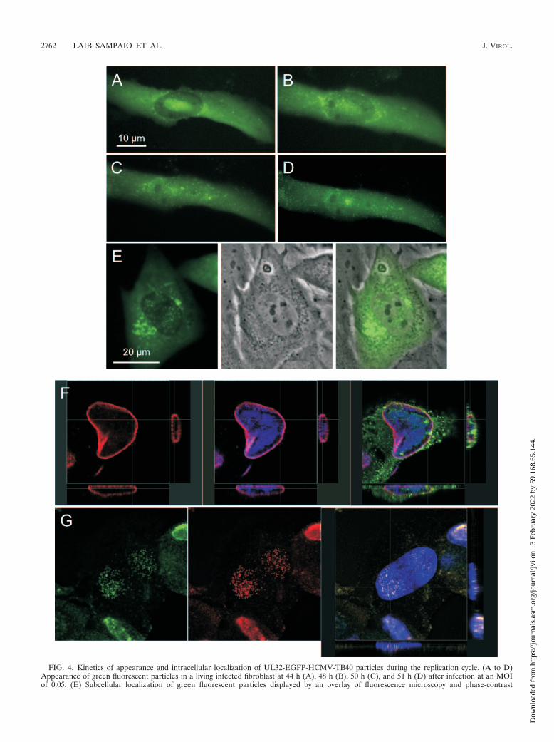
FIG. 4. Kinetics of appearance and intracellular localization of UL32-EGFP-HCMV-TB40 particles during the replication cycle. (A to D)Appearance of green fluorescent particles in a living infected fibroblast at 44 h (A), 48 h (B), 50 h (C), and 51 h (D) after infection at an MOIof 0.05. (E) Subcellular localization of green fluorescent particles displayed by an overlay of fluorescence microscopy and phase-contrast
2762 LAIB SAMPAIO ET AL. J. VIROL.
Dow
nloa
ded
from
http
s://j
ourn
als.
asm
.org
/jour
nal/j
vi o
n 13
Feb
ruar
y 20
22 b
y 59
.168
.65.
144.

microscopy of a living infected cell at 65 h p.i. (F) Subcellular localization of green fluorescent particles as detected by confocal laser scanningmicroscopy of an acetone-fixed infected cell at 72 h p.i. Indirect immunofluorescence of lamin B (Cy3, red) and EGFP (Alexa 488, green) versusDNA staining (DAPI, blue) is shown. (G) Intranuclear colocalization of the tegument protein pUL32 and the major capsid protein pUL86 byconfocal laser scanning microscopy of a paraformaldehyde-fixed infected cell at 72 h p.i. The native fluorescence of pUL32-EGFP is displayedversus indirect immunofluorescence of pUL86 (Cy3, red) and DNA staining (DAPI, blue). (H) Ultrastructural localization of the pUL32-EGFPfusion protein. Fibroblast cultures at 4 days after infection with UL32-EGFP-HCMV-TB40 or with wild-type virus were fixed and cryosectioned.EGFP was detected by immunogold labeling on ultrathin cryosections. Examples of viral capsids with associated pUL32-EGFP are indicated byarrowheads.
VOL. 79, 2005 EGFP-LABELED HCMV 2763
Dow
nloa
ded
from
http
s://j
ourn
als.
asm
.org
/jour
nal/j
vi o
n 13
Feb
ruar
y 20
22 b
y 59
.168
.65.
144.

FIG. 5. Live imaging of particle movements in fibroblasts infected by UL32-EGFP-HCMV-TB40 as documented by time-lapse fluorescencemicroscopy. (A) Centripetal and centrifugal movements in fibroblasts 1 h after infection at an MOI of 10. White arrows, actual position of theparticle; gray arrows, starting point of the particle. Selected frames from videos are shown. (B) Particle movements in an infected cell at 4 daysafter infection at an MOI of 0.1. The light gray arrow points to a particle moving parallel to the cell margin. The black arrow points to theperinuclear aggregation site. The dark gray arrow indicates the starting point of a particle appearing to be leaving the nucleus. The paired whitearrows indicate routes of saltatory movements. (C) Statistical analysis of particle movements in fibroblasts 3 days after infection with UL32-EGFP-HCMV-TB40. (D) Time-lapse series suggestive of release of a particle at the cell periphery of a fibroblast at 4 days after infection at anMOI of 0.1. Selected frames from a video are shown.
2764
Dow
nloa
ded
from
http
s://j
ourn
als.
asm
.org
/jour
nal/j
vi o
n 13
Feb
ruar
y 20
22 b
y 59
.168
.65.
144.

after about 2 days of infection, when progeny virus appeared,and at a late stage of infection when numerous progeny virusparticles were present. During the initial phase of high-doseinfections (MOI � 10), both centripetal and centrifugal salta-tory movements could be observed with a subset of inputparticles, whereas most input particles performed irregularmovements at their position (Fig. 5A and data not shown).Although no predominance of centripetal movements wasfound in fibroblasts during observation periods of about 500 s,an overall aggregation of particles at perinuclear sites occurredafter several hours. At about 2 days after low-dose infection,the number of particles within infected cells increased, reflect-ing the generation of progeny virus. Emerging virus particleswere first located in the inclusions of the nucleus but were soonalso detectable within the cytoplasm (Fig. 4), where they ag-gregated at perinuclear sites during late stages of infection.
During the late stage of infection, all kinds of movement ascharacterized for the initial phase of infection were alsopresent (Fig. 5B and data not shown). In particular, particlesseemed not to be destined for efficient egress from the cell. Asubset of particles reached the periphery of the cell by irregularmovements rather than by directed long-distance movements.Some of these particles performed back-and-forth movementsparallel to the cell margins. However, during a 300-s observa-tion sequence with a time frame of 2 s, events suggestive ofparticle release were a rare singular finding (Fig. 5D and datanot shown). Moreover, when late-stage infected cells werecocultured with an excess of uninfected cells, cell-to-cell trans-mission also could not be demonstrated unequivocally withinsequences of 300 s at a time frame of 2 s although numerousparticles were produced by these late-stage infected cells (datanot shown). Together these findings strongly indicated thatrelease of virions from late-stage infected cells is not a directedefficient process that would apply for the majority of cytoplas-mic particles. In summary, various kinds of short-distance orlong-distance movements with different directions occurredsimultaneously throughout the viral life cycle without a clearpredominance of a certain pattern at any time point.
DISCUSSION
Proper translocations within infected cells are critical eventsat various points during the replication cycle of viruses. Inparticular this applies to herpesviruses, which are known toreplicate their DNA and assemble their 100-nm large capsidsin the nuclei of infected cells. Thus, successful herpesviralreplication depends on penetration into the cell across theactin cortex, centripetal transport towards the nucleus, egressof viral progeny capsids from the nucleus, centrifugal transporttowards the cell periphery, and egress through the cell cortex.At present, most of these events are poorly understood. Al-though general shifts in the number and location of virus par-ticles can be documented by the staining of fixed infected-cellcultures at different time points after infection, there are lim-itations of such an approach that will prevent more detailedinsights. In particular, analyses of fixed cell cultures do notallow for (i) sequential analyses of a single cell, (ii) tracking ofindividual virus particles, and (iii) documentation of narrowtime frames. Such information can best be achieved by liveanalysis of cells infected by fluorescence-tagged virus. Here we
report the generation of green fluorescent human cytomega-lovirus particles expressing the tegument protein pp150(UL32) as an EGFP fusion protein.
As a target for EGFP fusion, we chose the viral tegumentprotein pp150 (UL32) for several reasons. We had successfullystained virus particles during viral entry and nuclear transportby indirect immunostaining of pUL32 (25). Evidence frombiochemical binding studies indicated that pUL32 binds di-rectly and strongly to capsids (1, 2). Steric constraints causedby an EGFP tag were expected to be better tolerated whenEGFP was fused to a capsid-associated tegument proteinrather than to a capsid protein itself. As pUL32 binds to thecapsid through its amino one-third (1), the carboxy terminuswas expected to be free and appeared to be an ideal target forEGFP fusion. In order to cause as little alteration of the viralgenome as possible, the EGFP gene was fused to the UL32gene directly before the last codon at the autologous positionwithout introducing any further selection marker. Microinjec-tion of a purified recombination fragment (consisting of theUL32 open reading frame truncated by one codon, the codingsequence of EGFP, and 1,622 bp of the UL31 open readingframe) into infected fibroblasts during the early phase of rep-lication resulted in the appearance of green fluorescent foci ofinfected cells, from which recombinant UL32-EGFP-HCMV-TB40 could be enriched by three sequential plaque purifica-tions. Limiting-dilution infections with plaque-purified viruspreparations and subsequent simultaneous immunofluores-cence of pUL32 and EGFP revealed that all infectious unitswithin the preparation expressed EGFP. Immunoblotting ofvirion proteins of UL32-EGFP-HCMV-TB40 with a pUL32-specific antibody demonstrated the lack of wild-type pUL32and the expected shift of the fusion protein towards a higherapparent molecular weight. Enzymatic DNA amplificationwith wild-type-specific sequences further confirmed thatplaque-purified preparations of UL32-EGFP-HCMV-TB40were free of contaminating infectious wild-type virus.
The recombinant UL32-EGFP-HCMV-TB40 appears to ex-press viral proteins and produce progeny virus with kineticsand titers similar to those of wild-type HCMV TB40. In par-ticular, pUL32-EGFP is expressed with the same kinetics andwith the same subcellular localization as wild-type pUL32, andall EGFP signals are colocalized with pUL32 signals. Infec-tious progeny virions released from infected cells displayedgreen fluorescence when analyzed as free extracellular parti-cles and during adsorption and entry in fibroblast cultures.Ultrastructural immunogold analyses demonstrated the incor-poration of pUL32-EGFP into progeny virion particles. Inconclusion, fusion of EGFP to the carboxy terminus ofHCMV-pUL32 was successful and resulted in green fluores-cent HCMV with growth properties that were indistinguish-able from those of wild-type virus. When applied in live-imag-ing approaches, this virus will enable live analyses of variouskinds of translocations of viral particles during the replicativecycle.
As a first application of UL32-EGFP-HCMV-TB40, we haveanalyzed the kinetics and localization of acquisition of pUL32by newly synthesized HCMV particles during the late phase ofinfection. Surprisingly, the first pUL32-containing particles ap-peared within the nucleus, indicating that binding of pUL32 tothe capsid is an early step of the tegumentation, which occurs
VOL. 79, 2005 EGFP-LABELED HCMV 2765
Dow
nloa
ded
from
http
s://j
ourn
als.
asm
.org
/jour
nal/j
vi o
n 13
Feb
ruar
y 20
22 b
y 59
.168
.65.
144.

before particles reach the nuclear membrane or lamina. Thelack of nuclear pUL32 signals in an earlier study (24) might bedue to limited sensitivity of the staining technique or to theusage of antibodies that might miss the nuclear form of pUL32.Consistent with our results, the nuclear localization of pUL32has been demonstrated by Hensel et al. (9). In principle,pUL32 might thus be important for any of the subsequentsteps, including transportation towards the nuclear lamina,egress from the nucleus, and regulation of the following tegu-mentation events within the cytoplasm. Targeted introductionof mutations into the pUL32 part of the UL32-EGFP fusiongene will now allow for a detailed analysis of pUL32 functionsin the context of viral morphogenesis and egress.
In addition, we have analyzed the intracellular movementsof green fluorescent particles during the late phase of infec-tion, when morphogenesis and egress from the infected celltake place. Again the results were surprising, as time-lapseseries of micrographs indicated a variety of different particlemovements with an almost chaotic appearance rather than thepredominance of any directed translocation pattern. In partic-ular, most particles within the cytoplasm performed short-distance back-and-forth or irregular movements, while asmaller fraction of particles performed centripetal and centrif-ugal long-distance movements or moved along the margins ofthe infected cell. Only very few events suggestive of particlerelease at the cell borders have been documented. Taken to-gether, the particle movements observed in productively in-fected cells do not support the assumption of an effectiveegress of progeny virus during the late stage of infection.Rather, release of virus by infected cells seems to be a rareevent. It is noteworthy, however, that this is in concordancewith the quantitative aspects of HCMV production by infect-ed-cell cultures. A culture of 4 � 106 late-stage infected fibro-blasts produces about 108 infectious units per day, which cor-responds to about 1 infectious unit per cell per h. On the basisof this calculation it is not surprising that release of greenfluorescent particles from infected cells was a rare event in ouranalysis.
Regarding the nature of the movements that were observed,the short-distance movements performed by the majority ofparticles could reflect active transportation of particles by thecellular transport machinery, movements of cellular vesiclesassociated with virus capsids during the process of tegumenta-tion and envelopment, or passive Brownian dynamics alongwith the surrounding cytoplasm. The long-distance movementscannot be explained by passive movements together with thesurrounding cytoplasm but are reminiscent of active transpor-tation along microtubular structures with regard to their meanapparent velocity of about 0.7 to 0.8 �m/s and peak velocitiesof 2 �m/s. In nerve axons, higher mean velocities of about 2�m/s have been reported for pseudorabies virus (27). In thecytoplasm of epithelial cells, however, adenoviruses moved at avelocity of about 0.5 �m/s, with peak velocities of about 2 �m/sin both directions (31). Likewise, vaccinia viruses were re-ported to move in epithelial cells at velocities of about 0.5 �m/s(7). In an in vitro system the velocities of microtubule-baseddynein-mediated and kinesin-mediated transport were 0.7 and1.0 �m/s, respectively (13). The movements described here forEGFP-tagged HCMV particles are therefore consistent withthe assumption of microtubule-based motility. Although it is
unclear whether naked capsids or enveloped particles aretransported, this indicates that the virus might use the cellulartransportation machinery in order to reach the periphery of thecell. Whereas a number of particles actually are translocatedtowards the cell periphery and particle movements along thecell margins were frequently detected, egress from the cellseems to be inefficient. It will now be interesting to studywhether egress from the cytoplasm is hampered by the physicalbarrier formed by actin cortex or by difficulties performing theenergy-consuming fusion of membranes.
In summary, a green fluorescent HCMV variant is now avail-able, enabling live analyses of viral particle translocations dur-ing various steps of the viral replication cycle. From such de-scriptive studies novel aspects of HCMV entry andmorphogenesis will arise and can be subjected to detailed cell-biological or biochemical analyses in the future.
ACKNOWLEDGMENT
This work was supported by the German Research Foundation(grants SI 779/2–1 and SI 779/3–1).
REFERENCES
1. Baxter, M. K., and W. Gibson. 2001. Cytomegalovirus basic phosphoprotein(pUL32) binds to capsids in vitro through its amino one-third. J. Virol.75:6865–6873.
2. Benko, D. M., R. S. Haltiwanger, G. W. Hart, and W. Gibson. 1988. Virionbasic phosphoprotein from human cytomegalovirus contains O-linked N-acetylglucosamine. Proc. Natl. Acad. Sci. USA 85:2573–2577.
3. Danscher, G. 1981. Histochemical demonstration of heavy metals. A revisedversion of the sulphide silver method suitable for both light and electron-microscopy. Histochemistry 71:1–16.
4. Desai, P., and S. Person. 1998. Incorporation of the green fluorescent pro-tein into the herpes simplex virus type 1 capsid. J. Virol. 72:7563–7568.
5. Dohner, K., A. Wolfstein, U. Prank, C. Echeverri, D. Dujardin, R. Vallee,and B. Sodeik. 2002. Function of dynein and dynactin in herpes simplex viruscapsid transport. Mol. Biol. Cell 13:2795–2809.
6. Elliott, G., and P. O’Hare. 1999. Live-cell analysis of a green fluorescentprotein-tagged herpes simplex virus infection. J. Virol. 73:4110–4119.
7. Geada, M. M., I. Galindo, M. M. Lorenzo, B. Perdiguero, and R. Blasco.2001. Movements of vaccinia virus intracellular enveloped virions with GFPtagged to the F13L envelope protein. J. Gen. Virol. 82:2747–2760.
8. Goodrum, F. D., C. T. Jordan, K. High, and T. Shenk. 2002. Human cyto-megalovirus gene expression during infection of primary hematopoietic pro-genitor cells: a model for latency. Proc. Natl. Acad. Sci. USA 99:16255–16260.
9. Hensel, G., H. Meyer, S. Gartner, G. Brand, and H. F. Kern. 1995. Nuclearlocalization of the human cytomegalovirus tegument protein pp150(ppUL32). J. Gen. Virol. 76:1591–1601.
10. Isomura, H., and M. F. Stinski. 2003. The human cytomegalovirus majorimmediate-early enhancer determines the efficiency of immediate-early genetranscription and viral replication in permissive cells at low multiplicity ofinfection. J. Virol. 77:3602–3614.
11. Iwata, M., J. Vieira, M. Byrne, H. Horton, and B. Torok Storb. 1999. Inter-leukin-1 (IL-1) inhibits growth of cytomegalovirus in human marrow stromalcells: inhibition is reversed upon removal of IL-1. Blood 94:572–578.
12. Kerman, I. A., L. W. Enquist, S. J. Watson, and B. J. Yates. 2003. Brainstemsubstrates of sympatho-motor circuitry identified using trans-synaptic tracingwith pseudorabies virus recombinants. J. Neurosci. 23:4657–4666.
13. King, S. J., and T. A. Schroer. 2000. Dynactin increases the processivity ofthe cytoplasmic dynein motor. Nat. Cell Biol. 2:20–24.
14. Mabit, H., M. Y. Nakano, U. Prank, B. Saam, K. Dohner, B. Sodeik, andU. F. Greber. 2002. Intact microtubules support adenovirus and herpessimplex virus infections. J. Virol. 76:9962–9971.
15. Mahy, B. W. J., and H. O. Kangro. 1996. Virology methods manual. Aca-demic Press, San Diego, Calif.
16. Marschall, M., M. Freitag, S. Weiler, G. Sorg, and T. Stamminger. 2000.Recombinant green fluorescent protein-expressing human cytomegalovirusas a tool for screening antiviral agents. Antimicrob. Agents Chemother.44:1588–1597.
17. Mettenleiter, T. C. 2002. Herpesvirus assembly and egress. J. Virol. 76:1537–1547.
18. Mocarski, E. S. J., and C. T. Courcelle. 2001. Cytomegaloviruses and theirreplication, p. 2629–2674. In D. M. Knipe and P. M. Howley (ed.), Fieldsvirology, 4th ed., vol. 2. Lippincott Williams & Wilkins, Philadelphia, Pa.
2766 LAIB SAMPAIO ET AL. J. VIROL.
Dow
nloa
ded
from
http
s://j
ourn
als.
asm
.org
/jour
nal/j
vi o
n 13
Feb
ruar
y 20
22 b
y 59
.168
.65.
144.

19. Muranyi, W., J. Haas, M. Wagner, G. Krohne, and U. H. Koszinowski. 2002.Cytomegalovirus recruitment of cellular kinases to dissolve the nuclear lam-ina. Science 297:854–857.
20. Murphy, E. A., D. N. Streblow, J. A. Nelson, and M. F. Stinski. 2000. Thehuman cytomegalovirus IE86 protein can block cell cycle progression afterinducing transition into the S phase of permissive cells. J. Virol. 74:7108–7118.
21. Potel, C., K. Kaelin, I. Gautier, P. Lebon, J. Coppey, and F. Rozenberg. 2002.Incorporation of green fluorescent protein into the essential envelope gly-coprotein B of herpes simplex virus type 1. J. Virol. Methods 105:13–23.
22. Pyner, S., J. Cleary, P. M. Buchan, and J. H. Coote. 2001. Tracing function-ally identified neurons in a multisynaptic pathway in the hamster and ratusing herpes simplex virus expressing green fluorescent protein. Exp. Physiol.86:695–702.
23. Sanchez, V., C. L. Clark, J. Y. Yen, R. Dwarakanath, and D. H. Spector.2002. Viable human cytomegalovirus recombinant virus with an internaldeletion of the IE2 86 gene affects late stages of viral replication. J. Virol.76:2973–2989.
24. Sanchez, V., K. D. Greis, E. Sztul, and W. J. Britt. 2000. Accumulation ofvirion tegument and envelope proteins in a stable cytoplasmic compartmentduring human cytomegalovirus replication: characterization of a potentialsite of virus assembly. J. Virol. 74:975–986.
25. Sinzger, C., M. Kahl, K. Laib, K. Klingel, P. Rieger, B. Plachter, and G.Jahn. 2000. Tropism of human cytomegalovirus for endothelial cells is de-
termined by a post-entry step dependent on efficient translocation to thenucleus. J. Gen. Virol. 81:3021–3035.
26. Sinzger, C., K. Schmidt, J. Knapp, M. Kahl, R. Beck, J. Waldman, H.Hebart, H. Einsele, and G. Jahn. 1999. Modification of human cytomegalo-virus tropism through propagation in vitro is associated with changes in theviral genome. J. Gen. Virol. 80:2867–2877.
27. Smith, G. A., S. P. Gross, and L. W. Enquist. 2001. Herpesviruses usebidirectional fast-axonal transport to spread in sensory neurons. Proc. Natl.Acad. Sci. USA 98:3466–3470.
28. Sodeik, B., M. W. Ebersold, and A. Helenius. 1997. Microtubule-mediatedtransport of incoming herpes simplex virus 1 capsids to the nucleus. J. CellBiol. 136:1007–1021.
29. Stierhof, Y. D., B. M. Humbel, and H. Schwarz. 1991. Suitability of differentsilver enhancement methods applied to 1 nm colloidal gold particles: animmunoelectron microscopic study. J. Electron Microsc. Technol. 17:336–343.
30. Strive, T., E. Borst, M. Messerle, and K. Radsak. 2002. Proteolytic process-ing of human cytomegalovirus glycoprotein B is dispensable for viral growthin culture. J. Virol. 76:1252–1264.
31. Suomalainen, M., M. Y. Nakano, S. Keller, K. Boucke, R. P. Stidwill, andU. F. Greber. 1999. Microtubule-dependent plus- and minus end-directedmotilities are competing processes for nuclear targeting of adenovirus.J. Cell Biol. 144:657–672.
VOL. 79, 2005 EGFP-LABELED HCMV 2767
Dow
nloa
ded
from
http
s://j
ourn
als.
asm
.org
/jour
nal/j
vi o
n 13
Feb
ruar
y 20
22 b
y 59
.168
.65.
144.


![Analysis of mRNA Expression by Fluorescent Labeled ......Analysis of mRNA Expression by Fluorescent Labeled Microbead Assay using Bioplex 200 [概 要] 本アッセイ法はフローサイトメトリーの原理を利用してLuminex](https://img.pdfslide.net/doc/110x75/6126f172350ac94ffa4eca58/analysis-of-mrna-expression-by-fluorescent-labeled-analysis-of-mrna-expression.jpg)