Embed Size (px)
Citation preview
Submitted 5 March 2015Accepted 8 May 2015Published 2 June 2015
Corresponding authorMatthew R. McCurry,[email protected]
Academic editorRobert Druzinsky
Additional Information andDeclarations can be found onpage 12
DOI 10.7717/peerj.988
Copyright2015 McCurry et al.
Distributed underCreative Commons CC-BY 4.0
OPEN ACCESS
The sensitivity of biological finiteelement models to the resolution ofsurface geometry: a case study ofcrocodilian craniaMatthew R. McCurry1,2, Alistair R. Evans2,3 and Colin R. McHenry1,4
1 Department of Anatomy and Developmental Biology, Monash University, Clayton,Melbourne, Australia
2 Geosciences, Museum Victoria, Carlton, Melbourne, Australia3 School of Biological Sciences, Monash University, Clayton, Melbourne, Australia4 School of Engineering, University of Newcastle, Callaghan, Australia
ABSTRACTThe reliability of finite element analysis (FEA) in biomechanical investigationsdepends upon understanding the influence of model assumptions. In producingfinite element models, surface mesh resolution is influenced by the resolution ofinput geometry, and influences the resolution of the ensuing solid mesh used fornumerical analysis. Despite a large number of studies incorporating sensitivitystudies of the effects of solid mesh resolution there has not yet been any investigationinto the effect of surface mesh resolution upon results in a comparative context. Herewe use a dataset of crocodile crania to examine the effects of surface resolution onFEA results in a comparative context. Seven high-resolution surface meshes wereeach down-sampled to varying degrees while keeping the resulting number of solidelements constant. These models were then subjected to bite and shake load casesusing finite element analysis. The results show that incremental decreases in surfaceresolution can result in fluctuations in strain magnitudes, but that it is possible toobtain stable results using lower resolution surface in a comparative FEA study.As surface mesh resolution links input geometry with the resulting solid mesh, theimplication of these results is that low resolution input geometry and solid meshesmay provide valid results in a comparative context.
Subjects Computational Biology, Evolutionary Studies, ZoologyKeywords Finite element analysis, Biomechanics, Resolution, Skull, Sensitivity
INTRODUCTIONComparative biomechanics applies mechanical theory and techniques to better understand
differences between biological structures and systems (Vogel, 2003). Finite element analysis
(FEA) is a computational modelling technique that has become a widely used method
in comparative biomechanics. The use of this method in anatomical studies (whether
biological, palaeontological or medical) has allowed for a better understanding of how
variation in morphology, material properties and loading conditions can influence the
How to cite this article McCurry et al. (2015), The sensitivity of biological finite element models to the resolution of surface geometry: acase study of crocodilian crania. PeerJ 3:e988; DOI 10.7717/peerj.988
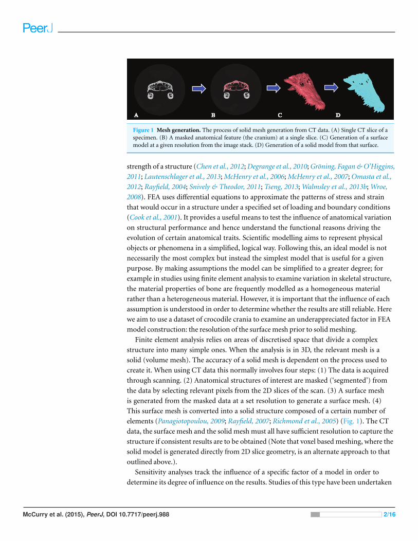
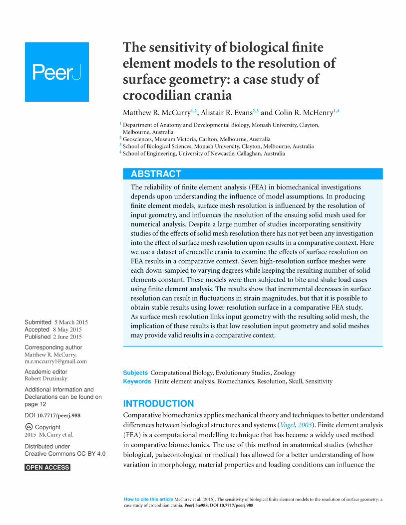
Figure 1 Mesh generation. The process of solid mesh generation from CT data. (A) Single CT slice of aspecimen. (B) A masked anatomical feature (the cranium) at a single slice. (C) Generation of a surfacemodel at a given resolution from the image stack. (D) Generation of a solid model from that surface.
strength of a structure (Chen et al., 2012; Degrange et al., 2010; Groning, Fagan & O’Higgins,
2011; Lautenschlager et al., 2013; McHenry et al., 2006; McHenry et al., 2007; Omasta et al.,
2012; Rayfield, 2004; Snively & Theodor, 2011; Tseng, 2013; Walmsley et al., 2013b; Wroe,
2008). FEA uses differential equations to approximate the patterns of stress and strain
that would occur in a structure under a specified set of loading and boundary conditions
(Cook et al., 2001). It provides a useful means to test the influence of anatomical variation
on structural performance and hence understand the functional reasons driving the
evolution of certain anatomical traits. Scientific modelling aims to represent physical
objects or phenomena in a simplified, logical way. Following this, an ideal model is not
necessarily the most complex but instead the simplest model that is useful for a given
purpose. By making assumptions the model can be simplified to a greater degree; for
example in studies using finite element analysis to examine variation in skeletal structure,
the material properties of bone are frequently modelled as a homogeneous material
rather than a heterogeneous material. However, it is important that the influence of each
assumption is understood in order to determine whether the results are still reliable. Here
we aim to use a dataset of crocodile crania to examine an underappreciated factor in FEA
model construction: the resolution of the surface mesh prior to solid meshing.
Finite element analysis relies on areas of discretised space that divide a complex
structure into many simple ones. When the analysis is in 3D, the relevant mesh is a
solid (volume mesh). The accuracy of a solid mesh is dependent on the process used to
create it. When using CT data this normally involves four steps: (1) The data is acquired
through scanning. (2) Anatomical structures of interest are masked (‘segmented’) from
the data by selecting relevant pixels from the 2D slices of the scan. (3) A surface mesh
is generated from the masked data at a set resolution to generate a surface mesh. (4)
This surface mesh is converted into a solid structure composed of a certain number of
elements (Panagiotopoulou, 2009; Rayfield, 2007; Richmond et al., 2005) (Fig. 1). The CT
data, the surface mesh and the solid mesh must all have sufficient resolution to capture the
structure if consistent results are to be obtained (Note that voxel based meshing, where the
solid model is generated directly from 2D slice geometry, is an alternate approach to that
outlined above.).
Sensitivity analyses track the influence of a specific factor of a model in order to
determine its degree of influence on the results. Studies of this type have been undertaken
McCurry et al. (2015), PeerJ, DOI 10.7717/peerj.988 2/16

on a wide number of factors used in FEA models including: the way in which the size
of models is standardised (Dumont, Grosse & Slater, 2009; Walmsley et al., 2013a), the
complexity of the material properties used in the model (Bright & Rayfield, 2011b; Cox et
al., 2011; Groning, Fagan & O’Higgins, 2012; Kupczik et al., 2007; Porro et al., 2011; Reed
et al., 2011; Strait et al., 2005; Tseng et al., 2011; Walmsley et al., 2013a), the role of sutures
(Bright, 2012; Kupczik et al., 2007; Porro et al., 2011; Reed et al., 2011; Wang et al., 2010), the
influence of ligaments (Groning, Fagan & O’Higgins, 2012; Groning, Fagan & O’Higgins,
2011), the effect of different orientations of muscle force (Bright & Rayfield, 2011b; Cox
et al., 2011; Groning, Fagan & O’Higgins, 2012; Groning, Fagan & O’Higgins, 2011; Grosse
et al., 2007), the effect of the type of solid elements (Bright & Rayfield, 2011a; Dumont,
Piccirillo & Grosse, 2005) and the influence of mesh density (Bright & Rayfield, 2011a; Tseng
et al., 2011). These studies have added to a growing collection of information on the effects
of model assumptions in FEA. Some of these studies have identified material properties
as having a comparatively large influence on the results (Cox et al., 2011; Porro et al., 2011;
Reed et al., 2011; Strait et al., 2005; Tseng et al., 2011) where as others have identified that
functional aspects of the simulation, such as bite point, are a large influence (Fitton et al.,
2012; Walmsley et al., 2013a).
A number of sensitivity studies have previously been undertaken to examine the
effects of altering solid resolution on FEA results (Bright & Rayfield, 2011a; Tseng et al.,
2011). Using models of a wolf mandible, Tseng et al. (2011) found that 300,000 solid
elements were sufficient to produce reaction forces and strain energy close to those of
high resolution models i.e., convergence. Higher levels of strain were observed in higher
resolution models. Bright & Rayfield (2011a) showed that element sizes of approximately
1 mm edge length (1,250,000 elements) achieved convergence in a pig skull. This study
also found that insufficiently dense models underestimated strain and displacement and
those areas of high strain converged faster than areas of low strain (Bright & Rayfield,
2011a). Tseng & Flynn (2015) conducted a detailed convergence analyses on a mongoose
skull; they found that higher resolution models did not provide more stable outputs, and
that convergence patterns differed across constraint types and bite positions simulated.
Together these studies show that the number of elements used in comparative analyses
does influence results and that the morphological complexity of the structure and loading
conditions are important in determining the number of elements required to obtain a
convergent result. Consequently the number of solid elements should be sufficiently high
in order to be consistent with a convergent result.
The creation of a volume mesh at a certain resolution is only the final step in model
generation however. The solid mesh is generated from a surface mesh, and the surface
mesh is generated from input geometry (the input geometry is derived from CT scanning,
surface laser scanning, photogrammetry, or computer aided design). A high resolution
solid mesh requires a high resolution surface mesh to accurately incorporate potential
geometric information; any solid mesh resolution exceeding the surface mesh resolution is
effectively wasted because it will incorporate a higher number of elements without altering
the geometric detail of the structure (although higher resolutions can improve accuracy
McCurry et al. (2015), PeerJ, DOI 10.7717/peerj.988 3/16
Figure 2 Simplified models. Example of the simplified models. C. moreletti models composed of 20k,30k, 90k and 300k surface elements.
in the displacement field of the model). Convergence tests on solid mesh resolution are
routinely undertaken as part of FEA studies, however, the effect of both input geometry
and surface mesh resolution have not been sufficiently investigated. Furthermore, most
finite element sensitivity studies have not assessed results in a comparative context. Many
studies using finite element analysis include specimens that, because of size or access
to data capture equipment, vary considerably in resolution. The effects of resolution
at various stages of the finite element model creation process are thus important in a
practical sense when planning a study. Here, as a first approach in determining the relative
influences of the steps used to prepare a FEA model from computed tomography (CT)
data, we aim to examine the degree to which the surface mesh resolution influences FEA
results in a comparative dataset of crocodilian crania.
MATERIALS AND METHODSData acquisition and surface mesh simplificationThe crania of seven species of crocodilian (comprising Mecistops cataphractus, Crocodylus
johnstoni, Crocodylus intermedius, Crocodylus moreletii, Gavialis gangeticus, Osteolaemus
tetraspis and Tomistoma schlegelii) were scanned using computer tomography (CT
scanning). These specimens varied greatly in size and morphology, details about the
collection of this data can be found in Walmsley et al. (2013b). The software package
“MIMICS” version 14 was used to segment the cranium from each scan and generate a
high resolution surface mesh of the cranium; the number of elements varied from 600,000
to 3,000,000 depending on specimen size and scanning parameters. Minimal manual
editing was undertaken to prevent bias being introduced between models. Using the
remesher in the FEA module of MIMICS, the high resolution surface meshes were then
down-sampled in number of elements using the “Reduce” function to approximately
20,000 (20k), 30,000 (30k), 90,000 (90k) and 300,000 (300k) surface elements for each of
the models (Fig. 2).
McCurry et al. (2015), PeerJ, DOI 10.7717/peerj.988 4/16
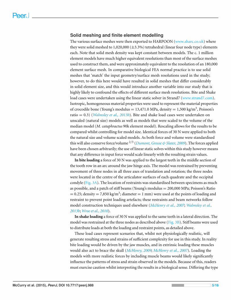
Solid meshing and finite element modellingThe various surface meshes were then exported to HARPOON (www.sharc.co.uk) where
they were solid meshed to 1,020,000 (±3.3%) tetrahedral (linear four node type) elements
each. Note that solid mesh density was kept constant between models. The c. 1 million
element models have much higher equivalent resolutions than most of the surface meshes
used to construct them, and were approximately equivalent to the resolution of an 180,000
element surface mesh. In comparative biological FEA normal practice is to use solid
meshes that ‘match’ the input geometry/surface mesh resolutions used in the study;
however, to do this here would have resulted in solid meshes that differ considerably
in solid element size, and this would introduce another variable into our study that is
highly likely to confound the effects of different surface mesh resolutions. Bite and Shake
load cases were undertaken using the linear static solver in Strand7 (www.strand7.com).
Isotropic, homogeneous material properties were used to represent the material properties
of crocodile bone (Young’s modulus = 13,471.0 MPa, density = 1,500 kg/m3, Poisson’s
ratio = 0.3) (Walmsley et al., 2013b). Bite and shake load cases were undertaken on
unscaled (natural size) models as well as models that were scaled to the volume of the
median model (M. cataphractus 90k element model). Rescaling allows for the results to be
compared whilst controlling for model size. Identical forces of 30 N were applied to both
the natural size and volume scaled models. As both force and volume were standardised
this will also conserve force/volume 2/3 (Dumont, Grosse & Slater, 2009). The forces applied
have been chosen arbitrarily; the use of linear static solves within this study however means
that any difference in input force would scale linearly with the resulting strain values.
In bite loading a force of 30 N was applied to the largest teeth in the middle section of
the tooth row in an arc around the jaw hinge axis. The model was restrained by preventing
movement of three nodes in all three axes of translation and rotation; the three nodes
were located in the centre of the articulator surfaces of each quadrate and the occipital
condyle (Fig. 3A). The location of restraints was standardised between specimens as much
as possible, and a patch of stiff beams (Young’s modulus = 200,000 MPa; Poisson’s Ratio
= 0.25; density = 7,850 kg/m3; diameter = 1 mm) were used at the points of loading and
restraint to prevent point loading artefacts; these restraints and beam networks follow
model construction techniques used elsewhere (McHenry et al., 2007; Walmsley et al.,
2013b; Wroe et al., 2010).
In shake loading a force of 30 N was applied to the same teeth in a lateral direction. The
model was restrained at the three nodes as described above (Fig. 3B). Stiff beams were used
to distribute loads at both the loading and restraint points, as detailed above.
These load cases represent scenarios that, whilst not physiologically realistic, will
generate resulting stress and strains of sufficient complexity for use in this study. In reality
bite loading would be driven by the jaw muscles, and in extrinsic loading these muscles
would also act to brace the skull (McHenry, 2009; McHenry et al., 2007). Loading the
models with more realistic forces by including muscle beams would likely significantly
influence the patterns of stress and strain observed in the models. Because of this, readers
must exercise caution whilst interpreting the results in a biological sense. Differing the type
McCurry et al. (2015), PeerJ, DOI 10.7717/peerj.988 5/16
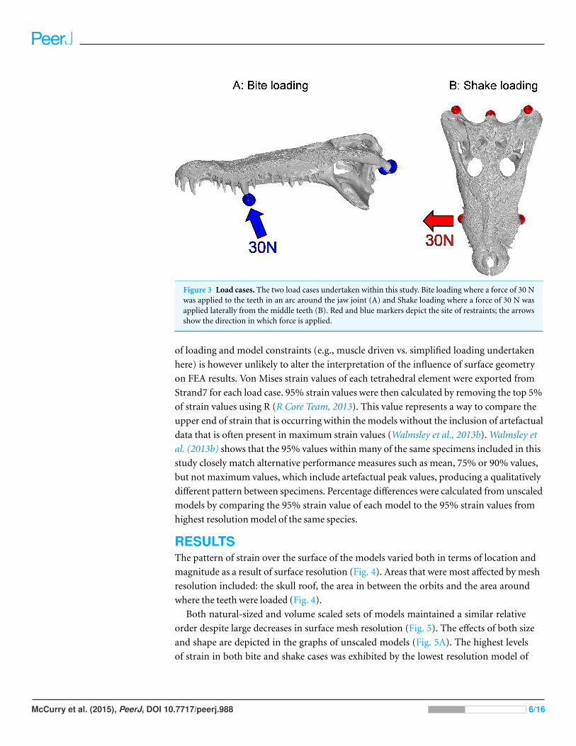
Figure 3 Load cases. The two load cases undertaken within this study. Bite loading where a force of 30 Nwas applied to the teeth in an arc around the jaw joint (A) and Shake loading where a force of 30 N wasapplied laterally from the middle teeth (B). Red and blue markers depict the site of restraints; the arrowsshow the direction in which force is applied.
of loading and model constraints (e.g., muscle driven vs. simplified loading undertaken
here) is however unlikely to alter the interpretation of the influence of surface geometry
on FEA results. Von Mises strain values of each tetrahedral element were exported from
Strand7 for each load case. 95% strain values were then calculated by removing the top 5%
of strain values using R (R Core Team, 2013). This value represents a way to compare the
upper end of strain that is occurring within the models without the inclusion of artefactual
data that is often present in maximum strain values (Walmsley et al., 2013b). Walmsley et
al. (2013b) shows that the 95% values within many of the same specimens included in this
study closely match alternative performance measures such as mean, 75% or 90% values,
but not maximum values, which include artefactual peak values, producing a qualitatively
different pattern between specimens. Percentage differences were calculated from unscaled
models by comparing the 95% strain value of each model to the 95% strain values from
highest resolution model of the same species.
RESULTSThe pattern of strain over the surface of the models varied both in terms of location and
magnitude as a result of surface resolution (Fig. 4). Areas that were most affected by mesh
resolution included: the skull roof, the area in between the orbits and the area around
where the teeth were loaded (Fig. 4).
Both natural-sized and volume scaled sets of models maintained a similar relative
order despite large decreases in surface mesh resolution (Fig. 5). The effects of both size
and shape are depicted in the graphs of unscaled models (Fig. 5A). The highest levels
of strain in both bite and shake cases was exhibited by the lowest resolution model of
McCurry et al. (2015), PeerJ, DOI 10.7717/peerj.988 6/16
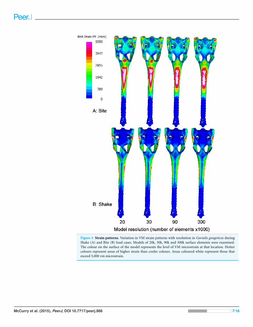
Figure 4 Strain patterns. Variation in VM strain patterns with resolution in Gavialis gangeticus duringShake (A) and Bite (B) load cases. Models of 20k, 30k, 90k and 300k surface elements were examined.The colour on the surface of the model represents the level of VM microstrain at that location. Hottercolours represent areas of higher strain than cooler colours. Areas coloured white represent those thatexceed 5,000 vm microstrain.
McCurry et al. (2015), PeerJ, DOI 10.7717/peerj.988 7/16
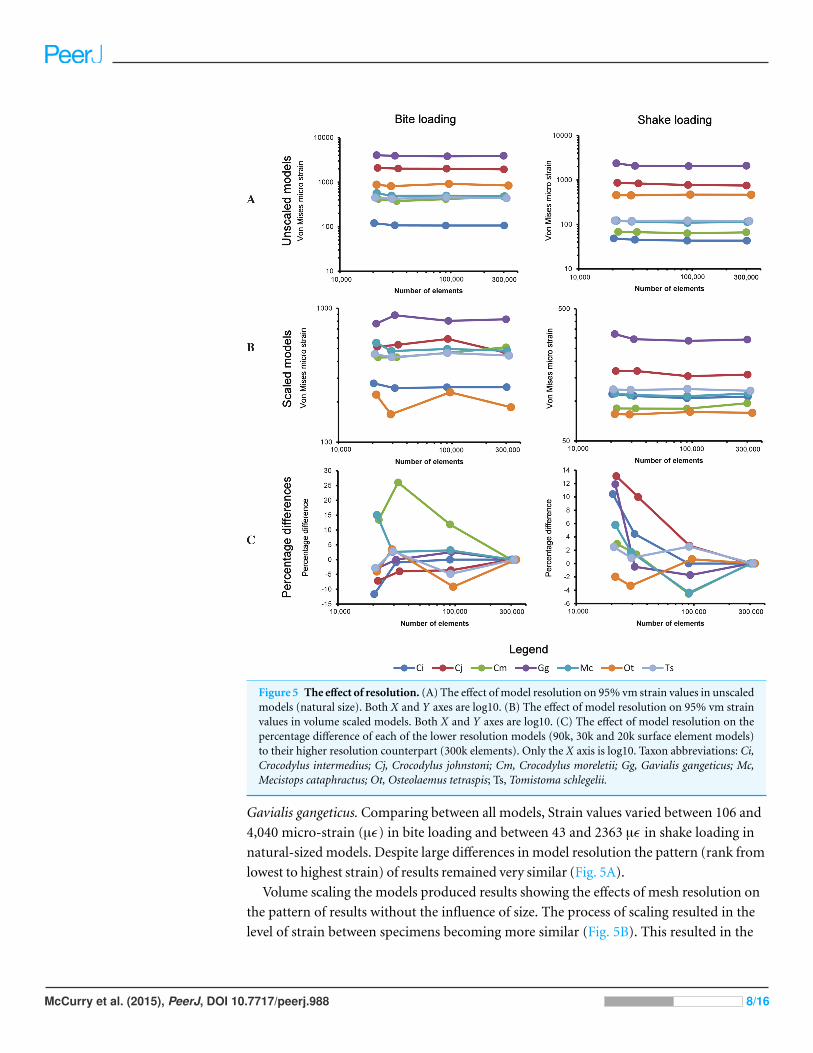
Figure 5 The effect of resolution. (A) The effect of model resolution on 95% vm strain values in unscaledmodels (natural size). Both X and Y axes are log10. (B) The effect of model resolution on 95% vm strainvalues in volume scaled models. Both X and Y axes are log10. (C) The effect of model resolution on thepercentage difference of each of the lower resolution models (90k, 30k and 20k surface element models)to their higher resolution counterpart (300k elements). Only the X axis is log10. Taxon abbreviations: Ci,Crocodylus intermedius; Cj, Crocodylus johnstoni; Cm, Crocodylus moreletii; Gg, Gavialis gangeticus; Mc,Mecistops cataphractus; Ot, Osteolaemus tetraspis; Ts, Tomistoma schlegelii.
Gavialis gangeticus. Comparing between all models, Strain values varied between 106 and
4,040 micro-strain (µϵ) in bite loading and between 43 and 2363 µϵ in shake loading in
natural-sized models. Despite large differences in model resolution the pattern (rank from
lowest to highest strain) of results remained very similar (Fig. 5A).
Volume scaling the models produced results showing the effects of mesh resolution on
the pattern of results without the influence of size. The process of scaling resulted in the
level of strain between specimens becoming more similar (Fig. 5B). This resulted in the
McCurry et al. (2015), PeerJ, DOI 10.7717/peerj.988 8/16
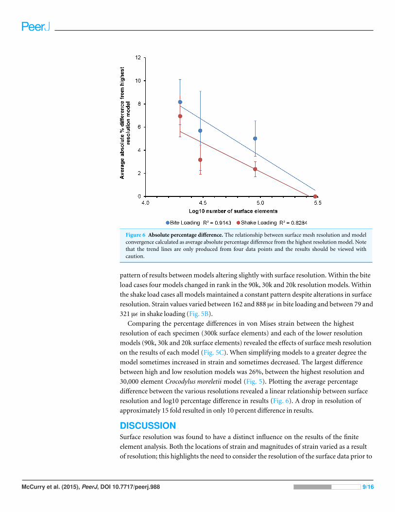
Figure 6 Absolute percentage difference. The relationship between surface mesh resolution and modelconvergence calculated as average absolute percentage difference from the highest resolution model. Notethat the trend lines are only produced from four data points and the results should be viewed withcaution.
pattern of results between models altering slightly with surface resolution. Within the bite
load cases four models changed in rank in the 90k, 30k and 20k resolution models. Within
the shake load cases all models maintained a constant pattern despite alterations in surface
resolution. Strain values varied between 162 and 888 µϵ in bite loading and between 79 and
321 µϵ in shake loading (Fig. 5B).
Comparing the percentage differences in von Mises strain between the highest
resolution of each specimen (300k surface elements) and each of the lower resolution
models (90k, 30k and 20k surface elements) revealed the effects of surface mesh resolution
on the results of each model (Fig. 5C). When simplifying models to a greater degree the
model sometimes increased in strain and sometimes decreased. The largest difference
between high and low resolution models was 26%, between the highest resolution and
30,000 element Crocodylus moreletii model (Fig. 5). Plotting the average percentage
difference between the various resolutions revealed a linear relationship between surface
resolution and log10 percentage difference in results (Fig. 6). A drop in resolution of
approximately 15 fold resulted in only 10 percent difference in results.
DISCUSSIONSurface resolution was found to have a distinct influence on the results of the finite
element analysis. Both the locations of strain and magnitudes of strain varied as a result
of resolution; this highlights the need to consider the resolution of the surface data prior to
McCurry et al. (2015), PeerJ, DOI 10.7717/peerj.988 9/16
solid meshing for use in finite element analysis. Size and shape were however found to have
a larger influence on the results than that of the range of surface resolutions tested (Fig. 5),
lending support for the use of a comparative approach in biomechanical analysis.
The location of strain varied as a result of resolution; areas under strain that were of high
geometric complexity such as the medial sides of the orbits were the most highly affected
(Fig. 4). Bright & Rayfield (2011a) reported that the area that took the longest to converge
in a domestic pig skull was close to a region of high geometric complexity. In order to
compare FEA results to validation data their study only measured strain at a number of
locations where strain gauges could be fitted. The results of this study support the findings
of Bright & Rayfield (2011a) and highlight that the level of geometric complexity of a
model should be considered when determining whether surface resolution is sufficient.
The high variation in strain observed between the specimens used in this study is likely
related to the large amount of interspecific variation present in size and morphology.
The Gavialis gangeticus specimen which exhibited the highest levels of strain in both
biting and shaking load cases is far smaller in size (approximately 190 mm in dorsal
cranial length) and has a far more gracile, elongate morphology compared to many of
the other specimens used in the study. The simplified loading methods used in this study
may also have contributed to the high levels of strain observed. Loading the skulls with
forces for simulated muscles, rather than with extrinsically applied loads, may considerably
decreased the strain levels observed.
This study employed low order solid elements (Tet4s) to make up the solid mesh.
These elements exhibit displacement behaviour described with linear equations and
omit constant state stress and strain over each element volume. Whilst these types of
elements can perform poorly in some bending applications, the approximately 1 million
solid elements used within each of the models here provided sufficient resolution to result
in many solid elements comprising each of the major bone structures, resulting in fairly
complex strain fields (Fig. 4). Previous studies have shown a high level of convergence in
models where this is the case (Bright & Rayfield, 2011a; Dumont, Piccirillo & Grosse, 2005).
When simplifying the model to a greater degree the strain magnitudes sometimes
resulted in higher levels of strain and other times resulted in lower levels of strain (Fig. 5).
This result is intuitive considering that when simplifying a model the meshing algorithm
is forced to choose whether to fill an area with material or not to. Previous studies of mesh
resolution have found that strain was most often underestimated in models of insufficient
resolution (Bright & Rayfield, 2011a). The findings of this study did not agree with this,
with approximately half of the models increasing in strain and half decreasing (Fig. 5). This
inconsistency could be a reflection of the use of a single model within previous analyses or
a result of methodological differences (e.g., the standardisation of solid mesh resolution
within this study or the use of 95% strain values instead of maximum and minimum
principal strain as a measure of model performance) between the two studies. Natural
sized models were used in this section as several studies have noted that the choice of
methods when scaling may influence results (Dumont, Grosse & Slater, 2009; Walmsley
et al., 2013a). The location of constraints in the models was chosen with a high level
McCurry et al. (2015), PeerJ, DOI 10.7717/peerj.988 10/16
of care; however we must note that the resolution of the models may have altered the
morphology of the surfaces at these locations, which may influence the strain patterns
observed. There were clear relationships between surface resolution and FEA results.
Decreasing surface resolution resulted in an average difference of approximately 8% in VM
strain values in the lowest resolution models (approx. 20k surface elements), compared to
values from the highest resolution meshes (Fig. 6). This is quite a small effect considering
the large decreases in resolution that were undertaken. Ideally our models would have been
validated in order to provide a benchmark of the real level of strain in each load case, this is
a clear opportunity for future research. Because boundary conditions can also alter strain
distributions and magnitudes it is likely that appropriate mesh resolutions may differ in
other studies. Furthermore, it is important to note that the crocodilians used within this
study have robust bone structure compared to other taxa; caution must be used when
trying to extrapolate these results to taxa that have thinner bones and hence may be more
susceptible to differences in surface resolution. Future studies should consider resolution at
the scanning, surface meshing and solid meshing stages in relation to the specific structures
and parameters being assessed.
Models with a surface resolution of 30k elements converged well with those with higher
numbers of elements, indicating that the usefulness of higher resolutions would be small.
We suggest that the results also have interesting implications for data collection methods
in the context of a comparative analysis. Quite simple geometries were found to perform
adequately in this instance. Importantly, the implications of this study for collecting data
in lower resolutions rely on the assumption that down sampling surface data will result in
a similar geometric simplification to initially collecting the same data in low resolution.
Although this makes logical sense, future studies will need to confirm this for their taxa, or
anatomical features of interest.
The advent of new scanning technologies has resulted in some studies scanning
specimens at quite high resolution (Dumont, Piccirillo & Grosse, 2005; Oldfield et al.,
2012; Tseng, 2009; Walmsley et al., 2013b; Wroe, Lowry & Anton, 2008) providing solid
meshes of over 2.5 million elements. Although this may be necessary in the examination
of the biomechanics of fine morphological features, it appears that low resolution data
is sufficient for the examination of large morphological differences. FEA studies should
consider whether scan, surface and solid mesh resolution is appropriate to answer the
questions of the study; our results suggest that very high resolutions are not always
necessary for meaningful comparative analysis.
The question of scanning and modelling resolutions is particularly important for
comparative studies that may include material that is logistically difficult to scan using
high-resolution CT. Although the influence of scan resolution is not directly tested within
this study, the results show that, for the specimens and loading scenarios examined here,
the level of geometric detail required to obtain reasonable results is quite low and hence
low resolution data collection methods could potentially be employed. However, this
result is may not hold true for studies interested in finer morphological variation such
as most intraspecific comparisons; further analyses will need to be undertaken before we
McCurry et al. (2015), PeerJ, DOI 10.7717/peerj.988 11/16
can determine what constitutes a large enough variation in morphology. The measure of
performance analysed here (95% VM strain) may also be under less influence by surface
mesh resolution than other performance measures (e.g., stress values) or levels of stress or
strain in smaller localised areas of the skull.
This result has important implications for data collection and FEA model construction,
in that collecting data in lower resolutions would speed up both data collection and
solve times. Many fossil specimens, due to their size, fragility or density, are logistically
unsuitable for high resolution CT scanning but if lower resolution surface meshes produce
meaningful results then the scope for including fossils specimens is greatly increased;
photogrammetry, surface scanning, or CAD approaches can be used to generate suitable
input geometry, and can be used with specimens where CT data is unavailable or
unsuitable. For incomplete or deformed fossils (i.e., the majority of vertebrate fossils),
digital reconstruction is required prior to finite element analysis, and these techniques
are much easier to use in lower resolution (McHenry, 2009). Our results suggest that low
resolution models used in this way will still produce meaningful results. Lower resolution
models are also easier and faster to construct, solve, and analyse.
This study adds to a body of literature on the influences of assumptions in FEA
models. The results document the effects of altering surface resolution and show that it
is possible to get reasonable results for a lower resolution surface if the solid elements
are standardised. This result also provides a basis for the use of simplified models in
comparative biomechanics. If studies are interested in large morphological differences,
such as those tested here, then considerable differences in surface resolution will not
influence the “take-home” results of a study.
ACKNOWLEDGEMENTSWe would like to thank Matthew Colbert and Jesse Maisano (Digital Morphology,
University of Texas), Christopher Brochu (University of Iowa), Mason Meers (University
of Tampa) and Bruno Frohlich (Smithsonian Institution) for access to specimens and CT
data. I would also like to thank Erich Fitzgerald and Chris Walmsley for useful discussion
on the topic of finite element analysis and Catherine Boisvert for the use of the MIMICS
software that was used in the study.
ADDITIONAL INFORMATION AND DECLARATIONS
FundingThis work was funded using an Australian Research Council Discovery Project grant (to
CRM) and an Australian Research Fellowship and Future Fellowship (to ARE), a grant
from The Joyce W. Vickery Scientific Research Fund (to MRM), a Museum Victoria 1854
student scholarship (to MRM) and Monash University internal funding (to CRM and
ARE). The funders had no role in study design, data collection and analysis, decision to
publish, or preparation of the manuscript.
McCurry et al. (2015), PeerJ, DOI 10.7717/peerj.988 12/16
Grant DisclosuresThe following grant information was disclosed by the authors:
Australian Research Council Discovery Project.
Australian Research Fellowship and Future Fellowship.
Joyce W. Vickery Scientific Research Fund.
Museum Victoria.
Monash University.
Competing InterestsMatthew McCurry is a Ph.D. student researcher at Museum Victoria and Alistair Evans
holds an honorary adjunct position at Museum Victoria. The authors do not consider these
affiliations to cause any competing interests.
Author Contributions• Matthew R. McCurry conceived and designed the experiments, performed the
experiments, analyzed the data, contributed reagents/materials/analysis tools, wrote
the paper, prepared figures and/or tables, reviewed drafts of the paper.
• Alistair R. Evans and Colin R. McHenry conceived and designed the experiments,
contributed reagents/materials/analysis tools, reviewed drafts of the paper.
Supplemental InformationSupplemental information for this article can be found online at http://dx.doi.org/
10.7717/peerj.988#supplemental-information.
REFERENCESBright JA. 2012. The importance of craniofacial sutures in biomechanical finite element models of
the domestic pig. PLoS ONE 7:e31769 DOI 10.1371/journal.pone.0031769.
Bright JA, Rayfield EJ. 2011a. The response of cranial biomechanical finite element models tovariations in mesh density. The Anatomical Record 294:610–620 DOI 10.1002/ar.21358.
Bright JA, Rayfield EJ. 2011b. Sensitivity and ex vivo validation of finite element models of thedomestic pig cranium. Journal of Anatomy 219:456–471 DOI 10.1111/j.1469-7580.2011.01408.x.
Chen DW, Lin C-L, Hu C-C, Wu J-W, Lee MS. 2012. Finite element analysis of different repairmethods of Vancouver B1 periprosthetic fractures after total hip arthroplasty. Injury43:1061–1065 DOI 10.1016/j.injury.2012.01.015.
Cook RD, Malkus DS, Plesha ME, Witt RJ. 2001. Concepts and applications of finite elementanalysis. 4th edition. New York: John Wiley & Sons, Inc.
Cox PG, Fagan MJ, Rayfield EJ, Jeffery N. 2011. Finite element modelling of squirrel, guineapig and rat skulls: using geometric morphometrics to assess sensitivity. Journal of Anatomy219:696–709 DOI 10.1111/j.1469-7580.2011.01436.x.
Degrange FJ, Tambussi CP, Moreno K, Witmer LM, Wroe S. 2010. Mechanical analysis of feedingbehavior in the extinct terror bird Andalgalornis steulleti (Gruiformes: Phorusrhacidae). PLoSONE 5:1–7 DOI 10.1371/journal.pone.0011856.
McCurry et al. (2015), PeerJ, DOI 10.7717/peerj.988 13/16

Dumont ER, Grosse IR, Slater GJ. 2009. Requirements for comparing the performance of finiteelement models of biological structures. Journal of Theoretical Biology 256:96–103DOI 10.1016/j.jtbi.2008.08.017.
Dumont ER, Piccirillo J, Grosse IR. 2005. Finite-element analysis of biting behavior and bonestress in the facial skeletons of bats. The Anatomical Record Part A: Discoveries in Molecular,Cellular, and Evolutionary Biology 283A:319–330 DOI 10.1002/ar.a.20165.
Fitton LC, Shi JF, Fagan MJ, O’Higgins P. 2012. Masticatory loadings and cranial deformation inMacaca fascicularis: a finite element analysis sensitivity study. Journal of Anatomy 221:55–68DOI 10.1111/j.1469-7580.2012.01516.x.
Groning F, Fagan M, O’Higgins P. 2012. Modeling the human mandible under masticatory loads:which input variables are important? The Anatomical Record: Advances in Integrative Anatomyand Evolutionary Biology 295:853–863 DOI 10.1002/ar.22455.
Groning F, Fagan MJ, O’Higgins P. 2011. The effects of the periodontal ligament on mandibularstiffness: a study combining finite element analysis and geometric morphometrics. Journal ofBiomechanics 44:1304–1312 DOI 10.1016/j.jbiomech.2011.01.008.
Grosse IR, Dumont ER, Coletta C, Tolleson A. 2007. Techniques for modeling muscle-inducedforces in finite element models of skeletal structures. The Anatomical Record: Advances inIntegrative Anatomy and Evolutionary Biology 290:1069–1088 DOI 10.1002/ar.20568.
Kupczik K, Dobson CA, Fagan MJ, Crompton RH, Oxnard CE, O’Higgins P. 2007. Assessingmechanical function of the zygomatic region in macaques: validation and sensitivity testing offinite element models. Journal of Anatomy 210:41–53 DOI 10.1111/j.1469-7580.2006.00662.x.
Lautenschlager S, Witmer LM, Altangerel P, Rayfield EJ. 2013. Edentulism, beaks, andbiomechanical innovations in the evolution of theropod dinosaurs. Proceedings ofthe National Academy of Sciences of the United States of America 110(51):20657–20662DOI 10.1073/pnas.1310711110.
McHenry C. 2009. Devourer of Gods: the palaecology of the Cretaceous pliosaur Kronosaurusqueenslandicus. University of Newcastle. Available at http://hdl.handle.net/1959.13/935911.
McHenry CR, Clausen PD, Daniel WJ, Meers MB, Pendharkar A. 2006. Biomechanics of therostrum in crocodilians: A comparative analysis using finite-element modeling. The AnatomicalRecord Part A 288:827–849 DOI 10.1002/ar.a.20360.
McHenry CR, Wroe S, Clausen PD, Moreno K, Cunningham E. 2007. Supermodeled sabercat,predatory behavior in Smilodon fatalis revealed by high-resolution 3D computer simulation.Proceedings of the National Academy of Sciences of the United States of America 104:16010–16015DOI 10.1073/pnas.0706086104.
Oldfield C, McHenry C, Clausen P, Chamoli U, Parr W, Stynder D, Wroe S. 2012. Finite elementanalysis of ursid cranial mechanics and the prediction of feeding behaviour in the extinct giantAgriotherium africanum. Journal of Zoology 286:163–170DOI 10.1111/j.1469-7998.2011.00862.x.
Omasta M, Palousek D, Navrat T, Rosicky J. 2012. Finite element analysis for the evaluation ofthe structural behaviour, of a prosthesis for trans-tibial amputees. Medical Engineering & Physics34:38–45 DOI 10.1016/j.medengphy.2011.06.014.
Panagiotopoulou O. 2009. Finite element analysis (FEA): applying an engineering methodto functional morphology in anthropology and human biology. Annals of Human Biology36:609–623 DOI 10.1080/03014460903019879.
McCurry et al. (2015), PeerJ, DOI 10.7717/peerj.988 14/16

Porro LB, Holliday CM, Anapol F, Ontiveros LC, Ontiveros LT, Ross CF. 2011. Free bodyanalysis, beam mechanics, and finite element modeling of the mandible of Alligatormississippiensis. Journal of Morphology 272:910–937 DOI 10.1002/jmor.10957.
R Core Team. 2013. R: a language and environment for statistical computing. Vienna: R Foundationfor Statistical Computing.
Rayfield E. 2004. Cranial mechanics and feeding in Tyrannosaurus rex. Proceedings of the RoyalSociety of London B 271:1451–1459 DOI 10.1098/rspb.2004.2755.
Rayfield EJ. 2007. Finite element analysis and understanding the biomechanics and evolutionof living and fossil organisms. Annual Review of Earth and Planetary Sciences 35:541–576DOI 10.1146/annurev.earth.35.031306.140104.
Reed DA, Porro LB, Iriarte-Diaz J, Lemberg JB, Holliday CM, Anapol F, Ross CF. 2011. Theimpact of bone and suture material properties on mandibular function in Alligatormississippiensis: testing theoretical phenotypes with finite element analysis. Journal of Anatomy218:59–74 DOI 10.1111/j.1469-7580.2010.01319.x.
Richmond BG, Wright BW, Grosse I, Dechow PC, Ross CF, Spencer MA, Strait D. 2005. Finiteelement analysis in functional morphology. The Anatomical Record Part A: Discoveries inMolecular, Cellular, and Evolutionary Biology 283:259–274 DOI 10.1002/ar.a.20169.
Snively E, Theodor JM. 2011. Common functional correlates of head-strike behavior in thepachycephalosaur Stegoceras validum (ornithischia, dinosauria) and combative artiodactyls.PLoS ONE 6:e21422 DOI 10.1371/journal.pone.0021422.
Strait DS, Wang Q, Dechow PC, Ross CF, Richmond BG, Spencer MA, Patel BA. 2005. Modelingelastic properties in finite-element analysis: how much precision is needed to producean accurate model? The Anatomical Record Part A: Discoveries in Molecular, Cellular, andEvolutionary Biology 283A:275–287 DOI 10.1002/ar.a.20172.
Tseng ZJ. 2009. Cranial function in a late Miocene Dinocrocuta gigantea (Mammalia: Carnivora)revealed by comparative finite element analysis. Biological Journal of the Linnean Society96:51–67 DOI 10.1111/j.1095-8312.2008.01095.x.
Tseng ZJ. 2013. Testing adaptive hypotheses of convergence with functional landscapes: a casestudy of bone-cracking hypercarnivores. PLoS ONE 8:e65305DOI 10.1371/journal.pone.0065305.
Tseng ZJ, Flynn JJ. 2015. Convergence analysis of a finite element skull model of Herpestesjavanicus (Carnivora, Mammalia): implications for robust comparative inferences ofbiomechanical function. Journal of Theoretical Biology 365:112–148DOI 10.1016/j.jtbi.2014.10.002.
Tseng ZJ, Mcnitt-Gray JL, Flashner H, Wang X, Enciso R. 2011. Model sensitivity and use of thecomparative finite element method in mammalian jaw mechanics: mandible performance inthe gray wolf. PLoS ONE 6:e19171 DOI 10.1371/journal.pone.0019171.
Vogel S. 2003. Comparative biomechanics: life’s physical world. New Jersey: Princeton UniversityPress.
Walmsley CW, McCurry MR, Clausen PD, McHenry CR. 2013a. Beware the black box:investigating the sensitivity of FEA simulations to modelling factors in comparativebiomechanics. PeerJ 1:e204 DOI 10.7717/peerj.204.
Walmsley CW, Smits PD, Quayle MR, McCurry MR, Richards HS, Oldfield CC, Wroe S,Clausen PD, McHenry CR. 2013b. Why the long face? The mechanics of mandibular symphysisproportions in crocodiles. PLoS ONE 8:e53873 DOI 10.1371/journal.pone.0053873.
McCurry et al. (2015), PeerJ, DOI 10.7717/peerj.988 15/16

Wang Q, Smith AL, Strait DS, Wright BW, Richmond BG, Grosse IR, Byron CD, Zapata U. 2010.The global impact of sutures assessed in a finite element model of a Macaque Cranium. TheAnatomical Record: Advances in Integrative Anatomy and Evolutionary Biology 293:1477–1491DOI 10.1002/ar.21203.
Wroe S. 2008. Cranial mechanics compared in extinct marsupial and extant african lions using afinite-element approach. Journal of Zoology 274:332–339DOI 10.1111/j.1469-7998.2007.00389.x.
Wroe S, Ferrara TL, McHenry C, Curnoe D, Chamoli U. 2010. The craniomandibular mechanicsof being human. Proceedings of the Royal Society of London B 277:3579–3586DOI 10.1098/rspb.2010.0509.
Wroe S, Lowry MB, Anton M. 2008. How to build a mammalian super-predator. Zoology111:196–203 DOI 10.1016/j.zool.2007.07.008.
McCurry et al. (2015), PeerJ, DOI 10.7717/peerj.988 16/16


![arXiv:1711.09078v2 [cs.CV] 11 Mar 2019 · less useful for video processing. We introduce a new dataset, Vimeo-90K, for a systematic evaluation of video process-ing algorithms. Vimeo-90K](https://img.pdfslide.net/doc/110x75/5fd463d1af98ba659a66980d/arxiv171109078v2-cscv-11-mar-2019-less-useful-for-video-processing-we-introduce.jpg)


![CASE STUDIES · 1/10/2015 · case study: $125m+ ch aritable foundation challenge solution result [+ ] $120k x + x = 25 x [+ $90k ] + 20 x $40k = $6m $120k x + x = 25 x [+ $90k ]](https://img.pdfslide.net/doc/110x75/5fd19c406e2b2c5b1c191d92/case-studies-1102015-case-study-125m-ch-aritable-foundation-challenge-solution.jpg)

![arXiv:1711.09078v3 [cs.CV] 10 Nov 2019 · less useful for video processing. We introduce a new dataset, Vimeo-90K, for a systematic evaluation of video process-ing algorithms. Vimeo-90K](https://img.pdfslide.net/doc/110x75/5fd462810ec7625d996cc261/arxiv171109078v3-cscv-10-nov-2019-less-useful-for-video-processing-we-introduce.jpg)