Embed Size (px)
Citation preview

The Skin Problems of the Lower-Extremity Amputee1
1 Based on a lecture presented before the University of California Pilot School in Lower-Extremity Prosthetics, August 25, 1955, at the U.S. Naval Hospital, Oakland, California.
S. WILLIAM LEVY, M.D.2
2 Clinical Instructor in Dermatology, School of Medicine, University of California Medical Center, San Francisco, and supervisor of the Study Group on Dermatology, Lower-Extremity Amputee Research Project, University of California, Berkeley and San Francisco.
Since the establishment, in the autumn of 1954, of the skin-study group of the Lower-Extremity Amputee Research Project at the University of California, other physicians within the Project have referred to us for observation and treatment those amputees having cutaneous problems associated with the wearing of a prosthesis. Out of this nidus, specific information regarding the various clinical problems has been assembled and correlated in an effort to benefit the individual amputee. Some of the clinical problems have aroused interest in basic dermatologic research, so that investigation has not been of a purely clinical nature.
The cutaneous difficulties associated with the wearing of a leg prosthesis have been evaluated in more than 200 patient-visits, and every effort has been made to classify cutaneous disorders specifically. Approximately the same number of above- and below-knee amputees have been carefully screened and examined. Complete histories have been taken, and physical examinations of the skin have been performed. Skin biopsies have been obtained in many instances, and histopathologic sections have been examined carefully in an effort to determine the course of a specific disorder.
Other laboratory aids, such as skin scrapings for fungi or patch tests for contact dermatitis, have been utilized. Stump hygiene is important in relation to many clinical disorders of the skin, and accordingly a specific hygienic program for the care of the stump is being developed.
Skin lesions, however minute they may appear, are nevertheless of great importance since they may be the beginning of an extensive cutaneous disorder that may be mentally, socially, and economically disastrous to a given individual. It is best to view any minor irritation as a potentially dangerous symptom and to deal with it as early as possible. Once the skin problem has begun, it should not be ignored in the hope that it will heal of its own accord. Nothing can be more frustrating to the lower-extremity amputee than to be told to remain off his prosthesis or to go on crutches because he has neglected a minor skin eruption.
This article is devoted to the common skin problems and danger signals associated with the wearing of a lower-extremity prosthesis. Most of our experience has been gained with the above-knee amputee using the suction-socket suspension, but it is believed that the same or similar problems arise in patients using the more conventional types of suspension.
STUMP HYGIENE
Hygienic measures are of the utmost importance in the daily care of amputation stumps and in the use of prostheses. A neglect of cleanliness can easily result in damage to the skin and thus open the door to a number of cutaneous disorders which can require temporary removal of the prosthesis. There is no
20

SKIN PROBLEMS
unanimity of opinion on exactly what measures should be employed routinely. Amputees have come to us with many varied and weird ideas. Some have used strong soaps and alkalies on their stumps, some alcohol, and others formaldehyde. These hygienic measures have been handed down from one person to another and frequently without reason or logical explanation. Some patients fail to wash either the stump or the socket, thereby giving rise to maceration and malodor.
A simple hygienic program using a sudsing detergent has in many instances prevented or eliminated a cutaneous disorder, and hence we frequently request an amputee to follow a given routine. He is advised against the use of any preparation which would leave a deposit in the socket or any solvent which might affect the interior finish. A simple procedure for cleaning the socket is to wash the inner surface with a lukewarm, soapy cloth or one containing a detergent, remove the soapy residue with a clean wet cloth, and then dry out the socket with a towel. The prosthesis should not be put on for several minutes so that it may have an opportunity to dry completely.
For the stumps of most individuals, a bland soap or liquid detergent provides a good cleansing without irritating the skin. Soaps or detergents containing hexachlorophene provide a bacteriostatic action, in addition to cleansing, and may aid in reducing the danger of infection. An amputee is frequently advised to purchase a plastic squeeze bottle of pHisoHex,® an item available in every drugstore, relatively inexpensive, and to be had without a prescription. He is instructed to spread over the amputation stump a small amount of this antibacterial sudsing detergent containing hexachlorophene. A little water is added and the material worked into a lather. More and more water is added to increase the amount of sudsing. He is told to avoid washing off the suds until ready for thorough rinsing. When well cleansed, the site is then rinsed off with lukewarm water, and the stump is dried by patting rather than by vigorous rubbing. This simple routine should be followed nightly, or every other night, depending upon the rate of perspiration, the degree of malodor, and the
bathing habits of the individual. In the treatment of some persistent eczemas of the stump, this simple hygienic program was found to be curative.
CLINICAL PROBLEMS
Some amputees go along for months or years without difficulty or irritation of the stump skin. In others, the skin is a weak tissue, and frequent difficulties arise. Persons concerned with amputees should be aware of certain pathologic conditions—certain danger signals—which are frequently the forerunners of seriously incapacitating cutaneous disorders. Early recognition and treatment of these conditions can avert much mental anguish and loss of social or economic activity. It should be remembered that, once on a prosthesis, the amputee desires to stay on, and it is of vital concern to the physician and prosthetist to prevent any disorder which may return him to crutches or bed rest. What, then, are some of these danger signals?
STUMP EDEMA SYNDROME
When an amputee first starts wearing a suction-socket prosthesis, he can expect to see edema or swelling and reddish-brown pigmentation, roughening, and drying of the skin of the terminal portion of the stump (Fig. 1). These changes are the almost inevitable result of the altered conditions forced upon the skin and subcutaneous tissues. They are relatively innocent, do not usually require therapy, and are partially prevented by gradually compressing the stump tissues with an elastic bandage prior to use of the prosthesis. An incorrectly fitted socket may predispose a leg amputee to this disorder.
In several of our patients the edema has been massive, and distal pigmentation has followed, with the formation of hemorrhagic papules and nodules. Superficial erosion of the skin in these regions is not uncommon, and, in rare instances, deep ulcers can result from the poor cutaneous nutrition (Fig. 2). Multiple biopsies have been taken in order to determine the pathogenesis of this disorder. Special staining of the sections revealed that the pigmentary changes were due to the blood pigment, hemosiderin, within the tissue (Fig. 3).
21
The collagen of the dermis was thickened by newly formed fibrous connective tissue, and there was an abnormal proliferation and dilatation of blood vessels. It may be that this kind of disorder is vascular in origin and that a venous and lymphatic congestion is productive of the edema and hemorrhage. It is hoped that the basic pathogenesis will be clarified as more patients with this syndrome are studied.
Edematous portions of the skin of the distal part of the stump may become pinched and strangulated within the socket (Fig. 4). Such areas may ulcerate or become gangrenous owing to impaired blood supply.
CONTACT DERMATITIS
Contact dermatitis (Fig. 5) is caused by contact of the skin with a chemical that acts either as a primary irritant or as a specific allergic sensitizer. As defined by Schwartz (10), "A PRIMARY CUTANEOUS IRRITANT is an
agent which will cause dermatitis by direct action on the normal skin at the site of contact if it is permitted to act in sufficient intensity or quantity for a sufficient length of time." Again using Schwartz' definition (10), "A CUTANEOUS SENSITIZER is an agent which does not necessarily cause demonstrable cutaneous changes on first contact but may effect such specific changes in the skin that, after five to seven days or more, further contact on the same or other parts of the body will cause dermatitis." Contact dermatitis may be acute, subacute, or chronic, and moderately severe to severe itching is present in most forms. In the acute and subacute types, diffuse erythema, edema, oozing, and crusting predominate. In addition, vesicles are often present if a specific allergic sensitizer is the cause. In chronic forms, erythema, scaling, and lichenification (thickening) prevail.
We have seen a number of patients with contact dermatitis of the amputation stump. In order to understand the problem, we have had to learn about the plastics and resins used in the external and internal finishes of the different types of prostheses. In some instances, we found only by carefully taken history that the use of a new cream, lubricant, or cleansing agent coincided with the onset of the dermatitis. Some amputees use a foam-rubber cushion,
others a plastic-covered pad on the bottom of their socket. These are also capable of producing a contact dermatitis of the skin weeks, months, or even years after use (Fig. 5, A and B).
On patients exhibiting the clinical manifestations of contact dermatitis, every attempt has been made to determine the exact contactant. Patch tests (Fig. 5, C and D) have been most informative with respect to specific substances as the cause of the dermatitis. In diagnostic patch-testing, a small amount of the suspected substance is applied to a site of normal skin on the patient. It is covered with an innocuous, impermeable material such as cellophane, which is then sealed to the skin by adhesive plaster. It is usually sufficient to leave the patch on for 24 to 48 hours. Upon removal of the patch, a positive reaction is signified by erythema, vesiculation, or blister formation at the site of application.
Because patch-testing with strong concentrations of known primary irritants will result in reactions on any skin, solutions of such substances are first diluted according to published lists (11) in order to prevent a false positive reaction and possible injury to the skin. A generalized eruption following the patch test indicates a high degree of sensitivity, but fortunately such eruptions are rare. Experience and good clinical judgment are necessary in choosing the correct chemical concentration of the irritant and the proper time for performing the patch test.
The sockets of leg prostheses are commonly finished on the inside by the application of a varnish or lacquer and on the outside by coating with plastics and resins. These complex organic substances are capable of causing a contact dermatitis in a given individual who has become sensitized. This sensitization is similar to that produced by poison oak or poison ivy, and the intensity of reaction may vary under different conditions of heat, humidity, and friction. The epoxy resins (8), if incompletely cured in their manufacture, may, in addition to being a specific allergic sensitizer, produce a primary-irritant dermatitis. These resins are frequently used to improve the appearance of a socket and to render it impervious to external agents. In the uncured state at room temper-
LEVY 22
ature they are viscous, amber-colored liquids. Curing agents, known as catalysts or hardeners, are added to solidify the plastic material. These agents are organic amines of various types and, when left in excess by incomplete baking or curing at high temperatures, are able to produce a primary-irritant dermatitis.
We have had several patients with proven contact dermatitis to Ambroid,® C-8 epoxy resin, polyethylene, foam-rubber pads, and plastic-covered cushions. Removal of the suspicious contactant resulted in a cure, and subsequent patch-testing proved the diagnosis.
In those instances of contact dermatitis where the irritant has not been obvious and the patch tests have been inconclusive, temporary therapy has alleviated the symptoms. Cool compresses, bland antipruritic lotions, and the topical use of hydrocortisone or fluorohydro-cortisone preparations have been most beneficial.
POST-TRAUMATIC EPIDERMOID CYSTS
Young (15), in 1951, described the appearance of multiple cysts in the skin of an amputee's thigh in association with the wearing of an artificial limb. Other authors (2,3,6,13,14, 16,17,18) have described similar nodules in the skin under the rim of the socket. In the typical case (Fig. 6), the cysts do not appear until the patient has worn an artificial limb for months or possibly years. They occur most frequently in above-knee amputees in the areas covered by the upper medial margin of the prosthesis but have also been described in below-knee amputees.
Characteristically, in the above-knee amputee small follicular keratin plugs develop in the skin of the adductor region of the thigh along the upper edge of the prosthesis. In the beginning they appear as small lumps or nodules and will, at times, disappear when the prosthesis is removed temporarily. Under the constant friction and pressure of the prosthesis, they become larger and more numerous. Some become pea-sized, round, or oval swellings deep within the skin. Gradually, with enlargement, they become sensitive and tender to the touch. The skin may break down and erode or ulcerate. With continued irritation by the prosthesis, the nodular swelling may suddenly
burst and discharge an opaque, purulent fluid. The discharging sinus may become chronic and thus make it impossible for the patient to use his prosthesis. In other instances, the break may take place within the deeper portion of the skin, and subcutaneous intercommunicating sinuses may develop.
The larger nodules become especially tender and necessitate removal of the prosthesis. These should not be confused with ordinary furuncles or boils (Fig. 7), which may occur on any portion of the stump. Surgical excision of the chronic, isolated, noninfected nodule may give relief, but no completely satisfactory method of treatment is known. In the acutely infected phase, hot compresses and antibiotics are indicated. As the process localizes, incision and drainage may be beneficial temporarily. Micrococcus pyogenes (Staphylococcus aureus) is frequently a secondary bacterial invader and at times resistant to many antibacterial agents. In some of the cystic lesions, the contained fluid is sterile.
The cysts range in size from microscopic papules to large nodules that can be palpated with the fingers. The microscopic picture, therefore, is variable, depending upon the size of the lesion and the extent of secondary irritation or infection. In the earliest phase, a keratin plug is seen to form. Later this plug invaginates the epidermis, and pockets of keratin appear in the subepidermal region of the skin. The invaginated epidermis containing keratin may be superficial or deep within the corium. As the keratin cyst enlarges and becomes secondarily infected, acute, subacute, and chronic inflammatory cells are seen. Foreign-body giant cells and newly formed capillaries and fibroblasts are not uncommon about the disintegrating cyst wall.
Many authors have written extensively on the cause of these so-called "prosthetic nodules and abscesses," so frequently the concern of the physician, the limbfitter, and the amputee. Their occurrence is not restricted to wearers of the suction-socket prosthesis, since amputees complained of these inflamed swellings long before the suction socket came into widespread use. In the first third of this century, German investigators (6,14,16,17,18) ascribed the lesions to foreign bodies and wrote of finding
SKIN PROBLEMS 23
"chamois-leather" particles, fine hairs, talc, and amorphous substances in the giant cells of the fully developed cyst. Other writers {2,3,15) disputed these foreign bodies as the cause and attributed the formation of the nodules to pressure and irritation from the socket and to epidermal keratin forced inward by this pressure. Some present-day investigators (12,13) regard the cysts as sebaceous adenomata and speak of sebaceous adenitis as being of frequent occurrence in the adductor region of the thigh stump. These and similar lesions have also been described in the hands and fingers following trauma (5).
Although our studies have been limited, and although we are only now beginning to see these nodules in various stages of development, it appears that the condition is one in which the surface keratin and epidermis becomes invaginated, acting as a "foreign body." Under the influence of friction and pressure from the prosthesis, the keratin plug and its underlying epidermis are displaced into the corium. The result is the production of nonspecific inflammatory tissue and implanted epidermoid cysts. These can remain quiescent for a long period of time or can, with secondary bacterial invasion, become abscessed and produce the characteristic clinical and pathologic picture previously described.
Recurrent and secondarily infected nodules may require the attention of a dermatologist or a surgeon. Some lesions necessitate incision and drainage. For others, total excision of the cyst under local anesthesia is the treatment of choice. These methods, however, do not solve the entire problem and may only succeed in alleviating an acute phase. The chronic problem can, in some instances, be mitigated or successfully eliminated by proper fit and alignment of the prosthesis.
At the present time we are attempting the clinical trial of topical agents in an effort to prevent or retard the formation of the keratin plug, which may be the precursor of the epidermoid cyst. We are endeavoring to develop a stump sock or adductor rim sock for use with the suction-socket prosthesis to prevent cyst formation, but to date this effort has been of an experimental nature only. In our experience, there is no completely satisfactory method of
treatment, and each amputee with the problem offers a therapeutic challenge.
FOLLICULITIS AND FURUNCLES
Folliculitis, usually caused by staphylococci, is a superficial bacterial infection of the hair follicle in which the primary lesion is an inflammatory papule or pustule. In contrast, a furuncle (Fig. 7) is a larger, more deep-seated, painful, bacterial infection of the pilosebaceous apparatus and is invariably caused by a staphylococcus or a streptococcus. Whereas folliculitis typically consists of multiple, small. itching, red papules, the furuncle, or "boil," is usually a tender, deep-red nodule which eventually rises to the surface of the skin and discharges its necrotic core.
Folliculitis is a commonly encountered problem in the amputee, particularly in dark-complexioned, hairy persons with an oily skin. The condition is aggravated by the use of an artificial leg (Fig. 8). It is usually worse in summer, when increased warmth and moisture from perspiration promotes maceration of the skin, which, in turn, favors invasion of the hair follicle by bacteria. Ordinarily this process is not serious, but sometimes it progresses to boil formation, cellulitis, or an eczem-atous, weeping and crusted, superficial pyoderma (1).
Folliculitis and boils may follow upon poor hygiene of the stump or the socket or both. In several patients, chronic recurrent folliculitis was essentially cured by having the amputee adhere to the routine hygienic program using pHisoHex.® The hexachlorophene in this product is a hundred times more effective than is soap in eliminating skin bacteria, and that circumstance possibly accounts for the effectiveness of this program. In other instances, therapy may require the use of wet dressings, the incision and drainage of boils after localization, the oral or parenteral use of antibacterial substances, and the application of local bactericides, but we do not subscribe to the use of epilating doses of roentgen-ray therapy, which has been reported by Heller (4). Since these conditions of the stump are frequently chronic or recurring, it is best to choose relatively nonsensitizing substances for topical application.
LEVY 24
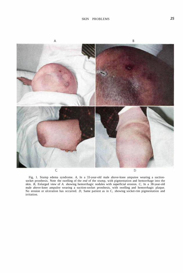
Fig, 1. Stump edema syndrome. A, In a 33-year-old male above-knee amputee wearing a suction-socket prosthesis. Note the swelling of the end of the stump, with pigmentation and hemorrhage into the skin. B, Enlarged view of A. showing hemorrhagic nodules with superficial erosion. C, In a 38-year-old male above-knee ampulee wearing a suction-socket prosthesis, with swelling and hemorrhagic plaque. No erosion or ulceration has occurred. D, Same patient as in C, showing socket-rim pigmentation and irritation.
SKIN PROBLEMS 25

Fig 2 Chronic ischemic ulcer, in a 43-year-okl male below-knee amputee. Poor prosthetic fit with venous obstruction was productive of this lesion
Fig. 3. Pigmentation following stump edema syndrome. A, Brown pigmentation of the skin of the distal portion of the stump. B, Microscopic section of .1, showing a marked increase in the thickness of the epidermis, with sclerosis of collagen and infiltration of pigment-laden cells.
Fig 4 Strangulated skin. Unusual view, showing the distal stump skin resting on the foam-rubber cushion, as seen through the valve opening of a suction-socket prosthesis. A portion of the skin has become partially strangulated, resulting in stasis, edema, and pain.
26
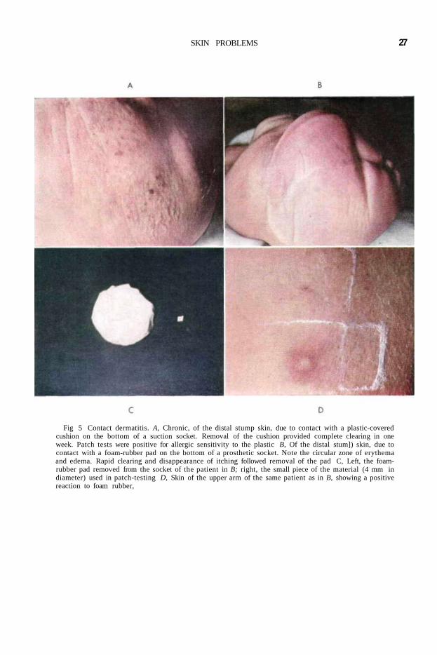
Fig 5 Contact dermatitis. A, Chronic, of the distal stump skin, due to contact with a plastic-covered cushion on the bottom of a suction socket. Removal of the cushion provided complete clearing in one week. Patch tests were positive for allergic sensitivity to the plastic B, Of the distal stum]) skin, due to contact with a foam-rubber pad on the bottom of a prosthetic socket. Note the circular zone of erythema and edema. Rapid clearing and disappearance of itching followed removal of the pad C, Left, the foam-rubber pad removed from the socket of the patient in B; right, the small piece of the material (4 mm in diameter) used in patch-testing D, Skin of the upper arm of the same patient as in B, showing a positive reaction to foam rubber,
SKIN PROBLEMS 27
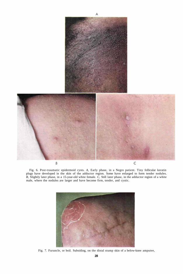
Fig. 6. Post-traumatic epidermoid cysts. A, Early phase, in a Negro patient. Tiny follicular keratin plugs have developed in the skin of the adductor region. Some have enlarged to form tender nodules. B, Slightly later phase, in a 15-year-old white female. C, Still later phase, in the adductor region of a white male, where the nodules are larger and have become firm, tender, and cystic.
Fig. 7. Furuncle, or boil. Subsiding, on the distal stump skin of a below-knee amputee,
28
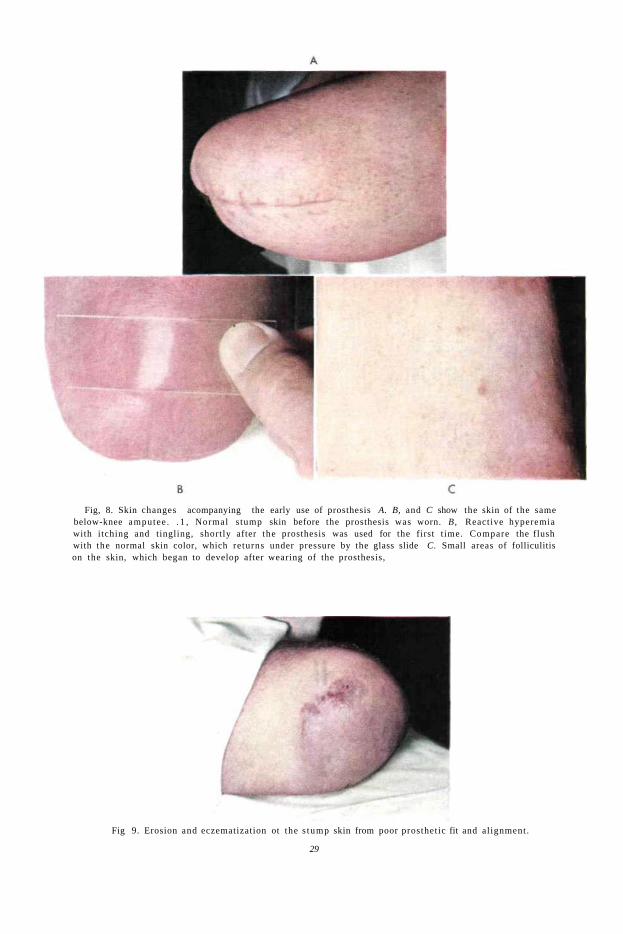
Fig, 8. Skin changes acompanying the early use of prosthesis A. B, and C show the skin of the same below-knee amputee . . 1 , Normal s tump skin before the prosthesis was worn. B, Reactive hyperemia with itching and tingling, shortly after the prosthesis was used for the first t ime. Compare the flush with the normal skin color, which returns under pressure by the glass slide C. Small areas of folliculitis on the skin, which began to develop after wearing of the prosthesis,
Fig 9. Erosion and eczematization ot the s tump skin from poor prosthetic fit and al ignment.
29

Fig. 10. Nonspecific eczematization. A, Of three months' duration on the stump skin of a 32-year-old above-knee amputee who presented unusually poor stump cleanliness. B, Enlarged view of A, showing erythema, edema, and vesiculation. After a simple hygienic program with a sudsing detergent containing hexachlorophene, the eczematous process disappeared completely.
Fig. 11. Skin irritation in the crotch area. A, Chronic, resulting from continued friction and pressure from the socket. B, Enlarged view of A, showing thickened (lichenified) and pigmented skin containing the early phase of post-traumatic epidermoid cysts. The skin of this area may become eroded or ulcerated. In some instances, these problems may be corrected by proper prosthetic fit and alignment.
30 LEVY
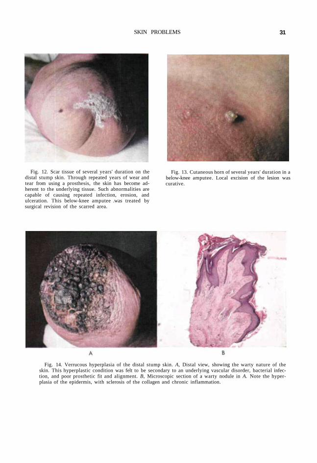
Fig. 12. Scar tissue of several years' duration on the distal stump skin. Through repeated years of wear and tear from using a prosthesis, the skin has become adherent to the underlying tissue. Such abnormalities are capable of causing repeated infection, erosion, and ulceration. This below-knee amputee .was treated by surgical revision of the scarred area.
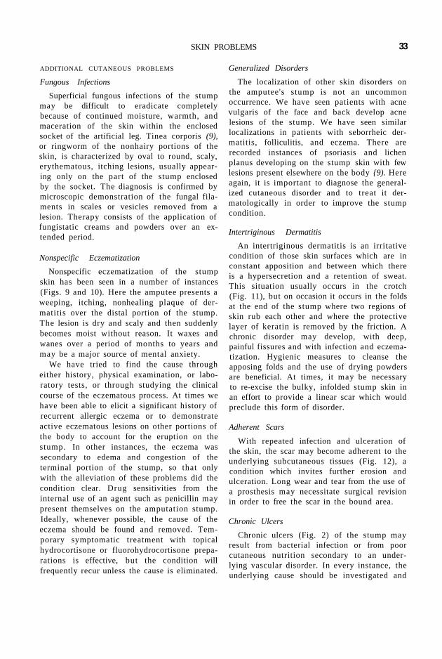
Fig. 13. Cutaneous horn of several years' duration in a below-knee amputee. Local excision of the lesion was curative.
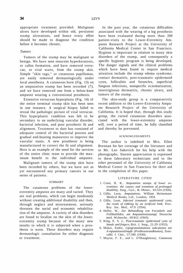
Fig. 14. Verrucous hyperplasia of the distal stump skin. A, Distal view, showing the warty nature of the skin. This hyperplastic condition was felt to be secondary to an underlying vascular disorder, bacterial infection, and poor prosthetic fit and alignment. B, Microscopic section of a warty nodule in A. Note the hyperplasia of the epidermis, with sclerosis of the collagen and chronic inflammation.
SKIN PROBLEMS 31
ADDITIONAL CUTANEOUS PROBLEMS
Fungous Infections
Superficial fungous infections of the stump may be difficult to eradicate completely because of continued moisture, warmth, and maceration of the skin within the enclosed socket of the artificial leg. Tinea corporis (9), or ringworm of the nonhairy portions of the skin, is characterized by oval to round, scaly, erythematous, itching lesions, usually appearing only on the part of the stump enclosed by the socket. The diagnosis is confirmed by microscopic demonstration of the fungal filaments in scales or vesicles removed from a lesion. Therapy consists of the application of fungistatic creams and powders over an extended period.
Nonspecific Eczematization
Nonspecific eczematization of the stump skin has been seen in a number of instances (Figs. 9 and 10). Here the amputee presents a weeping, itching, nonhealing plaque of dermatitis over the distal portion of the stump. The lesion is dry and scaly and then suddenly becomes moist without reason. It waxes and wanes over a period of months to years and may be a major source of mental anxiety.
We have tried to find the cause through either history, physical examination, or laboratory tests, or through studying the clinical course of the eczematous process. At times we have been able to elicit a significant history of recurrent allergic eczema or to demonstrate active eczematous lesions on other portions of the body to account for the eruption on the stump. In other instances, the eczema was secondary to edema and congestion of the terminal portion of the stump, so that only with the alleviation of these problems did the condition clear. Drug sensitivities from the internal use of an agent such as penicillin may present themselves on the amputation stump. Ideally, whenever possible, the cause of the eczema should be found and removed. Temporary symptomatic treatment with topical hydrocortisone or fluorohydrocortisone preparations is effective, but the condition will frequently recur unless the cause is eliminated.
Generalized Disorders
The localization of other skin disorders on the amputee's stump is not an uncommon occurrence. We have seen patients with acne vulgaris of the face and back develop acne lesions of the stump. We have seen similar localizations in patients with seborrheic dermatitis, folliculitis, and eczema. There are recorded instances of psoriasis and lichen planus developing on the stump skin with few lesions present elsewhere on the body {9). Here again, it is important to diagnose the generalized cutaneous disorder and to treat it der-matologically in order to improve the stump condition.
Intertriginous Dermatitis
An intertriginous dermatitis is an irritative condition of those skin surfaces which are in constant apposition and between which there is a hypersecretion and a retention of sweat. This situation usually occurs in the crotch (Fig. 11), but on occasion it occurs in the folds at the end of the stump where two regions of skin rub each other and where the protective layer of keratin is removed by the friction. A chronic disorder may develop, with deep, painful fissures and with infection and eczematization. Hygienic measures to cleanse the apposing folds and the use of drying powders are beneficial. At times, it may be necessary to re-excise the bulky, infolded stump skin in an effort to provide a linear scar which would preclude this form of disorder.
Adherent Scars
With repeated infection and ulceration of the skin, the scar may become adherent to the underlying subcutaneous tissues (Fig. 12), a condition which invites further erosion and ulceration. Long wear and tear from the use of a prosthesis may necessitate surgical revision in order to free the scar in the bound area.
Chronic Ulcers
Chronic ulcers (Fig. 2) of the stump may result from bacterial infection or from poor cutaneous nutrition secondary to an underlying vascular disorder. In every instance, the underlying cause should be investigated and
SKIN PROBLEMS 33
appropriate treatment provided. Malignant ulcers have developed within old, persistent stump ulcerations, and hence every effort should be made to diagnose the condition before it becomes chronic.
Tumors
Tumors of the stump may be malignant or benign. We have seen innocent hyperkeratosis, or callus formation, and have removed verru-cae, or viral warts, from the stump skin. Simple "skin tags," or cutaneous papillomas, are easily removed dermatologically under local anesthesia. A cutaneous horn (Fig. 13) on an amputation stump has been recorded (7), and we have removed one from a below-knee amputee wearing a conventional prosthesis.
Extensive verrucous hyperplasia (Fig. 14) of the entire terminal stump skin has been seen in one instance. A surgical biopsy failed to reveal the pathologic picture of viral verrucae. This hyperplastic condition was felt to be secondary to an underlying vascular disorder, bacterial infection, and poor prosthetic fit and alignment. Treatment to date has consisted of adequate control of the bacterial process and gradual end-bearing maneuvers to improve the vascular stasis. A new prosthesis is being manufactured to correct the fit and alignment. Here is an example of the need for the services of the entire clinic team to provide the maximum benefit to the individual amputee.
Malignant tumors of the stump skin have been recorded by others, but we have not as yet encountered any primary cancers in our series of patients.
SUMMARY
The cutaneous problems of the lower-extremity amputee are many and varied. They are real problems, which can begin insidiously without creating additional disability and then, through neglect and mistreatment, seriously threaten the social and economic rehabilitation of the amputee. A variety of skin disorders are found to localize on the skin of the lower-extremity stump because of the many new insults to which it is subjected when a prosthesis is worn. These disorders may require dermatologic consultation for either diagnosis or treatment.
In the past year, the cutaneous difficulties associated with the wearing of a leg prosthesis have been evaluated during more than 200 patient-visits to the Lower-Extremity Amputee Research Project at the University of California Medical Center in San Francisco. Hygiene is important in relation to many skin disorders of the stump, and consequently a specific hygienic program is being developed, The danger signals and the clinical problems which have been found to require medical attention include the stump edema syndrome, contact dermatitis, post-traumatic epidermoid cysts, folliculitis and furuncles, superficial fungous infections, nonspecific eczematization, intertriginous dermatitis, chronic ulcers, and tumors of the stump.
The skin-study group is a comparatively recent addition to the Lower-Extremity Amputee Research Project of the University of California. It is hoped that, through this study group, the varied cutaneous disorders associated with the lower-extremity amputee will, over a period of time, be fully classified and thereby be prevented.
ACKNOWLEDGMENTS
The author is indebted to Mrs. Ellen Brennan for her coverage of the literature and to Mr. Leo Sakovich for his help with the photographs. Sincere appreciation is expressed to these laboratory technicians and to the other personnel of the University of California Medical Center in San Francisco for their aid in the completion of this paper.
LITERATURE CITED
1. Conn, H. R., Amputation stumps of lower extremities: the causes and treatment of prolonged disability, Surg., Gyn., & Obstet., 43:524 (1926).
2. Gillis, Leon, Amputations, William Heinemann Medical Books, Ltd., London, 1954.
3. Gillis, Leon, Infected traumatic epidermoid cysts, the result of rubbing by an artificial limb, Proc. Roy. Soc. Med., 47:9 (1954).
4. Heller, W., Zur Behandlung von Furunkeln und Follikuliliden, am Amputationsstumpf, Deutsche med. Wchnschr., 69:812 (1943).
5. King, E. S. J., Post-traumatic epidermoid cysts of hands and fingers, Brit. J. Surg., 21:29 (1933).
6. Makai, Endre, Lipogranulomatosis subcutanea am A mputationssliimpfe (Prolhesenrandknoten), Zent-ralbl. f. Chir., 57:590 (1930).
7. Mayne, F. E., and L. O'Shaughnessy, Cutaneous
LEVY 34

horn on an amputation stump, Brit. Med. J., 1: 624 (1931).
8. Savitt, Leonard E., Contact dermatitis encountered in the production of epoxy resins, A. M. A. Arch. Dermat. & Syphilol., 71:212 (1955).
9. Schamberg, I. L., Dermatitis of lower limb amputation slump, J. Am. Med. Assoc, 150:1653 (1952).
10. Schwartz, Louis, Allergic occupational dermatitis in our war industries, Ann. Allergy, 2:387 (1944).
11. Schwartz, L., L. Tulipan, and S. M. Peck, Occupational diseases of the skin, 2nd ed., Lea & Febiger, Philadelphia, 1947.
12. Slocum, Donald B., An atlas of amputations, Mosby, St. Louis, 1949. pp. 254-288.
13. Thomas, A., and C. C. Haddan, Amputation prosthesis, Lippincott, Philadelphia, 1945. pp. 54-67.
14. Wohlvill, Fr., Über "Prothesenrandknoten," Vir-chows Arch. f. path, Anat.. 288:576 (1933).
15. Young, Freida, Post-traumatic epidermoid cysts, Lancet, 1:716 (1951).
16. zur Verth, [M.], Prothesenrandknoten und Pro-thesenrandabszesse, Zentralbl. f. Chir., 63:322 (1926).
17. zur Verth, [M.], Prothesenrandknoten und ihre Entstehung, Dermat. Wchnschr., 88:45 (1929).
18. zur Verth, M., and K. H. Vohwinkel, Prothesenrandknoten, Deutsche Ztschr. f. Chir., 205:302 (1927).
SKIN PROBLEMS 35