Embed Size (px)
Citation preview
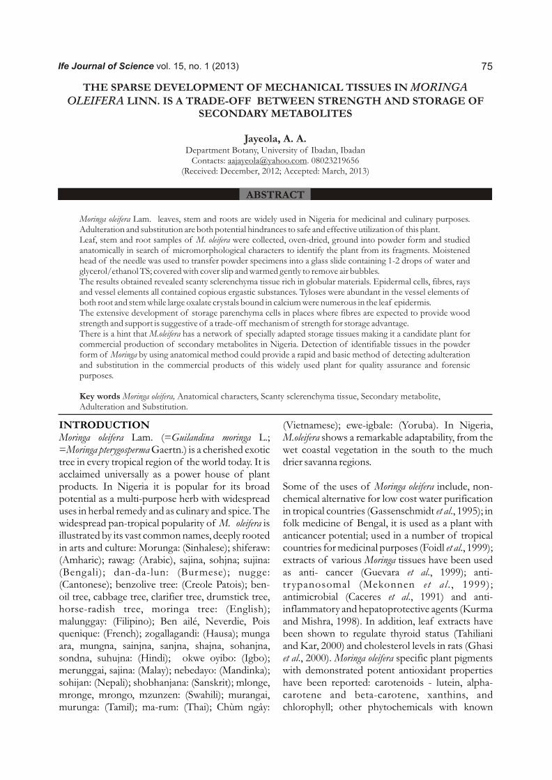
Ife Journal of Science vol. 15, no. 1 (2013) 75
THE SPARSE DEVELOPMENT OF MECHANICAL TISSUES IN MORINGA OLEIFERA LINN. IS A TRADE-OFF BETWEEN STRENGTH AND STORAGE OF
SECONDARY METABOLITES
Jayeola, A. A.Department Botany, University of Ibadan, Ibadan
Contacts: [email protected]. 08023219656(Received: December, 2012; Accepted: March, 2013)
ABSTRACT
Moringa oleifera Lam. leaves, stem and roots are widely used in Nigeria for medicinal and culinary purposes. Adulteration and substitution are both potential hindrances to safe and effective utilization of this plant.Leaf, stem and root samples of M. oleifera were collected, oven-dried, ground into powder form and studied anatomically in search of micromorphological characters to identify the plant from its fragments. Moistened head of the needle was used to transfer powder specimens into a glass slide containing 1-2 drops of water and glycerol/ethanol TS; covered with cover slip and warmed gently to remove air bubbles. The results obtained revealed scanty sclerenchyma tissue rich in globular materials. Epidermal cells, fibres, rays and vessel elements all contained copious ergastic substances. Tyloses were abundant in the vessel elements of both root and stem while large oxalate crystals bound in calcium were numerous in the leaf epidermis. The extensive development of storage parenchyma cells in places where fibres are expected to provide wood strength and support is suggestive of a trade-off mechanism of strength for storage advantage.There is a hint that M.oleifera has a network of specially adapted storage tissues making it a candidate plant for commercial production of secondary metabolites in Nigeria. Detection of identifiable tissues in the powder form of Moringa by using anatomical method could provide a rapid and basic method of detecting adulteration and substitution in the commercial products of this widely used plant for quality assurance and forensic purposes.
Key words Moringa oleifera, Anatomical characters, Scanty sclerenchyma tissue, Secondary metabolite, Adulteration and Substitution.
INTRODUCTIONMoringa oleifera Lam. (=Guilandina moringa L.; =Moringa pterygosperma Gaertn.) is a cherished exotic tree in every tropical region of the world today. It is acclaimed universally as a power house of plant products. In Nigeria it is popular for its broad potential as a multi-purpose herb with widespread uses in herbal remedy and as culinary and spice. The widespread pan-tropical popularity of M. oleifera is illustrated by its vast common names, deeply rooted in arts and culture: Morunga: (Sinhalese); shiferaw: (Amharic); rawag: (Arabic), sajina, sohjna; sujina: (Bengali); dan-da-lun: (Burmese); nugge: (Cantonese); benzolive tree: (Creole Patois); ben-oil tree, cabbage tree, clarifier tree, drumstick tree, horse-radish tree, moringa tree: (English); malunggay: (Filipino); Ben ailé, Neverdie, Pois quenique: (French); zogallagandi: (Hausa); munga ara, mungna, sainjna, sanjna, shajna, sohanjna, sondna, suhujna: (Hindi); okwe oyibo: (Igbo); merunggai, sajina: (Malay); nebedayo: (Mandinka); sohijan: (Nepali); shobhanjana: (Sanskrit); mlonge, mronge, mrongo, mzunzen: (Swahili); murangai, murunga: (Tamil); ma-rum: (Thai); Chùm ngây:
(Vietnamese); ewe-igbale: (Yoruba). In Nigeria, M.oleifera shows a remarkable adaptability, from the wet coastal vegetation in the south to the much drier savanna regions.
Some of the uses of Moringa oleifera include, non-chemical alternative for low cost water purification in tropical countries (Gassenschmidt et al., 1995); in folk medicine of Bengal, it is used as a plant with anticancer potential; used in a number of tropical countries for medicinal purposes (Foidl et al., 1999); extracts of various Moringa tissues have been used as anti- cancer (Guevara et al., 1999); anti-trypanosomal (Mekonnen et al . , 1999); antimicrobial (Caceres et al., 1991) and anti-inflammatory and hepatoprotective agents (Kurma and Mishra, 1998). In addition, leaf extracts have been shown to regulate thyroid status (Tahiliani and Kar, 2000) and cholesterol levels in rats (Ghasi et al., 2000). Moringa oleifera specific plant pigments with demonstrated potent antioxidant properties have been reported: carotenoids - lutein, alpha-carotene and beta-carotene, xanthins, and chlorophyll; other phytochemicals with known
76
powerful antioxidant ability such as kaempferol, quercetin, rutin and caffeoylquinic acids; powerful antioxidant vitamins, notably, C, E, and A and essential micronutrients with antioxidant activity, selenium and zinc (Fuglie, 1999).
Microscopic inspection of medicinal plant materials is indispensable for the identification of broken or powdered materials (WHO, 1998). It has been suggested that herbal medicine should be subjected to the same stringent scrutiny and controls as common drugs before their release on the market, and micromorphology is a rapid, cost effective and efficient method in quality control and assessment of herbal remedies (Jayeola, 2009, 2010).
In spite of the of the widespread commercial processing and use of Moringa, information is scanty on its structure and possible localization of its secondary metabolites. The objective of this research is to study the distribution of tissues in M.oleifera and to identify the specific tissues concerned with the storage of secondary products, using fresh samples and fragments recovered from processed commercial samples.
METHODOLOGYSamples of the fresh leaves, stem, fruits and roots of Moringa oleifera were collected from the two broad eco-floristic zones of Nigeria, comprising the forest (Akure and Ibadan) and savanna (Zaria and Katsina). For each zone, five accessions were collected and each separated into leaves, stem, roots, flowers and fruits. Altogether, samples consisted of 10 accessions each of leaves, stem, roots, flowers and fruits. Each organ was divided into two sub-samples. One sub-sample was dried, and ground while the other sub-sample was sectioned transversely and longitudinally at 25-50µ thickness using the Sledge Microtome. For the Moringa powder, the sample that adhered to the needle was transferred on the labeled glass slide containing 1 - 2 drops of water and glycerol/ethanol TS (ethanol Thin Stillage); covered with cover slip and warmed gently to remove air bubbles. This was repeated using phloroglucinol TS to detect lignified cell wall; Sudan Red TS for suberized cuticular cell walls and fats and oil; for myrosin cells sections were also stained in lactophenol-aniline blue. Slide preparation followed the classical method
recommended by WHO (1998).
For light microscopy, the sections were stained with safranin and mounted on slides using standard procedure (Wilkinson, 1979).
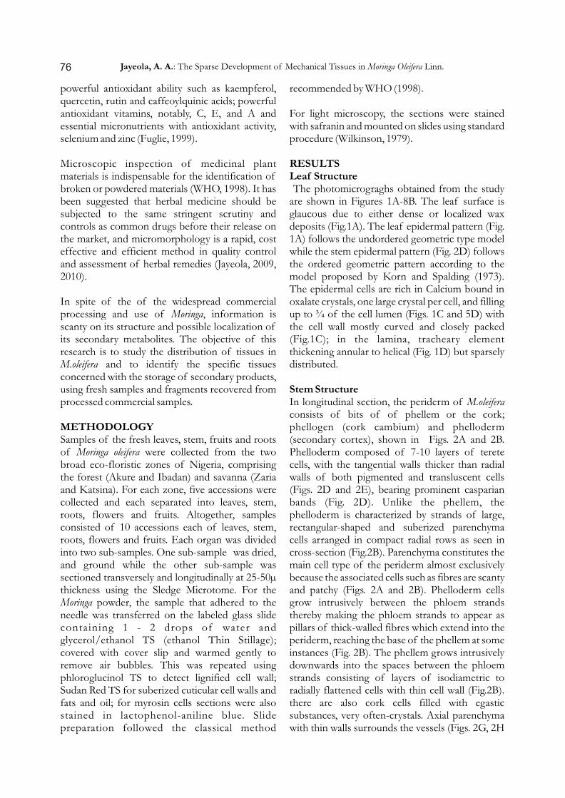
RESULTSLeaf Structure The photomicrograghs obtained from the study are shown in Figures 1A-8B. The leaf surface is glaucous due to either dense or localized wax deposits (Fig.1A). The leaf epidermal pattern (Fig. 1A) follows the undordered geometric type model while the stem epidermal pattern (Fig. 2D) follows the ordered geometric pattern according to the model proposed by Korn and Spalding (1973). The epidermal cells are rich in Calcium bound in oxalate crystals, one large crystal per cell, and filling up to ¾ of the cell lumen (Figs. 1C and 5D) with the cell wall mostly curved and closely packed (Fig.1C); in the lamina, tracheary element thickening annular to helical (Fig. 1D) but sparsely distributed.
Stem StructureIn longitudinal section, the periderm of M.oleifera consists of bits of of phellem or the cork; phellogen (cork cambium) and phelloderm (secondary cortex), shown in Figs. 2A and 2B. Phelloderm composed of 7-10 layers of terete cells, with the tangential walls thicker than radial walls of both pigmented and transluscent cells (Figs. 2D and 2E), bearing prominent casparian bands (Fig. 2D). Unlike the phellem, the phelloderm is characterized by strands of large, rectangular-shaped and suberized parenchyma cells arranged in compact radial rows as seen in cross-section (Fig.2B). Parenchyma constitutes the main cell type of the periderm almost exclusively because the associated cells such as fibres are scanty and patchy (Figs. 2A and 2B). Phelloderm cells grow intrusively between the phloem strands thereby making the phloem strands to appear as pillars of thick-walled fibres which extend into the periderm, reaching the base of the phellem at some instances (Fig. 2B). The phellem grows intrusively downwards into the spaces between the phloem strands consisting of layers of isodiametric to radially flattened cells with thin cell wall (Fig.2B). there are also cork cells filled with egastic substances, very often-crystals. Axial parenchyma with thin walls surrounds the vessels (Figs. 2G, 2H
Jayeola, A. A.: The Sparse Development of Mechanical Tissues in Moringa Oleifera Linn.
1A
1C
Trichome
20µ
1B
1D
1
A
4A 4B
D4C
C
4D
mx
mx
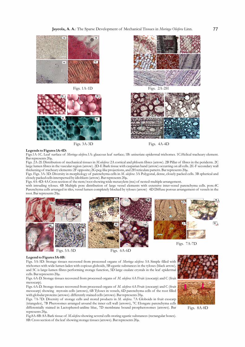
Legends to Figures 1A-4D:Figs.1A-1C. Leaf surface of Moringa oleifera.1A: glaucous leaf surface; 1B: uniseriate epidermal trichomes. 1C:Helical tracheary element. Bar represents 20µ.Figs. 2A-2I: Distribution of mechanical tissues in M.oleifera: 2A cortical and phloem fibres (arrow). 2B Pillar of fibres in the periderm. 2C large lumen fibres in the vascular region (arrow). 2D-E Bark tissue with casparian band (arrow) occurring on all cells. 2E-F secondary wall thickening of tracheary elements: 2F opposite; 2G peg-like projections, and 2H reticulate pattern. Bar represents 20µ.Figs. Figs. 3A-3D: Diversity in morphology of parenchyma cells in M. oleifera: 3A Polygonal, dense, closely packed cells. 3B spherical and closely packed cells interspersed by idioblasts (arrow). Bar represents 20µ.Figs. 4A-4D: 4A Cross section of the stem/root showing wide metaxylem (mx) of nested-multiple arrangement.with intruding tyloses. 4B Multiple pore distribution of large vessel elements with extensive inter-vessel parenchyma cells. pore.4C Parenchyma cells arranged in tiles, vessel lumen completely blocked by tyloses (arrow) 4D Diffuse porous arrangement of vessels in the root. Bar represents 20µ.
Figs. 1A-1D Figs. 2A-2H
Figs. 3A-3D Figs. 4A-4D
7A 7B
C 7D 7C
Myrosin cells
3A
3C 3D
3B
Continuous uniform band of Parenchyma cells Parenchyma cells differing in shapes and sizes
6A 6B
6C D
Myrosin cell
6D
Myrosin cell
1
5A 5B
5C
5D
Druse
8A
8B
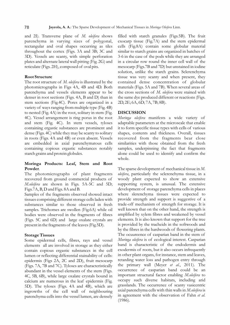
Legend to Figures 5A-8B:Figs. 5A-5D: Storage tissues recovered from processed organs of Moringa oleifera: 5A Simple filled with trichomes with wide lumen laden with copious globoids, 5B egastic substances in the tyloses (black arrow), and 5C is large-lumen fibres performing storage function, 5D large oxalate crystals in the leaf epidermal cells. Bar represents 20µ.Figs. 6A-D. Storage tissues recovered from processed organs of M. oleifera: 6A Fruit (exocarp) and C (fruit mesocarp).Figs. 6A-D. Storage tissues recovered from processed organs of M. oleifera: 6A Fruit (exocarp) and C (fruit mesocarp) showing myrosin cells (arrows), 6B Tyloses in vessels, 6D parenchyma cells of the root filled with globular proteins (arrows). differently stained cells (arrows). Bar represents 20µ.Figs. 7A-7D: Diversity of storage cells and stored products in M. oleifera. 7A Globoids in fruit exocarp (triangules), 7B Pherosomes arranged around the inner cell wall (arrows), 7C Elongate parenchyma cells differentially stained in Lactophenol-aniline blue, 7D membrane bound prospherosomes (arrows). Bar represents 20µ.Fig.8A-8B: 8A Bark tissue of M.oleifera showing several cells storing egastic substances (rectangular boxes). 8B: Cross section of the leaf showing storage tissues (arrows). Bar represents 20µ.
Figs. 5A-5D
Figs. 7A-7D
Figs. 6A-6D
Figs. 8A-8D
77Jayeola, A. A.: The Sparse Development of Mechanical Tissues in Moringa Oleifera Linn.
and 2I). Transverse plane of M. oleifera shows parenchyma in varying sizes of polygonal, rectangular and oval shapes occurring as tiles throughout the cortex (Figs. 3A and 3B; 3C and 3D). Vessels are scanty, with simple perforation plates and alternate lateral wall pitting (Fig. 2G) and reticulate (Figs. 2H), composed of oval pits.
Root StructureThe root structure of M. oleifera is illustrated by the photomicrographs in Figs 4A, 4B and 4D. Both parenchyma and vessels elements appear to be denser in root sections (Figs. 4A, B and D) than in stem sections (Fig.4C). Pores are organized in a variety of ways ranging from multiple type (Fig. 4B) to nested (Fig. 4A) in the root, solitary in stem (Fig. 4C). Vessel arrangement is ring porus in the root and stem (Fig. 4C). In stem vessels, tyloses containing ergastic substances are prominent and dense (Figs. 4C) while they may be scanty to solitary in roots (Figs. 4A and 4B) or even absent. Vessels are embedded in axial parenchymatous cells containing copious ergastic substances notably starch grains and protein globules.
Moringa Products: Leaf, Stem and Root Powder.The photomicrographs of plant fragments recovered from ground commercial products of M.oleifera are shown in Figs. 5A-5C and 5D; Figs.7A, B, D and Figs. 8A and B.Samples of the fragments observed showed intact tissues comprising different storage cells laden with substances similar to those observed in fresh samples. Trichomes are swollen (Fig.6A) while oil bodies were observed in the fragments of fibres (Figs. 5C and 6D) and large oxalate crystals are present in the fragments of the leaves (Fig.5D).
Storage TissuesSome epidermal cells, fibres, rays and vessel elements all are involved in storage as they either contain copious ergastic substances in the cell lumen or reflecting differential stainability of cells: epidermis (Figs 2A, 2C and 2D), fruit mesocarp (Figs. 7A, 7B and 7C). Tyloses are characteristically abundant in the vessel elements of the stem (Figs. 4C, 5B, 6B), while large oxalate crystals bound in calcium are numerous in the leaf epidermis (Fig. 5D). The tyloses (Figs. 4A and 4B), which are ingrowths of the cell wall of neighbouring parenchyma cells into the vessel lumen, are densely
filled with starch granules (Figs.5B). The fruit exocarp tissue (Fig.7A) and the stem epidermal cells (Fig.8A) contain some globular material similar to starch grains are organized in batches of 3-6 in the case of the pods while they are arranged in a circular row round the inner cell wall of the mesocarp (Figs.7B and 7D) but unstained in iodine solution, unlike the starch grains. Sclerenchyma tissue was very scanty and when present, they contained dense concentration of globular materials (Figs. 5A and 7B). When several areas of the cross sections of M. oleifera were stained with the same dye produced different or reactions (Figs. 2D, 2E; 6A, 6D; 7A, 7B; 8B).
DISCUSSIONMoringa oleifera manifests a wide variety of adaptable parameters at the microscale that enable it to form specific tissue types with cells of various shapes, contents and thickness. Overall, tissues recovered from the fragments bear close similarities with those obtained from the fresh samples, underpinning the fact that fragments alone could be used to identify and confirm the whole.
The sparse development of mechanical tissue in M. oleifera, particularly the sclerenchyma tissue, in a woody plant expected to show an extensive supporting system, is unusual. The extensive development of storage parenchyma cells in places where slerenchyma tissues were expected to provide strength and support is suggestive of a trade-off mechanism of strength for storage. It is well known that on the other hand, the strength is amplified by xylem fibres and weakened by vessel elements. It is also known that support for the tree is provided by the tracheids in the softwoods and by the fibres in the hardwoods of flowering plants. The ocuureence of casparian band in the stem of Moringa oleifera is of ecological interest. Casparian band is characteristic of the endodermis and exodermis of roots, but it also occurs infrequently in other plant organs, for instance, stem and leaves, retarding water loss and pathogen entry through the primary wall (Meyer et al., 2011). The occurrence of casparian band could be an important structural factor enabling M.oleifera to occupy such diverse habitats, including arid grasslands. The occurrence of scanty vasicentric axial parenchyma cells with thin walls in M.oleifera is in agreement with the observation of Fahn et al. (1986).
78 Jayeola, A. A.: The Sparse Development of Mechanical Tissues in Moringa Oleifera Linn.
Rather, there is a well developed system of storage tissue involving not just the traditional parenchyma cells, but also tissues that ordinarily perform supporting function such as fibre, vessel elements and epidermis. It has been suggested that heartwood forms as a repository for waste metabolic products (polyphenolics or tannins) or for surplus photosynthates. It is more likely, however, that heartwood forms to keep the amount of sapwood at an optimum thus confirming the fact that potassium, phosphorus, nitrogen and sulphur are reabsorbed from the sapwood as it is transformed into heartwood for nutritional balance in the living part of the tree.
In species with larger bordered pits, blockage of the vessels occurs as a result of the growth of adjacent parenchyma cells through the pit forming metabolite-containing balloon-like structures in the vessels known as tyloses. In species with small pits, blockage of the vessels occurs due to the secretion by the ray parenchyma of tannin or gum-like materials through the pits. In modern plants, vessel size is an important character in predicting where tyloses are formed (Zimmermann, 1983; Jaquish and Ewers, 2001): large vessels develop tyloses. But more commonly, many tyloses are formed in the vessel lumen, thus blocking the passage completely. Hence no conduction is possible and where tyloses are formed prematurely in response to infection, providing a defence mechanism and inhibit the spread of pathogen throughout the plant.
The presence of myrosin cells in M. oleifera is congruent with previous studies on systematic affinity of glucosinolate-producing plant families (Chase et al., 1998; APG III, 2009). Glucosinolates are a diverse class of plant-specific secondary metabolites synthesized mainly by myrosin cells producing plants. Myrosin cells are known to contain myrosinase which can break down glucosinolates to isothicynate, ntriles and elemental sulphur, thiocyanates, oxazolidines- -thiones and epithionitriles, as an evolution-driven chemical defence system in plants (Grubb and Abel, 2006). Glucosinolate derivatives not only contribute greatly to the distinctive flavour and aroma of cruciferous vegetables and condiments, but also possess profound biological activities that range from their participation in plant defense and auxin homeostasis to cancer prevention in humans
(Kliebenstein et al., 2005)
Plant cell and tissue cultures can be established routinely under sterile conditions from explants, such as plant leaves or stems. Strain improvement, methods for the selection of high-producing cell lines, and medium optimizations can lead to an enhancement in secondary metabolite production and utilization. Lack of progress in understanding of metabolic pathways had been shown to be the first barrier in developing commercial processes (Dornenburg and Knorr, 1995). It was expected that the biosynthetic capacity of plants could be exploited in vitro using plant cells and cell tissue systems analogous to microbial cells in fermentation processes.
There is a hint that M.oleifera has developed tissues that are adapted to store many secondary metabolites. One of the important requirements for the improvement of secondary metabolite production in plants is to identify candidate species with vast potentials for storage of these products. For this purpose, different strategies to increase biosynthesis of secondary metabolites and to alter the product spectrum have to be investigated. Those potentially useful species can then be induced with elicitors (molecules that stimulate secondary metabolism) either endogenously or exogenously. This will be applicable to both poisonous and non-poisonous plants. In Mentha species, for example, the epidermal oil glands on the leaves are the primary sites of monoterpene biosynthesis; these are the cells in which the cytotoxic substances are accumulated. If in cell cultures, specialized accumulation sites are supressed, then the plant is not able to accumulate high levels of monoterpenes. Moringa oleifera, with a network of specially adapted storage tissues could be one of the candidate plants for commercial production of secondary metabolites in Nigeria.
REFERENCESAPG III. 2009. An update of the Angiosperm
Phylogeny Group classification for the orders and families of flowering plants: APG III. Botanical Journal of the Linnean Society, 161: 105-121.
Cáceres A, López BR, Giron MA, Logemann H 1991. Plants used in Guatemala for the treatment of dermatophytic infections. 1. Screening for antimycotic activity of 44
79Jayeola, A. A.: The Sparse Development of Mechanical Tissues in Moringa Oleifera Linn.
plant extracts. Ethnopharmacology 31(3): 263-276.
Chase, M.W., Bremer, K., Stevens, P.F., Anderberg, A.A., Backlund, A., Bremer, B.G., Briggs, B.G., Endress, Fay M.F., Goldblatt, P, Gustafsson, M.H.G., Hoot, S.B., Judd, W.S., Kallersjo, M., Kellogg, E.A., Kron, K.A., Les, D.H., Morton, C.M., Nickrent, D.L., Olmstead, R.G., Price, R.A., Quinn, C.J., Rodman, J.E., Rudall, P.J., Savoleinen, V., Soltis, D.E., Soltis, P.S., Sytsma, K.J., Thulin, M. 1998. An Ordinal Classification for the Families of Flowering Plants. Annals of Missouri Botanical Gardens 85:531-553
Domenburg, H. and Knorr, D. 1995. Elicitation of chitinases and anthraquinones in Morinda cirrifolia cell cultures. Food Biotechnol. 8, 57-65.
Fahn, A., Werker E. and Baas, P. 1986. Wood anatomy and identification of trees and shrubs from Israel and adjacent regions. Isr.Acad. Sci. Hum., Jerusalem, Israel.
Foidl, N., Mayorga L., Vasquez W 1999. Utilization of marango (Moringa oleifera) as fresh forage for cattle. FAO Anim. Prod. Health Paper143, 341-346.
Fuglie, L.J. 1999. The Miracle Tree: Moringa oleifera: Natural Nutrition for the Tropics. Church World Service, Dakar.
Gassenschmidt, U., Jany, K.D., Tauscher, B., and Niebergall, H. 1995. Isolation and Characterization of a flocculating protein from Moringa oleifera Lam. Biochemica et Biophysica Acta, 124 (3), 477-481.
Ghasi S., Nwobodo E. and Ofili J. O. 2000. Hypocholesterolemic effects of crude extract of leaf of Moringa oleifera Lam in high fat diet fed wistar rats. Journal of Ethnopharmacology 69 (1), 21-25.
Grubb, C. Douglas and Abel Steffen 2006. Glucosinolate metabolism and its Control. Trends in Plant Science 11(2), 89-100.
Guevara, M. G., Oliva, C. R., Machinandiarena, M. & Daleo, G. R. 1999. Purification and properties of an aspartic protease from potato tuber that is inhibited by a basic chitinase. Physiol. Plant 106,164-169.
Jaquish, L. L. and Ewers, F. W. 2001. Seasonal conductivity and embolism in the roots and stems of two clonal ring-porous trees, Sassafras albidum (Laureaceae) and Rhus typhina (Anacardiaceae). American Journal of Botany 88, 206-212.
Jayeola, A.A. 2009. Micromorphological study of plant fragments in some powdered medicinal plants. Journal of Medicinal Plant Research 3(5), 438-442.
Jayeola, A.A. 2010. Anatomical identification of plant fragments in the powdered samples of Moringa oleifera Lam. (Moringaceae). Paper presented at the National Summit on Moringa Development organized by the Raw Material Research Development Council (RMRDC) Headquarters Abuja 16th-18th November, 2010.
Kliebenstein, D. J., Kroymann, J. and T. Mitchell-Olds 2005. The glucosinolate-myrosinase system in an ecological and evolutionary context. Curr. Opin. Plant Biol. 8, 264-271.
Korn, R. W. and Spalding, R. M. 1973. The geometry of plant epidermal cells. New Phytologist 72, 1357-1365
Kurma, S. R. and Mishra, S. H. 1998. Anti-inflammatory and hepatoprotective act iv i t ies of f r u i ts of Moringa pterygosperma Gaertn. Indian Journal of Natural Products 14(1), 3-10.
Mekonnen, Y., Yardleg V., Rock, P. and Croft, S 1999. In vitroantitrypanosomal activity of Moringa stenopetala leaves and roots. Phytother. Res. 13, 538-539.
Meyer, Chris J.; Peterson, Carol A. 2011. Casparian bands occur in the periderm of Pelargonium hortorum stem and root. Annals of Botany 107 (4), 591.
Tahiliani, P. and Kar, A. 2000. Role of Moringa oleifera leaf extract in the regulation of thyroid hormone status in adult male and female rats. Pharmacol. Res. 41, 319-323.
Wilkinson, H.P. 1979. The plant surface (Mainly Leaf). In: Metcalfe, C.R. and Chalk, L.. Anatomy of the Dicotyledons. Volume I: Systematic Anatomy of the Leaf and Stem. 2nd edition. Clarendon Press, Oxford. pp. 98-165
WHO 1998. Quality Control Methods for Medicinal Plant Materials.
Zimmerman, M. 1983. Xylem structure and the ascent of sap. Springer-Verlag. Berlin. pp1-143
80 Jayeola, A. A.: The Sparse Development of Mechanical Tissues in Moringa Oleifera Linn.