Embed Size (px)
Citation preview
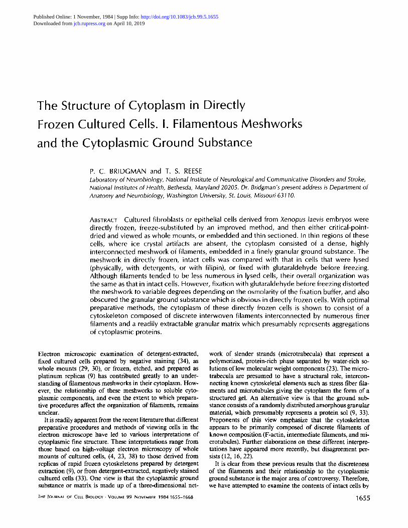
The Structure of Cytoplasm in Directly
Frozen Cultured Cells. I. Filamentous Meshworks
and the Cytoplasmic Ground Substance
P. C. BRIDGMAN and T. S. REESE Laboratory of Neurobiology, National Institute of Neurological and Communicative Disorders and Stroke, National Institutes of Health, Bethesda, Maryland 20205. Dr. Bridgman's present address is Department of Anatomy and Neurobiology, Washington University, St. Louis, Missouri 63110.
ABSTRACT Cultured fibroblasts or epithelial cells derived from Xenopus laevis embryos were directly frozen, freeze-substifuted by an improved method, and then either critical-point- dried and viewed as whole mounts, or embedded and thin sectioned. In thin regions of these cells, where ice crystal artifacts are absent, the cytoplasm consisted of a dense, highly interconnected meshwork of filaments, embedded in a finely granular ground substance. The meshwork in directly frozen, intact cells was compared with that in cells that were lysed (physically, with detergents, or with filipin), or fixed with glutaraldehyde before freezing. Although filaments tended to be less numerous in Iysed cells, their overall organization was the same as that in intact cells. However, fixation with glutaraldehyde before freezing distorted the meshwork to variable degrees depending on the osmolarity of the fixation buffer, and also obscured the granular ground substance which is obvious in directly frozen cells. With optimal preparative methods, the cytoplasm of these directly frozen cells is shown to consist of a cytoskeleton composed of discrete interwoven filaments interconnected by numerous finer filaments and a readily extractable granular matrix which presumably represents aggregations of cytoplasmic proteins.
Electron microscopic examination of detergent-extracted, fixed cultured cells prepared by negative staining (34), as whole mounts (29, 30), or frozen, etched, and prepared as platinum replicas (9) has contributed greatly to an under- standing of filamentous meshworks in their cytoplasm. How- ever, the relationship of these meshworks to soluble cyto- plasmic components, and even the extent to which prepara- tive procedures affect the organization of filaments, remains unclear.
It is readily apparent from the recent literature that different preparative procedures and methods of viewing cells in the electron microscope have led to various interpretations of cytoplasmic fine structure. These interpretations range from those based on high-voltage electron microscopy of whole mounts of cultured cells, (4, 23, 38) to those derived from replicas of rapid frozen cytoskeletons prepared by detergent extraction (9), or from detergent-extracted, negatively stained cultured cells (33). One view is that the cytoplasmic ground substance or matrix is made up of a three-dimensional net-
work of slender strands (microtrabecula) that represent a polymerized, protein-rich phase separated by water-rich so- lutions of low molecular weight components (23). The micro- trabecula are presumed to have a structural role, intercon- necting known cytoskeletal elements such as stress fiber fila- ments and microtubules giving the cytoplasm the form of a structured gel. An alternative view is that the ground sub- stance consists of a randomly distributed amorphous granular material, which presumably represents a protein sol (9, 33). Proponents of this view emphasize that the cytoskeleton appears to be primarily composed of discrete filaments of known composition (F-actin, intermediate fdaments, and mi- crotubules). Further elaborations on these different interpre- tations have appeared more recently, but disagreement per- sists (12, 16, 22).
It is clear from these previous results that the discreteness of the filaments and their relationship to the cytoplasmic ground substance is the major area of controversy. Therefore, we have attempted to examine the contents of intact cells by
THE JOURNAL OF CELL BIOLOGY • VOLUME 99 NOVEMBER 1984 1655-1668 1655
on April 10, 2019jcb.rupress.org Downloaded from http://doi.org/10.1083/jcb.99.5.1655Published Online: 1 November, 1984 | Supp Info:
combining the advantages for structural preservation offered by direct freezing and freeze-substitution with the apprecia- tion of structure in these dimensions afforded by examining whole mounts in high-voltage electron microscopes. Although some previous data using whole mounts after freezing and freeze-substitution is available (22, 38), our efforts to improve freezing and freeze-substitution appear to provide a substan- tially more realistic view of cytoplasm. This improvement justifies a new and detailed look at cytoplasmic fine structure.
In this paper, we concentrate on the relationship between filamentous meshworks and the ground substance in which they are suspended. The regional organization of the cyto- plasm, as determined by patterns of organelle and cytoplasmic movement, is described separately (2). Our interpretation of cytoplasmic fine structure differs in several important ways from those of previous studies using intact or extracted cells prepared by either fixation or direct freezing.
MATERIALS AND METHODS
Cultures: Dissociated cell cultures were derived from the somites or ectoderm of Xenopus laevis. Cultures from somites contained muscle cells and fibroblasts, while cultures from ectoderm contained flbroblasts and epithelial cells. Details of the culture methods have been published elsewhere (3, 21). Dissociated cells were pipetted onto gold or titanium grids coated with Formvar or carbon while immersed in culture medium (88% Steinberg's solution, 10% DI5, 1% fetal bovine serum, 10,000 IU per ml penicillin/streptomycin; os- molarity: 150 mOsmol). Glow-discharged or polylysine-coated carbon films on 300-mesh titanium grids were preferred because they were the most electron lucent and stable under the electron beam.
Albumin Mounts: Chicken egg albumin (Sigma Chemical Co., St. Louis, MO) solutions prepared in concentrations of 2-5% in distiUed water served as controls for freezing and other preparative steps. Coated grids were touched to the surface of a drop of the solution until enough liquid to cover one third to one half of the grid spread onto the grid surface. The grid was then frozen immediately.
Detergent and Filipin Extraction: Cells subjected to detergent or filipin extraction were first rinsed for 15 s in extraction buffer (30) that contained 30 mM PIPES, 25 mM HEPES, l0 mM EGTA, 1.7 mM MgSO4 and which was neutralized to pH 6.9 with KOH. The omsolarity of the buffer was 140 mOsmol. Cells were then lysed with extraction buffer supplemented with either 0.1% Triton X- 100, 0.1% saponin, or a mixture of 0.04% fllipin and 1% DMSO for 30-60 s. Lysis was terminated by freezing.
Freezing: Cultures on grids were frozen by rapid injection of the grid into a rapidly stirred mixture of propane/isopentane (3:1) (13) or propane/ ethane (3:1) cooled by liquid nitrogen. Injection of the grids into the freezing mixture was accomplished by means of a spriog-driven device (6, 8). Rapid stirring of the freezing mixture and careful removal of excess fluid from the grid immediately before freezing was critical to obtaining good freezing. Because the culture medium was rather viscous, grids were rinsed in Steinberg's solution (osmolarity: 134 mOsmol) before freezing. Some cultures grown on grids were also frozen by slamming them against a copper block cooled by liquid helium (10).
Freeze-Substitution: While under liquid nitrogen, grids to be freeze- substituted were placed in a stainless-stcel holder, which was then placed in a scintillation vial containing l0 ml of the primary substitution fluid covered with 10 ml of liquid nitrogen. The vial was usually transferred to a freezer maintained at -80* to -78"(2, though passive warmup in a well-insulated box was also successful. Temperature was monitored by a digital thermometer and chart recorder connected to a thermocouple placed in a dummy vial filled with l0 ml of the solvent. When changes or washes were to be made during the substitution procedure, the holder was quickly transferred from vial to vial with a pair of forceps in order to avoid warming or drying of the samples, or condensation of atmospheric water on the cold fluid during the transfers. Details as well as the rationale for the composition of the substitution fluid, times, and temperatures during warmup are presented in the Appendix.
Fixation: Some cultures were fixed before freezing. A rinse with Stein- berg's solution preceded fixation with 0.5% glutaraldehyde and 0.2% tannic acid in HEPES buffer (0.05-0.1 M), 7.4, at room temperature. The osmolarity of fixation buffers (as well as other solutions) was measured on a vapor pressure osmometer (Wescor Inc., Logan, UT). The glutaraldehyde was diluted from a high quality electron microscope grade 8% solution supplied under nitrogen
1656 THE JOURNAL OF CELL BIOLOGY • VOLUME 99, 1984
(Polysciences, Inc., Warrington, PA). After 1 h the cultures were rinsed with buffer and then further fixed for 2 h with 2% glutaraldehyde in the same buffer.
Critical-point Drying: After three final rinses in anhydrous acetone, the grid holder was quickly transferred to the chamber of a critical-point-drying apparatus (Balzers, Hudson, NH). To insure that all acetone was removed from the specimen and holder, we used a long flushing cycle (1 h) with agitation during the last part of the cycle (25). Instrument-grade carbon dioxide and two high pressure molecular sieve filters (Tousimis, Rockville, MD) were used to insure that the carbon dioxide was free of water or other contaminants (25). To test for water, we routinely placed a small piece of oven-dried water-sensitive indicating paper (Watesmo, Gallard-Schlesinger Chemical Mfg. Corp., Carle Place, NY) in the drying chamber. After critical-point drying the grid holder was immediately placed in a desiccator over phosphorus pentoxide and subse- quently handled under a dry nitrogen atmosphere in a glove box. Grids were vacuum-coated with a thin layer of carbon before viewing them in the electron microscope, after which they appeared to be somewhat less sensitive to atmos- pheric water. They were then stored over phosphorus pentoxide and transferred to the microscope specimen holder through room air. Some grids were refrozen after freeze-substitution to avoid critical-point drying. They were then freeze- dried in a Balzer's 301 to remove the frozen acetone and then allowed to warm to room temperature and coated with a layer of carbon.
Thin Sections: Following freeze-substitution, titanium grids containing cultures to be thin-sectioned were infiltrated with a graded series of tetrahydro- furan/Araldite and then vacuum-embedded in Araldite. The titanium grids could be pulled free from the plastic and sections were cut either parallel or perpendicular to the grid surface.
Electron Microscopy: Most of the electron microscopy for this study was performed on a JEOL-200 CX with a high magnification pole piece operating at 200 kV. In some instances, a tilting liquid nitrogen cold stage (Gatan, Inc., Warrendale, PA) was used to evaluate heating effects caused by the electron beam. We also used the JEOL 1,000-kV high-voltage electron microscope at Boulder, Colorado for parts of this study. Filament diameters were measured in high magnification micrographs (x 500,000) calibrated with a waffle grating using a digitizer connected to a desk-top Hewlett-Paekard computer (Hewlett-Packard Co., Salt Lake City, UT).
RESULTS
Selection of Optimal Preparations The cytoplasm of well-frozen cells appeared uniformly
dense at low magnifications (Fig. 1) but at higher magnifica- tions was finely textured (Fig. 2). Poorly frozen cells (not shown, but see Fig. 3) showed a distinct reticular pattern at low magnifications and at higher magnifications appeared to have a coarse texture that resulted from irregular-shaped spaces in the cytoplasm. The sizes of the irregular-shaped spaces in the cytoplasm of poorly frozen cells varied and were presumably dependent on the sizes of the ice crystals that were formed during freezing. Although poor-quality freezing was easily detectable, it was more difficult to make a precise judgment about marginal freezing quality.
To simplify interpretation of ice crystal-induced artifacts we divided the quality of freezing into four categories: (a) Poorest quality--large ice crystal-induced spaces (80 nm); membranes distorted, filaments grossly distorted or even un- recognizable. (b) Poor quality--large filaments and microtu- bules recognizable but distinct ice crystal-induced spaces (40- 50 nm) were uniformly distributed throughout the cytoplasm. Fine filaments and ground substance were distorted and halos were found around membrane-limited organdies. (c) Fair quality--large filaments were undistorted, small filaments were undistorted in some areas. Ice crystal-induced fissures (20-30-nm width) traveled through the cytoplasm, distorting ground substance and fine filaments. Areas between fissures might appear free of ice crystals. Halos were found around some organelles. (d) Good quality--very few spaces <5 nm detected which were attributable to ice crystals. All filaments appeared distinct and of uniform diameter along their lengths. Ground substance appeared uniformly dense with an ex-

FtGURE 1 Low magnification view of a portion of a cell that was directly frozen in liquid proprane/isopentane, freeze-substituted in acetone with a sequential fixative combination of acrolein/osmium/glutaraldehyde, and then critical-point-dried. The region shown is from an extended process that contains mitochondria (M} and stress fibers (SF) in its thicker portion and a dense filamentous meshwork (F) in its thinner portion. A fine granular marterial is ubiquitous. 1,000 kV. x 16,500.
tremely fine granular texture (Figs. 1 and 2). Well-spread fibroblasts or epithelial cells had relatively thick
organelle-packed central regions surrounded by extensive pe- ripheral regions that were usually very thin and contained few organelles (2). Precise assessments of freezing quality as well as the majority of the observations presented in this paper were made from the thin peripheral regions of cells. For assessments of freezing quality it was useful to compare whole mounts with thin sections because the latter gave the infor- mation in a form that was more easily interpreted and famil- iar.
Solutions of pure chicken egg albumin were also frozen and processed in the same way as the cell cultures. Frozen, freeze- substituted, and critical-point-dried albumin solutions (2-5 %) showed a range of ice crystal-induced spaces similar to those in the ground substance of whole mount cells. Filament-like structures also formed in the albumin when freezing was less than optimal. Their exact appearance varied depending on the size of the ice crystal-induced spaces (Fig. 3, B and C).
Optimal frozen albumin had a homogeneous, finely granular appearance (Fig. 3A). The best quality of freezing obtained by immersion in propane mixtures was equivalent to the best quality obtained by slam-freezing against a pure copper block cooled by liquid helium.
Artifacts associated with electron beam damage could also be seen in whole mount cells, especially those that were not coated with a layer of carbon. These artifacts ranged from stretching, tearing, or sometimes folding and shrinkage of the cytoplasm matrix or dense organelles, to a barely noticeable clearing of the cytoplasm. Viewing uncoated cells at -160"C using a liquid nitrogen cold stage (at 200 kV) slowed but did not prevent electron beam damage. Carbon coating could also help alleviate such effects but it was still necessary to minimize the electron beam dose.
Artifacts associated with critical-point drying have been described recently (25, 26). We initially observed similar artifacts when residual water or intermediate fluid (acetone) was not completely removed from the critical-point-drying
BRIDGMAN AND REESE Structure of Cytoplasm in Cultured Cells 1657
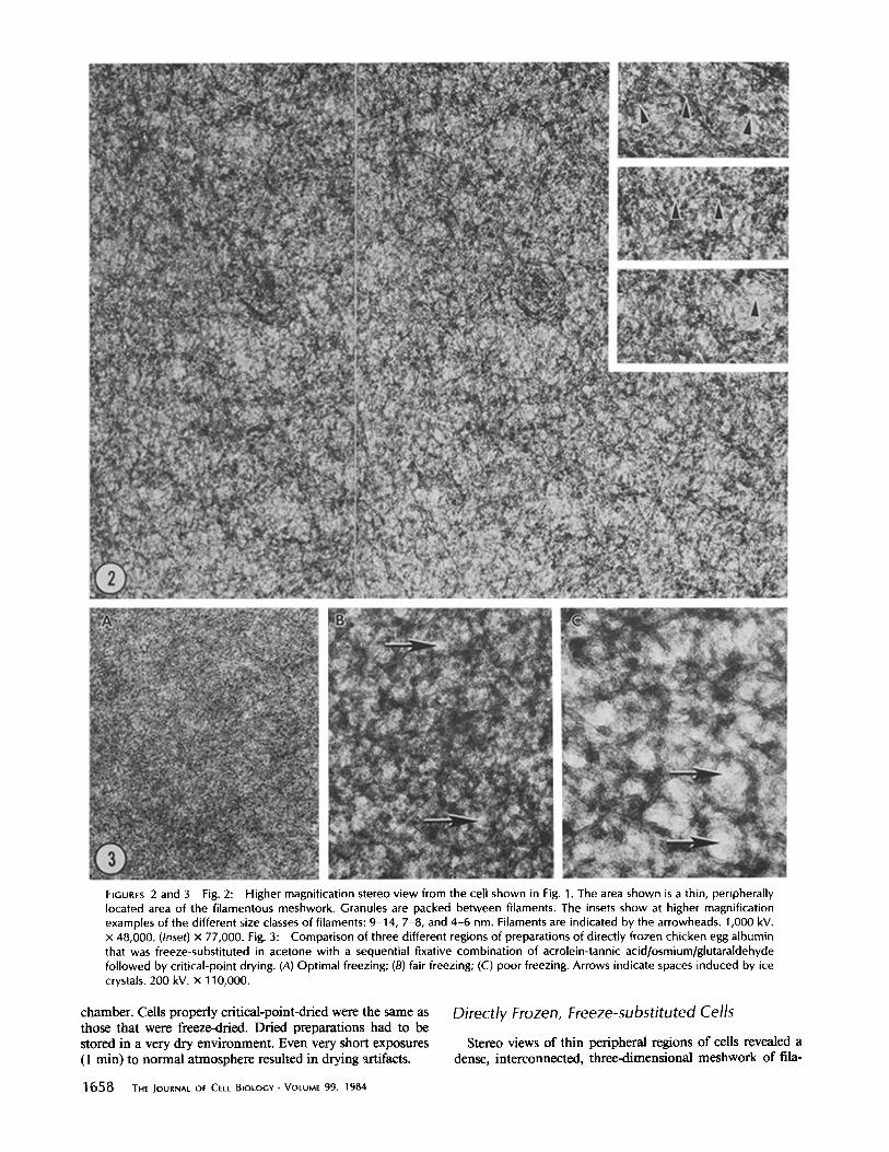
FIGURES 2 and 3 Fig. 2: Higher magnification stereo view from the cell shown in Fig. 1. The area shown is a thin, peripherally located area of the filamentous meshwork. Granules are packed between filaments. The insets show at higher magnification examples of the different size classes of filaments: 9-14, 7-8, and 4-6 nm. Filaments are indicated by the arrowheads. 1,000 kV. x 48,000. (Inset) x 77,000. Fig. 3: Comparison of three different regions of preparations of directly frozen chicken egg albumin that was freeze-substituted in acetone with a sequential fixative combination of acrolein-tannic acid/osmium/glutaraldehyde followed by critical-point drying. (A) Optimal freezing; (B) fair freezing; (C) poor freezing. Arrows indicate spaces induced by ice crystals. 200 kV. x 110,000.
chamber. Cells properly critical-point-dried were the same as those that were freeze-dried. Dried preparations had to be stored in a very dry environment. Even very short exposures (1 min) to normal atmosphere resulted in drying artifacts.
Directly Frozen, Freeze-substituted Cells
Stereo views of thin peripheral regions of ceils revealed a dense, interconnected, three-dimensional meshwork of ilia-
1 6 5 8 THE JOURNAL OF CELL BIOLOGY • VOLUME 99, 1984
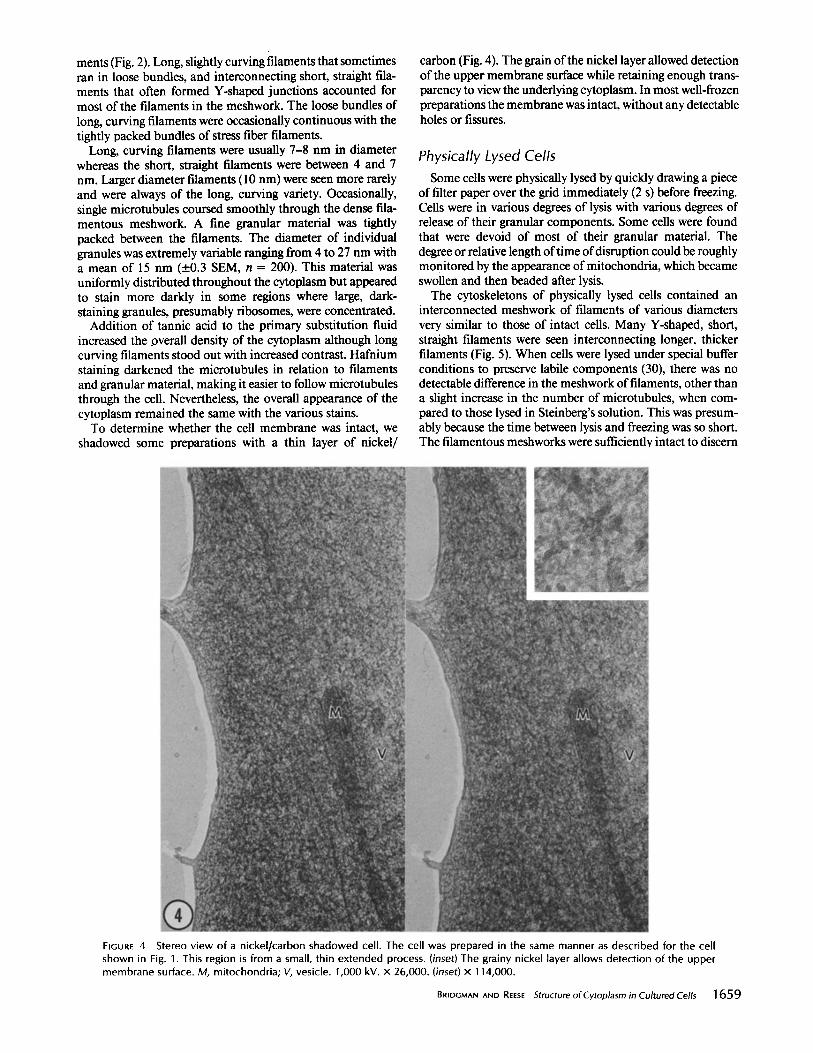
ments (Fig. 2). Long, slightly curving filaments that sometimes ran in loose bundles, and interconnecting short, straight fila- ments that often formed Y-shaped junctions accounted for most of the filaments in the meshwork. The loose bundles of long, curving filaments were occasionally continuous with the tightly packed bundles of stress fiber filaments.
Long, curving filaments were usually 7-8 nm in diameter whereas the short, straight filaments were between 4 and 7 nm. Larger diameter filaments (10 nm) were seen more rarely and were always of the long, curving variety. Occasionally, single microtubules coursed smoothly through the dense fila- mentous meshwork. A fine granular material was tightly packed between the filaments. The diameter of individual granules was extremely variable ranging from 4 to 27 nm with a mean of 15 nm (__.0.3 SEM, n = 200). This material was uniformly distributed throughout the cytoplasm but appeared to stain more darkly in some regions where large, dark- staining granules, presumably ribosomes, were concentrated.
Addition of tannic acid to the primary substitution fluid increased the overall density of the cytoplasm although long curving filaments stood out with increased contrast. Hafnium staining darkened the microtubules in relation to filaments and granular material, making it easier to follow microtubules through the cell. Nevertheless, the overall appearance of the cytoplasm remained the same with the various stains.
To determine whether the cell membrane was intact, we shadowed some preparations with a thin layer of nickel/
carbon (Fig. 4). The grain of the nickel layer allowed detection of the upper membrane surface while retaining enough trans- parency to view the underlying cytoplasm. In most well-frozen preparations the membrane was intact, without any detectable holes or fissures.
Physically Lysed Cells Some cells were physically lysed by quickly drawing a piece
of filter paper over the grid immediately (2 s) before freezing. Cells were in various degrees of lysis with various degrees of release of their granular components. Some cells were found that were devoid of most of their granular material. The degree or relative length of time of disruption could be roughly monitored by the appearance of mitochondria, which became swollen and then beaded after lysis.
The cytoskeletons of physically lysed cells contained an interconnected meshwork of filaments of various diameters very similar to those of intact cells. Many Y-shaped, short, straight filaments were seen interconnecting longer, thicker filaments (Fig. 5). When cells were lysed under special buffer conditions to preserve labile components (30), there was no detectable difference in the meshwork of filaments, other than a slight increase in the number of microtubules, when com- pared to those lysed in Steinberg's solution. This was presum- ably because the time between lysis and freezing was so short. The filamentous meshworks were sufficiently intact to discern
FIGURE 4 Stereo v iew of a nickel/carbon shadowed cell. The cell was prepared in the same manner as described for the cell shown in Fig. 1. This region is from a small, thin extended process. (inset) The grainy nickel layer allows detection of the upper membrane surface. M, mitochondria; V, vesicle. 1,000 kV. x 26,000. (inset) x 114,000.
BRIDGM^N AND REESE Structure of Cytoplasm in Cultured Cells 1659
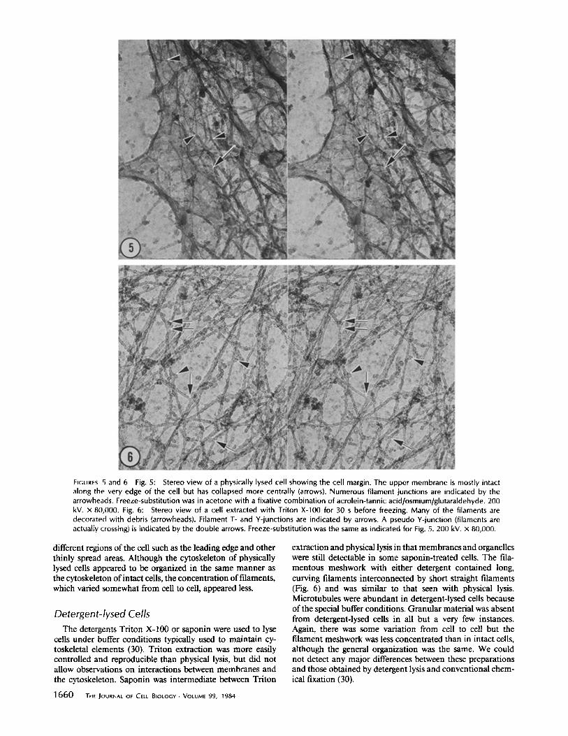
FIGURES 5 and 6 Fig. 5: Stereo view of a physically lysed cell showing the cell margin. The upper membrane is mostly intact along the very edge of the cell but has collapsed more centrally (arrows). Numerous filament junctions are indicated by the arrowheads. Freeze-substitution was in acetone with a fixative combination of acrolein-tannic acid/osmium/glutaraldehyde. 200 kV. × 80,000. Fig. 6: Stereo view of a cell extracted with Triton X-100 for 30 s before freezing. Many of the filaments are decorated with debris (arrowheads). Filament T- and Y-junctions are indicated by arrows. A pseudo Y-junction (filaments are actualty crossing) is indicated by the double arrows. Freeze-substitution was the same as indicated for Fig. 5. 200 kV. x 80,000.
different regions of the cell such as the leading edge and other thinly spread areas. Although the cytoskeleton of physically lysed cells appeared to be organized in the same manner as the cytoskeleton of intact cells, the concentration of filaments, which varied somewhat from cell to cell, appeared less.
Detergent-lysed Cells The detergents Triton X-100 or saponin were used to lyse
cells under buffer conditions typically used to maintain cy- toskeletal elements (30). Triton extraction was more easily controlled and reproducible than physical lysis, but did not allow observations on interactions between membranes and the cytoskeleton. Saponin was intermediate between Triton
extraction and physical lysis in that membranes and organeUes were still detectable in some saponin-treated cells. The fila- mentous meshwork with either detergent contained long, curving filaments interconnected by short straight filaments (Fig. 6) and was similar to that seen with physical lysis. Microtubules were abundant in detergent-lysed cells because of the special buffer conditions. Granular material was absent from detergent-lysed cells in all but a very few instances. Again, there was some variation from cell to cell but the filament meshwork was less concentrated than in intact cells, although the general organization was the same. We could not detect any major differences between these preparations and those obtained by detergent lysis and conventional chem- ical fixation (30).
1660 THE JOURNAL OF CELL BIOLOGY . VOLUME 99, 1984
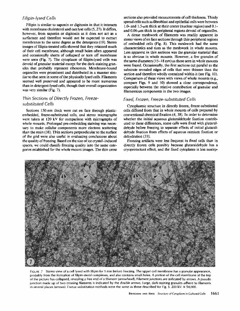
Filipin-lysed Cells Filipin is similar to saponin or digitonin in that it interacts
with membrane cholesterol and can lyse cells (5, 27). It differs, however, from saponin or digitonin as it does not act as a surfactant and therefore would not be expected to extract membranes to the same degree as the detergents (19). Stereo images of filipin-treated cells showed that they retained much of their cell membrane, although small holes often appeared and occasionally sheets of collapsed or torn off membrane were seen (Fig. 7). The cytoplasm of filipin-lysed cells was devoid of granular material except for the dark-staining gran- ules that probably represent ribosomes. Membrane-bound organelles were prominent and distributed in a manner sim- ilar to that seen in some of the physically lysed cells. Filaments seemed well preserved and appeared to be more numerous than in detergent-lysed cells, though their overall organization was very similar (Fig. 7).
Thin 5ections of Directly Frozen, Freeze- substituted Cells
Sections 150-nm thick were cut en face through plastic- embedded, freeze-substituted cells, and stereo micrographs were taken at 120 kV for comparison with micrographs of whole mounts. Prolonged pre-embedding staining was neces- sary to make cellular components more electron scattering than the resin (38). Thin sections perpendicular to the surface of the grid were also useful in evaluating conclusions about the quality of freezing. Based on the size of ice crystal-induced spaces, we could classify freezing quality into the same cate- gories established for the whole mount images. The thin cross
sections also provided measurements of cell thickness. Thinly spread cells such as fibroblast and epithelial cells were between 1.0- and 1.5-zm thick at their center (nuclear region) and 0.1- and 0.06-zm thick in peripheral regions devoid of organelles.
A dense meshwork of filaments was readily apparent in stereo views ofen face sections through thin peripheral regions of embedded cells (Fig. 8). This meshwork had the same characteristics and sizes as the meshwork in whole mounts. Less apparent in thin sections was the granular material that is so obvious in whole mounts. However, a few granules of the same diameters (15-18 nm) as those seen in whole mounts were found. Occasionally, the first sections cut parallel to the substrate revealed edges of cells that were thinner than the section and therefore wholly contained within it (see Fig. 10). Comparison of these views with views of whole mounts (e.g., compare Figs. 9 and 10) showed a close correspondence, especially between the relative contribution of granular and filamentous components in the two images.
Fixed, Frozen, Freeze-substituted Cells Cytoplasmic structure in directly frozen, freeze-substituted
cells differed from that in whole mounts of cells prepared by conventional chemical fixation (4, 38). In order to determine whether the initial aqueous glutaraldehyde fixation contrib- uted to these differences, some cells were fixed with glutaral- dehyde before freezing to separate effects of initial glutaral- dehyde fixation from effects of aqueous osmium fixation or dehydration (33).
Freezing artifacts were less frequent in fixed cells than in directly frozen cells possibly because glutaraldehyde has a cryoprotectant effect, and the fixed cytoplasm is less suscep-
FIGURE 7 Stereo view of a cell lysed with filipin for 1 min before freezing. The upper cell membrane has a granular appearance, probably from the formation of filipin-sterol complexes, and also contains small holes. A portion of the cell membrane at the top of the picture has collapsed, revealing a free end of a filament (arrowhead). Filament junctions are indicated by arrows. A pseudo junction made up of two crossing filaments is indicated by the double arrows. Large, dark-staining granules adhere to filaments in several places (arrows). Freeze-substitution methods were the same as those described for Fig. 5. 200 kV. x 54,000.
BRIDG/v~.N AND REESE Structure of Cytoplasrn in Cultured Cells 1661
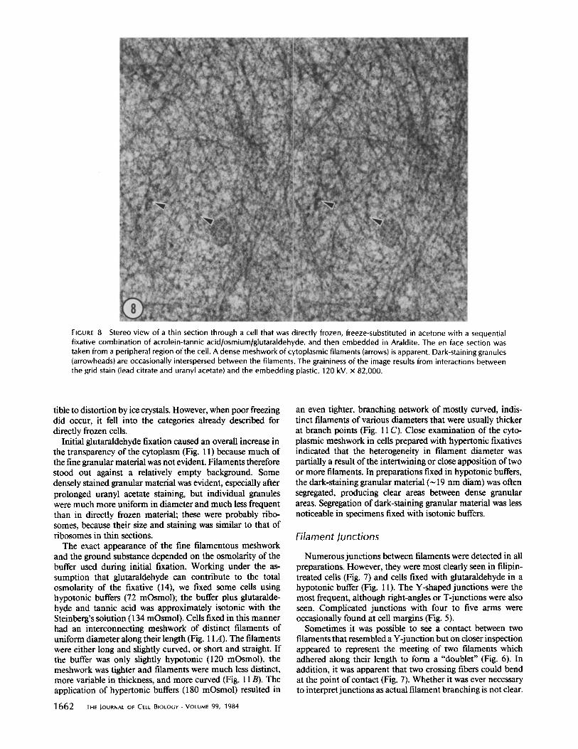
FIGURE 8 Stereo view of a thin section through a cell that was directly frozen, freeze-substituted in acetone with a sequential fixative combination of acrolein-tannic acid/osmium/glutaraldehyde, and then embedded in Araldite. The en face section was taken from a peripheral region of the cell. A dense meshwork of cytoplasmic filaments (arrows) is apparent. Dark-staining granules (arrowheads) are occasionally interspersed between the filaments. The graininess of the image results from interactions between the grid stain (lead citrate and uranyl acetate) and the embedding plastic. 120 kV. x 82,000.
tible to distortion by ice crystals. However, when poor freezing did occur, it fell into the categories already described for directly frozen cells.
Initial glutaraldehyde fixation caused an overall increase in the transparency of the cytoplasm (Fig. 11) because much of the fine granular material was not evident. Filaments therefore stood out against a relatively empty background. Some densely stained granular material was evident, especially after prolonged uranyl acetate staining, but individual granules were much more uniform in diameter and much less frequent than in directly frozen material; these were probably ribo- somes, because their size and staining was similar to that of ribosomes in thin sections.
The exact appearance of the fine filamentous meshwork and the ground substance depended on the osmolarity of the buffer used during initial fixation. Working under the as- sumption that glutaraldehyde can contribute to the total osmolarity of the fixative (14), we fixed some cells using hypotonic buffers (72 mOsmol); the buffer plus glutaralde- hyde and tannic acid was approximately isotonic with the Steinberg's solution (134 mOsmol). Cells fixed in this manner had an interconnecting meshwork of distinct filaments of uniform diameter along their length (Fig. 11A). The filaments were either long and slightly curved, or short and straight. If the buffer was only slightly hypotonic (120 mOsmol), the meshwork was tighter and filaments were much less distinct, more variable in thickness, and more curved (Fig. 11 B). The application of hypertonic buffers (180 mOsmol) resulted in
1662 THE JOURNAL OF CELL BIOLOGY - VOLUME 99, 1984
an even tighter, branching network of mostly curved, indis- tinct filaments of various diameters that were usually thicker at branch points (Fig. 11 C). Close examination of the cyto- plasmic meshwork in cells prepared with hypertonic fixatives indicated that the heterogeneity in filament diameter was partially a result of the intertwining or close apposition of two or more filaments. In preparations fixed in hypotonic buffers, the dark-staining granular material (~ 19 nm diam) was often segregated, producing clear areas between dense granular areas. Segregation of dark-staining granular material was less noticeable in specimens fixed with isotonic buffers.
Filament Junctions
Numerous junctions between filaments were detected in all preparations. However, they were most clearly seen in filipin- treated cells (Fig. 7) and cells fixed with glutaraldehyde in a hypotonic buffer (Fig. 11). The Y-shaped junctions were the most frequent, although fight-angles or T-junctions were also seen. Complicated junctions with four to five arms were occasionally found at cell margins (Fig. 5).
Sometimes it was possible to see a contact between two filaments that resembled a Y-junction but on closer inspection appeared to represent the meeting of two filaments which adhered along their length to form a "doublet" (Fig. 6). In addition, it was apparent that two crossing fibers could bend at the point of contact (Fig. 7). Whether it was ever necessary to interpret junctions as actual filament branching is not clear.
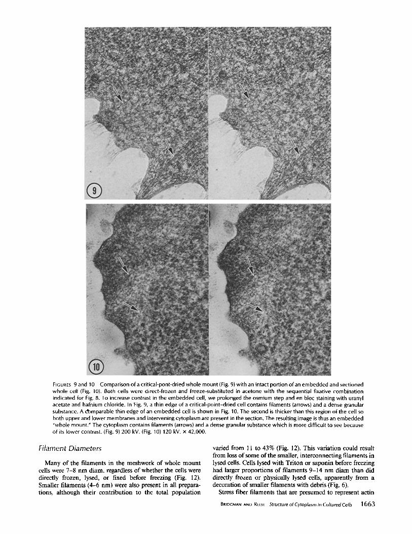
FIGURES 9 and 10 Comparison of a critical-pont-dried whole mount (Fig. 9) with an intact portion of an embedded and sectioned whole cell (Fig. 10). Both cells were direct-frozen and freeze-substituted in acetone with the sequential fixative combination indicated for Fig. 8. To increase contrast in the embedded cell, we prolonged the osmium step and en bloc staining with uranyl acetate and hafnium chloride. In Fig. 9, a thin edge of a crit ical-point-dried cell contains filaments (arrows) and a dense granular substance. A domparable thin edge of an embedded cell is shown in Fig. 10. The second is thicker than this region of the cell so both upper and lower membranes and intervening cytoplasm are present in the section. The resulting image is thus an embedded "whole mount." The cytoplasm contains filaments (arrows) and a dense granular substance which is more difficult to see because of its lower contrast. (Fig. 9) 200 kV. (Fig. 10) 120 kV. x 42,000.
Filament Diameters
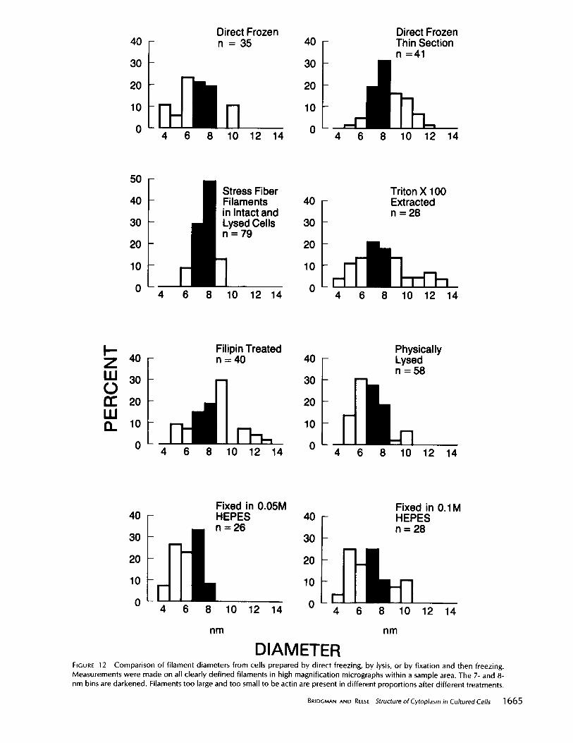
Many of the filaments in the meshwork of whole mount cells were 7-8 nm diam, regardless of whether the cells were directly frozen, lysed, or fixed before freezing (Fig. 12). Smaller filaments (4-6 nm) were also present in all prepara- tions, although their contribution to the total population
varied from 11 to 43% (Fig. 12). This variation could result from loss of some of the smaller, interconnecting filaments in lysed cells. Cells lysed with Triton or saponin before freezing had larger proportions of filaments 9-14 nm diam than did directly frozen or physically lysed cells, apparently from a decoration of smaller filaments with debris (Fig. 6).
Stress fiber filaments that are presumed to represent actin
BRIDGMAN AND REESE Structure of Cytoplasm in Cultured Cells 1663
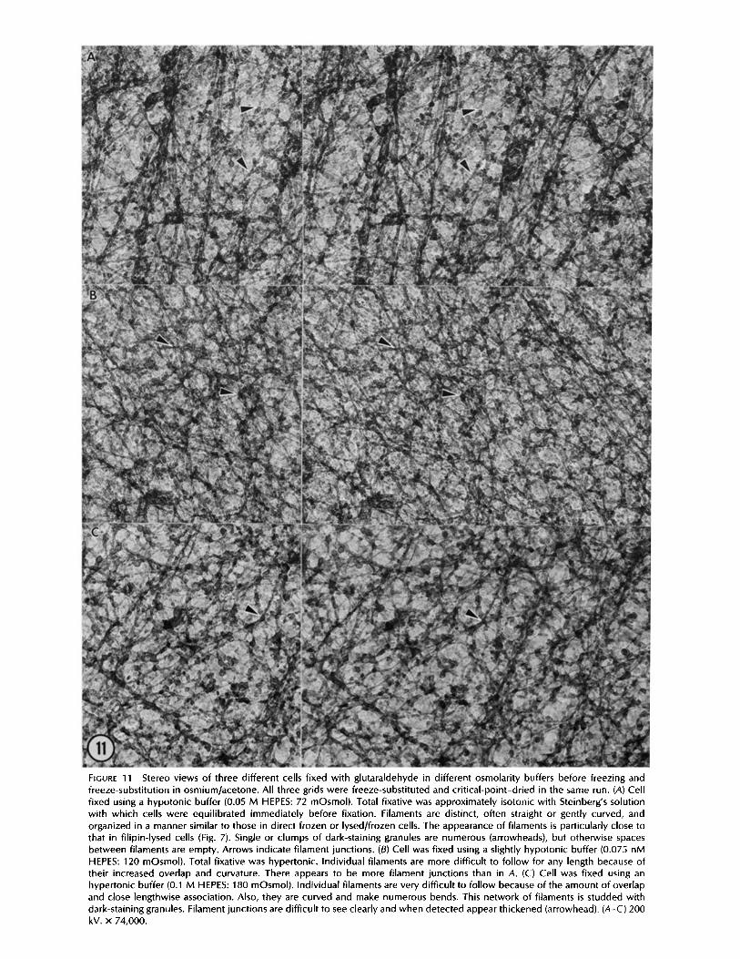
FIGURE 11 Stereo views of three different cells fixed with glutaraldehyde in different osmolarity buffers before freezing and freeze-substitution in osmium/acetone. All three grids were freeze-substituted and crit ical-point-dried in the same run. (A) Cell fixed using a hypotonic buffer (0.05 M HEPES: 72 mOsmol). Total fixative was approximately isotonic with Steinberg's solution with which cells were equilibrated immediately before fixation. Filaments are distinct, often straight or gently curved, and organized in a manner similar to those in direct frozen or lysed/frozen cells. The appearance of filaments is particularly close to that in filipin-lysed cells (Fig. 7). Single or clumps of dark-staining granules are numerous (arrowheads), but otherwise spaces between filaments are empty. Arrows indicate filament junctions. (B) Cell was fixed using a slightly hypotonic buffer (0.075 nM HEPES: 120 mOsmol). Total fixative was hypertonic. Individual filaments are more difficult to fol low for any length because of their increased overlap and curvature. There appears to be more filament junctions than in A. (C) Cell was fixed using an hypertonic buffer (0.1 M HEPES: 180 mOsmol). Individual filaments are very difficult to fol low because of the amount of overlap and close lengthwise association. Also, they are curved and make numerous bends. This network of filaments is studded with dark-staining granules. Filament junctions are difficult to see clearly and when detected appear thickened (arrowhead). (A-C) 200 kV. x 74,000.
40
30
20
10
0
Direct Frozen n = 3 5 40 -
30 -
20 -
10 -
0 -
Direct Frozen Thin Section n =41
4 6 8 10 12 14 4 6 8 10 12 14
50
40
30
20
10
0
;tress Fiber ilaments 40 = Intact and ysed Cells 30 = 79
20
Triton X 100 Extracted n = 28
10
0 4 6 8 10 12 14 4 6 8 10 12 14
l-- Z W 0 n" W O.
40 -
30 -
20 -
10 -
0
Filipin Treated n = 4 0 40
30
20
10
0
Physically Lysed n = 58
4 6 8 10 12 14 4 6 8 10 12 14
40
30
20
10
0
Fixed in 0.05M HEPES n = 2 6
40
30
20
10
0
Fixed in 0.1M HEPES n = 28
4 6 8 10 12 14 4 6 8 10 12 14
nm nm
D I A M E T E R FIGURE 12 Comparison of filament diameters from cells prepared by direct freezing, by lysis, or by fixation and then freezing. Measurements were made on all clearly defined filaments in high magnification micrographs within a sample area. The 7- and 8- nm bins are darkened. Filaments too large and too small to be actin are present in different proportions after different treatments.
BRIDGMAN AND REESE Structure of Cytoplasm in Cultured Cells 1665
filaments were also measured in intact and lysed cells. Stress fiber filaments had a uniform diameter within a preparation and when results from the different preparations were pooled, they still indicate a fairly homogeneous population of diam- eters, generally 7-8 nm. (Fig. 12).
Diameters of filaments in thin sections were close to those in whole mounts (Fig. 12), though a smaller proportion of filaments were in the 4-6-nm category. However, the finer filaments were difficult to measure because filament bound- aries are not as easy to see in the more grainy images obtained from thin sections. But it is still possible to confirm the contribution of fine filaments to the meshwork by a careful examination of stereo pairs of thin sections (Fig. 8).
DISCUSSION
The cytoplasm in thin peripheral areas of cells prepared as whole mounts by direct-freezing, freeze-substitution, and crit- ical-point drying contains a dense filamentous meshwork embedded in a granular ground substance. A major compo- nent of the highly interconnected meshwork are filaments with a uniform diameter of 7-8 nm. These filaments are presumably F-actin because stress fibers in our preparations were composed primarily of similar 7-8-nm filaments that are actin (33); other works on detergent-extracted fixed cells labeled with myosin SI fragments support this interpretation (24, 30). In addition, regions of the meshwork resemble the branching patterns in actin-actin binding protein gels viewed in shadowed, critical-point-dried preparations (18). However, we also find finer filaments, 4-6 nm diam, intercalated in the meshwork which may be related to the fine filaments that do not label with SI in detergent extracted, fixed cells (30). Recently, 5-nm filaments of the intestinal brush border have been identified as belonging to the spectrin/fodrin family ( 11 ). These results together with the present one suggest that spec- trin-like filaments could be ubiquitous and supports the inter- pretation that the 4-6-nm filaments belong to the spectrin/ fodrin family, although the presence of filaments made up of myosin monomers or stages of myosin monomer association can not be ruled out.
This view of cytoplasm differs in two important ways from the view derived from examination with high-voltage electron microscopy of fixed whole mounts of cultured cells (4, 23, 24, 38). The first difference is that the filaments that make up the dense meshwork are discrete rods of uniform diameter along their length, and there is no discernible difference, other than the total numbers of filaments, between the structural orga- nization of the meshwork in gently lysed cells and intact cells. A main feature of this organization are the many contacts between the individual discrete components. This aspect of the picture is similar to, if not the same as, that seen in cells extracted with detergents and fixed under appropriate condi- tions to preserve structural elements (30).
The structural organization of the cytoplasm as we observe it is not adequately characterized by an image of discrete filaments woven into a complex fabric, as has been suggested by examination of detergent-extracted, fixed, frozen, and etched cells (9), nor by the image of curvilinear strands in a continuous trabeculum as visualized on the basis of examin- ing whole mounts of fixed cells with the electron microscope (38). The former picture does not take into account the numerous interconnections between individual filaments and the latter does not describe the discrete nature of the individ-
1666 THE JOURNAL OF CELL BIOLOGY • VOLUME 99, 1984
ual structural elements. A second important difference is that our preparative meth-
ods show that the cytoplasm contains a fine granular material packed between filaments that is quickly lost upon cell lysis. This granular material effectively takes up all residual space between filaments and is distributed throughout the cyto- plasm. This is apparently the same granular material seen in replicas of directly frozen, etched cells (9, 28). The diameters of granules in optimally frozen cells, although variable, are all greater than those of granules seen in optimally frozen albu- min solutions. We therefore assume that the granular material as a whole represents a concentrated solution of protein whose individual components are complexes of soluble proteins.
Primary fixation in aqueous aldehyde obscures this granular material, with the exception of the large, dark-staining gran- ules that probably represent ribosomes. This finding suggests one reason why this granular material has not been previously detected in whole mounts of cells (4, 38). However, the granular material was also not detected in whole mounts of direct frozen, freeze-dried, or freeze-substituted PtK2 cells (22). We do not know the reason for this discrepancy, al- though we can suggest several possibilities. Unfixed and there- fore unsupported cytoplasm should be subject to the same artifacts during freeze-drying as it is during deep etching. Extensive etching of directly frozen material is known to give the impression of "clean" filaments and cross-bridges, and intervening granular material is lost (28). That granular ma- terial was not previously seen in freeze-substituted whole mounts may result from the choice of a highly polar solvent, 2-methoxyethanol, for freeze-substitution (22). The more po- lar solvents cause a dramatic decrease in the amount of detectable granular material which may be due to increased extraction of soluble components by the substitution fluid or may simply reflect differences in staining (Appendix).
Freezing that results in ice crystals >5 nm also produced images of clean, curving, relatively thick filaments without intervening granular material. Similar images resulted from poor freezing of solutions of albumin processed by freeze- substitution and critical-point drying. The formation of ice crystals concentrates soluble materials into boundary layers between crystals that give the impression of irregular filament- like structures bordering open spaces in the cytoplasm or protein solution (20).
The only difference between images of whole mounts of cells and thin-sectioned cells are accounted for by differences in specimen thickness and contrast. The decreased contrast in thin sections makes the lightly stained granular material more difficult to see. However, the organization of filaments seems to be the same with these two techniques, though the filamentous meshworks in directly frozen cells, whether viewed in whole mounts or thin sections, appear more distinct and uniformly spaced than in fixed cells. A major reason for this difference seems to be the osmotic effects of glutaralde- hyde (14), which, like glycerol, penetrates the cell slowly (36). If fixation occurs before equilibration, the cell will be fixed in a dehydrated state and will appear shrunken. This may be the explanation for why cells initially fixed in hypotonic buffer- fixative solutions have filamentous meshworks that are most similar to directly frozen cells.
Fixation of cultured cells in a saponin, tannic acid, and glutaraldehyde solution followed by a low concentration of osmium (16) supports our interpretation of the effects of fixation on cytoplasmic structure. By permeabilizing the cells
with saponin, osmotic effects are circumvented and it is possible that filaments are fixed in a relatively undistorted position. This could also be the explanation for why there have been such differences between the appearance of fila- mentous meshworks in whole mount cells fixed while intact as compared with those fixed after extraction with the deter- gent Triton X-100 (15, 24, 30). Clearly, cells can be initially fixed under appropriate conditions to give an accurate picture of the organization of their cytoplasmic filaments.
APPENDIX
Freeze-substitution Mixtures of osmium and acetone are typically used for freeze-substitution
of tissue to be thin-sectioned (37). However, we soon found that typical osmium-acetone combinations were inadequate for directly frozen cells because of their tendency to shrink during the critical-point drying. Osmium/acetone freeze-substitntion is also known to be an inadequate preparation for scanning electron microscopy (1). Shrinkage could be partially alleviated by using high concentrations of osmium (3-5%) and warming the specimens to room tem- perature while in the osmium/acetone. However, the dense staining that resulted obscured most of the details in cell whole mounts. Another concern was that osmium could have detrimental effects on actin fibers similar to those occurring in aqueous osmium solutions (17). We therefore decided to look for a better method of freeze-substituting specimens to be viewed as whole mounts. Several subjective criteria were used to determine the best methods: (a) The state of preservation of readily recognizable fibrous elements, such as micro- tubules and stress fibers; (b) the state of preservation of organelles such as mitochondria and endoplasmic reticulum; (c) the continuity of cell membranes; (d) the preservation of fine cell projections such as filopodia; and (e) the relative density and appearance of the cytoplasmic ground substance. To insure that artifacts induced by freeze-substitution were clearly distinguishable from freez- ing artifacts, we subjected at least six grids to each variation of the substitution procedure and then scanned them in the electron microscope. Recrystallization of ice (7) rarely occurred as long as high-grade solvents free of water were used. Recrystallization was distinguished from poor initial freezing by the ubiquity of large ice crystals in all the samples from a run. Recrystallization occurs when samples are warmed before substitution is complete (35).
Direct frozen whole mounts were subjected to over 60 different combinations of substitution solutions, temperatures, and times. In the most successful method, whole mounts were substituted in 10% acrolein in acetone usually with 0.2% tannic acid at -80"C for 10-18 h, warmed m -550C, and transferred (after a rinse in acetone) to 0.2% osmium tetroxide in acetone that was then warmed to -20"C. After 2-3 h at -20"C the whole mounts were rinsed, transferred to anhydrous 10% glutaraldehyde in a methanol/acetone mixture (40%/50%), warmed to 0"C, and maintained at that temperature overnight. Specimens were then washed with methanol (40 rain) and stained with 0.1- 0.5% uranyl acetate in methanol or acetone (30-45 min at 0"C) followed by (0.5-1.0%) hafnium chloride in methanol or acetone for 20-45 rain at room temperature.
When whole mounts were to be embedded and then thin-sectioned, the procedure was modified to increase the staining and fixation. The osmium concentration was increased to 0.4% and the time at -20"C in osmium was increased from 2 to 8 h. In addition, the length of staining with uranyl acetate was increased to -12 h and with hafnium chloride to 2 h. The results of the trials with different solvents, fixatives, and stains, which provide a rationale for this method, are detailed below.
Solvents Solvents or solvent combinations tried for freeze-substitution were tetrahy-
drofuran, acetone, ethanol, 90% ethanol/10% H20, 90% ethanol]10% glycerol, methanol, and 60% methanol/40% ethylene glycol (1).
The more polar solvents such as methanol and ethanol caused partial loss of cell membranes and some shrinkage and partial extraction of the cytoplasm even in the presence oftbe best fixative combinations (see below). The extrac- tion of cytoplasmic components was indicated by the decreased density of the cytoplasm compared with samples substituted in acetone or tetrahydrofuran. Addition of water, glycerol, or ethylene glycol to slow the rate of substitution prevented shrinkage but had no noticeable effects on preserving membranes or preventing extraction of cytoplasmic components.
No shrinkage could be detected when either acetone or tetrahydrofuran was used with the best fixatives. Cell membranes remained intact in these solvents
as long as substitution times at -80°C were <48 h. Substitution times >64 h caused loss of membranes, as did warming to >-40°C without osmium fixation.
Fixatives and Stains
Substantial chemical fixation during freeze-substitution was necessary to prevent shrinkage induced by critical-point drying. Osmium fixation alone was not adequate because concentrations, times, and temperature of osmium fixa- tion that gave well-stained preparations did not prevent shrinkage. For this reason, we tried various combinations of fixatives.
Acrolein (10% ) was the most effective fixative at cold temperatures and was the only fixative to give indications of f~xing (prevented shrinkage) at -80"C. However, it is difficult to give an accurate indication of the time necessary for fixation at -80"C becanse the time needed for substitution is unknown. The minimum time that was tried and found to be effective for substitution with 10% acrolein in acetone was 10 h. Although acrulein provided good structural preservation of the cytoplasm, it gave minimal fixation of membranes, even at temperatures of 0*C.
Osmium even at concentrations of 5%, had no apparent effect at the substitution temperature (-80"C) (37). The lowest temperature at which os- mium showed signs of fixing tissue (preserved structure and stained membranes) was between -40" and -50°C. By independently adjusting concentration, time, and temperature of exposure to osmium we came up with an exposure just sufficient for preservation and staining of membranes, in order to avoid possible detrimental effects on actin filaments. The useful concentrations were between 0.2 and 0.4% at temperatures up to -20°C and times ranging from 6 to 14 h.
Glutaraldehyde (10%) was the least effective fixative at cold temperatures. No fixation was observed at temperatures <-20"C (39), and from -20" to +4"C, fixation that helped prevent shrinkage occurred after fixation at colder temperatures by acrolein or osmium.
Tannic acid is not generally considered a fixative, but it is known to protect actin filaments from fragmentation by aqueous osmium (16). It also stains actin filaments and membranes directly, presumably by increasing the reduc- tion of osmium (16). Tannic acid was useful in freeze-substitution both as a stain and as a preserving agent. Addition of 0.2% tannic acid (desiccated for 4 h at 80°C) to the primary substitution fluid increased subsequent osmium staining of membranes, ground substance, and filaments. Membranes were also less likely m show the discontinuities that sometimes appeared when lower concentrations of osmium alone were used. Tannic acid was also an effective general stain when applied at higher concentrations and warmer temperatures after osmium fixation.
Uranyl acetate stabilizes membranes following aldehyde fixation (31, 32), and we found that it could help stabilize membranes at low temperatures depending on the solvent. Membranes were extracted in the more polar solvents but were generally intact when substituted in ethanol or acetone. Uranyl acetate increased the contrast of membranes and filaments after fixation by aldehydes and osmium. Its staining properties were not dependent imply upon concen- tration, time, and temperature but also on the solvent. For instance, 0.5% uranyl acetate in acetone applied for 30 min at 0"C gave more intense staining than the same concentration in methanol applied for the same time and at the same temperature. Hafnium chloride in methanol was especially useful for imparting contrast to microtubules when used in combination with uranyl acetate. Like uranyl acetate, it gave more intense, general staining when used in acetone than when used in more polar solvents such as methanol.
Conventional fixation by aqueous solution of fixatives produces the best results when used in combinations; no fixative has been found that will adequately fix all cellular constituents. The most widely used combination is aldehyde fixation followed by postfixation in osmium tetroxide. The best and most consistently reproducible fixation during the freeze-substitution process is a similar combination of fixatives. However, the constraints imposed by the freeze-substitution temperatures on the activity of the various fixatives partially dictate the particular combination.
We thank Dr. John Walrond for helpful discussions about freeze- substitution and in particular for sharing his experience with stains. We also thank Victor Frank for the design and construction of the spring-driven freezing device. George Wray gave valuable instruction and help with the high-voltage electron microscopy. Bryan Schroeder, Nicholas Christakis, and Janet Kinnane gave photographic assistance. We thank Sandra Cohen for processing the manuscript.
This investigation was supported in part by a postdoctoral fellow- ship from the Muscular Dystrophy Association to P. C. Bridgman and the Biotechnology Resources Program/Division of Research Resources National Institutes of Health grant No. RR-00592.
BRIDGMAN AND REESE Structure of Cytoplasm in Cultured Cells 1667
Received for publication 13 February 1984, and in revised form 12 July I984.
REFERENCES
1. Barlow, D. 1., and M. A. Sleigh. 1979. Freeze substitution for preservation of ciliated surfaces for scanning electron microscopy. J. Microsc. (Oxfi). 715:81-95.
2. Bridgman, P. C., B. Kachar, and T. S. Reese. 1983. The structure of cytoplasm in directly frozen cultured cells. II. Cytoplasmic domains associated with organelle move- ment. J. Cell Biol. 97(5, Pt. 2)266a. (Abstr.)
3. Bridgraan, P. C., S. Nakajima, A. Greenberg, and Y. Nakajima. 1984. Freeze-fracture and electmphysiological studies on newly developed acetylcboline receptors in Xenopus embryonic muscle cells. J. Cell Biol. 98:2160-2173.
4. Buckley, I. K., and K. R. Porter. 1975. Electron microscopy of critical-point dryed cultured cells. J. Microsc. (Oxf.). 104:107-120.
5. de Kruijff, K, W. J. Gerristen, A. O¢fiemans, R. A. Daniel, and L. L. M. van Deenen. 1974. Polyene antibiotic-sterol interactions in membranes of Acholeplasma laidlawii cells and lecithin liposomes. I. Specificity of the membrane permeability ohanges induced by the polyene antibiotics. Biochem. Biophys. Acta, 339:30--43.
6. Eseaig, T. 1982. New instruments which facilitate rapid freezing at 83K and 6K. Z Microsc. (Oxf). 126:221-229.
7. Franks, F. 1982. The properties of aqueous solutions at subzero temperatures. In Water: A Comprehensive Treatise. F. Franks, editor. Plenum Press, New York. 7:215-334.
8_ Handley, D. A., J. T. Alexander, and S. Chien. 1980. The desig~ and use of a simple device for rapid quanch-free-zing of biological samples. J. Microsc. (O.xf.). 121:273-282.
9. Heuser, J. E., and M. W. Kirschner. 1980. Filament organization revealed in platinum replicas of freeze-dried cytoskeletons. Z Cell Biol. 86:212-234.
10. Heaser, L E., T. S. Reese, M. J. Dennis, Y. Jan, L. Jan, and L Evans. 1979. Synaptic vesicle exocytosis captured by quick freezing and correlated with quantal transmitter release. Z Cell Biol. 81:275-300.
l I. Hirokawa, N., R. E. Cbeney, and M. Willard. 1983. Location of a protein of the fodrin- speetrin-TW260/240 family in the mouse intestinal brush border. Cell. 32:953-965.
12. Hirokawa, N., and J. E. Heuser. 1981. Quick-freeze, deep-etch visualization of the cytoskeleton beneath surface differentiations of inlestinal epithelial cells. J. Cell Biol. 91:399-409.
13. Jehl, B., R. Bauer, A. Dorge, and R. Rick. 1981. The use ofpropane/isopentane mixtures for rapid freezing of biological specimens. J. Microsc. (Oxf). 123:307-309.
14. Lee, R. M. K. W., R. McKenzie, K. Kobayaski, R. E. Garfield, J. B. Forrest, and E. E. Daniel. 1982. Effects of glutataldehyde fixative osmolaritias ou smooth muscle cell volume, and osmotic reactivity of the ceils aRer fixation. J. Microsc. (Oxf). 125:77-8g
15. Letoumeau, P. C. 1983. Differences in the organization of actin in the growth cones compared with the neurites of cultured neurons from chick embryos. J. Cell Biol. 97:963-973.
16. Maupin, P., and T. D. Pollard. 1983. Improved pre~rvation and staining of HeLa cell actin filaments, clathrin-coated membranes, and other cytoplasmic structures by tannic acid-glutaraldehyde-saponin fixation. J. Cell Biol. 96:51-62.
17. Maupin-Szamier, P., and T. D. Pollard. 1978. Actin filament destruction by osmium tctroxide. J. Cell Biol. 77:837-852.
18. Niederman, R., P. C. Amreinsi, and J. Hartwig. 1982. The three-dimensional structure of actin filaments and of an actin gel made with actin-binding protein. J. Cell Biol. 96:1400-1413.
19. Norman, A. W., R. M. Spielvogel, and R. G. Wong. 1976. Polyene antibiotic-sterol interaction. Adv. Lipid Res. 14:127-170.
20. Ornberg, R. L., and T. S. Reeso. 1979. Artifacts of freezing in Limulus ameboxytes. In Freeze Fracture: Methods, Artifacts and Interpretation. J. E. Rash and C. S. Hudson, editors. Raven Press, New York, 89-98.
21. Feng, H. B., and Y. Nakajima. 1978. Membrane particle aggregates in innervated and noninnervated cultures of Xenopus embryonic cells. Proc. Natl. Acad. ScL USA. 75:500- 504.
22. Porter, K. R., and K. L Andc~on. 1982. The structure of the cytoplasmic matrix preserved by fre'cze-drying and freeze-substitution. Eur. J. Cell Biol. 29:83-96.
23. Porter, K. R., and J. B. Tucker. 1981. The ground substance of the living cell. Sci. Am. 244:57-67.
24. Pryzwansky, K. B., M. Schliwa, and K. R. Porter. 1983. Comparison of the three- dimensional organization of unextracted and triton-extracted human neutrophilic pol- ymorpbonucleax leukocytes. Eur. J. Cell Biol. 30:112-125.
25. Ris, H. 1981. Morphology of cytoplasmic filaments in thick sections and critical point dried whole mounts. J. Cell Biol. 91 (5, Pt. 2):305a. (Abstr.)
26. Ris, H. 1980. The cytoplasmic ~microtmbeular lattice"--reality or artifact. 38th Ann. Proc. Electron Microsc. Soc. Am. 812-813.
27. Robinson, J. M., and M. J. Karnovsky. 1980. Evaluation of the polyene antibiotic filipin as a cytochemical probe for membrane cholesterol. J. Histochem. Cytochem. 28:161- 168.
28. Schnapp, B. J. and T. S. Reese. 1982. Cytoplasmic structure in rapid-frozen axons. J. Cell Biol. 94:667-679.
29. Schliwa, M. 1982. Action of cytochalasin D on cytoskeletal networks. J. Cell Biol. 92:79-91.
30. Schliwa, M., and L van Blerkom. 1981. Structural interaction of cytoskelctal compo- nents. J. Cell Biol. 90:222-235.
31. Silva, M. T., F. C. Guena, and M. M. Magalhaes. 1968. The fixative action of uranyl acetate in electron microscopy. Experientia. 24:1074.
32. Silva, M. T., J. M. S. Morn, J. C. V. Melo, and F. C. Gueua. 1971. Uranyl salts as fixatives for electron microscopy, study of the membrane ultrastructure and phospho- lipid loss in bacilli. Biochem. Biophys. Acta. 233:513-520.
33. Small, J. V. 1981. Organization of actin in the leading edge of cultured cells: influence of osmium tetroxide and dehydration on the ultrastructure of actin meshworks. J. Cell Biol. 91:695-705.
34. Small, J. V., and J. E. Cells. 1978. Filament arrangements in negatively stained cultured cells: the organization ofactin. Cytobiologie. 16:308-325.
35. Steinbreeht, R. A. 1982. Experiments on freezing damage with freeze substitution using moth antennae as test objects. Z Microsc (Oxf). 125:187-192.
36. Tao-Cheng, J. H., K. Hirosawa, and T. Nakajima. 1981. Ultrastroctum of the crayfish stretch receptor in relation to its function, d. Comp. NeuroL 20@ 1-21.
37. Van Hareeveld, A., S. Crowell, and S. K. Malhotra. 1965. A study of extracellular space in central nervous tissue by freeze-substitution. J. Cell Biol. 25: 117-137.
38. Wolosewick, S. L, and K. R. Porter. 1979. Microtrabecular lattice of the cytoplasmic ground substance. Artifact or reality. J. Cell Biol. 82:114-139.
39. Wooley, D. M. 1974. Freeze substitution: a method for the rapid arrest and chemical fixation of spermatozoa. J Microsc. (Oxf). 101:245-260.
1668 THE JOURNAL OF CELL BIOLOGY • VOLUME 99, 1984